1 Osteoarthritis | www.smgebooks.com Copyright Piróg KA.This book chapter is open access distributed under the Creative Commons Attribution 4.0 International License, which allows users to download, copy and build upon published articles even for commercial purposes, as long as the author and publisher are properly credited. Gr up SM Tissue Engineering Approaches for the Study and Therapeutic Intervention in Osteoarthritis CARTILAGE AGEING AND DISEASE Cartilage is a mechanically responsive tissue with morphologically complex structure, consisting entirely of one cell type (the chondrocyte) existing in various differentiation states. It plays important roles in developing bone (growth plate cartilage) and as protection against mechanical impacts on mature bone (articular cartilage) [1]. Pathological changes to articular cartilage structure and homeostasis lead to osteoarthritis (OA), which presents a heavy welfare burden on our rapidly aging society [2]. OA is thought to result from microdamage to cartilage and the subsequent repair/damage events which lead to OA progression [3,4]. Extracellular changes associated with ageing, such as increase in collagen fiber diameters and the incidence of collagen cross-links and a decrease in proteogly can content, lead to reduced tissue stiffness, making the tissue more prone to injury. Furthermore, during ageing the chondrocytes senesce, slow their metabolic activity and eventually die and the remodelling capabilities of the tissue decrease, leading to an increased OA susceptibility following injury related trauma [5–8]. In OA, diarthrodial joints undergo degradation towards the sub chondral bone leading to pain and formation of fibro cartilage with dramatically altered mechanical properties. OA is particularly noticeable in hips, knees, hands and feet [9,10]. Thais De Iasheras Ruiz and Katarzyna Anna Piróg* Institute of Genetic Medicine, Newcastle University, UK *Corresponding author: Katarzyna Anna Piróg, Institute of Genetic Medicine, Newcastle University, International Centre for Life, Central Parkway, Newcastle upon Tyne, NE1 3BZ, UK. Email: [email protected]Published Date: August 18, 2016
Transcript
1Osteoarthritis | www.smgebooks.comCopyright Piróg KA.This book chapter is open access distributed under the Creative Commons Attribution 4.0 International License, which allows users to download, copy and build upon published articles even for commercial purposes, as long as the author and publisher are properly credited.
Gr upSMTissue Engineering Approaches for the Study and Therapeutic Intervention in
Osteoarthritis
CARTILAGE AGEING AND DISEASECartilage is a mechanically responsive tissue with morphologically complex structure,
consisting entirely of one cell type (the chondrocyte) existing in various differentiation states. It plays important roles in developing bone (growth plate cartilage) and as protection against mechanical impacts on mature bone (articular cartilage) [1]. Pathological changes to articular cartilage structure and homeostasis lead to osteoarthritis (OA), which presents a heavy welfare burden on our rapidly aging society [2]. OA is thought to result from microdamage to cartilage and the subsequent repair/damage events which lead to OA progression [3,4]. Extracellular changes associated with ageing, such as increase in collagen fiber diameters and the incidence of collagen cross-links and a decrease in proteogly can content, lead to reduced tissue stiffness, making the tissue more prone to injury. Furthermore, during ageing the chondrocytes senesce, slow their metabolic activity and eventually die and the remodelling capabilities of the tissue decrease, leading to an increased OA susceptibility following injury related trauma [5–8]. In OA, diarthrodial joints undergo degradation towards the sub chondral bone leading to pain and formation of fibro cartilage with dramatically altered mechanical properties. OA is particularly noticeable in hips, knees, hands and feet [9,10].
Thais De Iasheras Ruiz and Katarzyna Anna Piróg*Institute of Genetic Medicine, Newcastle University, UK
*Corresponding author: Katarzyna Anna Piróg, Institute of Genetic Medicine, Newcastle University, International Centre for Life, Central Parkway, Newcastle upon Tyne, NE1 3BZ, UK. Email: [email protected]
Published Date: August 18, 2016
2Osteoarthritis | www.smgebooks.comCopyright Piróg KA.This book chapter is open access distributed under the Creative Commons Attribution 4.0 International License, which allows users to download, copy and build upon published articles even for commercial purposes, as long as the author and publisher are properly credited.
SURGICAL STRATEGIES FOR OA TREATMENTThe morphological complexity of cartilage makes it a difficult tissue to repair and to study.
Cartilage is an avascular tissue and so its healing properties are limited, making research into the novel strategies to repair articular cartilage of paramount importance [11]. Currently there are no routine Therapies available for OA, other than a full joint replacement (arthroplasty) or joint fusion (arthrodesis) at later stages of the disease. However, several endogenous or exogenous surgical techniques are currently being investigated and trialled in order to repair the damage and avoid the complete joint replacement. Endogenous therapies attempt to drive cartilage repair by enhancing the innate properties of the tissue and include introducing growth factors to promote the proliferation and differentiation of cells in situ, whereas exogenous approaches often use cells from another source or modify the cells harvested from the wounded region ex vivo before implanting them back into the joint [12,13].
Microfracture is one of the oldest and commonly applied endogenous techniques where by a surgical penetration into the subchondral bone leads to the disruption of blood vessels and a formation of a blood clot. The blood clotting attracts bone marrow mesenchymal stem cells (BM-MSC) which invade the microfracture and eventually differentiate into neo-cartilage. Despite the huge success of this technique, the main issue with it is that the neo-cartilage often resembles fibrous cartilage with undesired mechanical properties and so microfractures are currently not recommended as treatment for larger lesions or osteoarthritis [13–16]. Mosaicplasty is another keyhole surgery approach based on extracting osteochondral plugs from zones that tolerate moderate weight within the joint and applying them to fill the injured regions [17,18].Patients treated with mosaicplasty retained higher long-term activity levels compared to the microfracture patients. However, the new tissue still contains up to 20% fibro cartilage and the method requires further investigation [13,14].
Exogenous techniques for cartilage repair comprise autologous chondrocytes implantation (ACI) and stem cells implantation, both of which are not used routinely and currently only applied as part of a clinical trial. ACI was first mentioned by Brittberg et al 1994 [19], and is considered the first ever tissue engineering approach in cartilage repair consisting of extracting chondrocytes from less weight bearing areas and transplanting them to the injured area upon expansion in vitro in monolayer [12,13]. Initially this strategy showed great success and the implanted cells expresses the normal makers of healthy cartilage, however, the regenerative capacity largely depended on the age of the origin tissue and in time the new tissue showed signs of becoming more fibrous [13,14,20].Owing to their pluripotency and the capacity to differentiate into other cell lineages, stem cells represent an exciting alternative for exogenous therapies and an excellent source of material for regenerative medicine. Suitable cells can be harvested from synovium or bone marrow [11,21]. Synovium-derived stem cells (SDSCs) retain their chondrogenic potential and display articular cartilage mechanical properties when seeded in 3D environment. Despite
3Osteoarthritis | www.smgebooks.comCopyright Piróg KA.This book chapter is open access distributed under the Creative Commons Attribution 4.0 International License, which allows users to download, copy and build upon published articles even for commercial purposes, as long as the author and publisher are properly credited.
this, cell senescence is highly notable and further studies regarding the long term properties of SDSCs derived constructs are necessary [11,22]. Bone marrow stromal cells preserve the ability to differentiate into chondrocytes, and hence are a good candidate for cartilage repair. First studies using these cells showed their potential to synthesise cartilage-like tissue comparable to a healthy tissue, despite this other studies showed fibrocartilaginous formation and hypertrophy [11,12]. Recent studies have shown that induced pluripotent stem cells (iPSCs) may offer a more easily accessible autologous alternative to using stem cells. These cells were first mentioned in 2006 by Takahashi and Yamanaka whereby they dedifferentiated adult fibroblasts using 4 factors: Oct3/4, Sox2, c-Myc and Klf4 and thus generated cells which were capable of re-differentiate into other cell types [23]. Several studies since had differentiated skin and blood cells from patients with articular cartilage injuries and reintroduced them into the injuries showing a reparative potential. This novel technique presents some advantages such as lack of immune response post-implantation and ease to harvest, however, its main limitation is the risk of teratom a development due to presence of undifferentiated iPSCs [12].
TISSUE ENGINEERING FOR CARTILAGE REPAIR AND STUDYThe concept of applying tissue engineering to study cartilage biology and repair cartilage
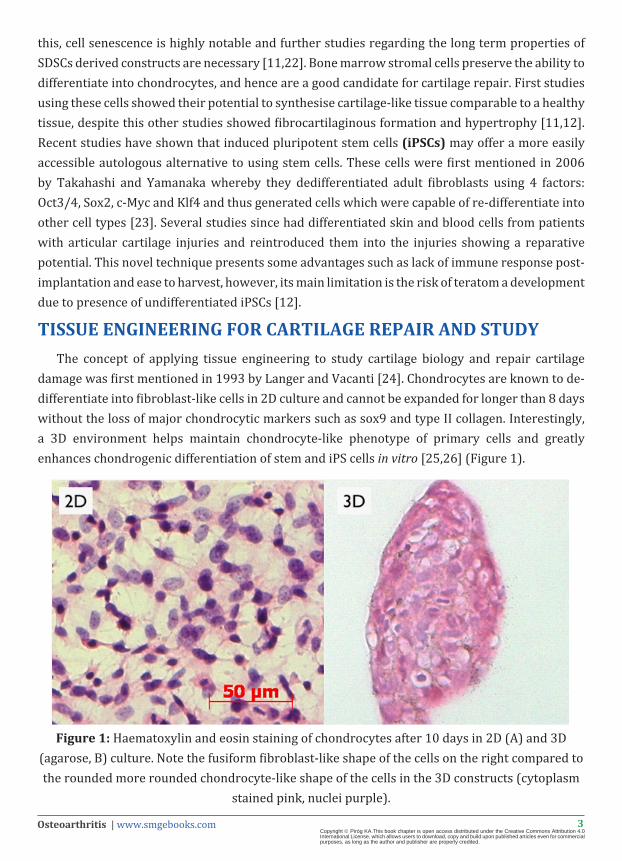
damage was first mentioned in 1993 by Langer and Vacanti [24]. Chondrocytes are known to de-differentiate into fibroblast-like cells in 2D culture and cannot be expanded for longer than 8 days without the loss of major chondrocytic markers such as sox9 and type II collagen. Interestingly, a 3D environment helps maintain chondrocyte-like phenotype of primary cells and greatly enhances chondrogenic differentiation of stem and iPS cells in vitro [25,26] (Figure 1).
Figure 1: Haematoxylin and eosin staining of chondrocytes after 10 days in 2D (A) and 3D (agarose, B) culture. Note the fusiform fibroblast-like shape of the cells on the right compared to the rounded more rounded chondrocyte-like shape of the cells in the 3D constructs (cytoplasm
stained pink, nuclei purple).
4Osteoarthritis | www.smgebooks.comCopyright Piróg KA.This book chapter is open access distributed under the Creative Commons Attribution 4.0 International License, which allows users to download, copy and build upon published articles even for commercial purposes, as long as the author and publisher are properly credited.
The dedifferentiation of chondrocytes in the two-dimensional environment has long been a recognised issue in cartilage study and repair. Pellet cultures have been proposed as a method for chondrocyte culture as early as 1980s and are the easiest way to culture chondrocytes in a three-dimensional environment. This method does not require an addition of a vehicle or a matrix to culture the cells and has been shown to maintain the chondrocytic phenotype in culture [27].Pellets are routinely used in cartilage studies, however, in prolonged culture they result in chondrocyte hypertrophy and the expression of undesired markers, such as type I and type X collagens [28]. This can be circumvented by addition of certain growth factors such as TGF-β, BMP 4/7/13 and other inhibitors of hypertrophy [29] or by culturing the cells in micromass cultures [30] that are formed by growing the cells in droplets of medium and result in larger and more homogenous hyaline cartilage-like constructs [29]. However, both matrix-free methods result in relatively small cartilage constructs which may not be suitable for surgical applications and due to the density of the cultures, retrieval of cells for post-culture applications is also difficult. Interestingly, recent studies show some successes of generating constructs combining 20-50 individual cartilage pellets into an engineered cartilage tissue that was successfully implanted into a lesion in an animal model [31]. However, in order to engineer the final constructs, the authors used not only expanded cells but also other matrices such as agarose, in order to provide the right microenvironment.
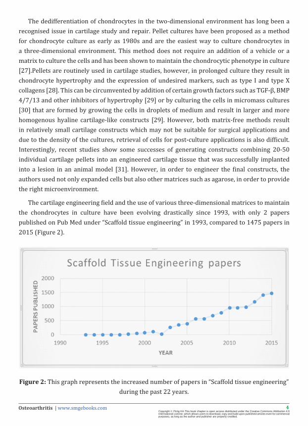
The cartilage engineering field and the use of various three-dimensional matrices to maintain the chondrocytes in culture have been evolving drastically since 1993, with only 2 papers published on Pub Med under “Scaffold tissue engineering” in 1993, compared to 1475 papers in 2015 (Figure 2).
Figure 2: This graph represents the increased number of papers in “Scaffold tissue engineering” during the past 22 years.
5Osteoarthritis | www.smgebooks.comCopyright Piróg KA.This book chapter is open access distributed under the Creative Commons Attribution 4.0 International License, which allows users to download, copy and build upon published articles even for commercial purposes, as long as the author and publisher are properly credited.
Three-dimensional environment supports and promotes chondrogenesis. However, in order to achieve a relevant and clinically applicable 3D model, constructs need to meet a long list of requirements to match the mechanical strength of the repaired tissue, permeability to nutrients, oxygen, metabolites and waste products, to enhance or maintain the cell viability, phenotype and adhesion, and to reproduce the biomechanical properties of the native articular cartilage. Moreover, scaffolds designed for clinical application need to be biocompatible with no induction of inflammation and biodegradable [13,32]. The use of naturally occurring or laboratory generated biodegradable polymers allows the control of pore size, construct density and seeding and assessment of the pre-implantation mechanical properties of the construct. There are three currently tested approaches for generating cartilage-like constructs, the use of polymer meshes, hydrogels and more recently, 3D printing.
Meshes are made up of polymer networks with engineered differences in diameter, volume, or orientation of the fibers which when seeded with cells lead to variations in cell morphology and gene synthesis [33]. Depending on the manufacturing protocol the meshes can be divided into non-woven or woven. Woven meshes are generated by intert wining the fibers at certain angles in a set pattern, whereas non-woven meshes are randomly interlaced with no orientation [34]. The different interlacing patterns give the scaffolds different properties, for instance higher strength, pore variability and resilience was found in woven meshes, whilst non-woven meshes presented a higher contact surface and voids within contiguous fibers [33,35].A wide range of methods to fabricate meshes is currently available. Electro spinning is the most commonly used approach, due to its capacity to reproduce the nanostructure of collagen fibers found in native cartilage [36,37]. Electro spinning consists of a polymer solution that is injected using a syringe into a high electric field. As the jet moves in the electric field a charge evaporates the solvent, leaving highly charged fibers and allowing drawing thin fibers that are collected typically on a metal screen or on a fibers collector drum. The size of the fibers can vary from 1000 nm to 1 µm depending on the properties of the solution and the technical parameters, such as conductivity of surface and tension, the diameter of the syringe, or electrical field applied [33,37].Polymers applied in electro spinning are poly(α-hydroxy esters) including poly(lactic acid) [PLLA], poly(glycolic acid) [PGA] and poly(lactic-co-glycolic acid) [PLGA], however, natural fibers such as collagen or silk fibroin can also be used. The properties of each polymer, such as lateral groups or hydrophobicity direct the properties of the scaffold, for instance PGA hydrophilic structure is degraded into natural products that presented non-toxicity and are re-absorbed, whereas hydrophobicity in groups from PLA gives it a moderate degradation rate [33,37]. Other methods for generating meshes, such as phase separation, consist of thermally or chemically inducing an instability in the thermodynamic properties of a polymer that lead to a separation of the polymer solution into two phases, one of which is rich in the polymer molecules. The rich phase then produces a matrix and the poor phase is eliminated [37,38]. Several studies proved that applying meshes could enhance cartilage regeneration and provide with enhanced mechanical properties by
6Osteoarthritis | www.smgebooks.comCopyright Piróg KA.This book chapter is open access distributed under the Creative Commons Attribution 4.0 International License, which allows users to download, copy and build upon published articles even for commercial purposes, as long as the author and publisher are properly credited.
providing a solid infrastructure [39,40]. Nevertheless, other studies show that the meshes are essentially providing a 2D microenvironment with the cells spreading on the substrate fibers [41] and suggest to combine meshes with hydrogel to maintain the cell phenotype or chondrogenesis and a 3D environment [42,43].
Sponges also provide an attractive porous structure suitable for tissue engineering. The porosity and interconnection of pores of many chemically manufactured sponges can vary and play a crucial role in the properties of the scaffold. The surface available for cell anchorage is determined by the quantity of pores, whereas the diameter of the pores and their interconnectivity influence the diffusion of waste and nutrients and the distribution of the matrix and cells [33,44]. Traditional methods of manufacturing sponges include salt-leaching, phase separation, freeze drying, gas foaming and lyophilisation, whereas the solid- free form fabrication comprises stereo-lithography, selective laser sintering and 3D printing. Despite the constantly growing variety of techniques the traditional methods are the most commonly applied in tissue engineering [44,45]. Salt-leaching is frequently used due to its simplicity, whereby salt particles are inserted within the polymer matrix and the removal of the salt by water during polymerisation creates a porous structure [46]. In freeze-drying, the polymer solution is initially frozen at low temperature (-70 to -80oC) and placed in progressively pressurised environment, leading to an evaporation of the ice and generation of a porous scaffold [37,47]. Sponges made up of poly(1,8 octanediol citrate) (POC), chitosan, collagen and silk fibroin have been previously applied to tissue engineering, and shown to support cell growth with enhancement of extracellular matrix (ECM) production and increased chondrogenesis [33,45]. Despite these promising results, other studies suggest that the cells cultured in sponges grow on the pore surface in a 2D microenvironment, adopting a flattened morphology and producing lower amounts of cartilage ECM [48,49].
Hydrogels consist of highly hydrated and hydrophilic networks, made up of natural or synthetic polymers. Depending on the chemistry, these networks can be held together byionic, hydrogen or covalent bonds. The covalent and ionic bonding provide more stability and are found in the most frequently employed hydrogels [32,33]. An additional advantage of certain hydrogels is that they due to their chemistry they can also be easily dissolved, acting as a perfect medium for the propagation of cells required for autologous implantation. Natural scaffolds consist of proteins and polypeptides or polysaccharides, and include collagen, gelatine, agarose, fibrin, chitosan and alginate [32]. Hydrogels mimic the relatively high water content of articular cartilage and represent a promising 3D environment for tissue engineering thanks to their biocompatibility, diffusion of oxygen, metabolites and waste, maintenance of the cell phenotype and an ability to with stand high mechanical loads [13,32,33]. Moreover, the encapsulation of chondrocytes in hydrogels provides a homogenous distribution and helps maintain a rounded morphology compared with sponges and meshes where a dedifferentiation effect of a 2D environment is often observed [32,49].
7Osteoarthritis | www.smgebooks.comCopyright Piróg KA.This book chapter is open access distributed under the Creative Commons Attribution 4.0 International License, which allows users to download, copy and build upon published articles even for commercial purposes, as long as the author and publisher are properly credited.
Natural scaffolds employ molecules naturally found in the body (Table 1). Collagen is a natural scaffold that has been developed and optimised for clinical purposes in products such as Carticel® and Matricel®. Collagen I and III hydrogels retain the chondrocyte phenotype and provide easy cell migration [33,45]. Gelatine is a scaffold derived from collagen using denaturation and hydrolysis and product routinely used in food industry it is very economically attractive. Initial studies have shown greater water solubility however, further studies suggest that some modifications may be required due to its thermosensitivity and unstable mechanical properties [33,50]. Hyaluronic acid is a molecule present in the native cartilage and forms part of the natural polysaccharide polymers. This scaffold is used in tissue engineering of injuries, and currently two scaffolds are being tested for clinical purpose. Hyalograft-C® and Hyalgan®. Hyalograft® has been shown to repair cartilage damages in short and long-term [32,51]. Hyaluronic acid matrix enhances cell proliferation, wound repair, bioactivity, morphogenesis and viscoelastic properties of the tissue [33,52], however, other studies showed a poor proteoglycan deposition [53]. Matrigel® is a natural and complex scaffold derived from Englebreth-Holm-Swarm tumors in mice, containing entactin, laminin and collagen type IV. Matrigel is widely used as a 3D environment in tissue engineering and experimental science where culturing cells in 3D environment is advantageous, such as blood vessel formation, mammary gland studies, cancer invasion etc. Both laminin and collagen type IV are natural components of the articular cartilage ECM making this matrix a desirable hydrogel for cartilage engineering. It’s been shown that using Matrigel as a 3D scaffold to stimulate chondrogenesis enhances the differentiation of stem cells, increases the cell attachment and promotes the synthesis of a complex ECM network [40,54,55]. Other studies suggest that some growth factors present in the Matrigel mixture may negatively influence the development of the new cartilage tissue, however, other soluble factors can also be applied to enhance ECM production [48,54].
Alginate and agarose are natural polysaccharide polymers derived from algae. Alginate is cross linked by bivalent cations and as a tissue engineering scaffold presents some setbacks such as lack of cell adhesion, nevertheless these limitations can be overcome by modifying the hydrogel with short peptide sequences such as Arg-Gly-Asp (RGD), providing a greater scaffold-cell interaction [13,33]. In addition to this, alginate has some advantageous properties such as high transport of nutrients, oxygen and waste, homogenous distribution of encapsulated cells, and stimulation of ECM synthesis thanks to the negative charges of the alginate mimicking those of native cartilage proteoglycans. Despite this, studies have also shown low mechanical stability of unmodified alginate constructs. Moreover, chondrocytes are usually embedded in alginate droplets rather than hydrogels, making it a useful pre-implantations scaffold for expansion and maintenance of cells rather than a therapeutic construct [33,45,56]. Alginate-derived sponges are also commercially available and show promise as a vehicle to expand chondrocytes [57] for autologous implantation as well as for generation of 3D cartilaginous constructs [58]. Agarose has been widely used in cartilage tissue engineering studies since first mentioned in 1982 [59]
8Osteoarthritis | www.smgebooks.comCopyright Piróg KA.This book chapter is open access distributed under the Creative Commons Attribution 4.0 International License, which allows users to download, copy and build upon published articles even for commercial purposes, as long as the author and publisher are properly credited.
due to its capabilities to reproduce the properties of articular cartilage, maintain the morphology of cells and its mechanical strength during compression [33,52]. Despite some constrains of this model, such as lower tensile properties and non-degradability [52,60] most of the studies showed the ability of agarose to stimulate long-term production of cartilage-like when seeded with either chondrocytes or stem cells and showing its capability of producing a similar tissue to articular cartilage [61,62].
Recent advances in 3D printing technology present an exciting new avenue in tissue engineering [63]. Bioprinting is based on the thermal inkjet technology and allows printing of zonally stratified cartilage tissue mimicking the physiological microniches and mechanical properties of the tissue and allowing togenerate tissue plugs for transplantation. The technology, currently in its early stages, utilises a mixture of cells and polymers such as poly(ethylene glycol) diacrylate (PEGDA) [64], poly (glycolic acid)/poly-L-lactic acid [65], glycosaminoglycan based hydrogels [66] or more composite mixes of polymeric framework with a chondrocyte-rich hydrogel [67–69], to generate neo-cartilage with a specific 3 dimensional structure. The composite printing mixtures are particularly of interest as they combine the advantageous properties of various tissue engineering approaches, with the polymers providing the necessary mechanical properties and the hydrogel maintaining the optimal environment for cartilage regeneration. Initial studies show good viability of cells encased in these constructs as well as great cell distribution, differentiation and matrix production, specifically, deposition of type II collagen and glycosaminoglycans, consistent with a hyaline cartilage signature [64].
9Osteoarthritis | www.smgebooks.comCopyright Piróg KA.This book chapter is open access distributed under the Creative Commons Attribution 4.0 International License, which allows users to download, copy and build upon published articles even for commercial purposes, as long as the author and publisher are properly credited.
Table 1: Clinical applications of cartilage tissue engineering.
Scaffold Commercial name Cell source Advantages Disadvantages References
Collagen I-III
Carticel® chondrocytes
Maintains chondrocyte phenotype in a 3D environment. Good
biomechanical properties
70
Matricel® chondrocytes
Maintains chondrocyte phenotype in a 3D environment. Good
biomechanical properties
Fibrocartilage in some graft areas
20
Peripheral Blood Marrow-derived
Mesenchymal cells
Formation of hyaline-like tissue
Fibrocartilaginous tissue in graft areas
71
Collagen I hydrogel
CaReS® Chondrocytes Improves the functionality of the articulation 72
Bone Marrow-derived
Improves the tissue with formation of hyaline-like
tissue
Some formation of fibrocartilaginous tissue. More studies needed.
73
Collagen scaffold Peripheral Blood Marrow-derived
Mesenchymal cellsClinical improvement
Formation of fibrocartilaginous tissue, low number of studies
74
Collagen membrane
Peripheral Blood Marrow-derived
Mesenchymal cells
Big sampling, long term study.
Clinical improvement with hyaline cartilage formation
Some fibrocartilage formation
75
76
Hyaluronic Acid
Hyalograft-C® chondrocytes
Maintains the chondrocyte phenotype, no inflammatory
response. Promotes the formation of hyaline
cartilage
Induction of chondrolysis, thin neocartilage tissue
77
Hyalgan® chondrocytes Improves the functionality of the articulation
Tissue formed is not homogenous
78
Bone Marrow-derived Reduces cartilage damage Follow up needed 79
Peripheral Blood Marrow-derived
Mesenchymal cellsHyaline cartilage formation 80
10Osteoarthritis | www.smgebooks.comCopyright Piróg KA.This book chapter is open access distributed under the Creative Commons Attribution 4.0 International License, which allows users to download, copy and build upon published articles even for commercial purposes, as long as the author and publisher are properly credited.
Fibrin
Tisseel® chondrocytes Enhances the integration of new cartilage
Low clinical application, poor mechanical
properties 20
Bone Marrow-derived
Improvement with formation of hyaline-like tissue Follow up needed 81
Bone Marrow-derived
Improvement with formation of hyaline-like tissue
Follow up needed, low power study
82
peripheral Blood Marrow-derived
Mesenchymal cellsClinical improvement Requires a long-term
follow up83
Platelet-rich fibrin glue with Hyaluronic acid
peripheral Blood Marrow-derived
Mesenchymal cells
Improvement and formation of hyaline-like tissue 84
Polylactic acid (PLA) chondrocytesHigh integration and
APPLICATION OF STIMULUS TO IMPROVE THE PROPERTIES OF THE ENGINEERED TISSUE
Solely seeding the chosen scaffold with cells is not sufficient to reproduce the complexity of cartilage ECM. However, it has been shown that manipulating the construct with external stimuli such as application of growth factors, changes in oxygen levels, or biomechanical pressure, enhance the formation of ECM improving the resemblance of constructs to the native tissue [33,90].
Studies confirm that anabolic growth factors combined with the scaffold can induce the production of ECM and promote chondrogenesis. The main growth factors applied in cartilage
11Osteoarthritis | www.smgebooks.comCopyright Piróg KA.This book chapter is open access distributed under the Creative Commons Attribution 4.0 International License, which allows users to download, copy and build upon published articles even for commercial purposes, as long as the author and publisher are properly credited.
tissue engineering include factors involved in the natural process of chondrogenesis and cartilage homeostasis, such as transforming growth factors (TGFs), bone morphogenetic proteins (BMPs) and insulin-like growth factor 1 (IGF-1) [13,48]. The TGF superfamily (TGFβ1, TGFβ2, and TGFβ3) play an important role in the stimulation of chondrogenesis of embryonic and adult MSCs, the maintenance of chondrogenic phenotype and ECM deposition [21,45]. IGF-1 has been shown to enhance the synthesis of proteoglycans and type II collagen [45,91,92] and the BMP protein family show potential to enhance the production of matrix and encouraging chondrogenes is [33,93], specifically, BMP2 and BMP7 induce the enhancement of Sox9 and the production of type II collagen, aggre can and proteoglycans [33,94]. BMP4 and BMP7 contributed to the maintenance of the cartilage phenotypeand induced chondrogenesis and synthesis of ECM molecules such as proteoglycans and type II collagen [93–95].
Cartilage is a mechano sensitive tissue and many cartilage genes are expressed in a mechano responsive fashion. Native tissue is constantly being compressed and bearing with high mechanical stresses and is highly implicated with the mechanical signalling and remodelling of the ECM. Interestingly, investigation of biomechanical responsiveness of chondrocytes is a relatively recent topic. Studies have shown that chondrocytes use a single non-motile primary cilium as a mechanosensor and regulator of several important signalling pathways including Ihh, BMP and wnt signaling [96,97]. Several options to mimic the biomechanically challenged environment in tissue engineering are to apply hydrostatic pressure, static or dynamic compression and/or shear stress. However, it is often difficult to ascertain the correct levels of the stimulus for cells encased in a hydrogel matrix and the range of dynamic compression may vary depending on the cycle of pressure, frequency and duration [32,98]. Dynamic compression can be applied in cell culture using compression systems such as Flexcell [99] and FMF Dynamic Compressor System (DCB) [100] which allow the culture of cells and constructs under mechanical stimulus. It has been shown that dynamic compression enhances the production of ECM molecules and increases the tissue resilience to compressive stresses [10,33,52] and a combination of dynamic compression and growth factors can dramatically enhance the resemblance of the engineered constructs to a complex native cartilage [101,102].
Native articular cartilage presents a low oxygen tension (less than 5%). Studies that applied hypoxia in cartilage tissue engineering reported induced chondrogenesis, and activation of pathways enhancing the production of matrix molecules such as type II collagen and proteoglycans, in addition to an increase inlysyl-oxidase that enhanced the biomechanical properties of the constructs by augmenting the cross linking in collagen [33,52,103]. Moreover, a combination of hypoxic culture conditions with growth factors such as TGF-β3 had a synergic effect on the deposition of ECM, especially collagen [104].
12Osteoarthritis | www.smgebooks.comCopyright Piróg KA.This book chapter is open access distributed under the Creative Commons Attribution 4.0 International License, which allows users to download, copy and build upon published articles even for commercial purposes, as long as the author and publisher are properly credited.
ENGINEERING A ZONALLY STRATIFIED CARTILAGEChondrocyte hypertrophy is not a desirable feature in engineered cartilage. The expression
of type X collagen and other hypertrophic markers induces cartilage mineralisation and ectopic bone formation. Therefore, the cartilage engineering techniques, especially when applied to the expansion of chondrocytes for autologous transplantation, are often modified to avoid further differentiation of cartilage into zonally distinct populations, similar to the ones found in native cartilage. However, generation of zonally stratified cartilage constructs presents an exciting possibility of repair of deeper cartilage lesions as well as a new tool to study cartilage homeostasis. Recent studies by Thorpe et al showed that confining and compressing 3D agarose constructs seeded with mesenchymal stem cells can induce a gradient in oxygen tension and a zonal stratification of cartilage. Dynamic compression induced a greater deposition of ECM [105]. Others have proposed the use of chondrocytes extracted from differentiated zones in order to generate stratified constructs [106] as well as varying the concentration and composition of the polymer (PEG, PEO) and hydrogel layers in order to induce zonal stratification [107]. Moreover, the onset of novel engineering techniques such as the precise 3D printing to generate zonally specific cellular niches, as well as a zonally specific distribution of growth factors, open new exciting avenues in cartilage research and repair.
CONCLUSIONS AND FUTURE PERSPECTIVESCartilage engineering is a dynamically evolving and multidisciplinary field. With the successes
of modern medicine our societies are rapidly ageing and the age-related complications such as cardiovascular disease, dementia and osteoarthritis are becoming a major burden on the healthcare systems worldwide. There is a huge need for prevention and therapeutic approaches to slow down the onset of these conditions and to develop less drastic surgical interventions. The onset of novel techniques such as 3D printing, large scale and single cell genomics and the integration of biomechanical approaches to the generation of novel scaffolds are furthering our understanding of cartilage biology and opening up a new exciting chapter in cartilage engineering.
References1. Karsenty G. The complexities of skeletal biology. Nature. 2003; 423: 316–318.
3. Yamamoto K, Shishido T, Masaoka T, Imakiire A. Morphological studies on the ageing and osteoarthritis of the articular cartilage in C57 black mice. J Orthop. Surg 2005; 13: 8–18.
4. Van der Kraan. P M Osteoarthritis year 2012 in review: biology. Osteoarthritis Cartilage. 2012; 20: 1447–1450.
5. Responte DJ, Lee JK, Hu JC, Athanasiou KA. Biomechanics-driven chondrogenesis: from embryo to adult. FASEB J. 2012; 26: 3614–24.
6. Flandry F, Hommel G. Normal anatomy and biomechanics of the knee. Sports Med. Arthrosc. 2011; 19: 82–92.
7. Lorenzo P, Bayliss MT, Heinegård D. Altered patterns and synthesis of extracellular matrix macromolecules in early osteoarthritis. Matrix Biol. 2004; 23: 381–391.
8. Li Y, Xu L, Olsen BR. Lessons from genetic forms of osteoarthritis for the pathogenesis of the disease. Osteoarthritis Cartilage. 2007; 15: 1101–1105.
13Osteoarthritis | www.smgebooks.comCopyright Piróg KA.This book chapter is open access distributed under the Creative Commons Attribution 4.0 International License, which allows users to download, copy and build upon published articles even for commercial purposes, as long as the author and publisher are properly credited.
9. Caramés B, Olmer M, Kiosses WB, Lotz M K. The Relationship of Autophagy Defects to Cartilage Damage during Joint Aging in a Mouse Model. Arthritis Rheumatol, 2015; 67: 1568–1576.
10. Mauck RL. Functional tissue engineering of articular cartilage through dynamic loading of chondrocyte-seeded agarose gels. J. Biomech. Eng. 2000; 122: 252–260.
11. Akkiraju H, Nohe A. Role of Chondrocytes in Cartilage Formation, Progression of Osteoarthritis and Cartilage Regeneration. J. Dev. Biol. 2015; 3: 177–192.
12. Tsumaki N. In A Tissue Regeneration Approach to Bone and Cartilage Repair. 2015; 85–98.
13. Moreira-Teixeira LS, Georgi N, Leijten J, Wu L, Karperien M. Cartilage Tissue Engineering. Endocr Dev. Basel. 2011; 21: 102–115.
14. Bhosale AM, Richardson JB. Articular cartilage: structure, injuries and review of management. Br Med Bull. 2008; 87: 77–95.
15. Peng G, McNary SM, Athanasiou KA, Reddi AH. Surface zone articular chondrocytes modulate the bulk and surface mechanical properties of the tissue-engineered cartilage. Tissue Eng. Part A. 2014; 20: 3332–3341.
16. Krych AJ, Harnly HW, Rodeo SA, Williams RJ. Activity levels are higher after osteochondral autograft transfer mosaicplasty than after microfracture for articular cartilage defects of the knee: a retrospective comparative study. J. Bone Joint Surg. Am. 2012; 94: 971–978.
17. Hangody L. Mosaicplasty for the treatment of articular cartilage defects: application in clinical practice. Orthopedics. 1998; 21: 751–756.
18. Robert, H. Chondral repair of the knee joint using mosaicplasty. Orthop. Traumatol. Surg. Res. 2011; 97: 418–429.
19. Brittberg M. Treatment of deep cartilage defects in the knee with autologous chondrocyte transplantation. N Engl. J Med. 1994; 331: 889–895.
20. Bartlett W. Autologous chondrocyte implantation versus matrix-induced autologous chondrocyte implantation for osteochondral defects of the knee: a prospective, randomised study. J. Bone Joint Surg. Br. 2005; 87: 640–645.
21. Bian L. Enhanced MSC chondrogenesis following delivery of TGF-β3 from alginate microspheres within hyaluronic acid hydrogels in vitro and in vivo. Biomaterials. 2011; 32: 6425–6434.
22. Sampat SR. Applied osmotic loading for promoting development of engineered cartilage. J. Biomech. 2013; 46: 2674–2681.
23. Takahashi K, Yamanaka S. Induction of pluripotent stem cells from mouse embryonic and adult fibroblast cultures by defined factors. Cell. 2006; 126: 663–676.
25. Takahashi T. Three-dimensional microenvironments retain chondrocyte phenotypes during proliferation culture. Tissue Eng. 2007; 13: 1583–1592.
26. Caron MMJ. Redifferentiation of dedifferentiated human articular chondrocytes: comparison of 2D and 3D cultures. Osteoarthritis Cartilage. 2012; 20: 1170–1178.
27. Bassleer C, Gysen P, Foidart JM, Bassleer R, Franchimont P. Human chondrocytes in tridimensional culture. Vitr. Cell. Dev. Biol. 1986; 22: 113–119.
28. Kitagawa F, Takei S, Imaizumi T, Tabata Y. Chondrogenic Differentiation of Immortalized Human Mesenchymal Stem Cells on Zirconia Microwell Substrata. Tissue Eng. Part C Methods, 2013; 19: 438–448.
29. Chen S, Fu P, Cong R, Wu H, Pei M. Strategies to minimize hypertrophy in cartilage engineering and regeneration. Genes Dis. 2015; 2: 76–95.
30. Zhang L. Chondrogenic differentiation of human mesenchymal stem cells: a comparison between micromass and pellet culture systems. Biotechnol. Lett. 2010; 32: 1339–1346.
31. Hoshi K. Production of three-dimensional tissue-engineered cartilage through mutual fusion of chondrocyte pellets. Int. J. Oral Maxillofac. Surg. 2016.
32. García-Carvajal Z. in Regenerative Medicine and Tissue Engineering. 2013.
35. Edwards SL, Church JS, Alexander DL, Russell SJ, Ingham E. Modeling tissue growth within nonwoven scaffolds pores. Tissue Eng Part C Methods. 2011; 17: 123-130.
36. Mo XM, Xu CY, Kotaki M, Ramakrishna S. Electrospun P (LLA-CL) nanofiber: a biomimetic extracellular matrix for smooth muscle cell and endothelial cell proliferation. Biomaterials. 2004; 25: 1883–1890.
14Osteoarthritis | www.smgebooks.comCopyright Piróg KA.This book chapter is open access distributed under the Creative Commons Attribution 4.0 International License, which allows users to download, copy and build upon published articles even for commercial purposes, as long as the author and publisher are properly credited.
37. Lu T, Li Y, Chen T. Techniques for fabrication and construction of three-dimensional scaffolds for tissue engineering. Int J Nanomedicine. 2013; 8: 337-350.
38. Liu X, Ma PX. Phase separation, pore structure, and properties of nanofibrous gelatin scaffolds. Biomaterials. 2009; 30: 4094-4103.
39. Jung Y. Cartilage regeneration with highly-elastic three-dimensional scaffolds prepared from biodegradable poly (L-lactide-co-epsilon-caprolactone). Biomaterials. 2008; 29: 4630–4636.
40. Levenberg S, Huang NF, Lavik E, Rogers AB, Itskovitz-Eldor J. Differentiation of human embryonic stem cells on three-dimensional polymer scaffolds. Proc Natl Acad Sci U S A. 2003; 100: 12741-12746.
42. Moutos FT, Guilak F. Composite scaffolds for cartilage tissue engineering. Biorheology. 2008; 45: 501-512.
43. Moutos FT, Freed LE, Guilak F. A biomimetic three-dimensional woven composite scaffold for functional tissue engineering of cartilage. Nat Mater. 2007; 6: 162-167.
44. Cho Y S, Kim BS, You HK, Cho YS. A novel technique for scaffold fabrication: SLUP (salt leaching using powder). Curr. Appl. Phys. 2014; 1: 371–377.
45. Bhattacharjee M. Tissue engineering strategies to study cartilage development, degeneration and regeneration. Adv. Drug Deliv. Rev. 2015; 8: 107–122.
46. Janik H, Marzec M. A review: fabrication of porous polyurethane scaffolds. Mater Sci Eng C Mater Biol Appl. 2015; 48: 586-591.
47. Haugh MG, Murphy CM, O’Brien FJ. Novel freeze-drying methods to produce a range of collagen-glycosaminoglycan scaffolds with tailored mean pore sizes. Tissue Eng. Part C. Methods. 2010; 887–894.
48. Mauck RL, Burdick JA. In Tissue Engineering. 2011; 493–520.
49. Zhang J. Cells behave distinctly within sponges and hydrogels due to differences of internal structure. Tissue Eng. Part A. 2013; 19: 2166–2175.
50. Hoch E, Tovar GE M, Borchers K. Biopolymer-based hydrogels for cartilage tissue engineering. Bioinspired, Biomim. Nanobiomaterials. 2016; 51–66.
51. Brix MO, Stelzeneder D, Chiari C, Koller U, Nehrer S. Treatment of Full-Thickness Chondral Defects With Hyalograft C in the Knee: Long-term Results. Am J Sports Med. 2014; 42: 1426-1432.
52. Huang BJ, Hu JC, Athanasiou KA. Cell-based tissue engineering strategies used in the clinical repair of articular cartilage. Biomaterials. 2016; 98: 1–22.
53. Alini M, Li W, Markovic P, Aebi M, Spiro RC. The potential and limitations of a cell-seeded collagen/hyaluronan scaffold to engineer an intervertebral disc-like matrix. Spine. 2003; 28: 446-454.
54. Hughes CS, Postovit LM, Lajoie GA. Matrigel: a complex protein mixture required for optimal growth of cell culture. Proteomics. 2010; 10: 1886-1890.
55. Jukes JM, Moroni L, van Blitterswijk CA, de Boer J. Critical Steps toward a tissue-engineered cartilage implant using embryonic stem cells. Tissue Eng Part A. 2008; 14: 135-147.
56. Kim IL, Mauck RL, Burdick JA. Hydrogel design for cartilage tissue engineering: a case study with hyaluronic acid. Biomaterials. 2011; 32: 8771-8782.
57. Yen CN. Use of porous alginate sponges for substantial chondrocyte expansion and matrix production: effects of seeding density. Biotechnol. 2008; 2: 452–457.
58. Miralles G, Baudoin R, Dumas D, Baptiste D, Hubert P. Sodium alginate sponges with or without sodium hyaluronate: in vitro engineering of cartilage. J Biomed Mater Res. 2001; 57: 268-278.
59. Benya PD, Shaffer JD. Dedifferentiated chondrocytes reexpress the differentiated collagen phenotype when cultured in agarose gels. Cell. 1982; 30: 215-224.
60. Bian L. Influence of Temporary Chondroitinase ABC-Induced Glycosaminoglycan Suppression on Maturation of Tissue-Engineered Cartilage. Tissue Eng. Part A, 2009; 2065–2072.
61. Yuan X, Serra RA, Yang S. Function and regulation of primary cilia and intraflagellar transport proteins in the skeleton. Ann N Y Acad Sci. 2015; 1335: 78-99.
62. Lima EG. Physiologic deformational loading does not counteract the catabolic effects of interleukin-1 in long-term culture of chondrocyte-seeded agarose constructs. J. Biomech, 2008; 3253–3259.
63. Lee M, Wu BM. Recent advances in 3D printing of tissue engineering scaffolds. Methods Mol Biol. 2012; 868: 257-267.
15Osteoarthritis | www.smgebooks.comCopyright Piróg KA.This book chapter is open access distributed under the Creative Commons Attribution 4.0 International License, which allows users to download, copy and build upon published articles even for commercial purposes, as long as the author and publisher are properly credited.
64. Cui X, Gao G, Yonezawa T, Dai G. Human cartilage tissue fabrication using three-dimensional inkjet printing technology. J Vis Exp. 2014.
65. Xu Y. Tissue engineering of human nasal alar cartilage precisely by using three-dimensional printing. Plast. Reconstr. Surg. 2015; 13: 451–458.
66. Kesti M, Müller M, Becher J, Schnabelrauch M, D’Este M. A versatile bioink for three-dimensional printing of cellular scaffolds based on thermally and photo-triggered tandem gelation. Acta Biomater. 2015; 11: 162-172.
67. Kang HW, Lee SJ, Ko IK, Kengla C, Yoo JJ. A 3D bioprinting system to produce human-scale tissue constructs with structural integrity. Nat Biotechnol. 2016; 34: 312-319.
68. Yi HG, Kang KS, Hong JM, Jang J, Park MN. Effects of electromagnetic field frequencies on chondrocytes in 3D cell-printed composite constructs. J Biomed Mater Res A. 2016; 104: 1797-1804.
69. Bakarich SE, Gorkin R, Panhuis M, Spinks GM. Three-dimensional printing fiber reinforced hydrogel composites. ACS Appl Mater Interfaces. 2014; 6: 15998-16006.
70. Bartlett W, Gooding CR, Carrington RW, Skinner JA, Briggs TW. Autologous chondrocyte implantation at the knee using a bilayer collagen membrane with bone graft. A preliminary report. J Bone Joint Surg Br. 2005; 87: 330-332.
71. Gigante A, Calcagno S, Cecconi S, Ramazzotti D, Manzotti S. Use of collagen scaffold and autologous bone marrow concentrate as a one-step cartilage repair in the knee: histological results of second-look biopsies at 1 year follow-up. Int J Immunopathol Pharmacol. 2011; 24: 69-72.
72. Schneider U. A prospective multicenter study on the outcome of type I collagen hydrogel-based autologous chondrocyte implantation (CaReS) for the repair of articular cartilage defects in the knee. Am. J. Sports Med. 2011; 39: 2558–2565.
73. Wakitani S, Mitsuoka T, Nakamura N, Toritsuka Y, Nakamura Y. Autologous bone marrow stromal cell transplantation for repair of full-thickness articular cartilage defects in human patellae: two case reports. Cell Transplant. 2004; 13: 595-600.
74. Kasemkijwattana C, Hongeng S, Kesprayura S, Rungsinaporn V, Chaipinyo K. Autologous bone marrow mesenchymal stem cells implantation for cartilage defects: two cases report. J Med Assoc Thai. 2011; 94: 395-400.
75. Skowronski J, Skowronski R, Rutka M. Large cartilage lesions of the knee treated with bone marrow concentrate and collagen membrane--results. Ortop. Traumatol. Rehabil. 2013; 1: 69–76.
76. SkowroAski J, Rutka M. Osteochondral lesions of the knee reconstructed with mesenchymal stem cells - results. Ortop Traumatol Rehabil. 2013; 15: 195-204.
77. Grigolo B. Evidence for redifferentiation of human chondrocytes grown on a hyaluronan-based biomaterial (HYAff 11): molecular, immunohistochemical and ultrastructural analysis. Biomaterials. 2002; 2: 1187–1195.
78. Solchaga LA, Dennis JE, Goldberg VM, Caplan AI. Hyaluronic acid-based polymers as cell carriers for tissue-engineered repair of bone and cartilage. J Orthop Res. 1999; 17: 205-213.
79. Lee KBL, Wang VTZ, Chan YH, Hui JHP. A novel, minimally-invasive technique of cartilage repair in the human knee using arthroscopic microfracture and injections of mesenchymal stem cells and hyaluronic acid--a prospective comparative study on safety and short-term efficacy. Ann. Acad. Med. Singapore. 2012; 511–517.
80. Saw KY. Articular cartilage regeneration with autologous peripheral blood progenitor cells and hyaluronic acid after arthroscopic subchondral drilling: a report of 5 cases with histology. Arthroscopy. 2011; 27: 493–506.
81. Nejadnik H, Hui JH, Feng Choong EP, Tai BC, Lee EH. Autologous bone marrow-derived mesenchymal stem cells versus autologous chondrocyte implantation: an observational cohort study. Am J Sports Med. 2010; 38: 1110-1116.
82. Mueller SM, Glowacki J. Age-related decline in the osteogenic potential of human bone marrow cells cultured in three-dimensional collagen sponges. J Cell Biochem. 2001; 82: 583-590.
83. Slynarski K, Deszczynski J, Karpinski J. Fresh bone marrow and periosteum transplantation for cartilage defects of the knee. Transplant Proc. 2006; 38: 318-319.
84. Buda R, Vannini F, Cavallo M, Grigolo B, Cenacchi A. Osteochondral lesions of the knee: a new one-step repair technique with bone-marrow-derived cells. J Bone Joint Surg Am. 2010; 92 Suppl 2: 2-11.
85. Giurea A, Klein TJ, Chen AC, Goomer RS, Coutts RD. Adhesion of perichondrial cells to a polylactic acid scaffold. J Orthop Res. 2003; 21: 584-589.
86. Liu Y. Repairing large porcine full-thickness defects of articular cartilage using autologous chondrocyte-engineered cartilage. Tissue Eng. 2002; 8: 709–721.
87. Cohen SB, Meirisch CM, Wilson HA, Diduch DR. The use of absorbable co-polymer pads with alginate and cells for articular cartilage repair in rabbits. Biomaterials. 2003; 24: 2653-2660.
16Osteoarthritis | www.smgebooks.comCopyright Piróg KA.This book chapter is open access distributed under the Creative Commons Attribution 4.0 International License, which allows users to download, copy and build upon published articles even for commercial purposes, as long as the author and publisher are properly credited.
88. Adachi N, Ochi M, Deie M, Ito Y. Transplant of mesenchymal stem cells and hydroxyapatite ceramics to treat severe osteochondral damage after septic arthritis of the knee. J Rheumatol. 2005; 32: 1615-1618.
89. Selmi TA, Verdonk P, Chambat P, Dubrana F, Potel JF. Autologous chondrocyte implantation in a novel alginate-agarose hydrogel: outcome at two years. J Bone Joint Surg Br. 2008; 90: 597-604.
90. Tur, K. Biomaterials and Tissue Engineering for Regenerative Repair of Articular Cartilage Defects. Arch. Rheumatol. 2009; 2: 206–217.
91. Frenkel SR, Di Cesare PE. Scaffolds for articular cartilage repair. Ann Biomed Eng. 2004; 32: 26-34.
92. Clemmons DR. The relative roles of growth hormone and IGF-1 in controlling insulin sensitivity. J Clin Invest. 2004; 113: 25-27.
93. Chen D, Li S, Li TF. In A Tissue Regeneration Approach to Bone and Cartilage Repair. 2015; 17–37.
94. Scarfì S. Use of bone morphogenetic proteins in mesenchymal stem cell stimulation of cartilage and bone repair. World J Stem Cells. 2016; 8: 1-12.
95. Jiang YZ, Qi YY, Zou XH, Wang LL, Ouyang HW. In 13th International Conference on Biomedical Engineering. 2009; 1285–1288.
97. De Andrea CE, Wiweger M, Prins F, Bovée JV, Romeo S. Primary cilia organization reflects polarity in the growth plate and implies loss of polarity and mosaicism in osteochondroma. Lab Invest. 2010; 90: 1091-1101.
98. Lima EG. The beneficial effect of delayed compressive loading on tissue-engineered cartilage constructs cultured with TGF-beta. Osteoarthritis Cartilage1. 2007; 1025–1033.
99. Carvalho RS, Yen EH, Suga DM. Glycosaminoglycan synthesis in the rat articular disk in response to mechanical stress. Am J Orthod Dentofacial Orthop. 1995; 107: 401-410.
100. Martinez-Ferro M, Fraire C, Bernard S. Dynamic compression system for the correction of pectus carinatum. Semin Pediatr Surg. 2008; 17: 194-200.
101. Mauck RL, Nicoll SB, Seyhan SL, Ateshian GA, Hung CT. Synergistic action of growth factors and dynamic loading for articular cartilage tissue engineering. Tissue Eng. 2003; 9: 597-611.
102. Jin M, Emkey GR, Siparsky P, Trippel SB, Grodzinsky AJ. Combined effects of dynamic tissue shear deformation and insulin-like growth factor I on chondrocyte biosynthesis in cartilage explants. Arch. Biochem. Biophys. 2003; 41: 223–231.
103. Makris EA, Hu JC, Athanasiou KA. Hypoxia-induced collagen crosslinking as a mechanism for enhancing mechanical properties of engineered articular cartilage. Osteoarthr. Cartil. 2013; 634–641.
104. Durant TJS. Mesenchymal stem cell response to growth factor treatment and low oxygen tension in 3-dimensional construct environment. Muscles. Ligaments Tendons J. 2014; 46–51.
105. Thorpe SD, Nagel T, Carroll SF, Kelly DJ. Modulating gradients in regulatory signals within mesenchymal stem cell seeded hydrogels: a novel strategy to engineer zonal articular cartilage. PLoS One. 2013; 8: e60764.
106. Klein TJ. Tissue engineering of stratified articular cartilage from chondrocyte subpopulations. Osteoarthritis Cartilage1. 2003; 595–602.
107. Sharma B, Williams CG, Kim TK, Sun D, Malik A. Designing zonal organization into tissue-engineered cartilage. Tissue Eng. 2007; 13: 405-414.