http://repository.osakafu-u.ac.jp/dspace/ Title Development of analytical and reduction methods of 2- and 3- monochlor opropanediol esters in oil products and estimation of their digestion and i ntestinal absorption Author(s) 風, 直樹 Editor(s) Citation Issue Date 2016-07 URL http://hdl.handle.net/10466/15037 Rights
Transcript
http://repository.osakafu-u.ac.jp/dspace/
Title
Development of analytical and reduction methods of 2- and 3- monochlor
opropanediol esters in oil products and estimation of their digestion and i
ntestinal absorption
Author(s) 風, 直樹
Editor(s)
Citation
Issue Date 2016-07
URL http://hdl.handle.net/10466/15037
Rights
Development of analytical and reduction methods of 2- and 3-monochloropropanediol esters in oil products and estimation of their digestion and intestinal absorption
Naoki Kaze 2016
1
Table of Contents Abbreviations…………………………..………………………..…….…3
Chapter 1 : General introduction…………………………..……….…4 Chapter 2 : Development of analytical methods of 2- and 3- monochloropropanediol esters in oil products
Chapter 2-1 : Bidirectional conversion between 3-monochloro- 1,2- propanediol and glycidol in course of the procedure of DGF standard method……………………………………………………...15 l Introduction
l Experimental procedures
l Results
l Discussion
l Conclusion
l References
Chapter 2-2 : Improvement of accuracy in quantification of 3- monochloropropane-1,2-diol and its esters by DGF Standard Methods C-III 18………………………………………………..……35 l Introduction
l Experimental procedures
l Results and Discussion
l Conclusion
l References
Chapter 2-3 : 2-Monochloro-1,3-propanediol (2-MCPD) Dynamics in DGF Standard Methods and Quantification of 2-MCPD....................................................................................43
2
l Introduction
l Experimental procedures
l Results and Discussion
l Conclusion
l References
Chapter 3 : Estimation of the intestinal absorption and metabolism behaviors of 2- and 3-monochloropropanediol esters…………...…65 l Introduction
l Experimental procedures
l Results
l Discussion
l Conclusion
l References
Chapter 4 : Estimation of the cause for fatty acid esters of 3-MCPD and glycidol formation and development of method to reduce their presence in refined oils…………………………………95 l Introduction
l Experimental procedures
l Results and Discussion
l Conclusion
l References
Chapter 5 : Summary…………………..………………….………....107 List of publication……………………..……………………………...113 Acknowledgment…………………...………………………………….114
3
Abbreviations
1,3-DCP 1,3-Dichloro-2-propanol
MCPD Monochloropropanediol
3-MCPD 3-Monochloropropane-1,2-diol
2-MCPD 2-Monochloropropane-1,3-diol
JECFA Joint FAO/WHO Expert Committee on Food Additives
IARC International Agency for Research on Cancer
DAG Diacylglycerol
LCMS Lipid chromatography mass spectrometry
GCMS Gas chromatography mass spectrometry
DGF Deutsche Gesellschaft fur Fettwissenschaft
MAG Monoacylglycerol
3-MCPD-d5 3-Monochloropropanediol-deuterated
2-MCPD-d5 2-Monochloropropanediol-deuterated
NMR Nuclear magnetic resonance
D2O Deuterium oxide
CDCl3 Deuterated chloroform
DMSO Dimethyl sulfoxide
HPLC High-performance lipid chromatography
t-BME tert-Butyl methyl ether
Tris Tris-(hydroxymethyl)-aminomethane
DMEM Dulbecco’s modified Eagle’s medium
EDTA Ethylenediaminetetraacetic acid
TEER Transepithelial electrical resistance
HBSS Hank's balanced salt solution
CAD Corona charged aerosol detection
TAG Triacylglycerol
FFA Free fatty acid
MCPD-FS MCPD-forming substances
4
Chapter 1 : General introduction
Ensuring the safety of a nation’s food supply is an important part of
protecting the health of its citizens. In recent years, there have been
increasing concerns about the safety of food imported into Japan, for
example, bovine spongiform encephalopathy (BSE) in beef, avian influenza,
food poisoning by Escherichia coli O157, melamine and other toxins, and
pesticides. Furthermore, food-born contaminants such as acryl amides,
furans, heterocyclic amines, and chloropropanols, which are generated in
food processing, have been detected, and this is a cause for concern owing
to their potential toxicity and negative effects on human health.
Chloropropanols, the food contaminants
1,3-Dichloro-2-propanol (1,3-DCP) and 3-monochloropropane-1,2-diol
(3-MCPD) are in the class of compounds called chloropropanols (Fig. 1),
and have been predicted to be toxic to humans. In the latter half of the
1970s, 3-MCPD has been detected in acid hydrolyzed vegetable protein
(HVP), which is used widely as an ingredient for seasoning and for related
products, such as soy sauce [1].
Figure 1. Structures of chloropropanols and glycidol.
5
Risk assessment and the development of reduction methods of 3-MCPD in
foods began immediately in Japan, because this harmful compound was
found in common foods that are consumed by many citizens on a daily basis.
In the seasoning-related industry, manufacturing methods were improved to
reduce MCPDs. Now, very little 3-MCPD can be detected in commercial
seasoning products used in Japan.
Toxicological profile of 3-MCPD
In vitro assays and animal bioassays were carried out to investigate the
toxicological profile of 3-MCPD [2, 3]. In vitro assays for mutagenicity in
bacteria and in mammalian cells were reported to be positive only in high
concentrations of 3-MCPD, and negative results were obtained in the
presence of an exogenous metabolic activation system from mammalian
tissue. The results of in vivo assays, including a test for micronucleus
formation in mouse bone marrow and an assay for unscheduled DNA
synthesis in rats, were negative. Taken together, the data indicated that
3-MCPD is not genotoxic in vivo.
The median lethal dose of 3-MCPD in rats after oral administration was
reported to be 150 mg/kg of body weight. In several short-term studies of
rats and mice, the kidney was shown to be the target organ for toxicity.
3-MCPD was reported to increase the weight of the kidneys relative to body
weight, in a 4-week study in rats treated by gavage at 30 mg/kg of body
weight per day, and in a 13-week study in rats given an oral dose of 9 mg/kg
of body weight per day.
In addition to the above studies, the weight of the kidney was reported to
be significantly increased by the administration of 3-MCPD in drinking
water, demonstrating a dose-response relationship, at all doses tested in the
pivotal long-term study in Fischer 344 rats. Overt nephrotoxicity was seen
at higher doses, 5.2 and 28 mg/kg of body weight per day.
In contrast to kidneys, there was no statistically significant increase in
6
malignant tumors, which would have indicated carcinogenicity of 3-MCPD.
There has been no report either that 3-MCPD in food has directly harmed
human health. Based on the above survey, the Joint FAO/WHO Expert
Committee on Food Additives (JECFA) set the maximum tolerable intake of
3-MCPD at 2 µg/kg body weight per day [2].
International Agency for Research on Cancer (IARC) further evaluated the
risk of carcinogenicity of 3-MCPD in 2012 based on the publications
reported after the assessment of JECFA, and concluded that there has not
been sufficient evidence to prove the carcinogenicity of 3-MCPD in humans,
though there has in vivo assays using animals. Thus, IARC categorized
3-MCPD as ‘probably carcinogenic to humans (Group 2A)’ [4].
3-MCPD in foods
Contamination of food with 3-MCPD has been investigated in cereals,
marine products, meats, dairy products, oils and fats, and confectionaries,
in addition to seasoning products [5, 6, 7]. 3-MCPD exists as a free form in
seasoning products, but exists as the ester form bound to fatty acids in oils
and fats. It has also been reported that the amount of 3-MCPD detected by
assays tended to increase after cooking, particularly flying in oil.
In Japan in 2009, testing of a commercial edible oil that consisted mostly
of diacylglycerols (DAG-oil) resulted in the detection of a relatively high
amount of 3-MCPD. Later, it was revealed that the standard method
established by the Deutsche Gesellschaft für Fettwissenschaft (DGF),
namely the only standard method to quantify 3-MCPD that time, had in fact
detected a fatty acid ester of glycidol as 3-MCPD. As 3-MCPD and glycidol
esters might cause adverse effects on health, production of DAG-oil was
discontinued. This change took place, even though the toxicity of 3-MCPD
and glycidol in fatty acid ester form had not been specifically demonstrated.
In vitro assays revealed that 3-MCPD and glycidol esters were hydrolyzed
by pancreatic lipase to produce free 3-MCPD and glycidol. Thus, it has been
7
reasonable to presume that 3-MCPD and glycidol esters would be
hydrolyzed to their free forms after intake and would have toxicity similar
to free 3-MCPD and glycidol.
The incident in 2009 sparked an explosion of the investigations and risk
assessments of 3-MCPD and glycidol contained in fat and oil products. The
oil and fat industry started to develop methods to reduce the concentration
of these compounds in processed oils and fat products.
The compounds 3-MCPD and glycidol esters are unintentionally generated
in the manufacturing processes of oils and fats, and fat products. It can be
assumed that we have been consuming these compounds in our foods for
many years, including before they were detected in DAG-oil incident.
Ironically, the major contributor to the finding of these compounds may
have been the development of microanalytical instruments and methods that
have a higher sensitivity for detection of trace amounts of food
contaminants.
Table 1 Properties of direct and indirect detection methods for 3-MCPD esters and glycidyl esters.
8
Analytical methods of MCPD and their problems
In the early days of MCPD study, there were two analytical methods for
measuring 3-MCPD esters and glycidyl esters in oils and fats, which were
called ‘the direct method’ and ‘the indirect method’ (Table 1). With the
direct method, MCPD-monoesters or diesters and their isomers with a
different fatty acid are separated and detected by LCMS [8, 9]. This method
is time-consuming and laborious, because it requires preparing the
calibration curve for each fatty acid ester, and the analytical operations are
complicated.
With the indirect method, MCPD esters and glycidyl esters are converted
to free form MCPD, and then derivatized by phenyl boric acid [10]. The
resulting MCPD phenyl borates were measured by GCMS, using only one
internal standard. This method is suitable for routine measurements of total
amount of MCPD and glycidyl esters, because its analytical operations are
Figure 2. Flow diagram of DGF standard method C-III 18 (09).
9
simple and less time-consuming than the direct method.
The DGF standard method C-III 18 (09) was the first and most widely used
indirect method. It is comprised of two analyses: option A, which measures
the total quantity of 3-MCPD esters and glycidyl esters irrespective of the
bound fatty acid types, and option B, which measurs only the quantity of
3-MCPD esters (Fig. 2). The glycidol content is calculated by subtracting
option B from option A. However, some problems have been reported with
this method; the sensitivity is extremely low, and the quantification of
3-MCPD is inaccurate due to the residual glycidyl esters in option B [11].
Aim of this study
In the work presented in chapter 2, we try to resolve the problems in DGF
method C-III 18 (09) to improve the sensitivity and the accuracy of
quantification. It was reported that 2-monochloropropane-1,3-diol
(2-MCPD), which is an isomer of 3-MCPD, could also be detected
simultaneously with 3-MCPD by the DGF method when oils and fats were
examined [12]. As of 2011, however, the reference compound of 2-MCPD
was not commercially available. Thus, a quantification method specific to
2-MCPD was not established. In this study, the synthesis of pure 2-MCPD
was successfully achieved by a novel method. Furthermore, we advanced a
new method for measuring 2-MCPD, using only 3-MCPD-d5 as an internal
standard. Our new method is advantageous that it does not require
expensive 2-MCPD-d5 as an internal standard or the preparation of a
2-MCPD calibration curve for every analysis.
2-MCPD was not commercially available until several years ago and is
still extremely expensive. Thus, there have been few reports on the
toxicological profile of 2-MCPD. Due to the lack of epidemiological and
experimental evidence concerning its carcinogenicity, 2-MCPD has not been
classified in the IARC monographs even in 2016.
10
Chapter 3 presents the results of our experiments on the hydrolysis of
synthesized 2-MCPD-oleates by pancreatic lipase and pancreatin, which has
not been reported before in the scientific literature. Based on the positional
specificity of pancreatic lipase, it has been presumed that
3-MCPD-2-acyl-monoester is in the main product of hydrolysis of 3-MCPD
diester. Nevertheless, 3-MCPD-1-acyl-monoester has been evaluated as the
substrate in hydrolysis experiments and animal bioassays [13, 14, 15]. This
study confirmed that the hydrolysis of 3-MCPD diester produces
3-MCPD-2-acyl-monoester mainly by analyzing the lipase hydrolysates
with HPLC-CAD that achieve separation of the two positional isomers of
the 3-MCPD monoester. In addition, synthesized 3-MCPD-2-acyl-monoester
was subjected to an in vitro absorption study using a Caco-2 cell monolayer
which is the widely used human epithelial cell model, to estimate its
absorption property in the small intestine.
Chapter 4 describes a method to decrease the amounts of the fatty acid
esters of 3-MCPD and glycidol produced during the oil refining process.
Unprocessed oils contain very low amounts of the fatty acid esters of
3-MCPD and glycidol, but they are detected in the refined oils. Thus, it
appears that they are produced during the refining steps applied to edible
oils, especially at the deodorization step [16, 17]. Among the refined
vegetable oils, there are oils in which very little MCPD is detected, such as
soy and rapeseed oils, and others that contain relatively high amounts of
MCPD, such as palm and corn oils [7, 12, 18, 19]. A common feature of
palm and corn oils is that they contain relatively high amounts of MAG and
DAG in their crude oil state. Thus, these partial acyl glycerols, MAG and
DAG, have been proposed to contribute the generation of MCPD and
glycidol, probably at the deodorization step. It was also hypothesized that
chloride in the crude oil contributes to the formation of MCPD [16].
However, there has been no direct proof that partial acyl glycerols or
11
chloride, are reagents for unintentional generation of MCPD and glycidol.
In this study, therefore, refined oils were spiked with MAG and DAG and
subjected to the deodorization step to determine whether MCPD would be
generated in proportion to the added partial acylglycerols. Moreover, it was
investigated whether the removal of the partial acylglycerols from the crude
oil prior to the deodorization step could suppress the generation of these
Chapter 2 : Development of analytical methods of 2- and 3- monochloropropanediol esters in oil products Chapter 2-1 : Bidirectional conversion between 3-monochloro-1,2- propanediol and glycidol in course of the procedure of DGF standard method
Introduction
3-Monochloropropane-1,2-diol (3-MCPD) has recently been a big issue due
to the concerns to the human health [1,2]. The recommended guideline for
its intake is 2 µg/kg bodyweight per day. In order to estimate the daily
intake from the diet, the quantification method is essential. The amount of
3-MCPD and its fatty acid esters (referred to as esters hereafter) in fat and
oil products is currently measured by the standard method established by
the German Society for Fat Science (DGF standard methods C-III 18(9) [3]).
In the method, it is stated that the method is not specific to 3-MCPD (esters)
and that glycidol and its esters are known to be detected as 3-MCPD. It has
thus been revised in 2009 to remove them by the acid treatment (option B)
prior to the conventional procedure (option A). The values obtained by
option B are defined as the true amount of 3-MCPD, whereas the difference
between the values obtained by option A and B is defined as the amount of
glycidyl esters, since glycidol is considered to be negligible in fats and oils.
Perplexingly, the amounts of glycidyl esters determined in the revised
standard method were not consistent to the amounts of those determined
directly by LC-MS method [4] when sample oils spiked with known amount
of glycidyl esters were analyzed in our laboratory. The revised standard
method is based on the assumption that glycidyl esters were completely
detected as 3-MCPD in option A and that the removal of glycidyl esters
were complete in option B. However, there was a possibility that the
assumption might not be true. Moreover, the mechanism of incorrect
16
detection of glycidyl esters as 3-MCPD by the standard method is not
clearly understood. This chapter reveals that bidirectional conversion
between 3-MCPD and glycidol was observed in the course of the analycical
procedure of DGF standard methods C-III 18 (09), and that the method was
not suitable for fats and oils which include glycidyl esters.
Determination of 3-MCPD forming substances by DGF standard
methods C-III 18 (09)
The contents of 3-MCPD forming substances were determined as described
in DGF standard methods C–III 18(09) with a slight modification. Option A:
soybean oil (0.1 g) mixed with 3-MCPD was dissolved in 0.5 mL solvent
consisted of t-buthyl methyl ether and ethyl acetate (=4:1, vol/vol). To the
sample, 3-MCPD-d5 (2 µg) and 0.5 M sodium methoxide/methanol solution
(1 mL) was added and left for 10 min at room temperature (step 2). The
mixture was extracted using n-hexane (3 mL) and water containing 16.7%
NaCl and 3.3% acetic acid (3 mL). The aqueous phase was rinsed with
n-hexane (3 mL, step 3). The aqueous phase was mixed with derivatization
reagent (0.125 g/mL phenylboronic acid solution, 0.5 mL) and left at 80 °C
for 20 min (step 4). Then, the extraction was conducted using n-hexane (3
mL, step 5). The organic phase was collected, evaporated to dryness, and
was dissolved again to 2,2,4-trimethylpentane (2 mL). The sample was
20
filtered by paper before it was brought to GC-MS analysis (step 6).
Option B: soybean oils (0.1 g) spiked with glycidyl esters were treated with
0.5% sulfuric acid/propanol solution (0.5 mL) at 45 °C for 15 min in the
ultrasonic bath (step 1). The samples were brought to the above-mentioned
procedure, steps 2-6.
GC-MS
GC-MS was conducted using GCMS QP 2010 (Shimadzu, Kyoto, Japan)
connected to DB-5 capillary column (30 m, 0.25 µm, Agilent Technologies,
Tokyo, Japan). The column temperature was controlled as follows; it was
kept at 60 °C for 1 min, raised at 6 °C /min to 190 °C, further raised at 20 °C
/min to 280 °C, and kept at 280 °C for 6 min. The temperature of
programmed-temperature vaporizer (PTV) injector was controlled as
follows; it was kept at 60 °C for 1 min, raised at 10 °C /min to 180 °C and
kept at 180 °C for 20 min. The temperatures of the interface and the ion
source were set at 250 °C and 200 °C. Other conditions for GC-MS were the
same with those described in DGF standard methods C–III 18(09).
LC-MS
Glycidyl esters were treated with 0.5% sulfuric acid/propanol solution (0.5
mL) at 45 °C for 15 min in the ultrasonic bath. To the sample, hexane (3
mL) and water (3 mL) was added and mixed by vortex. The organic phase
was collected and dried over sodium sulphate. It was evaporated to dryness
and dissolved to acetonitrile (1.5 mL). The resulting sample was then
analyzed by API 2000 LC/MS/MS system (Life Technologies Japan, Tokyo,
Japan) connected to YMC-Triart C18 column (2.0 x 50 mm, S-3 µm, 12 nm,
YMC Co. Ltd., Kyoto, Japan). The column temperature was set at 40 °C.
Elution was conducted at the flow rate of 0.2 mL/min, using mobile phase A
consisted of acetonitrile/methanol/water (=17:17:6, vol/vol/vol) and mobile
phase B consisted of 2-propanol. The binary gradient program was as
21
follows; mobile phase A, 98% and B, 2% at 0.0 min; a linear gradient
elution to A, 85% and B, 15% from 0.0 to 15.0 min; an isocratic elution with
A, 5% and B, 95% from 15.1 to 25.0 min. Mass chromatograms were
recorded by a triple stage quadrupole mass spectrometer equipped with an
atmospheric pressure chemical ionization (APCI) ionizer. The ion spray
voltage was +4500 V.
NMR 1H and 13C NMR spectra were recorded on a JEOL AL-300 spectrometer
(Tokyo, Japan) at 300 and 75 MHz, respectively and are referenced to
internal tetramethylsilane (CDCl3) or to the residual protonated solvent (for
D2O and methanol-d4).
Results
DGF standard methods C-III 18 (09) describes that glycidol and its fatty
acid esters are detected as 3-MCPD in the conventional procedure (option
A) and that they should be removed by acid treatment prior to the
conventional procedure (option B). As diagrammatically described in Fig. 1,
it consists of the following steps; 1) treatment of oil samples with 0.5%
sulfuric acid/propanol to open epoxide ring, 2) transesterification of
glycerides and other esters with sodium methoxide/methanol, 3)
fractionation of fatty acid esters from 3-MCPD using n-hexane/water
containing 16.7% NaCl and 3.3% acetic acid, 4) derivatization of 3-MCPD
in the aqueous phase with phenylboronic acid, 5) extraction of resulting
derivatives with n-hexane, and 6) GC-MS analysis. In order to verify the
effectiveness of the acid treatment (step 1), glycidyl esters were prepared
first.
22
Synthesis of glycidyl fatty acid esters
Schotten-Baumann reaction, which is the one between acid chlorides and
alcohols under the basic conditions, is generally applicable for fatty acid
ester synthesis. Palmitoyl chloride was reacted with glycidol to give not
only the corresponding glycidyl ester (70%) but also the unexpected
3-MCPD ester (9%). The epoxide ring of the ester was nucleophilically
substituted by the chloride ion which generated near the epoxide in the
reaction. On the other hand, esters are also obtained by the reaction between
free fatty acids and alcohols using condensation agents such as
Figure 1. Flow diagram of DGF standard method C-III 18. Major steps, reaction conditions, and conversions of target compounds were described.
23
carbodiimides under the basic conditions. The corresponding glycidyl ester
was afforded as the unique product (79%) using 1-ethyl-3-
(3-dimethylaminopropyl) carbodiimide hydrochloride (EDC•HCl) as the
condensation agent in spite that chloride ion existed in the reaction system.
Therefore, the 3-MCPD ester formation by Schotten-Baumann reaction
progresses concertedly with the glycidyl ester formation.
Palmitoyl chloride was reacted with 3-MCPD to give the monoester of
primary alcohol (3-MCPD ester, 64%), the one of the secondary alcohol
(3-MCPD ester isomer, 5%), and the diester (3-MCPD diester, 2%). This
result suggested that the first esterification underwent at the less hindered
1-position hydroxyl group. Interestingly, no glycidyl ester production was
observed in the EDC•HCl system. The glycidyl ester did not form due to too
low basicity of pyridine to deprotonate from the 2-position hydroxide. Thus,
the deprotonation may cause the conversion of 3-MCPD ester to glycidyl
ester, and the nucleophilicity of the chloride ion may cause the conversion
of glycidyl ester to 3-MCPD ester.
Evaluation of acid treatment
The resulting glycidyl esters were used to verify the effectiveness of the
acid treatment (step 1). Soybean oil spiked with glycidol or glycidyl esters
were treated according to the standard method, option B, which included the
treatment with 0.5% sulfuric acid/propanol solution (Table 1). 3-MCPD was
detected by GC-MS in the oil samples spiked with ≥10 ppm of glycidols but
was not in that with 5 ppm. Similar results were obtained with oils spiked
with glycidyl palmitate and oleate. These results indicated that the removal
of glycidol in addition to its esters were incomplete. The glycidyl esters of
≤ 5 ppm might be reduced to undetectable amount, but should remain still in
the samples after the acid treatment. The observation was consistent with
the observation of Shimizu et. al [5], though they did not directly quantify
the residual glycidyl esters.
24
In order to confirm the incomplete ring-opening of epoxides, the residual
glycidyl esters after the acid treatment were directly measured by LC-MS.
As shown in Table 2, 0.11, 0.42, and 1.72 ppm of glycidyl palmitate were
remained in the oils spiked with 1, 5, and 20 ppm, even after the acid
treatment. Likewise, removal of glycidyl oleate was not completed by the
acid treatment. The residual ratio was 10% approximately. Therefore, the
epoxide ring-opening of glycidyl esters was confirmed to be ca. 90% by the
acid treatment.
Table 1 Detection of 3-MCPD in oils spiked with glycidol or glycidyl esters by DGF standard methods C-III 18(09), option B a ).
Spiked compound spiked amount detected amount
as 3-MCPDb)
(ppm) (ppm)
Glycidol 5 n.d.c)
10 1.17±0.44
20 1.68±0.65
Glycidyl palmitate 5 n.d.c)
10 0.64±0.23
20 1.04±0.11
Glycidyl oleate 5 n.d.c)
10 0.63±0.17
20 1.31±0.30 a) Soybean oil was spiked with glycidol or its esters. The oil samples were treated as described in DGF standard method C-III 18(09), option B. b) The amount in soybean oil without any spike was 0.32 ppm. The amount was subtracted from those detected in the spiked oil samples. All analyses were conducted 3-4 times. The mean values and the standard deviations were presented. c) Not detected. The minimum limit of detection was 0.2 ppm
25
Table 2 Residual amount of glycidyl esters after treatment with 0.5% sulfuric
acid/propanola).
Compound amount detected amountb) residual
(ppm) (ppm) ratio (%) Glycidyl palmitate 1.0 0.11 11.5
5.0 0.42 8.5
20.0 1.72 8.6
Glycidyl oleate 1.0 0.11 10.6
5.0 0.52 10.3 a) Glycidyl esters were treated with 0.5% sulfuric acid/propanol solution at
45 °C for 15 min in the ultrasonic bath as described in DGF standard method
C-III 18(09), option B.
b) Glycidyl esters were analyzed by LC-MS.
Possibility of 1-glyceryl 1-propyl ether to cause the incorrect detection
of 3-MCPD
The treatment of glycidyl esters by sulfuric acid/propanol gives the
propane-2,3-diol (monoacylglycerol) is not expected, because sulfuric
acid/propanol does not contain considerable amount of water. Based on the
result above, 90% of glycidyl esters should be converted to
1-acyloxy-3-propyloxypropane- 2-ol. Its deacylation by the following
treatment with sodium methoxide (step 2) gives
3-propyloxypropane-1,2-diol (1-glyceryl 1-propyl ether). In general,
1-glyceryl 1-propyl ether is stable under basic conditions. However,
glycidol, which is responsible for the incorrect detection as 3-MCPD, could
be generated from the ether by the attack of neighboring 2-position alkoxide
to the epoxide-carbon if the alkoxide generated under the basic conditions.
In order to investigate the possibility of 1-glyceryl 1-propyl ether to cause
26
the incorrect detection of 3-MCPD, the compound was synthesized, and its
behavior under the treatment with sodium methoxide was monitored by
NMR. 13C NMR spectra were identical before and after the treatment (Fig.
2), since the peak at 49 ppm belonged to methanol. It was thus clarified that
glycidol was never generated by the sodium methoxide treatment of
1-glyceryl 1-propyl ether. Consequently, the acid treatment of
1-acyloxy-3-propyloxypropane-2-ol (step 1) did not cause the incorrect
detection of 3-MCPD by the standard procedure, option B.
Figure 2. 13C NMR spectra of 3-propoxypropane-1,2-diol.
A, 3-propoxypropane-1,2-diol (standard) dissolved in D2O B, 3-propoxypropane-1,2-diol (0.01 g) was dissolved in 0.5 M sodium methoxide/methanol solution (0.2 mL) and kept at room temperature for 10 min. Then, D2O (0.6 mL) was added to the solution. All reactions were conducted in NMR tubes.
Bidirectional conversion of 3-MCPD and glycidol
When soybean oil (0.1 g) spiked with 3-MCPD (1 µg) was treated according
to the DGF standard method, option A (without acid treatment, steps 2-6),
27
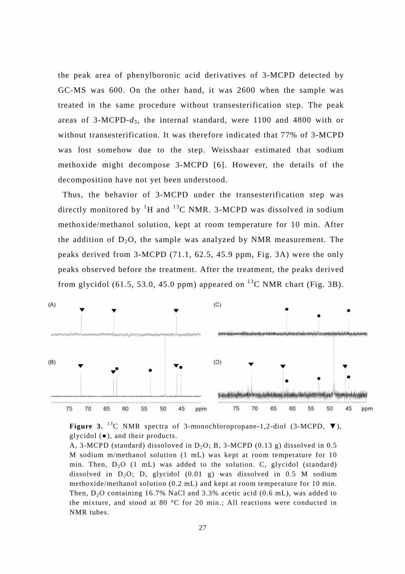
the peak area of phenylboronic acid derivatives of 3-MCPD detected by
GC-MS was 600. On the other hand, it was 2600 when the sample was
treated in the same procedure without transesterification step. The peak
areas of 3-MCPD-d5, the internal standard, were 1100 and 4800 with or
without transesterification. It was therefore indicated that 77% of 3-MCPD
was lost somehow due to the step. Weisshaar estimated that sodium
methoxide might decompose 3-MCPD [6]. However, the details of the
decomposition have not yet been understood.
Thus, the behavior of 3-MCPD under the transesterification step was
directly monitored by 1H and 13C NMR. 3-MCPD was dissolved in sodium
methoxide/methanol solution, kept at room temperature for 10 min. After
the addition of D2O, the sample was analyzed by NMR measurement. The
peaks derived from 3-MCPD (71.1, 62.5, 45.9 ppm, Fig. 3A) were the only
peaks observed before the treatment. After the treatment, the peaks derived
from glycidol (61.5, 53.0, 45.0 ppm) appeared on 13C NMR chart (Fig. 3B).
Figure 3. 13C NMR spectra of 3-monochloropropane-1,2-diol (3-MCPD, ▼), glycidol (●), and their products. A, 3-MCPD (standard) dissoloved in D2O; B, 3-MCPD (0.13 g) dissolved in 0.5 M sodium m/methanol solution (1 mL) was kept at room temperature for 10 min. Then, D2O (1 mL) was added to the solution. C, glycidol (standard) dissolved in D2O; D, glycidol (0.01 g) was dissolved in 0.5 M sodium methoxide/methanol solution (0.2 mL) and kept at room temperature for 10 min. Then, D2O containing 16.7% NaCl and 3.3% acetic acid (0.6 mL), was added to the mixture, and stood at 80 °C for 20 min.; All reactions were conducted in NMR tubes.
28
The conversion ratio was 37% calculated on their proton numbers obtained
by 1H NMR.The behavior of glycidol in the standard method option A was
also monitored by NMR. Glycidol was treated in the similar way to the
standard method steps 2-4 (transesterification to derivatization steps),
except n-hexane and phenylboronic acid was not added and D2O was used
instead of water (details of reaction conditions were given in the legend Fig.
3D). pH of the reaction mixture was 4.2. In addition to the peaks derived
from glycidol, those from 3-MCPD were newly observed by NMR after the
treatment (Fig. 3C, D). The conversion ratio from glycidol to 3-MCPD was
70%, when it was calculated on their proton numbers. Interestingly, the
conversion was not detected when the derivatization step (step 4) was
conducted at room temperature instead of 80 °C. On the other hand, the
conversion increased to nearly 100% when glycidol was directly dissolved
in D2O containing 16.7% NaCl and 3.3% acetic acid (pH 1.9), and stood at
80 °C for 20 min. It was thus indicated that the heating under the acidic
conditions at the derivatization step accelerated the conversion of glycidol
to 3-MCPD greatly.
Production of 3-MCPD derivatives at the derivatization step
As shown Fig. 3D, treatment of glycidol with steps 2-4 without
phenylboronic acid generated 3-MCPD with 70% of conversion. The
treatment was then conducted with phenylboronic acid. The reaction
conditions were the same with the standard method steps 2-4 except
n-hexane was not added and D2O was used instead of water. The conversion
of glycidol to 3-MCPD was again observed by 13C NMR (Fig. 4B). The
degree of conversion was 74% calculated on the proton numbers observed by
1H NMR. Suppose that the errors of integration value measured in 1H NMR
were ±5%, the conversion ratio were nearly the same with or without
phenylboronic acid at the derivatization step. On the other hand, the
3-MCPD phenylborate was not detectable (Fig. 4B). Association constant of
29
phenylboronic acid and diols was reported to drastically change at pH ~7.5,
and the phenylborate was hardly formed below pH 6.5 [7]. It was therefore
speculated that the 3-MCPD phenylborate were produced only in a small
amount in the experimental conditions of pH 4.2, and thus were undetectable
by NMR.
The reaction mixture was further extracted by n-hexane as the procedure
step 5. After the removal of the hexane phase, the aqueous phase was
analyzed by NMR again. Both of glycidol and 3-MCPD were observed by 13C
NMR (Fig. 4C), with the ratio of 13:87, calculated from the proton numbers
obtained by 1H NMR. These results indicated that the extraction of 3-MCPD
phenylborate, which was more hydrophobic than 3-MCPD, to the organic
phase was shifted the equilibrium of the three compounds, namely 3-MCPD
Figure 4. The conversion of glycidol to 3-MCPD under the condition of option A. The circle and triangle signs represent the peaks of glycidol and 3-MCPD, respectively. A, glycidol (standard) dissolved in D2O; B, glycidol (0.01 g) was dissolved 0.5 M sodium methoxide/methanol solution (0.2 mL) and kept at room temperature for 10 min. Then, D2O containing 16.7% NaCl and 3.3% acetic acid (0.6 mL) and of acetone-d6 containing 12.5% (w/v) phenylboronic acid (0.133 mL) were added to the mixture, and stood at 80 °C for 20 min.; C, After treatments described in B, the reaction mixture was washed by hexane (1 mL). The resulting aqueous phase was analyzed; all reactions were conducted in NMR tubes.
30
phenylborate, 3-MCPD, and glycidol in the aqueous phase. As a result, the
ratio of 3-MCPD against glycidol increased from 74% to 87%. It should be
noted that glycidol and 3-MCPD remained in the aqueous phase even after
the hexane extraction at step 5 of the standard method. The low efficiency of
the 3-MCPD phenylborate formation and extraction might explain the
relatively high standard deviations given in Table 1.
Discussion
It has been described that the epoxide ring-opening in glycidol and its esters
was incomplete by the acid treatment described in the DGF standard
methods C-III 18 (09), option B, and that the bidirectional conversion
between 3-MCPD and glycidol was observed by NMR in the course of the
method. The behaviors of 3-MCPD esters and glycidyl esters, which were
supposed to be in fats and oils, in the course of the standard method were
schematically shown in Fig. 5. 3-MCPD produced by the transesterification
using sodium methoxide was partly converted to glycidol in the step (37%).
There also is a possibility that 2-MCPD were converted to glycidol, though
it should be proven. The resulting glycidol, in addition to glycidol derived
from glycidyl esters, were partly converted to 3-MCPD in the following
steps, which were conducted in the presence of saturated NaCl under acidic
conditions at 80 °C (74%). Glycerol was not observed from glycidol under
the conditions, which could be explained that there were abandoned
chloride ions, which are highly nucleophylic, and little hydroxyl ions in the
solution. What important was that the degree of conversion from glycidol to
3-MCPD depended on the conditions of the procedure steps 2-5
(transesterification, derivatization, and extraction), and was not 100%. This
observation contradicted to that of Kuhlmann cited in ref. 8 that the
conversion was nearly complete. Based on our observation, the standard
method, option A, did not give combined amount of 3-MCPD esters and
glycidyl esters correctly. The removal of epoxides by the acid treatment
31
described in option B was not complete either, as shown in Table 2.
Therefore, the difference of the values obtained by options A and B did not
correspond to the amount of glycidyl esters.
The conversion of 3-MCPD to glycidol at the transesterification step was
estimated to be 37% by NMR, whereas that of glycidol to 3-MCPD at the
following steps was 74%. The loss of 3-MCPD in total was thus ca. 10%,
which was not consistent with the observation described in the section
‘bidirectional conversion of 3-MCPD and glycidol’, where the loss was
estimated to be 77% under the influence of the transesterification step. In
another report of ours, it was clarified that the loss was also caused by the
Figure 5. Behaviors of 3-MCPD esters and glycidyl esters under the procedure of DGF standard method C-III 18(09). R represents fatty acyl group. Arrows with solid lines represent the conventionally known/believed routes, whereas those with dotted lines represent routes newly confirmed in this study. Reagents and conditions: a) Sodium methoxide/methanol, rt, 10 min. b) Acetic acid, NaCl, water. c) Phenylboronic acid, 80 °C, 20 min. d) Hexane extraction.
32
low extraction capability of n-hexane used in the extraction step (step 5) [9].
The substitution of n-hexane to more polar solvent such as chloroform
increased the recovery of 3-MCPD derivatives. In the mean time, the
derivatives of 3-MCPD with phenylboronic acid were not observed in NMR
analysis in the aqueous phase at the derivatization step (step 4), although
they were detected by GC-MS in the hexane phase obtained in step 5. It was
thus estimated that the derivatives were rather formed in the course of
hexane extraction (Fig. 5), than at the derivatization step, and the polarity
of the solvent might affect to the production as well as the recovery of the
derivatives.
Conclusion
This chapter reports that bidirectional conversion was confirmed between
3-MCPD and glycidol in the course of the analycical procedure of DGF
standard methods C-III 18 (09), option A; 3-MCPD was partly converted to
glycidol at the transesterification step, and glycidol was converted partly to
3-MCPD at the derivatization step conducted at 80 °C under acidic
condition in the presence of NaCl. In addition, epoxide ring-opening of
glycidol and its esters was shown to be incomplete by the acid treatment
described in the method, option B. Thus, the standard method, option A, did
not give combined amount of 3-MCPD esters and glycidyl esters correctly,
and the difference of the values obtained by options A and B did not
correspond to the amount of glycidyl esters, either. The restricted
application of the standard method, option A, to glycidyl ester-free samples
is recommended. In addition, the conversion of 3-MCPD to phenylboronic
acid was not observed by NMR at the derivatization step. The derivatization
was estimated to rather occur in the following hexane extraction step. The
observations presented in this chapter are important for our understanding
to the standard method, and for the interpretation of the values so far given
by the standard method.
33
References
1. Food Advisory Committee 2000 (accessed Oct. 2010) Genotoxicity of
3-monochloropropane-1,2-diol, FdAC/Contaminants/48. Paper for
and ethyl acetate were purchased from Wako Pure Chemical Industries, Ltd.
(Osaka, Japan). NaCl and acetic acid were purchased from Nakalai Tesque
Co. Ltd. (Kyoto, Japan). Soybean oil was the product of Ueda Oils and Fats
MFG Co. Ltd. Other chemicals were of the analytical grade.
36
Quantification of 3-MCPD
The contents of 3-MCPD forming substances were determined based on DGF
standard methods C–III 18(09) with a slight modification. Typical procedure
for preparing samples was as follows; 0.1 g soybean oil mixed with 3-MCPD
was dissolved in 0.5 mL solvent consisted of t-buthyl methyl ether and ethyl
acetate (=4:1, vol/vol). To the sample, 2 µg of 3-MCPD-d5 and 1 mL of 0.5
M sodium methoxide/methanol solution was added and left for 10 min at
room temperature (step 1). The mixture was extracted using 3 mL n-hexane
and 3 mL water containing 16.7% NaCl and 3.3% acetic acid. The aqueous
phase was rinsed with 3 mL n-hexane (step 2). It was then mixed with 0.5 mL
derivatization reagent (ca. 0.125 g/mL phenylboronic acid solution) and left
at 80 °C for 20 min (step 3). Then, the extraction of 3-MCPD phenylborate
was conducted using 3 mL n-hexane, or other solvents (step 4). Organic
phase was collected, dried by evaporation and was dissolved again to 2 mL
2,2,4-trimethylpentane and filtered by paper before it was brought to GC-MS
analysis (step 5). Samples for calibration were prepared as follows; 2 µg of
3-MCPD-d5 dissolved in t-buthyl methyl ether was added to a test tube and
dried under N2 gas stream. Required amount of 3-MCPD and water
containing 16.7% NaCl were added to make the final volume of 3 mL. The
samples were subjected to steps 3-5 as described above. Analyses were
conducted 3 times independently and the mean values were presented.
GC-MS
GC-MS was conducted using GCMS QP 2010 (Shimadzu, Kyoto, Japan)
connected to DB-5 capillary column (30 m, 0.25 µm, Agilent Technologies,
Tokyo, Japan). The column temperature was controlled as follows; it was
kept at 60 °C for 1 min, then raised at 6 °C /min to 190 °C, further raised at
20 °C /min to 280 °C, and kept at 280 °C for 6 min. The temperature of
programmed-temperature vaporizer (PTV) injector was controlled as
follows; it was kept at 60 °C for 1 min, raised at 10 °C /min to 180 °C and
37
kept at 180 °C for 20 min. The temperatures of the interface and the ion
source were set at 250 °C and 200 °C. Other conditions for GC-MS were
same with those described in DGF standard methods C–III 18(09).
Results and Discussion
Comparison of sample preparation procedures by GC-MS
The analytical procedures for determination of 3-MCPD forming substances
by DGF standard methods C–III 18(09), option A consist of the following 5
steps; 1) transesterification of acyl glycerols and other esters by sodium
methoxide in the presence of 3-MCPD-d5, 2) removal of resulting fatty acid
methyl esters by hexane extraction, 3) derivatization of 3-MCPD in the
aqueous phase by phenylboronic acid, 4) extraction of 3-MCPD phenylborate
by n-hexane from aqueous phase, 5) GC-MS analyses of organic phase (for
experimental details, see ‘materiarls and methods quantificaton of
3-MCPD’ ). By conducting the analyses according to the standard method,
however, it was suspected that the detection limit of the method might be
different between the actual oil samples and the standard samples for
calibration.
Table 1 Comparison of procedure of DGF standard methods C-III 18.
Samples Peak area (A, arbitrary unit) Ratio
(Am/z196/Am/z 201)
Corrected
amount (µg)a m/z 196 m/z 201
Oil sampleb 090 1240 0.07 0.11
Calibration
samplec 260 4310 0.06 0.10
a) Correction was conducted based on the calibration curve given in Fig. 1. b) 0.1 g soybean oil mixed with 0.1 µg of 3-MCPD c) 1 µg of 3-MCPD
In order to confirm this, 0.1 µg 3-MCPD added to 0.1 g soybean oil was
analyzed by the standard method. As shown in Table 1, the peak areas of m/z
38
196 and 201, corresponding to 3-MCPD phenylborate and 3-MCPD-d5
phenylborate, were 90 and 1240, respectively. The peak areas were 260 and
4310, respectively, when the same amount of 3-MCPD was analyzed
according to the procedure to prepare samples for calibration, which
consisted of the aforementioned steps 3, 4, and 5. The ratio of the two peaks
were 0.07 (=90/1240) and 0.06 (=260/4310) and gave similar results (0.11
and 0.10 µg 3-MCPD) when they were corrected by the factor obtained by
the calibration (Fig. 1). It was thus confirmed that the peak areas in GC-MS
analyses of oil samples are 3-4 times smaller than those of the samples for
calibration.
Figure 1. Calibration curves using n-hexane and chloroform as extraction media. Analyses were conducted as described in section 2.2, using chloroform (●) instead of n-hexane (○) at step 4.
Effect of solvent in the recovery of 3-MCPD derivatives
The smaller peak areas obtained by the procedure of the standard method
(steps 1-5) implied the low recoveries of 3-MCPD phenylborate compared to
the procedure preparing samples for calibration. Here, we focused on step 4,
in which 3-MCPD phenylborate were extracted from the aqueous phase by
n-hexane. The recoveries might improve by substituting the conventional
solvent with more polar solvents (Table 2). As expected, n-butanol,
chloroform, and ethyl acetate, which have higher solvent polarity scale [4]
than n-hexane, successfully increased the recovery of 3-MCPD derivatives to
0
0.05
0.1
0.15
0.2
0.25
0.0 1.0 2.0 3.0 4.0 5.0
3-MCPD amount (x10-1µg)
Are
a 3-
MC
PD p
heny
lbor
ate
Are
a 3-
MC
PD-d
5ph
enyl
bora
te
0
0.05
0.1
0.15
0.2
0.25
0.0 1.0 2.0 3.0 4.0 5.00
0.05
0.1
0.15
0.2
0.25
0.0 1.0 2.0 3.0 4.0 5.0
3-MCPD amount (x10-1µg)
Are
a 3-
MC
PD p
heny
lbor
ate
Are
a 3-
MC
PD-d
5ph
enyl
bora
te
Are
a 3-
MC
PD p
heny
lbor
ate
Are
a 3-
MC
PD-d
5ph
enyl
bora
te
39
the organic phase to the relative extent of 5.6, 4.7, and 3.8, respectively.
Interestingly, when 0.1 g soybean oil containing 0.1 µg 3-MCPD was treated
by the standard method without transesterification step (step 1), the relative
recovery by n-hexane reached 4.9. The pH of the aqueous phase at step 4 was
4.2 and 1.9 with and without transesterification step, respectively. This
indicated that pH of the aqueous phase might affect the recovery of 3-MCPD
phenylborate. Weisshaar considered the low recovery of 3-MCPD-d5
(49-72%; coefficient of variation, 13.4%) by the standard method was caused
by the decomposition of 3-MCPD by sodium methoxide [5]. In another report
of ours, it was shown that the conversion of 3-MCPD to glycidol actually
observed by NMR [6]. However, it was shown in Table 2 that there should be
another reason; the low extraction capability of the organic phase caused the
low recovery. It might also explain the relatively large value of coefficient of
variation, 13.4%, described in ref. 5.
n-Butanol achieved the highest recovery. However, it co-extracted more
impurities, which had higher vaporization temperature and thus showed
Spiked amount (ppm)
0.00
0.01
0.02
0.03
0.04
0.05
0.06
0.07
0.0 0.2 0.4 0.6 0.8 1.0
0.07
0.00
0.01
0.02
0.03
0.04
0.05
0.06
0.0 0.2 0.4 0.6 0.8 1.0
Spiked amount (ppm)
(A) (B)
Are
a 3-
MC
PD p
heny
lbor
ate
Are
a 3-
MC
PD-d
5ph
enyl
bora
te
Are
a 3-
MC
PD p
heny
lbor
ate
Are
a 3-
MC
PD-d
5ph
enyl
bora
te
Spiked amount (ppm)
0.00
0.01
0.02
0.03
0.04
0.05
0.06
0.07
0.0 0.2 0.4 0.6 0.8 1.00.00
0.01
0.02
0.03
0.04
0.05
0.06
0.07
0.0 0.2 0.4 0.6 0.8 1.0
0.07
0.00
0.01
0.02
0.03
0.04
0.05
0.06
0.0 0.2 0.4 0.6 0.8 1.0
0.07
0.00
0.01
0.02
0.03
0.04
0.05
0.06
0.0 0.2 0.4 0.6 0.8 1.0
Spiked amount (ppm)
(A) (B)
Are
a 3-
MC
PD p
heny
lbor
ate
Are
a 3-
MC
PD-d
5ph
enyl
bora
te
Are
a 3-
MC
PD p
heny
lbor
ate
Are
a 3-
MC
PD-d
5ph
enyl
bora
te
Are
a 3-
MC
PD p
heny
lbor
ate
Are
a 3-
MC
PD-d
5ph
enyl
bora
te
Are
a 3-
MC
PD p
heny
lbor
ate
Are
a 3-
MC
PD-d
5ph
enyl
bora
te
Figure 2. Analyses of oils spiked with 3-MCPD. Soybean oil spiked with 3-MCPD was conducted as described in section 2.2. (A) n-hexane; (B) chloroform was used as extraction solvent at step 4. Expressions of the linear curve fittings were (A) y= 0.0457x + 0.0203, and (B) y = 0.0526x + 0.0118.
40
longer retention times than 3-MCPD phenylborate by GC analyses. The
impurities were undesirable for MS detector, though they would not disturb
the quantification of 3-MCPD phenylborate. In addition, n-butanol has the
highest boiling point (108 °C) among the four solvents tested, and was
hardest to be dried out before bringing the samples to GC-MS analyses.
the practical point of view, chloroform was chosen for further study.
Table 2 Effect of solvent on extraction of 3-MCPD derivatives.
Solvent Solvent polality scalea ) Relative peak area of
3-MCPD derivatives
n-Hexaneb 0.519 1.0
n-Butanolb 0.837 5.6
Chloroformb 0.786 4.7
Ethyl acetateb 0.795 3.8
n-Hexanec (without
transesterification step) 0.519 4.9
a) Ref. 3 b) 0.1 g soybean oil containing 1 µg of 3-MCPD was subjected to the procedure described in section 2.2. All analyses were conducted 3 times independently and the mean values were presented. c) The identical sample as a) was subjected to the procedure described in section 2.2, except that step 1 was omitted.
The calibration curve using chloroform as extraction solvent showed that
the solvent was as good as n-hexane for quantification of 3-MCPD
phenylborate (Fig. 1). In addition, analyses of oils spiked with 3-MCPD
(0.25~1.0 ppm) were conducted using the two solvents (Fig. 2). It was
obvious that the variations of data were smaller when chloroform was used.
In addition, the correlation coefficients (R-squared values) of the linear
curve fittings were 0.906 and 0.988, for n-hexane and chloroform,
respectively. The absolute values of x-intercepts (Fig. 2A, B) give the
estimated contents of 3-MCPD forming substances contained in the
41
un-spiked oil, which were calculated to be 0.44 and 0.22 ppm, respectively.
The latter value obtained using chloroform as solvents had higher reliability.
In conclusion, substitution of n-hexane with chloroform contributed to
increase the accuracy of DGF standard method, especially at the low
concentration.
Conclusion
By DGF standard methods C-III 18 for the determination of 3-MCPD, the
minimum limit of detection was lower in the case of actual oil samples
compared to the calibration samples. The problem was found to be lied in the
low recovery of 3-MCPD derivatives from the aqueous phase to the organic
phase at the extraction step of the standard procedure. The substitution of the
conventional solvent, n-hexane, with n-butanol, chloroform, and ethyl
acetate increased the recovery to the relative extent of 5.6, 4.7, and 3.9,
respectively. The modification contributed to improve the accuracy of the
method, especially at lower concentration (<1 ppm) of 3-MCPD.
This chapter provides the modification of DGF standard methods C-III
18(09) in order to improve the accuracy to quantify 3-MCPD at lower
concentration. It might be important for estimation and control of our daily
intake of 3-MCPD, and for the product control in the fat and oil processing.
References
1. Food Advisory Committee 2000 (accessed Oct. 2010) Genotoxicity of
3-monochloropropane-1,2-diol, FdAC/Contaminants/48. Paper for
Two synthetic methods for 2-MCPD have been reported, as far as we know,
although the evidential spectra were not assigned in either report. Ilczuk et
al. described the synthesis of 2-MCPD via glycerol 1,3-diacetate as a
synthetic intermediate from 1,3-dichloro-2-propanol. In this method [5],
however, 2,3-diacetoxy-1-propanol, the migrated isomer of the desired
product, was afforded as the main product by treatment with sodium acetate.
In contrast, Tsatsas et al. reported the preparation of 2-MCPD via
2-chloro-1,3-dibenzyloxypropane by aqueous or alcoholic hydrogen
chloride acidolysis [6]. 2-Chloro-1,3-dibenzyloxypropane was successfully
prepared, but no reaction was observed in the following deprotection step of
the benzyl ether and the original compound was recovered in good yield.
Other acidic treatments, for example, with trifluoroacetic acid, were not
effective. Hydrogenation using Pd/C was therefore used for deprotection.
The complete removal of thionyl chloride and the addition of formic acid
improved the deprotection by 50%. The NMR spectra and elemental
analysis of the resulting 2-MCPD (purity >99%) are described in the
Experimental Procedures section.
2- and 3-MCPD conversion to glycidol under basic conditions in the
course of DGF standard method C-VI 18 (10)
The dynamics of 3-MCPD and glycidol during DGF standard method C-III
18 (09) were monitored by 1H and 13C NMR in our previous research [3].
The results conclusively indicated that 3-MCPD and glycidol were
bidirectionally interconverted under different conditions: 3-MCPD to
50
glycidol under basic conditions, and glycidol to 3-MCPD under acidic
conditions. The dynamics of 2-MCPD has been assumed to be similar to that
of 3-MCPD, and 2-MCPD has been naturally believed to be converted to
glycidol under basic conditions, as in the case of 3-MCPD. However, this
has not yet been verified. The behavior of 2-MCPD in the transesterification
step was therefore monitored directly using 1H and 13C NMR.
When 2-MCPD (10 mg) was dissolved in D2O, the 13C peaks derived from
2-MCPD (63.4, 62.9 ppm) were the only peaks (Fig. 1a). 2-MCPD (10 mg)
was dissolved in sodium methoxide/methanol solution, and kept at room
temperature for 5 min (pH 10.8), the same as in the transesterification step.
After the addition of D2O, the sample was examined using NMR. After the
treatment, peaks derived from glycidol (61.5, 53.0, 45.0 ppm) appeared (Fig.
1b) in addition to those derived from 2-MCPD. The conversion was 3
Figure 1. 13C NMR spectra of 2-MCPD, 3-MCPD, glycidol, and their products. Diamonds, peaks derived from 2-MCPD; circles, glycidol; inverted triangles, 3-MCPD. (a) 2-MCPD (10 mg) dissolved in D2O (1 mL); (b) 2-MCPD (10 mg) dissolved in 0.5 M sodium methoxide/methanol solution (1 mL) was kept at room temperature for 4 min. Then, D2O (1 mL) was added to the solution. (c) 2-MCPD (10 mg) dissolved in 0.5 M sodium methoxide/methanol solution (0.2 mL) was kept at room temperature for 4 min. Then, D2O containing 20% sodium chloride and 0.9% sulfuric acid (0.6 mL) was added to the mixture, and it was allowed to stand for 5 min. (d) 3-MCPD (10 mg) dissolved in D2O (1 mL). (e) Method similar to (b), except using 3-MCPD (10 mg). (f) Method similar to (c), except using 3-MCPD (10 mg). All reactions were conducted in NMR tubes. The accumulation time for 13C NMR measurement took 20 min described in the section “Dynamics observation by NMR”, the signals served as an average for accumulation.
51
mole% after 5 min treatment, calculated from the proton numbers obtained
by 1H NMR (Fig. 2b). When 3-MCPD (10 mg) was treated similarly, the
peaks of 3-MCPD (71.2, 62.6, 45.9 ppm) and glycidol (61.5, 53.0, 45.0
ppm) were observed (Fig 1e). The conversion was 22 mole% after 5 min
treatment (Fig. 2b). When the sample concentration decreased to 2 mg, both
of their conversions increased to 4 mole% for 2-MCPD and 34 mole% for
3-MCPD (Fig. 2a). The conversions might differ depending on the sample
concentration. The treatment time (3.5-5.5 min) described in DGF standard
method C-VI 18 (10) is appropriate in order to minimize the generation of
glycidol.
Bidirectional conversion between 2- and 3-MCPD and glycidol under
standard method conditions
The samples in sodium methoxide/methanol solution were subsequently
treated with 20% NaCl and 0.9% sulfuric acid, and then allowed to stand for
Figure 2. Time dependence of 2-MCPD (triangle) and 3-MCPD (circle) conversion to glycidol. (a) MCPD (2 mg) and (b) MCPD (10 mg) dissolved in 0.5 M sodium methoxide/methanol-d4 solution (1 mL) was kept at room temperature. The conversion ratios were calculated from the proton numbers measured by 1H-NMR.
52
5 min, the same as in the extraction step of standard method C-VI 18 (10),
except that iso-hexane and phenylboronic acid were not added and D2O was
used instead of water. The pH of the reaction mixture was 3.9. In the case of
3-MCPD, only 3-MCPD (71.2, 62.6, 45.9 ppm) was generated from glycidol,
as reported in our previous research (Fig. 1f, ref. 3). In the case of 2-MCPD,
only peaks derived from 2-MCPD (65.8, 65.3 ppm) were observed (Fig. 1c),
though glycidol must be converted to only 3-MCPD. These results indicated
that both 2- and 3-MCPD were converted to glycidol under basic conditions,
and the generated glycidol was reconverted to the original MCPD under
acidic conditions.
In order to clarify the strange phenomenon, the memory effect, the same
treatment was conducted using a mixed system of 2-MCPD, 3-MCPD, and
glycidol. Equimolar amounts of 2-MCPD and glycidol treated under basic
conditions gave 2-MCPD (46 mole%) and glycidol (54 mole%), and the
(50 mole%) (Fig. 3e, 3f). This result indicated that both 2-MCPD and
3-MCPD were converted to glycidol, and the glycidol was reconverted to
the original 2- and 3-MCPD, respectively. Low pH treatment is essential for
the complete reconversion of glycidol to MCPD, as glycidol was clearly
observed in the samples that the acid treatment was conducted above pH 4.2
(eg. pH 5.5).
53
Overall, the above observations suggested that glycidol derived from 2- or
3-MCPD was chlorinated at the original position to mainly generate the
original MCPD by an unknown steric effect. Eliminated chloride anion from
MCPD might form a glycidol-chloride anion complex and recombined to
Figure 3. 13C NMR spectra of 2-MCPD, 3-MCPD, glycidol, and their products. Diamonds, peaks derived from 2-MCPD; circles, glycidol; inverted triangles, 3-MCPD; triangles, glycerol 2-methyl ether. (a) 2-MCPD (13 mg) and glycidol (13 mg) dissolved in 0.5 M sodium methoxide/methanol solution (1 mL) were kept at room temperature for 4 min. Then, D2O (1 mL) was added to the solution. (b) 2-MCPD (13 mg) and glycidol (13 mg) dissolved in 0.5 M sodium methoxide/methanol solution (0.2 mL) were kept at room temperature for 4 min. Then, D2O containing 20% sodium chloride and 0.9% acetic acid (0.6 mL) was added to the mixture, and it was allowed to stand for 5 min. (c) Method similar to (a), except using 3-MCPD (13 mg) and glycidol (13 mg). (d) Method similar to (b), except using 3-MCPD (13 mg) and glycidol (13 mg). (e) Method similar to (a), except using 2-MCPD (13 mg) and 3-MCPD (13 mg). (f) Method similar to (b), except using 2-MCPD (13 mg) and 3-MCPD (13 mg). The accumulation time for 13C NMR measurement took 20 min described in the section “Dynamics observation by NMR”, the signals served as average for accumulation.
54
glycidol to generate original MCPD. Unfortunately, the NMR observations
did not provide structural evidence of the glycidol-chloride anion complex.
Dynamics of MCPDs and glycidol in soybean oil
Soybean oil spiked with MCPDs was treated according to standard method
C-VI 18 (10), assay A. A small amount (< 2 mole%) of 3-MCPD was
observed in the sample of soybean oil spiked with 2-MCPD (Fig. 4a).
The correlation coefficients (R2 values) were >0.999 for 2-MCPD
phenylborate and 0.997 for 3-MCPD phenylborate. 3-MCPD was not
detected from 2-MCPD by NMR as described in the previous section, but
was detected by GC/MS, probably because of better sensitivity, although the
initial concentration was sufficiently high in the case of NMR. Similarly, a
small amount (<4 mole%) of 2-MCPD was observed in the sample spiked
with 3-MCPD (Fig. 4b). The interconversions of 2- and 3-MCPDs differed
by only a few per cent after treatments of the standard method. This could
Figure 4. Analyses of oils spiked with (a) 2-MCPD and (b) 3-MCPD. Soybean oil spiked with MCPDs was analyzed using DGF standard method C-VI 18 (10). Circles and triangles represent the concentration of detected 3- and 2-MCPD, respectively. (a) Linear curve-fitting equations are y = 0.02x + 0.064 for 3-MCPD, and y = 0.99x + 0.076 for 2-MCPD. (b) Linear curve-fitting equations are y = 1.37x − 0.085 for 3-MCPD and y = 0.06x − 0.053 for 2-MCPD. All analyses were carried out in duplicate.
55
be explained by supposing that the conversion of MCPDs to glycidol in the
transesterification step was approximately equal to that of glycidol to
MCPDs in the following extraction step.
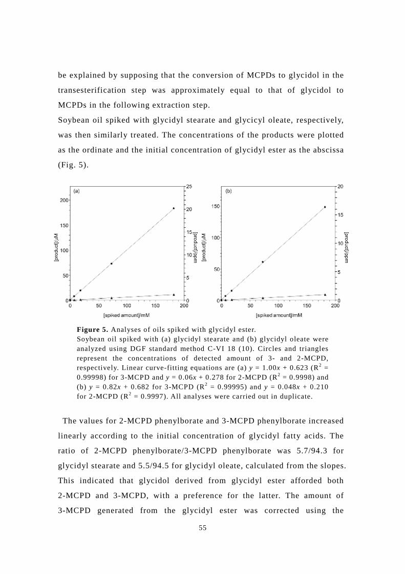
Soybean oil spiked with glycidyl stearate and glycicyl oleate, respectively,
was then similarly treated. The concentrations of the products were plotted
as the ordinate and the initial concentration of glycidyl ester as the abscissa
(Fig. 5).
The values for 2-MCPD phenylborate and 3-MCPD phenylborate increased
linearly according to the initial concentration of glycidyl fatty acids. The
ratio of 2-MCPD phenylborate/3-MCPD phenylborate was 5.7/94.3 for
glycidyl stearate and 5.5/94.5 for glycidyl oleate, calculated from the slopes.
This indicated that glycidol derived from glycidyl ester afforded both
2-MCPD and 3-MCPD, with a preference for the latter. The amount of
3-MCPD generated from the glycidyl ester was corrected using the
Figure 5. Analyses of oils spiked with glycidyl ester. Soybean oil spiked with (a) glycidyl stearate and (b) glycidyl oleate were analyzed using DGF standard method C-VI 18 (10). Circles and triangles represent the concentrations of detected amount of 3- and 2-MCPD, respectively. Linear curve-fitting equations are (a) y = 1.00x + 0.623 (R2 = 0.99998) for 3-MCPD and y = 0.06x + 0.278 for 2-MCPD (R2 = 0.9998) and (b) y = 0.82x + 0.682 for 3-MCPD (R2 = 0.99995) and y = 0.048x + 0.210 for 2-MCPD (R2 = 0.9997). All analyses were carried out in duplicate.
56
transformation factor (t) described in section 8.1.3 of standard method C-VI
18 (10), but the generation of 2-MCPD is currently neglected. The factor
relevant to 2-MCPD should be determined for accurate estimation of the
amount of glycidol ester because the value depends on the individual
experimental environment. Otherwise, removal of glycidol esters using an
adsorbent prior to the transesterification step [7] is recommended.
Quantification by GC/MS of 2-MCPD and its sensitivity compared with
3-MCPD
2-MCPD phenylborate, which forms a 1,3-cyclic ester, might have a
different sensitivity from that of 3-MCPD phenylborate because of the
different fragmentation patterns. Kuhlmann claimed that the sensitivity of
2-MCPD phenylborate calculated from FID intensities was approximately
twice that of 3-MCPD phenylborate. However, the true sensitivity should be
determined using a pure 2-MCPD standard. The quantification of 2-MCPD
phenylborate and its sensitivity compared with that of 3-MCPD
phenylborate were therefore investigated using two GC/MS systems,
SHIMADZU GCMS QP-2010 and Thermo Scientific ITQ 1100, in different
institutes. The mass unit of QP-2010 was composed of an electron impact
(EI) ionizer and a standard quadrupole (Q) detector, and that of ITQ 1100
was composed of an EI ionizer and an ion-trap unit before a Q detector
(Table 1).
Table 1. Peak area ratios of 2- and 3-MCPD phenylborates after treatment of MCPD spiked soybean oil (20 ng/mL) using DGF standard method C-VI 18 (10), assay A.
Instrument Peak area ratio of m/z 196 to m/z 201
2-MCPD 3-MCPDa
QP-2010 13.8 ± 0.1 4.6
ITQ1100 08.8 ± 0.0 ―
(a) A theoretical value is 4.0.
57
When soybean oil samples containing various concentrations of pure
2-MCPD and 3-MCPD-d5 were treated using standard method C-VI 18 (10),
the peak area ratio of m/z 196 (the precursor ion peak of 2-MCPD
phenylborate) to m/z 201 (the precursor ion peak of 3-MCPD-d5
phenylborate) was plotted against the spiked 2-MCPD concentrations. The
calibration curves were linear (Fig. 6). Although the slopes of the two lines
were different, both had correlation coefficients (R2 values) greater than
0.999.
From the slopes of the lines, the sensitivity of 2-MCPD phenylborate
compared with that of 3-MCPD phenylborate under the respective machine
conditions were found to be 3.26-fold (QP-2010) and 2.16-fold (ITQ 1100),
as shown in Table 2. The peak area ratio of the fragment ion of 3-MCPD-d5
phenyl borate (m/z 150) to the precursor ion of 3-MCPD-d5 phenyl borate
(m/z 201) gave constant values of 5.11 ± 0.20 (n = 9) and 3.30 ± 0.11 (n = 9),
Figure 6. Calibration curves of 2-MCPD obtained using two GC/MS systems. Circles and triangles represent the area ratio of 2-MCPD (m/z 196) phenylborate to 3-MCPD-d5 (m/z 201) phenylborate, detected using QP-2010 and ITQ1100, respectively. Linear curve-fitting equations are y = 3.26x + 0.306 for QP-2010 and y = 2.16x + 0.571 for ITQ1100. All analyses were carried out in duplicate.
58
measured by QP-2010 and ITQ 1100, respectively. The sensitivity of
2-MCPD phenylborate is referenced by the precursor ion of 3-MCPD-d5
phenylborate. Though 3-MCPD-d5 pheylborate fragments at different
efficiency for individual equipment due to the complex fragmentation, the
amount of 2-MCPD phenylborate ion is constant due to the stable precursor
ion.
Table 2. Ion ratio of the fragment to the precursor of 3-MCPD-d5 phenylborate and the sensitivity ratio of 2-MCPD phenylborate to 3-MCPD phenylborate obtained from GC/MS measurements.
Instrument
Ion-ratio of fragment
(m/z 150) to precursor
(m/z 201) of 3-MCPD-d5
Sensitivity ratio of 2-MCPD
phenylborate to 3-MCPD-d5
phenylborate
Correction
constant
QP-2010 5.11±0.20 3.26±0.02 0.64
QP-2010a 5.42±0.49 3.45±0.01 0.64
ITQ 1100 3.30±0.11 2.16±0.01 0.66
(a) The measurement conducted on a different day.
Thus, the sensitivity ratio of 2-MCPD phenylborate depends on the ion ratio
of the fragment (m/z 150) to the precursor (m/z 201) of 3-MCPD-d5
phenylborate. The ratio of the sensitivity of 2-MCPD to the ion-ratio of the
fragment to the precursor 201 of 3-MCPD-d5 is constant (0.65±0.01, Table
2). A measurement by QP-2010 in another day gave same results.
Since the ion-ratio of 3-MCPD-d5 can be adjusted as mentioned above, the
quantification of 2-MCPD without a pure 2-MCPD standard might be
achieved by using the sensitivity ratio (peak intensity ratio of 2-MCPD
phenylborate (m/z 196) to 3-MCPD-d5 phenylborate (m/z 201)) and the
ion-ratio of the fragment to the precursor (peak intensity ratio of the
fragment (m/z 150) to the precursor (m/z 201) of 3-MCPD-d5 phenylborate)
obtained using any instrument. The suggested formula is
59
(1)
The term SF i150/SF i201 in Eq. 1 is the observed fragmentation efficiency.
So,
(2)
where
w2-MCPD is the mass fraction, in mg/kg, of 2-MCPD;
w3-MCPD-d5 is the mass fraction, in mg/kg, of 3-MCPD-d5;
SF i201 is the area of 3-MCPD-d5 (m/z 201);
SF i150 is the area of 3-MCPD-d5 (m/z 150);
SF2 is the area of 2-MCPD (m/z 196);
SR in Eq. 1 is the sensitivity ratio of 2-MCPD phenylborate (m/z 196) to
3-MCPD-d5 phenylborate (m/z 201) obtained using our GC/MS instrument
(see Table 2);
IR in Eq. 1 is the ion-ratio of the fragment to the precursor of 3-MCPD-d5
phenylborate obtained using our GC/MS instrument (see Table 2); and
CC in Eq 2 is a correction constant (0.65 ± 0.01).
Suggested dynamics of MCPDs and glycidol in reported assays
The suggested dynamics of MCPDs and glycidol in previously reported
assays are discussed based on our NMR and GC/MS results in this section.
In the DGF standard method C-VI 18 (10), assay A (Fig. 7), 2-MCPD and
3-MCPD were partly converted to glycidol by basic treatment with
methanolic sodium methoxide, and the generated glycidols were mostly
(>96 mole%) converted to the original 2-MCPD and 3-MCPD by subsequent
acidic treatment with aqueous sulfuric acid and sodium chloride. The
glycidol was unchanged by the basic treatment, whereas the glycidol was
60
converted to 2-MCPD (5.6 mole%) and 3-MCPD (94.4 mole%) by the acidic
treatment. In the standard method, the true amount of 3-MCPD is calibrated
using the transformation factor (t). However, as the total amount of
2-MCPD and 3-MCPD is not consistent with the initial amount of glycidyl
Figure 7. Suggested dynamics of MCPDs and glycidol during proposed methods. Arrows and broken arrows represent the path observed using NMR and the minor path observed only in GC/MS, respectively. DGF standard method C-VI 18 (10), assay A: the first arrows represent basic treatment with methanolic sodium methoxide, and the second arrows represent acidic treatment with sulfuric acid/sodium chloride. DGF standard method C-VI 18 (10), assay B: the first arrows represent basic treatment with methanolic sodium methoxide, and the second arrows represent acidic treatment with sulfuric acid/sodium bromide. Ermacora method: the first arrows represent acidic treatment with sulfuric acid/methanol, and the second arrows represent neutral treatment with sodium sulfate.
61
ester (Fig. 5), the transformation factor needs to be further corrected,
considering the conversion to 2-MCPD, for accurate calibration.In the
course of the method using acidified sodium bromide solution at the
extraction step in assay B, glycidol was claimed to be converted to both
2-monobromopropnanediol (2-MBPD) and 3-MBPD [4]. There is a
possibility that MBPDs could also be generated by sodium bromide
treatment of glycidols, generated from MCPDs under the basic conditions in
the previous transesterification step. To investigate the possibility that
2-MCPD-derived glycidol is converted to 3-MBPD, NMR spectra were
measured after treatments with basic sodium methoxide and then acidic
sodium bromide. Peaks similar to 2-MCPD were obtained in the 13C-NMR
(Fig. 8) and 1H NMR spectra (data not shown). These peaks were assigned
to 2-MCPD, based on the chemical shifts, as unconverted 2-MCPD
remained from the previous step (Fig. 8b) and one chemical species was
observed after the treatment with sodium bromide. These results suggested
that glycidol generated from 2-MCPD might be reconverted to the same
2-MCPD by the recombination of the eliminated chloride ion under the
conditions of standard method C-VI 18 (10), assay B, even in the presence
of an excess amount of bromide ions. This means that the amounts of
MCPDs do not change before and after the treatment with sodium bromide,
and that the MCPDs can be quantified correctly. However, the amount of
glycidyl esters should be estimated based on both 2- and 3-MBPD
phenylborate because glycidyl-ester-derived glycidol is converted to both 2-
and 3-MBPD. If the peaks shown in Fig. 8c were assigned to 2-MBPD,
although the possibility is low, the amount of glycidyl esters cannot be
quantified by standard method C-VI 18 (10), assay B, but 3-MCPD can be
correctly quantified because the possibility of conversion of 3-MCPD to
3-MBPD should be equal to that of conversion of the reference 3-MCPD-d5.
2-MCPD can be quantified correctly for the same reason, but glycidol
cannot be quantified from the increase in MBPDs from MCPDs.
62
In the course of the method using methanolic sulfuric acid as an acidic
transesterification reagent, proposed by Ermacora et al. [8], 2-MCPD and
3-MCPD remain unconverted in the transesterification step. Complete
demineralization of chloride ions by a pre-cleaning step and elimination of
glycidol by acidic treatment of an oil sample enable the accurate and precise
quantification of MCPDs.
Conclusion
2-MCPD was synthesized and first quantitatively analyzed by DGF standard
method C-VI 18 (10). Preparation of a calibration curve using a pure
2-MCPD standard is not required for the quantification. The amount can be
determined by a calculation to correct the ion-ratio of the fragment to the
precursor of 3-MCPD-d5 phenylborate of the individual GC/MS in any
Figure 8. 13C NMR spectra of 2-MCPD and their products. (a) 2-MCPD (10 mg) dissolved in D2O. (b) 2-MCPD (10 mg) dissolved in 0.5 M sodium methoxide/methanol solution (1 mL) was kept at room temperature for 10 min. Then, D2O (1 mL) was added to the solution. (c) 2-MCPD (10 mg) dissolved in 0.5 M sodium methoxide/methanol solution (0.2 mL) was kept at room temperature for 10 min. Then, D2O containing 20% sodium bromide and 0.9% acetic acid (0.6 mL) was added to the mixture, and it was allowed to stand at 80 °C for 20 min. Peaks with same chemical shifts as those of 2-MCPD were obtained; all reactions were conducted in NMR tubes. Diamonds, peaks derived from 2-MCPD; circles, glycidol.
63
laboratory. In addition, the amounts of 2-MCPD in previous samples or
previous data could be re-analyzable using the peak intensity of 2-MCPD
and the peak are of 3-MCPD-d5 fragment (m/z 150).
The comprehensive dynamics and analyses of MCPDs and glycidol in the
course of the treatments in DGF standard method C-VI 18 (10) have been
presented in this chapter. The direct observation of MCPDs and glycidol by
NMR in the course of the analytical procedures of the standard methods and
indirect observations by GC/MS revealed the following dynamics: MCPDs
were partly converted to glycidol in the basic transesterification step, and
the glycidol was mainly converted to the original MCPD isomers in the
extraction step conducted under acidic conditions in the presence of NaCl
(or NaBr). Isomerization between 2- and 3-MCPDs was imperceptible in the
less-sensitive NMR observations. However, GC/MS analyses indicated that
2- and 3-MCPD spiked in soybean oil respectively were converted to
2-MCPD (98 mole%) and 3-MCPD (2 mole%), and 2-MCPD (4 mole%) and
3-MCPD (96 mole%), in the course of standard method C-VI 18 (10), assay
A. In addition, glycidyl ester spiked in soybean oil was converted to
2-MCPD (5.5-5.7 mole%) and 3-MCPD (94.3-94.5 mole%) in this method.
The amount of converted 2-MCPD should therefore be considered in the
quantification of glycidyl esters, or they should be removed prior to the
alkaline transesterification. Acidic transesterification is also recommended
instrument (Shimadzu, Kyoto, Japan) was equipped with an AOC-20i
auto-sampler and a DB-5ms capillary column (30 m × 0.25 mm, 0.25 µm;
Agilent Technologies, Santa Clara, CA). The column temperature was
controlled as follows: 85 °C for 0.5 min; 85 to 150 °C at a rate of 6 °C/min;
150 to 180 °C at a rate of 12 °C/min; 180 to 280 °C at a rate of 25 °C/min;
and 280 °C for 7 min. High-purity helium was used as the carrier gas at a
constant flow rate of 40 cm/s. Two microliters of sample were injected into
the system in the pulsed splitless mode. The temperature of the
programmed-temperature vaporizer injector for the Shimadzu GCMS
QP-2010 instrument was controlled as follows: maintained at 85 °C; 85 to
160 °C at a rate of 250 °C/min; maintained at 160 °C for 9.9 min; and 160 to
350 °C at a rate of 250 °C. The temperatures of the interface and the ion
source were set at 250 and 200 °C, respectively. A mass-selective detector
was used to monitor selected ions with m/z values of 147, 150, 196 and 201.
3-MCPD and 2-MCPD was quantified using 3-MCPD-d5 as an internal
standard by the following equation (1) [19] and equation (2) [18],
respectively;
× (1)
× × (2)
Where:
W3-MCPD is the mass fraction, in mg/kg, of 3-MCPD;
74
W2-MCPD is the mass fraction, in mg/kg, of 2-MCPD;
W3-MCPD-d5 is the mass fraction, in mg/kg, of 3-MCPD-d5;
SFi150 is the area of 3-MCPD-d5 (m/z 150);
SF3 is the area of 3-MCPD (m/z 196);
SF2 is the area of 2-MCPD (m/z 196);
CC is a correction constant (0.65 ± 0.01).
Results
Hydrolysis of MCPD dioleate by pancreatic lipase
2-MCPD esters are regioisomers of the corresponding 3-MCPD esters, and
these compounds have also been detected in fats and oils. However, to the
best of our knowledge, the metabolism of these compounds by pancreatic
lipase has not yet been studied in detail. With this in mind, we synthesized
2-MCPD-1,3-dioleate, which has been reported to be one of the most
common FA species found in oils and fats [6], and evaluated its hydrolysis
with porcine pancreatic lipase. This investigation was conducted to develop
a deeper understanding of the hydrolytic profiles of MCPD esters following
their ingestion, using 3-MCPD-1,2-dioleate as a control (Fig. 1). The
products of the hydrolysis reactions were analyzed by HPLC-CAD, which
allowed for the separation and detection of the MCPD esters, as well as the
different regioisomers of the MCPD mono- and di-esters, which were
produced as intermediates during the hydrolysis (Fig. 2).
The results revealed a significant decrease in the detected amount of
2-MCPD-1,3-dioleate from 18.0 to 8.4 µmol after 3 h of incubation with
75
pancreatic lipase at 37 °C, as well as the formation of free 2-MCPD (6.1
µmol) and free oleic acid (11.8 µmol) (Fig. 1a). The results also revealed
that the amount of 2-MCPD-1-oleate present in the mixture increased for the
first 0.5 h to reach approximately 3 µmol, where it reached a plateau.
The results for 3-MCPD-1,2-dioleate revealed a similar trend to the other
isomer, with the amount of substrate decreasing from 18.9 to 7.7 µmol after
3 h of incubation with pancreatic lipase at 37 °C. In contrast to the 2-isomer,
the hydrolysis of 3-MCPD-1,2-dioleate resulted in the formation of a large
amount of the monoester 3-MCPD-2-oleate (9.8 µmol), whereas the amount
Figure 1. Hydrolysis reactions of 2- and 3-MCPD dioleate with pancreatic lipase.Twenty micromoles of MCPD dioleate and 2 mg of pancreatic lipase in 1.2 mL of 50 mM Tris-HCl buffer (pH 8.0) containing 2.7 mM deoxycholic acid and 1 mM CaCl2 was stirred at 37 °C. Free MCPD was measured by GC-MS following the extraction of MCPD and the corresponding esters into a chloroform/methanol solution, whilst the MCPD esters were measured by HPLC-CAD. a, filled square, 2-MCPD-1,3-dioleate; open triangle, 2-MCPD-1-oleate; filled circle, 2-MCPD; diamond, oleic acid. b, filled square, 3-MCPD-1,2-dioleate; filled triangle, 3-MCPD-1-oleate; open triangle, 3-MCPD-2-oleate; filled circle, 3-MCPD; diamond, oleic acid. All of the reactions and analyses were conducted in triplicate and the results reported as the mean values ± SD.
76
of oleic acid (10.8 µmol) was similar in both cases (Fig. 1b). Notably, the
hydrolysis of 3-MCPD-1,2-dioleate resulted in the accumulation of a small
amount of the monoester 3-MCPD-1-oleate (0.7 µmol). The accumulation of
free 3-MCPD was also small (2.2 µmol) compared to that of free 2-MCPD
from 2-MCPD-1,3-dioleate.
These results indicated that the two acyl groups of 2-MCPD-1,3-dioleate
were being readily hydrolyzed by pancreatic lipase under these conditions
to produce 2-MCPD, and that the acyl group at the primary position of
3-MCPD-1,2-dioleate was being hydrolyzed preferentially to produce
3-MCPD-2-oleate.
Figure 2. Standard HPLC-CAD chromatogram for MCPD ester analysis. For HPLC conditions, see Materials and Methods. a, Tri-palmitate; b, 2-MCPD-dioleate; c, 3-MCPD-dioleeate; d, 3-MCPD-dilinolenate; e, glycidyl-oleate; f, glycidyl-linoleate; g, 1,3-dioleate; h, oleic acid; i, 1,2-dioleate; j, 3-MCPD-1-oleate; k, 2-MCPD-1-oleate; l, 3-MCPD-2-oleate; m, 3-MCPD-1-linolenate
77
Hydrolysis of MCPD monooleate by pancreatic lipase
To further investigate the hydrolytic characteristics of pancreatic lipase, we
synthesized the three different regioisomers of MCPD monooleate and
subjected them to the reaction with pancreatic lipase (Fig. 3).
The results revealed that the amount of 2-MCPD-1-oleate decreased with
time, whereas almost identical amounts of 2-MCPD and oleic acid were
produced (Fig. 3a). The amount of 3-MCPD-1-oleate decreased in a similar
manner to that of 2-MCPD-1-oleate, with similar amounts of 3-MCPD and
oleic acid being produced at the same time (Fig. 3b). The hydrolysis
reactions of 2-MCPD-1-oleate and 3-MCPD-1-oleate almost reached
completion in 3 h under the reaction conditions used in the current study. In
contrast, the hydrolysis of 3-MCPD-2-oleate occurred at a much slower rate
Figure 3. Hydrolysis reactions of 2- and 3-MCPD monooleate by pancreatic lipase. The conditions for the lipase reactions were the same as those described in Fig. 1, except MCPD monooleate was used instead for MCPD dioleate. a, filled square, 2-MCPD-1,3-dioleate; open triangle, 2-MCPD -1-oleate; filled circle, 2-MCPD; diamond, oleic acid. b and c, filled square, 3-MCPD-1,2-dioleate; open triangle, 3-MCPD-1-oleate; filled triangle, 3-MCPD-2-oleate; filled circle, 3-MCPD; diamond, oleic acid.
78
under these conditions, with only 4 µmol of the substrate being consumed
after 3 h (Fig. 3c), representing only 1/5 of the consumed amount of 2- and
3-MCPD-1-oleate. Furthermore, only 4 µmol of 3-MCPD was released from
3-MCPD-2-oleate after 3 h, and the amount of 3-MCPD-1-oleate was less
than 1.5 µmol after 3 h of hydrolysis. These results therefore suggested that
the hydrolysis of an acyl group at the sn-2 position of MCPD or the transfer
of an acyl group from the sn-2 position to the primary position of MCPD was
a slow process. To the best of our knowledge, this study represents the first
detailed evaluation of the hydrolytic behaviors of synthetic
2-MCPD-1-oleate and 3-MCPD-2-oleate in the presence of porcine
pancreatic lipase, as well as their reaction products.
Hydrolysis of MCPD dioleate by pancreatin
The hydrolysis reactions of the MCPD mono- and di-esters were further
studied using pancreatin from porcine pancreas (Fig. 4). The composition of
the products resulting from this reaction was expressed as the relative molar
concentration of each MCPD-containing compound to the sum of free
MCPD, MCPD monooleate and MCPD dioleate. The hydrolysis of
2-MCPD-1,3-dioleate under these conditions gave free 2-MCPD, which
reached 26 mol% after 3 h of hydrolysis, while the levels of
2-MCPD-1-oleate after 1 and 3 h were 17–18 mol% (Fig. 4a). In contrast,
3-MCPD-1,2-dioleate was mainly converted to 3-MCPD-2-oleate, with the
formation of only small amounts of 3-MCPD-1-oleate and 3-MCPD (Fig.
4b). This observation was therefore consistent with the above results
79
obtained using pancreatic lipase (Fig. 1).
Hydrolysis of MCPD monooleate by pancreatin
The three different regioisomers of MCPD monooleate were also hydrolyzed
with pancreatin (Fig. 5). 2-MCPD-1-oleate and 3-MCPD-1-oleate were
hydrolyzed by approximately 80% after 3 h of incubation to mainly afford 2-
and 3-MCPD, respectively (Fig. 5a and b). In contrast, 3-MCPD-2-oleate was
hydrolyzed by only 15% after 3 h (Fig. 5c). This observation was therefore
consistent with the previous result described above using pancreatic lipase
(Fig. 3).
Figure 4. Hydrolysis reactions of 2- and 3-MCPD dioleate by pancreatin. The conditions used for the pancreatin reactions were the same as those described in Fig. 1, except the amount of pancreatin was 0.4 mg. The compositions have been expressed as the relative molar contents of each MCPD-containing compound compared with the sum of free MCPD, MCPD monooleate and MCPD dioleate. a, gray box, 2-MCPD-1,3-dioleate; mesh-like pattern box, 2-MCPD-1-oleate; dotted box, 2-MCPD. b, gray box, 3-MCPD-1,2-dioleate; mesh-like pattern box, 3-MCPD-1-oleate; filled box, 3-MCPD-2-oleate; dotted box, 3-MCPD. All of the reactions and analyses were conducted in triplicate and the results reported as the mean values.
80
Notably, this reaction also lead to the formation of around 5 mol% of both
3-MCPD-1-oleate and 3-MCPD-dioleate, which indicated the occurrence of
intra- and/or intermolecular acyl transfer processes from the sn-2-position
to the primary of MCPD under the experimental conditions.
Penetration of 2- and 3-MCPD by Caco-2 cell monolayer
Caco-2 cells were cultured on Transwell® inserts under standard conditions
for 3 weeks to allow for the differentiation of the cells and the formation of
an epithelial cell like-monolayer. To the apical chamber was added a 0.1 or
1.0 mM (Fig. 6a or 6b) solution of 2- or 3- MCPD. The results of our
preliminary trials involving the addition of 2- or 3-MCPDs to the apical
chamber revealed that these substrates remained unchanged. The theoretical
concentrations of the MCPDs at equilibrium were 2.75 and 27.5 µg/mL for A
and B respectively, because the ratio of the volumes in apical and basolateral
Figure 5. Hydrolysis reactions of 2- and 3-MCPD monooleate by pancreatin. The conditions used for the pancreatin reactions were identical to those described in Fig. 4, except MCPD monooleate was used as a substrate instead of MCPD dioleate. The compositions have been expressed as the relative molar contents of each MCPD-containing compound compared with the sum of free MCPD, MCPD monooleate and MCPD dioleate. a, gray box, 2-MCPD-1,3-dioleate; mesh-like pattern box, 2-MCPD-1-oleate; dotted box, 2-MCPD. b and c, gray box, 3-MCPD-1,2-dioleate; mesh-like pattern box, 3-MCPD-1-oleate; filled box, 3-MCPD-2-oleate; dotted box, 3-MCPD.
81
chambers was 1:3. The results also revealed simultaneous decreases in the
levels of 2- and 3-MCPDs in the apical chamber, which were accompanied by
increases in the levels of these compounds in the basolateral chamber. This
result therefore indicated that MCPD had translocated from the apical
chamber to the basolateral chamber to reach equilibrium (Fig. 6a).
Minor differences were observed between the 2- and 3-MCPD, which were
consistent with those of previous reports [15, 16]. It is noteworthy that the
translocation profiles observed in the current study for the 2- and 3-MCPD
were similar when the concentration of the MCPD solution placed in the
Figure 6. Permeabilities of 2- and 3-MCPD by the Caco-2 cell monolayer method. a, 0.1 mM; b, 1.0 mM 2- or 3-MCPD solution in HBSS buffer (0.5 mL) was added to the apical chamber. After incubation at 37 °C, the solutions in the apical and basolateral chambers were collected, together with the Caco-2 cells. After the extraction of MCPD into a chloroform/methanol solution, the amount of free MCPD was measured by GC-MS. The theoretical concentration of each MCPD at equilibrium (a, 2.75 µg/mL; b, 27.5 µg/mL) was expressed by the dotted line. a and b, filled circle, 2-MCPD-apical; filled triangle, 3-MCPD-apical; open circle, 2-MCPD-basolateral; open triangle, 3-MCPD-basolateral. All of the sample preparations and analyses were conducted in triplicate and the results reported as the mean values ± SD.
82
apical chamber was increased from 0.1 to 1 mM (Fig. 6b). These results
therefore suggested that MCPD were passing through the Caco-2 cell
monolayer via a simple diffusion mechanism rather than an active
transportation system.
Permeability of the 2- and 3-MCPD monoester through a Caco-2 cell
monolayer
The results described above for the hydrolysis reactions of MCPD diesters
with pancreatic lipase and pancreatin suggested that 2-MCPD-1-oleate and
3-MCPD-2-oleate were the primary hydrolytic products to reach small
intestine (Fig. 1, 3-5). In the previous study, 3-MCPD-1-monoester but not
3-MCPD-2-monoester was subjected to the permeability study [15]. Thus,
3-MCPD-2-oleate as well as 2-MCPD-1-oleate and 3-MCPD-1-oleate were
subjected to a permeability study using a Caco-2 cell monolayer (Fig. 7a-c).
The total amount of 3-MCPD-2-oleate in the system decreased by half after 6
h (Fig. 7c), but it was not detected in the basolateral chamber and the cells.
On the other hand, the amount of 3-MCPD in the apical chamber increased
with time, and reached its maximum value of 15 nmol after 2 h, before
decreasing slightly. The amount of 3-MCPD in the basolateral chamber
increased after 2 h, reaching its maximum value of approximately 10 nmol
after 6 h. Similar trends were also observed for 2-MCPD-1-oleate (Fig. 7a)
and 3-MCPD-1-oleate (Fig. 7b).
83
In contrast that the hydrolysis of 3-MCPD-2-oleate by pancreatic lipase or
pancreatin was not observed (Fig. 3, 5), MCPD-monoesters, regardless of
their regioisomeric forms, were suggested to be hydrolyzed in the presence
of Caco-2 cell monolayer.
Even the amount of 2-MCPD-1-oleate or 3-MCPD-2-oleate added to the
apical chamber was increased 10-fold, and even though the detected amount
of MCPD monooleate decreased 170–190 nmol after 6 h (Supplementary Fig.
1a, c), they were still not detected in the basolateral chamber. The amount of
Figure 7. Permeabilities of the 2- and 3-MCPD-monooleates as determined using Caco-2 cells. The conditions for the transport experiments were the same as those described in Fig. 6, except 0.1 mM MCPD monoester solution were used instead for free MCPD after 5 min sonication. After the extraction of MCPD and its esters into a chloroform/methanol solution, the free MCPD was measured by GC-MS, and the MCPD esters were measured by HPLC-CAD. a, filled triangle, 2-MCPD-1-oleate-apical; filled circle, 2-MCPD-apical; open triangle, 2-MCPD-basolateral; open diamond, 2-MCPD-cell. b, filled triangle, 3-MCPD-1-oleate-apical; filled circle, 3-MCPD-apical; open triangle, 3-MCPD-basolateral; open diamond, 3-MCPD-cell. c, filled triangle, 3-MCPD-2-oleate-apical; filled circle, 3-MCPD-apical; open triangle, 3-MCPD-basolateral; open diamond, 3-MCPD-cell.
84
the corresponding 2- or 3-MCPD accumulated in the apical chamber was
around 30 nmol. When the experiments were conducted using Transwell
inserts in the absence of a Caco-2 cell monolayer, the amounts of
2-MCPD-1-oleate and 3-MCPD-2-oleate decreased similarly without
accumulation of free MCPD (Supplementary Fig. 1d-f). The lack of mass
balance in this experiments (Supplementary Fig. 1a-c) can thus be
explained by the incomplete recovery of the MCPD monoesters from the
Transwell chambers under the current experimental conditions. MCPD
dioleates were also subjected to the permeability study (data not shown).
Although the detected amount of 2-MCPD-1,3-dioleate or
3-MCPD-1,2-dioleate in the apical chamber decreased by half, they were
not detected in the basolateral chamber. 2- or 3-MCPD monooleate was not
detected in any of the fractions recovered from the Caco-2 cell monolayer
system, either. In addition, the detected amount of free MCPD was
negligible, approximately 0.5 mol% of the original amount of dioleate. In
addition, the detected amount of free MCPD was negligible, approximately
0.5 mol% of the original amount of dioleate. Thus, it was suggested that
MCPD diesters remained unchanged in the presence of Caco-2 cell
monolayer, and that they were not penetrated the monolayer. The results
were partly consistent with the previous reports [15, 16]; free MCPD was
detected from 2-MCPD-dipalmilate, but not from 3-MCPD-dipalmitate
under similar transport conditions using a Caco-2 cell monolayer.
85
Supplementary Figure 1. Permeabilities of the 2- and 3-MCPD-monooleates as determined using Caco-2 cells. The conditions for the transport experiments were the same as those described in Fig. 7, except the concentration of 2- and 3-MCPD monooleate was increased from 0.1 to 1.0 mM, and using Transwell inserts in the presence or in the absence of Caco-2 cell monolayer. a, filled triangle, 2-MCPD-1-oleate-apical; filled circle, 2-MCPD-apical; open triangle, 2-MCPD-basolateral; open diamond, 2-MCPD-cell. b and c, filled triangle, 3-MCPD-1-oleate-apical; filled diamond, 3-MCPD-2-oleate-apical; filled circle, 3-MCPD-apical; open triangle, 3-MCPD-basolateral; open diamond, 3-MCPD-cell. d, 2-MCPD-1-oleate-apical in the presence (filled triangle) and in the absence (open square) of Caco-2 cell monolayer. e, 3-MCPD-1-oleate-apical in the presence (filled triangle) and in the absence (open square) of Caco-2 cell monolayer. f, 3-MCPD-2-oleate-apical in the presence (filled diamond) and in the absence (open square) of Caco-2 cell monolayer.
86
Discussion
The European Food Safety Authority (EFSA) reported in 2013 that the major
contributors of the human exposure to 3-MCPD are processed oil and fats in
their fatty acid ester form [20]. EFSA also reported in 2011 that toxicity of
3-MCPD-dipalmitate was lower than free 3-MCPD in a 90-day toxicological
study on rats [11]. Recently, Onami et al. reported that 3-MCPD ester was
not in vivo genotoxin but had potential to be subchronically toxic to the
similar degree to their free form, with the no-observed-adverse-effect levels
of 3-MCPD ester to be 8-15 mg/kg body weight per day in rats [21, 22].
The toxicity of 3-MCPD-diester and 3-MCPD-1-monoester has been
evaluated in vitro and in vivo, previously [11, 16, 21, 22]. However, the
metabolism of 3-MCPD-diester after ingestion had not been monitored in
detail, let alone 2-MCPD-diester, which are also detected in fats and oils.
Moreover, 3-MCPD-1-monoester is not a natural metabolite of diester as we
presented in this study; we showed that pancreatic lipase mainly hydrolyzed
3-MCPD-dioleate to produce 3-MCPD-2-oleate, and that further hydrolysis
of 3-MCPD-2-oleate to produce 3-MCPD was slow. The hydrolysis of
3-MCPD-2-oleate was not significant by pancreatin, either. Pancreatin
prepared from pancreas is an enzyme mixture and known to contain
proteases, amylases and others in addition to lipases. Our results suggested
that pancreatin might not contain enzymes with esterase activity to
hydrolyze 3-MCPD-2-monoester, and that 3-MCPD-diester after injection
should reach to small intestine mainly as 3-MCPD-2-monoester in addition
to the remaining diester. In this report, it was presented that
87
3-MCPD-2-oleate was hydrolyzed to produce free 3-MCPD in the presence
of Caco-2 cells, implying the possibility of in vivo hydrolysis of
3-MCPD-2-monoester in the presence of epitherial cells of small intestine,
followed by the intestinal absorption as free 3-MCPD form (Fig. 8). Our
observation is consistent with the previous one that 3-MCPD-1-monoester
was hydrolyzed to produce 3-MCPD by the supernatant of Caco-2 cell
culture [15].
Abraham et al. reported that the oral administration of 3-MCPD to rats
increased free 3-MCPD levels in blood to the maximum within 10 min,
whereas the administration of 3-MCPD-dipalmitate increased free 3-MCPD
levels in blood to the maximum after 2 h [10]. Here, the maximum level was
1/5 compared to the administration of the free 3-MCPD. Orally administered
3-MCPD-dipalmitate was mainly recovered in small intestine first, then in
Figure 8. Estimated disposition of MCPD and their esters. FFA free fatty acid
88
large intestine with the maximum recovery of 70% in total. This could be
interpreted that roughly 20-30% of 3-MCPD-dipalmitate was digested and
absorbed as free form, whereas the rest remain in the intestine as ester
form. MCPD ester was not detected in any organs or blood, other than
intestine. In the report, however, it was not exactly identified if the
remaining MCPD ester was in di- or mono-ester form. Our in vitro
observation were consistent the above study [10], and provided additional
implication that the MCPD ester detected in the intestine after the oral
administration to rat might be a mixture of 3-MCPD diester and
3-MCPD-2-monoester mainly.
In conclusion, it was observed that the two acyl groups of
2-MCPD-1,3-dioleate were readily hydrolyzed by pancreatic lipase and
pancreatin to produce 2-MCPD. In contrast, the hydrolysis of
3-MCPD-1,2-dioleate occurred preferentially at the primary acyl position to
produce 3-MCPD-2-oleate. 2-MCPD-1-oleate and 3-MCPD-1-oleate were
further hydrolyzed to 2- and 3-MCPD by pancreatic lipase and pancreatin.
Meanwhile, the hydrolysis of 3-MCPD-2-oleate was approximately 80%
slower than that of 3-MCPD-1-oleate. The in vitro evaluation of the
intestinal absorption of these compounds using a Caco-2 cell monolayer
model revealed that the MCPD-monooleates were hydrolyzed to give the
free MCPD in the presence of Caco-2 cells, whereas the MCPD-dioleates
remained unchanged. The free MCPD permeated the Caco-2 monolayer,
most probably via a diffusion mechanism, because the permeation profile
was independent of the MCPD dose. Minor differences were observed
89
between the permeation profiles of 2- and 3-MCPD.
Overall, it was estimated that 2- and 3-MCPD-diesters would be digested to
free or monoester form by pancreatic lipase, and the resulting monoesters
would further be hydrolyzed, though not completely, to free form in the
presence of epitherial cells of small intestine to be absorbed. Absorption may
not occur in mono or di-ester form.
Conclusion
The regioisomers of the di- and mono-oleate of MCPD have been synthesized
and subsequently hydrolyzed with pancreatic lipase and pancreatin to
estimate the intestinal digestion and absorption of these compounds after
their intake. The hydrolysates were analyzed by HPLC using a corona
charged aerosol detection system, which allowed for the separation and
detection of the different regioisomers of the MCPD esters. The hydrolysates
were also analyzed by GC-MS to monitor the free MCPD. The results
indicated that the two acyl groups of 2-MCPD-1,3-dioleate were smoothly
hydrolyzed by pancreatic lipase and pancreatin to give free 2-MCPD. In
contrast, the hydrolysis of 3-MCPD-1,2-dioleate proceeded predominantly at
the primary position to produce 3-MCPD-2-oleate. 2-MCPD-1-oleate and
3-MCPD-1-oleate were further hydrolyzed to free 2- and 3-MCPD by
pancreatic lipase and pancreatin, although the hydrolysis of
3-MCPD-2-oleate was 80% slower than that of 3-MCPD-1-oleate. The
intestinal absorption characteristics of these compounds were evaluated in
vitro using a Caco-2 cell monolayer. The results revealed that the
90
MCPD-monooleates, but not the MCPD-dioleates, were hydrolyzed to
produce the free MCPD in the presence of the Caco-2 cells. The resulting
free MCPD permeated the Caco-2 monolayer most likely via a diffusion
mechanism because their permeation profiles were independent of the dose.
Similar permeation profiles were obtained for 2- and 3-MCPDs.
REFERENCES
1. List of classifications, Volumes 1–114, International Agency for
Research on Cancer (IARC) Monographs on the evaluation of
carcinogenic risks to humans, IARC,
http://monographs.iarc.fr/index.php., [accessed October 2015]
Chapter 4 : Estimation of the cause for fatty acid esters of 3-MCPD and glycidol formation and development of method to reduce their presence in refined oils
Introduction
Palm oil is produced at a rate of about 60 million tons per year and is one of
the most used edible oils in the world [1]. It accounted for 36% of the world
vegetable oil production in 2014 (Fig. 1). Palm oil is 40-50% palmitic acid.
The melting point of saturated fatty acids, such as palmitic acid, is higher
than unsaturated fatty acids, and palm oil is solid at room temperature. On
the other hand, a high content of saturated fatty acids, which do not contain
double bonds that are easily oxidized, contributes to the superior oxidative
stability of palm oil. Furthermore, palm oil is produced at a lower cost than
other oils, partly because palm trees require 10 times less land than other
oil-producing crops. For these properties, palm oil is widely used in foods,
for example, in margarine and shortening, and for the frying oil of instant
noodles and potato chips. Thus, palm oil is important for the oils and fats
industry.
Indonesia and Malaysia are the main producers of palm oil, and together
account for 85% of the world palm oil production. In these countries, crude
palm oil (CPO) is first pressed from palm fruits similarly to other oil crops,
and is subjected to the refining steps of degumming, bleaching, and
deodorization. These steps remove free fatty acids and colorants and the end
product can be qualified as edible oil.
The resulting oil is called Refined Bleached Deodorized Palm Oil
(RBDPO), and is exported to Japan, where it is further refined to remove
remaining free fatty acids and odors by deodorization (Fig. 2).
96
MCPD esters and glycidyl esters, which are undesirable compounds in
edible oils, are detected at very low concentrations in CPO, but at about 10
ppm in RBDPO, a much higher level. Thus, it has been deduced that the
majority of MCPD esters and glycidyl esters are generated during the
deodorization step [2, 3]. The industrial process for deodorization is
conducted at a high temperature (230-250°C) by reduced pressure with
steaming and removes impurities from the oil. Furthermore, palm oil refined
by ‘chemical refining’, in which free fatty acids are removed by
neutralization, contains very little MCPD esters and glycidyl esters,
compared to oil produced by ‘physical refining’ (Fig. 2)[2, 3]. This
observation is consistent with the conclusion that MCPD esters and glycidyl
esters are generated during deodorization, and it is the high temperature that
affects their formation [4, 5, 6].
In addition, analyses of MCPD esters in various edible oils have been
carried out both domestically and internationally. Palm oil as well as corn
oil was reported to contain relatively high amounts of 3-MCPD esters and
glycidyl esters, whereas soy and canola oils contained much less [7, 8, 9,
Figure 1. The world production of major vegetable oils (2014) (from Japan Oilseed Processors Association)
97
10]. Palm and corn oils contain relatively high levels of monoacylglycerols
(MAG) and diacylglycerols (DAG), whereas soy and canola oils do not.
Therefore, it has been presumed that the amount of partial acylglycerols in
crude oils might be related to MCPD ester formation during deodorization
[6, 11].
However, there has not been direct proof that these partial acylglycerols
are the source of MCPD/glycidyl esters in refined oil. In this study, refined
oil spiked with MAG or DAG was subjected to the deodorization test, to
evaluate the correlation between partial acylglycerols and the formation of
MCPD/glycidyl esters in the deodorization step. It was confirmed that
MCPD/glycidyl esters were generated in the deodorization process in
proportion to the MAG contents.
Furthermore, these results suggested that removal of MAG from oils might
be effective at preventing the formation of MCPD/glycidyl esters in the
Figure 2. Physical and Chemical refining of palm oil
98
deodorization process. To evaluate this effect, MAG was partially removed
from the samples by short path distillation. The amounts of MCPD/glycidyl
esters in oils were measured by the DGF standard method after
deodorization.
Experimental Procedures
Materials and Methods
3-MCPD was purchased from Kanto Chemical Co. (Tokyo, Japan).
glycerol and all of the other reagents used in the current study were
purchased from Kanto Chemical Co, Wako Pure Chemical Industries and
Nakalai Tesque Inc. (Kyoto, Japan). All of these materials were purchased
as the analytical grades unless otherwise specified.
High DAG Oil Production
High DAG oil was prepared by transesterification of canola oil (600 g) with
glycerol (27 g) using 14% sodium hydroxide solution (0.4 g). The mixture
was heated to 180 °C, at a pressure below 1.3 kPa for 30 min. The resulting
oil was washed with water, and bleached with activated clay (GALLEON
EARTH). High DAG oil contained 5.1% MAG, 41.5% DAG and 52.6%
triacylglycerol (TAG).
99
Oil Bleaching
Oil samples were heated to 110°C under ordinary atmospheric pressure with
3 vol % of activated clay. The mixture was stirred at 200 rpm for 5 min at
110°C, and filtered through paper to remove activated clay from the oil.
Bleached oil was subjected to short path distillation and deodorization.
Short Path Distillation
Short path distillation was carried out with Type2-03 WIPRENE
(KOBELCO ECO-SOLUTIONS Co. Ltd, Kobe, Japan). Oil was heated to
240°C and passed the distillator at a flow rate of 10 mL/min. The pressure
was controlled and kept below 2.7 Pa by a vacuum pump (PVD-180 pump,
ULVAC Inc., Kanagawa, Japan).
Deodorization
MAG and DAG were added to canola oil, which did not contain
MCPD-forming substances (MCPD-FS) (< 0.2 ppm), to give a final content
of 0.5, 1, 2, 5, and 10 wt %. The mixed oil (300 g) was subjected to the
deodorization test, which was carried out in a 1 kg scale deodorizer made of
glass. Oil was heated to the designated temperature (230/240/250 °C), and
deodorization was run for 60 min, at a pressure below 10 mmHg with
steaming.
Contents of Partial Acylglycerols
An oil sample (200 mg) was dissolved in hexane or hexane/aceton (1:1, by
vol). The contents of partial acylglycerols were determined using
GC-2014A device (Shimadzu, Kyoto, Japan) connected to a DB-1ht
capillary column (30 m, 0.25 mm, 0.25 µm, Agilent Technologies, Tokyo,
Japan). The column temperature was controlled as follows: it was kept at
140°C for 1 min, raised at 10°C/min to 250°C, kept at 250°C for 5 min,
further raised at 5°C/min to 300°C, and kept at 300°C for 19 min. The
100
carrier gas was high-purity helium at a constant flow of 25.5 cm/s. One
microliter of sample were injected in pulsed split mode. The temperature of
the injector and the detector were 350°C and 380°C, respectively. Peak
area % was considered to be equal to wt %.
Determination of MCPD-forming substances by DGF standard method
C-VI 18 (10)
The amounts of MCPD-forming substances (MCPD-FS) in oil samples were
determined as described in DGF standard method C-VI 18 (10) [13], which
expressed as the total amount of MCPD esters and glycidyl esters. The
protocol was slightly modified by using free-form 3-MCPD-d5 instead of
the ester-form of palmitic acid as an internal standard. Assay A: soybean oil
(100 mg) mixed with a spiking component was dissolved in 100 µL of
tert-butyl methyl ether. To the sample, a toluene solution of 3-MCPD-d5
(5.0 µg/mL) and 0.5 M sodium methoxide/methanol solution (200 µL) were
added, and the mixture was left for 3.5–4.5 min at room temperature.
3-MCPD-d5 was used instead of the esters in this work in order to neglect
the transesterification efficiency. Sodium chloride/sulfuric acid solution
(200 g/L with 0.9% H2SO4, 600 µL) was added to the solution, and then
iso-hexane (600 µL) was added and the mixture was left for 5 min. The
aqueous phase was washed twice with iso-hexane, and the products formed
were extracted three times with a mixed solvent (600 µL, ether/ethyl acetate
= 6/4, v/v). The organic phase was dried over sodium sulfate. A
phenylboronic acid/ether solution (10–100 µL) was added to the solution,
and solvent was removed using a gentle nitrogen stream. Isooctane (1.5 mL)
was added to the residue and filtered using paper with sodium sulfate. The
solution was analyzed by GC/MS.
101
GC/MS quantification
GC/MS was carried out using a SHIMADZU GCMS QP-2010 instrument
equipped with an AOC-20i auto-sampler, connected to a DB-5ms capillary
column (30 m, 0.25 mm, 0.25 µm, Agilent Technologies). The column
temperature was controlled as follows. It was maintained at 85 °C for 0.5
min, raised at rates of 6 °C/min to 150 °C, 12 °C/min to 180 °C, and
25 °C/min to 280 °C, and maintained at 280 °C for 7 min. The carrier gas
was high-purity helium at a constant flow of 40 cm/s. Two microliters of
sample were injected in pulsed splitless mode. The temperature of the
programmed-temperature vaporizer injector for the SHIMADZU GCMS
QP-2010 instrument was controlled as follows. It was maintained at 85 °C,
raised at a rate of 250 °C/min to 160 °C, maintained at 160 °C for 9.9 min,
and further raised at a rate of 250 °C/min to 350 °C. The temperatures of the
interface and the ion source were set at 250 °C and 200 °C, respectively.
Other conditions for GC/MS were the same as those described in DGF
standard method C-VI 18 (10). A mass-selective detector was used for
selected ion monitoring, focusing on ions with m/z values of 147, 150, 196,
and 201.
Results and Discussion
Deodorization of canola oil with added MAG or DAG
MCPD-FS were detected in processed oil made from crude oil contained
relatively high amounts of MAG and DAG. Thus, it has been suggested that
MAG and DAG were precursors of MCPD or glycidyl esters that formed
during the deodorization process. However, to the best of our knowledge,
there has not been direct proof that these partial acylglycerols are the
sources of MCPD/glycidyl esters in refined oil.
In this study, refined canola oil spiked with MAG or DAG was subjected to
the deodorization test, to evaluate the correlation between partial
acylglycerols and the formation of MCPD/glycidyl esters in the
102
deodorization step. First, it was confirmed that canola oil, MAG, and DAG
did not contain 3-MCPD-FS. The amounts of 3-MCPD-FS in oil samples
were measured by DGF standard method C-VI 18 (10), which expressed as
the total amount of MCPD esters and glycidyl esters.
When oil with MAG added to 0.5% was subjected to the deodorization
process conducted at 250 °C for 60 min, which is the standard condition of
oil refining, the detected amount of 3-MCPD-FS was 0.7 ppm. Furthermore,
the detected amounts of 3-MCPD-FS increased as more MAG was added,
and reached to 56.6 ppm when 10.0 % MAG was added to the oil (Fig. 3).
Thus, it was estimated that 3-MCPD-FS were generated in the deodorization
process in proportion to the MAG concentration. When the temperature of
deodorization was reduced to 230 °C, the amounts of 3-MCPD-FS generated
were 1.8 and 9.6 ppm in oil added with 5.0 % and 10.0 % MAG, respectively.
These results indicated that the generation of 3-MCPD-MS was dependent
both on the MAG content and on the temperature of the deodorization
process.
Figure 3. Deodorization of canola oil with MAG or DAG addition MAG (a) or DAG (b) was added to canola oil to give a final content of 0.5, 1, 2, 5, 10 wt%. Oil was deodorized at 230 or 250 °C for 60 min, at the pressure below 10 mmHg. The contents of MCPD-forming substances (MCPD-FS) in oil samples were measured by DGF standard method C-VI 18 (10), which expressed as the total amount of MCPD esters and glycidyl esters. a, b Open circle, distillation at 250 °C ; filled triangle, distillation at 230 °C. All of the tests and analyses were conducted once.
103
In contrast, the detected amounts of 3-MCPD-FS were only 3.0 ppm when
oil with DAG added to 10 % was subjected to the deodorization test at
250 °C for 60 min. Thus, the contents of DAG in oil affected the generation
of 3-MCPD-FS only weakly compared with MAG.
These results suggested that pre-processing to reduce MAG from oils and
fats might be effective at preventing 3-MCPD-FS formation during the
deodorization process.
Mitigation of 3-MCPD/glycidol esters formation during the
deodorization process, by removing MAG from oil
To evaluate the effect of MAG reduction on 3-MCPD/glycidol esters
formation during the deodorization process, the concentration of MAG was
reduced in the oil sample by short path distillation. The resulting oil was
subjected to the deodorization test, conducted at 240 °C for 60 min (Table
1).
After the deodorization test of CPO, which contained 0.2 % MAG and
4.9 % DAG, 1.8 ppm of 3-MCPD-FS was detected in CPO. When the
content of MAG and DAG was reduced to 0.1 % and 2.0 % by short path
Table 1 Mitigation of 3-MCPD/glycidyl esters formation by reducing the MAG content of oil before the deodorization process. MAG was partially removed from CPO and high DAG oil (DAG40), through short path distillation. The resulting oil samples were subjected to deodorization (CPO at 240 °C, DAG40 at 230 °C, for 60 min). The contents of MCPD-forming substances (MCPD-FS) in oil samples were measured by DGF standard method C-VI 18 (10), which expressed as the total amount of MCPD esters and glycidyl esters.
104
distillation of CPO prior to the deodorization test, the quantity of
3-MCPD-FS produced was reduced from 1.8 ppm to 0.8 ppm.
Next, high DAG oil (DAG 40), which contained 5.1 % MAG and 41.5 %
DAG, was subjected to the deodorization test. Even though the temperature
of the test was set low at 230 °C, 25.9 ppm of 3-MCPD-FS was generated.
Then, the content of MAG was reduced to below 0.1 % from high DAG oil
by short path distillation. The quantity of 3-MCPD-FS produced was
reduced to 11.7 ppm, after the deodorization test conducted under the same
condition.
These results indicated that preprocessing to reduce MAG was effective in
suppressing the generation of 3-MCPD-FS during the deodorization step.
Conclusion
In this study, refined canola oil spiked with MAG and DAG was
subjected to the deodorization process. The amount of MCPD esters and
glycidyl esters produced during deodorization increased depending on
the added quantity of MAG. The effect was smaller with DAG addition
compared to that with MAG addition. These results suggested that
preprocessing to reduce MAG in oils and fats might be effective at
suppressing the generation of those esters during the deodorization
process. In fact, after reducing the amount of MAG in crude palm oil
and high DAG oil using short path distillation, the production of MCPD
esters and glycidyl esters in the deodorization step was successfully
reduced. This study, for the first time, provides direct proof that MAG is
the main cause of the undesirable esters produced during the
deodorization process.
REFERENCES
1. Japan Oilseed Processors Association
http://www.oil.or.jp/kiso/seisan/seisan02_01.html [accessed June 2016]