Page 1
TITLE PAGE
ANTILIPIDEMIC EFFECT OF WATER (H20) EXTRACT OF
DESMODIUM VELUTINUM LEAVES ON ALBINO WISTAR RATS
BY
BARRATT MARY ENI
BC/2009/282
SUBMITTED TO THE
DEPARTMENT OF BIOCHEMISTRY
IN THE FACULTY OF NATURAL SCIENCES
CARITAS UNIVERSITY AMORJI NIKE, ENUGU STATE.
IN PARTIAL FULFILMENT OF THE REQUIREMENT FOR THE
AWARD OF BACHELOR OF SCIENCE (B.SC) DEGREE IN
BIOCHEMISTRY
AUGUST, 2013.
Page 2
CERTIFICATION
This is to certify that this report titled antilipidemic effect of water extracts
of Desmodium velutinum leaves on albino wistar rats submitted by Barratt
Mary Eni BC/2009/282 is a borne-fide record of the project work carried out
by her under my supervision.
………………………….. Date………..
Mr. Eze-Steven, Peter
(Supervisor)
…………………….. Date……………
Mr. Ezenwali Moses
Head of Department
………………………… Date: ……………
External Supervisor
Page 3
DEDICATION
This project work is dedicated to the TRINITY “GOD THE FATHER, GOD
THE SON AND GOD THE HOLY SPIRIT”, for their divine favour.
Page 4
ACKNOWLEDGMENT
My special thanks goes to the almighty God for his grace and provision to
finish this project.
My gratitude goes to CARITAS UNIVERSITY and the chancellor (Very
Rev. Fr. Prof. E.M.P.Edeh C.ss.p, for his noble institution.
My gratitude goes to my supervisor Mr. .Eze- Steven, Peter for his advice
and words of encouragement throughout this project.
My sincere gratitude goes to my lovely and caring parent (Mr. & Mrs. ENI
BARRATT), brothers and sisters for their financial support and
encouragement.
I acknowledge my H.O.D (Mr. Ezenwali Moses), my lecturers: Dr. Yusuf,
Dr. Ishiwu, Dr. Ikpe and Mr. Ugwudike and my friends for their contribution
has helped in the production of this work.
Page 5
LIST OF TABLES AND FIGURES
Table 1-- -- -- -- -- -- -- -- -- -- -- 14
Table 2-- -- -- -- -- -- -- -- -- -- -- 39
Table 3-- -- -- -- -- -- -- -- -- -- -- 43
Table 4-- -- -- -- -- -- -- -- -- -- -- 44
Figure 1-- -- -- -- -- -- -- -- -- -- -- 18
Figure 2-- -- -- -- -- -- -- -- -- -- -- 20
Page 6
TABLE OF CONTENTS
Title Page -- -- -- -- -- -- -- -- -- -- i
Certification -- -- -- -- -- -- -- -- -- ii
Dedication -- -- -- -- -- -- -- -- -- -- iii
Acknowledgment -- -- -- -- -- -- -- -- -- iv
List of Tables and Figures -- -- -- -- -- -- -- -- v
Table of Contents -- -- -- -- -- -- -- -- -- vi
Abstract -- -- -- -- -- -- -- -- -- -- ix
CHAPTER ONE
1.0 Introduction -- -- -- -- -- -- -- -- -- 2
1.2 Background of the Study - -- -- -- -- -- -- 2
CHAPTER TWO
2.0 Literature Review -- -- -- -- -- -- -- -- 3
2.1 Desmodium velutinum -- -- -- -- -- -- -- 3
2.1.2 Biological and Medicinal -- -- -- -- -- -- -- 5
2.1.3 Uses of other species Desmodium velutinum -- -- -- -- 5
2.2 Plants for Antilipidemic -- -- -- -- -- -- -- 7
2.2.1 Advantage of Medicinal Plants for Antilipidemic/
Cardiovascular Diseases over Synthetic Drugs -- -- -- 10
2.3 Lipids and Lipoprotein -- -- -- -- -- -- -- 11
2.3.1 Classification of Lipoprotein -- -- -- -- -- -- 13
Page 7
2.3.2 Function of lipoprotein -- -- -- -- -- -- -- 14
2.4 Lipoprotein Disorder -- -- -- -- -- -- -- -- 17
2.5 Antilipidemic Drug (Atorvastatin) -- -- -- -- -- 18
2.5.1 Other Antilipidemic Drugs -- -- -- -- -- -- 22
2.5.2 Other therapies -- -- -- -- -- -- -- -- 26
2.5.3 Health Side Effects of Atorvastatin -- -- -- -- -- 27
CHAPTER THREE
Materials and Methods -- -- -- -- -- -- -- -- 28
3.1 Materials -- -- -- -- -- -- -- -- -- -- 28
3.1.1 Chemicals and Reagents -- -- -- -- -- -- -- 29
3.2 Plant Material -Collection and Identification -- -- -- -- 30
3.3 Extraction -- -- -- -- -- -- -- -- -- 30
3.4 Phytochemical Analysis -- -- -- -- -- -- -- 31
3.5 Experimental Animal Model -- -- -- -- -- -- 35
3.6 Collection of Blood Samples -- -- -- -- -- -- 38
3.7 Lipid Profile Analysis -- -- -- -- -- -- -- 38
CHAPTER FOUR
RESULT
4.0 Statically Analysis -- -- -- -- -- -- -- -- 45
4.1 Phytochemical Results -- -- -- -- -- -- -- 45
Page 8
CHAPTER FIVE
DISCUSSION AND CONCLUSION
5.1 Discussion -- -- -- -- -- -- -- -- -- 48
5.2 Conclusion -- -- -- -- -- -- -- -- -- 51
References -- -- -- -- -- -- -- -- -- -- 52
Page 9
ABSTRACT
This study evaluated the antilipidemic activity of water extracts from leaves
of Desmodium velutinum on albino wistar rats. The phytochemical analysis
of the leaf extract showed the presence of tannins, saponins, alkaloids,
soluble carbohydrates, flavonoids, reducing sugar, steroids, cyanide and
terpenoids. The animals were treated with known drugs (atorvastatin 2ml).
There were significantly reductions in HDL 13.00 1.41 mg/dl LDL 1.20
0.14 mg/dl and triglyceride 39.00 0.14 mg/dl, compared with water extract
of Desmodium velutinum (0.5ml). when administered was found to
significantly reduce lipid plasma which was LDL 1.90 0.00mg/dl,
triglyceride 50.00 0.00mg/dl and increase HDL 25.00 0.00 mg/dl which
is the good cholesterol. The water extract of Desmodium velutinum leaf can
possibly normalize the plasma lipid when compared with the group given
atorvastatin. The phytochemicals analysis showed that the association
between these complexes and compounds and other constituent play an
important role in the biological activity of the leaf. This study suggested that
the water extract of D.velutinum leaf posses hypolipidemic as well as
antilipidemic effect.
Page 10
CHAPTER ONE
INTRODUCTION
1.1 BACKGROUND OF STUDY
Lipid and lipoprotein abnormalities play a major role in the
development and progression of coronary artery diseases. Low levels of high
density lipoprotein cholesterols have been identified as independent
coronary risk factors (Rodrigue et al., 2010). High level of blood cholesterol
is responsible for circulatory system disorder. Increase level of low density
lipoprotein (LDL) is alarming for cardiovascular diseases and their risk is
increased many times (Harman et al., 2011).
In developing countries, the occurrence of heart diseases increases
rapidly (Nordestgard et al., 2010). Medical studies show that about 70% of
adults over 50 years old suffer atherosclerosis. (Sherien and Azza, 2009). A
large number of synthetic hypolipidemic drugs are available in market. Long
term use of these drugs cause serious side effects, and are costly.
A medicinal plant is any plant which in one or more of its organ,
contains substance that can be used for therapeutic purpose or which is a
precursor for synthesis of useful drugs (Sivakumar et al., 2007). Plant
contains a large number of bioactive phytochemicals that are responsible for
pharmacological action of plants and used for development of drugs. Many
Page 11
medicinal plants have shown their antilipidemic effect and proved their
efficacy in cardiovascular diseases (Nordestgard et al., 2010; Wang, 1999).
One of such plants used very often in the management of the disease
by the traditional medicine practitioners of Eastern Nigeria is Desmodium
velutium, a perennial plant erect or semi-erect shrub or sub-shrub up to 3m
light. It is widely distributed in subtropical Asia and tropical Africa. (Amowi
and Azode, 2012).
Extracts of Desmodium velutium are used traditionally in some
disease conditions particularly aphrodisiac and headache. Hence,
Desomdium velutium may be a source of a pharmacological active agent
useful in the treatment of aches, pains and diarrhoea. In Ghana, native
doctors mix the root of Desmodium lasincarpum with some hot peppers and
use it as enema to cure blood in urine. In Eastern States of Nigeria, the plant
locally known as “Ikeagwuani”. (Onyegbule et al., 2012). In these present
studies, I investigated the antilipidemic activity of the water (H2O) extract of
Desmodium velutinum Leaves on albino wistar rats.
Page 12
CHAPTER TWO
2.0 LITEFATURE REVIEW
2.1 DESMODIUM VELUTIMUM
Desmodium velutinum is an upright woody perennial herb, sub-shrub
or shrub, up to 3m tall (Onyegbule, et al., 2012).
Leaflets are light green to blue, green or darker above, paler beneath
and with prominent pallid veins, roundish to elliptic to rhombic ovate, 3-
9.4cm long , 2-7cm wide, entire or repaid (indented at the termination of the
lateral veins) and ciliate, think, mostly soft velvety on both Surface with
long stiff golden, reddish or white hairs. Inflorescence arises in left axis, and
at the end of branches. They are densely flowered racemes up to 10cm or
mere long; at least the terminal ones often paniculately branched. Flowers
are white pink to blue, mauve or brilliant purple, usually appearing whitish
or pale when dry.
Common names – velvet leaf D, villous LD
Hind: Jagru, Lagavang, Lippa-Pank
Malayalam: Orial, Sanskrit, Prasnipani
Tamil: Akilametaki, Amcapatayilni, Ankachupati
Telugu: Cliua madu, chimanduri
India: Chilkiboota
Page 13
India: Lalkan while local name: Ikeagwuani (Igbos)
Botanical Name: Desmodium velutinum
Family: Fabaceae (Pea family)
Order: Fabales Class: Magnoliopsida, Division: Magnoliophyta, Genus:
Desmodium (Garg, 2006)
Other species of Desmodium include Desmodium lasiocarpum, Desmodium
gangeticm, Desmodium repandum, Desmodium styracitolium, Desmodium
diffusum, Desmodium heterophyllum, Desmodium triflorum, etc.
Desmodium velutinum can adapt to a wide range of soil PH, from very
acid (pH 4.0) to alkaline. It also prefers more humidity climates
of >1000>3000mm rainfall 1 year, tolerates up to 5 months dry season. It
grows at altitudes from 0.1-500m with average temperature above 200C and
has some shade tolerances as it grows in forest verges in New Guinea.
2.1.1 BIOLOGICAL AND MEDICINAL USE OF DESMODIUM
VELUTINUM
The water extract of Desmodium velutinum is use as an aphrodisiac (a
substance that increases sexual desire, example of such plants includes
tumera aphrodisiac, zingibar Offieinale, Mucuna Pruriens etc).
In South-East Asia, they are considered other prominent uses are the
treatment of diarrhea, dysentery and stomach ache. Desmodium velutinum
Page 14
may be a source of pharmacological active agent useful in the treatment of
aches, pains and pyretic. It is also have diuretic effects (a substance that
causes an increase in the flow of urine)
2.1.2 USES OF OTHER SPECIES OF DESMODIUM:
Desmodium shows a wide range of medicinal uses. In Ghana, native
doctors mix the roots of Desmodium lasiocarpum with some hot-peppers
and use it as enema to cure blood in urine. In the Philippines, a decoction of
Desmodium triflorum is used as a mouth wash and as a expectorant (a cough
medicine that helps you to get rid of thick liquid from the lungs). In India,
fresh leaves of Desmodium triflorum are used internally as a galactagogue (a
substance that increases lactation milk supply), some of the most commonly
used herbal galactagogues are fenugreek, blessed thistle, alfalfa; and in
Taiwan, the whole plant is used against fever, rheumatism, Jaundice and
gonorrhea. Desmodium incanum is used as a diuretic, stomachic, Febrifuge
and hemostatic in Central America. Desmodium heterocarpon are primarily
forages, but are also used medicinally in Malesia. The boiled roots of
Desmodium heterocarpon are used in Malaysia to poultice sore breasts, and
a decoction of the plant is regarded as a tonic and a bechic (a cough
suppressant). In Cambodia, the stems of Desmodium heterocarpon are
Page 15
applied to fracture and snake bite. In Taiwan, a decoction of the root is used
against rickets in children.
Desmodium heterophyllum is applied in Malaysia to treat sores,
earache, stomach-ache and abdominal complaints. In India, the roots are
considered carminative (a herb that either prevents formation of gas in the
gastrointestinal tract or facilitates the expulsion of gas, thereby combating
flatulence), tonic and diuretic, the leaves are used as a galactagogue and a
decoction of the whole part plant is used to treat stomach-ache and
abdominal problems. Desmodium gangeticum, Desmodium sequax,
Desmodium styracifolium, Desmodium repandum are use in treating wounds,
ulcers, toothache, stones in the gall bladder, kidneys or bladder and other
skin problems.
2.2 PLANTS FOR ANTILIPIDEMIC
Other plants for lowering lipoprotein level in the body.
Terminalia Arjuna: Botanical name
Terminalia arjuna is a big evergreen tree up to 25meters high, bark
grey, smooth leaves, flower small and fruits are 2.3-2.5m long. It has great
importance due to its curative properties in heart problems. Phytochemicals
belonging to different classes are present in the bark of terminalia arjuna
Page 16
including tannins, triterpenoids, saponina, arjunic acid, arjunolic acid,
arjungenin (Manna et al., 2007).
Experimental studies revealed its dark shaved significant antioxidant
(Vaidya et al., 2008; Shridhar and Gopal, 2009), antidiabetic (Raghavan
and Kumari, 2006) antigastric ulcer (Devi et al., 2007), antimutagenic
(Vaidya et al., 2008), anthelmintic, (Bachaya et al., 2009) activities.
The bark is useful in cardiovascular diseases, especially in disturbed
cardial rhythm angina or myocardial infraction. Clinical studies suggested
that it improves the blood circulation to heart, regulate blood pressure
(Nammi et al., 2003) is used for treatment of hypercholesterolemia (Jiwari et
al., 1990; Ram et al., 1997; Chander et al., 2004) and inhibit the platelet
aggregation (Namita et al., 2009). It protects liver and kidneys against the
harmful effect of free radicals.
Botanical Name- Trigonella foenum- graecum.
Trigonella foenum- graecum commonly know as fenugreek (methi) is a
widely cultivated aromatic herb varying in height from thirty or sixty
centimeter and used both as vegetable (leaves) and spice (seeds) (Toppo et
al., 2009). The seeds are known as “Maithray” is used in the preparation of
pickles, curry powders. The young leaves are eaten as vegetable and dried
Page 17
leaves (called Kasuri methi) have a bitter taste and strong characteristic
smell.
Experimental studies revealed that trigonella foenum seed and leaves
extracts possess strong antidiabetic activity (Sharma et al., 2009; Vats et al.,
2002). Fenugreek seeds contain high quantity of saponins and glactosamine,
whose cholesterol lowering effect is well established (Bahram et al., 2005;
Xue et al., 2007).
Cardioprotective and immunomodulatory potential of this important plant is
need to be explored.
Botanical Name: Rheum emodi
Rheum emodi is commonly known as rhubarb, revand chini. It is a perennial
plant that grows from Rhizomes. Rhubarb is an important herb used in
ayurvedic medicines. It is a very important hepatoprotective and showed
very good results against chemically induced elevated level of AST, ALT
and ALP in serum (Ibrahim et al., 2008, Akhtar et al., 2009). Antibacterial
(Babu et al., 2003), antifungal (Agarwal et al., 2000), hypoglycemic (Li and
Wang, 1997) nephroprotective (Alam et al., 2005), Laxative, appetite
stimulant, diuretic and anthelminthic activities.
Cardioprotective and immunomodulatory potential of this plant need to be
explored.
Page 18
Other examples of antilipidemic plants include coriandrum sativum,
Euophorbia tirvealli, cyperus rotundus etc.
2.2.1 ADVANTAGE OF MEDICINAL PLANT (ANTILIPIDEMIC
PLANTS) OVER SYNTHETIC DRUGS:
Medicinal plant is any plant which in one or more of its organ,
contains substance that can be used for therapeutic purpose or which is a
precursor for synthesis of useful drugs. Plants contains a large number of
bioactive phytochemicals that are responsible for pharmacological action of
plants and used for development of known drugs or a cheap source of known
drugs such as reserpine from ravwolfia species.
Medicinal plants (Antilipidemic plant) are receiving extra ordinary
importance and popularity as safe, efficacious and cost effective medicines
with extraordinary benefits due to combination of medicinal ingredients with
vitamins and minerals for antilipidemic activity. Many medicinal plants have
shown their antilipidemic effects and proved their efficacy in cardiovascular
diseases (Jain et al., 2007; Wang, 1999).
Synthetic drugs are those drugs with properties and effects similar to a
known hallucinogen or narcotic but having a slightly altered chemical
structure, especially such a drug created in order to evade restrictions against
illegal substance. In synthetic drugs, the efficacy is not 100% guarantee. The
Page 19
frequent use of some drugs has severe side effects for example, in
atorvastatin (Lipitor) drug, its side effect is diarrhea, headache etc. while
Desmodium velutinum is use in treating of diarrhea, headache and reduces
pain etc. In synthetic drugs, the costly of the latter is increased by modern
health technology which in many cases is inappropriate or irrelevant to the
immediate needs of people in developing countries.
2.3 LIPIDS AND LIPOPROTEIN:
Definition of Lipid:
Lipid is a broad group of naturally occurring molecules that include
fats, waxes, sterols, fat-soluble vitamins (such as vitamins A, D, E, and K),
monoglyerides, diglycerides, triglycerides, phospholipids and others. It is
hydrophobic or amphiphilic small molecules.
Definition of Lipoprotein:
Lipoproteins are complex aggregates of lipids and proteins that render
the lipids compatible with the aqueous environment of body fluids and
enable their transport throughout the body of all vertebrates and insects to
tissues where they are required. The proteins serve to emulsify the lipid
(otherwise called fat) molecules.
Page 20
Pathology/Structure of Lipoprotein:
The structure of lipoprotein is similar to plasminogen and TPA (tissue
plasminogen activator) and it competes with plasminogen for its binding site,
leading to reduced fibrinolysis. Because lipoprotein stimulates secretion of
PAL-1, it leads to thrombogenesis. Many enzymes transporters, structural
proteins, antigens, adhesions and toxins are lipoproteins. Examples include
the high-density (HDL) and low density (LDL) lipoprotein, which enable
fats to be carried in the blood stream, the transmembrane proteins of the
mitochrondrion and the chloroplast and bacterial lipoproteins.
Lipoproteins are assembles from polar and neutral lipids, as well as
specific proteins, which are referred to as apoproteins or apolipoproteins.
Apolipoproteins are amphiphilic proteins capable of interacting with both
lipids and the surrounding aqueous environment of the plasma. Lipoproteins
are synthesized mainly in the liver and intestines. Within the circulation,
these aggregates are in a state of constant flux, changing in composition and
physical structure as the peripheral tissues take up the various components
before the remnants return to the liver.
The most abundant lipid constituents are triacylglycerols, free
cholesterol, cholesterol and phospholipids (phosphatidylcholine and
sphingomyelin especially), though fat-soluble vitamins and antioxidants are
Page 21
also transported in this way. Free (unesterified) fatty acids and lyso
phosphatidycholine) are bound to the protein albumin by hydrophobic forces
in plasma and in effects are detoxified. The circulating lipoproteins are
structurally and metabolically distinct from the proteolipids containing
covalently linked fatty acids or other lipid moieties.
The lipoproteins present in plasma are: Chylomicrons (CM), very-
low-density lipoprotein (LDL), intermediate-density lipoproteins (IDL) and
high-density lipoprotein (HDL).
2.3.1 CLASSIFICATION OF LIPOPROTEINS
Lipoproteins can be classified according to larger and less dense to
smaller and denser density. Density is determined largely by the relative
concentrations of triacylglycerols and proteins and by the diameters of the
broadly spherical particles, which vary from about 6000A in CM to 100A or
less in the smallest HDL. Lipoproteins are larger and less dense when the fat
to protein ratio is increased. They are also classified on the basis of
electrophoresis and ultracentrifugation. It is also possible to classify
lipoproteins as “alpha” and “beta” according to the classification of proteins
in serum proteins electrophoresis.
0 0
o o
Page 22
Table 1
Major Classes of Human Plasma Lipoproteins: Some Properties
Composition (W %).
Lipoprotei
ns
Density
(g/ml)
Proteins Phospholi
pids
Free
Cholester
ol
Choleste
rol esters
Triacylglyc
erols
Chylomicro
ns
<1.006 2 9 1 3 85
VLDL 0.95-1.006 10 18 7 12 50
LDL 1.006-1.063 23 20 8 37 10
HDL 1.063-1.210 55 24 2 15 4
Principles of Biochemistry lehninger
Chapter 21, Lipid Biosynthesis Pg. 836.
2.3.2 FUNCTIONS OF LIPOPROTEINS
Lipoprotein and HDL especially play an important role also in host
defense as part of the innate immune system. Infection and inflammation
induce the acute-phase response, which leads to many changes in lipid and
lipoproteins metabolism and initially protects the host from the harmful
effects of bacterial, viruses and parasites, produced that the infections are not
prolonged.
For example an important defensive function is the ability of HDL
and other lipoproteins to binds the endotoxin lipopolysacharides, which are
Page 23
primary constituents of the outer membrane of Gram-negative bacteria and
so neutralize their toxic effects.
It may function as a multi-ligand binding protein capable of
transporting small hydrophobic molecules such as arachidonic acid, steroid
hormones and cholesterol for metabolism or signalling. The polar nature of
the source monolayer prevents the lipoprotein particles from aggregating to
form larger units.
For example, some are ligands for receptors on cell surfaces and
specify the tissues to which the lipid components are delivered, while others
are cofactors for lipase or regulate lipid metabolism in the plasma in various
ways.
The principal role of the chylomicrons and VLDL is to transport
triacylglycerol “forward” as a source of fatty acids from the intestine or liver
to the peripheral tissues. In contrast, the HDL removes excess cholesterol
from peripheral tissues and delivers it to the liver for excretion in bile in the
form of bile acids (reverse cholesterol transport). VLDL transports
endogenous triglycerides, phospholipids, cholesterol cholesteryl esters. A
function within the coagulation system seems plausible, given the aspects of
the high homology between apo (A) and plasminogen. The lipoprotein gene
derives from a duplication of the plasinogen gene.
Page 24
Another possibility, suggested by Linus Pauling, is that lipoprotein is
a primate adaptation to L-gulonolactone oxidase (GULO) deficiency, who
throughout that found only in certain lines of mammals. GULO is required
for converting glucose to ascorbic acid (vitamin C), which is needed to
repair arteries, following the loss of GULO, those primates that adopted
diets less abundant in vitamin C may have used lipoprotein as an ascorbic
acid surrogate to repair arterial walls.
In addition, lipoprotein transports the more atherogenic
proinflammatory oxidized phospholipids which attract inflammatory cells to
vessel walls with Mac-1 integrin, angiogenesis, wound healings and lead to
smooth muscle cell proliferation. Lipoprotein contains a number of
important enzymes, including lipases, acyl transferases, transport proteins
and some with anti-oxidation and anti-thrombotic effects.
2.4 LIPOPROTEIN DISORDER:
Familial hypertriglyceridemia is a lipoprotein disorder. It mechanism
is by decrease serum triglyceride removal, resulting from decreased
lipoprotein lipase (LPL) activity. Increased hepatic secretion of triglyceride-
rich VLDL. Its complications, pancreatitis at triglyceride concentrations >
2000mg per deciliter (22.6mmol/liter) low risk of coronary artery disease
(CAD).
Page 25
Familial hypoalphalipoproteinemia is a disorder which is
characterized by low concentration of HDL cholesterol and its complication
is CAD and peripheral vascular disease (PVD) (may be associated with
hypertriglyceridemia).
Familial combined hyperlipidemia is another disorder exacerbates the
condition of lipoprotein and its mechanism is by increase in hepatic
secretion of apolipoprotein B-containing VLDL and conversion to LDL.
Accumulation of VLDL, LDL, or both depending on efficiency of their
removal. It can lead to stroke, PVD and CAD.
Remnant removal disease (familiar dysbetalipoproteinemia), increased
secretion of VLDL, impaired removal of remnant lipoproteins resulting from
homozygosity (E2/E2) or heterozygosity (E2/E3 or E2/E4) for apolipoprotein E
E2 and can lead to PVD, CAD and stroke.
Polygenic hypercholesterolemia is a lipoprotein disorder which causes
the diminishing of LDL receptor activity and defective to apolipoprotein B
that is poorly recognized by LDL receptor and leads to CAD, occasionally
PVD, and stroke
Page 26
2.4 ANTILIPIDEMIC DRUG ATORVASTATIN (LIPITOR)
Fig. 1:
(3R, 5R) – 7-[2-(4=Fluorophenyl) -3-phenyl-4- (phenyl carbamoyl) -5-
propan -2-ylpyrrol-1-yl]-3, 5-dihydroxy heptanoic acid.
Atorvastatin marketed by pfizer as a calcium salt under the trade name
lipitor, is a member of the drug class known as statin, used for lowering
blood cholesterol. It also stabilizes plaque and prevents strokes through anti-
inflammatory and other mechanism. Like all statins, atorvastatin works by
inhibiting HMG-CoA reductase, an enzyme found in liver tissue that plays a
key role in production of cholesterol in the b
OH
II
OH
II
OH
II
OH
F
OH II
N H
N
Page 27
Mechanism of Action:Fig. 2:
H3C-C-S-CoA H3C-C-S-CoA
Acetyl CoA Acetyl CoA
CoA-S-C-CH3 H3C-C-CH2 –C-S-CoA
AcetylCoA AcetoacetylCoA
OOC - CH2 – C -CH2-C-S-CoA
3-Hydroxy-3-methylglutaryl CoA (HMG CoA)
OOC - CH2 – C -CH2-CH2-OH
O O
AcetocacetylCoA thiolase
O O O
H2O HMG-CoA synthase
H+ + HS-CoA
- O
OH O
CH3
- O
CH3
2NADPH + 2H + O
2NADP + O
HS - CoA
OH
Mevalonate
Page 28
As with other stains, atorvastatin is a competitive inhibitor of HMG-
CoA reductase. It is a completely synthetic compound. HMG-CoA reductase
catalyzes the reduction of 3-hydroxy-3- methyl glutaryl – Co-enzyme A
(HMG-CoA) to mevalonate, which is the rate-limiting step in hepatic
cholesterol biosynthesis. Inhibition of the enzyme decrease de nove
cholesterol synthesis, increasing expression of low-density lipoprotein
receptors (LDL receptors) on hepatocytes. This increases LDL uptake by the
hepatocytes, decreasing the amount of LDL-cholesterol in the blood.
It also reduces blood levels of triglycerides and slightly increased levels of
HDL-cholesterol.
Medical Uses:
Atorvastatin is used for the treatment of dyslipidemia and the prevention of
cardiovascular disease. It is recommended to be used only after other
measures such as diet, exercise and weight reduction have not improved
cholesterol levels.
Contraindications:
Active liver disease like cholestasis, hepatic encephalopathy, hepatitis and
jaundice unexplained elevations in AST or ALT levels and also pregnancy
or breastfeeding mother.
Page 29
Precaution must be taken when treating with atorvastatin, because rarely
may it lead to rhabdomyolysis, and it may be very serious leading to acute
renal failure due to myoglobinuria.
Dosing:
Atorvastatin is prescribed once daily. The usually starting dose is 10-20mg
per day, and the maximum dose is 80mg per day. Individuals who need more
than a 45% reduction in LDL cholesterol may be started at 40mg daily.
Atorvastation may be taken with or without food and at any time of day.
2.4.1 OTHER ANTILIPIDEMIC DRUGS:
Stains are useful in treating most of the major types if hyperlipidemias
but there are some other drugs which include fibrates, bile-acid-binding
resins, ezetimibe, Niacin (Nicotin Acid) etc.
Ezetimibe:
Ezetimibe inhibits cholesterol absorption in the small intestine. This reduces
absorption of dietary cholesterol, but also promotes cholesterol excretion,
since billiary cholesterol accounts for some of the cholesterol that passes
through the small intestine. Ezetimibe effectively lower LDL cholesterol.
However, clinical trials have called in to question whether further lowering
cholesterol with this drug is truly beneficial in reducing atherosclerosis and
heart disease.
Page 30
Fibrates:
Fibrates are the most effective triglyceride-lowering drugs. Fibrates enhance
the oxidation of fatty acid (FA) in liver and muscle and reduce the rate of
lipogenesis in the liver, thereby reducing hepatic secretion of very-low-
density lipoprotein (VLDL), triglycecrides. The increased uptake of
triglyceride derived fatty acids in muscle cells results from an increase in
lipoprotein lipase (LPL) activity in adjacent capillaries and a decrease in the
apoliprotein c-III(APO C-III) concentration mediated transcriptionally by
peroxisome proliferators-activated receptor alpha (PPAR ). The decrease in
apo C-III reduces the inhibition of LPL activity.
The enhanced catabolism of VLDL generates surface remnants, which are
transferred to high-density lipoprotein (HDL). HDL concentrations are
further augmented by an increase in PPAR -mediated transcription of
apoA-I and apo A-II. The rate of HDL-mediated reverse cholesterol
transport may increases. Fibrates activate PPAR , which binds to a PPAR
response element in conjunction with the retinoid X receptor. Other effects
of fibrates include an increase in the size of LDL particles, increased
removal of LDL and a reduction in the levels of plasminogen activator
inhibitor type I.
Page 31
Bile-Acid-Binding Resins:
Bile-acid-binding resins are cholesterol and colestipol. Resins bind bile acids
(not cholesterol) in the intestine, thereby interrupting the enterohepatic
circulation of bile acids and increasing the enterohepatic circulation of bile
acids in liver. Hepatic synthesis of cholesterol is also increased, which in
turn increase the secretion of VLDL in to the circulation, raises serum
triglyceride concentration, and limits the effect of the drug on LDL
cholesterol concentrations. The increase in serum triglyceride concentrations
can represent a major complication in patient who are prone to
hypertriglyceridemia.
A bile-acid-binding resin is to reduce serum LDL cholesterol
concentrations in patients who are already receiving a statin. The statin-
induced inhibition of cholesterol synthesis increases the efficacy of the bile-
acid-binding resin. In additional, serum HDL cholesterol concentration
increases by about 0.5mg per deciliter (0.04mmol per liter) when a bile-acid-
binding resin is added to the treatment regimen of patients who are already
receiving a statin.
Niacin (Nicotinic Acid):
Niacin is an essential nutrient of the vitamin B complex and is the
most effective drug for raising HDL levels. Nicotinic acid inhibits the
Page 32
mobilization of free fatty acid (FFA) from peripheral adipose tissue to the
liver. As a consequence of this decrease or an additional hepatic effect, the
synthesis and secretion of very-low-density lipoprotein (VLDL) are reduced,
and the conversion of VLDL to low-density lipoprotein (LDL) is decreased.
Nicotinic acids can also increase serum high-density lipoprotein (HDL)
cholesterol concentrations by up to 30 percent; the mechanism responsible
for this effect is unknown but in appears to inhibit an enzyme in the liver
that is involved in triacylglycerol synthesis, causing a decrease in VLDL
production.
2.4.2 OTHER THERAPIES:
Dietary supplementation which soluble fiber, such as psyllium lusk,
oat bran, gum and pectin, and fruit and vegetable fibers, lowers serum LDL
cholesterol concentration by 5 to 10 percent. Sitostanol, a plant sterol
incorporated in to margarine inhibits gastrointestinal absorption of
cholesterol. The n-3 fatty acids can lower serum triglyceride concentration
by up to 30 percent at a daily dose of 3g and by about 50 percent at a daily
dose of 9g.
In postmenopausal women, oral estrogen therapy can lower serum
LDL cholesterol concentration by approximately 10 percent and raise serum
Page 33
HDL cholesterol concentration by about 15 percent. Also an anabolic steroid
such as oxandrolone or stanozolol is used to reduce the hepatic secretion of
triglycerides.
2.4.3 HEALTH SIDE EFFECTS OF ATORVASTATIN:
Atrovastatin is generally well-tolerated. Minor side effects include
constipation, diarrhea, fatigue, gas, heart burn and headache. Atrovastatin
may cause liver and muscle damage. Serious liver damage caused by statins
is rare. Liver tests should be performed at the beginning of treatment then as
needed thereafter.
Inflammation of the muscle caused by statins can lead to serious
breakdown of muscle cells called rhabdomyolysis. Rhabdomyolysis causes
the release of muscle protein (myoglobin) in to the blood, and myoglobin
can cause kidney failure and even death.
Page 34
CHAPTER THREE
MATERIALS AND METHODS
3.1 MATERIALS
The materials used during identification and extraction, phytochemical
analysis and experimental animal model are:
Materials Manufacturers
Beakers Pyrex
Test tubes Pyrex
Pipettes Pyrex
Measuring cylinder Pyrex
Electric grinder Moulinex, 2000 France
Soxhlet apparatus Pyrex
Spectrophotometer Pee, medicals USA
Centrifuge Haracus Christ
Refrigerator Thermacool
Weighing balance Camry, China
Filter papers Whatman
Water bath Griffens
Lab mortar Gallen kamp
Dissecting kit Vernex, medicals
Page 35
Syringe Changzhou,medicals china
EDTA tubes
Hand glove Jinxiang
Nose masks Jinxiang
Cage Local made
3.11 CHEMICALS AND REAGENTS
The chemicals used during extraction, phytochemical analysis and
experimental animal model are:
Ethanol
Hydrogen chloride
Petroleum ether
Ethyl acetate
Ammonia slake
Phosphomolybdic acid
Formaldehyde
Methanol
Potassium ferricyanide
Alkaline picarate solution
Picric acid
Chloroform
Page 36
Alkaline copper reagent
Colour reagent
Distilled water
Atorvastatin drug (Lipitor)
3.2 PLANT MATERIAL-COLLECTION AND IDENTIFICATION:
Healthy fresh leaves of Desmodium velutinum were harvested at
Independence Layout, Enugu in the month of February 2013 from Prof. J.C.
Okafor‟s garden, a taxonomist with the Department of Biotechnology,
Enugu State University of Science and Technology, (ESUT) Enugu. The
leaves were also authenticated by him – Prof. J. C. Okafor.
3.3 EXTRACTION:
The leaves were dried at a room temperature for eighteen (18) days. The
dried leaves were later ground in to fine powder with the aid of a clean dry
electric grinder (moulinex, optiblend 2000, made in France). A 130g portion
of the ground leaves was soaked in 130ml of distilled water by hot-
continuous percolation method in a soxhlet, (this is a continuous procedure
used most frequently to extract active ingredients in the preparation of
tinctures and fluid extracts and the finely ground crude is placed in a porous
bag or thimble made of strong filter paper, which is place in chamber E of
Page 37
the soxhlet apparatus). The water solvent in the extract was then distilled off
in a distillatory and evaporated to dryness at 400C
.
The solid extract weighing 18.3g, was placed in a sterile container labeled
and stored at 400C
in a refrigerator. The 18.3g was later divided into two
containers (6.3g and 12g). The first container of 6.3g was used for
experimental animal model while the other used for 12g was used for
phytochemical analysis.
3.4 PHYTOCHEMICAL ANALYSIS:
Preliminary phytochemical test as described by Harbone (1973) and
Trease and Evans (1996) were carried out on the solid extract of Desmodium
velutinum. In general, tests for presence or absence of phytochemical
compounds using the above methods involved the addition of an appropriate
chemical agent to the solid extracts of the plant in a test tube. Summary of
the methods are as below:
Analysis for Steroid:
About 20mls of ethanol was added to 1g of the extract to macerate and was
filtered. 2mls of the filtrate was pipette and 2mls of colour reagent was
Page 38
added and allow standing for 30 minutes, measuring the absorbance at
550nM.
Analysis for Saponin:
Weigh 1g of the extract it was macerate with 10mls of petroleum ether.
Decant into a beaker and add another 10mls of petroleum ether. Decant it
into the beaker combine the filtrate, allow to evaporate to dryness and 6mls
of ethanol was added. Pipette 2mls into a test tube and add 2mls of colour
reagent. Allow it to stand for 30 minutes and measure the absorbance at
550nM.
Analysis for Flavonoids:
About 20mls of ethyl acetate was added to 1g of the extract to macerate
filter and pipette 5mls of the filtrate, add 5mls of dilute ammonia slake.
Collect the upper layer and measure the absorbance at 49nM.
Analysis for Reducing Sugar:
About 20mls of distilled water was added to 1g of the extract to macerate
and was filtered. Pipette 1ml of the filtrate, add 1ml of alkaline copper
reagent and boil for 5minutes, allow to cool. Add 1ml of phosphomolybdic
acid reagent and 7mls of distilled water, measure the absorbance of 420nM.
Analysis for Alkaloid:
Page 39
About 20mls of 20% H2SO4 in ethanol (1:1) was added to 1g of the
extract to macerate and was filtered. Pipette 1ml of the filtrate and add 5mls
of 60% H2So4 and 5mls of 0.5% formaldehyde in 60% H2So4. Mix and allow
to stand for 3 hours and measures the absorbance at 565nM.
Analysis for Terpenoid:
About 50mls of ethanol was added to 1g of the extract to macerate
and was filtered. Pipette 2.5mls of the filtrate; add 2.5mls of 5% aqueous
phosphomolybdic acid solution and 2.5mls of conc H2SO4 gradually. Mix
and allow to stand for 30minutes, make up to 12.5mls with ethanol and
measure the absorbance at 700nM.
Analysis for Glycoside:
About 2.5mls of 15% lead acetate was added to 1g of the extract to macerate
and was filtered. 2.5mls of chloroform was added and shake vigorously,
collect the lower layer and evaporate to dryness. Add 3mls of glacial acetic
acid and 0.1ml of 5% ferric chloride and 0.25ml conc H2SO4 and shake. Put
in the dark for 2 hours and measure the absorbance at 530nM.
Analysis for Tannin:
About 50mls of methanol was added to 1g of the extract to macerate and
was filter. Pipette 5mls of the filtrate and add 0.3mls of 0.1M ferric chloride
Page 40
in 0.1M HCL and 0.3mls of 0.0005M potassium ferricyanide. Measure the
absorbance at 720nM.
Analysis for Cyanide:
About 50mls of distilled water was added to 1g of the extract to macerate
and stand for 24hrs. Filter and pipette 1ml of the filtrate add 4mls of alkaline
picrate solution and boil for 5 minutes. Allow to cool and measure the
absorbance at 490nM.
Analysis for Soluble Carbohydrate:
About 50mls of distilled water was added to 1g of the extract to macerate
and was filter. Pipette 1ml of the filtrate and add 2mls of saturated picric
acid and measure the absorbance at 530nM.
3.5 EXPERIMENTAL ANIMAL MODEL:
Twelve (12) healthy male albino wistar rats with mean weight of
1.50 0.60kg were obtained locally from Nsukka, Enugu State. The rats
were randomly distributed into four (4) groups (i-iv) of three (3) rats each.
They were housed separately and fed with water and grower‟s mash (Guinea
feed Nigeria) and allowed for 3 days to acclimatize. A high lipoprotein food
(cow‟s brain) was prepared by dissolving 300g of fresh cow‟s brain in
500mls of distilled water forming a semi-solid mixture. A known
antilipidemic drug; atorvastatin (brand name-lipitor, 10mg) was prepared by
Page 41
dissolving 5mg (half of one tablet) in 2ml of distilled water. Also
Desmodium velutinum leaves water extract weighing 6.3g was dissolved in
17mls of distilled water forming a liquid drug extract.
Group I rats were fed orally with only growers mash and water for
also seven (7) days.
Group II rats were fed orally with 6mls of the lipoprotein food
mixture which contains 3.6g of the cow‟s brain twice a day (morning and
evening) for seven (7) days.
Rats in group III were also fed orally with 6mls of the lipoprotein
food mixture for seven (7) days (morning and evening)and later were
administered orally with the 2mls dissolved atorvastatin drug for the
following three (3) days (once each day) during which growers mash and
water was their food.
Rats in group IV were also fed orally with 6mls of the lipoprotein
food mixture for seven (7) days (morning and evening) and later were
administered orally with 0.5ml of the liquid drug extract. (Desmodium
velutinum leave water extract mixture) for the following three (3) days (once
a day) during which growers mash and water was their food.
NB: Both the lipoprotein food mixture and the extract doses were orally
administered by the use of a syringe (needle part removed).
Page 42
Nutritional value of the grower’s mash (Guinea feed Nigeria)
Crude protein…………………………………………....Min 20.00%
Crude fat……………………………………………..….Min 2.00%
Crude fiber…………………………………………..…..Max 4.5%
Calcium……………………………………………….…Actual 1.00%
Phosphorous…………………………………………….Actual 0.75%
Sodium………………………………………………….Actual 0.18%
Vitamin A……………………………………………….Min 12000 1U/kg
Vitamin D3………………………………………….…...Min 2000 1U/kg
Vitamin E…………………………………………….…..Min 20 1U/kg
Source: Guinea Feed Nigeria Label .Min- Minimum, Max- Maximum
Nutritional content of raw cow‟s brain
Water…………………………………. 76g in 100g of cow‟s brain
Protein………………………………... 10.9g in 100g of cow‟s brain
Fat…………………………………….. 10.3g (2.3g is saturated fat) in 100g
of cows brain .most of the is located in the myelin (which itself is 70-80 fat)
Carbohydrate……………………………….. 1g in 100g of cow‟s brain
There is no fiber or sugar content in cow‟s brain.
Source: United State Dietary Association, 2010
Page 43
3.6 COLLECTION OF BLOOD SAMPLES:
The collection of blood samples from the rats in each group was
simply done by dissecting of the rats, following by cardiac puncture after a
mild anesthesia with chloroform. About 5-9mls of blood samples was
collected in an EDTA tube from each group using a medical syringe. Serum
was separated from the blood after clothing by centrifugation and then used
for lipid analysis.
Blood samples were collected from group I rates and group II rats on
the following day of after the 7th day of orally feeding the rats with
lipoprotein food mixture and normal feed (growers mash and water)
respectively.
Blood samples were collected from group III and IV rats on the
following day of after the 3rd
day of orally administering a known drug
(atorvastatin, Liptor) and the liquid drug extract (Desmodium velutinum
leave water extract mixture) respectively.
3.7 LIPID PROFILE ANALYSIS:
In the lipid profile analysis, test is conducted for serum total
cholesterol (TC), high-density lipoprotein cholesterol (HDL-C), and
triacylgycerol. Low-density lipoprotein cholesterol (LDL-C) is then
calculated using a standard formula.
Page 44
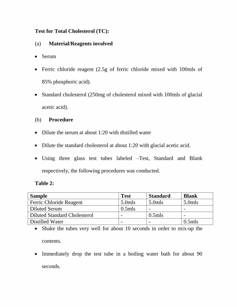
Test for Total Cholesterol (TC):
(a) Material/Reagents involved
Serum
Ferric chloride reagent (2.5g of ferric chloride mixed with 100mls of
85% phosphoric acid).
Standard cholesterol (250mg of cholesterol mixed with 100mls of glacial
acetic acid).
(b) Procedure
Dilute the serum at about 1:20 with distilled water
Dilute the standard cholesterol at about 1:20 with glacial acetic acid.
Using three glass test tubes labeled –Test, Standard and Blank
respectively, the following procedures was conducted.
Table 2:
Sample Test Standard Blank
Ferric Chloride Reagent 5.0mls 5.0mls 5.0mls
Diluted Serum 0.5mls - -
Diluted Standard Cholesterol - 0.5mls -
Distilled Water - - 0.5mls
Shake the tubes very well for about 10 seconds in order to mix-up the
contents.
Immediately drop the test tube in a boiling water bath for about 90
seconds.
Page 45
Cool for about 5 minutes with running tap water.
Read the absorbance at 560nM against the blank with a dry cuvette.
Meanwhile, the colour should be stable for 15minutes.
Finally, calculate using the formular:
Absorbance Test *250(constant value)
Absorbance Standard
The unit is mg/dl
Then to convert the result from mg/dl to mmol/l simply multiply the mg/dl
result by 0.0259 (constant value).
Test for HDL-Cholesterol (HDL-C):
Material/Reagents involved
Serum
Cholesterol standard (100mg of pure cholesterol mixed with 100mls of
glacial acetic acid).
Colour reagent (5.6g of 2,5-dimethyl benzene sulphuric acid mixed with
200ml of glacial acetic acid and 300ml of acetic anhydride).
Phosphotungstic acid reagent (4.5g of phosphotungstic acid mixed with
50ml of water, add 16ml of 1N NaOH and make up to 100ml with water).
Magnesium chloride (40.6g of magnesium chloride mixed with 100ml of
water).
Page 46
Tris buffer (1.21g of tris mixed with 90ml of water, when the PH was
reduced to about 7.6 with 1N HCL and diluted up to 100ml with water).
Procedure:
Pipette about 1ml of serum in a test tube
Drop 0.1ml of phosphotungstic acid reagent and mix very well
Add 0.05ml of magnesium chloride and mix very well
Centrifuge at 2500rpm or 1500g for 30minutes.
Carefully remove the clear supernatant with a Pasteur pipette.
Add 2 drops of the colour reagent and allow to stable for 15 minutes.
Read the absorbance at 560nM.
Finally, calculate using the formula:
HDL-C = Absorbance of Test *155(constant value)
Absorbance of Standard
Test for LDL –Cholesterol (LDL-C):
The LDL-Cholesterol is calculated using the standard formula:
Total Cholesterol – HDL-Cholesterol + 0.46(constant value)
Test for Triglycerides (TG):
(a) Material/Reagents involved
Serum
Page 47
Heptane
Isopropanol
Sodium methylate (50mg of sodium methylate diluted in 100mls of
isopropanol)
Sulphuric acid (0.08N)- prepared by mixing 2.25mls conc H2SO4 of
about 36.0N with 500ml of distilled water. The dilution should be up to 1
litre.
Periodate reagents (1.23g) of NaO4 was mixed with 100mls of 0.88N,
about 5% V/V/ action acid. Then stored in a brown bottle)
Acetylacetone reagent (0.75mls of acetyl acetone was dissolved with
2.5mls of isopropanol, then 2N of ammonium acetate (15.4%) was added
to make the volume up to 100ml, then stored in a brown at 40C
.
Triglyceride standard (200mg of pure triolein was mixed with 100ml of
isopropanol).
(b) Procedure:
Using three glass test tubes labelled-Test, Standard and Blank
respectively, the following procedures was conducted.
Table 3:
Sample Test Standard Blank
Serum 0.5ml - -
Triglyceride Standard - 0.5ml -
Page 48
Distilled Water - 0.5ml 0.5ml
Isopropanol 3.5mls 3.0mls 3.5mls
H2So4 (0.08N) 1.0ml 1.0ml 1.0ml
Heptane 2.0mls 2.0mls 2.0mls
Shake the tubes for 30 seconds to mix very well
The tubes should stand for 10minutes at room temperature for proper
separation of two layers
Prepare another set of three test tubes labeled-Test, Standard, and Blank
respectively and the following procedures was conducted:
Table 4:
Sample Test Standard Blank
Top Solvent Layer from respective tube 0.2ml 0.2ml 0.2ml
Sodium Methylate 3.0mls 3.0mls 3.0mls
Shake the tubes using vortex mixer to mix very well.
Incubation at 600C
for 10minutes and then cool at room temperature.
Colour development stage which involves dissolving 0.1ml of periodate
reagent to each tube and mix very well. Then cool at room temperature
after 10minutes of incubation at 600C.
Centrifuge the tubes and transfer the upper phase liquid to another fresh
tube.
Page 49
Using spectrophotometer, read the absorbance of test and standard at
420nM against blank.
Finally calculation is done by:
Triglyceride (mg/dl) = Absorbance of Test *200
Absorbance of Standard
Then to convert to mmol/dl is mg/dl *0.0113(standard value)
Page 50
CHAPTER FOUR
STATICALLY ANALYSIS
Statically analysis was carried out using statically package for social
sciences
PHYTOCHEMICAL RESULTS:
The modified Harbourne‟s (1973) phytochemical analysis revealed the
presence of soluble carbohydrate, cyanide, sugar, saponin, tannin, flavonoids,
alkaloids, steroids, terpenoids, in the extract in different concentration (table
1 and 2).
Table 1:
Quantitative analysis of phytochemical composition of water extract of
leaves of DV and other samples (mg/100g)
SAMPLE
SOLUBLE
CHO
CYANIDE
REDUCING
SUGAR
SAPONIN
TANNIN
FLAVON
OIDE
ALKALON
IOD
STERIOD
TERPENOI
DE
Water
Leaf
Extract
of DV
1.43 a
0.003
0.63 a
0.003
321.743
c 0.003
1.05 c
0.003
2.87 c
0.004
3.82 a
0.003
3.78 c
0.089
0.63 c
0.004
0.28 c
0.005
Data are means of triplicate determinations standard deviation (SD)
Data in the same column bearing different superscript differed significantly
(P 0.005).
Table 2:
Page 51
Qualitative analysis of phytochemical composition of water extract of leaves
of DV and other samples
Tannin +++
Alkaloid +++
Carbohydrate +
Saponin +
Steroid +
Hydrogen Cyanide +
Flavoniod ++
Reducing Sugar ++
Terpenoid +
Data:
+ = Present,
++ = Strongly present,
+++ = Fully Present
The effect of fresh cow‟s brain, atorvastatin and water extract of
D.velutinum on albino wistar rats is shown in table 3.
Table 3:
Lipid profile of rats fed with various samples (mg/dl)
Rats (Samples) Cholesterol
(mg/dl)
HDL (mg/dl) LDL (mg/dl) Triglyceride
(mg/dl)
Group 1 (Normal Feed) 140.00 1.41* 30.00 1.41
* 3.600.14
* 95.00 1.41
*
Group 2 (Cow‟s Brain) 145.00 1.41*
40.00 1.41*
3.90 0.14*
105.00 1.41*
Group 3 (Cow‟s Brain 110.00 0.00*
13.00 1.41*
1.20 0.14* 39.00 1.41
*
Page 52
+ Atorvastatin)
Group 4 (Cow‟s brain
+ Water extract of DV
leaf)
135.00 0.71* 25.00 0.00
* 1.90 0.00
* 50.000.00
*
Data are means of duplicate determinations standard Deviation (SD) –
Data in the same column bearing esterix (*) are significantly different
(P<0.05)
In group 1, animals, the normal feed, the LDL (3.600.14 mg/dl) and HDL
(30.00 1.41 mg/dl), in group 2, the LDL significantly increased to 3.90
0.14 mg/dl and HDL increase to 30.00 1.41 mg/dl because their were fed
with high lipoprotein food.
In the atorvastatin treated (2ml) group the LDL level was found to be 1.20
0.14 mg/dl decrease and HDL to 13.00 1.41 mg/dl decrease compared
with the group treated with the water extract of D.velutinum (0.5ml)
exhibited a progressively decrease in LDL to 1.90 0.00mg/dl by inhibiting
the HMG-CoA and significantly increased the HDL to 25.00 0.00mg/dl.
Page 53
CHAPTER FIVE
DISCUSSION AND CONCLUSION:
This study evaluated the antilipidamic activity of the water extract of
Desmodium velutinum in albino wistar rat. Hyperlipidemia comprises a state
of increased concentrations of TG, TC and LDL-C and is an important risk
factor for the development and progression of atherosclerosis and coronary
heart diseases (Viran et al., 2012).
In table 1, the data shows that, there is a significantly high content of
alkaloid, reducing sugar, soluble carbohydrate, flavonoid, and tannin in
water extract of D.V. leaf. These alkaloids often have pharmacological
effect and are used as medication, as recreational drug, exert anti-asthma,
anti-cancer, anti-arlythmia (Manske, 2006) and reduce unwanted side effects.
Tannin compounds are biomolecule, as in an astringent, bitter plant
polyhenolic compounds that binds to and precipitates proteins and various
other organic compounds including amino acids and alkaloids (Kadam et al.,
2005). Tannins and alkaloid may also be effective in reducing the risk of
cardiovascular disease. The content of cyanide present in water extract of
D.V leaf is very small. Even though cyanide is said to be toxic to the body,
its content in the extract is minimal (0.63 0.003) thus opposes no threat to
the body.
Page 54
In table II shows the presence of flavonoids, saponin, soluble
carbohydrate, cyanide, terpenoid, steroid, reducing sugar, alkaloid and
tannin, in extract of D.V. leaf .But tannin and alkaloid showed fully present,
flavonoid and reducing sugar showed strongly present significantly.
Flavonoids are most commonly known for their antioxidant, anti-
inflammatory, anti-microbial, anti-allergic, anti-cancer and anti-diarrheal
activities. (Cushine and Lamb, 2005). Flavonoids have medicinal properties
especially their putative role in inhibiting cancer and cardiovascular disease
(Frei, 2006).
Saponin involves the complexation with cholesterol to form pores in
cell membrane bilayer, e.g., in red cell (erythrocyte) membranes, where
complication leads to red cell lysis on intravenous injection (Yah et al., 2010)
and also show antioxidant, anticancer activity. Saponin have shown to
reduce cardiovascular and atherosclerosis. Polysaccharide and protein (e.g.,
glycoprotein complexes present in plant extracts (Oh et al., 2006) could also
be involved in the hypolipidemic and antiatherosclerotic effects of
D.velutinum. Therefore, the association between these complexes and
compounds and other constituents may play an important role in the
biological activity of the leaf. Soluble carbohydrate is said to help control
Page 55
body weight lower blood glucose and cholesterol (mayor, 2011) by
inhibiting the absorption of fats and cholesterol in intestine.
In table II, the group 1 fed normal feed the LDL.C is 3.60 0.14 and
HDL.C. is 30.00 V 1.41. In group II fed with cow‟s brain effectively
increase plasma lipid significantly to LDL-C 3.90 0.14 and HDL-C
40.00 1.41, thereby causing an increase in the activity of HMG-CoA
reductase and acting on cholesterol synthesis.
In group II treated with atorvastatin reduces the plasma lipid
significantly to LDL-C 1.20 0.14 and HDL-C 13.00 1.41 which can
possibly leads to hypocholesterolemia compared to group IV treated with
water extract of D.V. leaf reduce plasma lipid to LDL-C 1.90 0.00 and an
increase in HDL-C 25.00 0.00 which is the good cholesterol. The data
demonstrate that water extract of D.V. leaf can possibly normalize the
plasma lipid. The study suggests that the water extract of the leaf is effective
in reducing lipid plasma, thereby reducing the risk of cardiovascular and
atherosclerosis disease. The findings lend support to the folkloric use of
D.velutinum in the Eastern Nigeria as an antilipidemic agent.
Page 56
5.2 CONCLUSION:
The water extract of Desmodium velutinum leaves showed significant
antilipidemic effect in experimental rats.
Page 57
REFERENCES
Albers, J. J., Koschinsky, M. L. & Marcovina, S. M. (2007). „Evidence
Mounts for a Role of the Kidney in Lipoprotein Catabolism‟. Kidney Int.
71 (10): 961-962.
Deshmukh, H.A., Coilioun, H.M., Johnson, T. & Mckeigue, P. (2012).
Genome Wide Association Study of Genetic Determinants of LDL-C
Response to Atorvastatin Therapy Importance of LP(a) .J Lipid Res .53
(5): 1000-1011.
Gouni Berthold, I. & Berthold, H.K. (2011). „Lipoprotein: Current
Perspectives‟. Curr. Vasc. Pharmacol. 9 (6): 682-692.
Guan, X. & Wenk, M.R. (2008). „Biochemistry of Inositol Lipids.‟
Frontiers in Bioscience. 13(13): 3239-3251.
Harman, S.M., Vittinghoff, E., Brinton, E.A., Budoff, M.J., Cedars, M.I.,
Lobo, R.A., Merriam, G.R., Miller, V.M., NAftolin, F., Pal, L.,
Santoro, N., Tayl, H.S. & Black, D.M. (2011). „Timing and Duration
of Menopausal Hormone Treatment May Effect Cardiovascular
Outcomes‟. Am J Med. 124(3): 199-205.
Kamstrup, P.R. (2009). „Lipoprotein Should be Taken Much More
Seriously‟. Biomarkers in Medicine. 3(5): 439-441.
Nordestgard, B.G., Chapman, M.J., Ray, K., Boren, J., Andreotti, F., Watts,
G.F., Ginsberg, H., Amarenco, P., Catapano, A., Descamps, O.S.,
Fisher, E., Kovanen, P.T., Kuivenhoven, J.A., Lesnik, P., Masana, L.,
Reiner, Z., TAskinen, M.R., Tokogozoglu, L. & Tybjaerg-Hansen, A.
(2010). „Lipoprotein as a Cardio vascular Risk Factor: Current Status‟.
Eur Heart J. 31 (23): 2844-2853.
Parhofer, K.G. (2011). „Lipoprotein Medical : Treatment Options for an
Elusive Molecule‟.Curr Pharm Des. 17 (9) :871-876
Schwartz, J.B. (2009). „Effects of Vitamin D Supplementation in
atorvastatin Treated Patients, a New Drug Interaction with an
Unexpected Consequence‟. Clin Pharmacol Ther. 85 (2) :19
Page 58
Sotirious, S.N., Orlova, V.V., Al. Fakhri, N., Ihanus, E., Economopoulou,
M., Isermann, B., Bdeir, K., Nawroth, P.P., Preissner, K.T., Gahmberg,
C.G., Koschinsky, M.L. & Chavvakis, T. (2006). „Lipoprotein in
Atherosclerotic Plaques Recruits Inflammatory Cells Through Interaction
With Mac-I Integrin‟. FASEB J. 20 (3): 559-561.
Smolders, B., Lemmens, R. & Thijs, V. (2007). lipoprotein and Stroke: A
Meta-Analysis of Observational Studies‟. Stroke. 38 (6): 1959-1966.
Takagi, H. (2012). Atorvastatin Decreases Lipoprotein a Meta-Analysis of
Randomized Trials. Int J Cardiol .154 (2): 183-186
Tsimikas, S. & Witztum, J.L. (2008).„The Role of Oxidized Phospholipids
inMediating Lipoprotein Atherogenicity‟. Curr Opin Lipidol.19(4):
369-377
Villa, J. & Prattey, R.E. (2010). „Simvastatin or Atorvastatin for the
Treatment of Hypercholesterolemia in Patients with the metabolic
Syndrome the VYMET Study‟. Curr Diab Rep. 10 (3): 173-175.
Virani, S.S., Brautbar, A., Davis, B.C., Nambi, V., Hoogeveen, R.C., Sharrett,
A.R., Coresh, J., Mosley, T.H., Morrisect, J.D., Catallier, D.J., Folsom,
A.R., Boer Winkle, E. & Ballantyne, C.M. (2012). „Associations
between Lipoprotein Levels and Cardiovascular Outcomes in Black and
White Subjects‟. The Atherosclerosis Risk in Communities (ARIC) Study
Circulation. 125 (2):