Page 1
Title:Quality indicators in digestive endoscopy:introduction to structure, process, andoutcome common indicators
Authors:Julio López-Picazo , Fernando Alberca de lasParras, Antonio Sánchez del Río, ShirleyPérez Romero, Joaquín León Molina,Francisco Javier Júdez
DOI: 10.17235/reed.2017.5035/2017Link: PubMed (Epub ahead of print)
Please cite this article as:López-Picazo Julio, Alberca de las ParrasFernando, Sánchez del Río Antonio , PérezRomero Shirley, León Molina Joaquín, JúdezFrancisco Javier. Quality indicators indigestive endoscopy: introduction tostructure, process, and outcome commonindicators. Rev Esp Enferm Dig 2017. doi:10.17235/reed.2017.5035/2017.
This is a PDF file of an unedited manuscript that has been accepted for publication. As a service to ourcustomers we are providing this early version of the manuscript. The manuscript will undergocopyediting, typesetting, and review of the resulting proof before it is published in its final form.Please note that during the production process errors may be discovered which could affect thecontent, and all legal disclaimers that apply to the journal pertain.
Page 2
OR 5035 inglés
Quality indicators in digestive endoscopy: introduction to structure, process, and
outcome common indicators
Julio López-Picazo1, Fernando Alberca-de-lasParras2, Antonio Sánchez-del-Río3, Shirley
Pérez-Romero1, Joaquín León-Molina4 and Javier Júdez5
1Service of Health Care Quality. Hospital Clínico Universitario Virgen de la Arrixaca.
Murcia, Spain. 2Endoscopy Unit. Service of Digestive Diseases. Hospital Clínico
Universitario Virgen de la Arrixaca. Murcia, Spain. 3Service of Digestive Diseases.
Hospital San Juan de Dios. Santa Cruz de Tenerife, Spain. 4Service of Digestive Diseases.
Hospital Clínico Universitario Virgen de la Arrixaca. Murcia, Spain. 5Department of
Knowledge Management. SEPD. Madrid, Spain
Correspondence: Javier Júdez. Department of Knowledge Management. SEPD. C/
Sancho Dávila, 6. 20028 Madrid
e-mail: [email protected]
ABSTRACT
The general goal of the project wherein this paper is framed is the proposal of useful
quality and safety procedures and indicators to facilitate quality improvement in
digestive endoscopy units. This initial offspring sets forth procedures and indicators
common to all digestive endoscopy procedures.
First, a diagram of pre- and post-digestive endoscopy steps was developed.
A group of health care quality and/or endoscopy experts under the auspices of the
Sociedad Española de Patología Digestiva (Spanish Society of Digestive Diseases)
carried out a qualitative review of the literature regarding the search for quality
indicators in endoscopic procedures. Then, a paired analysis was used for the selection
of literature references and their subsequent review.
Twenty indicators were identified, including seven for structure, eleven for process
(five pre-procedure, three intra-procedure, three post-procedure), and two for
Page 3
outcome. Quality of evidence was analyzed for each indicator using the Grading of
Recommendations Assessment, Development and Evaluation (GRADE) classification.
Key words: Quality indicators. Endoscopy. Digestive system.
INTRODUCTION
It is well known that, in order to assess and improve care quality, the latter term must
be first defined (1) and its dimensions identified (2), including effectiveness, efficiency,
safety, accessibility, and patient-centered service (Table I). Once these dimensions,
their requirements for each undertaken activity, and the target results are established,
designing things so that they may be completed at the first go (for instance, by
describing procedures to be implemented and their requirements) significantly
facilitates work.
In order to acknowledge the quality level achieved, and to start making improvements
if needed, consistent information is necessary on the most relevant aspects of the care
provided, which may be summarized as indicators. Quality indicators may be divided
up into three categories: “structure”, “process” and “outcome or results” (3).
“Structure” includes all things related to the stable attributes wherein care is provided,
both material and organizational; “process” includes all things that are done for
patients and the skills involved; and “outcome” denotes any health status changes that
may be attributed to the received care, as well as patient satisfaction.
In this setting, the Sociedad Española de Patología Digestiva (SEPD) understood that
quality indicators associated with digestive endoscopy procedures should be analyzed
and assessed, both overall and specifically (the goal of this first report) for the three
main procedures in terms of volume and impact: gastroscopy, colonoscopy, and
endoscopic retrograde cholangio-pancreatography (ERCP). In a second stage,
echoendoscopy and balloon enteroscopy will also be analyzed. The reason is none
other than to safeguard practice quality in the field of gastroenterology in Spain by
providing quality indicators adapted to our setting and according to evidence levels.
The overall goal of this project is to suggest quality and safety procedures and
indicators useful to facilitate quality improvement in digestive endoscopy units. This
Page 4
paper puts forth procedures and indicators common to the various digestive
endoscopy tests.
METHODS
The study was structured in two clear-cut stages. First, a multidisciplinary task force
was set up and headquartered at the Hospital Clínico Universitario Virgen de la
Arrixaca (HCUVA), which reviewed the literature and the design of diagnostic
esophagogastroscopy, colonoscopy, and ERCP procedures. In a second stage proposals
were reviewed and discussed by an expert panel selected by the SEPD until a final
version was produced. Then data sheets were developed for each proposed indicator
to assess these procedures.
Search strategies and study selection
Two broad, systematic literature searches were performed. The first one was for
clinical practice guidelines (CPGs), the second was for original and review papers. CPGs
related to digestive endoscopy were obtained from three international sources
(Agency for Healthcare Research and Quality [AHRQ], National Institute for Health and
Care Excellence [NICE] and Scottish Intercollegiate Guidelines Network [SIGN]) and one
Spanish source (GuíaSalud), as well as from reviews in the web sites of the main
endoscopy and gastroenterology societies (American Society for Gastrointestinal
Endoscopy [ASGE], American Gastroenterological Association [AGA], European Society
of Gastrointestinal Endoscopy [ESGE], Sociedad Española de Endoscopia Digestiva,
SEED [SEPD], and Asociación Española de Gastroenterología [AEG]). Originals were
searched in the Web of Knowledge (WOK), PubMed, and Cochrane databases using the
following strategy. All documents were selected that were published between January
1, 2006 and August 10, 2016 and included any of the following descriptors: [digestive
endoscop*, gastrointestinal endoscopy, colonoscop*, gastroscop*, oesophagoscop*,
endoscopic retrograde cholangiopancreatography] and [informed consent, quality,
safety, (security), assessment, assurance, indicators, criteria]. (Active filters: clinical
trial, controlled clinical trial, meta-analysis, randomized controlled trial, review,
guideline, practice guideline, publication date from 2006/01/01 to 2016/08/10;
Page 5
humans; adults; language: English, Spanish).
A review of the references included in the selected, analyzed originals, published
guidelines and meta-analyses was also performed, and previously overlooked
references of interest were selected.
Once the search protocol was completed, eligible papers were split in two groups (n1:
other procedures [non-colonoscopy], and n2: colonoscopy). The documents selected
were reviewed and analyzed separately by two reviewers. Each pair of reviewers
screened their studies using any of the following criteria:
The document includes recommendations on appropriate preparation,
execution, and follow-up.
The document includes or suggests quality indicators for structure, process or
outcome.
Studies selected by just one reviewer were collated by the rest in order to decide
their final selection status.
A table listing information on structure, process or outcome indicators, and whether
such information was explicit, was developed to uniformly examine each document.
Furthermore, the table included the type of each referenced study (clinical trial,
observational study, meta-analysis, etc.), which was identified as pertaining to
endoscopy in general, gastroscopy, colonoscopy or ERCP, with the rest of procedures
being set aside. Document suitability for the intended goal (analysis of endoscopy-
related quality indicators) was also discussed.
Endoscopy procedure design
Based on the collected literature and the authors’ experience, the activities needed
for each procedure were recorded and sorted out. In the case of procedures common
to all endoscopic studies, the logical differences in structure, function, and
organization among clinical digestive endoscopy units restrict development down to
a minimum. Similarly, a description of specific techniques to be used in selected
situations was excluded, as it was not contemplated in the ongoing work. Results
were plotted in flow charts or parallel line diagrams. Proposals put forth by the group
Page 6
were reviewed and discussed by an expert panel selected by the SEPD until a final
version was produced.
Construction of indicators
In order to obtain valid indicators, the quality of the available knowledge was
assessed regarding the activities involved in the procedures and the documents
selected after the search. To this end, the quality of knowledge grading scheme
available within the GRADE model was used. In the GRADE system the quality of
“evidence” (this term will be hereafter used without quotation marks to denote the
“best proof-based knowledge available”) is initially classified as high or low, according
to its origin in experimental or observational studies; then, quality is rated as high,
moderate, low or very low depending on a number of considerations regarding items
that may downgrade or upgrade baseline quality (4,5).
To ensure reliability and to facilitate the estimation of the chosen indicators in clinical
units, each of them is associated with a data sheet including: use environment
(procedures wherein it is used); denomination; calculation formula; indicator type
according to Donabedian’s model (3); time relationship with test (pre-procedure,
procedure, post-procedure); quality dimension involved; justification, exclusions, and
clarifications; and supporting evidence level.
RESULTS AND DISCUSSION
Search results
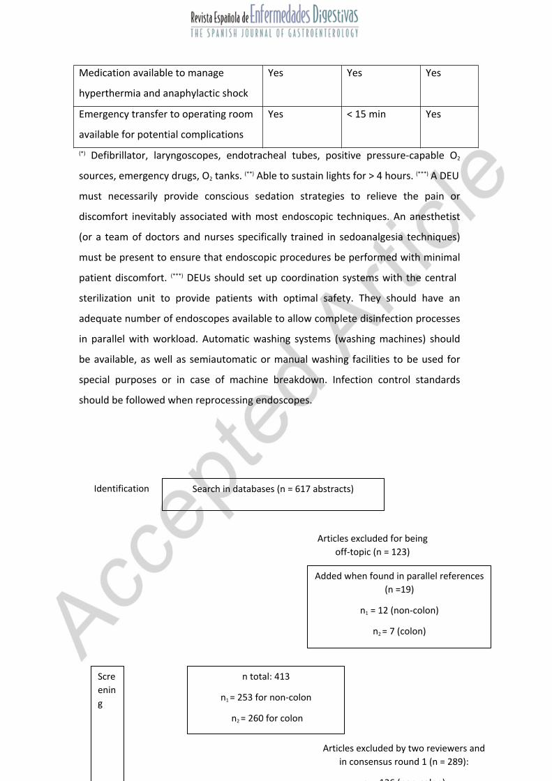
A total of 617 references were identified in the various databases according to the
designed search strategy. Once duplications were excluded, a total of 123 references
were discarded when titles/abstracts were examined (wrong references, abstract
only, obvious poor quality, unavailability, older than 2006, dealing with pediatric,
veterinary or non-digestive endoscopic topic, language other than Spanish or
English).
Post-hoc, 19 additional references were selected from the references found in other
already published papers, reviews, meta-analyses, and clinical practice guidelines.
Page 7
For the paired analysis, we examined a total of 413 papers (n1: 253; n2: 260) in full
text format, including both randomized and non-randomized clinical trials, as well as
224 full articles on high-quality case series, reviews, and meta-analyses (n1: 117; n2:
107).
Results are plotted in figure 1.
Common procedures
Two common procedures were implemented: admission to unit on the day scheduled
for endoscopy, and discharge from unit after the procedure. The inclusion of speed-up
procedures and waiting list management was dismissed on the grounds mentioned
under “Methods”.
The admission procedure (Fig. 2) is intended to confirm that the patient is fully eligible
for endoscopy, to ascertain the type of sedation that will be used, and to provide the
necessary pre-procedural nursing care. It lasts from the time the patient presents to
the endoscopy unit until endoscope insertion. It includes the following activities:
Before entering the room:
Confirm appointment and agenda availability (time/room) and mark as
present.
Confirm adequate preparation (see indicator below).
Check out informed consent.
In the endoscopy room:
The endoscopist must:
- Confirm informed consent and clarify last-minute concerns.
- Open up the appropriate form within the medical record.
- Offer sedation if applicable, with its related informed consent.
- Confirm key history data such as allergies, use of antithrombotic
drugs, or sedation contraindications.
The nurse must:
- Insert a peripheral venous access, install a pulse oximeter, and
provide oxygen through nasal prongs if sedation deeper than
Page 8
topical is required.
- Administer appropriate sedation.
The discharge from endoscopy unit process is intended to adequately document the
procedure’s course, findings, and potential incidences; to ensure the necessary care
until the patient leaves the unit; and to prepare the endoscopy room for a new
procedure. It lasts from endoscope withdrawal to patient leaving the unit. It includes
the following activities:
Ancillary personnel must clean out the equipment and recondition the
endoscopy room.
The endoscopist must write a medical report (see indicator below) and make
sure it is included in the patient’s medical records.
After patient transfer to the care room nurses must monitor vital signs, assess
level of consciousness, and provide appropriate post-procedural care (see
indicator below).
Patients must receive a copy of the endoscopy report before leaving the unit.
Indicators
Twenty indicators have been included, of which seven refer to structure, two refer to
outcome, and eleven are related to process. Table II lists all indicators used.
Structure indicators
A1. Valid informed consent
Definition and formula
Quality defects per document ratio
Numerator: no. of formal quality criteria unmet
Denominator: total of informed consent documents assessed
Type, temporal relationship, and quality dimension
Structure - Patient-centered care
Quality of evidence
Very low
Page 9
Providing patients with information regarding their therapy options and diagnostic
choices, in an understandable manner, so that they may take part in the decision-
making process involving their care, is both a duty health care professionals must fulfill
(6) and a right guaranteed to patients by law (7). Hence, there is no doubt that
informed consent forms are needed for digestive endoscopy procedures (8,9). These
provide patients with information on the involved procedure so that an informed
decision is made. Therefore, consent forms must meet formal quality requirements in
order to observe legal regulations, promote readability, and improve decision making.
In this regard the criteria put forward by the Murcia Region EMCA program have
proven useful in order to improve informed consent forms (10). Furthermore, it is also
necessary to ensure the readability and understandability of these forms, as well as
the validity of their contents (11,12). To achieve the former, the INFLESZ tool may be
used, which is a validated scale to assess ease of comprehension for documents
written in Spanish (scores of at least 55 are highly likely to provide the average
population with an understandable text) (12), as well as other items (11). To achieve
the latter it is key that texts and their supported evidence be up-to-date (13).
A2. Antithrombotic medication management plan
Definition and formula
The unit/service involved has a plan to manage antithrombotic and antiaggregant
medication that at least formulates recommendations pointing out their strength and
evidence level, and includes an explicit validity and review period
Qualitative indicator of presence/absence
Type, temporal relationship, and quality dimension
Structure – Safety
Quality of evidence
Very low
Although diagnostic endoscopies are usually considered to have a low bleeding risk,
the same does not apply to some therapeutic procedures (8). In these patients the risk
Page 10
of thromboembolic complications associated with treatment discontinuation must also
be assessed (14), and the timing of anticoagulant and/or antiaggregant drug
withdrawal and reinitiation has to be decided upon, together with the need for
monitoring during said periods (15). Because of this, several guidelines approach this
subject in the context of digestive endoscopy (16), and several scientific societies
recommend its inclusion among quality indicators (8).
About the minimal requirements an antithrombotic medication management plan
should meet, it is suggested that validity criteria, a key aspect of clinical guidelines, be
ascertained (17,18) and potentially assessed by checking out that recommendations
include their strength and evidence level, and the plan includes an explicit validity and
review period.
A3. Experienced endoscopist
Definition and formula
Qualitative indicator of the presence/absence of the experience and training required
of the endoscopist who performs the procedure (to establish for each procedure)
Type, temporal relationship, and quality dimension
Structure - Effectiveness
Quality of evidence
Low
An endoscopic procedure must reach its intended goal while minimizing the incidence
of adverse events. Evidence suggests that endoscopist inexperience and insufficient
training represent barriers to these ends (16,19,20).
Some scientific societies have established recommendations to assess expertise in
endoscopy, which include a minimum of procedures performed to meet basic quality
standards, as well as a minimum of procedures yearly (8,21,22).
Other factors to consider will depend upon the type of procedure and have to do with
diagnostic or therapeutic yield and safety (23). Expertise in a given procedure does not
qualify for other procedure types (20).
Page 11
A4. Discharge plan
Definition and formula
The unit/service performing the procedure has:
A recovery area other than the endoscopy room, with all necessary equipment
for post-anesthesia care
A patient management plan to facilitate recovery from procedure completion
to discharge, with an explicit validity and review period
A plan for patient information, instruction (allowed foods, activities not
permitted within the next 24 hours, etc.) and follow-up, including any adverse
events that might develop and their recognition
Qualitative indicator of presence/absence
Type, temporal relationship, and quality dimension
Structure – Safety
Quality of evidence
Very low
Trained nursing staff is usually responsible for patient recovery, including patient
monitoring and the assessment of patient discharge criteria. The plan must also
include a report on the procedure and its ensuing process, as well as patient
instructions (food ingestion, forbidden activities within 24 hours, etc.) and follow-up
assessments, including potential complications and their recognition (23,24).
The duration and frequency of monitoring should be individualized according to
sedation level and patient health status, and range from 30 minutes to two hours. In
this respect level of consciousness as assessed by response to verbal stimuli, vital signs
(heart rate, respiratory rate, oxygen saturation), and level of pain must be monitored
and recorded regularly until their return to normal (20,21,25).
Standard tools such as the modified Aldrete scale (22) are available to assess recovery
after sedation (respiration, oxygen saturation, blood pressure, level of consciousness,
activity). At discharge, patients must be fully aware and oriented with stable, normal
vital signs (heart rate, respiratory rate, blood pressure, oxygen saturation), with a
score of at least 9 points in the modified Aldrete scale (23,26).
Page 12
A5. Discharge report quality
Definition and formula
Quality defects per document ratio
Numerator: no. of formal quality criteria unmet
Denominator: total no. of discharge report documents (DRDs) assessed
Type, temporal relationship, and quality dimension
Structure - Patient-centered care
Quality of evidence
Very low
Providing accurate, timely information on the procedure performed, succinctly
including all relevant data, improves patient care. Both the ASGE (8,27) and the
Canadian Association of Gastroenterology (CAG) (10,28,29) recommend the inclusion
of selected items in the endoscopy report: date of procedure; patient identification;
endoscopist and other staff identification; relevant history and physical examination
data; conditions that may interfere with the procedure or with sedation; drug allergies;
medications, particularly anticoagulant and antiplatelet drugs; anesthetic risk
assessment (American Society of Anesthesiologists [ASA]) (28,30); proof of informed
consent; endoscopic procedure; indication; type of endoscope; medication during the
procedure (analgesia, anesthesia, sedation) (31) and during recovery (if applicable);
anatomical extent of the procedure; barriers and restrictions encountered during
preparation and procedure (9) (type, quality); samples obtained, including type,
location and sampling technique; findings, with a detailed description of
present/pertinent absent lesions; diagnosis, using as much as possible standardized,
coded terminology (32); therapies administered and outcomes, complications and
adverse events; recommended actions (new appointments, collection of results, etc.),
and recommended care.
A6. Disinfection procedure for endoscopy equipment
Definition and formula
Page 13
The unit/service performing the test has a disinfection procedure for endoscopy
equipment including an explicit validity and review period
Qualitative indicator of presence/absence
Type, temporal relationship, and quality dimension
Structure – Safety
Quality of evidence
Moderate
While the risk of infection is low (one case per 1.8 million endoscopies), digestive
endoscopy is an invasive procedure where infection may be transmitted, hence every
effort to minimize it must be in place. A consistently, appropriately executed cleaning
and disinfection procedure is effective to get rid of bacteria, mycobacteria, and viruses
(33,34).
To ensure adequate endoscope disinfection three steps should be followed:
mechanical cleaning, disinfection, and rinsing and drying.
Guidelines and consensus documents by different societies (35-48) recommend the
following:
Perform test to identify potential loss of tightness in channels or presence of
inner disruptions that may impair disinfection.
Suction out and submerge the endoscope in an enzymatic detergent solution.
Carefully clean and brush the whole endoscope, including valves and channels,
using an enzymatic detergent (49).
Use a device-friendly disinfectant agent of proven efficacy such as 2%
glutaraldehyde, 0.4-1% glutaraldehyde-phenate, or 0.2% peracetic acid
(48,50,51).
Submerge the endoscope in disinfectant filling up the channels. Exposure
duration and temperature vary according to product (52).
If an automatic machine is used, check it is properly connected and follow
manufacturer instructions.
Replace disinfectant after its activity period regardless of minimum effective
concentration.
Page 14
Once disinfected, rinse the endoscope using preferably sterile water and dry all
channels with air.
The use of 70% alcohol, followed by further air drying, may enhance
disinfection efficacy.
Store the endoscope vertically in a well-ventilated area. Valves and instrument
channel cap should be stored separately (9).
The irrigation bottle and tubing must undergo “high‐level” disinfection daily
(53).
Ancillary materials potentially in contact with blood must be sterilized following
careful mechanical cleaning (54).
If any material is stored under non-sterile conditions for over 48-72 hours,
reprocessing is advised in view of contamination risks (55).
A7. Structural and functional characteristics of an endoscopy unit
Definition and formula
The unit/service performing the test must meet the following structural and
functional requirements according to its regulatory background
Qualitative indicator of presence/absence
Type, temporal relationship, and quality dimension
Structure – Safety
Quality of evidence
Very low
The structural and functional requirements of endoscopy units may differ to a greater
or lesser extent according to location and type (10,53,56).
In this context, all of them must secure protocols, equipment, and trained,
experienced personnel to provide safe, effective conscious sedation.
Endoscopy unit accreditation is directly related to the ability to provide endoscopy
training (22).
In Spain, a document granted by the Ministry of Health and Consumption (57) includes
the structural and functional characteristics digestive endoscopy units must have
Page 15
according to their defined typology (Table III). It also points out that they must be
fitted with computer systems capable of processing and storing images, and of
facilitating the writing of standard reports.
B1. Appropriate indication
Definition and formula
Percentage of cases where the reason for endoscopy is recorded in the medical
history, and is also present in a list of appropriate indications
Numerator: 100 x cases with a recorded valid indication
Denominator: total of cases assessed
Type, temporal relationship, and quality dimension
Process - Pre-procedure - Effectiveness
Quality of evidence
Low
The test must be indicated when the information it may yield or the therapy it may
provide will improve the patient’s outcome with a good risk-benefit ratio, as evidence
suggests this improves cost-effectiveness (8,10,53,58) and provides a reference for
legal claims (30). A list of gastroscopy indications was published by the ASGE and
updated in 2012 (58). The European Panel on the Appropriateness of Gastrointestinal
Endoscopy (EPAGE) II criteria are used for colonoscopy (59-64). Consensus documents
also provide data for ERCP (58,65-67).
When a test is appropriately indicated, a higher rate of relevant diagnoses is obtained
(68-72).
B2. Signing the informed consent form
Definition and formula
Percentage of cases with confirmed delivery and signing of a valid informed consent
form (ICF)
Numerator: 100 x cases where a valid ICF was delivered and signed
Denominator: total of non-urgent cases assessed
Page 16
Type, temporal relationship, and quality dimension
Process - Pre-procedure - Patient-centered care
Quality of evidence
Very low
Providing patients with information on their therapeutic and diagnostic options in an
understandable way, so that it may be used to promote their involvement in the
decision making process regarding their care (8), is both a duty for health practitioners
and a right guaranteed to patients by law (7).
This indicator may be exempted in case of emergency. The signed ICF must meet
formal quality requirements or be accredited by the institution where the test is
performed or by its corresponding scientific society (see structure criteria)
(9,10,12,53,73).
In any case, the decision to undergo endoscopy should not be made under duress, and
the patient must acquiesce in writing by signing the consent form. The patient must be
allowed time and opportunities to pose additional questions before making a decision
(74). According to a recent systematic review (75), interventions to promote the
consent process in patients undergoing surgery or other invasive procedures such as
digestive endoscopy usually enhance comprehension.
B3. Clinical assessment
Definition and formula
Percentage of cases with a confirmed appropriate assessment before endoscopy
- Numerator: 100 x cases with previous appropriate assessment
- Denominator: total of cases assessed
Type, temporal relationship, and quality dimension
Process - Pre-procedure - Safety
Quality of evidence
Very low
Page 17
Assessing a patient’s clinical status improves test safety. This implies that all risk
factors must be considered to minimize complications during the procedure. Since
many adverse effects during endoscopy are associated with sedation, it is advisable
that individual risk for sedation be evaluated to adjust the regimen to be administered.
The following must be verified before the procedure (8,9,30,76,77):
The patient has been informed about the procedure and has no concerns.
Consent signed by both the patient and the physician.
Scheduled preparation is appropriately followed.
Fasting time as instructed.
Antiplatelet or anti-inflammatory drugs (past seven days), or anticoagulants
(past three or four days).
Drug allergies.
Removal of all metallic objects and dentures, when applicable.
Issues in prior examinations, when present.
The patient is accompanied and will not need to drive.
Another poorly clarified concept is the need to pause of the endoscopy team before a
procedure in order to ensure the patient is aware of the procedure’s reason, course
and goals.
B4. Sedation plan
Definition and formula
Percentage of cases with a specified sedation plan before procedure onset
Numerator: 100 x cases with specified sedation plan
Denominator: total of cases assessed
Type, temporal relationship, and quality dimension
Process - Pre-procedure - Patient-centered care
Quality of evidence
Moderate
Page 18
Sedation should be offered by levels to all patients according to their clinical status.
The patient will choose one among the available options once adequately briefed,
which is associated with greater satisfaction.
The level of sedation (from none to deep) to be used during the procedure must be
recorded (53,78).
B5. Antithrombotic medication management
Definition and formula
Percentage of cases with antiplatelet or anti-inflammatory medication specified in the
medical records, together with a risk minimization plan if present
Numerator: 100 x cases where the use of antiplatelet or anti-inflammatory
drugs is explicitly specified, together with a risk minimization plan if present
Denominator: total of cases assessed
Type, temporal relationship, and quality dimension
Process - Pre-procedure - Safety
Quality of evidence
Very low
Diagnostic endoscopies are usually deemed to entail a low bleeding risk. This is not the
case for some therapeutic procedures where a graded risk assessment applies (79).
These patients must also have their risk for thromboembolic complications after
therapy discontinuation assessed (8-10,14,16-18,80-82). Therefore, the timing for
anticoagulant and/or antiaggregant discontinuation and reintroduction must be
defined, as well as the need for monitoring during that period (79). Furthermore,
decisions must be agreed with the patient (83).
C1. Graphical documentation
Definition and formula
Percentage of cases with a graphically documented test (pictures, video)
Numerator: 100 x cases with graphical documentation (pictures, video)
Denominator: total of cases assessed
Page 19
Type, temporal relationship, and quality dimension
Process - Procedure - Effectiveness
Quality of evidence
Very low
No studies have approached the effectiveness of including graphical documentation on
the procedure, but it represents a universally accepted good practice requirement.
Thus, the ASGE (8) and other guidelines (9,10,53,84,85) recommend that photographs
be taken of the cecum as a quality parameter for colonoscopy (86). They are also
recommended for other procedures since lesion’s pictures enhance patient
understanding and facilitate inter-consultations and second opinions (87).
C2. Sedated patient monitoring
Definition and formula
Percentage of cases where sedated patient monitoring is recorded
Numerator: 100 x cases with at least pulse oximetry, heart rate, and blood
pressure monitoring
Denominator: total of cases where sedation is used
Type, temporal relationship, and quality dimension
Process - Procedure – Safety
Quality of evidence
Very low
Patient monitoring improves procedure safety but not clinical outcome.
According to the ASGE (8,26,78) and CAG (10), monitored parameters should include
oximetry, heart rate, and blood pressure (the latter two at intervals no longer than five
minutes), which help identify potentially life-threatening cardiovascular changes
during sedation (53,88,89).
Evidence is insufficient to recommend capnography for patients sedated with propofol
(9,26,90).
Page 20
C3. Recording of immediate adverse events
Definition and formula
Percentage of cases with recorded presence/absence of adverse events, and their
nature when present
Numerator: 100 x cases with recorded presence/absence of adverse events
and their nature
Denominator: total of cases assessed
Type, temporal relationship, and quality dimension
Process - Procedure - Safety
Quality of evidence
Very low
Patient safety requires that damaging or potentially damaging events be identified and
followed up (30,91). The recording of events emerging during the procedure or before
leaving the endoscopy unit is therefore key in the effective implementation of
programs to improve patient safety (30).
Efforts have been made to grade the severity of adverse events (92), and to establish a
coherent classification (93). The following adverse events must be included:
1. Medication-related (94):
a) Need for cardiopulmonary resuscitation. Use of reversal medication, such as
flumazenil and naloxone, to antagonize the sedative effects of benzodiazepines
and opiates, respectively, which indicates excess sedation. Elective use to
speed up recovery is inconsistent with better practice, and not recommendable
in view of potential rebound sedation when the patient is no longer supervised
(68,95,96).
a) Hypoxemia (< 85%). The risk of sequelae from hypoxemia is poorly understood,
but an association with higher risk for late adverse events and longer recovery
has been reported.
b) Hypotension (< 90/50 mmHg or ≤ 20% from baseline) or hypertension (>
190/130 mmHg or ≥ 20% from baseline) may trigger endoscopy termination,
require direct intervention, have adverse consequences, and/or delay recovery.
Page 21
c) Allergic reactions including laryngospasm/bronchospasm.
2. Procedure-related (97-99):
a) Perforation (100).
b) Immediate bleeding after polypectomy. Uncertain clinical outcome. Some
authors suggest that even with immediate hemostasis significant complications
may develop, this being a risk factor for subsequent bleeding events (101).
c) Need for admission or transfer to the Emergency Room for any procedure-
emergent event.
d) Tube impaction, which may require surgery.
e) Persistent, severe abdominal pain requiring careful assessment to rule out
perforation. In a randomized trial, 45% and 31% of patients undergoing
colostomy experienced abdominal pain after one and six hours. While this pain
usually subsides, in some patients it is persistent to the extent of requiring
medical care. Pain from air retention during or after colonoscopy may be
reduced when CO2 insufflation (102,103) or water immersion are used (104).
f) Instrument failures requiring a repeat procedure.
D1. Patient recovery
Definition and formula
Percentage of cases where monitoring and discharge criteria assessments are
recorded
Numerator: 100 x cases with adequate monitoring according to discharge plan
Denominator: total of cases assessed
Type, temporal relationship, and quality dimension
Process - Post-procedure - Safety
Quality of evidence
Very low
Recovery is usually supervised by trained nurses who perform the monitoring and
assess discharge criteria.
Page 22
The duration and frequency of the monitoring must be tailored to each patient
according to sedation level and general health status (8,9), and should range from 30
minutes to two hours.
The level of consciousness as assessed by the response to verbal stimuli, together with
vital signs (heart rate, respiratory rate, oxygen saturation) and pain level, must be
regularly monitored and recorded until their return to normal (10).
Standard scales (modified Aldrete) (105,106) are available to assess recovery from
sedation (breathing, oxygen saturation, blood pressure, level of consciousness,
activity). Patients at discharge must be aware and oriented, with normal, stable vital
signs (heart rate, breathing rate, blood pressure, oxygen saturation), with a score of at
least 9 points on the modified Aldrete scale (31,107,108).
D2. Information on discharge
Definition and formula
Percentage of cases where a discharge report was generated on the procedure day
Numerator: 100 x cases with discharge report generated on procedure day
Denominator: total of cases assessed
Type, temporal relationship, and quality dimension
Process - Post-procedure - Patient-centered care
Quality of evidence
Very low
Information on discharge must include the procedure’s course and findings, process
follow-up (further appointments, reviews), and patient instructions, as well as
potential adverse events and how to recognize them. This is known to reduce anxiety,
to enhance recall of test conclusions and recommendations, and to improve patient
adherence (8,9,30).
This is verified by ascertaining a high-quality report was written, discussed, and
delivered before patient leave (10,27,31,53,109,110).
D3. Recording delayed adverse events
Page 23
Definition and formula
Percentage of cases where the presence/absence of delayed adverse events was
recorded, together with their nature if present
Numerator: 100 x cases with recorded presence/absence of adverse events
and their nature
Denominator: total of cases assessed
Type, temporal relationship, and quality dimension
Process - Post-procedure - Safety
Quality of evidence
Very low
Patient safety requires the identification and follow-up of damaging or potentially
damaging events (91). While the recording of adverse events occurring during the
procedure is widespread, the recording of those developing after leaving the
Endoscopy Unit is also key for the effective implementation of programs to enhance
patient safety (15,30,94,97).
According to the CAG, adverse events that should be recorded include (93):
Death within 30 days after the procedure.
Unscheduled admission or emergency visit within 14 days after the procedure.
Gastrointestinal bleeding within 14 days after the procedure.
Infection, both acute and chronic.
Symptomatic metabolic complication (hypoglycemia or hyperglycemia,
electrolyte disorders). Colonoscopy requires colon cleansing with a laxative
preparation. There is evidence of associated metabolic disorders such as
hypokalemia, hyponatremia, hypocalcemia, and renal failure. Phosphate-
containing preparations have been associated with acute phosphate
nephropathy and their use is limited. Furthermore, preparation for
colonoscopy may interfere with the management of conditions such as
diabetes mellitus.
During ERCP the development of procedure-induced pancreatitis depends upon
the expertise of the endoscopist and the techniques performed.
Page 24
E1. Incidence of adverse events
Definition and formula
Percentage of cases with delayed adverse events, both total and per type
Numerator: 100 x cases with adverse events
Denominator: total procedures per observation period
Type, temporal relationship, and quality dimension
Outcome – Safety
Quality of evidence
Very low
Indicators C3 and D3 refer to the recording of immediate and delayed events
(30,89,94,97,107). The purpose of such recording is twofold: on the one hand, to know
the magnitude, typology, and transcendence of adverse events emerging from
digestive endoscopy procedures; and on the other, to assess their potential for
prevention and to establish specific improvement actions.
E2. Perceived quality and patient satisfaction
Definition and formula
Satisfaction. An 11-item (0-10) Likert scale is suggested for measurement
Concurrent measurement of associated perceived quality dimensions
Type, temporal relationship, and quality dimension
Outcome - Patient-centered care
Quality of evidence
Very low
A measure of care quality cannot be considered as fulfilled without an assessment of
the patient perceived quality and patient satisfaction with the care received. In fact,
the Institute of Medicine (IOM) highlights patient-centered care as one of the six major
values of health care (108). Therefore, satisfaction measurements in the digestive
endoscopy setting have been repeatedly proposed by authors and scientific societies
Page 25
alike (8,10,53,106,111). The above suggested measurement involves an 11-item (0-10)
Likert scale that we feel better suited to Spanish context as compared to the 5- or 7-
item scales usually seen in other settings and cultures. With this scale, useful
measurements may be obtained to understand its evolution and set a ground for the
analysis of conditioning factors such as the mean, median or percentage of excellent
scores, the latter understood as the number of scores equal to or greater than 8 over
the total of scores, as has already been tested in other environments (112,113).
However, measuring satisfaction is not enough. In order to implement an ongoing
improvement approach and know where interventions are needed, conditioning
factors must also be learned. Some derive from the other indicators suggested above.
Others will have to be explored based on perceived quality items. In this respect, the
Endoscopy Global Rating Scale (GRS) initiative, successfully developed in the United
Kingdom, may represent a good starting point for their identification, as one
dimension refers to perceived quality (114). GRS is a web-based assessment tool that
provides statements requiring a yes or no answer, the usefulness of which has been
tested in various settings and health systems (115,116). The six level-graded items of
patient-centered care include equality and equity, opportunity (waiting times), ability
to select dates (accessibility), privacy and dignity, post-procedural care, and capacity to
generate return information to the endoscopy unit.
ACKNOWLEDGEMENTS
We are grateful to the SEPD Task Force on endoscopy quality indicators and the
Murcian Health Service Task Force.
CONFLICTS OF INTEREST
The undersigned authors signed the present paper on behalf of the Sociedad Española
de Patología Digestiva (SEPD). Neither the SEPD nor any of the task force members
have any relationship with manufacturers of endoscopy equipment. Neither the SEPD
nor any of the members of the task force have any financial interest in the companies
that played a role in the research and provision of digestive endoscopy devices, albeit
both the SEPD and the task force members have a sustained relationship with said
Page 26
companies for training, research, and improved clinical care purposes in order to
promote digestive health. Finally, both the SEPD and the undersigned authors declare
that the initial efforts of both local and nation-wide groups on which this review study
was based were supported by SimMédica Pentax and BostonScientific, who had no
influence on the undertaken research, and that no third parties were involved in the
discussions or development of the present paper, or had access to the contents of the
final manuscript before effective publication in the Revista Española de Enfermedades
Digestivas.
REFERENCES
1. Donabedian A. The definition of quality and approaches to its assessment.
Explorations in quality assessment and monitoring. Michigan: Health Administration
Press; 1980.
2. America IoMUCoQoHCi. Crossing the quality chasm: A new health system for the
21st century. Washington: Press NA; 2000.
3. Donabedian A. Basic approaches to assessment: What to assess. Exploration,
structure, process and outcomes - Quality assessment and monitoring. Vol. 1.
Michigan: Health Administration Press Ann Arbor; 1980. pp. 79-122.
4. Atkins D, Eccles M, Flottorp S, et al. Systems for grading the quality of evidence and
the strength of recommendations I: Critical appraisal of existing approaches The
GRADE Working Group. BMC Health Serv Res 2004;4(1):38. DOI: 10.1186/1472-6963-4-
38
5. Atkins D, Briss PA, Eccles M, et al. Systems for grading the quality of evidence and
the strength of recommendations II: Pilot study of a new system. BMC Health Serv Res
2005;5(1):25.
6. Godolphin W. The role of risk communication in shared decision making. BMJ
2003;327(7417):692-3. DOI: 10.1136/bmj.327.7417.692
7. Ley 41/2002, de 14 de noviembre, Básica Reguladora de la Autonomía del Paciente y
de Derechos y Obligaciones en Materia de Información y Documentación Clínica
Page 27
(2002). Disponible en: https://www.boe.es/buscar/pdf/2002/BOE-A-2002-22188-
consolidado.pdf
8. Rizk MK, Sawhney MS, Cohen J, et al. Quality indicators common to all GI endoscopic
procedures. Gastrointest Endosc 2015;81(1):3-16.
9. Jover R, Herráiz M, Alarcón O, et al. Clinical practice guidelines: Quality of
colonoscopy in colorectal cancer screening. Endoscopy 2012;44(4):444-51. DOI:
10.1055/s-0032-1306690
10. Armstrong D, Barkun A, Bridges R, et al. Canadian Association of Gastroenterology
consensus guidelines on safety and quality indicators in endoscopy. Can J
Gastroenterol 2012;26(1):17-31. DOI: 10.1155/2012/173739
11. Gargoum FS, O’Keeffe ST. Readability and content of patient information leaflets
for endoscopic procedures. Ir J Med Sci 2014;183(3):429-32. DOI: 10.1007/s11845-013-
1033-8
12. Calle-Urra JE, Parra-Hidalgo P, Saturno-Hernández PJ, et al. Formal quality
assessment of informed consent documents in 9 hospitals. Rev Calid Asist
2013;28(4):234-43. DOI: 10.1016/j.cali.2013.01.006
13. Barrio-Cantalejo IM, Simón-Lorda P, Melguizo M, et al. Validation of the INFLESZ
scale to evaluate readability of texts aimed at the patient. An Sist Sanit Navar
2008;31(2):135-52.
14. Becker RC, Scheiman J, Dauerman HL, et al. Management of platelet-directed
pharmacotherapy in patients with atherosclerotic coronary artery disease undergoing
elective endoscopic gastrointestinal procedures. Am J Gastroenterol
2009;104(12):2903-17. DOI: 10.1038/ajg.2009.667
15. Allen JI. Quality assurance for gastrointestinal endoscopy. Curr Opin Gastroenterol
2012;28(5):442-50.
16. Anderson MA, Ben-Menachem T, Gan SI, et al. Management of antithrombotic
agents for endoscopic procedures. Gastrointest Endosc 2009;70(6):1060-70.
17. Veitch AM, Baglin TP, Gershlick AH. Guidelines for the management of
anticoagulant and antiplatelet therapy in patients undergoing endoscopic procedures.
Gut 2008;57(9):1322-9.
Page 28
18. Parras FA. Clinical practice guidelines for managing coagulation in patients
undergoing endoscopic procedures. Rev Esp Enferm Dig 2010;102(2):124-38.
19. Boustière C, Veitch A, Vanbiervliet G, et al. Endoscopy and antiplatelet agents.
European Society of Gastrointestinal Endoscopy (ESGE) Guideline. Endoscopy
2011;43(5):445-61. DOI: 10.1055/s-0030-125631720.
20. James PD, Antonova L, Martel M, et al. Measures of trainee performance in
advanced endoscopy: A systematic review. Best Pract Res Clin Gastroenterol
2016;30(3):421-52. DOI: 10.1016/j.bpg.2016.05.003
21. Dominitz JA, Ikenberry SO, Anderson MA, et al. Renewal of and proctoring for
endoscopic privileges. Gastrointest Endosc 2008;67(1):10-6. DOI:
10.1016/j.gie.2007.06.020
22. Anderson JT. Assessments and skills improvement for endoscopists. Best Pract Res
Clin Gastroenterol 2016;30(3):453-71.
23. Burls A. AGREE II-improving the quality of clinical care. Lancet
2010;376(9747):1128-9. DOI: 10.1016/S0140-6736(10)61034-3
24. Lorenzo-Zúñiga V, Moreno de Vega V, Doménech E, et al. Endoscopist experience
as a risk factor for colonoscopic complications. Colorectal Dis 2010;12(10 Online):e273-
7. DOI: 10.1111/j.1463-1318.2009.02146.x
25. Fracchia M, Senore C, Armaroli P, et al. Assessment of the multiple components of
the variability in the adenoma detection rate in sigmoidoscopy screening, and lessons
for training. Endoscopy 2010;42(6):448-55. DOI: 10.1055/s-0029-1244131
26. Calderwood AH, Chapman FJ, Cohen J, et al. Guidelines for safety in the
gastrointestinal endoscopy unit. Gastrointest Endosc 2014;79(3):363-72.
27. Lieberman D, Nadel M, Smith RA, et al. Standardized colonoscopy reporting and
data system: Report of the Quality Assurance Task Group of the National Colorectal
Cancer Roundtable. Gastrointest Endosc 2007;65(6):757-66. DOI:
10.1016/j.gie.2006.12.055
28. Beaulieu D, Barkun AN, Dubé C, et al. Endoscopy reporting standards. Can J
Gastroenterol 2013;27(5):286-92. DOI: 10.1155/2013/145894
29. De Lange T, Moum BA, Tholfsen JK, et al. Standardization and quality of endoscopy
text reports in ulcerative colitis. Endoscopy 2003;35(10):835-40.
Page 29
30. Gurudu SR, Ramírez FC. Quality metrics in endoscopy. Gastroenterol Hepatol (NY)
2013;9(4):228-33.
31. Simón MA, Bordas JM, Campo R, et al. Consensus document of the Spanish
Association of Gastroenterology on sedoanalgesia in digestive endoscopy.
Gastroenterol Hepatol 2006;29(3):131-49.
32. Groenen MJ, Kuipers EJ, Van Berge Henegouwen GP, et al. Computerization of
endoscopy reports using standard reports and text blocks. Neth J Med 2006;64(3):78-
83.
33. Nelson DB. Infectious disease complications of GI endoscopy: Part II, exogenous
infections. Gastrointest Endosc 2003;57(6):695-711. DOI: 10.1067/mge.2003.202
34. Spach DH, Silverstein FE, Stamm WE. Transmission of infection by gastrointestinal
endoscopy and bronchoscopy. Ann Intern Med 1993;118(2):117-28.
35. Cleaning and disinfection of equipment for gastrointestinal endoscopy. Report of a
Working Party of the British Society of Gastroenterology Endoscopy Committee. Gut
1998;42(4):585-93.
36. Banerjee S, Shen B, Nelson DB, et al. Infection control during GI endoscopy.
Gastrointestinal Endoscopy 2008;67(6):781-90.
37. Bordas JM, Pou Fernández JM, Nieto M, et al. Disinfection in digestive endoscopy.
Current state and recommendations. Gastroenterol Hepatol 1999;22(3):157-9.
38. Alvarado CJ, Reichelderfer M. APIC guideline for infection prevention and control in
flexible endoscopy. Association for Professionals in Infection Control. Am J Infect
Control 2000;28(2):138-55.
39. Kruse A, Rey JF, Beilenhoff U. The European Society of Gastrointestinal Endoscopy
(ESGE) - The European Society of Gastroenterology and Endoscopy Nurses and
Associates (ESGENA) - Guidelines on Cleaning and Disinfection in GI Endoscopy -
Update 1999 - Protocol for the Reprocessing of Endoscopy Accessories. Endoscopy
2000;32(1):76-83.
40. Rey JF. Protocol for reprocessing endoscopic accessories. European Society of
Gastrointestinal Endoscopy. Endoscopy 2000;32(1):81-3.
41. Nelson DB, Barkun AN, Block KP, et al. Technology status evaluation report.
Transmission of infection by gastrointestinal endoscopy. May 2001. Gastrointest
Page 30
Endosc 2001;54(6):824-8.
42. Rey JF, Kruse A, Neumann C, et al. ESGE/ESGENA technical note on cleaning and
disinfection. Endoscopy 2003;35(10):869-77.
43. Rey JF, Kruse A, Endoscopy EESoG. Cleaning and disinfection in Europe according to
the Endoscopic Societies’ Guidelines. Endoscopy 2003;35(10):878-81.
44. Endoscopy ASfG. Multi-society guideline for reprocessing flexible gastrointestinal
endoscopes. Gastrointest Endosc 2003;58(1):1-8.
45. Argaña A, Hernández‐Soto E. Recomendaciones AEEED Limpieza y Desinfección en
Endoscopia Gastrointestinal. Madrid: Digestiva AEEE; 2013.
46. Society of Gastroenterology Nurses and Associates Ic. Guideline for the use of high-
level disinfectants and sterilants for reprocessing of flexible gastrointestinal
endoscopes. Gastroenterol Nurs 2000;23(4):180-7.
47. Society of Gastroenterology Nurses and Associates Ic. Standards of infection
control in reprocessing of flexible gastrointestinal endoscopes. Gastroenterol Nurs
2000;23(4):172-9.
48. Santolaria S, Ducons J, Bordas JM, et al. Cleaning and disinfection in
gastrointestinal endoscopy. Gastroenterol Hepatol 2007;30(1):25-35. DOI:
10.1157/13097448
49. Cronmiller JR, Nelson DK, Salman G, et al. Antimicrobial efficacy of endoscopic
disinfection procedures: A controlled, multifactorial investigation. Gastrointest Endosc
1999;50(2):152-8.
50. Foliente RL, Kovacs BJ, Aprecio RM, et al. Efficacy of high-level disinfectants for
reprocessing GI endoscopes in simulated-use testing. Gastrointest Endosc
2001;53(4):456-62. DOI: 10.1067/mge.2001.113380
51. Bordas JM, Marcos-Maeso MA, Perez MJ, et al. GI flexible endoscope disinfection:
“In use” test comparative study. Hepatogastroenterol 2005;52(63):800-7.
52. Santos SJ, Montoro M. Medidas de esterilización de endoscopios y material
endoscópico accesorio. GH Continuada 2004;2(4):167-70.
53. González Thompson J, De la Torre Bravo A, Abdo Francis J, et al. Primer Consenso
Mexicano sobre Calidad en Endoscopia Gastrointestinal. Asociación Mexicana de
Endoscopia Gastrointestinal. Endoscopia 2011;23(4):195-201.
Page 31
54. Croffie J, Carpenter S, Chuttani R, et al. ASGE Technology Status Evaluation Report:
Disposable endoscopic accessories. Gastrointest Endosc 2005;62(4):477-9. DOI:
10.1016/j.gie.2005.07.005
55. Alfa MJ, Sepehri S, Olson N, et al. Establishing a clinically relevant bioburden
benchmark: A quality indicator for adequate reprocessing and storage of flexible
gastrointestinal endoscopes. Am J Infect Control 2012;40(3):233-6. DOI:
10.1016/j.ajic.2011.02.023
56. Facilities IH. Clinical Practice Parameters and Facility Standards. Ontario: Ontario
CoPaSo; 2006.
57. Palanca I, Colomer J. Unidades asistenciales del aparato digestivo. Estándares y
recomendaciones de calidad y seguridad. Madrid: Ministerio de Sanidad, Servicios
Sociales e Igualdad, Centro De Publicaciones; 2013. Disponible en:
http://www.msssi.gob.es/organizacion/sns/planCalidadSNS/EEyRR_org.htm
58. Early DS, Ben-Menachem T, Decker GA, et al. Appropriate use of GI endoscopy.
Gastrointest Endosc 2012;75(6):1127-31.
59. Juillerat P, Peytremann-Bridevaux I, Vader JP, et al. Appropriateness of
colonoscopy in Europe (EPAGE II). Presentation of methodology, general results, and
analysis of complications. Endoscopy 2009;41(3):240-6. DOI: 10.1055/s-0028-1119644
60. Arditi C, Gonvers JJ, Burnand B, et al. Appropriateness of colonoscopy in Europe
(EPAGE II). Surveillance after polypectomy and after resection of colorectal cancer.
Endoscopy 2009;41(3):209-17. DOI: 10.1055/s-0028-1119646
61. Arditi C, Peytremann-Bridevaux I, Burnand B, et al. Appropriateness of colonoscopy
in Europe (EPAGE II). Screening for colorectal cancer. Endoscopy 2009;41(3):200-8.
DOI: 10.1055/s-0028-1119644
62. Schussele Filliettaz S, Gonvers JJ, Peytremann-Bridevaux I, et al. Appropriateness of
colonoscopy in Europe (EPAGE II). Functional bowel disorders: Pain, constipation and
bloating. Endoscopy 2009;41(3):234-9. DOI: 10.1055/s-0028-1119644
63. Peytremann-Bridevaux I, Arditi C, Froehlich F, et al. Appropriateness of
colonoscopy in Europe (EPAGE II). Iron-deficiency anemia and hematochezia.
Endoscopy 2009;41(3):227-33. DOI: 10.1055/s-0028-1119644
Page 32
64. Schusselé Filliettaz S, Juillerat P, Burnand B, et al. Appropriateness of colonoscopy
in Europe (EPAGE II). Chronic diarrhea and known inflammatory bowel disease.
Endoscopy 2009;41(3):218-26. DOI: 10.1055/s-0028-1119627
65. Adler DG, Lieb JG, Cohen J, et al. Quality indicators for ERCP. Gastrointest Endosc
2015;81(1):54-66.
66. Ekkelenkamp VE, Koch AD, Haringsma J, et al. Quality evaluation through self-
assessment: A novel method to gain insight into ERCP performance. Frontline
Gastroenterol 2014;5(1):10-6. DOI: 10.1136/flgastro-2013-100334
67. Costamagna G, Familiari P, Marchese M, et al. Endoscopic biliopancreatic
investigations and therapy. Best Pract Res Clin Gastroenterol 2008;22(5):865-81. DOI:
10.1016/j.bpg.2008.05.004
68. Froehlich F, Harris JK, Wietlisbach V, et al. Current sedation and monitoring
practice for colonoscopy: An International Observational Study (EPAGE). Endoscopy
2006;38(5):461-9. DOI: 10.1055/s-2006-925368
69. De Bosset V, Froehlich F, Rey JP, et al. Do explicit appropriateness criteria enhance
the diagnostic yield of colonoscopy? Endoscopy 2002;34(5):360-8. DOI: 10.1055/s-
2002-25277
70. Bersani G, Rossi A, Ricci G, et al. Do ASGE guidelines for the appropriate use of
colonoscopy enhance the probability of finding relevant pathologies in an open access
service? Dig Liver Dis 2005;37(8):609-14. DOI: 10.1016/j.dld.2005.03.008
71. Morini S, Hassan C, Meucci G, et al. Diagnostic yield of open access colonoscopy
according to appropriateness. Gastrointest Endosc 2001;54(2):175-9. DOI:
10.1016/S0016-5107(01)70102-2
72. Charles RJ, Chak A, Cooper GS, et al. Use of open access in GI endoscopy at an
academic medical center. Gastrointest Endosc 1999;50(4):480-5.
73. López-Picazo JJ, Tomás-Garcia N, Calle-Urra JE, et al. Introduction of an
accreditation system for hospital informed consent forms. Rev Calid Asist
2015;30(2):55-63.
74. Kopacova M, Bures J. Informed consent for digestive endoscopy. World J
Gastrointest Endosc 2012;4(6):227-30. DOI: 10.4253/wjge.v4.i6.227
Page 33
75. Kinnersley P, Phillips K, Savage K, et al. Interventions to promote informed consent
for patients undergoing surgical and other invasive healthcare procedures. Cochrane
Database Syst Rev 2013;(7):CD009445.
76. American Society for Gastrointestinal Endoscopy. Quality improvement of
gastrointestinal endoscopy: Guidelines for clinical application. From the ASGE.
Gastrointest Endosc 1999;49(6):842-4.
77. Faigel DO, Pike IM, Baron TH, et al. Quality indicators for gastrointestinal
endoscopic procedures: an introduction. Am J Gastroenterol 2006;101(4):866-72.
78. Lichtenstein DR, Jagannath S, Baron TH, et al. Sedation and anesthesia in GI
endoscopy. Gastrointest Endosc 2008;68(5):815-26. DOI: 10.1016/j.gie.2008.09.029
79. Alberca-de-Las-Parras F, Marín F, Roldán-Schilling V, et al. Management of
antithrombotic drugs in association with endoscopic procedures. Rev Esp Enferm Dig
2015;107(5):289-306.
80. Eisen GM, Baron TH, Dominitz JA, et al. Guideline on the management of
anticoagulation and antiplatelet therapy for endoscopic procedures. Gastrointest
Endosc 2002;55(7):775-9.
81. Zuckerman MJ, Hirota WK, Adler DG, et al. ASGE guideline: The management of
low-molecular-weight heparin and nonaspirin antiplatelet agents for endoscopic
procedures. Gastrointest Endosc 2005;61(2):189-94. DOI: 10.1016/S0016-
5107(04)02392-2
82. Alberca de Las Parras F, Egea Valenzuela J, Carballo Álvarez F. Bleeding risk in
endoscopic retrograde cholangiopancreatography. Impact of the use of antithrombotic
drugs. Rev Esp Enferm Dig 2017;109(3):202-10. DOI: 10.17235/reed.2017.4358/2016
83. Devereaux PJ, Anderson DR, Gardner MJ, et al. Differences between perspectives
of physicians and patients on anticoagulation in patients with atrial fibrillation:
Observational study. BMJ 2001;323(7323):1218-22.
84. Rex DK, Petrini JL, Baron TH, et al. Quality indicators for colonoscopy. Gastrointest
Endosc 2006;63(Suppl 4):S16-28.
85. Rey JF, Lambert R, Committee EQA. ESGE recommendations for quality control in
gastrointestinal endoscopy: Guidelines for image documentation in upper and lower GI
endoscopy. Endoscopy 2001;33(10):901-3. DOI: 10.1055/s-2001-42537
Page 34
86. Rex DK. Still photography versus videotaping for documentation of cecal
intubation: A prospective study. Gastrointest Endosc 2000;51(4 Pt 1):451-9. DOI:
10.1016/S0016-5107(00)70447-0
87. Asfeldt AM, Straume B, Paulssen EJ. Impact of observer variability on the
usefulness of endoscopic images for the documentation of upper gastrointestinal
endoscopy. Scand J Gastroenterol 2007;42(9):1106-12.
88. Non-Anesthesiologists ASoATFoSaAb. Practice guidelines for sedation and
analgesia by non-anesthesiologists. Anesthesiol 2002;96(4):1004-17.
89. Waring JP, Baron TH, Hirota WK, et al. Guidelines for conscious sedation and
monitoring during gastrointestinal endoscopy. Gastrointest Endosc 2003;58(3):317-22.
90. Barnett S, Hung A, Tsao R, et al. Capnographic monitoring of moderate sedation
during low-risk screening colonoscopy does not improve safety or patient satisfaction:
A prospective cohort study. Am J Gastroenterol 2016;111(3):388-94. DOI:
10.1038/ajg.2016.2
91. Adler DG. Consent, common adverse events, and post-adverse event actions in
endoscopy. Gastrointest Endosc Clin N Am 2015;25(1):1-8. DOI:
10.1016/j.giec.2014.09.001
92. Cotton PB, Eisen GM, Aabakken L, et al. A lexicon for endoscopic adverse events:
Report of an ASGE workshop. Gastrointest Endosc 2010;71(3):446-54. DOI:
10.1016/j.gie.2009.10.027
93. Borgaonkar MR, Hookey L, Hollingworth R, et al. Indicators of safety compromise in
gastrointestinal endoscopy. Can J Gastroenterol 2012;26(2):71-8. DOI:
10.1155/2012/782790
94. Romagnuolo J, Cotton PB, Eisen G, et al. Identifying and reporting risk factors for
adverse events in endoscopy. Part I: Cardiopulmonary events. Gastrointest Endosc
2011;73(3):579-85. DOI: 10.1016/j.gie.2010.11.022
95. Lugay M, Otto G, Kong M, et al. Recovery time and safe discharge of endoscopy
patients after conscious sedation. Gastroenterol Nurs 1996;19(6):194-200. DOI:
10.1097/00001610-199611000-00002
96. Sharma VK, Nguyen CC, Crowell MD, et al. A national study of cardiopulmonary
unplanned events after GI endoscopy. Gastrointest Endosc 2007;66(1):27-34.
Page 35
97. Romagnuolo J, Cotton PB, Eisen G, et al. Identifying and reporting risk factors for
adverse events in endoscopy. Part II: Noncardiopulmonary events. Gastrointest Endosc
2011;73(3):586-97. DOI: 10.1016/j.gie.2010.11.023
98. Levin TR, Zhao W, Conell C, et al. Complications of colonoscopy in an integrated
health care delivery system. Ann Intern Med 2006;145(12):880-6.
99. Ko CW, Riffle S, Michaels L, et al. Serious complications within 30 days of screening
and surveillance colonoscopy are uncommon. Clin Gastroenterol Hepatol
2010;8(2):166-73. DOI: 10.1016/j.cgh.2009.10.007
100. Fehmi SMA, Choksi N, Saini SD, et al. Risk of perforation during colonoscopy: A
systematic review and meta-analysis. Gastroenterol 2009;136(5):A39-A. DOI:
10.1016/S0016-5085(09)60181-5
101. Nelson DB, McQuaid KR, Bond JH, et al. Procedural success and complications of
large-scale screening colonoscopy. Gastrointest Endosc 2002;55(3):307-14.
102. Sumanac K, Zealley I, Fox BM, et al. Minimizing postcolonoscopy abdominal pain
by using CO2 insufflation: A prospective, randomized, double blind, controlled trial
evaluating a new commercially available CO2 delivery system. Gastrointest Endosc
2002;56(2):190-4. DOI: 10.1016/S0016-5107(02)70176-4
103. Shi H, Chen S, Swar G, et al. Carbon dioxide insufflation during endoscopic
retrograde cholangiopancreatography: A review and meta-analysis. Pancreas
2013;42(7):1093-100. DOI: 10.1097/MPA.0b013e3182909da5
104. Rabenstein T, Radaelli F, Zolk O. Warm water infusion colonoscopy: A review and
meta-analysis. Endoscopy 2012;44(10):940-51. DOI: 10.1055/s-0032-1310157
105. Aldrete JA. The post-anesthesia recovery score revisited. J Clin Anesth
1995;7(1):89-91. DOI: 10.1016/0952-8180(94)00001-K
106. Aldrete JA, Kroulik D. A postanesthetic recovery score. Anesth Analg
1970;49(6):924-34. DOI: 10.1213/00000539-197011000-00020
107. Willey J, Vargo JJ, Connor JT, et al. Quantitative assessment of psychomotor
recovery after sedation and analgesia for outpatient EGD. Gastrointest Endosc
2002;56(6):810-6. DOI: 10.1067/mge.2002.129609
108. White PF, Song D. New criteria for fast-tracking after outpatient anesthesia: A
comparison with the modified Aldrete’s scoring system. Anesth Analg
Page 36
1999;88(5):1069-72. DOI: 10.1097/00000539-199905000-00018
109. Fatima H, Rex DK. Minimizing endoscopic complications: Colonoscopic
polypectomy. Gastrointest Endosc Clin N Am 2007;17(1):145-56viii. DOI:
10.1016/j.giec.2006.10.001
110. Spodik M, Goldman J, Merli K, et al. Providing an endoscopy report to patients
after a procedure: A low-cost intervention with high returns. Gastrointest Endosc
2008;67(1):103-11. DOI: 10.1016/j.gie.2007.08.035
111. Rasool S, Ahmed S, Siddiqui S, et al. Evaluation of quality and patient satisfaction
during endoscopic procedure: A cross sectional study from south Asian country. J Pak
Med Assoc 2010;60(12):990-5.
112. Parra Hidalgo P, Bermejo Alegría RM, Más Castillo A, et al. Factors related to
patient satisfaction with hospital emergency services. Gac Sanit 2012;26(2):159-65.
113. López-Picazo JJ, De Dios Cánovas-García J, Antúnez C, et al. Perceived quality in a
dementia unit: Patients’ caregivers as information providers. Neurologia 2016;pii:
S0213-4853(16)30195-5.
114. Group JA. Global Rating Scale. Disponible en: https://www.jagaccreditation.org.
115. Sint Nicolaas J, De Jonge V, De Man RA, et al. The Global Rating Scale in clinical
practice: A comprehensive quality assurance programme for endoscopy departments.
Dig Liver Dis 2012;44(11):919-24. DOI: 10.1016/j.dld.2012.06.021
116. MacIntosh D, Dubé C, Hollingworth R, et al. The endoscopy Global Rating Scale-
Canada: Development and implementation of a quality improvement tool. Can J
Gastroenterol 2013;27(2):74-82. DOI: 10.1155/2013/165804
Table I. A multi-dimensional quality model with its corresponding indicators
Quality
dimension
Definition Examples of indicators
Effectiveness Extent to which digestive endoscopy
improves health status in the target
population
Complete colonoscopy
Efficiency Relates digestive endoscopy results
to incurred costs
All polyps smaller than 20
mm excised
Page 37
Safety Absence of unnecessary, actual or
potential damage associated with
digestive endoscopy
Adverse effects after
endoscopy
Patient-centered
care
Extent to which patient expectations
are met
Satisfaction
Accessibility Ease of access to digestive
endoscopy despite barriers
(organizational, financial, cultural,
emotional)
Waiting times
Table II. Quality indicators common to all endoscopic procedures
A. Structure
01. Valid informed consent
02. Antithrombotic therapy management plan
03. Experienced endoscopist
04. Discharge plan
05. Discharge report quality
06. Endoscopy equipment disinfection procedure
07. Structural and functional endoscopy unit characteristics
B. Process - Pre-procedure
01. Appropriate indication
02. Signed informed consent form
03. Clinical assessment
04. Scheduled sedation
05. Antithrombotic medication management
C. Process - Procedure
01. Graphic documentation
02. Sedated patient monitoring
Page 38
03. Recording of immediate adverse events
D. Process - Post-procedure
01. Patient recovery
02. Information on discharge
03. Recording of delayed adverse events
E. Result
01. Incidence of adverse events
02. Perceived quality and patient satisfaction
Table III. Structural and functional characteristics of digestive endoscopy units (DEUs)
(57)
In-hospital or
independent
with
general/loco-
regional
anesthesia
Independent,
without
general/loco-
regional
anesthesia
Satellite
Medical staff certified to perform
digestive endoscopies through a
training program validated by the
Comisión Nacional de Especialidades
(National Specialties Commission)
Yes Yes Yes
Adequate space and facilities Yes Yes (less
demanding)
Yes
Monitoring equipment
(sphygmomanometer, ECG, pulse
oximeter) should be checked the day
before
Yes Yes Yes
At least an endoscopy post for
digestive endoscopy only
Yes Yes Yes
Page 39
Anesthesia delivery equipment will be
readily available, with appropriate
maintenance and cleaning
Yes Yes Yes
Aspiration devices available in
endoscopy and recovery rooms. A
backup aspiration device is required
Yes Yes Yes
Resuscitation equipment (*) Yes Yes Yes
Means to perform cricothyroidotomy
or tracheotomy
Yes Yes Yes
IV administration equipment Yes Yes Yes
Emergency power system for
illumination (**) (operating status to be
checked weekly)
Yes Yes Yes
For all procedures under local/general
anesthesia, an anesthesiologist will be
present until consciousness is
recovered by the last patient (***)
Yes No Yes
Anesthesia machine using non-
explosive anesthetics only
Expired CO2 analyzer
Yes No Yes
Pathogen-free endoscopy room
(sampling twice a year)
Yes No Yes
Instrument sterilization
Use of Bacillus stearothermophilus to
verify sterilization (****)
Yes Yes Yes
Post-anesthesia recovery room with
adequate space
Oxygen and vacuum outlets
Not used for other purposes
Yes Recovery posts Yes
Page 40
Identification Search in databases (n = 617 abstracts)
Screening
n total: 413
n1 = 253 for non-colon
n2 = 260 for colon
)
Articles included Selected for analysis (n = 224)
n1 = 117 (non-colon)
n2 = 107 (colon)
Articles excluded by two reviewers andin consensus round 1 (n = 289):
n1 = 136 (non-colon)
n2 = 143 (colon)
Articles excluded for beingoff-topic (n = 123)
Added when found in parallel references(n =19)
n1 = 12 (non-colon)
n2 = 7 (colon)
Medication available to manage
hyperthermia and anaphylactic shock
Yes Yes Yes
Emergency transfer to operating room
available for potential complications
Yes < 15 min Yes
(*) Defibrillator, laryngoscopes, endotracheal tubes, positive pressure-capable O2
sources, emergency drugs, O2 tanks. (**) Able to sustain lights for > 4 hours. (***) A DEU
must necessarily provide conscious sedation strategies to relieve the pain or
discomfort inevitably associated with most endoscopic techniques. An anesthetist
(or a team of doctors and nurses specifically trained in sedoanalgesia techniques)
must be present to ensure that endoscopic procedures be performed with minimal
patient discomfort. (***) DEUs should set up coordination systems with the central
sterilization unit to provide patients with optimal safety. They should have an
adequate number of endoscopes available to allow complete disinfection processes
in parallel with workload. Automatic washing systems (washing machines) should
be available, as well as semiautomatic or manual washing facilities to be used for
special purposes or in case of machine breakdown. Infection control standards
should be followed when reprocessing endoscopes.
Page 41
Fig. 1. Study selection flow.
Fig. 2.