Tlie Structure of the Golgi Apparatus In the Tissues of Amphibia. By Arthur Wagg Pollster Department of Zoology, Columbia University, New York. With Plates 17-21 and 3 Text-figures. INTRODUCTION. STUDIES of the Golgi apparatus in the cells of Vertebrata greatly outnumber those dealing with that aspect of the cytology of the tissues of Invertebrata. Yet, with regard to actual details of structure of the Golgi apparatus our informa- tion appears to be more accurate in the latter group. It seems from the researches of Hirschler, Monne, Nassonova, and others that the osmiophilic substance in the cells of Invertebrata is almost invariably lamellar in form. The lamellae may con- stitute the boundaries of closed vesicles, or may be more or less irregularly folded structures, that do not enclose a definite region of non-osmiophilic material. Exact descriptions of the form of the osmiophilic substance in vertebrate tissues are exceptional. Most recent workers have been interested primarily in the orientation of the apparatus with respect to some cellular function, and have either ignored the question of structure or have been satisfied to speak of it as a network, a single black mass, or a number of separate black bodies. The small number of recent studies that have attempted exact description of the structure of the osmiophilic and argentophilic Golgi apparatus in vertebrate cells has indicated that in these, as in Invertebrata, the structure is lamelliform. Examination offiguresin a number of other works strongly suggests that the authors demonstrated a lamelliform Golgi apparatus, though this fact is not emphasized in the text. It is especially important to determine whether this sort of morphology is generally characteristic of the Golgi apparatus of all animal cells, because the writings of Parat and
Transcript
Tlie Structure of the Golgi Apparatus In theTissues of Amphibia.
By
Arthur Wagg PollsterDepartment of Zoology, Columbia University, New York.
With Plates 17-21 and 3 Text-figures.
INTRODUCTION.
STUDIES of the Golgi apparatus in the cells of Vertebratagreatly outnumber those dealing with that aspect of thecytology of the tissues of Invertebrata. Yet, with regard toactual details of structure of the Golgi apparatus our informa-tion appears to be more accurate in the latter group. It seemsfrom the researches of Hirschler, Monne, Nassonova, and othersthat the osmiophilic substance in the cells of Invertebrata isalmost invariably lamellar in form. The lamellae may con-stitute the boundaries of closed vesicles, or may be more or lessirregularly folded structures, that do not enclose a definiteregion of non-osmiophilic material. Exact descriptions of theform of the osmiophilic substance in vertebrate tissues areexceptional. Most recent workers have been interested primarilyin the orientation of the apparatus with respect to some cellularfunction, and have either ignored the question of structure orhave been satisfied to speak of it as a network, a single blackmass, or a number of separate black bodies. The small numberof recent studies that have attempted exact description of thestructure of the osmiophilic and argentophilic Golgi apparatus invertebrate cells has indicated that in these, as in Invertebrata,the structure is lamelliform. Examination of figures in a numberof other works strongly suggests that the authors demonstrateda lamelliform Golgi apparatus, though this fact is not emphasizedin the text. It is especially important to determine whetherthis sort of morphology is generally characteristic of the Golgiapparatus of all animal cells, because the writings of Parat and
236 ARTHUR WAGG POLLISTBR
others have recently raised the question of whether the sub-stance called the Golgi apparatus is strictly comparable in allcells. It seems to the present writer that this scepticism hasbeen fostered especially by the vagueness of our informationconcerning the structure of the Golgi apparatus in a widevariety of tissue cells. The present contribution is the reportof an extensive survey of the Golgi apparatus in tissue cells ofone class of Vertebrata to determine whether its structure inall these cells conforms to one general type, that may be com-pared with that in the cells of Invertebrata.
MATERIAL AND METHODS.
The study has been made entirely from the tissues of Am-phibia, which long have been recognized as the best vertebratecytological material. The Golgi apparatus is most easily demon-strated in a variety of tissues by dropping intact live larvaeinto the fixing reagent. A large quantity of the fluid is alwaysswallowed; the animal is well fixed throughout; and subsequentosmication blackens the Golgi apparatus in nearly every cell.Preparations have been made of larvae of Eana p ip iens ,Tr i tu rus torosus, and various species of Amblys toma;with such uniform results that most of the tissues could havebeen described from any one of these forms. The last genushas been used in most cases, because it was more frequentlyavailable and because the cells are slightly larger than in thefrog or the newt. Larvae of Amblystoma, from the timethey begin feeding until they have grown to 25 mm., respondexcellently to technical treatment as entire animals. An exten-sive check has been made on the tissues of adult urodeleamphibians. One or more organs of each of the following adultswas prepared: Amblystoma opacum, Amblystomap u n c t a t u m , Tr i tu rus v i r idescens , T r i t u rus toro-sus, T r i tu rus py r rhogas te r , Sa lamandra sala-mandra , P s e u d o t r i t o n ruber , Siren l a c e r t i n a ,Cryp tobranchus a l leghaniens i s , Necturus macu-losus, and Amphiuma t r i d a c t y l u m . Drawings arepresented from only part of this material. It is sufficient to say
GOLGI APPARATUS IN AMPHIBIA 237
of the remainder that it shows no exceptions to the factsdescribed below.
The methods for demonstration of the Golgi apparatus havebeen described and critically compared in a series of articlesby Bowen (1928 a, b, c). The present investigation has employedno new methods, but has been made entirely from materialprepared by four of the most widely used procedures; silverimpregnation, according to Cajal or Da Fano, and ostnicationaccording to Weigl (Mann-Kopsch) or Kolatchev. The pro-cedures have been carried through almost exactly as describedby Bowen (loc. cit.). Osmication in the Kolatchev method hasbeen in a 2-per-cent. solution at 35° C. For success with thesilver methods the writer is indebted to Doctor Jose Nonidez forsuggesting the use of Merck Eeagent Grade Chemicals for allreagents. Glass-distilled water wTas used throughout the Cajaland Da Fano procedures, except in the final washing. Theauthor wholly agrees with Bowen that the silver methods are de-finitely outmoded. They are exceedingly capricious, not highlyspecific, and the quality of general fixation rarely approachesthat by the Kolatchev method. They have been employed inthis study merely as a control, for greater certainty in identifica-tion of the Golgi apparatus; and, as a matter of fact, it nowseems best to regard silver methods as useful only in thatregard. Certainly it is extremely hazardous to report upon themorphology of the Golgi apparatus from material studied onlyby silver impregnation; for these pictures are rarely, if ever,free from artifact, either due to defective fixation or to erraticdeposition of silver. The Weigl method succeeds in many in-stances with vertebrate tissues, but the general cell fixation isdistinctly inferior to that by Champy's fluid in the Kolatchevmethod, especially with respect to the preservation of chondrio-somes. This last brings us to one of the chief reasons for use ofChampy's fluid wherever possible, namely, the fact that anycytoplasmic study is incomplete unless it involves paralleldemonstration of both chondriosomes and Golgi apparatus—and surely the most obvious method of doing this is to fixmaterial in Champy's fluid and to subsequently divide it intotwo parts, one to be carried through the Kolatchev technique
238 ARTHUR WAGG POLLISTEB
and the other through the Kull procedure. This is the author'sinvariable habit, which has been followed in all the technicalwork incidental to this study. Accordingly, there is a comparableset of all this material, in which the chondriosomes are dem m-strated. It is planned to report in detail upon this in a later work.It seems pertinent at this time, however, to state that theseChampy-Kull preparations show very clearly that there is nopossibility that the structures described below as the Golgiapparatus are any part of the chondriome.
For the reasons just set forth, most of the study has beenmade from Kolatchev material. It should be added that onemust exercise a certain amount of selection from the osmicatedmaterial. Tissues that have been osmicated for 6—7 days at35° 0. are nearly always correctly prepared. Four days or lessgives insufficient blackening; 9 days or over results in over-osmication. These times are not arbitrary; and there is anothercriterion which is the final one in determining whether one isstudying tissues that are correctly blackened; namely whenthe Golgi a p p a r a t u s is b lackened to the r i g h tdegree for ana lys i s of s t r u c t u r e one sees bo thb lack and grey a r e a s , never a uni form b l a c k .The reason for this last is inherent in the microscopy of theGolgi apparatus as described below.
OBSERVATIONS.
In order to appreciate the significance of the figures drawnfrom osmic and silver preparations, it is important to understandthe microscopic appearance given by the delicate structuresimpregnated. On these slides a structure seems uniformly blackonly when one is looking through a considerable thickness—over one-half micron. Thus only a massive body of that thick-ness offers a considerable expanse of uniform black. In theslides selected for this study such an appearance is never seen.The Golgi substance, as actually observed, presents two aspects,black regions and adjacent grey areas. The grey areas occupyonly a shallow focal plane, the black regions may be followedthrough considerable focal depth; the latter in nearly every case
GOLGI APPARATUS IS AMPHIBIA 239
are narrow lines; the former are broad areas. Obviously suchan appearance can only be presented by various aspects of alamellar structure or platework, viewed by transmitted light.The black lines represent the lamella as seen on edge. Focusingshows that they extend in the vertical plane, and such verticalparts of the platework can frequently be found to become con-tinuous at some level with a broad grey area of shallow focaldepth, which is the lamella extended in the horizontal plane.Such an optical effect can never result from viewing a networkof filamentous elements. In such a case the black regions mustalways be points (the filaments seen on end), while the filamentsin the horizontal plane will be grey lines. The latter microscopicalappearance is very familiar to cytologists who study lightlystained chromosomes or chondriosom.es. Indeed, next to thesmall spherule, the filament, generally cylindrical, seems to bethe most common geometrical form assumed by structuralelements of the cell—a fact which makes their microscopyfamiliar to all. Lamellae or plateworks, on the other hand, seemto be very rare, and accordingly the microscopic appearanceof such a structure is strange to most observers. There is anobvious corollary to the above conclusions regarding the micro-scopic appearances of lamellar structures, which it is importantto keep in mind in interpreting the figures of this paper, or ofothers: namely, if one encoun te r s a Golgi a p p a r a t u st h a t gives a p r e d o m i n a n t a p p e a r a n c e of b lackl ines and shows no black po in t s th i s can only bea p l a t ework or l amel l a r s t r u c t u r e . This follows eventhough because of the light osmication or their thinness onecannot observe the grey areas that are the lamellae in thehorizontal plane.
Briefly to summarize the microscopy of the Golgi apparatus:where one sees thin black lines of considerable focal depth andadjacent wide grey areas of shallow focal depth he is observinga lamella or platework; where there are grey lines of shallowfocal depth and black points of considerable focal depth, onehas a filamentous network. Text-figs. 1 and 2 diagrammaticallyillustrate the contrast between these two types of structure incorrectly impregnated material. Text-fig. 1 is a hypothetical
240 ABTHUB WAGG POIiHSTEB
structure, never present in a columnar cell (though seen incartilage and notochordal tissue). Text-fig. 2 is the appearanceencountered in nearly every type of cell of amphibian tissues.1
TBXT-ITG. l.
A series of diagrams to show the expected microscopic appearanceof an epithelial cell, with Golgi apparatus in the form of a net-work of threads or filaments. A. Entire cell. B. Optical sectionof upper third of cell (i.e. section nearest observer), c. Opticalsection of middle third of cell. D. Optical section of lower thirdof cell (i.e. section farthest from observer), E. Cross-section of A,in region marked X. Whatever aspect of a filamentous Golgiapparatus is studied, one sees but two possible images: eitherblack dots (or points), which have considerable focal depth; or greylines of shallow focal depth. The former are parts of the filamen-tous network that run vertically; the latter are horizontallyoriented filaments. No epithelial cell of Amphibia contains thisfilamentous sort of Golgi apparatus. I t is characteristic only ofcells of the specialized connective tissues, cartilage, bone, noto-ehord, and odontoblasts.
1 I t should now be clear why it is important to select correctly preparedmaterial. If the tissue is under-impregnated one sees only the black lines,since in surface view the plates are invisible because of insufficient blacken-ing. Unless studied very critically, such a slide gives the impression thatthe Golgi apparatus is composed of filamentous elements that constitutea network. Osmication for four days or less (Kolatchev) frequently givesthis appearance. If, on the other hand, the preparation is over-impregnatedthe surface aspects of the plates will appear solidly black and indistinguish-able from the edgewise views—and the observer receives a very definiteimpression that the Golgi apparatus is a massive, lumpish body. Theauthor has many Kolatchev preparations that give just this appearance;they have resulted from allowing material to remain for nine days or morein the osmic acid.
GOIiGI APPARATUS IN AMPHIBIA 241
OBSEBYATIONS.
E p i t h e l i a l Cells .
In euboidal and columnar epithelial cells the Golgi apparatusis easily resolved into a vertical collar, or sleeve-like structure,most frequently encircling the distal1 end of the nucleus and
2A 2B 2CTEST-FIG. 2.
2D
A series of diagrams to show the expected microscopical appearanceof an epithelial cell, with Golgi apparatus in the form of a lamellaror plate-like collar, A. Diagram of entire cell. B. Optical sectionof upper third of cell. c. Optical section of middle third of cell.D. Optical section of lower third of cell. E. Cross-section of A inregion marked X. In optical section one sees but two sorts ofimages: black lines of considerable focal depth; or grey lines ofshallow focal depth. The former are parts of the plate-workrunning vertically; the latter are horizontally oriented plates.This lamellar Golgi apparatus is found in all Amphibian cellsexcept those of specialized connective tissues.
extending for some distance into the cytoplasm distal to thenucleus.
Fig. 1, PI. 17, shows one-half of a cell of the epithelial liningof the pancreatic duct. The grey band across the nucleus is thesurface view of the collar. It is continuous at either side of thenucleus with a heavy black line, which is, of course, an edgewiseview of the collar as it passes around the nucleus in the verticalplane. Pig. 2, PL 17, shows a similar structure from the liningof the gall-bladder. In mucous cells from the lower end of theoesophagus (fig. 3, PI. 17) is the same simple type of collar, herelocated between nucleus and lumen of the oesophagus. A
1 The term 'distal' is here used to mean towards the free surface of thecell, and 'proximal' towards its base.
NO. 322 E
242 ARTHUR WAGG POLMSTER
section transverse to the long axis of the cell (fig. 4, PL 17) showsthat the structure is a complete ring, which encloses droplets ofsecretion within its circumference. In mucous cells of theintestine of the Amblys toma larva the collar .; similarbut is somewhat less regular. Four focal planes of one such cellare shown in fig. 5 a-d, PL 17; while successive cross-sectionsof the same cell are seen in figs. 6 a-c, PL 17. In this and in thevery elongate goblet-cell of the intestine of N e c t u r u s (fig. 7.PL 17) there appear to be perforations in the plate-like Golgiapparatus, a condition encountered in some other cells as well.In the non-mucous cells of stomach and small intestine theirregular collar-like Golgi apparatus encircles the upper part ofthe nucleus and extends into the distal cytoplasm (figs. 8 and 9,PL 17). A similar structure occurs in the columnar cell of thegastric mucosaof Amphiuma (figs. 10-13, PL 17). The lattertwo figures are from a Da Fano silver preparation, showing thatessentially similar structures are demonstrated by osmium andsilver techniques.1 The effect of alteration of the cell shape onthe form of the Golgi apparatus has been studied in the super-ficial gastric epithelium of the larval A m b l y s t o m a (length,17 mm.). In the empty stomach these cells appear like thoseillustrated in figs. 10-13, PL 17. When the stomach is distendedby a solid mass of food (2 hours after swallowing a 3-mm.section of T u b i f e x) the epithelial cells are greatly compressed,and even the nucleus is flattened horizontally. The Golgiapparatus also suffers distortion, parts of the platework beingactually folded back upon themselves, see right side of fig. 14,PL 17. Specimens fixed after gastric digestion is completed(15 hours after feeding) and food has all passed into the duo-denum, again show the high columnar epithelium with the
1 The darker reticulation seen in fig. 12, PI. 17, is often observed in silverpreparations. I t is projected on a uniformly grey background, the plate-work in surface view, and these two together seem the obvious equivalentof the osmiophilic substance revealed by Kolatchev preparations. Thedelicate network described by D'Agata, 1910, in the gastric epithelium ofT r i t o n was only the reticulation. The older observations of Golgi, 1909,on the frog's stomach agree exactly with the results of the present study,except that I have not observed such great variation in position of theapparatus with respect to the nucleus.
GOLGI APPARATUS IK AMPHIBIA 243
normal type of Golgi apparatus. Similar phenomena have beenbriefly studied in the epithelium of the bladder.
In most parts of the uriniferous tubule the Golgi apparatusis a collar around the distal part of the nucleus. Fig. 15 a-d,PL 17, are successive focal planes through one type of cell ofthe proximal segment of the mesonephric tubule ofNecturus ,showing the continuous, though very irregular, collar. Certaincells of the distal segment of the tubule have the very rarevertebrate condition of a Golgi apparatus in the form of smallseparate platelets around the nucleus (figs. 16-18, PI. 17). Thecollar in the ciliated cell of the neck of the mesonephric tubuleprojects slightly from its circumnuclear position into the distalend of the cell (fig. 19, PL 17). Ciliated cells of the trachealepithelium also have a Golgi apparatus much like this (fig. 20,PL 17).
The structure of the Golgi apparatus has not been studiedin the skin, but in other stratified squamous epithelia, and intransitional epithelium it is not greatly different from that inthe simple epithelia described above. In mucous cells of theupper part of the oesophagus the apparatus is an extensivelyperforated, collar-like structure, within which are droplets ofsecretion (figs. 21 and 22, PL 18). In the superficial non-mucouscells of the pharyngeal epithelium the apparatus is a ringencircling and extending distally to the nucleus (fig. 23, PL 18)(circular grey bodies are fat droplets); and much the same struc-ture is found in cells of the second, deeper layer of the two-layered epithelium (fig. 24, PL 18). When compared with thesetwo cells it appears that ameloblasts have the Golgi apparatusconsiderably hypertrophied (fig. 25, PI. 18). In the lining of thegill chamber the two-layered epithelium is extremely thin. Insuch a flattened cell the Golgi apparatus takes a positionparallel to the plane of surface of the epithelium and surroundingthe nucleus (figs. 26 and 27, PL 18). This is a significant typeof modification since, in effect, we here have flattened the cellof fig. 24, PL 18, until the collar-like Golgi apparatus, somewhatdifficult exactly to resolve in fig. 24, PL 18, is spread out ina single plane, making its lamellar structure evident at a glance.In the epithelium lining the bladder, the Golgi apparatus is an
244 ARTHUR WAGG POIiLISTEB
extremely irregular, collar-like structure. Parts of the plate-work are so narrow as to approximate the thickness of the plate.Thus parts of the Golgi apparatus are filamentous, such partsappearing like black dots, when they are seen on end (fig ;. 28,29, and 30, PI. 18).
If the simple squamous epithelia are not subjected to con-siderable pressure the Golgi apparatus, lamellar in structure,assumes a position between the nucleus and the adjacent cavity.This is shown in the endothelial cells of a small larval vessel infig. 32, PI. 18, in which the Golgi apparatus is located close tothe centre of that nuclear surface adjacent to the lumen of thevessel. It has been shown that this is also the location of thecentrioles (Pollister, 1933), and hence it appears that in theseendothelial cells the Golgi apparatus is in close topographicalrelationship to. the centrioles. In this respect they differ fromthe other epithelia so far described; and they are more strictlycomparable with the cells like leucocytes, described below. Inmany peritoneal epithelial cells the Golgi apparatus is likewisebetween nucleus and lumen (fig. 33, PL 18). In more flattenedepithelium, however, it appears as if the apparatus had slippedinto the zone of less pressure at the side of the nucleus (fig. 34,PL 18), somewhat as one may interpret the situation in thecells lining the gill chamber.
Compared with the simple conditions described above, thestructure of the Golgi apparatus is often extremely complicatedin some types of glandular cells. This has already been describedin the liver (Makarov, 1931, and Pollister, 1932), where theextensive development of intercellular bile canaliculi is accom-panied by a distribution of the Golgi apparatus along thiscomplicated surface of secretion. In this instance the Golgiapparatus is probably not a continuous band, but is in the formof a number of separate, more or less complicated platelets(fig. 35, PL 18). The same structure is observed in well-fixedDa Fano preparations (fig. 36, PL 18).
Another case where the Golgi apparatus is oriented, not withreference to the nucleus, but along the zone of discharge ofsecretion from the cell is seen in the zymogenic cells of thecardiac glands of the stomach. These large cells are roughly
APPARATUS IN AMPHIBIA 245
pyramidal, the tips of all the cells being in contact with thedilated terminal portion of the common duct, which is continuouswith the neck of the gland. On two opposite sides of each cellis a straight canaliculus, which extends from the tip of the cellto near its proximal (basal) end. This canaliculus is sharedwith one adjacent cell. It is difficult to demonstrate the Golgiapparatus in these cells when they are full of secretion, a factknown since 1909, when Golgi failed to find it in the frog'sstomach, and concluded that this was one type of cell thatlacked the apparatus. It is beautifully demonstrated, however,in an exhausted cell like that drawn in fig. 37 a-c, PL 19 (froma 14-mm. larva of T r i t u r u s t o rosus which had beenfeeding heavily shortly before it was fixed). The large greycircular masses in the basal part of the cell (left) are fat droplets,always present in many cells of a well-fed amphibian. TheGolgi apparatus consists of a number of extended, more or lessirregular plates, much like that already noted in the hepaticepithelium. Careful study of these three sections of one cellshows the orientation of the Golgi plates with reference to thesecretory zone, which, marked by the presence of small secretorygranules, is seen to extend along two sides, where the canaliculiare located, and around the tip of the cell (fig. 37 a, PL 19).In a and b the band of Golgi platelets is adjacent only to thedistal ends of the canaliculi; in c it can be seen that the bandis continuous with a group of Golgi platelets that extends outalong the canaliculi and around the tip of the cell. Kt no pointis the Golgi apparatus between nucleus and lumen. It is alwayslateral to the nucleus; and careful study of the course of thecapillary at the base of the cell has made it clear that theorientation of the Golgi apparatus in this cell is most accuratelyexpressed by stating that it is located along a course betweenthe blood capillary and the secretory surface. With this inmind, it seems pertinent to remark that this is one way ofdescribing the orientation of the Golgi apparatus in all theepithelia described above, except the simple squamous.
The pancreas cell has recently appeared as a prime favouritefor studying the relation of the Golgi apparatus to secretion.There can be no doubt, however, that it is one of the most
246 ARTHUE WAGG POLLISTEB
difficult cells in which to demonstrate this cell component.The problem is of the same nature as that encountered with thezymogenic cell of the gastric gland; when the cell is full ofsecretory granules, which is usually the case, the Golgi apparatusis not definitely demonstrable. In the writer's slide collection ismore material of the pancreas than of any other organ. Inmuch of this there is perfect preservation and impregnationof the Golgi apparatus in endothelial cells, duct cells, leuco-cytes, fibroblasts—in short, in every type of cell in the organexcept the exocrine glandular cells. Fixation appears good,with the minimum of shrinkage or distortion, but within thecytoplasm of the glandular cell there is no trace of structurethat could be identified as the Golgi apparatus. Aside from anoccasional obvious artifact, all one can see is a diffuse greynessin the region of the so-called secretogenous zone. It is verydifferent, however, in the few cases where an exhausted pan-creas has been preserved. Then one duplicates the pictures thathave been so beautifully presented by Nassonov, of an extensiveplate-work, in close association with which are often vesicles,which have been interpreted as a stage in the synthesis of thesecretory product (figs. 38 and 39, PI. 19). As a whole theapparatus forms a nearly complete collar.
Blood Tissue .
The structure of the Golgi apparatus of erythrocytes has notbeen determined. From the observations of Cajal, 1915, onbirds, and of Cowdry, 1921, on mammals, it seems unlikely thatit is greatly different from that of the leucocyte. In the latterit is very easily demonstrated; and in any cell the black linesand grey areas clearly indicate that here also the structure isessentially a lamellar one (figs. 40, 41, 42, and 43, PI. 19) (allosmic preparations), and fig. 44, PI. 19, a Da Fano preparation.The Golgi apparatus is located in the approximate centre of themain mass of cytoplasm. In fig. 42, PI. 19, the chondriosomesare likewise shown. Fig. 45, PI. 19, is a non-granular leucocytethat was compressed between aorta and notochord so that thecell was somewhat flattened. In comparison with the sphericalcells of figs. 40-4, PI. 19, it appears that the Golgi apparatus is
GOLGI APPARATUS IK AMPHIBIA 247
very little altered in shape, obviously much less so than thenucleus. In figs. 40-5, PL 19, it is difficult to determine theform of the apparatus as a whole. In fig. 41, PL 19, there isa central space in the apparatus, a feature which is character-istic of many of the cells, and is a clue to the interpretation ofthe scheme of structure of the Golgi apparatus in all leucocytes.Selection of cells in which the structure can be definitely deter-mined shows that as a whole it is in the form of a disk with acentral perforation—perhaps more clearly comparable with theGolgi apparatus of epithelial cells if one describes it as a hori-zontal collar. This is shown with great clarity in fig. 46, PL 19,a rare condition where the collar is undistorted and extendedin one plane. Comparatively common are cells like those drawnin figs. 47 and 48, PL 19, in which the collar is more or lessfolded. Fig. 49, PL 19, shows a collar like that of fig. 48, PL 19,as seen from the edge. Study of figs. 47-9, PL 19, which havebeen drawn with the greatest possible regard for accuracy, willshow that the individual bends of the plate-work, or collar, areroughly radial to the central perforation, and are always a right-angle or less. This latter characteristic of the distortion of thelamellar structure, so far as I can determine, is an almostinvariable rule not only in the case of the leucocyte but for theGolgi apparatus of all types of amphibian cells. The singlenotable exception is the distortion of the Golgi apparatus underconsiderable pressure, as in the flattened gastric epithelial cellof fig. 14, PL 17. Once this collar-like structure of the Golgiapparatus has been clearly determined for certain selectedleucocytes, as above, it becomes clear enough that the commonerappearances illustrated in figs. 40-5, PL 19, are produced bya slightly greater degree of distortion of the collar. The authorwas able to convince himself, by careful focusing, that theGolgi apparatus of every leucocyte studied was a distortedcollar, fundamentally like that shown by analysis of figs. 46-9,PL 19. Only occasionally does one find the collar incompleteat one point as in fig. 47, PL 19.
The orientation of the Golgi apparatus with reference to thenucleus is to a certain extent variable. The nucleus is roughlythe shape of a thick biscuit, frequently somewhat concavo-
248 ARTHUR WAGG POLLISTER
convex on the broader surfaces. In many cases there is a per-foration through the approximate centre of the biscuit, in whichtype of cell the disk-like Golgi apparatus is oriented with itshorizontal dimension adjacent to and parallel with tLe concavesurface of the nucleus, and with its central perforation nearlyopposite the central hole in the nucleus. This orientation withreference to the shape of the nucleus is approximated in all thespherical leucocytes to the following extent: the collar maybe somewhat oblique to the adjacent concave surface of thenucleus, but the obliquity rarely exceeds a forty-five degreeangle; and in no case observed is it a right-angle. The apparentdiscrepancy between this statement and some of the figures isdue to the fact that no drawing has been made that includesa whole cell.
This cell, with a Golgi apparatus localized in the approximatecentre of the main mass of cytoplasm, has long been familiar.Its prototype is the epithelium of Descemet's membrane, inwhich it has been demonstrated that the Golgi apparatus sur-rounds the centrioles (Ballowitx, 1900). It has always beenassumed that the Golgi apparatus of leucocytes was in closetopographical relationship with the centrioles, but no previousdemonstration of the exact relationship between the two isknown to the present author. Fig. 51, PI. 19, is a leucocytefrom a preparation fixed in Helly's fluid and stained with ironhaematoxylin. By this method an aster is demonstrated tooccupy a large region of the centre of the main cytoplasmic mass.At the centre of this aster is a more heavily stained mass, towhich the term centrosome has been applied (Pollister, 1933 a).On the surface of the centrosome are located the two minutecentrioles. In lightly impregnated large leucocytes of Nec-t u r u s the centrosome may be stained with thionin.1 It canthen be seen that the centrosome, and hence the centrioles onits surface, lies in the middle of the central perforation of thecollar-like Golgi apparatus (fig. 50, PI. 19.)
The structure and orientation of the Golgi apparatus of theleucocyte have been described at length because of the fact that
1 This method of staining the centrosome was discovered by Mr.Robert L. Bowman, one of my students.
GOLGI APPARATUS IN AMPHIBIA 249
this cell, as will subsequently be shown, may be very plausiblyregarded as a type of a variety of cells as great as the epitbeliadescribed above. It is profitable at this point to comparebriefly the leucocyte and epithelial cell types. Fundamentallythe structure of the Golgi apparatus itself is much alike in thetwo. In each it is a lamellar body, in most instances constitutinga single mass. In the epithelial cell the mass is a cylindricalstructure, a .vertical collar; in the leucocyte it is a perforateddisk, a horizontal collar. These are two morphological featuresthat appear much alike. When the analogy between the twocell types is extended to inelude the organization of the entirecell, however, it becomes apparent that the two cell types arefundamentally very different. The vertical collar of the epi-thelial cell usually partially surrounds the nucleus; and inevery case it is far removed from the centrioles which have beenshown to be in the distal end of the cell (Pollister, 1933 b). Bycontrast, the Golgi apparatus of the leucocyte is in the centreof the main mass of cytoplasm, not closely adjacent to thenucleus; and the centrioles are in very close topographicalrelationship with it. Text-fig. 3 summarizes this comparisonin diagrammatic form.
Connec t ive T issues .In mesenchyme cells (fig. 52, PI. 19) and fibroblasts the lamel-
lar Golgi apparatus is localized like that of the leucocyte. It isvery likely here also in the form of a collar, since in flattenedcells (fig. 54, PI. 20), or those with a considerable volume ofendoplasmic cytoplasm (fig. 53, PI. 19), the Golgi apparatus isof this character. In the more highly differentiated connectivetissue cells the Golgi apparatus becomes considerably altered,but it is clear that this is a specialized condition derived fromthe less complicated Golgi apparatus that is characteristic ofmesenchyme cells and fibroblasts. This series is fully illustratedin the case of hyaline cartilage. A cell of a blastemal rudimentof a trunk vertebra of a 19-mm. Amblys toma larva is shownin fig. 55, PI. 19. The Golgi apparatus is localized near the cellcentre and is not greatly different from that of the fibroblast.The area of the plate-work is greatly increased in lateral extent
250 ABTHUR WAGG POLLISTBR
in a more highly differentiated cartilage cell, e.g. in the some-what flattened outer cells of a branchial arch rudiment (figs. 56and 57, PI. 19). The swelling of the cell that accompanies the
.̂
G.Ar -
TEXT-ITS. 3.
The two types of topographical relationship between Golgi apparatusand centrioles. A. Epithelial Cell Type, Golgi apparatus (G.A.)in the form of an irregular vertical collar, that usually surroundsthe distal end of the nucleus (N.) and extends into the adjaeentcytoplasm. Centrioles (G.) in the extreme distal end of the cell,some distance from the Golgi apparatus. With this type aregrouped all epithelial cells except simple squamous. B. LeucocyteType, Golgi apparatus a somewhat bent horizontal collar locatednear the centre of the main mass of cytoplasm. Centrioles withinthe space in the centre of the collar. With this type are groupedcells of blood, connective, muscle, nerve, and germinal tissues.
development of the matrix seems to involve especially the regionof the Golgi apparatus, which thus becomes extended through-out a considerable volume of the cell. This extension is notaccompanied by a compensatory growth in area of the Golgiplate-work. Instead, the width of the individual regions of the
GOLGI APPABATUS IN AMBHIEIA 251
plate-work appears much narrower. Indeed, this reduction pro-ceeds to such an extent that the width of the plate equals itsthickness, the result being that the Golgi apparatus appearsas a f i l amen tous ne twork rather than the typical plate-work. One section of a typical cartilage cell in which this typeof Golgi apparatus has been developed is shown in fig. 58,PI. 20, from the centre of a branchial arch rudiment. In thisinstance some plate-like portions of the Golgi apparatus stillpersist, but in many cartilage cells the entire apparatus is anextensive network, all the elements of which are uniformlyfilamentous, as in the cell of notochordal tissue (fig. 59, PL 20).The Golgi apparatus is likewise of this character in bone cells,and in all odontoblasts except the terminal one, where it appearsto have a complex lamellar structure.
Cajal, 1914, has reported a similar series of changes in develop-ment of hyaline cartilage of the chick. Pensa, 1913, has shownthat the apparatus surrounds the centrioles. The Golgi appara-tus of the single-layered perichondrium of amphibian cartilageis shown hi two views in figs. 60 and 61, PL 20. It is a smallplate-work opposite the centre of one flat side of the nucleus.
Germina l T i ssue .In somewhat flattened cells of the gonadial ridge of Ambly-
s torn a the Golgi apparatus is clearly a complicated plate-work, of considerable extent (fig. 62, PL 20). In rounded cellsof the same type the apparatus is localized in the centre of themain cytoplasmic mass, and is unquestionably lamellar (fig. 68,PL 20); but its exact form as a whole is very difficult to resolve.It seems most likely that this appearance is given by a structurelike that in fig. 62, PL 20, which is here, however, spread outupon the surface of a sphere, the idiosomic mass. Essentiallythe same structure has been noted in the early spermatocytesof N e c t u r u s . This is a condition that is thoroughly familiarto students of spermatogenesis and oogenesis; where the Golgiapparatus is on the periphery of a spherical body, the idiozome,which is known to contain the centrioles. The arrangement isclearly but one variation of the Golgi apparatus-centriolerelationship characteristic of leucocytes and kindred cells.
252 AETHUE WAGG POLLISTBK
Muscle T i ssue .Smooth muscle is developed by modification of mesenchyme
cells. Consequently one would expect the Golgi apparatus ofthese fibres to show resemblances to that of the parent cell;and such is the case. In the fibre of the intestinal wall ofa 17-mm. Amblys toma larva (fig. 64, PI. 20). the Golgiapparatus is the familiar collar-like body, located midway alongthe length of the nucleus. The centrioles are known to have thissame relation to the nucleus in this cell (Pollister, 1932 and 1933);hence, it is quite possible that they are enclosed within the Golgicollar, as in the leucocyte. In adult fibres the ring or collarpersists in this same region, but is extended to and beyond eitherend of the nucleus as a narrow ribbon (figs. 65 and 66, PL 20)from the uncontracted circular muscle of the intestine ofA m b l y s t o m a . Study of cross-sections of the fibres, likefig. 67, PI. 20, shows that in the inner layer of circular fibresthe Golgi apparatus is invariably on the side of the fibreadjacent to the submucosa. In the longitudinal layer thereappears to be no constant relationship to adjacent parts of theorgan. Figs. 65-7, PI. 20, are clearly uncontracted fibres. Theadjacent longitudinal muscle-layer of this specimen was fixedin the contracted state, as indicated by the irregular nuclearcontours and the increased girth of both nucleus and fibre. Inthese contracted fibres the ribbon-like Golgi apparatus is con-siderably folded in contrast to the uncontracted state (fig. 68,PI. 20).
The Golgi apparatus of cardiac muscle is extremely compli-cated and very extensive. In my preparations nearly all partsof the myocardium appear, from analogy with conditions insmooth muscle, to have been fixed in the contracted state.The region occupied by the Golgi apparatus is similar to thatin smooth muscle; it is along one side of the nucleus and pro-longed into the cones of sarcoplasm at either end of the nucleus(figs. 69-72, PL 21). In cardiac muscle tissue, however, theapparatus is in the form of several parallel, more or less twistedlong ribbons, that anastomose with one another. This situationmakes it doubtful whether one can reasonably compare a
GOLGI APPARATUS IN AMPHIBIA 258
collar-like section of the apparatus, like that in fig. 69, PL 21,with the collar of smooth muscle. However, due to the natureof the myocardium of Nec tu rus it is possible to determinequite definitely that the Golgi apparatus of the cardiac muscleis to be regarded as a modification of that of the leucocyte.There is a peripheral layer of undifferentiated tissue containingno myofibrillae and directly continuous with the more centraltissue that contains the typical cross-striated myofibrillae.Adjacent to each nucleus of this non-myofibrillar outer myo-cardium is a collar-like Golgi apparatus, extremely like thatalready noted in many types of cells (fig. 73, PI. 21). Suchstructures are also common in parts of the ventricular myo-cardium of the Amblys toma larva.
These structures of amphibian cardiac muscle are strikinglysimilar to those noted by MacDougald, 1936, in the chickmyocardium. MacDougald studied the behaviour of the Golgiapparatus in histogenesis and concluded that in the earlieststage of differentiation it was in the form of a ring or knot. Inlater stages he traced the elongation of this ring towards andbeyond either end of the nucleus. One intermediate stage wasalmost exactly like the adult condition in the fibre of Nec-t u r u s . In the adult fibre of the bird, however, the differentia-tion proceeds to one more stage, when the apparatus disappearsentirely from its original position alongside the nucleus andthere remain only the parts at either end of the nucleus, a con-dition which agrees with that reported by Luna (1911) in thecardiac muscle of Cavia . In the turtle heart Eastlick (1937)has reported what may be an intermediate condition, wherethere are still some remnants beside the nucleus, in additionto the portions at either end. It is also of some interest thatRojas and Carrea, 1936, report that in the presumably lessspecialized tissue of the atrioventricular bundle of the adultbeef heart the Golgi apparatus remains of the localized type,similar to that described by MacDougald as characteristic ofyoung, relatively unspecialized cardiac muscle.
Tissues of the Nervous S y s t e m .Those tissues of the nervous system that are epithelial closely
254 ARTHUR WAGG POLLISTEE
resemble the other cells of that type; they contain a Golgiapparatus in the approximate form of a vertical collar aroundthe distal end of the nucleus (figs. 74-6, PL 21). In the thinnerepithelia the plate-work is between nucleus and hunt i (fig. 77,PL 21). In the thin ciliated epithelium of the roof of the fourthventricle the cell processes are restricted to one small regionof the free surface of the cell. The Golgi apparatus consistsof several separate platelets that are found only in the part ofthe cell immediately below these cilia (fig. 78, PL 21). TheGolgi apparatus of the sheath cell is a complicated lamellarstructure the exact form of which has not been determined(fig. 79, PL 21).
In the nerve-cell of a small mesenteric ganglion (fig. 80,PL 21), the Golgi apparatus takes the form of a plate-work inthe part of the perikaryon closely adjacent to one side of thenucleus. Smaller cells of cranial and of dorsal spinal gangliashow a similar Golgi apparatus, though often less definitelylocalized (figs. 81 and 82, PL 21). The same complicated plate-work is in the cells of the larval spinal cord (fig. 83, PL 21) andthe larval medulla. Large cells in the common ganglion of theninth and tenth cranial nerves show a unique condition ofhaving a Golgi apparatus in the form of small plates scatteredthroughout the whole perikaryon (fig. 84, PL 21). This lastclosely resembles the sort of Golgi apparatus that has beendescribed in many invertebrate ganglionic cells.
In the spinal ganglia of the adult frog, Dornesco and Busnitza,1935, have described the Golgi apparatus in the form of separatebodies, or dictyosomes. Their figures are so much like fig. 84,PL 21, that they must have been studying the same sort ofstructure. Their interpretation, however, is that they aredealing with a black osmiophilic rodlet and an adjacent greyosmiophobic (really less osmiophilic) material, of a differentsort. In view of the present demonstration of unmistakableplate-works unaccompanied by any non-osmiophilic materialin a great variety of amphibian cells, it seems warranted toassume that the interpretation of Dornesco and Busnitza waserroneous; that in the spinal ganglia of the frog the Golgiapparatus is in the form of separate osmiophilic platelets, which
GOLGI APPABATUS IN AMPHIBIA 255
appear solid black when seen on edge and grey in face view.Wholly aside from this difference in interpretation, it wouldappear, if Domesco and Busnitza's results on the frog are con-firmable in adult Urodela (which has not been attempted inthe present study), that between the early larva and the adultthe Golgi apparatus of the spinal and cranial ganglionie cellsundergoes a fragmentation into a number of separate bodies.If that is the case the large cell of fig. 84, PL 21, is best regardedas more highly differentiated than the smaller cells of the sameganglion (figs. 81 and 82, PI. 21).
Th ickness of the L a m e l l a e .
Allowing for differences in the magnification and for difficultyof making accurate drawings at a table-level projection of3,050 diameters, examination of the figures of Plates I-V showsthat there is a remarkable approach to uniformity in thicknessof the lamellae, that is, in the width of the black lines that areviews of the lamellae on edge. An effort has been made to mea-sure this thickness in nearly all the cell types, by projectionwith an Abbe camera lucida to table-level (giving a magnifi-cation of 1,600, the highest at which the projected image issharp), and comparison of this projected image with a series ofmeasured black lines drawn with a ruling pen. For suchmeasurements were selected only views of platelets whichappeared as straight black lines that shifted neither to left norright within a focal depth of approximately one micron (asdetermined by the scale on the fine adjustment of Zeiss ModelLWoG). The thickness of the plates is on the very border ofresolution, and such micrometry is very difficult. All deter-minations were made in a room completely darkened exceptfor illumination of the card on which the black lines wereinscribed, and this illumination was carefully adjusted in amountand quality to give optimum conditions for comparison withthe microscopic image. Not more than ten measurements weremade on any one day, since eye fatigue sets in very soon, andseems to be unrelieved by a brief rest. It is obvious fromthe above that the measurement is attended by considerable
256 ARTHUR WAGG POLMSTER
difficulty, but nevertheless the uniformity of the result is a strik-ing confirmation of the impression of similarity in thickness thatone gains from study of the figures, or even more from pro-longed study of the material from which the figures A eredrawn.
In all, measurements have been made of plates in 144different cells, representing all the major types of tissues, andtaken from six genera of Urodela and one anuran genus.Of these 124 were classified as comparing most closely witha line 0-326 mm. in width, and 20 compared most closely witha 0-396 mm. line. These groups represent a width of the actualplate of 0-0002 mm. and 0*00025 mm. respectively (0-2 micronand 0-25 micron). The theoretical limit of resolution in micro-scopic observation is usually stated to be in the neighbourhoodof 0-2 micron,, and the author's practical experience fits in withthis theory. Hence, strictly speaking, the majority of thesemeasurements only indicate that the thickness of the platesis not over 0-2 micron. The fact that 16-1 per cent, of the platesappeared slightly greater than 0-2 micron may be interpretedas an indication that the thickness is very little less than that.An unexceptionable method of describing these results in termsof microscopy is to state that the thickness of the plate is suchthat in a focal depth of approximately one micron it gives theappearance of a solid black image 0-2 micron wide.
From the measurements certain conclusions may be drawnconcerning the nature of the technical demonstration of theGolgi apparatus. Measurements of the plates in Da Fano pre-parations give results identical with those on the same tissues(leucocytes, hepatic epithelium, and superficial gastric epithe-lium) that were osmicated by the Kolatchev method, indicatingthat in each case the deposition of silver or of osmium has beenwithin the same region. Very lightly osmicated preparations(where surface views of the plates could not be detected) andthe very heavily osmicated preparations show plates identicalin thickness with those in an intermediate stage of blackening.Evidently longer immersion in osmic acid does not change theregion of distribution of the deposit, but leads to increase indensity of the deposit within the same locality—presumably
GOLGI APPARATUS IS AMPHIBIA 257
either on the surface of, or •within the substance of the Golgiapparatus.1
It did not seem necessary to extend the micrometric studyfarther within the amphibian material at hand, since experiencein observation and measurement of the plates at a uniformmagnification had made the author so familiar with theirstandard thickness that any significant deviation from thenormal would have been at once apparent. For the amphibiantissues studied, it can be unhesitatingly stated that the Golgiapparatus consists of a material arranged in lamellae not overO2 microns in thickness. Within this group of tissues, then, theGolgi apparatus is a substance that varies widely in two dimen-sions (although for the most part within certain morphologicalschemata), while the third dimension remains unvaryingly con-stant. This is a unique property, possessed by no other cellcomponent, and it does not seem unduly optimistic to hope thatit may prove a clue that will presently lead to determination ofthe approximate chemical nature of the substance of the Golgiapparatus. Beyond question, it offers a new point of departurefor study of the details of the mode of functioning of the Golgiapparatus within the cell.
DISCUSSION.
The foregoing observations show that in a great variety oftissues of Amphibia the osmiophilic and argentophilic material
1 The reaction of osmic acid with the Golgi apparatus seems to be ofa distinctly different sort from that with fat droplets. In material osmicatedfor six days the latter never appear solid black even when in the form ofspheres far larger than one micron in diameter, e.g. see figs. 37, 39, 23,Pis. 19 and 17. Partington and Huntingford, 1921, have concluded thatthe blackening of olein is due to a reduction of the osmium tetroxide to ahydrated form of osmium dioxide. Possibly the black reaction that demon-strates the Golgi apparatus involves further reduction. Certainly it seemsunjustified to conclude, as many have done, that the substance of theGolgi apparatus is of a fatty nature, in view of the difference in appearanceof fat and Golgi material on the slide, and in view of the further fact thatthe Golgi apparatus will reduce silver nitrate with the precipitation ofmetallic silver—a reaction which never occurs in the presence of substancesdemonstrably lipoidal in nature.
NO. 322 S
258 ARTHUR WAGG POLMSTER
known as the Golgi apparatus is in various forms which haveas a common basis the lamella, probably approximately 0-2microns in thickness. The demonstration of a lamellar structureagrees substantially with many observations on the tissues ofInvertebrata. It remains to consider to what extent this struc-ture is characteristic of the Golgi apparatus in the tissues ofother Vertebrata.
Golgi called the structure' apparato reticolare'. To many whohave written reviews of the Golgi apparatus this has apparentlyimplied a reticulum of which the strands were filamentous.However, a reference to the French edition of Golgi's earliestdescription of the apparatus (1899), in nerve-cells of the owl,S t r ix f l ammea, makes it clear that his concept of thestructure was not that of a predominantly filamentous networkbut was rather essentially as it is seen in the smaller ganglioniccells of Amphibia. For he states, p. 63, 'L'aspect caracteristiquede cet appareil reticulaire interne peut provenir de la formepredominante en ruban, des fils, du mode de se diviser, des'anastomoser et du cours de ceux-ci (specialement dans lesgrandes cellules on observe un cours nettement tortueux), dela presence dans cet appareil de minces plaquettes ou de petitsdisques arrondis....' Golgi's accompanying figure of a Purkinjecell clearly shows this sort of structure. It seems fairly certainthat this concept of an apparatus in the form of a ribbon-likereticulum was that of all the workers of the Golgi school, sincethe customary method of describing the apparatus was to statethat it was a network like that described in the nerve-cells.Golgi's (1909) figures of the apparatus in the gastric mucosaof the frog are fine representations of a plate-like, irregularcollar, exactly like that described for the same tissue in thepresent paper.
If one interprets the microscopic appearances as outlined onpp. 239-41 it becomes clear, from study of the figures and inmany cases from the written descriptions, that there have beenmany observers who have demonstrated a Golgi apparatus oflamellar structure in vertebrate tissues. Thus most of the figuresin Cajal's (1914) report of his comprehensive survey of the Golgiapparatus in vertebrate embryonic and adult tissues indicate
GOLGI APPARATUS IN AMPHIBIA 259
that the elements of the Golgi apparatus are plate-like orribbon-like, a notable exception being the filamentous networkof the inner cartilaginous cells—as in the present study. Inthe careful studies of Nassonov, 1923, 1926, on a variety ofepithelial tissues the Golgi apparatus is clearly shown to belamellar; and the same is true of the works of Jasswoin, 1925,and of Makarov, 1931. From study of a great variety of verte-brate glandular cells Bowen, 1926 i, concluded that in nearlyevery ease he was observing a structure that was essentiallya plate-work. Some of the plainest figures of a lamellar osmio-phih'c material are given in Morelle's (1927) paper on thepancreas. In many human tissues Kopsch, 1926, has figuredthe Golgi apparatus in a manner that indicates it is plate-like;and with respect to the Golgi apparatus of nerve-cells of thespinal ganglion he speaks of it thus, '. . . es besteht aus rund-lichen oder bandartigen . . . Paden'. Severinghaus, 1933, givesan exceedingly accurate description of a lamellar Golgi apparatusof very specific form in the secretory cells of the anterior lobeof the mammalian pituitary gland. One noteworthy exceptionto the above is the work of von Bergen, 1904, who was one ofthe first to apply the Kopsch osmic reaction to demonstration ofthe Golgi apparatus in a wide variety of tissues. In one cellhe figured a plate-work, but in most cases the apparatus wasdescribed as consisting of slender fibres of uniform diameter.Some of these tissues, e.g. nerve-cells (Esterman and Gitlitz,1927), have since been shown to have a lamellar apparatus.In the epithelial tissues it seems very likely that his study wasmade from slides that were of the type considered in the presentpaper as under-impregnated. Such preparations will show onlythe black lines which might be interpreted as elements of afilamentous network (see p. 240, footnote), though careful focus-ing will always show that they are edgewise views of plates.As a matter of fact, demonstration of a Golgi apparatus of net-like type with filamentous elements in a few cell types in noway invalidates the view that in general its structure is lamellar(cf. the cartilage cell) unless it is also shown that the diameterof the filaments is appreciably different from the thickness ofthe plates.
260 ARTHUR WAGG POLLISTER
It is of importance to realize that the evidence is predomi-nantly in favour of the view that the Golgi apparatus in verte-brate tissues is some variation of a plate-like structure, becausestudents of the tissues of Invertebrata have been almostunanimous in ascribing such a structure to the osmiophilicsubstance: e.g. Hirschler, 1918, in embryonic and larval tissuesof Lymnaea ; Monne, 1930, in adult tissues of Gastropoda;Nassonova, 1927, in tissues of Hirudinea; Krjukowa, 1929, andBeams and Goldsmith, 1930, in salivary glands of Chirono-mus; Beams and King, 1932, in nerve-cells of Orthoptera;Polusxynski, 1911, and Dornesco, 1934, in nerve-cells of Crus-tacea; Weiner, 1925, in germinal epithelium of Tegena r i a ;Dumitresco and Dornesco, 1933, in nerve-cells of spiders;Wilson and Pollister, 1937, in epithelium of sperm duct ofscorpion, Ce 'ntrurus; Sokolska, 1931, in various tissues ofAscidians; Schlottke, 1931, in various tissues of Hydra. Inthese tissues it seems the more general condition for the Golgisubstance to be in the form of separate, frequently ratherwidely scattered Golgi bodies, often incorrectly called dictyo-somes; a condition that is exceptional in amphibian cells. Inmany invertebrates, however, epithelial tissues have beenreported that have a Golgi apparatus in the form of a singlecomplicated plate-work, approaching the condition that is moreusual in Amphibia. The separate Golgi bodies of Invertebrataare often in the form of cups or closed spheres. Hirschler hasbeen much impressed by the ubiquity of the latter structure.He has pointed out that the closed sphere isolates a definiteregion from the general cytoplasm, and he has suggested, 1927,that the Golgi apparatus in all animal cells may be of thisduplex structure, consisting of osmiophilic substance enclosinga special sort of non-osmiophilic material. Very recently thisview seems to have been partially adopted by Hirsch, 1937,as the basis of an elaborate theory of the method of functioningof the Golgi apparatus. The tissues of Amphibia offer noevidence of the presence of a special non-osmiophilic regionaccompanying the osmiophilic Golgi apparatus.
On the basis of orientation of the Golgi apparatus it is possibleto classify fairly logically the great majority of the tissues of
GOLGI APPABATUS IN AMPHIBIA 261
Amphibia into one or the other of two classes: A, the epithelialtype, in which the Golgi apparatus is in the form of a collarusually surrounding the nucleus, and with the centrioles fardistant from the Golgi apparatus—in the extreme distal endof the cell (including columnar, cuboidal, and stratified epi-thelia); and B, the leucocyte type, in which the Golgi apparatusis a horizontal collar, or some modification of this form, in closetopographical relationship with the centrioles (including endo-thelium, blood-cells, connective tissue cells, muscle-fibres, nerve-cells, and germmal cells). One is led to speculate on whetherthere are also marked physiological differences between thesetwo types of cells. An obvious difference is in the relationshipto the blood-stream or to the tissue fluid. In type A one notesthe familiar epithelial polarization; the cell in contact with theblood-stream at its proximal or basal end; the opposite endin contact with the lumen of some cavity containing a fluiddifferent from blood or tissue fluid; and the sides of the cell incontact only with adjacent elements of the epithelium. Bycontrast the leucocyte is unpolarized in its relation to blood-stream or body fluid; it is bathed in the same medium on allsurfaces. Clearly the unpolarized B type of physiologicalrelationship is characteristic of connective, muscle, and germinaltissues. The endothelial cell likewise has essentially similarfluids on either surface. It is also true that the nerve-cell isessentially unpolarized with respect to the blood and tissuefluid, and accordingly we should expect evidence from itsinternal architecture to show that it can be placed in Group B.The present study offers no clear data on this point, apparentlybecause it has not included study of early neuroblasts. ForCajal, 1914, figures a neuroblast of the chick embryo with whatappears to be a small collar-like Golgi apparatus, which hedoes not hesitate to suggest is in close topographical relationwith the centrioles. This type of neuroblast apparatus has like-wise been observed by Alexenko, 1930. As the nerve-cellbecomes differentiated from the neuroblast, the typical com-plicated type of Golgi apparatus appears as a result of growthof this simple ring. This is very much like the changes of theGolgi apparatus in the histogenesis of smooth and cardiac
262 ARTHUR WAGG POLLISTEB
muscle; and it gives the same sort of basis for classifying thenerve-cell in Group B, along with the other tissue cells thatare physiologically unpolarized. In this connexion it is alsointeresting to note that the medullary cells of tht mam-malian adrenal gland, which are considered to be closelyrelated genetically to the nerve-cells, have a Golgi apparatussharply localized about the centrioles (see especially Pilat,1912).
If cells originally in the epithelial physiological relationshipwere to lose this relation it would be expected that the internalstructures would adopt the unpolarized orientation. Such maybe assumed to be the course of events in the differentiation ofthe epithelioid glandular cells of the anterior lobe of thepituitary gland; and it seems certain from the observations ofZimmerman, 1898, on the centrioles, and from the recent workof Severinghaus, 1933, that the compact centralized type ofGolgi apparatus within the B type of pituitary glandular cellsurrounds the centrioles.
The cells of the mammalian adrenal cortex are arranged inepithelial cords around a virtual lumen. The blood capillariesare basal to the cells, and on their sides the cells are in contactwith other members of the epithelium. Thus from a purelymorphological aspect these cells are much like the typicalexocrine glandular epithelia; but physiologically there is theall-important difference that the lumen with which the distalend of the cell is in contact is not continuous with any ductsystem; and, so far as can be told, no secretion is ever passedout of that end of the cell. Consideration of the observationsof Pilat, 1912, and of Tsehassownikow, 1929, makes it clearlyevident that the sharply localized Golgi apparatus surroundsthe centrioles, so that internally this cell is like those that arephysiologically unpolarized. This indicates that the orientationof the Golgi apparatus and centrioles is not primarily a conse-quence of the cell merely assuming the shape and externalmorphological relations of an epithelial cell. To bring aboutthe type A polarized condition it is furthermore necessary thatthe cells should have specific physiological relationships, suchthat one end is in contact with the blood or tissue fluid while
GOLGI APPARATUS IN AMPHIBIA. 263
the other is in relation with a region containing a considerablydifferent fluid.1
The above discussion is presented as an attempt to bringsome order to the chaotic and unsatisfactory state of our know-ledge concerning the relation of the cytological structure oftissue cells to their function in the organism as a whole. It is,of course, obvious that the relation of Golgi apparatus and cen-trioles must be examined in a number of other cell types—e.g.the impermeable epidermis, the hepatic epithelium—and thatthere must be more exact studies of histogenesis before it can beconsidered as an established fact that, on the basis of orientationof the cytoplasmic components, all vertebrate tissue cells aredivisible into two main groups; such orientation being a conse-quence of the relationship of the cell to the body fluids.2
1 The posthumous work of S. G. Tschassownikow, 1929, pertains to thematter of the position of the centrioles. This observer described a secretorycycle in the pancreas cell, in the course of which, immediately subsequentto the discharge of the secretory granules, the centrioles assumed a tem-porary position in the region of the Golgi apparatus. Later, as the processof elaboration of a new batch of secretory granules got under way, thecentrioles moved away from the Golgi apparatus, back to the usualepithelial position at the distal end of the cell. This suggests that fora brief time following discharge of secretion there is a sort of resting period,when the normal proximo-distal flow of materials through the cell isinterrupted—the cell then being in the same physiological relation to theblood-stream as is normally characteristic of the cells of the adrenal cortex.The consequent movement of the centrioles to a region near the centre ofthe main mass of cytoplasm, adjacent to the Golgi apparatus, brings aboutan orientation of eytoplasmic components that is much like that of thecells of the adrenal cortex—and of unpolarized cells in general. To date,these interesting results of Tschassownikow remain unconfirmed.
2 The possibility of classifying vertebrate tissue cells into these twogroups was briefly indicated five years ago (Pollister, 1933 a). It seemspertinent to repeat here that this classification is also supported by evidencefrom a study of chondriosomes in the same cell types. In the polarizedepithelia these rod-like bodies are oriented with their long axes parallelto the course of flow of materials through the cell, that is from bloodcapillary to lumen, fig. 17, PL 17. In the unpolarized type, where thereis considerable volume of endoplasmic cytoplasm, chondriosomes arearranged with their long axes radial to the point where the centrioles arelocated. It now seems likely that these facts are explicable by the assump-tion that the watery phase of the cytoplasm is in the form of channels
264 ARTHUR WAGG POLLISTER
Some students of the Golgi apparatus have considered it tobe of fluid consistency, while to others it has appeared solid(see especially Bowen, 1926 a, for discussion of this point).Evidence from the amphibian tissues favours the ;econd view.The thin lamella is an extremely unlikely form to be assumedby a material more fluid than its surroundings. A liquid undersuch conditions rather would be expected to approximate aspherical shape. Consideration of the normal form of thelamellae in the various types of cells leads to the conclusion thatthe substance is not a highly plastic solid, for such folds asoccur are merely slight bends, rarely over forty-five degrees.Only under conditions of pressure, such as that exerted by dis-tention of the stomach, do we find instances of the platesbeing actually folded back upon themselves; and here thenormal form is, resumed upon removal of the distorting forceand the return of the cell to its usual shape. It seems probablethat the lamellae are of the same elastic nature as has beendemonstrated for the Golgi apparatus (acroblast) of the sperma-tid of Gerris (Pollister, 1930). In this experiment the cell wasruptured, allowing the Golgi apparatus, a sac-like structure toone end of which the acrosome is attached, to escape into thesurrounding medium, Einger's fluid. There the apparatusremained unchanged in form for nearly an hour, though com-pletely separate from the remainder of the cell, which hadpromptly disintegrated. The form of this isolated Golgi appara-tus could be altered by pressure, and it regained its originalshape when the distorting force was removed. Prom all thisit was concluded that the sac-like Golgi apparatus (acroblast)of the spermatid of Gerris was a solid, with the property ofelasticity. The assumption that the Golgi apparatus of am-phibian tissues consists of lamellae of a solid, elastic natureseems thoroughly compatible with its normal appearance and
which are parallel to the line of flow of materials through the epithelial cell(see Jasswoin, 1925), and radial to the centre of the cytoplasm in leuco-cytes, &c. (the latter giving rise to the appearance of an aster). The restric-tion of the chondriosomes to the more stainable, presumably less aqueouscytoplasm between these watery channels will explain the definite orienta-tion of the chondriosomes in these cells.
GOLGI APPARATUS IN AMPHIBIA 265
its behaviour under abnormal conditions when the cell is dis-torted by pressure.
SUMMARY.
The Golgi apparatus has been studied in nearly all tissues oflarvae and adults of a number of Amphibia. In every case theapparatus is lamellar in structure. In all epithelia, except thesimple squamous and the specialized cells of the liver and pepticglands, the Golgi apparatus approximates the form of a verticalcollar, most commonly encircling the distal part of the nucleusand projecting into the distal end of the cell. In leucocytes andflbroblasts the Golgi apparatus takes the form of a horizontalcollar near the approximate centre of the main cytoplasmicmass. The Golgi apparatus of smooth muscle-fibres and ofmyocardium is developed from the condition in leucocytes byelongation of the ring. The apparatus is greatly extendedthroughout the central region of the cytoplasm in cartilage andbone cells, and in odontoblasts; and in these types there isa condition where the width of the Golgi plates approximatesthe thickness, resulting in a filamentous structure. In nerve-cells of the spinal cord and medulla, and in the smaller nerve-cells of cranial, spinal, and sympathetic ganglia, the Golgiapparatus is in the form of a single complex plate-work. Inlarge cranial ganglionic cells of larvae the apparatus consistsof a considerable number of separate platelets.
Measurements of the lamellae that are the basis of structureof the Golgi apparatus, in all types of cells of all animals studied,shows that the thickness is very uniform, approximating 0-2micron, regardless of the shape of the apparatus as a whole.Data on the form of the apparatus, and on the mode of distor-tion of the lamellar structure when the cell shape is altered bypressure, indicate that the lamellae have the properties of anelastic solid.
On the basis of orientation of centrioles and Golgi apparatusall cells of Amphibia that have been studied may be assignedto one of two groups: A, the epithelial, or physiologicallypolarized type; or B, the leucocyte, or physiologically un-polarized type.
2 6 6 ABTHUB WAGG POLLISTER
E B F B E E N C E S .
Alexenko, B., 1930.—"Morphogenese des 'Apparato reticulare interno'Golgi der Nervenzellen der Riickenmarksganglien des Huhnchers",'Zeit. Zellf.', 11.
Ballowitz, E., 1900.—"Epithel der Membrana elastica posterior des Auges,seine Kerne, und eine merkwiirdige Struktur seiner grossen Zellspharen",'Arch. mikr. Anat.', 56.
Beams, H. W., 1929.—"Golgi apparatus of insect muscle", 'Anat. Rec.', 42.Beams, H. W., and Goldsmith, J. B., 1930.—"Golgi bodies, vacuome, and
mitochondria in the salivary glands of the Chironomus larva", 'Journ.Morph.', 50.
Beams, H. W., and King, R. L., 1932.—"Cytoplasmic structures in theganglion cells of certain Orthoptera", ibid., 53.
von Bergen, F., 1904.—"Z. K. gewisser Strukturbilder im Protoplasmaverschiedener Zellenarten", 'Arch. mikr. Anat.', 64.
Bowen, R. H., 1926a.—"The Golgi apparatus—its structure and func-tional significance", 'Anat. Rec.', 32.
19266.-—"Critique of the topography, structure, and function of theGolgi apparatus in glandular tissue", 'Quart. Journ. Micr. Sci.', 70.
——1928a, 6, c.—"Methods for demonstration of the Golgi apparatus.I, II, I I I" , 'Anat. Rec.', 38 and39.
Cajal, S. R., 1914.—"Algunas variaciones fisioWgicas y patol6gicas delaparato reticular de Golgi", 'Trab. Lab. Invest. Biol.', 12.
Cowdry, E. V., 1921.—"Reticular material of developing blood cells",'Journ. Exp. Med.', 33.
D'Agata, G., 1910.—"Modifications de l'appareil reticulaire interne dansI'6pith61ium de la muqueuse gastrique", 'Arch. ital. Biol.', 54.
Dornesco, G. Th., 1934.—"Les constituants cytoplasmiques des cellulesnerveuses de Potamobius astacus", 'Bull, d'hist. appl.', 11.
Dornesco, G. Th., et Busnitza, Th., 1935.—"Nature de l'appareil de Golgides cellules nerveuses des ganglions spinaux de la grenouille", 'Arch.d'anat. micr.', 31.
Dumitresco, M., et Dornesco, G. Th., 1933.—"Constituants cytoplasmiquesdes cellules nerveuses des Araignees", 'C. R. Soc. Biol.', 114.
Eastlick, H. L., 1937.—"Cytological study of the Golgi substance ofstriated muscle of vertebrates", 'Journ. Morph.', 61.
Esterman, B., and Gitlitz, A. J., 1927.—"Golgi apparatus in spinalganglion cella of the cat", 'Anat. Rec.', 36.
Golgi, C, 1898.—"Structure des cellules nerveuses", 'Arch. ital. Biol.', 30.Golgi, C, 1909.—"Di una minuta particolarita di struttura dell' epitelio
della mucosa gastrica ed intestinale di alouni Vertebrati", 'Arch. per.Sci. Med.', 33.
Hirsch, G. C, 1937.—"Grundlinien e. Theorie der Golgikorper. I. DieGolgikorper im Raum", 'Proc. Kon. Akad. Wetensoh.', 40..
1927.—"Stud. ii. d. sich mit Osmium schwarzenden Plasmakom-ponenten einiger Protozoenarten", 'Zeit. Zellf.', 5.
Jasswoia, G., 1925.—"Histophysiologie der Tubuli contorti der Ampiri-bienniere", ibid., 2.
Kopsch, Fr., 1926.—"Binnengerfist in den Zellen einiger Organe desMenschen", 'Zeit. mikr.-anat. Forsch.', 5.
Krjukowa, Z. I., 1929.—"Observ. eytol. sur les glandes salivaires de lalarve du Chironome", 'Arch. Russ. d'Anat.', 8.
MacDougald, T. J., 1936.—"Cytology of muscle", 'Zeit. Zellf.', 24.Makarov, P., 1931.—"Speicherung des Gallenfarbstoffes in Leberzellen
und die Bedeutung der Fe-Salze b. d. Granulabildung", ibid., 13.Morelle, J., 1927.—"Constituants du cytoplasme dans le pancreas",
'Cellule', 37.Monne, L., 1930.—"Vergl. Unters. ti. d. Golgi-Apparat", 'Bull. Acad.
Pol. Sci.', B.Nassonov, D. N., 1923.—"Golgische Binnennetz u. seine Beziehungen zu
der Sekretion", 'Arch. mikr. Anat.', 97.1926.—"Physiologische Bedeutung des Golgi-Apparats im Lichte
der VitalfSrbungsmethode", 'Zeit. Zellf.', 3.Nassonova, S., 1927.—"Golgi-Apparat in einigen somatischen Hirudinea-
Zellen", 'Arch. Russ. d'Anat.', 6.Partington, J. R., and Huntingford, D. B., 1921.—"Reduction of osmic
acid by lipoids",' Journ. Roy. Micr. Sci.'Pensa, A., 1913.—"Struttura della cellula cartilaginea", 'Arch. Zellf.', 11.Pilat, M., 1912.—"Der 'intracellulare Netzapparat' in den Epithelzellen
der Nebenniere vom Igel (Erinaeeus europaeus)", 'Arch. mikr. Anat.', 80.Pollister, A. W., 1929.—"Notes on cell division in pancreas of dogfish",
'Anat. Rec.', 44.1930.—"Cytoplasmic phenomena in the spermatogenesis of Gerris",
'Journ. Morph.', 49.1932a.—"The centriole of the Amphibian leucocyte", 'Science', 75.19326.—"Cytology of the liver of Amphiuma", 'Amer. Journ. Anat.',
50.1932c.—"Mitosis in non-striated muscle cells", 'Anat. Ree.', 53.1933a.—"Cytology of Amphibia", 'Biol. Bull.', 65.
• 19336.—"Centrioles of amphibian tissue cells", ibid., 65.Poluszynski, G., 1911.—"Unters. ii. d. Golgi-Kopschen Apparat in den
Ganglienzellen der Crustaceen", 'Bull. Acad. Sci. Cracow'.Rojas, P. y Carrea, R. M. E., 1936.—"El aparato de Golgi del sistema de
conduction auriculo-ventricular", 'Rev. Soc. Argent. Biol.', 12.Schlottke, E., 1931.—"Zellstudien an Hydra. II . Cytoplasmakomponen-
ten", 'Zeit. mikr.-anat. Forsch.', 24.Severinghaus, A. E., 1933.—"Cytological study of anterior pituitary of
rat", 'Anat. Rec.', 57.
268 ARTHUR WAGG POLLISTER
Sokolska, J., 1931.—"Constituants cytoplasmiques des cellules somatiqueschez les Ascidies", 'Fol. Morph.', 3.
Tschassownikow, S. G., 1929.—"Beziehung der Golgi-Netze zum Proto-plasma der Drusenzellen b. d. Bedeutung der Zentralkorperchen -furden Sekretionsprozess", 'Arch. Russ. d'Anat.', 8.
Weiner, P., 1925.—"Contributions a 1'etude des noyaux vitellins", ibid., 4.Wilson, E. B., and Pollister, A. W-, 1937.—"Sperm formation in the
Centrurid scorpions", 'Journ. Morph.', 60.Zimmermann, K. W., 1898.—"Beitr. z. K. einiger Driisen und Epithelien",
'Arch. mikr. Anat.', 52.
EXPLANATION OP PLATES
All the figures were outlined with the Abbe camera lucida, withproj ection at table level. The actual magnification, after reductionin reproduction, of all the figures is (as fig. 1) x 1,525, unless other-wise stated. In most of the drawings the details of nuclear struc-ture have been purposely omitted. Unless otherwise stated, allfigures are from Kolatchev preparations. Epithelial cells areoriented with the free surface towards the top of the plate.
PLATE 17.
EXPLANATION OF FIGURES.
Fig. 1.—Pancreatic duct, 19 mm. larva, Amblystoma puncta-tum. x 1,525.
Fig. 2.—Gall-bladder, 19 mm. larva, Amblystoma punc ta tum.Fig. 3.—Mucous cell, oesophagus, 15 mm. larva, Rana pipiens .Fig. 4.—Like fig. 3, cross-section through Golgi apparatus.Figs. 5 a-d.—Successive focal planes of goblet-cell, intestine, 17 mm.
larva, Amblystoma opacum. x 1,000.Figs. 6 a-c.—Successive focal planes perpendicular to plane of fig. 5.
X 1,000.Fig. 7.—Goblet-cell, intestine, Necturus maculosus. X 1,800.Fig. 8.—Non-mucous cell, intestine, Necturus maculosus. x 1,800.Fig. 9.—Sections perpendicular to long axis, non-mucous cells, intestine,
17 mm. larva, Amblystoma punc ta tum. x 1,000.Fig. 10.—Cells of superficial epithelium, stomach, Amphiuma t r i -
dactylum. X800.Fig. 11.—Like 10, transverse to long axis of cell.Fig. 12.—Like 10, Da Fano preparation, x 800.Fig. 13.—Like 11, Da Fano preparation.Fig. 14.—Superficial epithelium, stomach, 17 mm. larva, Ambly-
stoma punota tum. Cell distorted by distension of stomach.Figs. 15 a-d.—Successive focal planes, proximal segment of mesonephric
tubule, Necturus . X 1,000.
GOLGI APPARATUS IN AMPHIBIA 269
Fig. 16.—Pronephric tubule, 17 mm. larva, Amblystonia opaenm.Fig. 17.—Distal segment, mesonephric tubule, Neeturus . X 1,000.Fig. 18.—Distal segment, mesonephric tubule, Necturus . xl,GG0.Fig. 19.—Ciliated cell, neck of mesonephric tubule, 19 mm. larva,
Amblystoma punc ta tum.Fig. 20.—Ciliated cell from trachea, 19 mm. larva, Amblystoma
punc ta tum.PLATE 18.
Figs. 21 and 22.—Mucous cells, upper oesophagus, 19 mm. larva,Amblystoma punc ta tum.
Fig. 23.—Superficial cell, pharyngeal epithelium, fat droplets grey, Golgiapparatus black, 19 mm. larva, Amblystoma punc ta tum.
Fig. 24.—Like 23, from deeper layer of cells.Fig. 25.—Ameloblast, left side of cell was in contact with tooth, 19 mm.
larva, Amblystoma punc ta tum. .Fig. 26.—Cell of deeper layer of two-layered epithelium, branchial
chamber, 17 mm. larva, Amblystoma puncta tum.Fig. 27.—Like 26, section perpendicular to epithelial surface.Figs. 28 and 29.—Superficial cells, epithelium of urinary bladder,
Necturus .Fig. 30.—Deeper cell, epithelium of urinary bladder.Fig. 31.—Epitheliumoflung, 19mm.larva, Amblystoma punctatum.Fig. 32.—Cross-section of branch of external jugular vein, 17 mm.
Amblystoma opacum. X 1,000.Fig. 34.—Surface view of part of peritoneal cell, Golgi apparatus and
enclosed mass of pigment granules (latter lighter grey), 17 mm. larva,Amblystoma punc ta tum. (Only part of cell shown, since cellboundaries were not distinct.) x 800.
Fig. 35.—Hepatic epithelium, Amphiuma t r idac ty lum, darkmarginal line indicates secretory surface, adjacent to bile canaliculus. X 800.
Fig. 36.—Like 35, Da Fano preparation. X 800.
PLATE 19.
Figs. 37 ar-c.—Successive sections through zymogenic cell of gastricgland, 14 mm. larva, Tr i turus torosus . Large spheres are fat droplets;smaller spheres are secretory granules adjacent to secretory canaliculus.
Fig. 38.—Acinar cell of pancreas, 17 mm. larva, Amblystomaopacum.
Fig. 39.—Pancreatic acinar cell, 14 mm. larva, Tr i turus torosus .Fig. 40.—Non-granular leucocyte, hepatic capsule, 17 mm. larva,
Amblystoma opacum. X 1,800.Fig. 41.—Eosinophilic leucocyte, capsule of liver, Amphiuma t r i -
dactylum. x 1,000.
270 ARTHUR WAGG POLLISTER
Fig. 42.—Non-granular leucocyte, peri-hepatic zone, Amphiumatr idaeylum. Shows both Chondriosomes (grey filaments) and Golgiapparatus, x 1,000.
stoma opacum. Drawn to show collar-like Golgi apparatus in edgewiseview. Fig. 49 is oriented approximately at right-angle to figs. 40-8.
Fig. 50.—Non-granular leucocyte, leucopoietic zone of testis of Nec-t u r u s. Centrosome in centre of perforation in collar-like Golgi apparatus.
Fig. 51.—Non-granular leucocyte, perihepatic zone, Amphiumatr idactylum. Fixed in Helly's fluid, stained with iron hematoxylin,showing centrosome at centre of aster, and the two centrioles on surfaceof centrosome.
Fig. 52.—Cell of head mesenchyme, Amblystoma punc ta tum,Harrison's stage 43.
Fig. 53.—Fibroblast, atrioventricular valve, 17 mm. larva, Ambly-stoma opacum.
Fig. 55.—Cell of blastemal rudiment of trunk vertebra, 19 mm. larva,Amblystoma punc ta tum.
Figs. 56 and 57.—Two views of sub-perichondrial cartilage cells, 17 mm.larva, Amblystoma punc ta tum.
PLATE 20.
Fig. 54.—Portion of fibroblast, that was flattened between peritoneumand ventral rectus muscle, 17 mm. larva, Amblystoma punc ta tum.
Fig. 58.—Cartilage cell from centre of branchial bar, 19 mm. larva,Amblystoma puncta tum. x800.
Fig. 59.—Portion of notochordal cell, 19 mm. larva, Amblystomapunc ta tum. Golgi apparatus in form of filamentous network.
Figs. 60 and 61.—Two different aspects, surface and transverse, of peri-ehondrial cells. Nucleus not included in 60.17 mm. larva, Amblystomaopacum.
Fig. 62.—Germinal cell from gonadial ridge, 17 mm., Amblystomaopacum. Golgi apparatus flattened in one plane between nucleus andcell membrane.
GOLGI APPARATUS IN AMPHIBIA 271
Fig. 63.—Like 62, but more typical condition, Golgi apparatus interpretedas like that in 62, but on the surface of a sphere, the idiozome.
Fig. 64.—Smooth muscle-fibre, intestine, 17 mm. larva, Amblystomaopacum. Simple, collar-like Golgi apparatus. X 1,000.
Figs. 65 and 66.—Two aspects of Golgi apparatus of uncontraetedsmooth muscle-fibre, circular layer of intestinal muscularis, adult Ambly-stoma punc ta tum.
Fig. 67.—Like 65 and 66, cross-section of fibre.Fig. 68.—Contracted smooth muscle-fibre, longitudinal layer of intestinal
museularis, adult Amblystoma punc ta tum.
PLATE 21.
Fig. 69.—Portion of myocardium, Necturus , showing part of Golgiapparatus on surface of nucleus.
Fig. 70.—Like 69, but more complicated Golgi apparatus. X 3,050.Fig. 71.—Myocardium, Necturus , showing Golgi apparatus in region
at end of nucleus.Fig. 72.—Myocardium, Neeturus , portion of Golgi apparatus drawn
with great care to show details of twisting of the ribbon-like structureunder conditions of fibre contraction.
Fig. 73.—Peripheral non-fibrillar myocardium and adjacent fibrillarregion with which it is continuous, Necturus . Note especially collar-like Golgi apparatus.
Fig. 74.—Hair-cell, of crista ampullaris, 19 mm. larva, Amblystomapunc ta tum. (Isolated oval grey mass above nucleus is clump ofgranules of pigment.)
Fig. 75.—Supporting cell from vicinity of cell drawn in 74.Fig. 76.—Glandular cell, ehorioid plexus of fourth ventricle, 19 trim,
larva, Amblystoma punc ta tum.Fig. 77.—Cell of non-nervous portion of lining of ampulla, 19 mm. larva,
Amblystoma punctatum.Fig. 78.—Ciliated cell from lining of fourth ventricle, 19 mm. larva,
Amblystoma punctatum.Fig. 79.—Sheath cell and nerve fibre. Vagus nerve, 19 mm. larva,
Amblystoma punc ta tum. (Light grey granular mass is clump ofpigment.)
Fig. 80.—Nerve cell, small mesenteric ganglion, adult Amblystomapunc ta tum.
Figs. 81 and 82.—Small nerve-cells, vago-glossopharyngeal ganglion,19 mm. larva, Amblystoma punc ta tum.
Fig. 83.—Nerve cell, spinal cord, 19 mm. larva, Amblystomapunc ta tum.
Fig. 84.—Large nerve cell from dorso-lateral area of vago-glossopharyn-geal ganglion, 19 mm. larva, Amblystoma punc ta tum. x800.


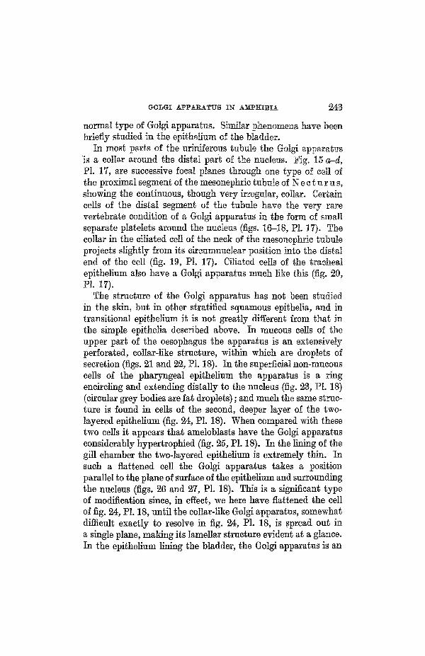

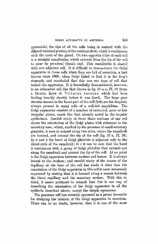


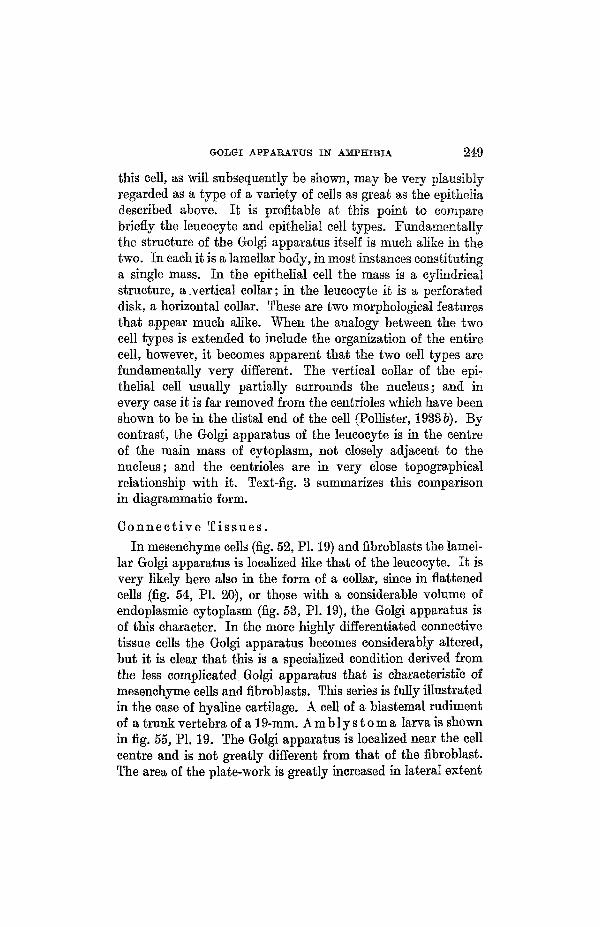


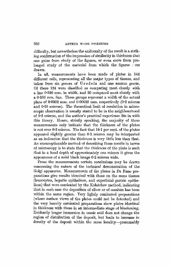


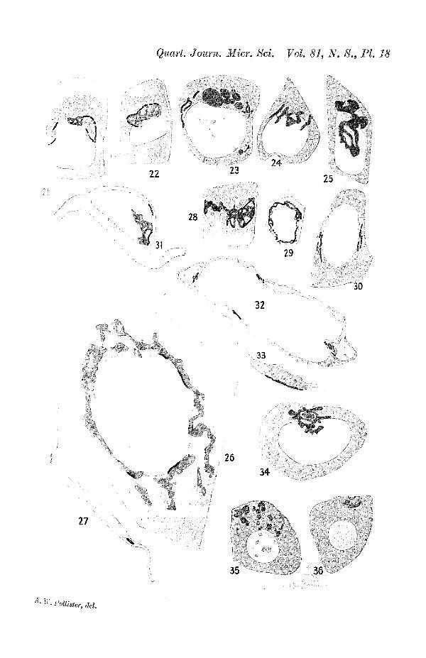


![A Golgi-Released Subpopulation of the Trans-Golgi · A Golgi-Released Subpopulation of the Trans-Golgi Network Mediates Protein Secretion in Arabidopsis1[OPEN] Tomohiro Uemura,a,b,2,3,4](https://static.documents.pub/doc/80x56/5eda9f5a09f66a09130ba5a1/a-golgi-released-subpopulation-of-the-trans-golgi-a-golgi-released-subpopulation.jpg)


