91
TLR-AGONIST TREATMENT AND SALMONELLA INFECTION
| Date post: | 05-Feb-2018 |
| Category: |
Documents |
| Upload: | truongduong |
| View: | 217 times |
| Download: | 0 times |

TLR-AGONIST TREATMENT AND SALMONELLA INFECTION

THE ROLE OF TOLL-LIKE RECEPTOR AGONIST TREATMENT
ON
SALMONELLA INFECTION IN MACROPHAGES
By
CHRISTINE ELIZABETH WONG, B. Sc.
A Thesis
Submitted to the School of Graduate Studies
in Partial fulfillment of the Requirements
for the Degree
Masters of Science
McMaster University
© Copyright by Christine Elizabeth Wong, September 2008

Master of Science (2008) McMASTER UNIVERSITY
Biochemistry and Biomedical Sciences Hamilton, Ontario
TITLE:
AUTHOR:
SUPERVISOR:
Number of Pages:
The Role of Toll-Like Receptor agonist Treatment on Salmonella Infection in Macrophages
Christine Elizabeth Wong, B. Sc. (University ofToronto)
Dr. Brian K. Coombes
xii, 78
ii

ABSTRACT
Salmonella is a Gram-negative intracellular pathogen that causes gastroenteritis and
typhoid fever in humans. Salmonella can survive and replicate within host cells and has
adapted several mechanisms to evade host immune defenses. The innate immune system
plays an important role as a first-line of defense against pathogens such as Salmonella,
and is mediated in part by toll-like receptors (TLRs). TLRs recognize fundamental
components of pathogenic microorganisms and activation ofTLRs leads to downstream
signaling cascades eventually resulting in the expression of pro-inflammatory cytokines
( 4) and also has a role in activating adaptive immunity through presentation of antigens to
lymphocytes (86). There are several lines of evidence that suggest that TLR activation
may have therapeutic potential in therapies against infectious disease and several TLR
agonists have been shown to protect against both bacterial and viral infection in mice (7;
8; 38; 66; 75; 84; 89; 121). To understand how TLR-agonist treatment of host cells
affects Salmonella pathogenesis, RAW 264.7 murine macrophages were treated with the
TLR agonists liposaccharide (LPS), poly(I:C), peptidoglycan, and CpG-ODN.
Treatment ofmacrophages with all TLR-agonists results in increased phagocytosis of
Salmonella compared to control-treated macrophages. These increases in phagocytic
activity, however, do not enhance macrophage anti-microbial activity, since Salmonella
infection ofTLR-treated macrophages results in increased intracellular replication
compared to control-treated cells. Infection with Salmonella mutants indicates that
increased intracellular replication of Salmonella in TLR-treated macrophages is
1ll

dependent on a functional SPI-2 type III secretion system. This also indicates that there
was not a generalized defect in macrophage anti-bacterial function. These data exemplify
how interactions between macrophage defense mechanisms and bacterial virulence
factors can result in evasion of the innate immune response. Studying how TLR-agonist
treatment affects Salmonella pathogenesis will give us a better understanding of the host
pathogen relationship and may provide insight into novel strategies to fight intracellular
microorganisms.
Key words: Salmonella, bacterial pathogenesis, macrophage, Toll-like receptor,
innate immunity
lV

ACKNOWLEDGEMENTS
I would like to acknowledge all past and present members of the Coombes lab, especially
Ana Tomljenovic Berube, Suzanne Osborne, Colin Cooper, and Kun Zhang for their
friendship, assistance and support. I would like to thank my friends (especially fellow
4H13 members) and family for their support. Thank you to my favourite sister and to
Joseph, for coming to the lab with me for late-night experiments, listening to me as I
sorted my thoughts, and taking care of me during the busy times. Thank you to Dr. Tony
Collins for his assistance and training with the microscopes in the McMaster
Biophotonics Facility and to Veronica Canadien and Dr. John Brumell for training,
reagents, and invaluable troubleshooting advice for my immunofluorescence experiments.
Thank you to friends and Coombes lab members who provided helpful feedback and
suggestions for the preparation of this paper. I would like to thank my committee
members Dr. Ali Ashkar, Dr. Lori Burrows, Dr. Murray Junop and Dr. Christian Baron
for their helpful comments and feedback. Finally, I would like to thank my supervisor
Dr. Brian Coombes for allowing me to pursue this project and for his continuous
inspiration, support, and guidance.
v

TABLE OF CONTENTS
Abstract ............................................................................................................................ iii
Acknowledgements ........................................................................................................... v
Table of Contents ............................................................................................................. vi
List ofFigures and Tables ................................................................................................ ix
List of Abbreviations ....................................................................................................... xi
1. INTRODUCTION
1.1 Salmonella enterica ....................................................................................... 1
1.1.1 Bacterial invasion ............................................................................ 2
1.1.2 Salmonella pathogenicity island-2 (SPI-2) ...................................... 3
1.1.3 Intracellular survival within the Salmonella-containing vacuole ..... 4
1.1.4 SPI-1 and SPI-2: Complex interactions and coordination within the host ............................................................................................ 5
1.2 Innate immunity ............................................................................................. 6
1.3 Toll-like receptors .......................................................................................... 7
1.4 Toll-like receptor signal transduction ............................................................ 8
1.5 Toll-like receptor agonists ........................................................................... 10
1.6 Potential of Toll-like receptor agonist treatment against infection .............. 12
1. 7 Rational and objectives ................................................................................ 13
Vl

2. MATERIALS AND METHODS
2.1 Materials ..................................................................................................... 15
2.2 Methods
2.2.1 Bacterial strains and growth conditions .......................................... 15
2.2.2 Mammalian cell lines and growth conditions ................................ 15
2.2.3 Toll-like receptor agonist treatment.. ............................................. 15
2.2.4 Gentamicin protection assay .......................................................... 16
2.2.5 Fluorescent microscopy ................................................................. 17
2.2.6 Antibodies and reagents ................................................................. 17
2.2. 7 Phagocytosis of fluorescent microspheres ..................................... 18
2.2.8 Recombinase-based in vivo expression technology (RIVET) ....... 19
2.2.9 Transmission electron microscopy ................................................ 19
3. RESULTS
3.1 Treatment with TLR-agonists leads to increased uptake of S. Typhimurium ........................................................................................ 23
3.2 Treatment with TLR-agonists does not affect phagocytosis of Fluorescent microspheres ........................................................................ 24
3.3 Treatment with TLR-agonists leads to increased intracellular replication of S. Typhimurium ................................................................ 24
3.4 Increased bacterial replication is observed even with reduced multiplicity of infection ............................................................................ 25
Vll

3.5 Stimulation with interferon-gamma results in net killing of Salmonella within TLR-agonist treated cells ........................................... 25
3.6 The increase inS. Typhimurium replication in TLR-agonist treated macrophages is dependent on a functional SPI-2 T3SS ........................... 27
3.7 SPI-2 promoter activity is not enhanced in TLR-agonist treated cells early in infection ....................................................................................... 27
3.8 Bacteria are not found in significant levels in the cytosol but are found within Lamp !-positive vacuoles .................................................... 29
4. DISCUSSION
4.1 TLR agonist treatment and phagocytosis ................................................. 31
4.2 TLR-agonist treatment increases intracellular bacterial replication ......... 34
4.3 TLR agonist-dependent increase in Salmonella replication through Cdc42, Rae and actin remodeling ............................................................. 36
4.4 TLR-agonist treatment and SCV maturation ............................................ 37
4.5 Host-pathogen interaction in TLR-agonist treated macrophages ............ 39
4.6 Model for TLR agonist-dependent increase in Salmonella replication through scavenger receptor-A, Hook3, and SPI-2 effector SsaB ............. 40
4. 7 Summary ................................................................................................. 42
5. REFERENCES ........................................................................................................... 58
6. APPENDICES
6.1 Transmission electron micrographs ofTLR-agonist treated macrophages 2 hours following infection with Salmonella ..................... 73
6.2 Sample images from ImageJ software program ....................................... 74
6.3 Macro for phagocytosis of fluorescent beads assay ................................ 75
Vlll

LIST OF FIGURES AND TABLES
Table 1: Chemicals and Reagents .................................................................................. 21
Figure 1: Salmonella redirects phagosome maturation .................................................. 43
Figure 2: TLR signaling pathway .................................................................................. 44
Figure 3: Treatment with TLR agonists results in an increase in Salmonella uptake at 2 hours ............................................................................................................ 45
Figure 4: Immunofluorescence ofTLR-agonist treated cells shows an increase in Salmonella uptake at 2 hours .......................................................................................... 46
Figure 5: Treatment with TLR-agonists does not affect the phagocytosis of fluorescent microspheres ................................................................................................. 4 7
Figure 6: Treatment with TLR agonists results in increased intracellular replication of Salmonella .................................................................................................................. 48
Figure 7: Reducing the multiplicity of infection still results in increased intracellular replication of Salmonella ............................................................................ 49
Figure 8: Net killing of bacteria in cells treated with TLR-agonist plus interferon-gamma ............................................................................................................ 50
Figure 9: Increase in Salmonella intracellular replication RAW 264.7 cells is dependent on functional SPI-2 type 3 secretion .............................................................. 51
Figure 10: Schematic representation of recombinase-based in vivo expression technology (RIVET) ....................................................................................................... 52
Figure 11: SPI-2 promoter activity is not enhanced in TLR-agonist treated cells early in infection ............................................................................................................. 53
Figure 12: Immunofluorescent staining for cytosolic bacteria in LPS-treated cells ...... 54
Figure 13: Immunofluorescent staining for bacteria within Lampl-positive vacuoles. 55
ix

Figure 14: Most bacteria reside within Lamp1-positive vacuoles ................................. 56
Figure 15: Hypothesized interaction map between TLRs, SRs, SsaB, and Hook3 ....... 57
X

LIST OF ABBREVIATIONS
AP-I Arp2/3 CD DMEM EEAI ERK GPI IFN IKB IKK IL iN OS IRAK IRF3 JAK Lamp LBA LBP LPS LRR M6PR MAL MAPK MDR MEK MyD88 NADPH NF-KB NO Nrampl PAMP PBS PG Poly(I:C) PRR RIP1 ROS scv SEM
activator protein-1 actin-related protein 2/3 clusters of differentiation Dulbecco's Modified Eagle's Medium early endosome antigen- I extracellular signal-regulated kinase glycosylphosphatidylinositol interferon inhibitor ofNF-KB inhibitor of KB kinase interleukin inducible nitric oxide synthase interleukin-1 receptor-associated kinase IFN regulatory factor-3 Janus kinase lysosomal-associated membrane protein lysobisphosphatidic acid LPS binding protein lipopolysaccharide leucine rich repeat mannose-6-phosphate receptor MyD88 adaptor-like mitogen-activated protein kinase multi-drug resistance MAPK and ERK kinase myeloid differentiation protein-88 nicotinamide adenine dinucleotide phosphate nuclear factor KB nitric oxide natural resistance-associated macrophage protein 1 pathogen-associated molecular pattern phosphate-buffered saline peptidoglycan polyinosine-polycytidylic acid pattern recognition receptor receptor-interacting protein-1 reactive oxygen species Salmonella-containing vacuole standard error of the mean
Xl

Sif SNARF SPI SR TAB TAK TANK TBKl TF TGF(3 TIR TIRAP TLR TNF TRAF6 TRIF
TRAM V-ATPase WASP
Salmonella-induced filament seminaphtharhodafluor Salmonella pathogenicity island scavenger receptor TAKl-binding TGF-P activated kinase TRAF family member-associated NF-KB activator TANK-binding k:inase-1 transferrin receptor transforming growth factor-(3 Toll/IL-lR homology TIR-associated protein toll-like receptor tumour necrosis factor tumour necrosis factor receptor-associated factor 6 TIR -domain-containing adapter-inducing IFN -P/TLR -associated activator ofiFN TRIP-related adaptor molecule vaculolar adenosine triphosphatase Wiskott-Aldrich Syndrome protein
Xll
M. Sc. Thesis- C. E. Wong McMaster- Biochemistry and Biomedical Sciences
1. INTRODUCTION
1.1 Salmonella enterica
Salmonella enterica is an intracellular Gram-negative bacterium that infects
multiple hosts and causes a variety of diseases (141 ). There are over 2500 known
serovars of Salmonella. Serovars such as Salmonella Typhi or Salmonella Paratyphi are
human-restricted and are the infective agents of typhoid fever (65). Other serovars such
as Salmonella enterica serovar Typhimurium (S. Typhimurium) have a broad host range,
infecting a wide variety of species including humans, mice, reptiles, and birds (65).
Salmonella is usually acquired following ingestion of contaminated food or water. In
humans, S. Typhimurium typically causes self-limiting gastroenteritis and enteric fever
(141), whereas in mice it causes a systemic infection which resembles the human-specific
typhoid fever (65). Therefore murine infection with S. Typhimurium serves as an
excellent animal model for typhoid fever.
An important part of Salmonella's virulence strategy is its ability to survive and
replicate within eukaryotic host cells. Once inside host cells, the bacteria reside within a
specialized membrane-enclosed compartment, termed the Salmonella-containing vacuole
or SCV, which provides a niche where Salmonella is able to survive and replicate. Type
III secretion systems (T3SSs) play an important role in the ability of Salmonella to infect
host cells and cause disease. These systems consist of over 20 proteins that form a
needle-like structure that spans both the inner and outer bacterial membrane and extends
to the host cell membrane, allowing for the translocation of bacterial effectors into the
host cell cytosol (78). While several Gram-negative bacteria possess similar T3SSs, the
1

M. Sc. Thesis- C. E. Wong McMaster- Biochemistry and Biomedical Sciences
effector proteins secreted vary. Salmonella has two distinct T3SSs encoded within two
different horizontally acquired Salmonella pathogenicity islands (SPI), termed SPI-1 and
SPI-2.
1.1.1 Bacterial invasion
The SPI-1 T3SS plays an important role in the bacteria's ability to gain entry into
the host and cause disease. Salmonella is able to enter its host through invasion of the
epithelial cells of the small intestine. Mutations that prevent the formation of the SPI-1
T3SS result in a 10 to 100-fold decrease in virulence (11; 49; 83). Following contact
with the host cell the SPI-1 T3SS iiijects effectors such as SopB, SopE, SopE2 and SopD
which activate the Rho GTPases Cdc42, Rac-1, and RhoG in the host cell (10; 45; 67;
112; 115; 139; 160). Once activated, Rho GTPases activate Wiskott-Aldrich Syndrome
protein (WASP) family members N-WASP and W A VE2, which in turn recruit the actin
related protein 2/3 (Arp2/3) complex to the site of entry (33; 134; 139; 143). This leads
to membrane ruffling and ultimately causes the host cell to engulf the bacterium (50).
Actin cytoskeleton rearrangement is critical to Salmonella entry since treatment with
drugs that disrupt actin dynamics prevent bacterial uptake into epithelial cells ( 42).
Interestingly, recent studies have shown that treatment with TLR-agonists can also lead
activation of the Rho GTPases Cdc42 and Rae, resulting in an increase in phagocytosis
(88).
Salmonella entry also activates mitogen activated protein kinases (MAPKs) such
as Erk, Jnk and p38, which in turn activate the transcription factors nuclear factor KB
(NF-KB) and activator protein-1 (AP-1), leading to the production of pro-inflammatory
2

M. Sc. Thesis- C. E. Wong McMaster- Biochemistry and Biomedical Sciences
cytokines (50). These signaling events are significant because they contribute to
inflammatory diarrhea, which is a characteristic symptom of Salmonella infection (50).
1.1.2 Salmonella pathogenicity island-2 (SPI-2)
The SPI-2 T3SS is prominently induced after the bacterium has entered host cells
and plays an important role in intracellular survival and replication. The SPI-2 locus
encodes for a T3SS apparatus and SPI-2 genes are regulated by SsrAB, a two-component
regulatory system (113; 133). SPI-2 is required for prevention of fusion between the
SCV and the lysosomes (142), for avoidance of macrophage killing through NADPH
oxidase (51; 146), and for prevention of delivery of inducible nitric oxide synthase
(iNOS) to the SCV (27). All of these processes are carried out by secreted bacterial
effector proteins that are encoded both within and outside ofSPI-2 and are important for
the survival ofthe bacterium within the host cell (153).
Previous studies have suggested that bacteria can upregulate expression of genes
required for intracellular survival when infecting cells that are better equipped to kill
bacteria (159). Natural resistance-associated macrophage protein- I (Nrampl, also known
as Sic 11 al) is a divalent cation transporter that localizes to the late endocytic/lysosomal
vacuole (60). Nrampl can restrict the growth of Salmonella by removing ions from the
SCV (148). Nrampl has an important role in host defense and infection of mice lacking
Nrampl with various intracellular bacteria such asS. Typhimurium and Mycobacterium
bovis bascillus Calmette-Guerin results in high levels of bacterial replication and fatality
(16). Zaharik et al. examined potential crosstalk between host Nrampl status and
Salmonella virulence effectors (159). They found that when Salmonella is infecting an
3

M. Sc. Thesis - C. E. Wong McMaster - Biochemistry and Biomedical Sciences
Nrampl+t+ host there was an increase in expression ofSPI-2 genes such as sseA and ssrA
compared to infections ofNramp 1-/- hosts (159). This suggests that bacteria can sense the
immune status of its host, and upregulate SPI-2 gene expression accordingly.
1.1.3 Intracellular survival within the Salmonella-containing vacuole (SCV)
Within the SCV, bacteria are able to redirect the normal phagosomal maturation
process to allow for survival (22) (Figure 1 ). The SCV maturation process involves
interaction with the endosomal system in a highly orderly and selective manner (1 ). The
SCV interacts briefly with early endosomes (138), acquiring marker proteins such as
early endosome antigen- I (EEAI), transferrin receptor (TF), and the GTPases Rab5 and
Rabll (123; 136; 138). However, the interaction with these proteins is only transient,
and after EEAl and Rab5 dissociate, the SCV then acquires select markers of the late
endosome/lysosome such as Rab 7 ( 1 01 ), which in turn attracts lysosomal-associated
membrane proteins (Lamps) Lampl, Lamp2, and Lamp3 and vacuolar ATPase, which
acidifies the SCV (37; 52; 138). Despite the presence of these late endosome/lysosome
markers, the SCV does not merge with lysosomes. This is confirmed by the absence of
mannose-6-phosphate receptors (M6PR), cathepsin D, and lysobisphosphatidic acid
(LBA) (27; 50; 52; 54; 68). Therefore, Salmonella is able to redirect the normal
phagosome maturation process to its advantage, creating an environment that is
conducive to its survival and growth. Interestingly, some studies have suggested that
TLR signaling may also have an effect on phagosome maturation and proteolytic capacity
in macrophages (17; 155).
4

M. Sc. Thesis- C. E. Wong McMaster- Biochemistry and Biomedical Sciences
Approximately 4-6 hours after invasion in epithelial cells, the SCV extends
elongated tubular structures from its surface called Salmonella-induced filaments (Sifs)
(53). This structural change corresponds with the beginning of bacterial replication and
like the SCV, Sifs are associated with Lamps and vacuolar ATPase (23; 53). Sifs were
originally thought to be restricted to epithelial cells, however studies using interferon
gamma (IFN-y)-primed macrophages demonstrated that Salmonella forms Sifs in
macrophages as well (87). Though their exact physiological purpose is not known, it is
possible that they function to increase the size of the SCV in a specific direction that is
more conducive for bacterial replication. Additionally, it is thought that they may deliver
nutrient-containing vacuoles and organelles to the SCV (65).
One method that macrophages have developed to combat infection by intracellular
pathogens is the generation of reactive oxygen species (ROS) and nitrogen species such
as nitric oxide (NO), superoxide and hydrogen peroxide. Superoxide is created by the
membrane-associated NADPH oxidase complex. This process of generating reactive
oxygen species is called the respiratory burst and is usually effective at killing microbes
within the phagosome (79; 1 06). SPI-2 effectors enable Salmonella to avoid the
respiratory burst and survive within the macrophage (51; 146). Thus, Salmonella has
evolved to survive within several cell types and has acquired multiple mechanisms to
resist host immune defenses.
1.1.4 SPI-1 and SPI-2: Complex interactions and coordination within the host
It was previously believed that SPI-1 and SPI-2 were expressed at distinct points
of infection with SPI -1 being responsible for induction of an inflammatory response in
5

M. Sc. Thesis - C. E. Wong McMaster - Biochemistry and Biomedical Sciences
the gut, invasion of epithelial cells, and colonization of the intestine and with SPI-2 being
responsible for establishing systemic infection and for intracellular survival in
macrophages. However, an increasing number of studies are concluding that this view is
too simplistic and that there is likely more interplay between these pathogenicity islands.
For example, it was shown that SPI-1 is also required for S. Typhimurium survival and
replication within epithelial cells (137), which expands the role ofSPI-1 from beyond the
initial invasion of the gut. Also, SPI-2 has been shown to be expressed in the intestine,
prior to intestinal colonization (21). Lack of functional SPI-2 T3SS can reduce the
expression of some SPI-1 T3SS genes and be detrimental to colonization, which provides
further evidence for the importance ofSPI-1 and SPI-2 T3SS interplay (34). In addition,
several groups have shown that some SPI-1 effectors are present in host cells long after
the initial invasion (65). Therefore, there is much more spatio- and temporal-overlap
between these two pathogenicity islands than previously thought and this is further
evidence for the complex interactions that have evolved between Salmonella and its host.
1.2 Innate immunity
The mammalian immune system has evolved to combat numerous pathogens that
pose a daily threat. The immune system in mammals consists of two main branches:
innate immunity and adaptive immunity. The innate immune system plays an important
role in early host defense against pathogens (9; 13-15). It involves the recognition of
microorganisms through germline-encoded pattern-recognition receptors (PRRs) (4).
PRRs recognize pathogen-associated molecular patterns (P AMPs) that are conserved
components of microorganisms such as lipopolysaccharide (LPS), flagellin,
6

M. Sc. Thesis - C. E. Wong McMaster- Biochemistry and Biomedical Sciences
peptidoglycan, bacterial DNA, and viral RNA (4). PRRs recognize specific PAMPs and
this interaction results in the activation of downstream signaling cascades and distinct
anti-microbial responses by the host cell (4). Unlike the adaptive immune system, this
form of recognition is nonclonal and is not associated with immunological memory (4).
These basic recognition mechanisms are found in all classes of species, in both plants and
mammals (4).
1.3 Toll-like receptors
Toll-like receptors (TLRs) are a family ofPRRs that are conserved in many
species (4). Toll was first discovered in Drosophila as a critical component involved in
establishing embryonic dorsal-ventral patterning (6; 107) and was later found to play an
important role in resisting fungal infections (94). One year later, the first human
homologue of the Drosophila Toll protein (hToll) was identified by Janeway and
colleagues and was shown to induce both innate and adaptive immune responses through
activation of the NF-KB pathway (99). This receptor was later renamed TLR4 and mice
with defective TLR4 were found to be hyporesponsive to LPS, further supporting a role
for TLRs in mammalian immunity (76; 118). To date, 13 mammalian TLRs have been
identified along with numerous TLR ligands, signaling molecules, and regulatory
molecules (55).
TLRs are type I integral membrane glycoproteins that contain leucine rich repeat
(LRR) motifs in the extracellular domains and a Toll!IL-lR homology (TIR) signaling
domain in the intracellular domain (18). TLRs are expressed by both immune cells such
as macrophages, dendritic cells and B cells, as well as on non-immune cells such as
7

M. Sc. Thesis - C. E. Wong McMaster - Biochemistry and Biomedical Sciences
epithelial cells and fibroblasts (4). Some TLRs such as TLRs 1, 2, 4, 5, and 6 are
expressed on the surface of cells where they can sample extracellular ligands such as
lipopeptides, peptidoglycan, LPS, and flagellin. Other TLRs such as TLRs 3, 7, 8, and 9
are localized to an intracellular compartment for recognition of internalized bacterial and
viral DNA (4).
1.4 Toll-like receptor signal transduction
The molecules involved in TLR signaling are the same as those that are used for
IL-lR signaling (3). The TLR signaling cascade begins with the recruitment ofTIR
containing adaptor molecules such as Myeloid differentiation factor-88 (MyD88), MyD88
adaptor-like (MAL, also known asTIR-associated protein, TIRAP), TIR domain
containing adaptor inducing IFNP (TRIF), and TRIF-related adaptor molecule (TRAM)
(86). The different TLRs have specificity for different signaling pathways by recruiting
different adaptor proteins (36).
MyD88 is a shared adaptor molecule in all TLR signaling pathways with the
exception ofTLR3 (5). MyD88 contains a C-terminal TIR domain, which allows it to
interact with the TIR domain in the cytoplasmic tail ofTLRs (36). MyD88 also has N
terminal death domains, which interact with the death domains of the interleukin-1
receptor-associated kinase (IRAK) family members (36). There are two main TLR
signaling pathways, the MyD88-dependent pathway and the MyD88-independent
pathway.
In the MyD88-dependent pathway, MyD88 interacts with the cytoplasmic tail of
TLRs, recruits IRAK4 and together they mediate IRAK1 phosphorylation (97). Once
8

M. Sc. Thesis - C. E. Wong McMaster - Biochemistry and Biomedical Sciences
phosphorylated, IRAK1 interacts with and activates tumour necrosis factor receptor~
associated factor-6 (TRAF6) and together they interact with a membrane complex
consisting ofTGF-~ activated kinase (TAK1), and the TAK1-binding (TAB) family
members, TABl, and TAB2 (36). The complex is activated following TRAF6 and TAKl
ubiquitination, resulting in the phosphorylation ofTAKl and TAB2 and the dissociation
of the complex from the membrane (81; 122; 151). In the cytosol IRAK1 is degraded and
activated TAK1 can phosphorylate the inhibitor of KB kinase (IKK) complex, leading to
the phosphorylation and degradation ofiKB and the release ofNF-KB (100). Free NF-KB
translocates to the nucleus where it promotes the expression of pro-inflammatory genes.
TLR3 and TLR4 activate the MyD88-independent pathway which is also known
as the TRIF-dependent pathway (86). TLR4 recruits TRAM, which subsequently recruits
TRIF, whereas TLR3 can recruit TRIF directly (43). TRIF can bind to receptor
interacting protein-1 (RIP1), which recruits TRAF6, resulting in the activation of the IKK
complex (103). Direct binding ofTRIF and TRAF6 can result in IRAK1/4-independent
activation ofTAKl (80; 82). TRIF can bind TANK-binding kinase-1 (TBKl) and IKKs,
which activates the transcription factor Interferon regulatory factor-3 (IRF3) which plays
an important role in the production of type 1 interferons and defense against viral
infections (129) (Figure 2).
TLR4 is unique in its ability to participate in both the MyD88-dependent and
TRIF-dependent signaling pathways. It was recently shown that MyD88-dependent and
TRIF-dependent signaling are not initiated simultaneously in TLR4 (85). The MyD88-
dependent signaling is initiated after TLR4 activation by LPS on the plasma membrane
9

M. Sc. Thesis - C. E. Wong McMaster- Biochemistry and Biomedical Sciences
whereas the TRIF -dependent signaling pathway is initiated in the early endosome after
TLR4 has been endocytosed (85). Therefore, it can be generalized that TRIP-dependent
signal transduction is initiated intracellularly for both TLR3 and TLR4.
1.5 Toll-like receptor agonists
Several TLR-agonists have been reported and studied, however for the purpose of
this study I focused on lipopolysaccharide (LPS), poly(I:C), peptidoglycan, and CpG
oligodinucleotides. These ligands are all commercially available and their effects on the
innate immune response have been studied previously, but not in the context of
Salmonella infection of macrophages.
One ofthe strongest immunostimulants identified to date is LPS from the Gram
negative cell wall. LPS is made up ofthree main components -lipid A (endotoxin), a
core oligosaccharide, and an 0-antigen (26). Lipid A is the active component of LPS and
is responsible for many Gram-negative pathologies such as endotoxin shock. The initial
clue that TLR4 was the receptor for LPS was through the observation that C3H/HeJ mice
were hyporesponsive to LPS. It was later discovered that this was due to a mutation in
the TLR4 gene (118). The TLR4 signaling pathway requires the accessory proteins CD14
and MD-2 (4). Free LPS binds to the LPS binding protein (LBP), which is an acute-phase
protein that is found in the bloodstream. LPS-LBP then binds to CD14, which is a
glycosylphosphatidylinositol (GPI) linked protein that is expressed on the cell surface of
phagocytic cells. Next, LPS is transferred to MD-2, which associates with the
extracellular portion ofTLR4. This leads to TLR4 oligomerization and downstream
signaling.
10

M. Sc. Thesis - C. E. Wong McMaster- Biochemistry and Biomedical Sciences
Many viruses produce double stranded RNA as part of their life cycle. Double
stranded RNA and its synthetic analog, polyinosine-polycytidylic acid, (poly(I:C)), are
both recognized by TLR3 (5). TLR3 is expressed intracellularly in several types of cells
and its expression is upregulated significantly when cells are treated with poly(I:C) ( 4).
Another strong stimulator of the innate immune system is peptidoglycan (PG, also
known as murein). Peptidoglycan is a polymer of ~-(1,4) linked N-acetylglucosamine
and N-acetylmuramic acid. A small peptide chain consisting of 3-5 amino acids is
attached to the N-acetylmuramic acid. Peptidoglycan makes up approximately 10% of
the dry weight of the Gram-negative cell wall and 90% of the Gram-positive cell wall.
TLR2 is involved in the recognition of peptidoglycan (132; 157). TLR2 can recognize a
wide variety of ligands due to its ability to form heterodimers with TLRI and TLR6 (26).
Bacterial DNA is also highly immunostimulatory. In mammalian DNA, CpG
dinucleotides are highly methylated (4). In contrast, bacterial DNA contains
unmethylated CpG dinucleotides, which is referred to as CpG-DNA (4). Unmethylated
CpG-DNA is found approximately twenty times more frequently in bacterial DNA than in
mammalian DNA (150) and is recognized by TLR9, which is an intracellular receptor
located in endosomes (72; 90). In order to stimulate a response, the bacterial DNA must
be delivered to the endosome where it is degraded into smaller single-stranded segments
which are then able to interact with TLR9 (2; 93). Synthetic oligonucleotides that contain
the CpG motif, such as CpG-oligodeoxynucleotide (CpG-ODN), are also able to stimulate
an immune response (4).
11

M. Sc. Thesis- C. E. Wong McMaster- Biochemistry and Biomedical Sciences
1.6 Potential of Toll-like receptor agonist treatment against infection
There are several lines of evidence that suggest that TLR activation may have
therapeutic potential. In fact, development of synthetic TLR ligands in cancer, vaccine
improvement, infectious disease and allergies are already in development, with some
already in the early clinical trial phase (91). For example, treatment with
oligonucleotides that contain the CpG motif has been shown to provide protection against
several pathogens in mice including Listeria monocytogenes (38; 89), Francisella
tularensis (38), Herpes Simplex Virus-2 (HSV-2) (7; 66; 121) and Mycobacterium
tuberculosis (84). Treatment with the TLR3 ligand, poly(I:C), can protect against
infection with HSV-1 and HSV-2 (8; 75). The TLR7 agonist imiquimod has been
approved for use as a topical treatment against viral infections such as human
papillomavirus ( 41 ). One advantage of enhancing innate immunity through TLR
stimulation is that it will not result in direct selective pressure on the pathogens ( 41 ). The
effect ofTLR stimulation on Salmonella infection is an area that requires further study.
12

M. Sc. Thesis - C. E. Wong McMaster- Biochemistry and Biomedical Sciences
1. 7 Rationale and objectives
Worldwide, Salmonella is one of the most widespread sources of food-borne
illness and disease. Every year, millions of people are infected with Salmonella, resulting
in thousands of fatalities (59). In developing countries, Salmonella infection continues to
be a significant public health concern (77), especially in children, the elderly, and patients
with HIV (59). In addition, the emergence ofmultidrug-resistant strains of Salmonella
are threatening to become a serious public health problem, not just in developing
countries, but in developed nations such as Canada as well. For example, the Salmonella
Typhimurium strain DT1 04 is resistant to several antibiotics including ampicillin,
chloramphenicol, florfenicol, streptomycin, spectinomycin, sulfonamides, and
tetracycline (71). It has been hypothesized by many groups that drug-resistant strains of
Salmonella are more virulent than drug-susceptible strains since infection with multi-drug
resistant (MDR) Salmonella is associated with increased morbidity and mortality
compared to the drug-susceptible strains (57; 108; 145). The multidrug-resistance region
ofDT104 is located on the chromosome allowing for more stable maintenance in the
absence of selective-pressures (19).
Based on these observations, there is a strong need to examine alternative
therapies to antibiotics. Scientists have proposed a variety of strategies as novel
alternatives to antibiotic therapy, some of which include enhancing immunity, the
creation of novel anti-bacterials, prebiotics and probiotics, as well as bacteriophage
therapy. For my graduate studies, I have focused on investigating the potential to
13
M. Sc. Thesis- C. E. Wong McMaster - Biochemistry and Biomedical Sciences
enhance or modulate the innate immune system through TLRs as a strategy to combat
bacterial infection.
Modulating the innate immune response through TLR -agonist treatment is an area
of research that has received increased attention. Several reviews have examined the
effects ofTLR agonists and signaling on animal models of infectious disease (58; 91;
114). However, few have examined the specific role ofTLRs on macrophages, which
are important since macrophages play a dual role in immunity by acting as both a first
line of defense through destruction of pathogens as well as antigen-presenting cells to
initiate the adaptive immune response (98). Therefore macrophages are truly at the
interface between the innate and adaptive branches of the immune system. In addition,
few have examined the role of innate immune modulation on Salmonella pathogenesis.
Studying Salmonella infection of macrophages is of particular importance since mutants
that are unable to survive within macrophages are also unable to cause systemic disease in
mice (40).
For my graduate research, I have focused on the role ofTLR-agonist treatment on
the host response to Salmonella infection. My objectives were: (i) To understand how
treatment with TLR-agonists affects phagocytosis, phagosome maturation, and
bactericidal ability of host macrophages (ii) To understand the host-pathogen
relationship in TLR-agonist primed macrophages.
Studying how TLR-agonist treatment affects Salmonella pathogenesis will
provide us with a better understanding of this complex host-pathogen relationship and
may provide insight into novel strategies to fight intracellular microorganisms.
14

M. Sc. Thesis - C. E. Wong McMaster - Biochemistry and Biomedical Sciences
2. MATERIALS AND METHODS
2.1 Materials
Please refer to Table 1 for a list of all chemicals and reagents used.
2.2 Methods
2.2.1 Bacterial strains and growth conditions
Salmonella enterica serovar Typhimurium strain SL1344 (154) was used
throughout. Bacteria were cultured in Luria-Bertani (LB) broth and on LB agar plates
containing streptomycin (50 J,tg/ml), ampicillin (100 J,tg/ml), or chloramphenicol (15
J,tg/ml) where appropriate. The ssaR deletion mutant (D.ssaR) was derived from SL1344
(23). The recombinase-based in vivo expression technology (RIVET) reporter strains to
measure SPI-2 promoter activity were derived from SL1344 (21).
2.2.2 Mammalian cell lines and growth conditions
RAW 264.7 murine macrophages (ATCC) were maintained in Dulbecco's
modified Eagle's medium (DMEM, Gibco) supplemented with 10% fetal bovine serum
(HyClone). Cells were maintained in 75-cm2 culture flasks at 3TC in 5% C02 without
antibiotics.
2.2.3 Toll-like receptor agonist treatments
RAW 264.7 cells were treated with TLR agonists approximately 20 hours before
infection with 100 ng/mllipopolysaccharide from Escherichia coli (Sigma), 10 J.tg/ml
poly(I:C) (Sigma), 10 J,tg/ml peptidoglycan from Staphylococcus aureus (Sigma), or 10
15

M. Sc. Thesis - C. E. Wong McMaster - Biochemistry and Biomedical Sciences
J.Lg/ml CpG-ODN 5'-TCGTCGTTTTGTCGTTTTGTCGTT-3' (Mobix Labs, McMaster
University, Canada). Cells treated with phosphate buffered saline (PBS) were used as a
control.
2.2.4 Gentamicin protection assay
RAW 264.7 cells were seeded 24 hours before infection at a density of 2.0 x 10⁵
cell per well. S. Typhimurium SL1344 was grown overnight in 3 ml ofLB broth in
culture tubes, with shaking at 225 rpm. RAW cells were infected in triplicate with
human-serum opsonized stationary phase bacteria at a multiplicity of infection (MOl) of
approximately 50 bacteria per cell. After a 30 minute incubation period to allow for
bacterial uptake, cells were washed 3 times with PBS and incubated in DMEM containing
100 J.Lg/ml gentamicin, which kills extracellular bacteria. After 2 hours, the gentamicin
concentration was reduced to 10 f.tg/ml. At 2 and 20 hours post-infection, cells were
washed 2 times with PBS and then lysed with 250 J..Ll of 1% Triton X-100 and 0.1%
sodium dodecyl sulfate (SDS) in PBS. Lysates were diluted in PBS and plated onto LB
agar containing the appropriate drug-selection and incubated overnight at 3TC. Colonies
were counted and expressed as colony forming units (cfu). Two hour uptake levels were
normalized by dividing the 2 h cfu values of each treatment group by the average 2 h cfu
value for the PBS control. The fold increase in the number of intracellular bacteria was
calculated by dividing the cfu values at 20 h by the cfu values at 2 h post infection for
each treatment group. Fold increase was normalized by dividing by the fold increase for
each treatment group by the average fold increase of the PBS control. All experiments
16
M. Sc. Thesis - C. E. Wong McMaster- Biochemistry and Biomedical Sciences
were performed in triplicate, on at least three separate occasions. Error bars represent the
standard error of the mean,p s 0.5.
2.2.5 Fluorescent microscopy
RAW 264.7 cells were seeded onto 1.2 em coverslips at a density of 2.0 x 10⁵
cells/well in 24-well culture plates. S. Typhimurium SL1344 was grown overnight in 3
ml of LB broth in culture tubes, with shaking at 225 rpm. RAW cells were infected in
triplicate with human-serum opsonized stationary phase bacteria at a multiplicity of
infection of approximately 50 bacteria per cell. Samples were fixed with 2.5%
paraformaldehyde in PBS for a minimum of 30 min at 37°C. Samples were washed twice
with PBS and blocked and permeabilized with 10% goat serum and 0.2% saponin in PBS
(SS-PBS) for 30 minutes. Primary and secondary antibodies were spread over the surface
of the coverslips in SS-PBS for 1 hour, followed by three washes with PBS. Coverslips
were mounted onto glass slides using fluorescent mounting medium (Dako ). Widefield
microscopy was performed using a Leica DMI 600B (lOOx 1.4NA objective) and
Velocity 4 software. Images were taken with a Hamamatsu Orca ER-AG camera,
imported into Adobe Photoshop 7.0 and assembled in Adobe Illustrator CS for labeling.
2.2.6 Antibodies and reagents
Rat anti-Lampl (1D4B) antibody developed by August, J.T. was obtained from
the Developmental Studies Hybridoma Bank developed under the auspices of the
National Institute of Child Health and Human Development and maintained by The
University oflowa (Department of Biological Sciences, Iowa City, IA 52242). 1D4B
17

M. Sc. Thesis - C. E. Wong McMaster- Biochemistry and Biomedical Sciences
was used at 1:100 . Mouse Anti-Mono- and Polyubiquitinylated Conjugates, Monoclonal
(clone FK2) (Biomol International) was used at 1:500. Rabbit anti-Salmonella 0
Antiserum Group B Factors 1, 4, 12, 27 (Difco) was used at 1:100. Alexa-488-
conjugated donkey anti-rat and goat anti-mouse antibodies and Alexa-568-conjugated
donkey anti-rabbit and goat anti-rabbit antibodies were used at 1 :200 and were purchased
from Molecular Probes. 4',6-diamidino-2-phenylindole, dihydrochloride (DAPI) was
used at 1 :2500 and was purchased from Molecular Probes.
2.2. 7 Phagocytosis of fluorescent microspheres
RAW 264.7 cells were seeded onto 2.5 em coverslips at a density of 1.0 x 10⁵
cells/ml in 35 x 10 mm petri dishes. 1.0 J.tm diameter green-yellow fluorescent
microspheres (Molecular Probes) were opsonized in 20% human serum for 30-60
minutes. RAW 264.7 cells were washed with PBS and incubated with 1.0 x lOi beads/ml
for 3.5 hours and then washed 3 times with PBS and transferred to Attofluor cell
chambers (Invitrogen) in HEPES buffer for live cell imaging. Samples were treated with
3 J.tM 5- (and 6)-carboxy SNARF-1 acetoxymethyl ester, acetate (Molecular Probes) to
label the cytoplasm. Confocal microscopy was performed using a Leica DMI 600B
microscope with the Leica TCS SP5 AOBS scanner (63x 1.3NA objective) and Leica
Application Suite Advanced Fluorescence Software. The 100 m W 488 nm Argon ion and
10 mW 561 nm DPSS lasers were used. Internalized beads were identified using ImageJ
software and through enumeration of at least 200 cells per treatment group. Images were
imported into Adobe Photoshop 7.0 and assembled in Adobe Illustrator CS for labeling.
18

M. Sc. Thesis- C. E. Wong McMaster- Biochemistry and Biomedical Sciences
2.2.8 Recombinase-based in vivo expression technology
A recombinase-based in vivo expression technology (RIVET) reporter was used to
measure the transcriptional activity ofthe sseA promoter (21). RAW 264.7 cells were
cultured and seeded as described previously and infected with human serum opsonized
stationary phase RIVET strains for 30 minutes and then washed and incubated in DMEM
containing 100 ~g/ml gentamicin. Macrophages were lysed at 15 minute intervals post
infection with 1% Triton X-100 and 0.1% SDS . Resolution was determined by plating
bacteria on streptomycin-containing LB-agar and then replica stamping onto
chloramphenicol-containing LB-agar.
2.2.9 Transmission electron microscopy
RAW 264.7 cells were seeded in 24-well plates at a density of 2 x 10⁵ cells/well
and treated with TLR agonists approximately 20 hours before infection. S. Typhimurium
SL1344 was grown overnight in 3 ml ofLB broth in culture tubes, with shaking at 225
rpm. RAW cells were infected in triplicate with human-serum opsonized stationary phase
bacteria at a multiplicity of infection of approximately 50 bacteria per cell. After a 30
minute incubation period to allow for bacterial uptake, cells were washed 3 times with
PBS and incubated in DMEM containing 100 ~g/ml. Two hours post-infection, cells were
fixed with 2% glutaraldehyde (Canemco) in 0.1 M sodium cacodylate (NaCac)
(Canemco) pH 7.4 for one hour at room temperature. Sample dehydration, embedding,
sectioning, and staining were performed by the Integrated Microscopy Facility in the
Health Science Centre at McMaster University. Following fixation, samples were
incubated in 0.2 M NaCac pH 7.4 two times for five minutes and post fixed with 1%
19

M. Sc. Thesis - C. E. Wong McMaster- Biochemistry and Biomedical Sciences
osmium tetroxide (Os04) (Canemco) in 0.1 M NaCac buffer for 30 minutes. Samples
were dehydrated in an ethanol series (50%, 70%, 95%, and 100%) and infiltrated with
TAAB 812 (Canemco) in 1:1 (TAAB 812: 100% ethanol) for one hour and 3:1 (TAAB
812: 100% ethanol) for one hour. Samples were then incubated in TAAB 812 twice for
one hour. Excess TAAB 812 was aspirated offthe samples and small BEEM capsules
were placed into the surface of the wells and polymerized at approximately 65°C
overnight. BEEM capsules (Canemco) were topped off with TAAB 812 and polymerized
at approximately 65°C overnight. Capsules were snapped off and ultra-thin sections
(approximately 70 nm think) were cut with an Ultracut T (Leica) and placed onto 200
mesh Cu!Pd grids. Sections were stained with saturated aqueous uranyl acetate
(Canemco) for 5 minutes and rinsed with distilled water followed by lead citrate
(Canemco) for 2 minutes. Sections were viewed and representative micrographs were
taken using a 1200 EX TEMSCAN (JEOL, Tokyo).
20

M. Sc. Thesis - C. E. Wong McMaster- Biochemistry and Biomedical Sciences
Table 1: Chemicals and Reagents
Reagent Catalogue# Source
1D4B-s (rat anti-LAMP-1 antibody) 1D4B Hybridoma Bank
4 ',6-diamidino-2-phenylindole, D1306 Molecular Probes
dihydrochloride (DAPI)
5- (and 6)-carboxy SNARF-1 acetoxymethyl C1272 Molecular Probes
ester, acetate
Agar AGROOI.l Bioshop
Alexa Fluor 488 donkey anti-rat IgG A21208 Molecular Probes
Alexa Fluor 488 goat anti-mouse lgG A11029 Molecular Probes
Alexa Fluor 568 donkey anti - rabbit lgG A10042 Molecular Probes
Alexa Fluor 568 goat anti-rabbit lgG A11036 Molecular Probes
Ampicillin Sodium Salt A0166 Sigma
Chloramphenicol CLR201 Bioshop
CpG Oligodinucleotide Mobix Labs
5'TCGTCGTTTTGTCGTTTTGTCGTT-3'
Dimethylsulfoxide DMS666 Bioshop
Dulbecco's modified Eagle's medium 11995 Gibco
Fetal bovine serum SH30397.03 HyClone
Fluorescence mounting media S3023 Dako
Fluospheres polystyrene microspheres F13081 Molecular Probes
Gentamicin Sulfate GTA202 Bioshop
Glutaraldehyde 111-30-8 Can em co
Goat serum 053-150 Wisent
HEPES Sodium Salt HEP002 Bioshop
HyQ Trypan Blue Solution SV30084.01 HyClone
21

M. Sc. Thesis- C. E. Wong McMaster- Biochemistry and Biomedical Sciences
Lead Citrate 512-26-5 Can em co
Lipopolysaccharide from Escherichia coli L2654 Sigma
Mouse Anti-Mono- and Polyubiquitinylated PW-8810
Biomol Conjugates, Monoclonal (clone FK2) International
Osmium tetroxide 20816-12-0 Can em co
Electron Paraformaldehyde (16%) 15710 Microscopy
Sciences
Peptidoglycan from Staphylococcus aureus 77140 Sigma
Peptone PEP403.1 Bioshop
Phosphate buffered saline (+Mg, +Ca) 14080-055 Invitrogen
Phosphate buffered saline 405.4 Bioshop
Pluronic F-127 P3000MP Molecular Probes
Poly(I:C) P1530 Sigma
Rabbit anti-Salmonella 0 Antiserum Group 229731 Difco
B Factors 1, 4, 12, 27
Saponin CA80058-630 VWR
Sodium cacodylate 124-65-2 Can em co
Sodium chloride SOD001.205 Bioshop
Sodium dodecyl sulfate SDS001 Bioshop
Streptomycin sulfate STP101 Bioshop
TAAB 812 CE001-3 Canemco
Triton X-100 TRX777 Bioshop
Uranyl acetate 541-09-3 Canemco
Yeast extract YES401.500 Bioshop
22

M. Sc. Thesis - C. E. Wong McMaster- Biochemistry and Biomedical Sciences
3. RESULTS
3.1 Treatment with TLR-agonists leads to increased uptake of S. Typhimurium
To understand how treatment of host cells with TLR-agonists affects the host cell
response to Salmonella infection I performed gentamicin protection assays using RAW
264.7 murine macrophages. Macrophages were treated approximately 20 hours prior to
infection with the TLR-agonists LPS, poly(I:C), peptidoglycan, CpG or with PBS as a
control as a similar time-point was defined previously to be effective against HSV -2
infection (7). Serum-opsonized S. Typhimurium cells were incubated with the
macrophages for 30 minutes to allow for phagocytic uptake, and then extracellular
bacteria were washed off and killed with a 1.5 hour gentamicin treatment. The number of
intracellular bacteria were then enumerated by colony counting following lysis of the
macrophages. Treatment with all TLR-agonists significantly increases the bacterial
uptake after 2 hours compared to the control treatments (Figure 3). Specifically,
treatment with LPS, poly(I:C), peptidoglycan, or CpG increased bacterial uptake at 2
hours post-infection by 3.0, 2.3, 4.6, 5.0-fold, respectively, as compared to control cells
treated with PBS (Figure 3). Similar results were observed using immunofluorescence
(Figure 4) and transmission electron microscopy (Appendix 1). Therefore, TLR-agonist
treatment results in an increased uptake of S. Typhimurium by RAW 264.7 cells.
23
M. Sc. Thesis - C. E. Wong McMaster- Biochemistry and Biomedical Sciences
3.2 Treatment with TLR-agonists does not affect phagocytosis of fluorescent microspheres
Treatment with all TLR-agonists tested results in an increase of S. Typhimurium
uptake two hours post-infection (Figure 3, Figure 4). To see if this increased uptake was
specific to Salmonella or a more general increase in phagocytic capacity, I examined the
effect ofTLR-agonist treatment on the phagocytosis of fluorescent microspheres. Human
serum-opsonized 1.0 J..Lm green-yellow fluorescent microspheres were incubated with
TLR-agonist treated macrophages for 3.5 hours to allow for phagocytic uptake. The
cytoplasm was labeled with SNARF-1 dye and live cells were viewed under a confocal
microscope. Following z-stack acquisition, ImageJ software was used to distinguish
internalized beads from external beads (Appendix 2, Appendix 3). There was not a
significant increase in the number of cells phagocytosing beads in macrophages treated
with LPS, peptidoglycan, or CpG compared to the PBS-control (Figure 5). This
demonstrates that while TLR -agonists treatment results an increase in S. Typhimurium
uptake, it does not lead to an increased uptake of inert microspheres.
3.3 Treatment with TLR-agonists leads to increased intracellular replication of S. Typhimurium
Since TLR-agonist treatment results in increased phagocytic capacity, I next
examined whether it also leads to increased intracellular killing of the bacteria. TLR-
agonist treated macrophages were infected with Salmonella and the macrophages were
lysed at 2 and 20 hours post infection. The number of intracellular bacteria was
determined by colony counting and the fold increase in Salmonella colony forming units
was calculated (20 hour CFU/2 hour CFU) and normalized to the PBS control. Under all
24

M. Sc. Thesis- C. E. Wong McMaster - Biochemistry and Biomedical Sciences
TLR-agonist treatment conditions there was a significant increase in intracellular
replication compared to the PBS control (Figure 6). Specifically, treatment with LPS,
Poly(I:C), peptidoglycan, and CpG increased bacterial replication by 3.0, 2.0, 3.7, 6.1-
fold, respectively, as compared to the PBS treated control (Figure 6). Thus, while
treatment with TLR-agonists increases bacterial uptake, it does not result in increased
bacterial killing ability since S. Typhimurium replicates better in TLR-agonist treated
cells.
3.4 Increased bacterial replication is observed even with reduced multiplicity of infection
To ensure that the increase in replication is not due to the burden of an initially
high bacterial load, infecti()lls were perfQrmed with the MOl reduced frQffi 50:1 tQ 25:1,
10: 1, or 5: 1 so that the number of bacteria at the 2 hour time point was similar to the PBS-
control (Figure 7 A). Despite the reduced bacterial load, there was still a significant
increase in bacterial replication fQr all Qfthe TLR-ag<mist treated macmphages (Figure
7B). This suggests that the increase in bacterial replication following TLR-agonist
treatment is not due to the burden of an initially high bacterial load.
3.5 Stimulation with interferon-gamma results in net killing of Salmonella within TLR-agonist treated cells
Interferon-gamma (IFN-y) is a dimeric cytokine that plays an important role in the
immune response by participating in antigen presentation and in the Th1 response (109).
IFN-y is the prototypic cytokine for activating macrophages (61). It was long thought
that IFN-y production and secretion was restricted to natural killer (NK) cells and T
25

M. Sc. Thesis- C. E. Wong McMaster- Biochemistry and Biomedical Sciences
lymphocytes and was not produced by macrophages themselves (131). However several
groups have shown that upon stimulation with certain cytokines and immune-stimulatory
ligands, IFN-y can be produced by some murine and human macrophages (39; 48; 1 09;
119; 120; 127). In macrophages, stimulation with IFN-y leads to increased expression of
majQf histocompatibility ~001plex {MHC) dass II nrole~ules, nitric oxide (NO)
production, cytokine production, and cytolytic activity (131 ). IFN-y signaling can prime
macrophages to respond faster and more efficiently against bacteria (125) and in vitro
treatment of macrophages with IFN-y allows them to control intracellular repli~ation of S.
Typhimurium (124).
Since IFN-y is a key activator ofmacrophages both in vivo and in vitro and plays
an important role in resistance against intracellular pathogens such as Salmonella (124)
and Mycobacterium tuberculosis (31; 44), I examined whether the addition ofiFN-y to
the TLR-agonist treatments would increase macrophage bactericidal ability. RAW 264.7
macmphages were treated with TLR-agonist, IFN-"f, or TLR-agonist in conjunction with
IFN-y approximately 20 hours before infection with S. Typhimurium. Macrophages
were lysed at 2 and 20 hours post-infection and intracellular bacteria were enumerated by
rolony counting to calculate the fold-increase. Unlike treatment with PBS or TLR
agonists alone (Figure 6), treatment with IFN-y and IFN-y in conjunction with TLR
agonist resulted in a net-killing of internalized bacteria (Figure 8). Therefore, addition of
IFN-v can activate macrophages to effectively kill Salmonella.
26

M. Sc. Thesis - C. E. Wong McMaster- Biochemistry and Biomedical Sciences
3.6 The increase in S. Typhimurium replication in TLR-agonist treated macrophages is dependent on a functional SPI-2 type 3 secretion system
SPI-2 is known to be important for intracellular survival of S. Typhimurium
within host macrophages. To determine whether SPI-2 effectors played a role in the
increased bacterial replication within TLR-agonist treated macrophages, the gentamicin
protection assay was performed using a strain with a deletion of the ssaR gene (D..ssaR).
The ssaR gene is within SPI-2 and has homology to the yscR gene in Yersinia
enterocolitica, which encodes for membrane-bound component of the T3SS (73; 117).
The D..ssaR strain does not have a functional SPI-2 T3SS and SPI-2 effectors cannot be
translocated into the host cell. RAW 264.7 cells were treated with PBS, LPS, poly(I:C),
peptidoglycan, or CpG-ODN approximately 20 hours prior to infection with wild-type
SL1344 or SL1344 D..ssaR. Cells were lysed at 2 and 20 hours post-infection and fold
increase was determined by colony counting {2 hour CFU/20 hour CFU). TLR-agonist
treatment resulted in a significant increase in uptake of both the wildtype and D..ssaR
strains (Figure 9A). However, unlike the wild-type strain, there was a net killing of
mutants locking the SPI-2 T3SS (Figure 9B), suggesting that SPI-2 type 3 secretion is
required for the increase in bacterial replication following treatment with TLR-agonists.
3.7 SPI-2 promoter activity is not enhanced in TLR-agonist treated cells early in infection
As mentioned earlier, previous studies have suggested that bacteria can sense the
immune status of the host, and regulate gene expression oc~ordingly {159). Since SPI-2
T3SS is required for increased bacterial replication following TLR-agonist treatment
27

M. Sc. Thesis - C. E. Wong McMaster- Biochemistry and Biomedical Sciences
(Figure 9), I wanted to examine whether SPI-2 gene expression was increased to improve
bacterial survival within these activated macrophages. To do this, the recombinase-based
in vivo expression technology (RIVET) system was used to examine the promoter activity
of the SPI-2 gene, sseA. SseA is the first gene in one of the SPI-2 operons and its
expression is required f-or the secretion of other SPI-2 effectors {W). SseA is a chaperone
for two other SPI-2 proteins, SseB and SseD (126; 161) and the sseA mutant strain is
attenuated for virulence and has a severely impaired ability to replicate within cells (74).
In the RIVET system, activation of the sseA promffier leads to transcription and
translation of tnpR, which encodes for a site-specific recombinase. TnpR directs
recombination between two res sites that flank a chloramphenicol resistance cassette on
the chrQfilosome, resulting in the permanent loss of the chloramphenicol resistance
(Figure 1 0). Thus, sseA promoter activation can be measured by determining the
percentage of bacteria that gain chloramphenicol sensitivity. This can be achieved by
replka plating onto media with or without "Chloramphenicol. RAW 264.7 cells were
treated with PBS, LPS, or CpG approximately 20 hours before infection with wild-type
SL1344. Cells were lysed at 15-minute intervals post-infection and replica plating was
used to determine SPI-2 promoter activation by calculating the percentage of bacteria that
had resolved the chloramphenicol resistance cassette and thereby gained chloramphenicol
sensitivity. The LPS- and CpG-treated macrophages shared a similar trend to the PBS
control over the 90-minute time course (Figure ll ). Therefore SPI-2 promoter activity is
not enhanced in TLR -agonist treated cells early in infection.
28

M. Sc. Thesis - C. E. Wong McMaster- Biochemistry and Biomedical Sciences
3.8 Bacteria are not in the cytosol but are found within Lampl-positive vacuoles
Since TLR-stimulation appears to have an effect oo. phagocytosis, I chose to
investigate its role in phagosome maturation. Loss of SCV integrity can result in
increased bacterial replication in the cytosol of epithelial cells (24). Therefore, it is
possible that the increase inS. Typhimurium replication observed in TLR-agonist treated
macrophages could be due to disruption of the SCV, bacterial escape, and replication in
the cytosol. Alternatively, since Salmonella is able to redirect phagosome maturation
(22), it is possible that Salmonella is altering the SCV maturation process in TLR-agonist
treated cells.
To test this hypothesis, macrophages treated with TLR -agonists and infected with
wild-type S. Typhimurium were fixed at two hours post infection and co-stained with
antibodies against S. Typhimurium, FK2, and DNA. The FK2 monoclonal antibody
recognizes mono- and polyubiquinated proteins in the cytosol, but not free ubiquitin (47).
Therefore, the FK2 antibody labels ubiquinated bacteria found within the cytosol.
Immunofluorescence experiments revealed that approximately 10% of bacteria in the
TLR-ag{)nist treated cells and the contr{)l cells are found in the cytoplasm and not within
the SCV (Figure 12). This was confirmed by immunofluorescence with antibodies
against Lamp 1, a lysosomal glycoprotein that is a marker of SCV (Figure 13). In
untreated cells, 93.5%ofbacteria were found with Lampl-postive vacuoles. This was
comparable to the LPS, poly(I:C), peptidoglycan, or CpG-treated cells, in which had 92.4,
93.3, 94.0, and 93.4% of bacteria were in Lamp1-postive vacuoles, respectively (Figure
14). Therefore, at 2 hours post-infection, approximately 90% of S. Typhimurium are
29

M. Sc. Thesis - C. E. Wong McMaster- Biochemistry and Biomedical Sciences
within the SCV in both the TLR-agonist treated cells and the PBS control. Thus,
treatment with TLR -agonist does not affect the percentage of bacteria within SCVs or the
percentage of bacteria in the cytosol.
30

M. Sc. Thesis - C. E. Wong McMaster- Biochemistry and Biomedical Sciences
4. DISCUSSION
4.1 TLR-agonist treatment auld phagocytosis
This work has shown that treatment with a range ofTLR-agonists such as LPS,
poly(I:C), CpG oligonucleotides, and peptidoglycan leads to an increase in phagocytosis
Qf Salmonella enterica semvar Typhimurium using the gentamicin pmtection assay
(Figure 3), immunofluorescence (Figure 4) and transmission electron microscopy
(Appendix 1). Treatment with TLR-agonists LPS, peptidoglycan, and CpG, however,
does not lead to a signifiCant increase in phagocytosis of floorescent microspheres (Figure
5). This suggests that enhanced phagocytosis following TLR-stimulation is restricted to
Salmonella. Since both the bacteria and the beads were opsonized with human serum,
this also suggests that the increase in phagocytoois is not due to an increase in F~
receptors or complement receptor 3 (CR3), since IgG and complement are major
components of serum.
Phagocytoois is a characteristic function Qf macmphages and mher immune cells
such as neutrophils and dendritic cells. It plays an important role in host defense because
it ultimately results in the destruction of phagocytosed pathogens and presentation of
antigens in the rontext Qf MHC tQ activate an adaptive immune response ( 149).
Phagocytosis is a complex and highly regulated process, involving a variety of molecules
including receptors, protein kinases and Rho GTPases. It begins with recognition and
binding Qf pathogens tQ ceH surface receptQrs on the phagocyte. Ligand-receptor
interaction activates a series of downstream signaling events, leading to actin
cytoskeleton rearrangements and phagocytic uptake (149). Macrophages express a
31

M. Sc. Thesis - C. E. Wong McMaster- Biochemistry and Biomedical Sciences
variety of cell-surface receptors, and the underlying molecular mechanisms behind
complement receptor 3 (CR3) and the Fe-y receptor ligand binding and phagocytosis, have
been well characterized (32; 62). However, macrophages also express TLRs on their cell
surface and there is a growing body of evidence suggesting a possible link between TLR
stimulati<m and phagocytosis (17; 28; 35; 88; 96; 111; 144).
Several other studies have also found that TLR signaling has an effect on
phagocytosis. Impaired phagocytosis of Escherichia coli has been observed in
macmphages lacking the adapter mole~ule MyD88, suggesting a ~onnection between
TLR-signaling and phagocytosis (17). Treatment with TLR-agonists has also been shown
to affect phagocytic capacity. Flow cytometric analysis of murine macrophages treated
with CpG oligodinucleotides showed increased phagocytosis of Burkholderia
pseudomallei, Salmonella enterica serovar Typhi, and E. coli in a dose-dependent manner
(144). Doyle eta/. expanded on these studies by showing that treatment ofmacrophages
with TLR agooists CpG, lipid A, peptidoglycan and poly{I:C) increases the phagocytosis
of E. coli and Staphylococcus aureus, but not latex beads (35). Recently, Kong and Ge
have also found that stimulation ofRA W 264.7 cells with LPS, peptidoglycan, CpG and
poly{I:C) leads to an increase in the phagocytosis of GFP-E. coli and that LPS-stimulation
increases uptake ofFITC-dextran, but not latex beads (88). However, they did not find
that the degree of phagocytosis enhancement differed with the various TLRs as we
(Figure 3) and Doyle et al. (35) have observed, with TLR9 providing the strongest
increase in phagocytosis and TLR3 providing the weakest. This is possibly due to the
fact that Kong and Ge treated with TLR-agonists for 2 hours (88), compared to the 20-24
32

M. Sc. Thesis - C. E. Wong McMaster- Biochemistry and Biomedical Sciences
hour treatment times performed by us and others (35). Other recent studies have also
linked TLR2 to phagocytosis of amyloid f3 peptide by microglia cells (28) and fungi by
macrophages (96), and TLR4 to bacterial phagocytosis by enterocytes (111). Two
mechanisms for this increase in phagocytic capacity following TLR-agonist treatment
have been pr{)}Xlsed, one that is dependent oo. C<k4 2, Rae and actin {88) and another that
is dependent on scavenger receptors (SR) and MyD88-IRAK4-p38 signal transduction
(35).
Koo.g and Ge f<Rlnd that stimulation with LPS result-ed in an increase in
phagocytosis of E. coli in a manner that is dependent on the small GTPases Rae and
Cdc42 (88). Rae and Cdc42 have long been known to play an important role in
phagocytosis {25). When Rae and C<k42 are inhibited either by ~hemkal inhibitilis ili
through RNAi knockdown, LPS-stimulated E. coli phagocytosis is reduced (88). This
pathway is independent of MyD88 but requires actin (88). It is interesting to note that
while enhanced phagocytosis of E. coli occurs 12-24 hours following LPS stimulation,
Rae and Cdc42 are activated less than one hour following TLR stimulation (88). This
suggests that signaling events downstream of these Rho GTPases play a role in TLR
stimulated phagocytosis.
Doyle et a/. found that TLR-agonist treatment induced the upregulation of
scavenger receptors in a manner that depended on the MyD88, IRAK4, and p38 signaling
pathway {35). This suggests overlap between TLR and SR intracellular events. Due to
the high number of molecules involved in the signal transduction and regulation
33

M. Sc. Thesis - C. E. Wong McMaster - Biochemistry and Biomedical Sciences
of these pathways, this demonstrates that intracellular events are more intricately
connected them previously thought.
4.2 TLR-agonist treatment increases intracellular bacterial replication
The increase in bacterial uptake following TLR -agonist treatment does not result
in enhanced bactericidal activity of the macrophages. Treatment with LPS, poly(I:C),
peptidoglycan, and CpG all lead to increased bacterial replication within macrophages
compared to the PBS control (Figure 6). This increase in bacterial replication is not due
to an initially high bacterial load because there was still a significant increase when the
multiplicity of infection was reduced to establish similar levels of bacteria at two hours
(Figure 7). Wang et al. also observed an increase in Salmonella enterica serovar Dublin
and Listeria replication following treatment with CpG (152). CpG treatment, however,
was able to partially inhibit the growth of M tuberculosis (152), suggesting that TLR
agonist stimulation may have different effects on uptake and survival of different
bacteria.
Salmonella's ability to survive within macrophages activated through TLR
stimulation may be due to the fact that Salmonella has multiple effectors that are able to
subdue a host inflammatory response (65). For example, the SPI-1 effector SptP has a
role in antagonizing the pro-inflammatory signaling cascade that is activated following
invasion (110). SptP can downregulate activation of the MAPK Erk through its tyrosine
phosphatase activity (110). SspHl has also been implicated in downregulating the innate
immune response. SspHl contains leucine rich repeat motifs and has been shown to
localize to the nucleus and inhibit NF-KB gene expression (64; 105). AvrA, another SPI-
34
M. Sc. Thesis - C. E. Wong McMaster - Biochemistry and Biomedical Sciences
1 effector, has also been shown to inhibit NF-KB-dependent cytokine secretion (29). The
presence of multiple mechanisms for antagonizing the host immune response may explain
why Salmonella is able to survive and replicate within macrophages activated through
TLR -agonist treatment.
Where treatment with TLR-agonist akme results in an increase in Salmonella
replication (Figure 6), treatment with TLR-agonist in combination with IFN-y results in a
net killing of intracellular bacteria (Figure 8). TLR-agonists are able to activate
m~mphages, resulting in the productiQil Qf cytoones such as TNF-a, IFN-{3, IL-6, IL-5
and IL-12. IFN-y is one of the most well characterized activators ofmacrophages, so
much so that it was previously known as "macrophage activating factor" (131 ). While
productiQil ofiFN-y has been reported in some m~mphages {39; 48; W9; 119; 12{); 127),
it is not produced by RAW 264.7 cells treated with these TLR-agonists. IFN-y treatment
inhibits Salmonella replication through activation ofMEK!Erk kinase signal transduction
and phagocyt-e NADPH Qxidase {124). Since addition ofiFN-y results in a net killing of
bacteria in all treatment groups (Figure 8), this suggests that the production of the
inflammatory cytokines TNF-a, IFN-p, IL-6, IL-5 and IL-12 is not sufficient to launch an
aggressive defense against Salmonella infection.
Another reason for the increase in bacterial replication following TLR-agonist
treatment could be the due to limitations of the cell culture model. In vivo, macrophage
activation by TLR-agonist treatment would also result in the recruitment of other immune
cells and the initiation of an adaptive immune response (4). It is likely that the production
35

M. Sc. Thesis - C. E. Wong McMaster - Biochemistry and Biomedical Sciences
of inflammatory cytokines is insufficient to combat Salmonella infection in the absence of
other immune cells.
4.3 TLR agonist-dependent increase in Salmonella replication through Cdc42, Rae and actin remodeling
Recent studies have shown that treatment with TLR -agonists results in an increase
in phagocytosis in a manner that is dependent on actin and the Rho GTPases C<k42 and
Rae (88). Interestingly, Salmonella has many effectors that activate Cdc42 and Rae
within the host cell as well as effectors that interact with actin.
Salmonella effect{}fs SopB, Sop£, SopE2 and SopD have been shown to activate
the Rho GTPases Cdc42 and Rac-1 in the host cell (10; 45; 67; 115; 139; 160). While
they have mainly been characterized for their role in invasion, a recent study has shown
that SopB, SopD and SopE2 are all synthesized at later stages of infectioo. in mice {56).
If both TLR-agonist treatment and Salmonella are able to activate Cdc42 and Rae signal
transduction, then a combination ofTLR-agonist treatment followed by Salmonella
infection may result in conditions that are favourable for Salmonella survival.
Alternatively, the Salmonella effector SptP has been shown to antagonize Cdc42 and
Rac-1 (46; 92). Therefore it is possible that any beneficial effects ofRac-1 and Cdc42
activation following TLR -stimulation could be dampened, instead leading to an
environment that is advantageous for the bacteria.
Actin cytoskeletal rearrangements are important for bacterial invasion in the gut,
however, they also play a role in later stages of infection since treatment of infected cells
with actin polymerization inhibitors leads to a decrease in bacterial replication (1 02). The
SPI-1 T3SS effectors SipA and SipC (also known as SspA and SspC, respectively) are
36

M. Sc. Thesis - C. E. Wong McMaster- Biochemistry and Biomedical Sciences
actin-binding proteins that are able to directly modulate actin remodeling (65). SipA has
a role in bacterial entry by reducing the critical concentration required for the initiation
of actin polymerization and SipC can nucleate and bundle actin and interact with
intermediate filaments (70; 130; 160). Even though they are SPI-1 effectors, SipA has
been shown to persist after entry and to have a mle in phagosome maturation and
bacterial replication (20; 56). SPI-2 T3SS can induce actin polymeration around the
SCV, though this may not be due to the action of effectors but rather may represent a host
reactiQll to the translocQll insertion into the vacoole since a similar response has been
observed with Yersinia T3SSs (147). In fact, there is evidence to suggest that some SPI-2
effectors may function to inhibit vacuole-associated actin polymerization (V AP) (65).
SpvB and SspH2 have been reported to have roles in promoting actin depolymerization
and reducing actin polymerization, respectively (95; 1 04). Therefore, since both
Salmonella and TLR signaling can affect actin dynamics, perhaps Salmonella is able to
benefit from actin rearrangements indueed by TLR-agonist treatment. Alternatively,
Salmonella may be able to tolerate and modify any changes in actin dynamics through the
use of multiple effectors that result from TLR-agonist treatment if they are unfavourable
for its survival.
4.4 TLR-agonist treatment and SCV maturation
The results from these experiments show that approximately 90% of bacteria were
within Lampl-positive vacuoles, in all ofthe TLR-agonist treated cells as well as the PBS
control (Figure 13). These results are comparable to those observed by others (12; 24).
While there was no significant difference in Lamp !-association between the TLR-
37

M. Sc. Thesis - C. E. Wong McMaster- Biochemistry and Biomedical Sciences
agonists and the PBS control, this does not exclude the possibility that TLR-agonist
treatment modulates SCV maturation in a manner that allows for increased bacterial
replication.
The downstream consequences of TLR signaling on phagosome maturation is an
area of some controversy. Medzhitov and Blander found a role for TLR signaling in
phagosome maturation since MyD88-deficient and TLR2/4-deficient macrophages
display delayed acidification and phagosome-lysosome fusion following uptake of
bacteria (17). They concluded that innate sensing and the ability to initiate TLR signaling
can induce phagosome maturation. A later study by Yates and Russell disputed these
results and instead concluded that phagosome maturation is in fact unaffected by TLR
stimulation (156). They argue that there is no difference in the kinetics of phagosome
maturation in MyD88-deficient macrophages stimulated with the TLR ligands LPS or
Pam3Cys ( 156). Further studies are necessary to clarify the effect of TLR signaling on
phagosome maturation and the role this may have on Salmonella infection. Also,
phagosome-lysosome fusion and acidification kinetics are not the only markers of
phagosome maturation. It would be interesting to see if acquisition of other SCV markers
other than Lamp 1 is altered following TLR-agonist treatment. Since Salmonella is
already known to modulate phagosome maturation on its own (22), any additional
alterations to phagosome maturation due to treatment with TLR-agonists may result in an
additive effect that is more conducive for Salmonella growth.
Additionally, a study by Yates et al. in 2007 found that treatment with LPS and/or
IFN-y resultoo in reduced proteolytic capacity of the early phagosome (155). Reduced
38

M. Sc. Thesis - C. E. Wong McMaster- Biochemistry and Biomedical Sciences
lipase activity was also observed in cells treated with LPS or LPS with IFN-y (155). The
authors hypothesize that the activated macrophages have a general decrease in
degradative capacity. This may allow for epitope preservation so the macrophages are
better able to present antigen through MHC class II receptors and subsequently initiate
the adaptive immune response (156). It is possible that this is also the <;ase f{)f the TLR
agonist treated cells in this study. Activation through TLR-agonist treatment may result
in cells that favour antigen presenting over degradation. This may create conditions that
are more favoo.rable f{}r Salmonella survival. Examining the effect {}f LPS, poly(I:C),
peptidoglycan, and CpG treatment on protease and lipase activity could confirm this
hypothesis and may provide insight into macrophage function.
4.5 Host-pathogen interaction in TLR-agonist treated macrophages
In this study I used the RIVET system to examine SPI~2 promoter octivity in
TLR-agonist treated cells and found that there was no noticeable difference between the
LPS- and CpG- treated macrophages and the PBS controls over the 90-minute time
course (Figure 11 ). However, this result does not exclude the possibility that SPI-2 gene
expression is not increased, particularly at later time-points in infection. This reporter
system is extremely sensitive, and ultimately perhaps too sensitive for this experiment.
One ofthe drawbacks of this system is that activation ofthe promoter of interest results in
the permanent loss of chloramphenicol resistance. Therefore, any subsequent fluctuations
in gene expression cannot be measured. Other reporter systems such as i)-galactosidase,
green fluorescent protein (GFP), or lux could be used to measure changes in SPI-2 gene
39

M. Sc. Thesis - C. E. Wong McMaster- Biochemistry and Biomedical Sciences
expression in TLR -agonist treated macrophages. The implementation of these other
systems will allow for promoter activation to be measured over a longer period of time.
4.6 Model for TLR agonist-dependent increase in Salmonella replication through scavenger receptor-A, Hook3 and SPI-2 effector SsaB
Macrophage scavenger receptor-A (SR-A) plays a role in innate immunity through
recognition and phagocytosis ofboth Gram-positive and Gram-negative bacteria (116;
140) as well as in inactivating toxic microbial products (63; 69). Recently Sano et al.,
found that the ~ytoplasmic tail of SR-A interacts with the mkrotubule-binding protein
Hook3 and that cells transfected with Hook3 siRNA have increased surface and total SR-
A expression (128). They suggest that Hook3 may play a role in intracellular trafficking
and dearance of SR-A following endocytosis (128). Hook3 has also been implkated in
endocytic trafficking through interaction with the Salmonella effector SsaB (135).
SsaB (also known as SpiC) is encoded within SPI-2 (113) and is required for
virulence in mi~ and for replkation within macrophages { 142). SsaB is thought to be
part of the SPI-2 type 3 secretion apparatus. Some groups have demonstrated that SsaB is
also translocated into the host cytosol (135), however other groups using less sensitive
methods have daimed that SsaB is not translocated {158). Hook3 is foond in infected and
uninfected macrophages and Shotland et al demonstrated that full length Hook3 from
macrophages binds to SsaB (135). Since expression ofSsaB leads to Golgi and late-
endosome/lysosome disruption in a manner that resembles a Hook3 dominant negative
mutant, it is likely that SsaB interacts with Hook3, inactivating it, and thereby resulting in
the inhibition of phagosome-lysosome fusion (135).
40

M. Sc. Thesis - C. E. Wong McMaster- Biochemistry and Biomedical Sciences
Taken together, there is a possible explanation for why TLR-agonist treatment
leads to an increase in bacterial uptake and intracellular replication (Figure 15).
Treatment with TLR-agonists leads to an increase in SR-A expression and an increase in
phagocytosis of bacteria (35). Though the bacteria used in that study were E. coli and S.
aureus, it is likely that Salmonella would share a similar pathway. The ~ytoplasmi~ tail
of SR-A interacts with the microtubule-binding protein Hook3 (128), a protein that has
also been shown to bind to the translocated Salmonella effector, SsaB (135). Hook3 is
involved in SR-A traffi~king (128) and the effect on Hook3 availability and activity
following an increase in SR-A expression due to TLR-agonist treatment has not yet been
examined. It is possible that an increase in SR-A expression results in a decrease in
active and available Hook3, whkh is also observed following interaction with SsaB
(135). It has previously been hypothesized that Hook3 inactivation by SsaB results in
decreased phagosome-lysosome fusion and therefore enables Salmonella replication
within the phagosome (135). I hypothesize that this effect is exacerbated folwwing TLR
agonist treatment since additional Hook3 would be needed for SR-A recycling. Therefore
a combination ofTLR-agonist treatment and Salmonella infection leads to less active and
available Hook3 than either would alone. This may explain why TLR-agonist treatment
results in increased bacterial uptake, and also increased bacterial replication.
41

M. Sc. Thesis- C. E. Wong McMaster- Biochemistry and Biomedical Sciences
4.7 Summary
This study has shown that treatment Qf RAW 264.7 rells with LPS, poly{I:C),
peptidoglycan, and CpG-ODN results in increased phagocytosis of S. Typhimurium. This
increase in bacterial uptake, however, does not result in increased bactericidal ability
since bacterial replication is higher in TLR -agonist treated macrophages compared to the
PBS-control. The increase in bacterial replication following TLR-agonist treatment is
dependent on functional SPI-2 type 3 secretion, but can be abolished when macrophages
are also activated with IFN-y. A recent study has suggested that TLR stimulation can
lead to an increase in phagocytosis in a Rae, Cdc42, and actin-dependent manner (88).
Interestingly, Salmonella has several effectors that have been shown to interact with Rae,
Cdc42, and actin (65), and this may represent a connection between TLR signaling,
phagocytosis, and Salmonella pathogenesis. TLR signaling has also been shown to lead
to an increase in phagocytQsis through scavenger receptors (35). I hypothesize that the
observed increase in bacterial replication may be due to interactions between the SPI-2
effector SsaB and the host protein Hook3, which has also been implicated in scavenger
receptor recyding (128). The more we learn about TLR signaling, the more apparent it
becomes that there is extensive interplay between intracellular signaling pathways
involved in immunity and in macrophage function. Understanding these pathways, how
they interact with one another, and the role they have on Salmonella pathogenesis may
present novel opportunities to combat infection and disease.
42

M. Sc. Thesis - C. E. Wong McMaster- Biochemistry and Biomedical Sciences
EEAl TF
l Bacteria are killed
Figure 1: Salmonella redirects phagosome maturation. The SCV maturation process involves interaction with the endosomal system in a highly orderly and selective manner (1). The SCV interacts briefly with early endosomes (138), acquiring marker proteins such as early endosome antigen- I (EEAl), transferrin receptor (TF), and the GTPases Rab5 and Rabll (123 ; 136; 138). However, the interaction with these proteins is only transient, and after EEAl and Rab5 dissociate, the SCV then acquires select markers of the late endosome/lysosome such as Rab7 (101), which in turn attracts lysosomal membrane glycoproteins (Lamps) Lampl , Lamp2, and Lamp3 and vacuolar ATPase, which acidifies the SCV (37; 52; 138). Despite the presence of these late endosome/lysosome markers, the SCV does not merge with the lysosomes. This is confirmed by the absence of mannose-6-phosphate receptors (M6PR), cathepsin D, and lysobisphosphatidic acid (LBA) (27; 50; 52; 54; 68). Adapted from (149).
43

M. Sc. Thesis - C. E. Wong
1 B / 1~
IL-l TNF r:t IL-6
TLR 1 / 2 TLR2/6
McMaster - Biochemistry and Biomedical Sciences
TLR4 TLR5
TLR l l
IL-l TNF r:t IL-6
TLR3 TLR 7, 8, 9
Figure 2: TLR signaling pathway. Myeloid differentiation factor-88 (MyD88) interacts with the cytoplasmic tail of TLRs and recruits interleukin-1 receptor-associated kinase-4 (IRAK4) and together they mediate IRAKI phosphorylation (97). Once phosphorylated, IRAKI interacts with and activates tumour necrosis factor receptorassociated factor-6 (TRAF6) and together they interact with a membrane complex consisting ofTGF-~ activated kinase (TAKI), and the TAKI-binding (TAB) family members, TABI , and TAB2 (36). The complex is activated following TRAF6 and TAKI ubiquitination, which leads to the phosphorylation of TAK 1 and T AB2 and the dissociation ofthe complex from the membrane (81; 122; 151). In the cytosol IRAKI is degraded and activated T AKl can phosphorylate the IKK complex, leading to the phosphorylation and degradation ofiKB and the release ofNF-KB (100). NF-KB translocates to the nucleus where it promotes the expression of pro-inflammatory genes. TLR3 and TLR4 activate the TRIF-dependent pathway (86). TLR4 recruits TRIF-related adaptor molecule (TRAM), which subsequently recruits TRIF, whereas TLR3 can recruit TRIF directly (43). TRIF can bind to receptor-interacting protein- I (RIPI), which recruits TRAF6, resulting in the activation of the inhibitor of KB kinase (IKK) complex (1 03). Direct binding of TRIF and TRAF6 can result in IRAKI/4-independent activation ofTAKl (80; 82). TRIF can bind TANK-binding kinase- I (TBKI) and IKKe, which activate the transcription factor IRF3 (129). Adapted from (26) and (36).
44

M. Sc. Thesis - C. E. Wong McMaster- Biochemistry and Biomedical Sciences
- 6 -""0 * Q) N
ro E
4 s.... 0 c -
* T T j_ l.
-* T
Q) ~ l. * ro -=-....... ....L. c. 2 :::J
-s.... :::J 0 .c.
N 0 I I I I I
PBS LPS poly(I:C) PG CpG
Treatment
Figure 3: Treatment with TLR agonists results in an increase in Salmonella uptake at 2 hours. RAW 264.7 cells were treated with PBS, LPS, poly(I:C), peptidoglycan or CpG-ODN approximately 20 hours before infection with wild-type SL1344. Two hour uptake was calculated as the intracellular cfu count, normalized to the PBS control. Bars represent mean ± SEM normalized to the PBS control. All experiments were performed in triplicate on at least 3 separate occasions. Asterisks represent significant difference from the PBS control (p :S 0.05) using a one-way ANOV A.
45

M. Sc. Thesis - C. E. Wong McMaster- Biochemistry and Biomedical Sciences
..- 5 "0
-Q) N
ro 4 E
- -, -r-
"- -0
3 c::: - T --'-
- j_ -'-Q) (.) 2 -ro
-·c Q)
t5 1 ro -
co ~ 0 I I I I I
PBS LPS poly(I:C) PG CpG
Treatment
Figure 4: Immunofluorescence of TLR-agonist treated cells shows an increase in Salmonella uptake at 2 hours. RAW 264.7 cells were treated with PBS, LPS, poly(I:C), peptidoglycan or CpG-ODN approximately 20 hours before infection with wild-type SL1344. Cells were fixed with 2% paraformaldehyde 2 hours post infection and stained with antibodies against Salmonella. The number of bacteria per cell was counted for at least 100 cells and normalized to the PBS control. Bars represent mean ± SEM normalized to the PBS control. All experiments were performed in duplicate, on at least 3 separate occasions.
46

M. Sc. Thesis - C. E. Wong
800
600
C/)
(3 400 =t:t:
200
0
-~
-
- ~
--
I
~
I
McMaster - Biochemistry and Biomedical Sciences
-
~
n I
~
I
D Beads D No Beads
Control LPS PG CpG
Treatment
Figure 5: Treatment with TLR-agonists does not affect the phagocytosis of fluorescent microspheres. 1.0 f.!m fluorescent microspheres were opsonized in 20% human serum and incubated with RAW 264.7 macrophages to allow for phagocytosis. Cells were treated with SNARF-1 dye for 30 minutes to stain the cytoplasm and then viewed under a confocal microscope. Software was used to determine if beads were internalized or on the surface of the cell and internalized beads were counted and plotted. Experiments were performed on at least 3 separate occasions. No significant difference between treatment groups as determined by Chi-squared test.
47

M. Sc. Thesis - C. E. Wong McMaster - Biochemistry and Biomedical Sciences
- 8 -"C Q) * N r
co 6 E -'- '-0 c - * -r
Q) 4 (/) co Q) '-
-* T
-'-..1. * (.)
2 c T - ..L
"C 0 I.L.
0 I I I I I I I
PBS LPS poly(I:C) PG CpG
Treatment
Figure 6: Treatment with TLR agonists results in increased intracellular replication of Salmonella. RAW 264.7 cells were treated with PBS, LPS, poly(!: C), peptidoglycan or CpG-ODN approximately 20 hours before infection with wild-type SL1344. Fold increase was calculated as cfu count at 20 hours divided by cfu count at 2 hours. Bars represent mean ± SEM normalized to the PBS control. All experiments were performed in triplicate on at least 3 separate occasions. Asterisks represent significant difference from the PBS control (p :S 0.05) using a one-way ANOV A.
48

M. Sc. Thesis - C. E. Wong McMaster- Biochemistry and Biomedical Sciences
-1 .50 A -a B -c -g * ~ 1.25 .!::!
ro ~ 6 E 0 1.00 0 .s .s * Ql 0.75 Ql 4 ~ (/)
* * ro ro "§-0.50 Ql .... .... g 2 6 0.25 -c .s::. 0
C\1 0.00 u. 0 PBS LPS Poly(I:C) PG CpG PBS LPS Poly(I :C) PG CpG
Treatment Treatment
Figure 7: Reducing the multiplicity of infection still results in increased intracellular replication of Salmonella. RAW 264.7 cells were treated with PBS, LPS, poly(I:C), peptidoglycan, or CpG-ODN approximately 20 hours before infection with wild-type SL1344 with MOl of50:1 , 25:1, 10:1, or 5:1. (A) MOl was modified so that macrophages treated with TLR-agonists had similar number of bacteria at 2 hours post infection (B) Fold increase (20 hour/2 hour CFU), normalized to PBS-control that had similar number of bacteria at 2 hours. All experiments were performed in triplicate, on at least 3 separate occasions. Bars represent mean ± SEM. Asterisks represent significant difference from PBS control (p ::; 0.05) using a one-way ANOV A.
49

M. Sc. Thesis- C. E. Wong McMaster- Biochemistry and Biomedical Sciences
~1.5 Q) N
ro E 0 1.0 c
Q) CJ) ro ~ 0.5 (.) c
""C 0
Ll.. 0.0
-
-
-
~ -
* ""T'" -
I I
Control PBS
* T 1 * T
* * .1.. ...... -r ....... ...J...
I I I I
LPS Poly(I:C) PG CpG
IFN-y
Figure 8: Net killing of bacteria in cells treated with TLR-agonist plus interferongamma. RAW 264.7 cells were treated with PBS alone or with IFN-y in addition to PBS, LPS, poly(I:C), peptidoglycan or CpG-ODN approximately 20 hours before infection with wild-type SL1344. Fold increase was calculated as cfu count at 20 hours divided by cfu count at 2 hours. Bars represent mean ± SEM normalized to the PBS control with no IFN -y treatment. All experiments were performed in triplicate on at least 3 separate occasions. Asterisks represent significant difference from the control (p :S 0.05) using one-way ANOV A.
50

M. Sc. Thesis - C. E. Wong McMaster- Biochemistry and Biomedical Sciences
- 8 A :0 1.5 "0
.I§ Q)
.!:::! iii iii 6 * E E .... 1.0 .... 0 0 .s .s *
Q) 4 * Q) rn ..:.<: ro _g Q)
0.5 a. .... ()
:J 2 .£ .... :J "0 0 0 ..c: LL
N 0 0.0 Control PBS LPS poly(I :C) PG CpG Control PBS LPS poly(I:C) PG CpG
tlssaR tlssaR
Figure 9: Increase in Salmonella intracellular replication RAW 264.7 cells is dependent on functional SPI-2 type 3 secretion. RAW 264.7 cells were treated with PBS, LPS, poly(I:C), peptidoglycan, or CpG-ODN approximately 20 hours before infection with wildtype SL1344 or mutant strain SL1344 !lssaR. (A) Two hour uptake normalized to wildtype PBS control. (B) Fold increase normalized to wildtype PBS control. All experiments were performed in triplicate, on at least 3 separate occasions. Bars represent mean ± SEM. Asterisks represent significant difference from the wildtype PBS control (p ~ 0.05) using a one-way ANOV A.
51

M. Sc. Thesis - C. E. Wong McMaster- Biochemistry and Biomedical Sciences
jl J9'A tnpR::IacZ ~--~.l~_s_l~ ___ ca_t ____ ~l~-5~1 J,f
tn R::lacZ
tnpR::/acZ
Figure 10: Schematic representation of recombinase-based in vivo expression technology (RIVET). Activation of the sseA promoter results in the transcription and translation of the site-specific recombinase gene, tnpR. TnpR will direct recombination between the two res sites flanking the chloramphenicol resistance cassette, resulting in the permanent loss of the chloramphenicol resistance. Percent resolution can then be calculated by replica stamping onto plates with or without chloramphenicol.
52

M. Sc. Thesis- C. E. Wong
110
100 c 0
:.;::; 90 ::J 0 (/)
<1> 80 0:: cf!.
70
60 0 20
McMaster - Biochemistry and Biomedical Sciences
40 60 80 100
• Control
• LPS .a. CpG
Time post-infection (min)
Figure 11: SPI-2 promoter activity is not enhanced in TLR-agonist treated cells early in infection. SPI-2 promoter activation in early time-points post-infection was measured using recombinase-based in vivo expression technology. Replica plating was used to determine the percentage of bacteria in which SPI-2 promoter activation resulted in permanent resolution of the chloramphenicol resistance cassette. Experiments were performed in triplicate, on two separate occasions and shown as mean ± SEM.
53

M. Sc. Thesis - C. E. Wong
Q)
0"1 .... Q)
E
McMaster - Biochemistry and Biomedical Sciences
Figure 12: Immunofluorescent staining for cytosolic bacteria in LPS-treated cells. RAW 264.7 cells were treated with TLR-agonists approximately 20 hours before infection with wild-type S. Typhirnuriurn. Cells were fixed with 2% paraformaldehyde 2 hours post-infection and co-stained with antibodies against Salmonella (top panel, red in merge) or FK2 (middle panel, green in merge). Bottom panel represents merged colourized image with red representing Salmonella, green representing FK2 and blue representing DNA. Arrows suggest possible colocalization of Salmonella with FK2.
54

M. Sc. Thesis - C. E. Wong
V) co Cl..
V) Cl.. ....J
Q ~ 0 Q.
l9 Q. u
Salmonella
McMaster - Biochemistry and Biomedical Sciences
Lampl merge
Figure 13: Immunofluorescent staining for bacteria within Lampl-positive vacuoles. RAW 264.7 cells were treated with TLR-agonists approximately 20 hours before infection with wild-type S. Typhimurium. Cells were fixed with 2% paraformaldehyde 2 hours post-infection and co-stained with antibodies against Salmonella (left column, red in merge), Lampl (middle column, green in merge), and DNA (blue in merge) . Experiment was performed in triplicate, on at least three separate occasions and representative images are shown.
55
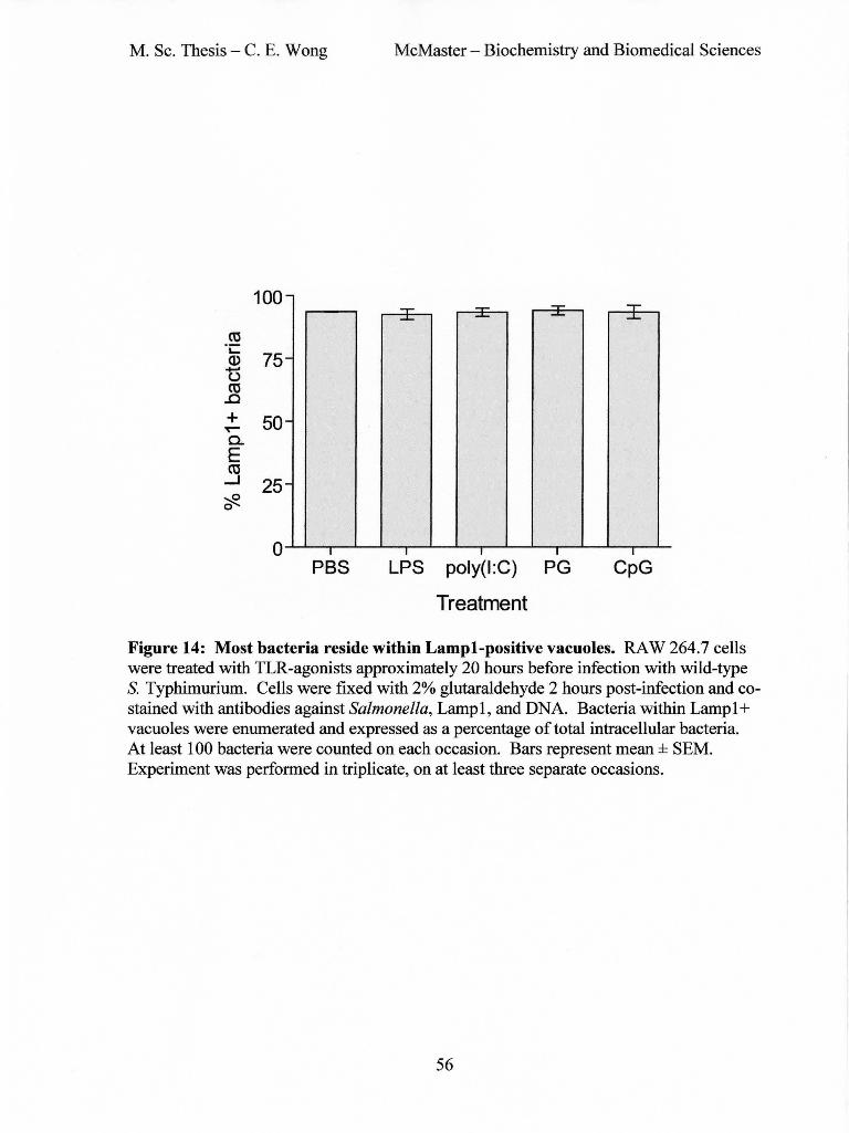
M. Sc. Thesis - C. E. Wong McMaster- Biochemistry and Biomedical Sciences
100 -"'T"" "'T"" ~- ... ....... - ....... ..J...
ro "i::
75 Q)
t5 -
ro ..c + 50 ~
-0.. E ro
....J 25 -~ 0
0 I I I I I
PBS LPS poly(I:C) PG CpG
Treatment
Figure 14: Most bacteria reside within Lampl-positive vacuoles. RAW 264.7 cells were treated with TLR-agonists approximately 20 hours before infection with wild-type S. Typhimurium. Cells were fixed with 2% glutaraldehyde 2 hours post-infection and costained with antibodies against Salmonella, Lamp 1, and DNA. Bacteria within Lamp 1 + vacuoles were enumerated and expressed as a percentage of total intracellular bacteria. At least 1 00 bacteria were counted on each occasion. Bars represent mean ± SEM. Experiment was performed in triplicate, on at least three separate occasions.
56

M. Sc. Thesis - C. E. Wong McMaster - Biochemistry and Biomedical Sciences
Upregulation of Scavenger receptor A through the
MyD88-IRAK4-p38 pathway (Doyle et at., 2003)
( t Scavenger Receptor A )
Treatment with TLRagonists can increase
phagocytosis of bacteria in a MyD88, p38, and SR
dependent manner (Doyle eta/., 2003)
Hook3 may be involved in SR recycling after endocy
tosis. Hook3 may play a role in SR expression and
ligand uptake (Sa no et at., 2007)
( t Phagocytosis )
? ( Salmonella effector SsaB )
SsaB interacts with and inactivates Hook3 (Shetland et at., 2003)
( t active and available Hook3)
Dominant negative mutant of Hook3 disrupts Golgi and lateendosomes/lysosomes (Shetland et at., 2003)
( t Phagosome-lysosome fusion )
l (t Salmonella replication)
Figure 15: Hypothesized interaction map between TLRs, SRs, SsaB, and Hook3. Treatment ofmacrophages with TLR-agonists leads to an increase in phagocytosis, in a manner that is dependent on MyD88, p38, and SR (35). Hook3 interacts with the cytoplasmic tail of SR and may play a role in SR expression and trafficking (128). The Salmonella protein SsaB also interacts with Hook3 and inactivates it, resulting in a decrease in phagosome-lysosome fusion (135) and a subsequent increase in Salmonella replication.
57

M. Sc. Thesis - C. E. Wong McMaster - Biochemistry and Biomedical Sciences
6. REFERENCES
1. Abrahams, G. L., and M. Hensel. 2006. Manipulating cellular transport and immune responses: Dynamic interactions between intracellular salmonella enterica and its host cells. Cell. Microbiol. 8:728-737.
2. Ahmad-Nejad, P., H. Hacker, M. Rutz, S. Bauer, R. M. Vabulas, and H. Wagner. 2002. Bacterial CpG-DNA and lipopolysaccharides activate toll-like receptors at distinct cellular compartments. Eur. J. Immunol. 32:1958-1968.
3. Akira, S., and K. Takeda. 2004. Toll-like receptor signalling. Nat. Rev. Immunol. 4:499-511.
4. Akira, S., S. Uematsu, and 0. Takeuchi. 2006. Pathogen recognition and innate immunity. Cell124:783-801.
5. Alexopoulou, L., A. C. Holt, R. Medzhitov, and R. A. Flavell. 2001. Recognition of double-stranded RNA and activation ofNF-kappaB by toll-like receptor 3. Nature 413:732-738.
6. Anderson, K. V., L. Bolda, and C. Nusslein-Volhard. 1985. Establishment of dorsalventral polarity in the drosophila embryo: The induction of polarity by the toll gene product. Cell 42:791-798.
7. Ashkar, A. A., S. Bauer, W. J. Mitchell, J. Vieira, and K. L. Rosenthal. 2003. Local delivery of CpG oligodeoxynucleotides induces rapid changes in the genital mucosa and inhibits replication, but not entry, of herpes simplex virus type 2. J Virol 77:8948-8956.
8. Ashkar, A. A., X. D. Yao, N. Gill, D. Sajic, A. J. Patrick, and K. L. Rosenthal. 2004. Toll-like receptor (TLR)-3, but not TLR4, agonist protects against genital herpes infection in the absence of inflammation seen with CpG DNA. J Infect Dis 190: 1841-1849.
9. Ashkar, A. A., and K. L. Rosenthal. 2003. Interleuk:in-15 and natural killer and NKT cells play a critical role in innate protection against genital herpes simplex virus type 2 infection. J. Virol. 77:10168-10171.
10. Bakshi, C. S., V. P. Singh, M. W. Wood, P. W. Jones, T. S. Wallis, and E. E. Galyov. 2000. Identification ofSopE2, a salmonella secreted protein which is highly homologous to SopE and involved in bacterial invasion of epithelial cells. J. Bacteriol. 182:2341-2344.
58

M. Sc. Thesis- C. E. Wong McMaster- Biochemistry and Biomedical Sciences
11. Baumler, A. J., R. M. Tsolis, P. J. Valentine, T. A. Ficht, and F. Heffron. 1997. Synergistic effect of mutations in inv A and lpfC on the ability of salmonella typhimurium to cause murine typhoid. Infect. Immun. 65:2254-2259.
12. Beuzon, C. R., G. Banks, J. Deiwick, M. Hensel, and D. W. Holden. 1999. pHdependent secretion ofSseB, a product ofthe SPI-2 type III secretion system of salmonella typhimurium. Mol. Microbiol. 33:806-816.
13. Biron, C. A. 2001. Interferons alpha and beta as immune regulators--a new look. Immunity 14:661-664.
14. Biron, C. A. 1998. Role of early cytokines, including alpha and beta interferons (IFNalphalbeta), in innate and adaptive immune responses to viral infections. Semin. Immunol. 10:383-390.
15. Biron, C. A., and L. Brossay. 2001. NK cells and NKT cells in innate defense against viral infections. Curr. Opin. Immunol. 13:458-464.
16. Blackwell, J. M., T. Goswami, C. A. Evans, D. Sibthorpe, N. Papo, J. K. White, S. Searle, E. N. Miller, C. S. Peacock, H. Mohammed, and M. Ibrahim. 2001. SLC11A1 (formerly NRAMP1) and disease resistance. Cell. Microbiol. 3:773-784.
17. Blander, J. M., and R. Medzhitov. 2004. Regulation of phagosome maturation by signals from toll-like receptors. Science 304:1014-1018.
18. Bowie, A., and L.A. O'Neill. 2000. The interleukin-1 receptor/Toll-like receptor superfamily: Signal generators for pro-inflammatory interleukins and microbial products. J. Leukoc. Bioi. 67:508-514.
19. Boyd, D., G. A. Peters, A. Cloeckaert, K. S. Boumedine, E. Chaslus-Dancla, H. Imberechts, and M. R. Mulvey. 2001. Complete nucleotide sequence of a 43-kilobase genomic island associated with the multi drug resistance region of salmonella enterica serovar typhimurium DT104 and its identification in phage type DT120 and serovar agona. J. Bacteriol. 183:5725-5732.
20. Brawn, L. C., R. D. Hayward, and V. Koronakis. 2007. Salmonella SPil effector SipA persists after entry and cooperates with a SPI2 effector to regulate phagosome maturation and intracellular replication. Cell. Host Microbe 1:63-75.
21. Brown, N. F., B. A. Vallance, B. K. Coombes, Y. Valdez, B. A. Coburn, and B. B. Finlay. 2005. Salmonella pathogenicity island 2 is expressed prior to penetrating the intestine. PLoS Pathog. 1:e32.
22. Brumell, J. H., and S. Grinstein. 2004. Salmonella redirects phagosomal maturation. Curr. Opin. Microbiol. 7:78-84.
59

M. Sc. Thesis - C. E. Wong McMaster - Biochemistry and Biomedical Sciences
23. Brumell, J. H., C. M. Rosenberger, G. T. Gotto, S. L. Marcus, and B. B. Finlay. 2001. SifA permits survival and replication of salmonella typhimurium in murine macrophages. Cell. Microbiol. 3:75-84.
24. Brumell, J. H., P. Tang, M. L. Zaharik, and B. B. Finlay. 2002. Disruption of the salmonella-containing vacuole leads to increased replication of salmonella enterica serovar typhimurium in the cytosol of epithelial cells. Infect. Immun. 70:3264-3270.
25. Caron, E., and A. Hall. 1998. Identification oftwo distinct mechanisms of phagocytosis controlled by different rho GTPases. Science 282: 1717-1721.
26. Carpenter, S., and L.A. O'Neill. 2007. How important are toll-like receptors for antimicrobial responses? Cell. Microbiol. 9:1891-1901.
27. Chakravortty, D., I. Hansen-Wester, and M. Hensel. 2002. Salmonella pathogenicity island 2 mediates protection of intracellular salmonella from reactive nitrogen intermediates. J. Exp. Med. 195:1155-1166.
28. Chen, K., P. Iribarren, J. Hu, J. Chen, W. Gong, E. H. Cho, S. Lockett, N. M. Dunlop, and J. M. Wang. 2006. Activation of toll-like receptor 2 on microglia promotes cell uptake of alzheimer disease-associated amyloid beta peptide. J. Biol. Chern. 281:3651-3659.
29. Collier-Hyams, L. S., H. Zeng, J. Sun, A. D. Tomlinson, Z. Q. Bao, H. Chen, J. L. Madara, K. Orth, and A. S. Neish. 2002. Cutting edge: Salmonella AvrA effector inhibits the key proinflammatory, anti-apoptotic NF-kappa B pathway. J. Immunol. 169:2846-2850.
30. Coombes, B. K., N. F. Brown, S. Kujat-Choy, B. A. Vallance, and B. B. Finlay. 2003. SseA is required for translocation of salmonella pathogenicity island-2 effectors into host cells. Microbes Infect. 5:561-570.
31. Cooper, A.M., D. K. Dalton, T. A. Stewart, J.P. Griffin, D. G. Russell, and I. M. Orme. 1993. Disseminated tuberculosis in interferon gamma gene-disrupted mice. J. Exp. Med. 178:2243-2247.
32. Cox, D., and S. Greenberg. 2001. Phagocytic signaling strategies: Fc(gamma)receptor-mediated phagocytosis as a model system. Semin. Immunol. 13:339-345.
33. Criss, A. K., and J. E. Casanova. 2003. Coordinate regulation of salmonella enterica serovar typhimurium invasion of epithelial cells by the Arp2/3 complex and rho GTPases. Infect. Immun. 71:2885-2891.
60

M. Sc. Thesis - C. E. Wong McMaster - Biochemistry and Biomedical Sciences
34. Deiwick, J., T. Nikolaus, J. E. Shea, C. Gleeson, D. W. Holden, and M. Hensel. 1998. Mutations in salmonella pathogenicity island 2 (SPI2) genes affecting transcription of SPil genes and resistance to antimicrobial agents. J. Bacteriol. 180:4775-4780.
35. Doyle, S. E., R. M. O'Connell, G. A. Miranda, S. A. Vaidya, E. K. Chow, P. T. Liu, S. Suzuki, N. Suzuki, R. L. Modlin, W. C. Yeh, T. F. Lane, and G. Cheng. 2004. Toll-like receptors induce a phagocytic gene program through p38. J. Exp. Med. 199:81-90.
36. Doyle, S. L., and L.A. O'Neill. 2006. Toll-like receptors: From the discovery of NFkappaB to new insights into transcriptional regulations in innate immunity. Biochem. Pharmacol. 72:1102-1113.
37. Drecktrah, D., L.A. Knodler, D. Howe, and 0. Steele-Mortimer. 2007. Salmonella trafficking is defined by continuous dynamic interactions with the endolysosomal system. Traffic 8:212-225.
38. Elkins, K. L., T. R. Rhinehart-Jones, S. Stibitz, J. S. Conover, and D. M. Klinman. 1999. Bacterial DNA containing CpG motifs stimulates lymphocyte-dependent protection of mice against lethal infection with intracellular bacteria. J Immunol 162:2291-2298.
39. Fenton, M. J., M. W. Vermeulen, S. Kim, M. Burdick, R. M. Strieter, and H. Kornfeld. 1997. Induction of gamma interferon production in human alveolar macrophages by mycobacterium tuberculosis. Infect. Immun. 65:5149-5156.
40. Fields, P. I., R. V. Swanson, C. G. Haidaris, and F. Heffron. 1986. Mutants of salmonella typhimurium that cannot survive within the macrophage are avirulent. Proc. Natl. Acad. Sci. U.S. A. 83:5189-5193.
41. Finlay, B. B., and R. E. Hancock. 2004. Can innate immunity be enhanced to treat microbial infections? Nat. Rev. Microbiol. 2:497-504.
42. Finlay, B. B., S. Ruschkowski, and S. Dedhar. 1991. Cytoskeletal rearrangements accompanying salmonella entry into epithelial cells. J. Cell. Sci. 99 ( Pt 2):283-296.
43. Fitzgerald, K. A., S.M. McWhirter, K. L. Faia, D. C. Rowe, E. Latz, D. T. Golenbock, A. J. Coyle, S.M. Liao, and T. Maniatis. 2003. IKKepsilon and TBK1 are essential components of the IRF3 signaling pathway. Nat. Immunol. 4:491-496.
44. Flynn, J. L., J. Chan, K. J. Triebold, D. K. Dalton, T. A. Stewart, and B. R. Bloom. 1993. An essential role for interferon gamma in resistance to mycobacterium tuberculosis infection. J. Exp. Med. 178:2249-2254.
61

M. Sc. Thesis- C. E. Wong McMaster- Biochemistry and Biomedical Sciences
45. Friebel, A., H. IIebmann, M. Aepfelbacher, K. Ehrbar, W. Machleidt, and W. D. Hardt. 2001. SopE and SopE2 from salmonella typhimurium activate different sets of RhoGTPases of the host cell. J. Biol. Chern. 276:34035-34040.
46. Fu, Y., and J. E. Galan. 1999. A salmonella protein antagonizes rac-1 and Cdc42 to mediate host-cell recovery after bacterial invasion. Nature 401:293-297.
47. Fujimuro, M., H. Sawada, and H. Yokosawa. 1994. Production and characterization of monoclonal antibodies specific to multi-ubiquitin chains of polyubiquitinated proteins. FEBS Lett. 349:173-180.
48. Fultz, M. J., S. A. Barber, C. W. Dieffenbach, and S. N. Vogel. 1993. Induction of IFN-gamma in macrophages by lipopolysaccharide. Int. Immunol. 5:1383-1392.
49. Galan, J. E., and R. Curtiss 3rd. 1989. Cloning and molecular characterization of genes whose products allow salmonella typhimurium to penetrate tissue culture cells. Proc. Natl. Acad. Sci. U. S. A. 86:6383-6387.
50. Galan, J. E., and D. Zhou. 2000. Striking a balance: Modulation of the actin cytoskeleton by salmonella. Proc. Natl. Acad. Sci. U.S. A. 97:8754-8761.
51. Gallois, A., J. R. Klein, L.A. Allen, B. D. Jones, and W. M. Nauseef. 2001. Salmonella pathogenicity island 2-encoded type III secretion system mediates exclusion ofNADPH oxidase assembly from the phagosomal membrane. J. Immunol. 166:5741-5748.
52. Garcia-del Portillo, F., and B. B. Finlay. 1995. Targeting of salmonella typhimurium to vesicles containing lysosomal membrane glycoproteins bypasses compartments with mannose 6-phosphate receptors. J. Cell Bioi. 129:81-97.
53. Garcia-del Portillo, F., M. B. Zwick, K. Y. Leung, and B. B. Finlay. 1993. Salmonella induces the formation of filamentous structures containing lysosomal membrane glycoproteins in epithelial cells. Proc. Natl. Acad. Sci. U. S. A. 90:10544-10548.
54. Garvis, S. G., C. R. Beuzon, and D. W. Holden. 2001. A role for the PhoP/Q regulon in inhibition of fusion between lysosomes and salmonella-containing vacuoles in macrophages. Cell. Microbiol. 3:731-744.
55. Gerold, G., A. Zychlinsky, and J. L. de Diego. 2007. What is the role of toll-like receptors in bacterial infections? 19:41-47.
56. Giacomodonato, M. N., S. Uzzau, D. Bacciu, R. Caccuri, S. H. Sarnacki, S. Rubino, and M. C. Cerquetti. 2007. SipA, SopA, SopB, SopD and SopE2 effector
62

M. Sc. Thesis - C. E. Wong McMaster- Biochemistry and Biomedical Sciences
proteins of salmonella enterica serovar typhimurium are synthesized at late stages of infection in mice. Microbiology 153:1221-1228.
57. Glynn, M. K., C. Bopp, W. Dewitt, P. Dabney, M. Mokhtar, and F. J. Angulo. 1998. Emergence of multidrug-resistant salmonella enterica serotype typhimurium DT104 infections in the united states. N. Engl. J. Med. 338:1333-1338.
58. Goldman, M. 2007. Translational mini-review series on toll-like receptors: Toll-like receptor ligands as novel pharmaceuticals for allergic disorders. Clin. Exp. lmmunol. 147:208-216.
59. Gordon, M.A. 2008. Salmonella infections in immunocompromised adults. J. Infect. 56:413-422.
60. Govoni, G., and P. Gros. 1998. Macrophage NRAMP1 and its role in resistance to microbial infections. lnflamm. Res. 47:277-284.
61. Gray, P. W., and D. V. Goeddel. 1982. Structure of the human immune interferon gene. Nature 298:859-863.
62. Groves, E., A. E. Dart, V. Covarelli, and E. Caron. 2008. Molecular mechanisms of phagocytic uptake in mammalian cells. Cell Mol. Life Sci ..
63. Hampton, R. Y., D. T. Golenbock, M. Penman, M. Krieger, and C. R. Raetz. 1991. Recognition and plasma clearance of endotoxin by scavenger receptors. Nature 352:342-344.
64. Baraga, A., and S. I. Miller. 2003. A salmonella enterica serovar typhimurium translocated leucine-rich repeat effector protein inhibits NF -kappa B-dependent gene expression. Infect. lmmun. 71:4052-4058.
65. Baraga, A., M. B. Ohlson, and S. I. Miller. 2008. Salmonellae interplay with host cells. Nat. Rev. Microbial. 6:53-66.
66. Harandi, A.M., K. Eriksson, and J. Holmgren. 2003. A protective role oflocally administered immunostimulatory CpG oligodeoxynucleotide in a mouse model of genital herpes infection. J Virol 77:953-962.
67. Hardt, W. D., L. M. Chen, K. E. Schuebel, X. R. Bustelo, and J. E. Galan. 1998. S. typhimurium encodes an activator of rho GTPases that induces membrane ruffling and nuclear responses in host cells. Cell93:815-826.
68. Hashim, S., K. Mukherjee, M. Raje, S. K. Basu, and A. Mukhopadhyay. 2000. Live salmonella modulate expression of rab proteins to persist in a specialized compartment and escape transport to lysosomes. J. Bioi. Chern. 275:16281-16288.
63

M. Sc. Thesis - C. E. Wong McMaster- Biochemistry and Biomedical Sciences
69. Haworth, R., N. Platt, S. Keshav, D. Hughes, E. Darley, H. Suzuki, Y. Kurihara, T. Kodama, and S. Gordon. 1997. The macrophage scavenger receptor type A is expressed by activated macrophages and protects the host against lethal endotoxic shock. J. Exp. Med. 186:1431-1439.
70. Hayward, R. D., and V. Koronakis. 1999. Direct nucleation and bundling of actin by the SipC protein of invasive salmonella. EMBO J. 18:4926-4934.
71. Helms, M., S. Ethelberg, K. Molbak, and DT104 Study Group. 2005. International salmonella typhimurium DT104 infections, 1992-2001. Emerg. Infect. Dis. 11:859-867.
72. Hemmi, H., 0. Takeuchi, T. Kawai, T. Kaisho, S. Sato, H. Sanjo, M. Matsumoto, K. Hoshino, H. Wagner, K. Takeda, and S. Akira. 2000. A toll-like receptor recognizes bacterial DNA. Nature 408:740-745.
73. Hensel, M., J. E. Shea, B. Raupach, D. Monack, S. Falkow, C. Gleeson, T. Kubo, and D. W. Holden. 1997. Functional analysis ofssaJ and the ssaKIU operon, 13 genes encoding components of the type III secretion apparatus of salmonella pathogenicity island 2. Mol. Microbiol. 24:155-167.
74. Hensel, M., J. E. Shea, S. R. Waterman, R. Mundy, T. Nikolaus, G. Banks, A. Vazquez-Torres, C. Gleeson, F. C. Fang, and D. W. Holden. 1998. Genes encoding putative effector proteins of the type III secretion system of salmonella pathogenicity island 2 are required for bacterial virulence and proliferation in macrophages. Mol. Microbiol. 30:163-174.
75. Herbst-Kralovetz, M. M., and R. B. Pyles. 2006. Quantification ofpoly(I:C)mediated protection against genital herpes simplex virus type 2 infection. J Virol 80:9988-9997.
76. Hoshino, K., 0. Takeuchi, T. Kawai, H. Sanjo, T. Ogawa, Y. Takeda, K. Takeda, and S. Akira. 1999. Cutting edge: Toll-like receptor 4 (TLR4)-deficient mice are hyporesponsive to lipopolysaccharide: Evidence for TLR4 as the Ips gene product. J. Immunol. 162:3749-3752.
77. Huang, D. B., and H. L. DuPont. 2005. Problem pathogens: Extra-intestinal complications of salmonella enterica serotype typhi infection. Lancet Infect. Dis. 5:341-348.
78. Hueck, C. J. 1998. Type III protein secretion systems in bacterial pathogens of animals and plants. Microbiol. Mol. Bioi. Rev. 62:379-433.
79. Ischiropoulos, H., L. Zhu, and J. S. Beckman. 1992. Peroxynitrite formation from macrophage-derived nitric oxide. Arch. Biochem. Biophys. 298:446-451.
64

M. Sc. Thesis - C. E. Wong McMaster- Biochemistry and Biomedical Sciences
80. Jiang, Z., T. W. Mak, G. Sen, and X. Li. 2004. Toll-like receptor 3-mediated activation ofNF-kappaB and IRF3 diverges at toll-IL-l receptor domain-containing adapter inducing IFN-beta. Proc. Natl. Acad. Sci. U. S. A. 101:3533-3538.
81. Jiang, Z., J. Ninomiya-Tsuji, Y. Qian, K. Matsumoto, and X. Li. 2002. Interleukin-1 (IL-l) receptor-associated kinase-dependent IL-l-induced signaling complexes phosphorylate TAKl and TAB2 at the plasma membrane and activate TAKl in the cytosol. Mol. Cell. Bioi. 22:7158-7167.
82. Jiang, Z., M. Zamanian-Daryoush, H. Nie, A. M. Silva, B. R. Williams, and X. Li. 2003. Poly(I-C)-induced toll-like receptor 3 (TLR3)-mediated activation ofNFkappa B and MAP kinase is through an interleukin-1 receptor-associated kinase (IRAK)independent pathway employing the signaling components TLR3-TRAF6-TAK1-T AB2-PKR. J. Bioi. Chern. 278:16713-16719.
83. Jones, B. D., N. Ghori, and S. Falkow. 1994. Salmonella typhimurium initiates murine infection by penetrating and destroying the specialized epithelial M cells of the peyer's patches. J. Exp. Med. 180:15-23.
84. Juffermans, N. P., J. C. Leemans, S. Florquin, A. Verbon, A. H. Kolk, P. Speelman, S. J. van Deventer, and van der Poll, T. 2002. CpG oligodeoxynucleotides enhance host defense during murine tuberculosis. Infect Immun 7:147-152.
85. Kagan, J. C., T. Su, T. Horng, A. Chow, S. Akira, and R. Medzhitov. 2008. TRAM couples endocytosis oftoll-like receptor 4 to the induction of interferon-beta. Nat. Immunol. 9:361-368.
86. Kawai, T., and S. Akira. 2007. TLR signaling. Semin. Immunol. 19:24-32.
87. Knodler, L.A., B. A. Vallance, M. Hensel, D. Jackel, B. B. Finlay, and 0. SteeleMortimer. 2003. Salmonella type III effectors PipB and PipB2 are targeted to detergentresistant microdomains on internal host cell membranes. Mol. Microbial. 49:685-704.
88. Kong, L., and B. X. Ge. 2008. MyD88-independent activation of a novel actinCdc42/Rac pathway is required for toll-like receptor-stimulated phagocytosis. Cell Res. 18:745-755.
89. Krieg, A.M., L. Love-Homan, A. K. Yi, and J. T. Harty. 1998. CpG DNA induces sustained IL-12 expression in vivo and resistance to listeria monocytogenes challenge. J Immunol161:2428-2434.
90. Krieg, A. M. 2002. CpG motifs in bacterial DNA and their immune effects. Annu. Rev. Immunol. 20:709-760.
65

M. Sc. Thesis- C. E. Wong McMaster - Biochemistry and Biomedical Sciences
91. Krieg, A.M. 2006. Therapeutic potential of toll-like receptor 9 activation. Nat. Rev. Drug Discov. 5:471-484.
92. Kubori, T., and J. E. Galan. 2003. Temporal regulation of salmonella virulence effector function by proteasome-dependent protein degradation. Cell115:333-342.
93. Latz, E., A. Schoenemeyer, A. Visintin, K. A. Fitzgerald, B. G. Monks, C. F. Koetter, E. Lien, N.J. Nilsen, T. Espevik, and D. T. Golenbock. 2004. TLR9 signals after translocating from the ER to CpG DNA in the lysosome. Nat. Immunol. 5: 190-198.
94. Lemaitre, B., E. Nicolas, L. Michaut, J. M. Reichhart, and J. A. Hoffmann. 1996. The dorsoventral regulatory gene cassette spatzle/Toll/cactus controls the potent antifungal response in drosophila adults. Cell86:973-983.
95. Lesnick, M. L., N. E. Reiner, J. Fierer, and D. G. Guiney. 2001. The salmonella spvB virulence gene encodes an enzyme that ADP-ribosylates actin and destabilizes the cytoskeleton of eukaryotic cells. Mol. Micro bioi. 39:1464-14 70.
96. Luther, K., A. Torosantucci, A. A. Brakhage, J. Heesemann, and F. Ebel. 2007. Phagocytosis of aspergillus fumigatus conidia by murine macrophages involves recognition by the dectin-1 beta-glucan receptor and toll-like receptor 2. Cell. Microbial. 9:368-381.
97. Lye, E., C. Mirtsos, N. Suzuki, S. Suzuki, and W. C. Yeh. 2004. The role of interleukin 1 receptor-associated kinase-4 (IRAK -4) kinase activity in IRAK-4-mediated signaling. J. Bioi. Chern. 279:40653-40658.
98. Ma, J., T. Chen, J. Mandelin, A. Ceponis, N. E. Miller, M. Hukkanen, G. F. Ma, andY. T. Konttinen. 2003. Regulation of macrophage activation. Cell Mol. Life Sci. 60:2334-2346.
99. Medzhitov, R., P. Preston-Hurlburt, and C. A. Janeway Jr. 1997. A human homologue of the drosophila toll protein signals activation of adaptive immunity. Nature 388:394-397.
100. Mercurio, F., H. Zhu, B. W. Murray, A. Shevchenko, B. L. Bennett, J. Li, D. B. Young, M. Barbosa, M. Mann, A. Manning, and A. Rao. 1997. IKK-1 and IKK-2: Cytokine-activated IkappaB kinases essential for NF -kappaB activation. Science 278:860-866.
101. Meresse, S., 0. Steele-Mortimer, B. B. Finlay, and J.P. Gorvel. 1999. The rab7 GTPase controls the maturation of salmonella typhimurium-containing vacuoles in HeLa cells. EMBO J. 18:4394-4403.
66

M. Sc. Thesis - C. E. Wong McMaster- Biochemistry and Biomedical Sciences
102. Meresse, S., K. E. Unsworth, A. Habermann, G. Griffiths, F. Fang, M. J. Martinez-Lorenzo, S. R. Waterman, J.P. Gorvel, and D. W. Holden. 2001. Remodelling of the actin cytoskeleton is essential for replication of intravacuolar salmonella. Cell. Microbiol. 3:567-577.
103. Meylan, E., K. Burns, K. Hofmann, V. Blancheteau, F. Martinon, M. Kelliher, and J. Tschopp. 2004. R1P1 is an essential mediator of toll-like receptor 3-induced NFkappa B activation. Nat. Immunol. 5:503-507.
104. Miao, E. A., M. Brittnacher, A. Haraga, R. L. Jeng, M.D. Welch, and S. I. Miller. 2003. Salmonella effectors translocated across the vacuolar membrane interact with the actin cytoskeleton. Mol. Microbiol. 48:401-415.
105. Miao, E. A., C. A. Scherer, R. M. Tsolis, R. A. Kingsley, L. G. Adams, A. J. Baumler, and S. I. Miller. 1999. Salmonella typhimurium leucine-rich repeat proteins are targeted to the SPil and SPI2 type III secretion systems. Mol. Microbiol. 34:850-864.
106. Miller, R. A., and B. E. Britigan. 1997. Role of oxidants in microbial pathophysiology. Clin. Microbiol. Rev. 10:1-18.
107. Morisato, D., and K. V. Anderson. 1994. The spatzle gene encodes a component of the extracellular signaling pathway establishing the dorsal-ventral pattern of the drosophila embryo. Cell 76:677-688.
108. Mulvey, M. R., D. A. Boyd, A. B. Olson, B. Doublet, and A. Cloeckaert. 2006. The genetics of salmonella genomic island 1. Microbes Infect. 8:1915-1922.
109. Munder, M., M. Mallo, K. Eichmann, and M. Modolell. 1998. Murine macrophages secrete interferon gamma upon combined stimulation with interleukin (IL )-12 and IL-18: A novel pathway ofautocrine macrophage activation. J. Exp. Med. 187:2103-2108.
110. Murli, S., R. 0. Watson, and J. E. Galan. 2001. Role of tyrosine kinases and the tyrosine phosphatase SptP in the interaction of salmonella with host cells. Cell. Microbiol. 3:795-810.
111. Neal, M.D., C. Leaphart, R. Levy, J. Prince, T. R. Billiar, S. Watkins, J. Li, S. Cetin, H. Ford, A. Schreiber, and D. J. Hackam. 2006. Enterocyte TLR4 mediates phagocytosis and translocation of bacteria across the intestinal barrier. J. Immunol. 176:3070-3079.
112. Norris, F. A., M.P. Wilson, T. S. Wallis, E. E. Galyov, and P. W. Majerus. 1998. SopB, a protein required for virulence of salmonella dublin, is an inositol phosphate phosphatase. Proc. Natl. Acad. Sci. U.S. A. 95:14057-14059.
67

M. Sc. Thesis- C. E. Wong McMaster- Biochemistry and Biomedical Sciences
113. Ochman, H., F. C. Soncini, F. Solomon, and E. A. Groisman. 1996. Identification of a pathogenicity island required for salmonella survival in host cells. Proc. Natl. Acad. Sci. U. S. A. 93:7800-7804.
114. Parkinson, T. 2008. The future of toll-like receptor therapeutics. Curr. Opin. Mol. Ther. 10:21-31.
115. Patel, J. C., and J. E. Galan. 2006. Differential activation and function of rho GTPases during salmonella-host cell interactions. J. Cell Bioi. 175:453-463.
116. Peiser, L., P. J. Gough, T. Kodama, and S. Gordon. 2000. Macrophage class A scavenger receptor-mediated phagocytosis of escherichia coli: Role of cell heterogeneity, microbial strain, and culture conditions in vitro. Infect. Immun. 68:1953-1963.
117. Pfeifer, C. G., S. L. Marcus, 0. Steele-Mortimer, L.A. Knodler, and B. B. Finlay. 1999. Salmonella typhimurium virulence genes are induced upon bacterial invasion into phagocytic and nonphagocytic cells. Infect. Immun. 67:5690-5698.
118. Poltorak, A., X. He, I. Smirnova, M. Y. Liu, C. Van Huffel, X. Du, D. Birdwell, E. Alejos, M. Silva, C. Galanos, M. Freudenberg, P. Ricciardi-Castagnoli, B. Layton, and B. Beutler. 1998. Defective LPS signaling in C3H/HeJ and C57BL/1 OScCr mice: Mutations in Tlr4 gene. Science 282:2085-2088.
119. Puddu, P., M. Carollo, I. Pietraforte, F. Spadaro, M. Tombesi, C. Ramoni, F. Belardelli, and S. Gessani. 2005. IL-2 induces expression and secretion ofiFN-gamma in murine peritoneal macrophages. J. Leukoc. Bioi. 78:686-695.
120. Puddu, P., L. Fantuzzi, P. Borghi, B. Varano, G. Rainaldi, E. Guillemard, W. Malorni, P. Nicaise, S. F. Wolf, F. Belardelli, and S. Gessani. 1997. IL-12 induces IFN-gamma expression and secretion in mouse peritoneal macrophages. J. Immunol. 159:3490-3497.
121. Pyles, R. B., D. Higgins, C. Chalk, A. Zalar, J. Eiden, C. Brown, G. Van Nest, and L. R. Stanberry. 2002. Use ofimmunostimulatory sequence-containing oligonucleotides as topical therapy for genital herpes simplex virus type 2 infection. J Virol 76:11387-11396.
122. Qian, Y., M. Commane, J. Ninomiya-Tsuji, K. Matsumoto, and X. Li. 2001. IRAK-mediated translocation ofTRAF6 and TAB2 in the interleukin-1-induced activation ofNFkappa B. J. Bioi. Chern. 276:41661-41667.
123. Roberts, R. L., M.A. Barbieri, J. Ullrich, and P. D. Stahl. 2000. Dynamics of rab5 activation in endocytosis and phagocytosis. J. Leukoc. Bioi. 68:627-632.
68

M. Sc. Thesis - C. E. Wong McMaster- Biochemistry and Biomedical Sciences
124. Rosenberger, C. M., and B. B. Finlay. 2002. Macrophages inhibit salmonella typhimurium replication through MEKIERK kinase and phagocyte NADPH oxidase activities. J. Biol. Chern. 277:18753-18762.
125. Rosenberger, C. M., and B. B. Finlay. 2003. Phagocyte sabotage: Disruption of macrophage signalling by bacterial pathogens. Nat. Rev. Mol. Cell Biol. 4:385-396.
126. Ruiz-Albert, J., R. Mundy, X. J. Yu, C. R. Beuzon, and D. W. Holden. 2003. SseA is a chaperone for the SseB and SseD translocon components of the salmonella pathogenicity-island-2-encoded type III secretion system. Microbiology 149:1103-1111.
127. Salkowski, C. A., G. R. Detore, and S. N. Vogel. 1997. Lipopolysaccharide and monophosphoryllipid A differentially regulate interleukin-12, gamma interferon, and interleukin-10 mRNA production in murine macrophages. Infect. Immun. 65:3239-3247.
128. Sano, H., M. lshino, H. Kramer, T. Shimizu, H. Mitsuzawa, C. Nishitani, andY. Kuroki. 2007. The microtubule-binding protein Hook3 interacts with a cytoplasmic domain of scavenger receptor A. J. Biol. Chern. 282:7973-7981.
129. Sato, S., M. Sugiyama, M. Yamamoto, Y. Watanabe, T. Kawai, K. Takeda, and S. Akira. 2003. Toll/IL-l receptor domain-containing adaptor inducing IFN-beta (TRIF) associates with TNF receptor-associated factor 6 and TANK-binding kinase 1, and activates two distinct transcription factors, NF-kappa Band IFN-regulatory factor-3, in the toll-like receptor signaling. J. Immunol. 171:4304-4310.
130. Scherer, C. A., E. Cooper, and S. I. Miller. 2000. The salmonella type III secretion translocon protein SspC is inserted into the epithelial cell plasma membrane upon infection. Mol. Microbiol. 37:1133-1145.
131. Schroder, K., M. J. Sweet, and D. A. Hume. 2006. Signal integration between IFNgamma and TLR signalling pathways in macrophages. lmmunobiology 211:511-524.
132. Schwandner, R., R. Dziarski, H. Wesche, M. Rothe, and C. J. Kirschning. 1999. Peptidoglycan- and lipoteichoic acid-induced cell activation is mediated by toll-like receptor2. J. Bioi. Chern. 274:17406-17409.
133. Shea, J. E., M. Hensel, C. Gleeson, and D. W. Holden. 1996. Identification of a virulence locus encoding a second type III secretion system in salmonella typhimurium. Proc. Natl. Acad. Sci. U. S. A. 93:2593-2597.
134. Shi, J., G. Scita, and J. E. Casanova. 2005. WA VE2 signaling mediates invasion of polarized epithelial cells by salmonella typhimurium. J. Bioi. Chern. 280:29849-29855.
69

M. Sc. Thesis - C. E. Wong McMaster- Biochemistry and Biomedical Sciences
135. Shotland, Y., H. Kramer, and E. A. Groisman. 2003. The salmonella SpiC protein targets the mammalian Hook3 protein function to alter cellular trafficking. Mol. Microbiol. 49:1565-1576.
136. Smith, A. C., J. T. Cirulis, J. E. Casanova, M.A. Scidmore, and J. H. Brumell. 2005. Interaction of the salmonella-containing vacuole with the endocytic recycling system. J. Bioi. Chern. 280:24634-24641.
137. Steele-Mortimer, 0., J. H. Brumell, L.A. Knodler, S. Meresse, A. Lopez, and B. B. Finlay. 2002. The invasion-associated type III secretion system of salmonella enterica serovar typhimurium is necessary for intracellular proliferation and vacuole biogenesis in epithelial cells. Cell. Microbiol. 4:43-54.
138. Steele-Mortimer, 0., S. Meresse, J.P. Gorvel, B. H. Toh, and B. B. Finlay. 1999. Biogenesis of salmonella typhimurium-containing vacuoles in epithelial cells involves interactions with the early endocytic pathway. Cell. Microbiol. 1:33-49.
139. Stender, S., A. Friebel, S. Linder, M. Rohde, S. Mirold, and W. D. Hardt. 2000. Identification of SopE2 from salmonella typhimurium, a conserved guanine nucleotide exchange factor for Cdc42 of the host cell. Mol. Microbiol. 36:1206-1221.
140. Thomas, C. A., Y. Li, T. Kodama, H. Suzuki, S.C. Silverstein, and J. El Khoury. 2000. Protection from lethal gram-positive infection by macrophage scavenger receptor-dependent phagocytosis. J. Exp. Med. 191:147-156.
141. Tsolis, R. M., R. A. Kingsley, S.M. Townsend, T. A. Ficht, L. G. Adams, and A. J. Baumler. 1999. Of mice, calves, and men. comparison of the mouse typhoid model with other salmonella infections. Adv. Exp. Med. Biol. 473:261-274.
142. Uchiya, K., M.A. Barbieri, K. Funato, A. H. Shah, P. D. Stahl, and E. A. Groisman. 1999. A salmonella virulence protein that inhibits cellular trafficking. EMBO J. 18:3924-3933.
143. Unsworth, K. E., M. Way, M. McNiven, L. Machesky, and D. W. Holden. 2004. Analysis of the mechanisms of salmonella-induced actin assembly during invasion of host cells and intracellular replication. Cell. Microbiol. 6:1041-1055.
144. Utaisincharoen, P., W. Kespichayawattana, N. Anuntagool, P. Chaisuriya, S. Pichyangkul, A.M. Krieg, and S. Sirisinha. 2003. CpG ODN enhances uptake of bacteria by mouse macrophages. Clin. Exp. Immunol. 132:70-75.
145. Varma, J. K., K. Molbak, T. J. Barrett, J. L. Beebe, T. F. Jones, T. RabatskyEhr, K. E. Smith, D. J. Vugia, H. G. Chang, and F. J. Angulo. 2005. Antimicrobialresistant nontyphoidal salmonella is associated with excess bloodstream infections and hospitalizations. J. Infect. Dis. 191:554-561.
70

M. Sc. Thesis - C. E. Wong McMaster- Biochemistry and Biomedical Sciences
146. Vazquez-Torres, A., Y. Xu, J. Jones-Carson, D. W. Holden, S.M. Lucia, M. C. Dinauer, P. Mastroeni, and F. C. Fang. 2000. Salmonella pathogenicity island 2-dependent evasion ofthe phagocyte NADPH oxidase. Science 287:1655-1658.
147. Viboud, G. I., and J. B. Bliska. 2001. A bacterial type III secretion system inhibits actin polymerization to prevent pore formation in host cell membranes. EMBO J. 20:5373-5382.
148. Vidal, S.M., D. Malo, K. Vogan, E. Skamene, and P. Gros. 1993. Natural resistance to infection with intracellular parasites: Isolation of a candidate for beg. Cell 73:469-485.
149. Vieira, 0. V., R. J. Botelho, and S. Grinstein. 2002. Phagosome maturation: Aging gracefully. Biochem. J. 366:689-704.
150. Wagner, H. 1999. Bacterial CpG DNA activates immune cells to signal infectious danger. Adv. Immunol. 73:329-368.
151. Wang, C., L. Deng, M. Hong, G. R. Akkaraju, J. Inoue, and Z. J. Chen. 2001. TAK1 is a ubiquitin-dependent kinase ofMKK and IKK. Nature 412:346-351.
152. Wang, J.P., T. Hayashi, S. K. Datta, R. S. Kornbluth, E. Raz, and D. G. Guiney. 2005. CpG oligonucleotides partially inhibit growth of mycobacterium tuberculosis, but not salmonella or listeria, in human monocyte-derived macrophages. FEMS Immunol. Med. Microbiol. 45:303-310.
153. Waterman, S. R., and D. W. Holden. 2003. Functions and effectors of the salmonella pathogenicity island 2 type III secretion system. Cell. Microbiol. 5:501-511.
154. Wray, C., and W. J. Sojka. 1978. Experimental salmonella typhimurium infection in calves. Res. Vet. Sci. 25:139-143.
155. Yates, R. M., A. Hermetter, G. A. Taylor, and D. G. Russell. 2007. Macrophage activation downregulates the degradative capacity of the phagosome. Traffic 8:241-250.
156. Yates, R. M., and D. G. Russell. 2005. Phagosome maturation proceeds independently of stimulation of toll-like receptors 2 and 4. Immunity 23:409-417.
157. Yoshimura, A., E. Lien, R. R. Ingalls, E. Tuomanen, R. Dziarski, and D. Golen bock. 1999. Cutting edge: Recognition of gram-positive bacterial cell wall components by the innate immune system occurs via toll-like receptor 2. J. Immunol. 163:1-5.
158. Yu, X. J., M. Liu, and D. W. Holden. 2004. SsaM and SpiC interact and regulate secretion of salmonella pathogenicity island 2 type III secretion system effectors and translocators. Mol. Microbiol. 54:604-619.
71

M. Sc. Thesis- C. E. Wong McMaster - Biochemistry and Biomedical Sciences
159. Zaharik, M. L., B. A. Vallance, J. L. Puente, P. Gros, and B. B. Finlay. 2002. Host-pathogen interactions: Host resistance factor Nrampl up-regulates the expression of salmonella pathogenicity island-2 virulence genes. Proc. Natl. Acad. Sci. U.S. A. 99:15705-15710.
160. Zhou, D., M.S. Mooseker, and J. E. Galan. 1999. Role ofthe S. typhimurium actin-binding protein SipA in bacterial internalization. Science 283:2092-2095.
161. Zurawski, D. V., and M.A. Stein. 2003. SseA acts as the chaperone for the SseB component of the salmonella pathogenicity island 2 translocon. Mol. Microbial. 47:1341-1351.
72

M. Sc. Thesis - C. E. Wong McMaster- Biochemistry and Biomedical Sciences
6. APPENDIX
PBS LPS poly(I:C)
PG
Appendix 1: Transmission electron micrographs ofTLR-agonist treated macrophages 2 hours following infection with Salmonella. RAW 264.7 cells were treated with TLR-agonists approximately 20 hours before infection with Salmonella. Cells were fixed with 2% glutaraldehyde 2 hours post-infection and prepared for viewing under the transmission electron microscope at 5000x magnification. Bars represent 1 ~m.
73

M. Sc. Thesis - C. E. Wong McMaster - Biochemistry and Biomedical Sciences
Appendix 2: Sample images from ImageJ software program. Raw images from the original z-stacks using the red channel (A) and green channel (B). (C) Program identifies maximum intensities for the red channel and fills in gaps to create a mask to represent the cytoplasm. (D) Program identifies maximum intensities for the green channel and marks location of beads. (E) Combining the results from C and D, the program labels the beads that colocalize within the mask as internal, and those that do not as external. (F) Compressed z-stack where internal beads are marked with a white dot and external beads with a black dot.
74

M. Sc. Thesis- C. E. Wong McMaster- Biochemistry and Biomedical Sciences
Appendix 3: Macro for phagocytosis of fluorescent beads assay
Created by: Dr. Tony Collins, McMaster Biophotonics Facility Manager For use in ImageJ software
originalTitle=getTitle(); origiD=getlmageiD();
//setBatchMode(true ); steps = nSlices/2; width= getWidth(); height = getHeight();
newlmage("red", "8-bit", width, height, steps); rediD = getlmageiD();
for (i=l; i<=(steps); i++) { selectlmage( origiD); setSlice((i*2)-1 ); run(" Copy"); selectlmage(rediD ); setSlice(i); run("Paste"); }
newlmage("green", "8-bit", width, height, steps); greeniD= getlmageiD();
for (i=l; i<=(steps); i++) { selectlmage( origiD); setSlice((i*2)); run(" Copy"); selectlmage(greeniD ); setSlice(i); run("Paste"); }
I /setBatchMode( false);
//get unique image ID #s I /selectlmage(nlmages-1 ); I /rediD=getlmageiD(); I /selectlmage(nlmages ); //greeniD=getlmageiD();
75

M. Sc. Thesis - C. E. Wong McMaster- Biochemistry and Biomedical Sciences
I /subtract green from red to correct bleedthru imageCalculator("Subtract stack" ,rediD ,greeniD ); redProcessiD=getlmageiD(); rename(" red proc"); run("Duplicate ... ", "title=red-copy duplicate"); steps=nSlices;
//make mask cells using SNARF channle then fill in bead holes setThreshold(15, 255); //run("OtsuThresholding 8Bit"); run(" Convert to Mask", " black");
run("Fill Holes", "stack"); maskiD=getlmageiD(); rename("mask");
//subtract cell outlines from green bead image to get 'outside beads' imageCalculator("Subtract create stack", greeniD,maskiD); outsideiD=getlmageiD(); rename(" outside");
I /make a projection of the 'outside bead' stack run("Z Project ... ", "start=l stop="+steps+" projection=[Max Intensity]"); maxProjiD=getlmageiD(); rename("max");
//subtract outside bead image from green beads stack =all beads imageCalculator("Subtract create stack", greeniD,max:ProjiD); insideiD=getlmageiD(); rename("inside");
//analyse //count all beads
selectlmage(greeniD ); run("Z Project...", "start=l stop="+steps+" projection=[Max Intensity]"); greenMax = getlmageiD(); rename(" greenMax");
setThreshold(60, 255); I /run("OtsuThresholding 8Bit"); run("Find Maxima ... ", "noise=IO output=Count exclude above"); run("Find Maxima ... ", "noise=lO output=[Single Points] exclude above"); run("Dilate");
76

M. Sc. Thesis - C. E. Wong
allPointsiD=getlmageiD();
selectlmage(greenMax ); close();
selectlmage( outsideiD );
McMaster - Biochemistry and Biomedical Sciences
run("Z Project...", "start=l stop="+steps+" projection=[Max Intensity]"); outsideMax = getlmageiD(); rename("outsideMax"); setThreshold(60, 255); I /run("OtsuThresholding 8Bit");
run("Find Maxima ... ", "noise=lO output=Count exclude above"); run("Find Maxima ... ", "noise=lO output=[Single Points] exclude above"); outsidePointsiD=getlmageiD(); run("Dilate");
selectlmage( outside Max); close();
//merge images for colour overlay selectlmage( redProcessiD ); redTitle=getTitle(); selectlmage(greeniD ); greenTitle=getTitle(); selectlmage( outsideiD ); blueTitle=getTitle(); run("RGB Merge ... ", "red=["+redTitle+"] green=["+greenTitle+"] blue=["+blue Title+"]"); rename("RGB- "+ originalTitle); run("Z Project...", "start=l stop="+steps+" projection=[Max Intensity]"); rgbiD=getimageiD();
imageCalculator("Add stack", rgbiD,allPointsiD); imageCalculator("Subtract stack", rgbiD,outsidePointsiD);
rename("RGB proj - "+ original Title);
//tidy up selectlmage( maskiD ); close(); selectlmage( maxProjiD ); close(); selectimage(insideiD);
77

M. Sc. Thesis- C. E. Wong
close(); selectlmage( outsidePointsiD ); close(); selectlmage( allPointsiD ); close();
McMaster- Biochemistry and Biomedical Sciences
78


![DOI: 10.4014/jmb.1512.12082 Manuscript Number: JMB15-12082 ... · Salmonella contains 2 species, 7 subspecies and approximately 269 2500 serovars [27]. Dozens of Salmonella serovars](https://static.documents.pub/doc/80x56/5f71895c71cf3e62e42f3e04/doi-104014jmb151212082-manuscript-number-jmb15-12082-salmonella-contains.jpg)


