Page 1
To comply with professional boards/associations standards:• I declare that I (or my family) do not have a financial relationship in any amount, occurring in the last 12 months with a commercial interest whose products or services are discussed in my presentation. Additionally, all planners involved do not have any financial relationship.•Requirements for successful completion are attendance for the full session along with a completed session evaluation.•Vyne Education and all current accreditation statuses does not imply endorsement of any commercial products displayed in conjunction with this activity.
Session 105: Postural & Movement Deficiencies in the Neck & Scapular Region
Sue Dupont, MS, MBA, PT, ATC, LAT
Leading the Way in Continuing Education and Professional Development. www.Vyne.com
Neck & Shoulder pain affects >60% of adult population
Strong correlation between:• Neck & Shoulder pain ► Scapular dysfunction
Lack of Diagnostic Evidence:• X‐ray findings often do not correlate w/
symptoms.
Cools, et al. 2014; Cleland, et al. 2007; Olson, et al. 2000
Page 2
“To be effective, treatment must somehow reach
& reverse the painful process at its source
in a lasting fashion.”
--Dr. James Cyriax, MD
ICF Impairment‐based Classification for Neck Pain:
Neck Pain
Mobility Deficits
Headaches
Movement Coordination Impairments
Radicular Pain
Neck Pain with Mobility Deficits
• Diagnosis: Neck pain, Thoracic pain
• Findings:
• Restricted Upper Cervical spine mobility
• Poor performance on CCFT test (deep neck flexors)
• Limited rotation in Upper Cervical Spine
Page 3
Neck Pain with Movement Coordination Impairments
• Diagnosis: Cervical Spine sprain/strain
• Findings:
• Chronic (>12 weeks)
• Strength, Coordination, Endurance deficits in Scapular Stabilizers (Mid‐& Lower Trap, Serr. Ant.)
• Flexibility deficits (scalenes, UT, Lev Scap, Pecs)
• Postural imbalances (“Upper Crossed Syndrome”‐Janda)
Importance of Alignment, Posture & Muscle Activation in Upper Quadrant Pain:
Neck Pain due to:
1.Mobility Deficits
2. Movement Coordination Impairment
Faulty Alignment
C/T region
Uncoordinated Movements
Weak Stabilizer Muscles
Muscle Imbalances
2 “SCHOOLS” FOR
MUSCULOSKELETAL MEDICINE:
1. Structural Pathology–• Based on anatomy & biomechanics
• Able to visualize lesion w/ static diagnostic testing
• Treat lesion with immobilization, surgery, rehab.
• If “structural” treatment fails or inconclusive:
2. Functional Pathology• Impaired ability of structure or physiologic system to perform
it’s normal function.
• Can’t be observed directly with structural methods.
Page 4
Functional movement assessment
REHAB based on Movement Dysfunction, not PAIN or Dx:
• Muscle Weakness vs. Muscle Inhibition?
• Weakness in Prime Mover Result of dysfunctional Stabilizer?
• Poor Agonist function Antagonist dysfunction?
• Muscle tightness Protective tone, guarding, poor coordination?
• Faulty technique Only option for performance?
G. Cook, 2010.
Importance of movement
“When there is variety in stresses & directions of movement, the
supporting tissues are more likely to retain optimal kinesiologic
behavior.”
Sahrmann, 2002.
2 Effects of sustained forces & postures
1. Time‐Dependent deformation of soft tissues
2. Soft tissue adaptations (involving protein synthesis)
• “Creep” of soft tissues after 20 min. in position of sustained flexion.
• Requires >40 min. for full recovery!!!
• Maintaining precise movement patterns–
• minimizes abnormal stresses on body surfaces in motion.
(Sahrmann, 2002)
Page 5
Can movement cause pain?
PATHO‐KINESIOLOGIC MODEL
• related to abnormal movement
• Pathologic abnormality is the source of the pain
• Disease & injury produces impaired movements
• Results in disability & dysfunction
KINESIO‐PATHOLOGIC MODEL
• Repetitive movements, (includ. fitness & sports), & sustained postures cause tissue stress & overload.
• Tissue overload & movement impairments Microtrauma.
• Eventually Macrotrauma.
• Results in pain & disability.
Sahrmann, 2002
Effect of chronic pain & dysfunction
Hypertonicity & Inhibition
Structural Pathology
Muscle Imbalance Response
Functional Pathology
Altered Movement Patterns & Adaptive
ChangePage et al, 2002.
What is Typical Posture?POSTURAL MODELINFLUENCE OF GENET ICSINFLUENCE OF B IRTH & DEVELOPMENT
Page 6
Aspects of Good Posture
• Minimum of muscle force
• Balance between agonist & antagonist muscle groups
• Sufficient “relative flexibility”
• Adequate coordination of movement
• Well‐developed postural reflexes
Ideal Head Posture
Dependent on horizontal orientation of 3 parallel lines of reference:
• Bi‐pupilar plane
• Vestibular plane
• Transverse Occlusal Plane
• Permit visual gaze & vestibular system to remain level w/ ground.
Rocabado, 1984
Ideal head posture & Body Asymmetry
• Check level of ears, eyes & mouth.
• Any change in normal horizontal & parallel relationship of 3 planes to each other & to ground–
• Results in compensatory adaptations in spine
Example:
• Patients with shorter R leg develop L ‐sided loss of vertical dimension in jaw; altered alignment shoulders, hips & pelvis
Page 7
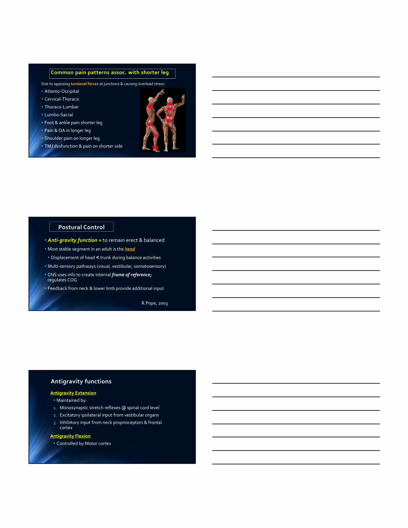
Common pain patterns assoc. with shorter leg
Due to opposing torsional forces at junctions & causing overload stress:
• Atlanto‐Occipital
• Cervical‐Thoracic
• Thoraco‐Lumbar
• Lumbo‐Sacral
• Foot & ankle pain shorter leg
• Pain & OA in longer leg
• Shoulder pain on longer leg
• TMJ dysfunction & pain on shorter side
Postural Control
• Anti‐gravity function = to remain erect & balanced
• Most stable segment in an adult is the head
• Displacement of head < trunk during balance activities
• Multi‐sensory pathways (visual, vestibular, somatosensory)
• CNS uses info to create internal frame of reference; regulates COG
• Feedback from neck & lower limb provide additional input
R.Pope, 2003
Antigravity functions
Antigravity Extension
• Maintained by:
1. Monosynaptic stretch reflexes @ spinal cord level
2. Excitatory ipsilateral input from vestibular organs
3. Inhibitory input from neck proprioceptors & frontal cortex
Antigravity Flexion
• Controlled by Motor cortex
Page 8
Effect of Dominant Hand/Foot
• With general ADLs:
• one leg/hand is used for postural support (vestibular dominance)
• other leg/hand is used for voluntary activities (motor dominance)
Posterior & Anterior Spiral Spring System• Need stretch on Iliopsoas during gait for spring effect
• Swing phase/pushoff – Utilize AOS and POS myofascial slings
• DLS active @ Heel strike (eccentric loading)
• Ipsilateral gluteals co‐contract with opposite Lat. Dorsi
• Counter‐rotation of arms assists with this energy spring system
• OBSERVE:
• Patient walking, or marching in place– Is arm swing symmetrical?
• If not, problem with Anti‐Gravity Spiral Spring System!!
www.erikdalton.com
IF joint dysfunction or malalignment exists:
• Lose our anti‐gravity spring!!!
• Muscle spindles –
• > concentration in Posturalmuscles; slow twitch, oxidative
• Better designed for sustained, compressive loads
• Muscle Spindle Dysfunction in deep, postural stabilizers = SPASM!!• Anti‐gravity function shifts to Globalmuscles; fast‐twitch
• Fatigue quickly, then shift load back to overloaded Postural muscles
Page 9
Origins & Causes of Compensatory Postures & Malalignment
Origin of Compensatory Posture:
• Genetic Potential
• Stresses on Fetus during childbirth
• Developmental Influence – crawl to walk
• Structural Asymmetries– from injuries, leg length difference, etc.
• Tight muscles unopposed by inhibited muscles–leads to imbalance & compensation.
3 Common Regions for Postural Dysfunction
1. Lumbo‐sacral junction
2. Lower extremities
• leg length, foot arches & malalignment
3. Cranio‐cervical mandibular junction (CCMJ)
• Occlusion & mandibular rest position closely related to posture of head & neck.
• Fascial strains prod. by structural asymmetries can directly contribute to CCMJ dysfunction
www.erickdalton.com “Puzzle of Perfect Posture”
Page 10
Other Biomechanical & Structural Changes
• Untreated joint laxity & torsional deformities
• Prolonged computer use & sedentary jobsCreate possible structural changes in soft tissue:
• Collagen shortening; muscle fibrosis• Reduced “Flexion-Relaxation Phenomenon”
(FRP)
• Produce symptoms due to premature DJD • Caused by overload & break-down of joint structures• Asymptomatic DDD common in Cervical spine >30 y.o.
Olson et al. 2000; Won-Gyu Yoo 2014; Ali, A.H.A. et al, 2015; Yong, et al. 2016
Upper Crossed Syndrome— Janda
Asymmetry between line A & line B allows postural dysfunction:
• Line (a): Line of Hypertonicity• passes through neck extensors, levator scapulae, upper trap & pectorals
• Sustained hypercontraction in typically tonic muscles elevates & protracts shoulders.
• Line (b): Line of Inhibition • Passes through deep neck flexors & lower shoulder stabilizers
•
Page, 2003
Chronic pain linked to:
•Reduced movement speed
• Increased background activity of stabilizing muscles to minimize use of painful muscle
•Temporal isolation of painful joint movement outside of kinetic chain.
Page 11
Effects of Neck Pain (NP) & Cervico‐Cranio‐Facial Pain (CCFP) :
• n= 64 subjects with 1) chronic, mechanical neck pain (NP), 2)those with CCFP, 3) asymptomatic controls
• Evaluated Cervical ROM, Mandibular opening, Neck Disability Index (NDI)
• Higher NDI scores for NP and CCFP vs. controls
• Significant difference for NP and CCFP for all ROM measurements, especially Cervical Extension & Rotation
• Mandibular opening signif. lower in NP & CCFP
Munoz‐Garcia, et al. 2016
Whiplash
• Soft tissues injured by sudden "whipping" of the head.
• Flex/Ext ROM most limited (sagittal plane)
Symptoms:• Pain & stiffness in neck & shoulder• Altered muscle activation in Upper & lower trap, Ser. Ant
• Dizziness & Headaches
Jull 2011; Helgadottir 2011; Sterling, 2014
Altered Cervical APA with Chronic NP & Whiplash Disorder (WAD)
• 3 groups (n=173): 1) WAD, 2) CNP and 3) controls
• Measure max Neck AROM, conjunct motion in other planes, and Joint Position Sense error (JPE)
RESULTS:
• Conjunct motion > during rotation, and flexion/extension.
• Reduced conjunct motion in pain groups vs. controls, especially for SB during rotation
• No difference in JPE testing.Woodhouse & Vasseljen, 2008
Page 12
Age‐Related Variance in MaxCervical ROM (53%) & Conjunct Motions (9%)
Possible Reasons:
1. Age‐dependent Muscle atrophy
2. Decrease in # motor units
3. Changes in muscle fiber composition
4. Degen. Alterations in flexibility
5. Impaired vestibular function
6. CNS altered motor planning
7. Fear‐avoidance behaviors
Niederer, et al. Eur Spine J 2015
Proprioception & KinesthesiaEFFECTS OF NECK & SHOULDER PAIN POSTURAL MALALIGNMENTSMOTOR CONTROL ADAPTATIONS (MCA’S)
Proprioception & Postural Control
• Contributes to posture & body alignment
• Includes:
1. Joint Position Sense
2. Kinesthesia
3. Sense of tension
Page 13
Motor Control Adaptations (MCAs) with Neck & Shoulder Pain
• Linked to movement repetition, posture & muscle fatigue.
• Local joint & global movement patterns change over time with repetitive‐motion‐induced fatigue:
• Increased co‐activation of Agonist‐Antagonist
• Decreased inhibition between Synergist muscles
• Changes in inter‐muscular coordination b/w Agonist groups
Motor Control Adaptations (MCAs) with Neck Pain
• Loss of proprioception in neck alone may not explain MCAs
• Altered response in Motor Cortex related to chronic pain may contribute to MCA’s.
Head Repositioning Test
• Test for Proprioception & Kinesthesia of Head & Neck
• Use of head‐mounted laser pointer
• Target chart 90 cm away from patient’s head
• Test ability of patient to return to “neutral” target with eyes closed
• Performs max ROM, then asked to return to target
• Cervical Rotation is most sensitive for testing
www.rehabmeasures.org
Page 14
Motor Control Adaptations (MCAs) & Neck Pain
• Conjunct motion: used to assess motor coordination during primary plane movement.
• Cervical rotation requires >motor control of facet joints
• With Chronic NP:
• Stiffer, more guarded movement patterns with
< conjunct motion
Altered Cervical APA with Chronic NP & Whiplash Disorder (WAD)
• 3 groups (n=173): 1) WAD, 2) CNP and 3) controls
• Measure max Neck AROM, conjunct motion in other planes, and Joint Position Sense error (JPE)
RESULTS:
• Conjunct motion > during rotation, and flexion/extension.
• Reduced conjunct motion in pain groups vs. controls, especially for SB during rotation
• No difference in JPE testing.Woodhouse & Vasseljen, 2008
Forward Head Posture (FHP)
• Defined as the head being anterior to a vertical line through the center of gravity
• Correlated to:
• Headaches
• TMD
• Myofascial Pain
• Scapular Dyskinesia
• Neck & Shoulder pain
Page 15
Other Problems induced by FHP:
•Loss of reposition sense
•Dizziness
•Lack of coordination
•Balance problems
Effects of FHP on Proprioception
FHP EFFECTS:
• Increased extension in Upper Cervical Spine
• Shortened SCM & Ant. Scalene
• Lengthening of Post. Extensors (Lev. Scap, Semi spinalis)
• Altered activity in Scapular Stabilizers (Trapezii, Serr. Ant)
PRODUCES PROBLEMS IN PROPRIOCEPTION:
1. Mechanoreceptor dysfunction
2. Altered muscle spindle sensitivity
3. Loss of kinesthetic acuity with neck ROM
FHP Affects Neck Kinematics & SCM activity
• FHP can induce changes in frontal plane motion & SCM activation during neck rotation.
• STUDY: n=28, with & without FHP
• Tested Neck rotation R & L; SCM activity via sEMG
• RESULTS:
1. Maintaining FHP increased rotation/lat flex ratios in both directions
2. FHP group had faster onset time for lat. flexion motion in both directions
3. EMG values for SCM > in FHP group for contra‐lateral SCM
M‐S Kim. J Phys Ther Sci. 2015
Page 16
Clinical Application of Results:
• Consider Quality and Quantity of Cervical ROM
• Primary problem with FHP = shortening & hyperactivity in SCM!
• Induces Neck Rotation/contralateral sidebending with ipsilateral activation
• Induces lower cervical flexion with bilateral activation
• Global muscles are overactivated!!
• Produce large movements vs. precise motion
• Local, deep stabilizers of neck are inhibited or weak
• Can cause overload, overuse, mechanical dysfunction
Why are SCM & Scalenes a Problem???
• SCM and Ant. Scalene are superficial muscles• typically substituted for weak deep neck flexors
• BUT:• SCM has an Extensor Moment
• Ant. Scalene is NOT attached to cranium• Can’t flex head on the neck• Can’t control FHP
0
0.125
0.25
0.375
0.5
22 mm 24 mm 26 mm 28 mm 30 mm
Thi
ckne
ss v
aria
tion
(cm
)
Control SCM NP SCM Control DCF NP DCF
NP-SCM
Control-DCFControl-SCM
NP-DCF
Jun et al, 2013.CCFT: Change in Muscle Thickness SCM vs DCF
Page 17
FHP associated with Loss of Reposition Sense
• Used Head Repositioning Accuracy (HPA) Test
• Head‐to‐neutral test
• n=41, age 20‐30, with FHP, no neck pain
• Subjects seated, hips & knees at 90 deg, feet hip‐width apart
• Laser headset used to locate neutral target at eye level
• Subject closed eyes & instructed to remember target• performed full AROM (flexion, extension, SB, Rotation) held for 5 sec, and return to neutral position
Results of HPA Test:
•Found significant change in Lumbar posture assoc. with compensatory change in Cervical position.
•Neck SB assoc. with pelvic side tilt angle.
Lee, et al. 2015
HPA Results: Why Posture Changed
• Tonic Neck Reflex– alters tone of trunk & extremities via Muscle Spindles & Upper Cervical afferents
• Myofascial Chain Reaction– (Tom Myers’ Anatomy Trains)
• Fascia: agonist muscle of Neck SB & Pelvis side tilt angle are connected via fascia on Lateral Line
• Quad Lumborum: not directly on Lateral line, but works as Lateral flexor of trunk
• Scalenes: also work as lateral flexor of Neck
Page 18
Deep muscles of Spine & Thorax
• Quadratus Lumborum
• Lateral flexion and extension of spine
• Lateral Line – balances posture in frontal & sagittal planes; helps create lateral flexion, hip ABD, Ankle EV; decelerates trunk lat. Flex & rotation
• Spiral Line – wraps body into double spiral to maintain balance in all 3 planes; mediates oblique spiral & rotational movements
• Superficial Front & Back Lines – provides postural support in standing
• Deep Front Line – plays major role in support of arch, LE’s, lumbar spine, abdominal‐pelvic region, chest/breathing, head & neck.
Tom Myer’s Myofascial “Anatomy Trains”
Consider Trunk Posture w/ Neck & Shoulder Problems!!
• Over 20 muscles attach to head/neck; 30 muscles to scapula
• Lat Dorsi attaches thru T‐L fascia, lumbar & sacral vertebrae, pelvis, scapula
• Quadratus Lumborum attaches to lower ribs, lumbar vertebrae, and pelvis
• Trapezius attaches to occiput, cervical & thoracic vertebrae, scapula
• Innervated by C2‐3‐4
• Serratus Anterior attaches to upper 8‐9 ribs, medial border of scapula
• Innervated by C5‐6‐7‐8
• Levator Scapula attaches to T.P. C1‐4, and medial border of scapula
• Innervated by C3‐4‐5
Kendall & Kendall, 3rd Ed., 1983
Page 19
Muscle Imbalance Continuum
Muscle Imbalance
Tissue Damage
& Pain
Altered Movement
Pattern
Effect of Arm Position & Sitting Posture
• Pain, fatigue & joint position affect proprioception
• Changes in body posture affect proprioception
• STUDY: Measured Cervical‐Cephalic Kinesthetic Sensibility (CCKS) Test
• Test subjects Cervical AROM (Rot, Flex, Ext) in 3 positions:
1. Slouched sitting w/ arms at side (SS)
2. Slouched sitting w/ arms supported (SS‐AS)
3. Upright Sitting w/ arms at side (US)
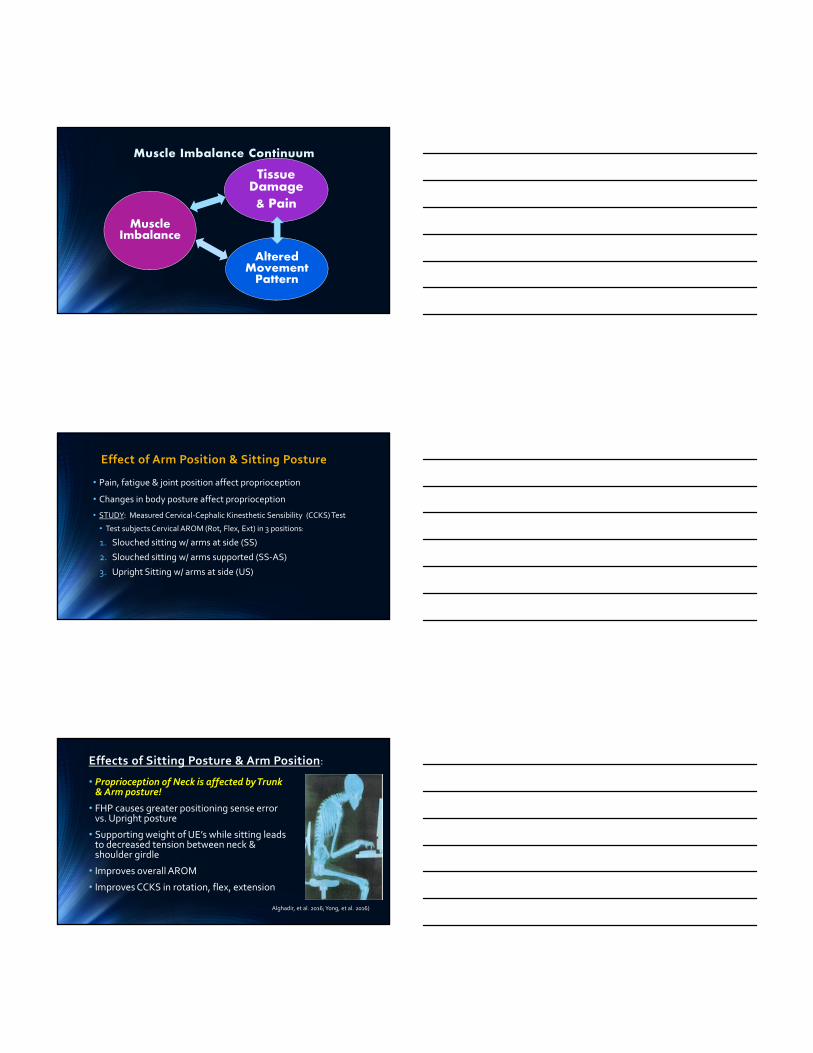
Effects of Sitting Posture & Arm Position:
• Proprioception of Neck is affected by Trunk & Arm posture!
• FHP causes greater positioning sense error vs. Upright posture
• Supporting weight of UE’s while sitting leads to decreased tension between neck & shoulder girdle
• Improves overall AROM
• Improves CCKS in rotation, flex, extension
Alghadir, et al. 2016; Yong, et al. 2016)
Page 20
Effect of Sitting Posture on Spine Stability
• Recruitment of DCF & Lumbar Multifidi are affected by sitting posture.
• 2 sitting positions tested (n=10; surface EMG):
1. Self‐corrected sitting “up straight”
2. Therapist‐facilitated sitting via verbal & manual cues to correct neutral lumbo‐pelvic position
• Activation of DCF & Multifidi muscles were signif. > with Therapist‐facilitated postural correction
Falla, et al. 2007
Strategies Used for Sitting Posture
• Self‐Corrected:
• Tend to initiate movement with Thoraco‐Lumbar Extension
• Less activation of deep spinal stabilizers (Multifidus, DCF)
• Therapist‐Facilitated:
• Increased activation of deep spinal stabilizers
• No change in Thoraco‐Lumbar extensor activity
Measuring Motor Control Adaptations DELAYED ANTICIPATORY POSTURAL ADJUSTMENTS (APA’S)
Page 21
Dynamic stability
• “The ability to be balanced, stable, lengthened, centered & free to move.” ~ Medoff, 2014.
• Allows postural corrections to take place to perturbations.
To TEST:
• Give small perturbation, observe new behavior of stability system.
• If it differs from previous behavior, then system is
UNSTABLE !
Anticipatory Postural Adjustments (APAs)
• Involuntary adjustments that occur prior to predictable postural perturbations (PPP)
• Delayed APAs have been identified in neck & spine patients
• Evidence: (n=64; 18‐50 y.o. recurrent LBP vs. controls)
• 8‐week training program: Specific Ex Program vs. General Ex
• Use of rapid arm flexion movements for PPP
• Measure sEMG: Rect. Abd, Lumb Ext, Tran Abd, Int. Obliq, Ant. Deltoid
Results of Study on APAs
• Pain levels reduced in both groups, but 30% > in Specific Ex Group
• Self‐rated disability improved in Specific Ex Group only
• APAs not delayed in LBP patients at baseline
• APAs changed in both groups after training, show > coordination
Clinical Application:
• Abdominal bracing (“drawing in”) should NOT be mandatory inclusion in Specific Ex Group.
Page 22
Problem with “routine exercise”
• Routine Exercises create GLOBAL muscle dominancy over DEEP Local Stab. System (DSS)
• Alter muscle coordination & increase PAIN!
• Do not increase endurance or cross‐sectional area of DSS.
• Specific Stab. Ex. can correct movement pattern & then DECREASE PAIN. (via Motor Control ex)
McGill, 2009; Javadian et al, 2012; Sahrmann, 2002; Lee, 2010
Evidence for stabilization • DYNAMIC stab. @ varied movement rate vs. Conventional tx. (n=141)
• Both groups signif. improvement in QOL (SF‐36)
& Pain (VAS):
• Stab group better!
Kumar, 2010
0
10
20
30
SF‐36 VAS
24.6
3.9510.7
2.87
< Change in Scores
Stab Ex Control
• SF‐36 >50% difference• VAS >25% difference
Testing for Postural Asymmetry
Page 23
Initial Postural Assessment of Patient:
• Assess alignment of COG
• Assess participation of muscles based on line of gravity
Example: Acquired Thoracic Kyphosis
• Alignment of thorax is major factor in neck pain
• Dynamic condition; can be changed!
• Requires compensation of head/neck extension to maintain horizontal plane of eyes and mouth
Dynamic Posture Test– Slant BoardNORMAL STANCE STANCE ON SLANT BOARD
Core stabilizers engage on slant board. Improved alignment of COG, more neutral pelvis, head/neck.
Poor postural alignment, FHP & kyphosis. Leans back into excessive lordosis, pelvis pushed forward, standing back on heels, COG is posterior.
“Extensor Bias”
•Hyper‐extension Lumbar
•Hyper‐extension Knees
•Plantar flexion Ankles
•Anterior Pelvic Tilt
•COG shifted anterior
69
Page 24
Flexion Bias
• Flattened lumbar spine
• Increased kyphosis
• Posterior pelvic tilt
• Flexed knees & hips
• Ankles flexed (DF)
• COG moves posterior
Evidence of Motor Recruitment Changes
•Hypertonic T/L extensors
• Classic sign of global muscle over‐activation & inhibition of deep stabilizer system (DSS)
•Slumped Posture• Inhibits diaphragm, affecting DSS function
•Hypertonic Quadratus Lumborum and/or Piriformis• Classic sign of lack of hip extension & “Gluteal Amnesia”
Liebenson 2001; Janda 2004; McGill 2009; Sahrmann 2010; Brookbush 2013.
Standing Arm Elevation Test
Look for compensation:
•Forward head
•Increased cervical extension
•Increased lumbar lordosis
•Rib cage elevation
•Inhalation
•Scapula abduction/depression
Page 25
Scapular Balancing Index
6-part test— degree of Scapular Control:1. Lateral Scapular Slide Test2. Neuromuscular Evaluation (PNF)3. Strength & Endurance (10 reps)4. Cervical Posture5. Thoracic Posture6. Thoracic Segmental MobilityTOTAL SCORE: Part 1-6= 0 - 20 points
Brownstein & Bronner, 1997)
Scapular Balancing Index
Scores:
0-10 pts Possible neurologic Involvement
11-12 pts Poor Scapular Control
13-16 pts Fair Scapular Control
17-18 pts Normal Scapular Control
19-20 pts Excellent Scapular Control
Testing for Scapular DyskinesiaSAT, SRT, & DYNAMIC SDT
Page 26
Movements that Stabilize the ScapulaUpward/Downward Rotation
• Axis perpendicular to spine of scapula; movement of inferior angle of scapula
• Controlled by force couple: UT, LT, SA
Anterior/Posterior Tilting
• Occurs around axis thru spine of scapula; movement of superior border
• Controlled by force couple: LT, SA (lower fibers)
Internal/External Rotation
• Occurs around vertical axis; movement of lateral border of scapula
• Controlled by MT, LT
Superior/Inferior Translation
• Occurs at SCJ; clavicular elevation or depression
What is Scapular Dyskinesia (SD)?
• 61% of overhead athletes have evidence of SD vs. 33% non‐overhead athletes
• Movement Dysfunction of Scapula:
• Change in Post. Tilt (+/‐)• Change in Upward Rotation (+/‐)
• Increased Internal Rotation/ Medial winging
• Increased Superior Translation at SCJ
(Burn, et al. 2016; McClure, et al. 2012; www.pthaven.com)
3 Tests for Scapular Dyskinesia
1. Dynamic Scapular Dyskinesia Test (SDT)
2. Scapular Assistance Test (SAT)
3. Scapular Relocation Test (SRT)
Kibler, et al. 2013; McClure, et al. 2012
Page 27
Scapular Dyskinesia Test
• Patient actively elevates arms to 90‐100 deg flexion or abduction
• Observe dysrhythmia, non‐smooth or ratcheting motion, medial winging of scapula, rotation of inferior angle of scapula
• Yes or No result
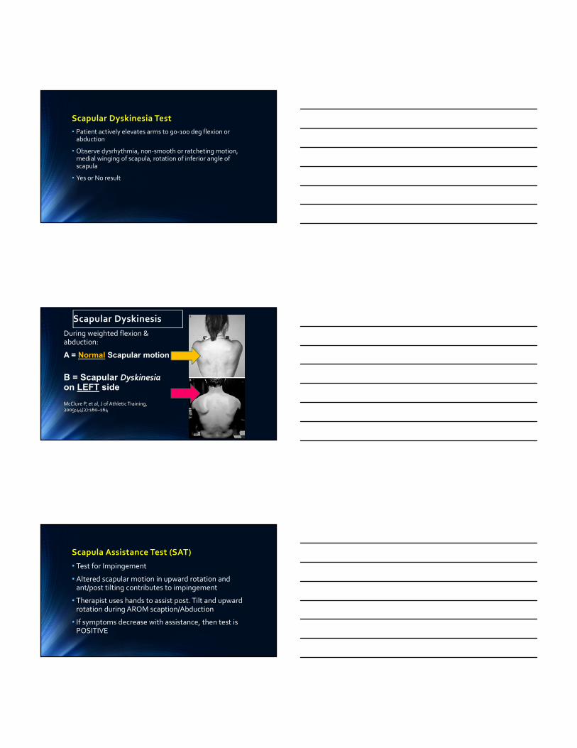
Scapular Dyskinesis
During weighted flexion & abduction:
A = Normal Scapular motion
B = Scapular Dyskinesia on LEFT side
McClure P, et al, J of Athletic Training, 2009;44(2):160–164
Scapula Assistance Test (SAT)
• Test for Impingement
• Altered scapular motion in upward rotation and ant/post tilting contributes to impingement
• Therapist uses hands to assist post. Tilt and upward rotation during AROM scaption/Abduction
• If symptoms decrease with assistance, then test is POSITIVE
Page 28
Scapular Relocation/Retraction Test (SRT)
• Test for GHJ instability, dislocation, subluxation
• Used in conjunction with Apprehension Test
• In supine, Therapist repositions scapula by applying post. force to humeral head during max ER
• If Apprehension symptoms improve with SRT, then test is POSITIVE
Motor Retraining for Postural Asymmetry
• With decreased proprioception after injury or inactivity:– ↓ tonic (SMU) muscle recruitment – hyperactivity in global phasic (FMU) muscles
– » INSTABILITY!
• Need to retrain SMU to improve stability• Recognize substitution patterns & retrain early and often
Neuromuscular Re‐Education
Page 29
Common Substitution Patterns in Cervical & Scapular Regions
• Hyperactivity in extensor muscles
• Weakness in deep neck flexors
• Increased upper trap tone & scapular elevation/ant. tilt/ abduction
• Poor control of scapular depression, retraction, & adduction
• Rib cage elevation
• Kyphosis or flattened Thoracic spine
Progression of spinal stability programs
Motor Control
Static Stabilization
Dynamic Stabilization
Motor control vs. stabilization ex.
• Motor control training (Isolated activation) of DSS necessary to restore altered activation patterns.
• Increased activation of DSS occurs w/ functional & loadedpostures.
Bystrom, 2013; Crommert, 2011.
Page 30
Pressure biofeedback for motor control DSS
0
1
2
3
4
5
6
7
Core Stab Press Bfbk
6.76.2
4.6
3.5
2.3
0.9
Change in Pain (VAS)
Baseline 15 days 30 days
Core Stab vs. Pressure Biofeedback (n=30)• Significant diff. in
Pain levels after 8‐wk training.
Uddin & Ahmed, 2013.
EBP for pressure biofeedback (cont.)
0
5
10
15
20
25
30
35
40
45
50
Core Stab Press Bfbk
40.446.6
30.624.8
19.8
2.0
Change in Function (ODI)
Baseline 15 days 30 daysUddin & Ahmed, 2013
Treatment for Postural & Movement Deficiencies in Neck & Scapular RegionMANUAL TECHNIQUES
Page 31
Core rehab vs core strengthening
• You can’t strengthen something that isn’t working!
• PAIN— inhibits optimal motor patterns, prevents re‐establishing “healthy” patterns.
• CORE STRENGTHENINGwith altered movement patterns can create injury!
• Reinforces “inappropriate” movement patterns & motor control errors.
• CORE REHAB improves motor control & re‐establishes correct movement patterns.
Functional Rehab focus:Functional Rehab focus:
• Establish proper postural alignment
• Establish proper motion at all involved segments
• Work proximal to distal for sequential muscle activations in UE & LE.
• Utilize closed chain exercise early
• Work in multiple planes
Proper Progression is the KEY!
• Identify abnormal motion or motor pattern.
• Restore normal joint & tissue mobility.
• Perform corrective exercises to retrain neuromuscular patterns & improve proprioception.
• 1st Rep IS the provocation test!
• Do NOT allow compensation—perform with PERFECTform!
McGill, 2010
Page 32
Rehab Progression: Stage 2
• Retrain normal movement & motor patterns to ensure stability.
• Two levels of Stab:
1. Joint Stability– Spinal Stability
2. Whole Body Stability
• Use Co‐contractions for > durations (10 sec. or less)
• Maintain perfect form
Soft Tissue Mobilization
• Muscle Energy Release
• Scalenes
• Tissue Unwinding• Suboccipital triangle
Mobility Testing
TMJ Measurement:
•Mandibular open
(35‐40 mm)
•Lateral excursion (~1cm)
Rib Cage mobility
• Palpate over First rib with inhale/ exhale
• Apply downward pressure w/ exhale
Page 33
C‐T Junction Mobilization• With cervical rotation & arm elevation
• “Arm Pit Sniff” MET
Thoracic Spine Mobilization (TSM)
• Evidence for immediate improvement of neck pain after TSM
• Evidence for immediate, significant increase in strength of LT after TSM
• Neurophysiological Effects:
• Inhibition of hypertonic muscles
• Reduction of pain
• Improved scapular position & function
Cleland, et al. 2007; Puntumetakul, et al. 2015
Cervical Thoracic Junction
Cleland, et al. (2007):
• 6 criteria for neck patients who may benefit from thoracic spine mobilization:1. Duration < 30 days
2. No symptoms distal to shoulder
3. Cervical Extension does not increase sx’s
4. FABQPA score <12 (fear‐avoidance beliefs)
5. Diminished T‐spine kyphosis (T3‐T5)
6. Cervical Extension AROM < 30 deg.
Page 34
Biomech. Link in C‐T Junction
• If pt had 4 of 6 criteria = 93% prob. Success
• If 5 or 6 of 6 criteria = 100% prob. Success
•NDI and Pain levels significantly decreased with single (T6‐7) & multi‐level mobilization @ 1‐week post treatment.
Cleland, et al. 2007; Puntumetakul, et al. 2015
PNF Scapular Pattern
Exercises for Postural & Movement Impairments
NECK REPOSITIONING DEEP NECK FLEXORTMJ COMPLEX
Page 35
Joint Position Sense Training for C‐spine
• Use laser pointer attached to head piece
• Have patient move head to various targets in mid‐ROM
• Use cervical rotation especially for conjunct motions.
• Practice with eyes open, and eyes closed to return to neutral target.
Rocabado’s 6 x 6 TMD Complex exercises
1. Tongue “clucking”
• Tongue on roof of mouth; position for nasal & diaphragmatic breathing
• Teeth slightly apart, lips closed; prevents excess jaw retrusion& mouth breathing
2. Controlled TMJ Rotation on Opening
• Tongue “cluck” position; slowly open and close mouth
3. Mandibular Rhythmic Stabilization
• Apply light resistance w/ opening/closing/lateral excursion with jaw in rest position
Rocabado’s 6 x 6 TMD Complex exercises
4. Upper Cervical Distraction
• Perform upper cervical flexion (“chin nod”) while stabilizing cervical spine with “hand collar”
• Relieves neurovascular compression in upper C‐spine by distracting A‐O joint
5. Axial Extension C‐spine
• Cervical retraction (upper C‐spine flexion with simultaneous lower C‐spine Extension
• Goal to normalize upper quadrant posture & mechanical relationship
6. Shoulder Girdle Retraction
• Scapular retraction and depression of scap relative to rib cage (“Cobra” position or standing shoulder extension with retraction at wall)
Page 36
Biofeedback for Neuromuscular Re‐education
Pressure Biofeedback
• teaches patients how to activate stabilizers with right amount of force
Stabilizer for TA Bracing
CCFT ‐ Deep Neck Flexors
• Fold Stabilizer in three sections or BP cuff at bladder.
• Place Stabilizer under neck ‐ inflate to 20 mmHg. • Gently nod head, "look down" without lifting head. Increase the pressure in 2mm increments and hold steady.
• Relax and repeat. Increase up to 22‐30 mmHg.
• GOAL: Hold for 10 seconds, repeat 10 times on highest pressure target that can be held steady without compensation of SCM or Ant. Scalenes.
Page 37
Other uses for Stabilizer
• Scapular stabilization with arm elevation:• Place cuff under spine of
scapula• Inflate to ~30 mm Hg
• Maintain with AROM flexion.
• Scapular stabilization with External Rotation:
Other Neck Stabilizer Exercises
1. CCFE–Cranio Cervical Flexion Exercise
• Supine chin “nods” with head, neck and thoracic spine in neutral
• Teach patient to monitor for SCM and Ant. Scalene excess activation
2. CCEE–Cranio Cervical Extension Exercise
• Prone with forehead on hands stacked
• Perform chin nods with retraction
• Monitor for scapular depression/retraction
3. Quadraped “Synergy Retraining” of deep neck flexors
• Advanced position for CCEE
Hidalgo‐Perez, et al. 2015
Cervicogenic Headache Exercise Progression1. Start with the CORE!
• Diaphragmatic breathing to decrease activity in accessory muscles (SCM, Scalenes)
2. Cervical Stabilization w/ Chin Nod (Forehead on mini ball against wall)
• Resistance band at wrists, elbows flexed; perform bilateral scap retract in ER
3. Dynamic Cervical Extension Exercise
• Seated in hip hinge with (red) resistance band around head; resist 4 directions
• Hold 3‐5 sec
4. Sensorimotor Training on unstable surfaces
• Promotes reflexive stabilization & postural stability for head righting
Page, 2011
Page 38
Exercises for Postural & Movement Impairments
SCAPULAR REBALANCINGSCAPULAR STRENGTHENINGKINETIC CHAIN MOTOR REPROGRAMMING
0
20
40
60
80
100
120
Uni Shrug Scaption >120 Prone "Y" uni Prone HorExt/ER
Prone ER @ 90 Uni Row Scaption <80 Uni Press DiagFlx/HorFlx/ER
Top 3 Exercise for % MVIC for Trapezii & Serr. Anterior
UT MT LT SAEkstrom, et al. 2003
2 Best Exercise for Upward Rotation of Scapula
• Requires simultaneous activation of Trapezius & Serr. Anterior
1. Prone “Y”
• All 3 parts of Trapezius @ 79-101% MVIC
• SA levels could not be reliably measured
2. Scaption >120 deg
• Trapezius levels 49-79% MVIC
• SA @ 96%
Ekstrom, et al. 2003
Page 39
Is Max % MVIC the GOAL for Scapular Stab?
• Low levels of SA & LT @ 20-40% MVIC are associated with kinematics of scapular dyskinesia
• Scap Dyskinesia is a motor control issue, not strength!
If >20-40% MVIC:
• Produce strength, & recruit other, global muscles
• Contribute to adaptive movements & imbalances!
• Scap Stab Exercises should maintain 20-40% MVIC in LT & SA
Barreto, et al. 2012
Scap Stab: Ideal Exercises for 20‐40% MVIC in LT & SA
7
27
6 4.5
26.5
15
53.5
29
13 10.5
0
10
20
30
40
50
60
Mod Crucifix Scaption 120 Mod Mil Press Pullover Low Row
% MVIC for LT & SA for 5 Key Exercises
LT SA
Why not the Prone “Y”?
• Prone “Y” recruitment % MVIC >20-40% for motor control:
• SA = 43%
• LT = 97%
• Excess activation could lead to co‐activation of UT & increased cervical extension
• Best exercises for LT & SA co‐activation include combination movements for Flexion/Adduction/ER
Page 40
Other Ideal Exercises for Lower Trap (LT):
•Mod. Prone Cobra = 44.7% MVIC
•Prone Row = 36.5% MVIC
•Lat Pulldown = 35.2% MVIC
% Muscle Contributions for Shoulder Abduction
Mid Deltoid, 50
Subscapularis, 30
Supraspinatus, 25
Infraspinatus, 10
Ant Deltoid, 2
Mid Deltoid Subscapularis Supraspinatus Infraspinatus Ant Deltoid
Escamilla, et al. 2009
69.4
29.436 33.4
79.6
28.6
43.2 40.3
56.3
12.9
48.1
27.7
78.3
15.6
60.4
33.2
0
10
20
30
40
50
60
70
80
90
Quad Shld Flex Lawn Mower Robbery Abduction
LT‐3 LT‐5 SA‐3 SA‐5
Compare % MVIC for LT & SA w/ Free Motion Exercises
Tsuruike & Ellenbecker, 2015.
Page 41
Force Couples During Scap Stab Exercises
With injury & during early phase rehab, important to:
1. minimize UT activity
2. promote MT, LT, & SA activity
• Establish balance among these muscles early to promote better motor activation of lower scapular stabilizers.
Moeller, et al. 2014.
% MVIC of Trapezii & Serr. Ant. w/ GHJ Injury
17.921.9
42.7
73
27.2
18.822.4
37.5
20.815.6 13
23.4
39.3
24.3 23.119
0
10
20
30
40
50
60
70
80
ER Scap Squeeze Lawnmower Robbery Bow Arrow
UT MT LT SA
Bottom Line: Does Scapular Stabilization work?
• Teaching conscious correction of scapular orientation during rehab to balance force couples:
• UT/LT
• UT/MT
4 Exercises for training Trapezii:
1. Prone extension
2. Prone Hor. Ext/ER
3. Sidelying ER
4. SL Forward Flexion
DeMey, et al. 2013
Page 42
% Change in % MVIC of Trapezii after Conscious Correction of Scapular Position
5.9
2.2
13.8
6.7
9.8
13.3
0
2
4
6
8
10
12
14
16
Prone Ext SL ER
UT MT LT
Other BEST Exercises for Scapular Rehab
• Scap Squeezes in “Row” position (MT, LT, Rhomboids)
• Low Row – Shoulder extension bilateral w/ scap squeeze (LT)
• Dynamic Hug – bilateral standing chest press w/ arms forward as if hugging a tree (SA & LT)
• Scap Punches – band @ shoulder ht., punch w/ full protraction (SA)
• Cheerleader Exercise – perform alternate diagonal patterns w/ both arms pulling outward w/ one horiz. ABD motion b/w each diagonal (LT & Rhomboids)
Paine & Voight, 2013
Summary for Neck & Scap Rehabilitation
• Start with the Core– DCF, Diaphragmatic breathing
• Minimize SCM and Scalene accessory activity
Focus on Main Scapular Stabilizers:
• Serratus Ant– scapula abduction/protraction
• Rhomboids– scapula adduction/retraction; stabilize medial border
• Lev Scapula– elevate /rotate scapula downward
• Upper Trap–Upward rotation/elevation of scapula
• Mid Trap– Retraction of scapula
• Lower Trap– Upward rotation/depression of scapula
Page 43
• Focus on GHJ “Protectors” –Rotator cuff muscles
• Rebalance tissues, maintain alignment & mobility of structures
Summary for Neck & ScapRehabilitation
Summary & Conclusions
• QUESTIONS & ANSWERS
Thank YOUSUE DUPONT, MS, MBA, PT, ATCEMAIL: [email protected]