Giuseppe D'Ippolito, Denise Tokechi, David Carlos Shigueoka, Sergio Ajzen Tomographic aspects of xanthogranulomatous pyelonephritis and related complications Department of Imagery Diagnosis, Paulist School of Medicine, Federal University of Siio Paulo - Siio Paulo, Brazil The authors present their experience involving seven patients with histopathologic diagnosis of xanthogranulomatous pyelonephritis who were submitted to preoperative computed tomography (CT).The results are the following: a) stones (86 percent of the cases), b) increase in renal volume, c) hydronephrosis, d) density measurements (from 14 to 29 HU), e) enhancement found in all cases, f) extrarenal involvement (all cases). CT has shown to be a reliable method in characterizing xanthogranulomatous xyelonephritis and extrarenal involvement. UNITERMS: Pyelonephritis. Computed Tomography. INTRODUCTION X anthogranulomatous pyelonephritis (XGP) is an atypical variety of a chronic renal infection which is usually unilateral and frequently associated with urinary obstruction and stones 13.15. Before ultrasonography (US) and computed tomography, (CT) preoperative diagnosis was not certain in 44-64 percent of all cases, due especially to uncharacterized symptoms, and laboratory exams that showed no alterations 5 . 13. 16. IX. Radiographic exams such as excretory urogram (EU) and antegrade pyelography (APG) allow the diagnosis of a renal mass with or without functional elimination; however, they do not indicate the degree of inflammation caused by the disease 13 • Although it is relatively rare (820 Address for correspondence: Giuseppe D'Ippolito Rua Alceu de Campos Rodrigues, 165 - V. N. Concei9ao Sao Paulo/SP - Brasil- CEP 05409-001 cases described up to 1993), the tomographic aspects have already been described and have been said by some authors to be pathognomonic 5 . 9. Therefore, it is possible not only to diagnose the disease, but also to evaluate its extrarenal extension and to differentiate the focal and the diffuse forms, allowing for precise surgical planning. Treatment consists of total nephrectomy in the diffuse form and a partial nephrectomy when only a limited area is affected II. The purpose of our study is to present seven cases, describing their tomographic aspects and frequency, so as to help differentiate this disease from others. MATERIAL AND METHODS We reviewed CT studies of seven patients with an histopathologic diagnosis ofXGP made between 1991 and 1993. Six of these seven patients were female and one was male, with ages ranging from 13 to 71 (mean age - 41 years). D'IPPOLITO, G.; TOKECHI, D.; SHIGUEOKA, D.C.; AJZEN, S. - Tomographic aspects of xanthogranulomatous pyelonephritis and related complications Sao Paulo Medical Journal/RPM 114(1): 1091-1096, 1996
Transcript
Giuseppe D'Ippolito, Denise Tokechi,David Carlos Shigueoka, Sergio Ajzen
Tomographic aspects of xanthogranulomatouspyelonephritis and related complications
Department of Imagery Diagnosis, Paulist School of Medicine, Federal University of Siio Paulo - Siio Paulo, Brazil
The authors present their experience involving seven patients with histopathologic diagnosis of xanthogranulomatous pyelonephritiswho were submitted to preoperative computed tomography (CT). The results are the following: a) stones (86 percent of the cases), b)increase in renal volume, c) hydronephrosis, d) density measurements (from 14 to 29 HU), e) enhancement found in all cases, f)extrarenal involvement (all cases). CT has shown to be a reliable method in characterizing xanthogranulomatous xyelonephritis andextrarenal involvement.
UNITERMS: Pyelonephritis. Computed Tomography.
INTRODUCTION
Xanthogranulomatous pyelonephritis (XGP) is anatypical variety of a chronic renal infection whichis usually unilateral and frequently associated with
urinary obstruction and stones 13.15. Before ultrasonography(US) and computed tomography, (CT) preoperativediagnosis was not certain in 44-64 percent of all cases,due especially to uncharacterized symptoms, andlaboratory exams that showed no alterations5. 13. 16. IX.
Radiographic exams such as excretory urogram (EU) andantegrade pyelography (APG) allow the diagnosis of arenal mass with or without functional elimination;however, they do not indicate the degree of inflammationcaused by the disease13• Although it is relatively rare (820
Address for correspondence:Giuseppe D'IppolitoRua Alceu de Campos Rodrigues, 165 - V. N. Concei9aoSao Paulo/SP - Brasil- CEP 05409-001
cases described up to 1993), the tomographic aspects havealready been described and have been said by someauthors to be pathognomonic5. 9.
Therefore, it is possible not only to diagnose thedisease, but also to evaluate its extrarenal extension andto differentiate the focal and the diffuse forms, allowingfor precise surgical planning. Treatment consists of totalnephrectomy in the diffuse form and a partial nephrectomywhen only a limited area is affected II.
The purpose of our study is to present seven cases,describing their tomographic aspects and frequency, soas to help differentiate this disease from others.
MATERIAL AND METHODS
We reviewed CT studies of seven patients with anhistopathologic diagnosis ofXGP made between 1991 and1993. Six of these seven patients were female and onewas male, with ages ranging from 13 to 71 (mean age - 41years).
D'IPPOLITO, G.; TOKECHI, D.; SHIGUEOKA, D.C.; AJZEN, S. - Tomographic aspects ofxanthogranulomatous pyelonephritis and related complications
Sao Paulo Medical Journal/RPM 114(1): 1091-1096, 1996
1092
Table 1 . blurring of peritoneal fat due to
Patients according to the presence and the site of stones, degree of hyperdense thick layers, masshydronephrosis, and measurements of the less dense area of renal lesion images with the aspect of soft tissue
or circundating liquid collections.Patient Stones Coraliform Site Degree of Density Heterogeneity and asymmetric
Stones Hydronephrosis (UH) enlargement of paravertebralAR + pyeloureteral junction IV 21-25 muscle and or s ubcu taneousBFS + + pyelo-calix IV 17-29 cellular tissue were consideredFLN + parenchyma/ureteral I indications of abdominalJCM pyelo-calix IV 14 involvement.+ +LACC + ureteral IV 22-24MLF + + pyelo-calix IVMVS + IV 23-25 RESULTS
The exams were carried out using the Somatom DR(Siemens Medical System), with 8 mm wide cuts and an 8or 16 mm increment, before and after one endovenousinjection of 100 -150 ml iodine hydrosoluble contrast,measuring densities before and after contrast injection.
The tomographic parameters assessed were:a) presence of stones and their sites.b) renal dimensions. We considered the kidney
"enlarged" when its longitudinal diameter exceeded13 cm4•
c) degree of hydronephrosis, classification I - IV2•
d) density measured in low-density areas.e) presence of enhancement after endovenous contrast
injection.t) extra-renal damage (perirenal, pararenal and
abdominal sites), with presence of heterogeneity or
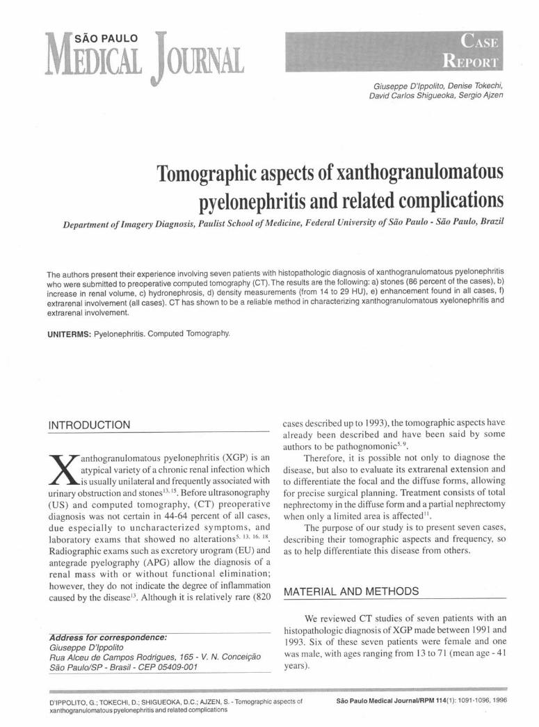
Figure 1 - Increase of left kidney volume showing peripheralenhancement and dilation of the collecting system with centralcalcification. This patient showed a normal inferior segment ofthe kidney.
Unilateral renal malfunction was evident in allpatients; left malfunction in 5 (71 percent) and right in 2(29 percent). In six cases, kidney enlargement wasobserved with grade IV hydronephrosis (86 percent), whilein only one case (14 percent) there was a volumetricdecrease with hydronephrosis grade I (Fig. 5). In six (86percent), there were stones iIi the collecting system; intwo (28 percent), there were stones in the renalparenchyma; and only one patient (14 percent) did notpresent any stones. In three of the six patients with stones inthe collecting system, the stone was coraliform (Table I).
The density of the low-density component of thelesion, measured from 14 to 29 UH, with no fat or gas-type densities being observed. After the intravenousinjection of contrast, there was a peripheral enhancementof the affected kidney (Fig. 10), as well as extrarenal
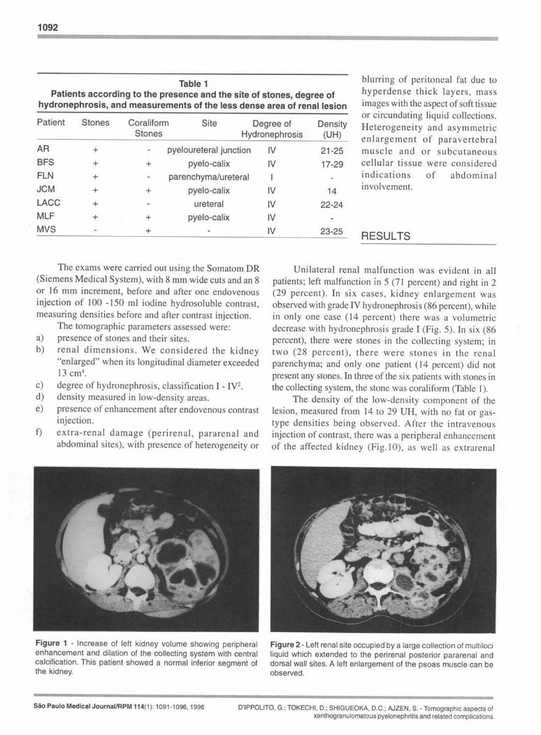
Figure 2 - Left renal site occupied by a large collection of multilociliquid which extended to the perirenal posterior pararenal anddorsal wall sites. A left enlargement of the psoas muscle can beobserved.
sao Paulo Medical Journal/RPM 114(1): 1091-1096, 1996 D'IPPOLITO, G.; TOKECHI, D.; SHIGUEOKA, D.C.; AJZEN, S. - Tomographic aspects ofxanthogranulomatous pyelonephritis and related complications
1093
Table 2Patients according to presence and site of extrarenal
XGP was described in 1916 by Schlogenhaufer, whocalled it staphylomycosis due to its resemblance toactinomycosis and to the presence of staphylococci 17. Theterm XGP was used by Oberling, in 1935, due to its yellowcolor and to its granulomatous characteristic. XGP can bedefined as a rare form of a chronic renal infection with
damage extending to perirenal and posterior pararenal sitesin all cases. In 4 cases (57 percent), the abdominal wallwas involved (Table 2). There were no signs ofinvolvement of retroperitoneal garylia, damage to otherabdominal organs, or fistulae. All the cases were classifiedas XGP of the diffuse form, with one exception, which,due to the damage of the superior segment of the kidney,was considered to be a segmented form (Fig. 1). Observedtomographic signs and their relative and absolutefrequency are listed in Table 3.
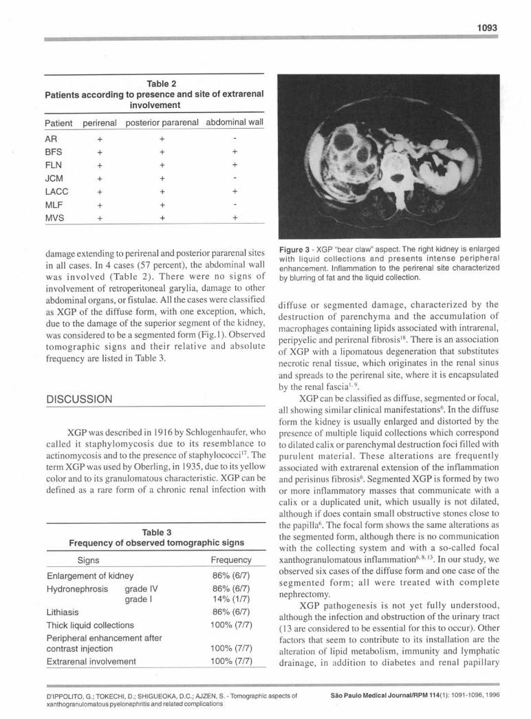
Figure 3 - XGP "bear claw" aspect. The right kidney is enlargedwith liquid collections and presents intense peripheralenhancement. Inflammation to the perirenal site characterizedby blurring of fat and the liquid collection.
diffuse or segmented damage, characterized by thedestruction of parenchyma and the accumulation ofmacrophages containing lipids associated with intrarenal,peripyelic and perirenal fibrosislH• There is an associationof XGP with a lipomatous degeneration that substitutesnecrotic renal tissue, which originates in the renal sinusand spreads to the perirenal site, where it is encapsulatedby the renal fascial. 9.
XGP can be classified as diffuse, segmented or focal,all showing similar clinical manifestations6• In the diffuseform the kidney is usually enlarged and distorted by thepresence of multiple liquid collections which correspondto dilated calix or parenchymal destruction foci filled withpurulent material. These alterations are frequentlyassociated with extrarenal extension of the inflammationand perisinus fibrosis6• Segmented XGP is formed by twoor more inflammatory masses that communicate with acalix or a duplicated unit, which usually is not dilated,although if does contain small obstructive stones close tothe papilla6. The focal form shows the same alterations asthe segmented form, although there is no communicationwith the collecting system and with a so-called focalxanthogranulomatous inflammation6. H. 13. In our study, weobserved six cases of the diffuse form and one case of thesegmented form; all were treated with completenephrectomy.
XGP pathogenesis is not yet fully understood,although the infection and obstruction of the urinary tract(13 are considered to be essential for this to occur). Otherfactors that seem to contribute to its installation are thealteration of lipid metabolism, immunity and lymphaticdrainage, in addition to diabetes and renal papillary
D'IPPOLITO, G.; TOKECHI. D.; SHIGUEOKA, D.C.; AJZEN, S. - Tomographic aspects ofxanthogranulomatous pyelonephritis and related complications
sao Paulo Medical Journal/RPM 114(1): 1091-1096, 1996
1094
necrosis 15.The incidence of XGP is variable, occurring atany age, with the description of a case at 48 days of age!l.l!l; however, it is more frequent in women in their fiftiesand sixties 12.
Clinical manifestations are colic pain in the lowerback (84 percent)13, fever (55 percent), macroscopichematuria (24 percent), weight loss (10 percent) 12 and apalpable mass in the lower back (39 percent) (3, 13, 14,18); all these symptoms may be acute or subacute and occurwithout a previous history of urinary tract infection5.
Laboratory exams may be as non-speci fic as theclinical manifestations, with a normal uroculture in 40percent of the patients, especially due to the renal exclusionfound in these cases 10. On the other hand, positiveuroculture more frequently indicates the presence of E.coli (49-67 percent), P. mirabilis (26-31 percent), S. aureus(19 percent) and P. aeruginosa (20 percent)l3. 14.16.l!l.
Other laboratory alterations found are increased ESR(100 percent), leukocytosis (70 percent), a decrease inhematocrit (67 percent), and creatinine alterations (46percent)14. 16.l!l.
Imagery diagnosis such as EU, APG, US andangiography suggest XGP, although there are somelimitations which are especially due to the extrarenalextension of the disease (I).
EU may show renal exclusion (71-96 percent),nephromegaly (100 percent), lithiasis (71-82 percent) andfocal dilation of the collecting system (9 percent)3. 13.14.APG is used for cases involving renal exclusion, anddemonstrates the level of obstruction, which is morefrequently located at the ureteropyelic junctionl3. 15.
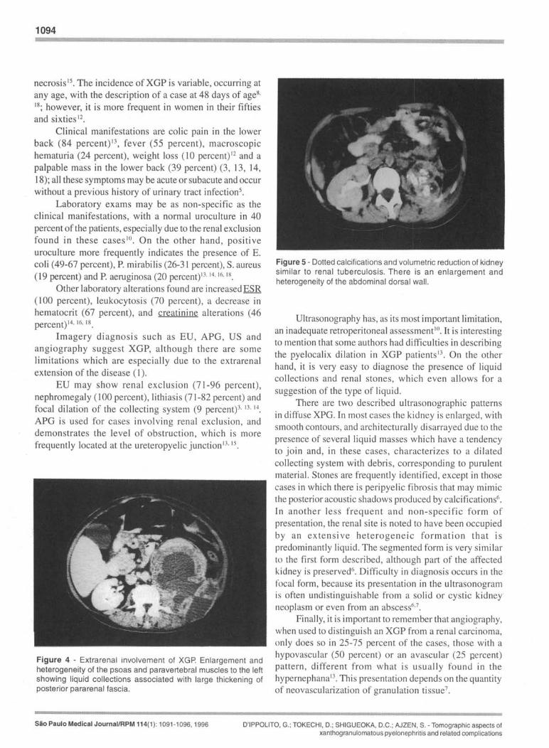
Figure 4 - Extrarenal involvement of XGP. Enlargement andheterogeneity of the psoas and paravertebral muscles to the leftshowing liquid collections associated with large thickening ofposterior pararenal fascia.
Figure 5 - Dotted calcifications and volumetric reduction of kidneysimilar to renal tuberculosis. There is an enlargement andheterogeneity of the abdominal dorsal wall.
Ultrasonography has, as its most important limitation,an inadequate retroperitoneal assessmentlO. It is interestingto mention that some authors had difficulties in describingthe pyelocalix dilation in XGP patientsl3. On the otherhand, it is very easy to diagnose the presence of liquidcollections and renal stones, which even allows for asuggestion of the type of liquid.
There are two described ultrasonographic patternsin diffuse XPG. In most cases the kidney is enlarged, withsmooth contours, and architecturally disarrayed due to thepresence of several liquid masses which have a tendencyto join and, in these cases, characterizes to a dilatedcollecting system with debris, corresponding to purulentmateria!. Stones are frequently identified, except in thosecases in which there is peripyelic fibrosis that may mimicthe posterior acoustic shadows produced by calcifications6,In another less frequent and non-specific form ofpresentation, the renal site is noted to have been occupiedby an ex tensi ve heterogenei c form ati on th at ispredominantly liquid. The segmented form is very similarto the first form described, although part of the affectedkidney is preserved6. Difficulty in diagnosis occurs in thefocal form, because its presentation in the ultrasonogramis often undistinguishable from a solid or cystic kidney'neoplasm or even from an abscess6.7•
Finally, it is important to remember that angiography,when used to distinguish an XGP from a renal carcinoma,only does so in 25-75 percent of the cases, those with ahypovascular (50 percent) or an avascular (25 percent)pattern, different from what is usually found in thehypernephana13, This presentation depends on the quantityof neovascularization of granulation tissue7,
Sao Paulo Medical Journal/RPM 114(1): 1091-1096, 1996 D'IPPOLITO, G.; TOKECHI, D.; SHIGUEOKA, D.C.; AJZEN, S. - Tomographic aspects ofxanthogranulomatous pyelonephritis and related complications
Magnetic resonance has not shown any significantadvances up to this moment in the diagnosis of XGP,and is not able to provide any information beyond thatgiven by CT (9). However, it is important to rememberthat the low toxicity of the paramagnetic contrast, aswell as the possibility of receiving orthogonal imagesin three planes, may make MR useful for those patientswho are allergic to iodine or need more detailed surgicalplanning.
In our study, CT showed a constant pattern similarto those described by other authorsl2. 13.14.We observed adiffuse increase in renal volume in most of the patients6.7,except for one who presented the segmented form of thedisease. In this case, besides being enlarged, the kidneykept its usual form, and peripheral enhancement was notedthat may correspond to compressed residual parenchyma orto a capsule of inflammatory tissueS (Fig. 1 ).
It is important to observe that the radial distributionof the liquid cavities which were found in the kidneyresembles the distribution of the collecting system and hasthe aspect of a "bunch of grapes" or a "bear claw" asdescribed by some authors 12.13(Figs. 2 and 3).
The density measures obtained (14-29 UH) do notdiffer significantly from those found by Goldman et al.(10 -15 UH)S, if we remember that all these rates indicatea thick liquid and that different calibrations of theequipment used may lead to small differences. On the otherhand, we emphasize that in no case did we find densitiessimilar to fat, as was suggested by Acunas et al., whoconsidered this infrequent I.
It was not surprising to find a frequent extrarenalextension of XGP that was drained in all studied casesand easily identified in several retroperitoneal sites anddorsal wall (Fig. 4). The importance in defining extrarenaldamage resides in adequate surgical planning, thusavoiding any undesirable fistulae'2.'3, which has evensuggested a classification of the XGP through CT in: StateI when the disease is restricted to renal parenchyma, inState II when there is perirenal involvement, and in StateIII when there is peri and pararenal involvement3.Therefore, it may be interesting to define a State IV whenthere is damage to the abdominal wall.
Despite the characteristic aspect of the XGP in theCT, a differential diagnosis with hypernephroma, renal
tuberculosis (Tb), and pyohydronephrosis must be made.Hypernephroma may be similar to XGP in its focal formwhen studied by the US and CT, and, even more, 50 whenin its cystic form, although this is relatively rare in adults.In these cases, a more crude type calcification with acoraliform aspect may help in the diagnosis of XGPI3.16.When this is not possible and there are no retroperitonealganglia in the tomographic exam (rare in XGP) or othermalignity signs (e.g. hepatic metastases), selectivearteriography may be useful if it demonstrates an avascularpattern therefore ruling out the blastomatose origin of theprocess. On the other hand, a hypo or hypervascular patternmay be found in XGP and hypernephroma's.
Renal Tb usually evolves with a decrease in renalvolume and with calcifications somewhat different fromXGP which are more pointed in shape. When XGP leadsto renal reduction, differential diagnosis with Tb may beextremely difficult as in one of the studied cases (Fig. 5).
Finally, pyohydronephrosis is considered by manyauthors to be an initial stage or a precursor of XGP9, andhas a very similar pattern.
Other differential diagnoses, which are more rare thatshould be remembered, are lipoma and liposarcoma, inwhich XGP coexists with an intense gradual replacementof granulation tissue for adipose tissue of an unknownorigin'.
CONCLUSION
Although some authors suggest that preoperati vediagnosis through imagery exams should not be done in
I
any case, but cannotjustify this3, we believe that, accordingto the results found in our study and literary review, thatcomputed tomography is a method which allowsidentification of very characteristic signs indicating apreoperative diagnosis ofXGP. These signs are: a) increasein renal volume; b) hydronephrosis; c) renal/ureterallithiasis; c) collections of thick fluid; d) peripheralenhancement after contrast injection; f) extrarenalin vol vement of peri renal, posterior pararenal andabdominal wall sites.
D'IPPOLITO, G.; TOKECHI, D.; SHIGUEOKA, D.C.; AJZEN, S. - Tomographic aspects ofxanthogranulomatous pyelonephritis and related complications
Sao Paulo Medical Journal/RPM 114(1): 1091-1096, 1996
1096
RESUlVlO
Os autores apresentam a sua experiencia em 7 pacientes com diagn6stico ancHomopatol6gico de PielonefriteXantogranulomatosa (PXG) submetidos a tomografia computadorizada (TC) pre-operat6ria. Os parametros estudados e seusresultados foram: a) presenc;:ade calculos em 86% dos casos, b) volume renal frequentemente aumentado, c) hidronefrose, d)medidas de dens idade variando entre 14 e 29 HU, e) presenc;:ade realce em todos os casos, f) comprometimento extra-renal,tambem presente em todos os casos. A TC demonstrou ser um metodo bastante fidedigno na caracterizac;:ao da PXG e suaextensao extra-renal.
3. Chuang CK, Lai MK, Chang P, et al. XantogranulomatousPyelonephritis: Experience in 36 cases. 1 Urol1992; 147:333-6.
4. Emamian SA, Nielsen MB, Pedersen IF. Kidneydimensions at sonography: Correlation with age, sex andhabitus in 665 adults volunteers. AlR 1993; 160:83-6.
6. Hartman DS, Goldman SM, Davis Cl, Isbister SS, SandersRC. Xantogranulomatous Pyelonephritis: Sonographicpathologic correlation of 16 cases. 1 Ultrasound Med1984;3:481-8.
7. Hayes WS, Hartman DS, Sesterhenn IA. In: Archives ofthe AFIP Xantogranulomatous Pyelonephritis.Radiographies 1991; II :485-498.
8. Hughes PM, Gupta SC, Thomas NB, et al. Case report:Xantogranulomatous Pyelonephritis in childhood. ClinicalRadiology 1990;41 :360-2.
9. Laugareil P, BIery M, Chagnon S, et at. PyelonephritisXantogranulomateuse avec proliferation graisseuse dela loge renale. J Radiol 1989;70:295-7.
10. Madero JM, Garcia JA, Sexmilo IE. PyelonefritisXantogranulomatosa: posibilidades de diagnostico porla imagem. An Esp Pediatr 1990;33( 1):50-3.
II. Malek RS, Elder JS. XantogranulomatousPyelonephritis: A critical analysis of 26 cases and ofthe literature. J Urol 1978; 119:589-593.
12. Paez AB, Silmi AM, Diego AG, et at. PyelonefritisXantogranulomatosa: Estudio Retrospectivo. Arch Espde Urol 1990;43:843-9.
13. Parker MD, Clark RL. Evolving concepts in the diagnoisof Xantogranulomatous Pyelonephritis. Urol Radiol1989;11:7-15.
14. Petronic V. Xantogranulomatous Pyelonephritis. Br 1Urol 1989;64:336-8.
15. Rafael RB, Kosovsky PA, Markisz JA. Xantong.Pyelonephritis in an infant. Urology 1991 ;36:553-6.
16. Shah M, Haagan R. Focal XantogranulomatousPyelonephritis simulating a renal tum9r: CT characteristics.J Comput Assist Tomogr 1989; 13:712-3.
17. Schlagenhaufer F. Uber eigentumliche Staphylomykosender Nieven und des pararenalen Bindegewebes.Frankfurt Z Pathol 1916; 19: 139-148.
18. Zafaranhas S, Gerard PS, Bryk D. Xant Pyelon inChildren: Analysis by diagnostic modalities. Urol Radiol1990; 12: 18-21.
Sao Paulo Medical Journal/RPM 114(1): 1091-1096, 1996 D'IPPOLITO, G.; TOKECHI, D.; SHIGUEOKA, D.C.; AJZEN, S. - Tomographic aspects ofxanthogranulomatous pyelonephritis and related complications