“…it is now my constant practice, where defective vision is complained of, to ascertain almost at the first instant the state of tension in the eye.… It is easy enough to estimate the tension of the eye, though there is a right and a wrong way of doing even so simple a thing…. With medical men, the touch is already an educated sense, and a very little practice should suffice to apply it successfully to the eye.” (Bowman, 1826)
Transcript
“…it is now my constant practice, where defective vision is complained of, to ascertain almost at the first instant the state of tension in the eye.… It is easy enough to estimate the tension of the eye, though there is a right and a wrong way of doing even so simple a thing…. With medical men, the touch is already an educated sense, and a very little practice should suffice to apply it successfully to the eye.” (Bowman, 1826)
IOP has been associated with glaucoma for a long time and
clinicians managing glaucoma patients have a love hate relationshipwith IOP
As clinicians we look for data that is helpful in managing a disease, consistent, reproducible and accurate.
High IOP doesn’t always mean glaucoma, Low IOP doesn’t always rule out the glaucoma.
The factors that contribute to short term fluctuations in IOP are diurnal variations, body posture, exercise, eye movements, activities causing valsalva maneuver and food and drug effects.
Despite these arguments, IOP remains the single most important alterable risk factor in the management of glaucoma
TONOMETRYSREEKANTH RAMACHANDRAN
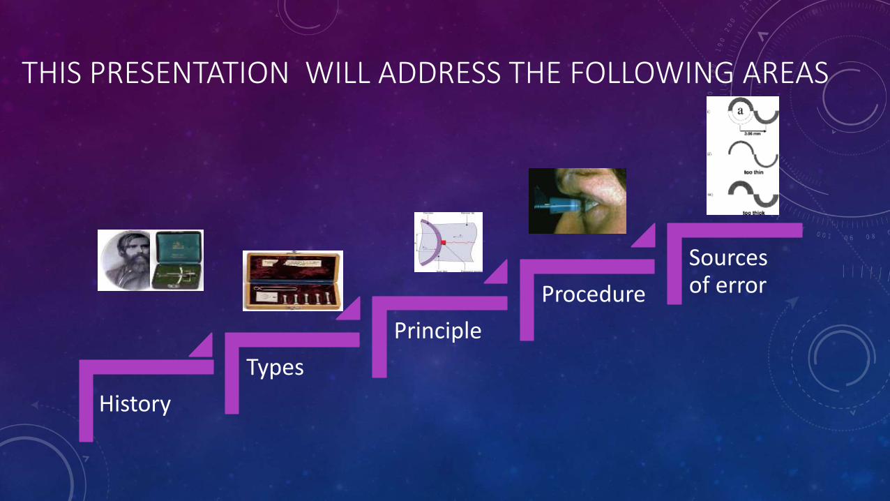
THIS PRESENTATION WILL ADDRESS THE FOLLOWING AREAS
History
Types
Principle
Procedure
Sources of error
HISTORY
1826: William Bowman used digital tonometry as a routine examination test.
1863: Albrecht von Grafe designed the first instrument to attempt to measure intraocular pressure.
Further instruments followed, notably by Donders in 1865 and Preistly-Smith in 1880.These instruments were all of the indentation type and rested on the sclera (no anaesthetic
was used until 1884).
1885: Maklakov designed an applanation tonometer. This was refined in 1892. Used for a number of years in Russia and Eastern Europe. This was used till 1959.
1905: Hjalmar Schiotz produced his indentation tonometer. This made tonometry a simple and routine clinical test.
Goldmann 1954 prototype Appl. T (constant area)
IDEAL TONOMETER
Should give accurate and reasonable IOP measurement
Convenient to use
Simple to calibrate
Stable from day to day
Easier to standardize
Free of maintenance problems
CLASSIFICATION
Tonometry
Indirect
Indentation Applanation
Direct
Manometer
Applanation
Fixed area
Goldmann tonometerMACKAY-MARG
TONOMETER PROTOTYPE
Fixed force
MaklakoutonometerGlucotest,
Applanometer
• Maklakov type
• Planometer
• Tonomat
• Halberg
• Barraquer
APPLANATION TONOMETER
GOLDMANN APPLANATION TONOMETER PROTOTYPE
PERKINS APPLANATION TONOMETER
MACKAY-MARG TONOMETER PROTOTYPE
TONOPEN
PNUEMATIC TONOMETER
1. Maklakov –tonometer
VARIABLE FORCE VARIABLE AREA
Non contact tonometer (NCT)
X –pert TGrolman airblastTKeeler pulsair T (hand held)
• Miscellaneous T
• Continuous IOP monitoring devices
• Self tonometer
• Impact tonometer
• Vibrational tonometer
• Newer tonometers
• Trans –palpebral T
• Disposable tonometer
• Tonosafe – acrylic biprism
• Tonoshield- silicone shield
• Dynamic contour tonometer
INDENTATION
Schiotz ,Mercurial ,Electronic
,Scleral tonometry
OCULAR RIGIDITYMeasure of distensibility or resistance to deformation of
ocular coats.
Important in indentation tonometer
Increase in ocular rigidity–
increase IOP
Long standing
glaucomaARMD
Hyperopic eyes
Decrease in ocular rigidity-decrease in IOP
Acutely elevated IOP
Osteogenesis imperfecta
Miotic therapy
Vasodilator therapy
Vitrectomy Myopic eyes
CORNEAL RIGIDITY
Ability of the corneal tissue to resist deformation
Important in applanation tonometers
Provided by collagen lamellae – 90% of corneal thickness
Increased corneal thickness–increased rigidity– increase in IOP
Manometry
Intraocular pressure is higher than atmospheric pressureif a small hollow needle is inserted into the anterior chamber…aqueous humor flows out through the needle. ……If the needle is attached to a reservoir of fluid that is raised just high enough to prevent any loss of aqueous, the height of the column of fluid, usually calibrated in cm of water or mm of mercury, reflects the intraocular pressure
DIGITAL TONOMETRY
Palpation Method/
digital tonometry
.Hard to touch –high IOP
indents easily –low IOP
Firm to touch –
normal IOP
Intraocular pressure (IOP) is estimated by response of eye to pressure applied by finger pulp
INDENTATION TONOMETRY
It is based on fundamental fact that plunger will indent a soft eye more than
hard eye
The indentation tonometer in
current use is that of Schiotz
It was devised in 1905 and
continued to refine it through
1927
BASIC CONCEPT AND THEORY OF INDENTATION
As soon as tonometer is
placed on cornea different forces come into play
W - weight of tonometer
A -Area Vc –volume
displaced after indentation
T- tensile force, set up in outer coats of eye at everywhere
tangentially to corneal surface
The weight of tonometer on the eye increases the actual IOP (Po) to a higher level (Pt). The change in
pressure from Po to Pt is an expression of the resistance of the eye (scleral rigidity) to the displacement of fluid.
P(t) = P(o) + E
IOP with Tonometer in position
Pt = Actual IOP Po + Scleral Rigidity E
Determination of Po from a scale reading Pt requires conversion which is done according to Friedenwald conversion
tables.
• Friedenwald generated formula for linear relationship between the log function of IOP and the ocular distension.
• Pt = log Po + C ΔV
• This formula has ‘C’ a numerical constant, the coefficient of ocular rigidity which is an expression of distensibility of eye. Its average value is 0.025
• ΔV is the change in volume
The original conversion tables referred to as 1948 tables, calculated using average K 0.0245 (coefficient of ocular rigidity)
The Friedenwald later revised average K to 0.0215 known as 1955 tables
Subsequent studies indicate 1948 tables agree more closely with measurement by goldmann AT
Plunger Load
Scale Reading 5.5 g 7.5 g 10 g 15 g
3.0 24.4 35.8 50.6 81.8
3.5 22.4 33.0 46.9 76.2
4.0 20.6 30.4 43.4 71.0
4.5 18.9 28.0 40.2 66.2
5.0 17.3 25.8 37.2 61.8
5.5 15.9 23.8 34.4 57.6
6.0 14.6 21.9 31.8 53.6
6.5 13.4 20.1 29.4 49.9
7.0 12.2 18.5 27.2 46.5
7.5 11.2 17.0 25.1 43.2
8.0 10.2 15.6 23.1 40.2
8.5 9.4 14.3 21.3 38.1
9.0 8.5 13.1 19.6 34.6
9.5 7.8 12.0 18.0 32.0
10.0 7.1 10.9 16.5 29.6
SCHIOTZ TONOMETRY
• Schiotz (1905, Modified 1924/1926)
PARTS OF SCHIOTZ TONOMETER
scale
needle
Weight 5.5g
plunger
holder
Foot plate
lever
3mm diameter
ROC 15mm
Tonometer weight = 11g
Additional weights
7.5,10,15g
SCHIOTZ TONOMETRY - CHARACTERISTICS
• The extent to which cornea is indented by plunger is measured as the distance from the foot plate curve to the plunger base and a lever system moves a needle on calibrated scale.
• The indicated scale reading and the plunger weight are converted to an IOP measurement.
• More the plunger indents the cornea, higher the scale reading and lower the IOP
• Each scale unit represents 0.05 mm protrusion of the plunger.
ERRORS OF INDENTATION TONOMETRY
1)Errors inherent in the instrument
• These may be due to difference in weight, size ,shape and curvature of footplate
2)Errors due to contraction of extra ocular muscles
- tend to increase IOP
3) errors due to accommdation
• patient look at the tonometer and thus accommodation comes into play
• Contraction of ciliary muscle increases the facility of aqueous outflow by pulling on trabecule
• Thus causes some lowering of IOP
4)Errors due to ocular rigidity
5) Errors due to variation in corneal curvature
-Steeper or thicker cornea will cause greater displacement of fluid
-Causes false low IOP readings
• Errors may arise in cases of –
-Microphthalmos
-Buphthalmos
-High myopia
-Corneal scars
• 6)Moses effect
- At low scale reading the cornea may mould into space between Plunger and hole
- Pushing the plunger up and leading to falsely high pressure reading
CALIBRATION
• The instrument should be calibrated before each use by placing it on a polished metal sphere and checking to be sure that the scale reading is zero.
• If the reading is not zero, the instrument must be repaired.
STERILIZATION
• The tonometer is disassembled between each use and the barrel is cleaned with 2 pipe cleaners, the first soaked in isopropyl alcohol 70% or methylated spirit and the second dry.
• The foot plate is cleaned with alcohol swab.
• All surfaces must be dried before reassembling.
• The instrument can be sterilized with ultraviolet radiation, steam, ethylene oxide.
• As with other tonometer tips, the Schiotz can be damaged by some disinfecting solutions such as hydrogen peroxide and bleach.
DIFFERENTIAL TONOMETRY
• It is done to get rid from ocular rigidity.
• A reading is taken with one weight on the Plunger and then a second reading' in taken with a different weight.
• Making a diagnosis of glaucoma in a pt. with myopia presents unusual difficulties. The low ocular rigidity in these eyes result in Schiotz readings within normal limits.
5.5g
10g Ocular rigidity
IOP
18 mmHg
15 mm Hg
lower
>18
18 mmHg
21 mm Hg
higher
<18
18 mmHg
18 mmHg
equal
18
APPLANATION TONOMETER
APPLANATION TONOMETER
Biprism(measuring prism)
Feeder arm
Housing
Adjusting knob
Connects to the slit
lamp
Control weight insert
PRINCIPLE
• Imbert-Fick principle
• “the pressure in a sphere filled with fluid and surrounded by an infinitely thin and flexible membrane is measured by the counter-pressure which just flattens the membrane to a plane.”
• P can be determined if
Force F is fixed or
Area A is fixed
• The ideal sphere is dry, thin-walled and flexible.
• The cornea is not ideal sphere
• Cornea being aspherical, wet, and slightly inflexible fails to follow the law.
• Moisture creates surface tension (S) or capillary attraction of tear film for tonometry head.
• Lack of flexibility requires force to bend the cornea (B) which is independent of internal pressure.
• The central thickness of cornea is about 0.55 mm and the outer area of corneal flattening differs from the inner area of flattening (A1). It is this inner area which is of importance.
IMBERT FICKS LAW & MODIFIED IMBERT FICKS LAW
W=PA W+S=PA1+B
MODIFIED IMBERT-FICK LAW IS
• W + S = PA1 + B
• When A1 = 7.35 mm2, S balances B and W =P.
• This internal area of applanation is achieved when the diameter of the external area of corneal applanation is around 3.06 mm.
• Grams of force applied to flatten 3.06 diameter of the cornea multiplied by 10 is directly converted to mmHg.
APPLANATION TONOMETERS
1) Goldman tonometer
2)Perkins AT
3)Pneumatic tonometer
4)Pulse air tonometer
5)Tono pen
GOLDMANN TONOMETER
• Most popular and accurate tonometer
• It consists of double prism mounted on slit lamp
• The prism applanates the cornea in an area of 3.06 mm diameter
The two beam-splitting prism
within the applanating unit
optically convert the circular
area of corneal contact in to
semicircles
The instrument is mounted on a standard slit lamp in
such a way that the examiners view is directed through the
centre of a plastic Biprism.
Biprism is attached by a rod to a housing which
contains a coil spring and series of levers that are used to
adjust the force of the biprism against the cornea.
Two beam splitting prisms within applanating unit
optically convert circular area of corneal contact in 2
semicircles.
PROCEDURE
The angle between the illumination and the microscope should be approximately 60°.
The room illumination is reduced.
A fixation light may be placed in front of the fellow eye.
The tension knob is set at 1 g. If the knob is set at 0, the prism head may vibrate when it touches the eye and damage the corneal epithelium.
The 1 g position is used before each measurement.
PROCEDURE CONT..
• The palpebral fissure is a little wider if the patient looks up. However, the gaze should be no more than 15° above the horizontal to prevent an elevation of IOP.
• After instilling topical anaestheia, Edge of corneal contact is made apparent by instilling fluorescein while viewing in cobalt blue light.
• The biprism should not touch the lids or lashes because this stimulates blinking and squeezing.
• The patient should blink the eyes once or twice to spread the fluorescein-stained tear film over the cornea, and then should keep the eyes open wide.
Do not to place any pressure on the globe because this raises
IOP.
PROCEDURE CONT..• In some patients, it is necessary for the examiner to hold the eyelids open with
the thumb and forefinger
of one hand against the
orbital rim.
• By manually rotating a dial calibrated in grams, the force is adjusted by changing the length of a spring within the device.
• The prisms are calibrated in such a fashion that inner margin of semicircles touch when 3.06 mm of the cornea is applanated.
• The Intra ocular pressure is then read directly from a scale on the tonometry housing.
CONT….
The fluorescent semicircles are viewed
through the biprism and the force against
the cornea is adjusted until the inner
edges overlap.
The fluorescein rings should be
approximately 0.25–0.3 mm in
thickness – or about one-tenth the
diameter of the flattened area.
POTENTIAL ERRORS
• Patient related
• Thin cornea
• Thick cornea
• Astigmatism
• Irregular cornea
Technical
Tonometer out of calibrationRepeated tonometryPressing on the eyelids or globeSqueezing of the eyelidsObserver bias (expectations and even numbers)
EFFECT OF CENTRAL CORNEAL THICKNESS (CCT): • A thinner cornea may require less force to applanate it, leading to
underestimation of true IOP while a thicker cornea would need more force to applanate it, giving an artificially higher IOP.
• The Goldmann applanation tonometer was designed to give accurate readings when the CCT was 550 μm.
• The deviation of CCT from 550 μm yields a change in applanationreadings of 0.7 mm Hg per 10 μm.
• IOP measurements are
also modified after PRK and
LASIK.
• Thinning of the central
cornea is gives lower readings
on applanation.
APPLANATION - POSSIBLE ERRORS
• Falsely low IOP
• too little flouroscein
• thin cornea
• corneal edema
• with the rule astigmatism
• 1mm Hg per 4 D
• prolonged contact
• Repeated tonometry
• Falsely high IOP
• too much flouroscein
• thick cornea
• steep cornea
• against the rule astigmatism
1mm Hg per 3D
• Widening the lid fissure excessively
• Elevating the eyes more than 15°
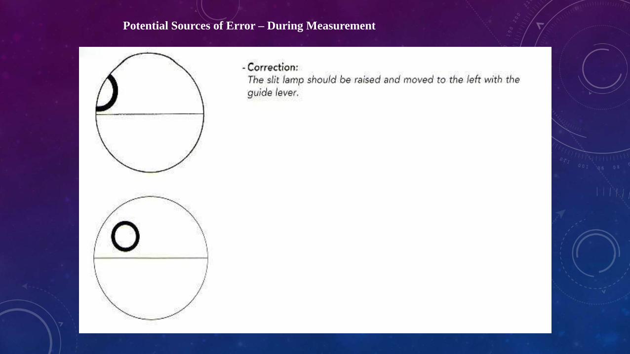
Potential Sources of Error – During Measurement
If the fluorescein rings are too wide, the patient’s eyelids should be blotted
carefully with a tissue, and the front surface of the prism should be dried with
lint-free material.
An excessively wide fluorescein ring can cause IOP to be overestimated
Potential Sources of Error – During Measurement
If the rings are too narrow, the patient should blink two or three times to
replenish the fluorescein; additional fluorescein may be added if necessary.
If the fluorescein rings are too narrow,IOP is underestimated.
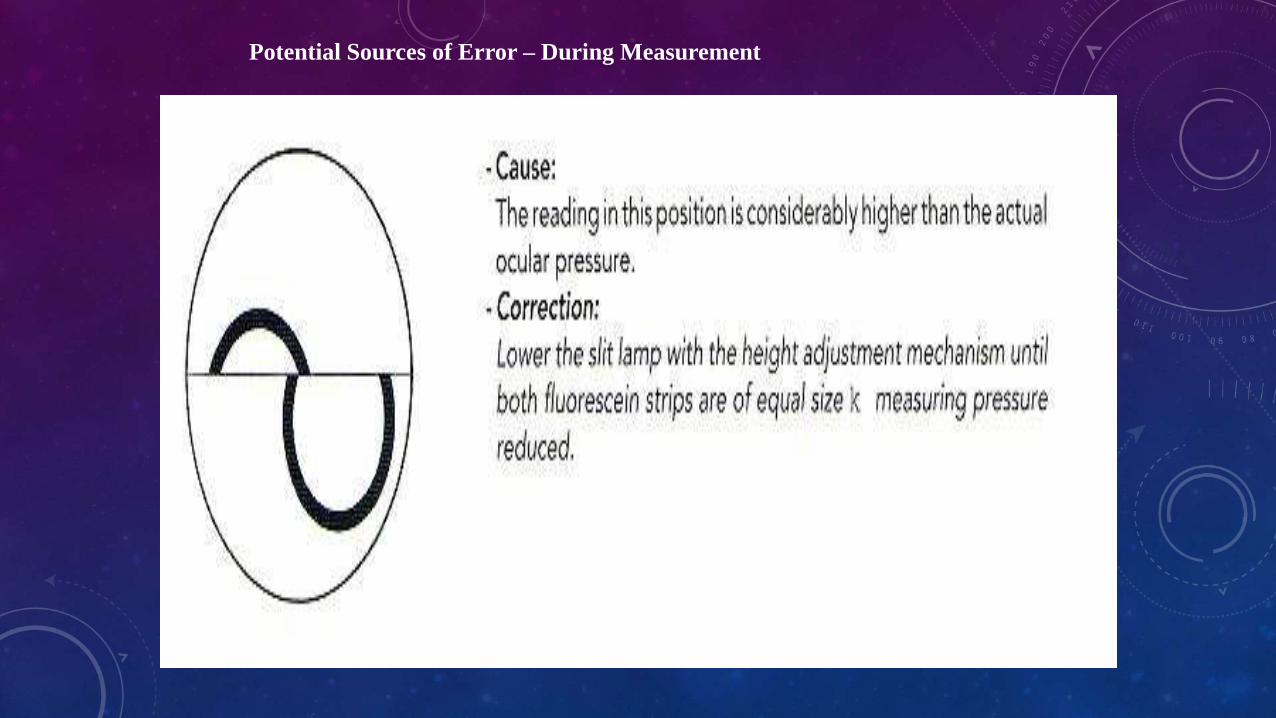
Potential Sources of Error – During Measurement
Potential Sources of Error – During Measurement
Potential Sources of Error – During Measurement
Potential Sources of Error – During Measurement
Potential Sources of Error – During Measurement
Potential Sources of Error – During Measurement
Potential Sources of Error – During Measurement
CALIBRATION
• GAT should be calibrated periodically, at least monthly. If the GAT is not within 0.1 g (1 mmHg) of the correct calibration, the instrument should be repaired; however, calibration errors of up to 2.5 mmHg may still be tolerated clinically.
• Following checks are necessary:
• • Check position 0: Turn the zero calibration on the measuring drum downwards by the width of one calibration marking, against the index marker.
• When the feeler arm is in the free movement zone, it should then move itself against the stop piece in the direction of the examiner.
• • Check position 0.05: Turn the zero calibration on the measuring drum upwards by the width of one calibration marking, against the index marker.
• When the feeler arm is in the free movement zone, it should then move itself against the stop piece in the direction of the patient.
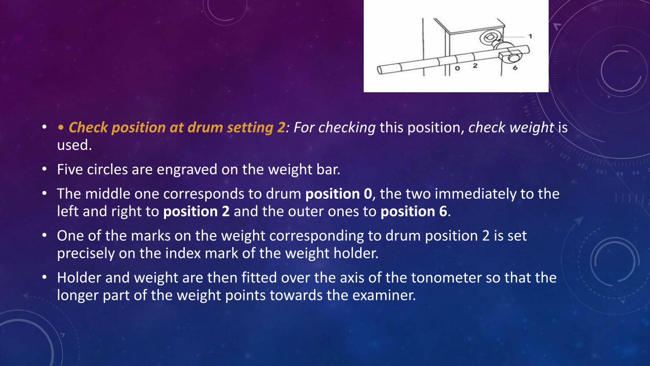
• • Check position at drum setting 2: For checking this position, check weight is used.
• Five circles are engraved on the weight bar.
• The middle one corresponds to drum position 0, the two immediately to the left and right to position 2 and the outer ones to position 6.
• One of the marks on the weight corresponding to drum position 2 is set precisely on the index mark of the weight holder.
• Holder and weight are then fitted over the axis of the tonometer so that the longer part of the weight points towards the examiner.
• Check position 1.95: The feeler arm should move towards the examiner.
• Check position 2.05.The feeler arm should move in the direction of the patient.
• • Check at measuring drum setting 6: Turn the weight bar to scale calibration 6, the longer part shows in the direction of the examiner.
• • Check position 5.9/6.1 as performed for drum setting 2.
STERILIZATION
• Applanation tip should be soaked for 5-15 min in diluted sodium hypochlorite, 3% H2O2 or 70% isopropyl alcohol or by wiping with alcohol, H2O2, povidone iodine or 1: 1000 merthiolate.
• Other methods of sterilization include: 10 min of rinsing in running tap water, wash with soap and water, cover the tip with a disposable film, and exposure to UV light.
• Disposable tonometer tips may also be used
When using disposable tips, they have a smooth
applanating surface.
The acrylic disposable tips seem to be somewhat
more accurate than the silicone ones.
While disposable shields or tips may be safer
than disinfection solutions, they are not 100%
protective against prion disease.
• It is possible to transfer bacteria, viruses, and other infectious agents with the tonometer head, including such potentially serious infections as epidemic keratoconjunctivitis, hepatitis B, Jacob-Kreutzfeld and, theoretically, acquired immunodeficiency syndrome.
• Care must be taken to be sure any sterilizing solution has been completely rinsed off the tonometer tip, as some of these solutions may be toxic to the corneal epithelium, especially after LASIK or other corneal procedures.
• If the tonometer tip is not mechanically wiped after each use, epithelial cells may stick to the tip with the small but serious risk of transmitting Jacob-Kreutzfeld virus.
SAFETY REGULATIONS
• No examination should be undertaken in case of eye infections (or) injured corneas.
• Only clean and disinfected measuring prism should be used.
• No damaged prisms should be used.
• If the measuring prism come in to contact with the cornea without the drum having previously been correctly set, vibration can occur in the feeler arm, which will produce unpleasant feeling for the patient.
• The tonometer tips should be examined periodically under magnification as the antiseptic solutions and mechanical wiping may cause irregularities in the surface of the tip that can, in turn, injure the cornea.
PERKINS TONOMETER
• It uses same prisms as Goldmann
• It is counterbalanced so that tonometry is performed in any position
• The prism is illuminated by battery powered bulbs.
• Being portable it is practical when measuring IOP in infants / children, bed ridden patients and for use in operating rooms.
DRAEGER TONOMETER
• Draeger tonometer is similar to Perkins
• It has a different set of prisms
• It operates with a motor.
MACKAY MARG TONOMETER
• Plunger plate has diameter of 1.5mm
• Surrounding Sleeve has 3 mm
• Force required to keep the plate flush with the sleeve is electronically monitored – recorded on a paper strip
At 1.5 mm of corneal area applanation, tracing reaches a peak and the
force applied = IOP + force required to deform the cornea.
At 3 mm flattening, force required to deform cornea is transferred from
plunger to surrounding sleeve, creating a dip in tracing corresponding to IOP.
Flattening of >3 mm of area gives artificial elevation of IOP.
Source of error->3 mm flattening – high IOP
Multiple readings to compensate ocular pulsation
Specific utility- irregular and edematouscornea
TONO PEN
• This is handheld Mackay Marg type tonometer
• It is a computerised pocket tonometer
• It converts IOP into electric waves
The wave form is internally analyzed by a microprocessor.
Three to six estimations of the pressure are then averaged.
The instrument is 18 cm in length and weighs 60 g.
For pressures from 6 to 24 mmHg, it measured an average of 1.7 mm higher than the Goldmann tonometer.
Above 24 mmHg, the readings were similar.
PNEUMATIC TONOMETER
PNEUMATIC TONOMETER
• Cornea is applanated by touching apex by silastic diaphragm covering sensing nozzle
• It is connected to central chamber containing pressurized air
• There is pneumatic to electronic transducer
• It converts the air pressure to recording on paper strip and IOP is read
• instrument is useful with edematous and irregular corneas
PRINCIPLE
• The principle is similar to the MacKay-Marg tonometer.
• Corneal contact of the pencil-like tip records both the IOP and the force required to bend the cornea.
• advancement of the tip transfers the latter force to the surrounding “collar.”
• The “plunger” is replaced by a column of air and the contact surface is a Silastic membrane
MAKLAKOV TONOMETER (FORCE CONSTANT)
•Pt supine
Dumb-bell-shaped metal cylinders with
flat end plates of polished glass
Diameter of 10 mm
The surface of the weight is painted
with a dye, such as mild silver protein
(Argyrol) mixed with glycerin.
1 sec contact
imprint on end plate
• IOP = W / π (d/2) 2
• weight (W) diameter of the area of applanation (d)
• Intraocular pressure is measured in grams per square centimeter and is converted to millimeters of mercury by dividing by 1.36.
• widely in Russia and China
• This instrument displaces a greater volume of aqueous humor and thus IOP readings are more influenced by ocular rigidity.
• It does not correct for corneal bending, capillary attraction, or tear encroachment on the layer of dye.
• Many instruments similar to the Maklakow device have been described,like the Applanometer, Tonomat, Halbergtonometer, and GlaucoTest.
REBOUND TONOMETER
It is a new and updated version of an indentation tonometer Portablecan be used without anesthetizing the eye.A very light, disposable, sterile probe is propelled forward into the cornea .The time taken for the probe to return to its resting position and the characteristics of the rebound motion are indicative of the IOP.The time taken for the probe to return to its resting
position is longer in eyes with lower IOP and faster in eyes with higher IOP.
CONTD…
• It is comparable to the GAT.
• It correlates with central corneal thickness like the Goldmann, .
• used in screening situations, when patients are unable to be seated or measured at the slit lamp, or when topical anesthetics are not feasible or usable.
• Not useful in scarred corneas (as does the Goldmann).
TRANS PALPEBRAL TONOMETRY
used in situations where other, more accurate, devices are not practical, such as in young children,
demented patients and severely developmentally-challenged patients.
In addition to all the problems facing indentation tonometry, such as scleral rigidity, transpalpebral
tonometry adds variables such as the thickness of the eyelids, orbicularis muscle tone and potential
Intra palpebral scarring.
• Portable. patients can measure their own IOP at home, pressure on the eyelid in most eyes produces retinal phosphenes.
• The pressure on the eyelid required to induce these phosphenes is proportional to the intraocular pressure.
• It is not accurate always. inter observer and intra observer variability was large.subsequent studies failed to confirm the accuracy of this device.
NONCONTACT TONOMETER
• It is an applanation tonometer and works on the principle of a time interval.
• Measuring the time from initial generation of the puff of air to cornea gets flattened (in milliseconds) to the point where the timing device stops.
• It takes less time for the puff of air to flatten a soft eye than it does a hard eye.
• Three subsystems:
• Alignment system
• Optoelectric applanation monitoring system
• Transmitter
• Receiver and detector
• Pneumatic system
• Time for max light detection= time to applanate the cornea = corelatedwith IOP
• Limitations
• Ocular pulse
• Glaucomatous eyes
• Average of 3 readings
DYNAMIC CONTOUR TONOMETER
• The PASCAL (DCT) is a slit lamp–mounted device
• It measures IOP independent of corneal rigidity or thickness.
• It was commercially launched in
August 2004.
PRINCIPLE
• DCT uses the principle of contour matching instead of applanation.
• The tip contains a hollow miniature pressure sensor in its centre.
• when the contours of the cornea and tonometer match, then the pressure measured at the surface of the eye equals the pressure inside the eye (B).
PRINCIPLE
• The probe is placed on the pre-corneal tear film on the central cornea
• The integrated piezoelectrical ( electricity resulting from pressure) pressure sensor records data, measuring IOP 100 times per second.
• The tonometer tip rests on the cornea with a constant appositional force of one gram.
• When the sensor is subjected to a change in pressure, the electrical resistance is altered
• The PASCAL's computer calculates a change in pressure according to the change in resistance.
• A complete measurement cycle requires about 8 seconds of contact time.
• It is less influenced by corneal thickness than other methods
• As the tip shape is designed for the shape of a normal cornea, it is more influenced by corneal curvature.
OCULAR RESPONSE ANALYZER
• It is similar to Reichert’s current generation NCT and provides a Goldmann-equivalent IOP reading.
• It analyzes the signal obtained from the corneal response to measure the biomechanical properties of the corneal tissue.
PRINCIPLE
• It utilizes a dynamic bi-directional applanation process to measure pressure of the eye.
• During measurement, a precisely metered collimated-air-pulse applies force to the cornea.
PRINCIPLE
• Under the force of the air pulse, the cornea moves inwards, past applanation, and into a slight concavity
• As the air pulse pressure decreases, the cornea return to its normal configuration.
• In the process, it once again passes through an applanation state.
PRINCIPLE
• An advanced electro- optical system monitors the changes in curvature of the cornea
• Two independent pressure values are derived the inward and outward applanation events.
• Due viscous damping in the cornea causes delays, resulting in the different pressure values.
• The average of these two pressure values provides Goldman-Correlated IOP value (IOPG).
• The difference between these two pressure values is Corneal Hystersis.
HYSTERESIS
• The phenomenon was identified, and the term coined, by Sir James Alfred Ewing in 1890.
• Hysteresis is a property of physical systems that do not instantly follow the forces applied to them, but react slowly, or do not return completely to their original state.
CORNEAL HYSTERESIS
• It is the "energy absorption capability" of the cornea
• This because of the speed at which the cornea is deformed during the dynamic bi-directional applanation process in ORA