11/5/2015 1 MICRO 6 TOOLS OF THE LABORATORY most habitats harbor microbes in complex associations often necessary to separate microbes in order to identify and study them – need to be grown in artificial conditions need an oil immersion microscope to view procedures for investigating and characterizing microorganisms – the six I’s Inoculation Incubation Isolation Inspection Information gathering identification METHODS OF MICROBIAL INVESTIGATION
Transcript
11/5/2015
1
MICRO 6TOOLS OF THE LABORATORY
most habitats harbor microbes in complex associations often necessary to separate microbes in order to
identify and study them – need to be grown in artificial conditions
need an oil immersion microscope to view
procedures for investigating and characterizing microorganisms – the six I’s Inoculation Incubation Isolation Inspection Information gathering identification
METHODS OF MICROBIAL INVESTIGATION
11/5/2015
2
sample the object of interest (almost anything)
Common sources Bodily fluids
Foods
Water
Soil
Plants
Animals
Icebergs
Volcanoes
Rocks
SPECIMEN COLLECTION
sample is placed on a medium that will support its growth (human biological samples – blood agar plate)
medium may be solid or l iquid – held in tubes, plates, flasks and even eggs
sample delivery tool is usually a loop, needle, swab or syringe
INOCULATION
11/5/2015
3
inoculated media are placed in a controlled environment (incubator) to promote growth (body temp / aerobic or anaerobic)
culture of visible colonies develop on the media
INCUBATION
Inoculation technique that separates microbes into distinct colonies that contain a single type of microbe – want a pure culture
Urine culture – pink colonies on McConkeyGram stain – GNR (E coli)
11/5/2015
4
cultures are observed for macroscopic appearance of growth characteristics
samples of the culture are stained and viewed under the microscope – cell type and shape
INSPECTION
Identify the microorganism using info gathered from inspection and investigation
accomplished through keys, charts and computer programs that analyze data
IDENTIFICATION
11/5/2015
5
optical microscopy
sample is mounted on a glass slide that sits on the stage between the condenser and the objective lens
the manner in which the specimen (mount) is prepared depends upon the condition of the specimen (living or preserved)
aim of the examiner – observe overall structure, identify microorganisms or see movement
type of microscopy available – bright field,
dark field, phase contrast or fluorescence
PREPARING SPECIMENS FOR OPTICAL MICROSCOPES
l ive samples – wet mounts – microbes can be observed as close to their natural state as possible
cells are suspended in a fluid (water, broth, saline) –temporarily maintains viabil ity and provides a medium to see locomotion a drop of liquid culture placed between a glass slide and a
coverslip (glass cover)
FRESH LIVING PREPS
Trichomonas vaginalis
11/5/2015
6
hanging drop slide – special concave (depression) slide, an adhesive (sealant) and coverslip from which a small drop of sample is suspended
short term mount – true assessment of the size, shape, arrangement, color and motility of cells
sreater cellular detail with phase-contrast or interference microscope
Trichomonas in urine samples
permanent mount for long-term study – fixed, stained specimen
smear technique (Robert Koch)
spread a thin film of specimen on a slide and air dry
heat fixation (heat gently) – simultaneously kills the specimen and secures it to the slide – preserves cellular components
stain for cell differentiation
FIXED, STAINED SMEARS
11/5/2015
7
dyes impart color to cells by affixing to them through chemical reactions
basic (cationic) dyes – positive charge
acidic (anionic) dyes – negative charge
Negative / Positive Staining most procedures involve positive staining – dye sticks to
cells and gives them color
negative stain – dye does not stick to specimen but dries around its outer boundary (silhouette) – cells do not stain b/c negative stain is repelled by negative charge of cell surface
smear is not fixed – simple stain to show cellular shape, size, arrangement and accentuate capsules
Nigrosin (blue / black)
India ink (black suspension of carbon
particles)
11/5/2015
8
Simple vs Differential Staining
positive staining methods are classified as simple, differential or structural
simple stains – require only a single dye that readily bind to bacterial cells (malachite green, crystal violet, basic fuchsin, safranin, methylene blue)
differential stains – require two different colored dyes (primary dye and counterstain) to distinguish between cell types / parts
uses dyes of contrasting color to clearly emphasize differences between cells types / parts (red and purple / red and green/ pink and blue)
show characteristics of size, shape and arrangement
gram stain, acid fast, endospore stains
DIFFERENTIAL STAINS
11/5/2015
9
GRAM STAIN developed by Has Christian Gram (1884)
two cell reactions: gram-pos (purple) and gram-neg (red)
difference in staining due to structural variations in the cell wall
crystal violet stains all cell purple
Gram’s iodine (mordant (IKI) – key differentiating step) causes the dye to form large crystals that get trapped in the cell wall (b/c cell wall in gm+ cells is thicker, the entrapment of the dye is more extensive than gm- cells)
application of alcohol dissolves lipids in the outer membrane of gm- cells (removes the dye from them)
application of a counterstain (safranin) dyes the colorless gm- cells red
11/5/2015
10
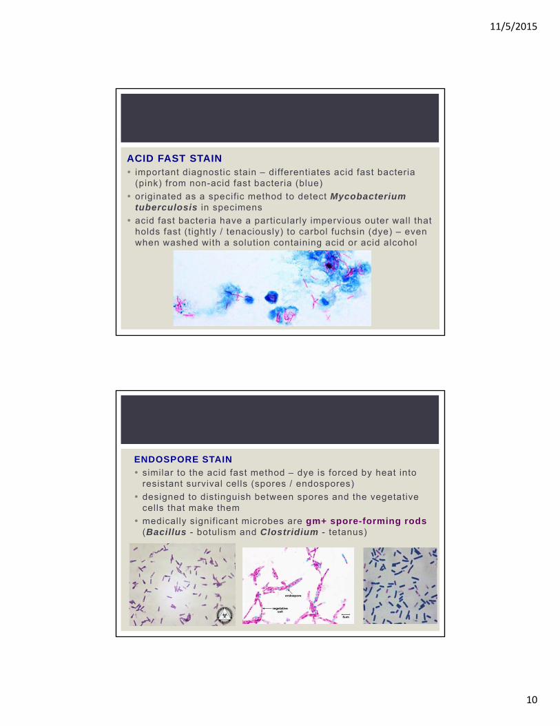
ACID FAST STAIN important diagnostic stain – differentiates acid fast bacteria
(pink) from non-acid fast bacteria (blue)
originated as a specific method to detect Mycobacterium tuberculosis in specimens
acid fast bacteria have a particularly impervious outer wall that holds fast (tightly / tenaciously) to carbol fuchsin (dye) – even when washed with a solution containing acid or acid alcohol
ENDOSPORE STAIN
similar to the acid fast method – dye is forced by heat into resistant survival cells (spores / endospores)
designed to distinguish between spores and the vegetative cells that make them
medically significant microbes are gm+ spore-forming rods (Bacillus - botulism and Clostridium - tetanus)
11/5/2015
11
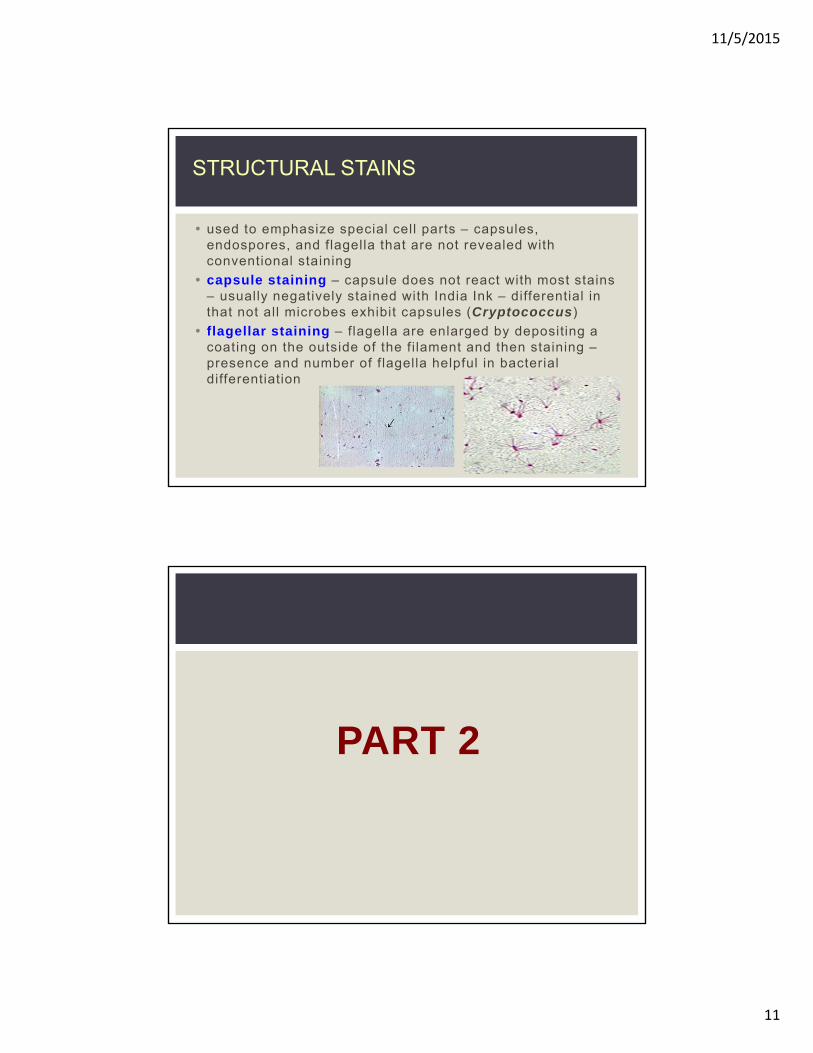
used to emphasize special cell parts – capsules, endospores, and flagella that are not revealed with conventional staining
capsule staining – capsule does not react with most stains – usually negatively stained with India Ink – differential in that not all microbes exhibit capsules (Cryptococcus)
flagellar staining – flagella are enlarged by depositing a coating on the outside of the fi lament and then staining –presence and number of flagella helpful in bacterial differentiation
STRUCTURAL STAINS
PART 2
11/5/2015
12
inoculation (to culture bacteria) - a tiny amount of sample (inoculum) into medium (media) which provides an environment for multiplication
culture – the observable growth on the media
the nature of the sample being cultured depends on the objectives of the analysis
clinical specimens for determining cause of infectious disease – obtained from body fluids (blood, CSF) discharges (urine, feces, sputum) or diseased tissue (wounds, burns, ulcers)
other samples for analysis can include nearly any natural material (soil, water, sewage, foods, air and inanimate objects)
INOCULATION, GROWTH AND IDENTIFICATION OF CULTURES
Special Requirements of Culturing
Sterile (complete absence of viable microbes), aseptic(prevention of disease) and pure culture (growth of a single culture) techniques
contamination during inoculation is a constant problem –sterile techniques (media, transfer equipment) help ensure that only microbes from the sample are cultured
concern – possible release of infectious agents from cultures into the environment – prevented by aseptic technique (keeping species in a pure culture for further study, ID or biotechnology applications
11/5/2015
13
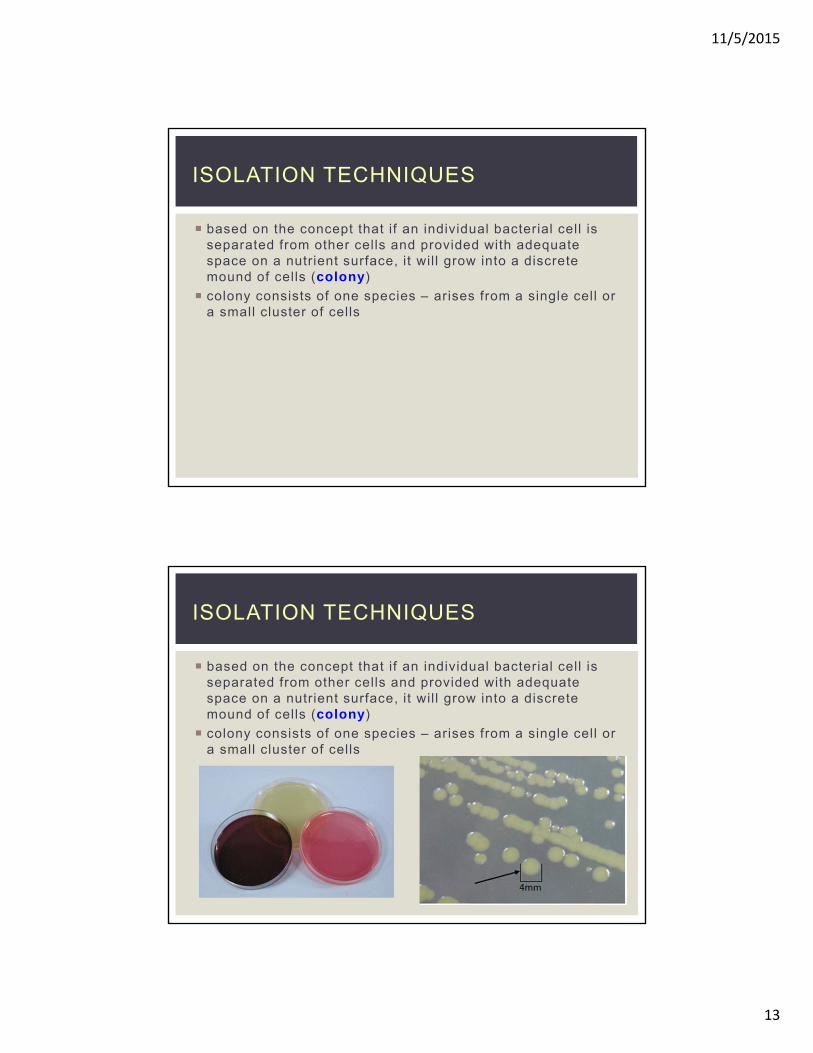
based on the concept that if an individual bacterial cell is separated from other cells and provided with adequate space on a nutrient surface, it wil l grow into a discrete mound of cells (colony)
colony consists of one species – arises from a single cell or a small cluster of cells
ISOLATION TECHNIQUES
11/5/2015
14
streak plate method mall droplet / amount of sample is spread with an inoculating loop over the surface of the medium in a pattern that gradually thins out the sample – separates cells spatially over several sections of the plate
loop dilution or pour plate technique sample is inoculated into a series of cooled but sti l l l iquid
agar tubes so as to dilute the number of cells in each successive tube in the series
inoculated tubes are plated (poured) out in to sterile petri dishes and allowed to solidify (harden)
some colonies may develop deep in the medium on not just on the surface
11/5/2015
15
spread plate technique small volume of l iquid from a diluted sample is pipetted onto
the surface of the medium and spread around evenly by a sterile spreading tool (hockey stick, glass spreader)
as with the streak plate, cells are spread over separate areas on the surface to form individual colonies
11/5/2015
16
once the medium has been inoculated, it is incubated in a temp-controlled chamber (incubator) to encourage microbial growth
usual temps used in the clinical lab fall between 20-40°C
Incubators can also control the content of atmosphere gases (O2 / CO2) required by some bacterial pathogens
Incubation period ranges from a few hours to several weeks
culture multiples to produce macroscopically visible colonies
pure culture (axenic) – culture free of other l iving things except for the one being studied – only one species of bacteria
used to identify pathogenic bacteria in a lab setting
mixed culture – contains two or more easily differentiated species of bacteria
may contain contaminants – species other than the pathogen
subculture – method of taking a
small sample of a distinct colony of
bacteria and transferring it to new
media to obtain a pure culture
11/5/2015
17
gram stain colony growth on differential media
biochemical tests – determine fundamental chemical characteristics, products produced during growth, presence of enzymes, and mechanisms for deriving energy
immunologic testing – test isolate against antibodies animal studies DNA profiles
IDENTIFICATION TECHNIQUES
traditional pathway in bacterial identification uses flowcharts or keys that apply the results of tests to a selection process (points of separation are based on a positive or negative result)
this process of “keying out” the microorganism can simplify the identification process
11/5/2015
18
PART 3
some microbes require only a few simple organic nutrients for growth – others need a complex list of specific compounds
at least 500 types of media used in culturing and identifying microorganisms
media contained in tubes, flasks, or petri dishes –inoculated with tools l ike loops, needles, pipettes and swabs
media are varied in nutrient content, consistency and can be formulated for a specific purpose – sterile technique imperative
MEDIA THE FOUNDATIONS OF CULTURING
11/5/2015
19
LIQUID MEDIA
water-based solutions that do not solidify at temps above freezing and that tend to flow freely when the container is ti lted
termed broths, milks or infusions – nutrient solutes are dissolved in water
growth occurs throughout the container – dispersed, cloudy or flakey appearance
common lab medium – nutrient broth – beef extract and peptone (amino acid) in sterile water
methylene blue milk / litmus milk (whole milk and dyes)
fluid thioglycollate (slighlty viscous broth used to determine patterns of growth in O2)
PHYSICAL STATES OF MEDIA
11/5/2015
20
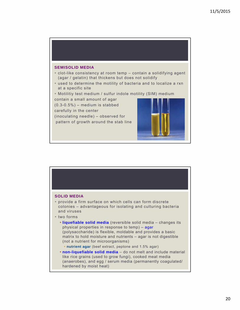
SEMISOLID MEDIA
clot-l ike consistency at room temp – contain a solidifying agent (agar / gelatin) that thickens but does not solidify
used to determine the motil ity of bacteria and to localize a rxnat a specific site
Motil it iy test medium / sulfur indole motil ity (SIM) medium
contain a small amount of agar
(0.3-0.5%) – medium is stabbed
carefully in the center
(inoculating needle) – observed for
pattern of growth around the stab line
SOLID MEDIA
provide a firm surface on which cells can form discrete colonies – advantageous for isolating and culturing bacteria and viruses
two forms
liquefiable solid media (reversible solid media – changes its physical properties in response to temp) – agar(polysaccharide) is flexible, moldable and provides a basic matrix to hold moisture and nutrients – agar is not digestible (not a nutrient for microorganisms) nutrient agar (beef extract, peptone and 1.5% agar)
non-liquefiable solid media – do not melt and include material like rice grains (used to grow fungi), cooked meat media (anaerobes), and egg / serum media (permanently coagulated/ hardened by moist heat)
11/5/2015
21
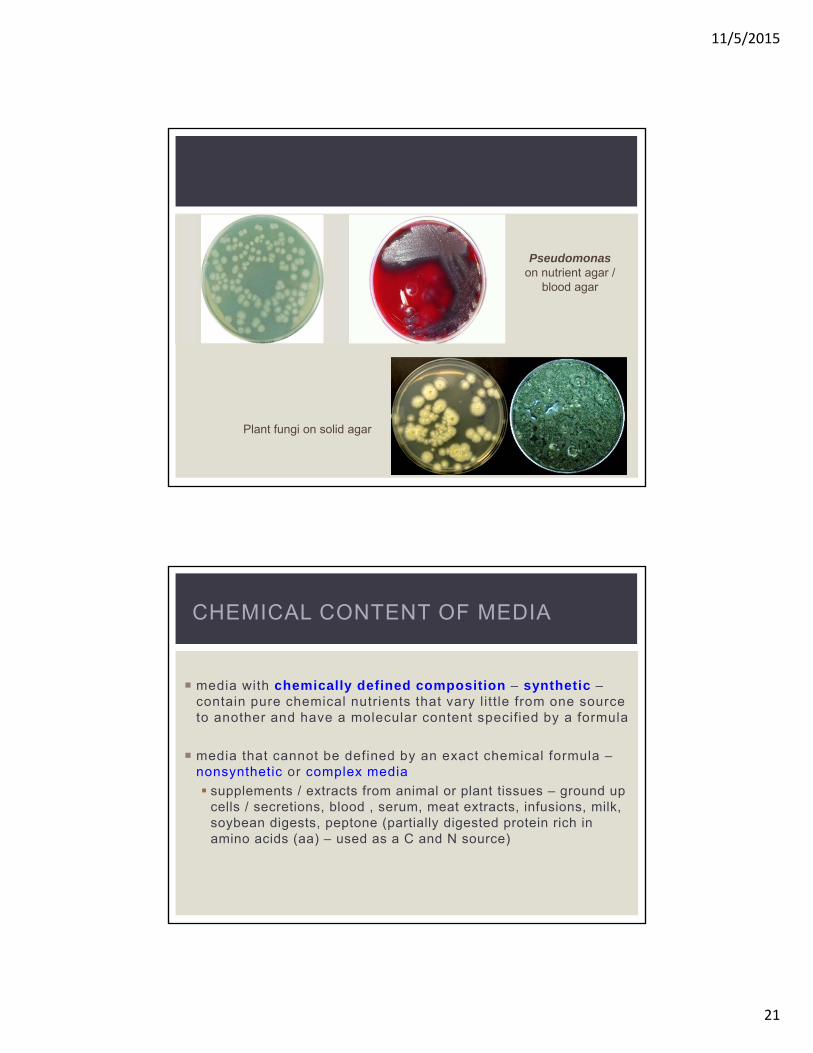
Pseudomonas on nutrient agar /
blood agar
Plant fungi on solid agar
media with chemically defined composition – synthetic –contain pure chemical nutrients that vary l itt le from one source to another and have a molecular content specified by a formula
media that cannot be defined by an exact chemical formula –nonsynthetic or complex media
supplements / extracts from animal or plant tissues – ground up cells / secretions, blood , serum, meat extracts, infusions, milk, soybean digests, peptone (partially digested protein rich in amino acids (aa) – used as a C and N source)
CHEMICAL CONTENT OF MEDIA
11/5/2015
22
General Purpose Media
designed to grow a broad spectrum of microbes that do not have special growth requirements
Nonsynthetic (complex) and contain a mix of nutrients that can support a variety of bacteria and fungi
nutrient agar / broth, brain-heart infusion, trypticase soy agar (TSA – contains partially digested mil protein (casein), soybean digest, NaCl and agar)
Enriched Media
contains complex organic substances – blood, serum, hemoglobin, or special growth factors (vitamins, amino acids that bacteria cannot synthesize) that certain species need in order to grow
fastidious bacteria – require growth factors and complex nutrients (blood agar / chocolate agar)
11/5/2015
23
SELECTIVE AND DIFFERENTIAL MEDIA
• designed for special microbial groups
• can provide a preliminary ID of a genus / species in a single step
selective medium
• contains one or more agents that inhibit the growth of certain microbes – selects a microbe and allows it to grow by itself
• important in the primary isolation of a specific type of organism from samples containing a mixture of organisms (feces, urine, saliva, skin, water, soil) – suppress the unwanted organisms and allow growth of the desired ones
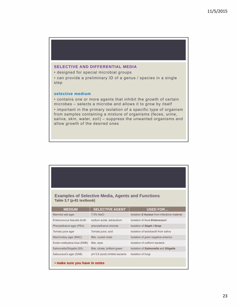
MEDIUM SELECTIVE AGENT USED FOR…
Mannitol salt agar 7.5% NaCl Isolation S Aureus from infections material
Enterococcus faecalis broth sodium azide, tetrazolium Isolation of fecal Enterococci
Phenylethanol agar (PEA) phenylethanol chloride Isolation of Staph / Strep
Tomato juice agar Tomato juice, acid Isolation of lactobacilli from saliva
MacConkey agar (MAC) Bile, crystal violet Isolation of gram negative enterics
Eoisin-methylene blue (EMB) Bile, dyes Isolation of coliform bacteria
Salmonella/Shigella (SS) Bile, citrate, brilliant green Isolation of Salmonella and Shigella
Sabouraud’s agar (SAB) pH 5.6 (acid) inhibits bacteria Isolation of fungi
Examples of Selective Media, Agents and FunctionsTable 3.7 (p-81 textbook)
• make sure you have in notes
11/5/2015
24
differential media
• grow several types of microorganisms but are designed to bring out visible differences among those organisms – colony size and color, formation of gas bubbles or precipitates
• variations come from the types of chemicals contained in the media and the ways the microbes react to them
11/5/2015
25
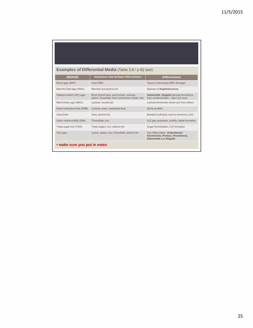
MEDIUM Substances that facilitate differentiation Differentiates
Blood agar (BAP) Intact RBC Types of hemolysis (RBC damage)
Mannitol Salt Agar (MSA) Mannitol and phenol red Species of Staphylococcus
Hektoen enteric (HE) agar Brom thymol blue, acid fuchsin, sucrose, salicin, thiosulfate, ferric ammonium citrate, bile
Salmonella, Shigella (lactose fermentersfrom nonfermenters – also H2S rxns)
MacConkey agar (MAC) Lactose, neutral red Lactose fermenters (lower pH) from others
Eosin-methylene blue (EMB) Lactose, eosin, methylene blue Same as MAC
Urea broth Urea, phenol red Bacteria hydrolyze urea to ammonia (↑pH)
Sulfur indole motility (SIM) Thiosulfate, iron H2S gas producers, motility, indole formation
Triple-sugar iron (TSIA) Triple sugars, iron, phenol red Sugar fermentation, H2S formation
XLD agar Lysine, xylose, iron, thiosulfate, phenol red Can differentiate: Enterobacter, Escherichia, Proteus, Providencia, Salmonella and Shigella
Examples of Differential Media (Table 3.8 / p-82 text)


![H - Optical equipments B - General use C · 6 FCD - Laboratory jacks. page [73] [FC] assemblies - LABORATORY ASSEMBLIES AND TOOLS laboratory equipment LABORATORY ASSEMBLIES AND TOOLS](https://static.documents.pub/doc/80x56/5b9357e909d3f206218d2a5f/h-optical-equipments-b-general-use-c-6-fcd-laboratory-jacks-page-73.jpg)


