1 TOPIC – 1 Initial Assessment & Management of Poly-Trauma Patients Objectives • To know correct sequence and triage • To know primary and secondary survey • To know history of trauma incident and thus assume the type of injury • To set Guidelines for initial resuscitation and definitive treatment Trauma Care • Management of trauma requires broad knowledge, sound judgment, technical skills and leadership capabilities • Most trauma victims are healthy, young individuals who, if salvaged, have a normal life expectancy • Critical care specialists play a vital role in stabilization and diagnostic phases of trauma care • “what happens in this period often determines the outcomes of care” Trauma Mortality: A Trimodal Distribution Immediate deaths • Occur within first hour (half of all trauma related deaths) • Major vascular, brain & cardiac injuries are usual causes Early deaths • Occur within hours after injury–Hemorrhage & breathing problems are common causes Late deaths • Occur after 3 days and peak at 3-4 weeks–Sepsis & multi organ failure are causes of death Mechanism of Traumatic Injury Blunt Trauma • Distributed dissipation of kinetic energy either by concussion or by deceleration • Leads to direct contusive injury, shearing, vascular disruption, & indirect laceration secondary to fractures Penetrating trauma • More focal dissipation of a projectile’s kinetic energy • Leads to direct impact laceration and fractures Trauma Score (TS) TS evaluates • Respiratory rate and expansion • Systolic blood pressure • Capillary refill • Glasgow coma scale score Numbers assigned to each parameter, with higher number representing normal function Underestimation of head injury severity lead to the revision of the score Revised Trauma Score (RTS) RTS has been shown to be a more reliable predictor of outcome RTS is based on three physiologic measures • Glasgow coma scale score • Respiratory rate • Systemic BP RTS remains in common use today
Transcript
1
TOPIC – 1
Initial Assessment & Management of Poly-Trauma Patients
Objectives • To know correct sequence and triage • To know primary and secondary survey • To know history of trauma incident and thus assume the type of injury
• To set Guidelines for initial resuscitation and definitive treatment
Trauma Care
• Management of trauma requires broad knowledge, sound judgment, technical skills and leadership capabilities
• Most trauma victims are healthy, young individuals who, if salvaged, have a normal life expectancy • Critical care specialists play a vital role in stabilization and diagnostic phases of trauma care • “what happens in this period often determines the outcomes of care”
Trauma Mortality: A Trimodal Distribution
Immediate deaths • Occur within first hour (half of all trauma related deaths) • Major vascular, brain & cardiac injuries are usual causes
Early deaths • Occur within hours after injury–Hemorrhage & breathing problems are common causes
Late deaths
• Occur after 3 days and peak at 3-4 weeks–Sepsis & multi organ failure are causes of death
Mechanism of Traumatic Injury
Blunt Trauma • Distributed dissipation of kinetic energy either by concussion or by deceleration
• Leads to direct contusive injury, shearing, vascular disruption, & indirect laceration secondary to fractures
Penetrating trauma • More focal dissipation of a projectile’s kinetic energy
• Leads to direct impact laceration and fractures
Trauma Score (TS)
TS evaluates • Respiratory rate and expansion • Systolic blood pressure • Capillary refill • Glasgow coma scale score
Numbers assigned to each parameter, with higher number representing normal function Underestimation of head injury severity lead to the revision of the score
Revised Trauma Score (RTS)
RTS has been shown to be a more reliable predictor of outcome RTS is based on three physiologic measures
• Glasgow coma scale score • Respiratory rate • Systemic BP
RTS remains in common use today
2
Trauma Index (TI)
Contains five variables: • Region of body injured • Type of injury • Cardiovascular status • CNS status • Respiratory status
Low scores correlated with minor injuries, whereas multiple system trauma victims scored higher with higher mortality
CRAMS
10-point scale that measures 5 parameter: • Circulation • Respiratory • Abdomen • Motor • Speech
Each parameter scored as normal (2), mild abnormal (1) and severely abnormal (0)
Stimuli that initiate the physiologic response to trauma
• Perception of pain • Shock • Blood loss • Hypoxia • Acidosis
Trauma Management: Golden Hour Time to reach operating room (or other definitive treatment) Patients in their Golden Hour must
• Be recognized quickly
3
• Have only immediate life threats managed • Be transported to an appropriate facility
Survival depends on assessment skills Good assessment results from
• An organized approach • Clearly defined priorities
• Understanding available resources
Principles of Trauma Management
Organized team approach • Complexity of multiple trauma patients • Trauma victims are best managed by a team approach
Assumption of most serious injury
• assume that the worst possible injury has occurred and act accordingly until the diagnosis is confirmed
Treatment before diagnosis • urgency of situation often demands treatment based on an initial brief assessment
Thorough examination • initial survey of vital organ systems, followed by resuscitative interventions • Most missed injuries occur in unconscious patients
Frequent assessment
• helps detecting early changes in physical findings and thus lead to prompt corrective actions
Low-Priority Areas • Neurologic • Abdominal • Cardiac
• Musculoskeletal Soft tissue injury
Phases
“A systematic approach should always be practiced” I. Be prepared II. Triage III. Primary Survey (ABC’s) IV. Resuscitate V. Secondary survey VI. Monitoring, definitive treatment & transfer
“TIME IS ESSENCE”
Be Prepared
Pre-hospital Phase : The receiving hospital should be notified about the patients condition to help in “Advanced planning”
In-hospital Phase
Bleep the trauma team and keep ready
• Airway equipment • Warmed IV fluids (crystalloids/colloids)
4
• Monitoring facilities with lines etc. • Procedural equipment Inform Laboratory department • Inform Radiology department • Bed made available for transfer (Transfer agreements with other hospital)
Primary Survey: Circulation • Is the heart beating? • Is there serious external bleeding? • Is the patient perfusing? • Does patient have radial pulse?
- Absent radial = systolic BP < 80 • Does patient have carotid pulse?
- Absent carotid = systolic BP < 60 • No carotid pulse?
- Extricate, CPR, MAST, Run • Survival rate from cardiac arrest secondary to trauma is very low • Serious external bleeding?
- Direct pressure (hand, bandage, MAST) - Tourniquet as last resort
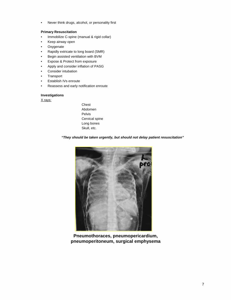
• Never think drugs, alcohol, or personality first Primary Resuscitation • Immobilize C-spine (manual & rigid collar) • Keep airway open • Oxygenate • Rapidly extricate to long board (SMR) • Begin assisted ventilation with BVM • Expose & Protect from exposure • Apply and consider inflation of PASG • Consider intubation • Transport • Establish IVs enroute • Reassess and early notification enroute Investigations X rays:
Chest Abdomen Pelvis Cervical spine Long bones Skull, etc.
“They should be taken urgently, but should not delay patient resuscitation”
Secondary Survey “The secondary survey does not begin until the primary survey (ABC) is completed, resuscitation is initiated and the patients ABCs are reassessed” “Secondary survey is a complete head-to-toe evaluation along with vital signs” “A more detailed survey is required in the unresponsive patient” History : A Allergies M Medications currently taken P Past illness L Last meal E Events/Environment related to the injury Physical examination:
Tension pneumothorax
Pneumomediastinum
9
A Complete “Head to Toe’ examination in mandatory Depending on the injuries it should include4:
• Glasgow Coma Scale • Vision • Cervical spine examination + stabilization • Chest for flail element • Blind peritoneal lavage • Appropriate X-rays, lab studies, USG/CT scans, etc.
Secondary Survey ‘Tubes and fingers in every orifice’ Secondary Survey: Neurologic Evaluation • Head and face are carefully inspected and palpated to detect any evidence of injury • Rectal tone must be evaluated even in alert patients as its absence indicates spinal cord injury • Spinal cord injury is more difficult to diagnose in patient with altered mental status • If spinal cord injury is diagnosed, high-dose methylprednisolone should be administered Secondary Survey: Thoracic Evaluation • The entire thorax should be reexamined, including adequacy and rate of respirations • Seatbelt or other contusions should be inspected and the ribs and sternum palpitated for
- bony crepitus - flail segments - emphysema
• A repeat chest radiograph should be obtained to confirm placement of the endotracheal and/or thoracostomy tubes
Secondary Survey: Abdominal Evaluation • Complaints of abdominal pain or findings of tenderness or ecchymosis raise the possibility of
intraabdominal injury • Nasogastric tube permits the detection of gastric bleeding & decompression of stomach • Presence of hematuria may indicate renal injury DO’S AND DON'TS
DO DON'T
• Bleep the trauma team • Follow algorithm • Hard collar for suspected cervical spine trauma • Jaw thrust • Large bore needles • Involve as many personnel as possible
• Manage haphazardly • Soft collar • Chin lift, head tilt • Small bore needles • Do it alone • Give up resuscitation earlier
Algorithm
• Be prepared • Triage • Primary survey ABC Identify life-threatening conditions and tackle them • Resuscitate ABC • Secondary survey Complete “Head to Toe” evaluation {clinical & lab} • Definitive treatment Surgery
10
TOPIC – 2
INITIAL RESUSCITATION OF THE INJURED PATIENT
Experience of the American College of Surgeons Advanced Trauma Life Support Course Programme (ATLS®) has shown the value of a structured approach to the resuscitation of the patient with multiple injuries, the significance of the "Golden hour" of resuscitation, and the "Platinum ten minutes" of pre-hospital care. The ATLS® scheme has four components:
Primary Survey Resuscitation Secondary Survey Definitive Care
It is important to try and achieve as much of this as possible simultaneously. Primary Survey Preparation Triage A Airway with cervical spine control
B Breathing to achieve adequate oxygenation of tissues
C Circulation with haemorrhage control
D Disability
E Events and environment.
Assume a cervical spine injury in any patient with multisystem trauma, especially with an altered level of consciousness, or a blunt or penetrating injury above the level of the clavicle.
Any patient with multisystem trauma, especially with injury above the clavicle, or where the patient is unable to give a clear history, is at risk due to the possibility of a cervical spine fracture. Excessive neck movement can convert an unstable spinal injury without neurological fallout, to one with paraplegia or quadriplegia. The patient's head should be controlled so that there is no movement causing hyperextension of flexion, especially during efforts to establish an airway.
11
TOPIC – 3
AIRWAY
Initial attempts to open the airway in an unconscious patient should include the jaw thrust, Sauction should be at hand. Ultimately, patients are very likely to need a definitive airway.
A definitive airway is a cuffed tube in the trachea
Indications for a definitive airway: A Airway Obstructed airway Inadequate gag reflex
B Breathing Inadequate breathing Oxygen saturation less than 90% C Circulation Inadequate circulation Systolic BP less than 75 mm. Hg. despite adequate fluid resuscitation D Disability Coma Glasgow Coma Scale less than 8/15
E Environment Hypothermia Core temperature less than 33° C.
Airway access may be Nasal Oral Surgical
A clear distinction must be made between an adequate airway and adequate breathing. Oxygenation and Ventilation It is essential to maintain adequate oxygenation of the tissues. For this reason, maximum possible oxygen should be given, either by a reservoir type mask, bag/valve/mask combination, or a definitive airway. The oxygen delivery system must be capable of delivering at least an inspired oxygen level of 60% by mask, or 100% be tube. If the patient cannot maintain adequate tissue oxygenation breathing adequate quantities of oxygen spontaneously, then artificial ventilation should be instituted. Circulation
12
Obvious external haemorrhage should be identified. The treatment of bleeding is to stop the Bleeding
Any hypotension after injury must be assumed to be due to hypovolaemia until proved otherwise.
Shock is classified according to stages: Class 1 Class II Class III Class IV
% Blood Loss 15% 30% 40% >40%
Vol. Blood Loss (70Kg)
750ml 1500 ml 2000 ml >2000ml
Pulse Raised Raised Raised Raised / Bradycardia
Colour Normal Pale Pale Pale Blood pressure Normal Pale Low Absent!
Recognition that shock is present, and appropriate early treatment is critical. Access to the circulation with at least two large bore cannulae (14G) is essential. Initial fluid management is commenced with Lactated Ringers, and then followed by appropriate available fluids and blood if available. Disability The patient's level of consciousness is assessed. This ideally should occur at least:
Pre-hospital On admission On leaving resuscitation
Glasgow Coma Scale Score Eye opening Best Motor Response Best Verbal Response
6 Obeys commands 5 Localises pain Orientated 4 Spontaneous Flexed to pain Confused 3 To speech Abnormal flexion (decorticate) Inappropriate words
2 To pain Extension Incomprehensible 1 None None
Events and Environment A very careful history should be obtained. The patient should be fully undressed in order to perform a full examination including a rectal examination. Monitoring During the resuscitation phase, the patient's condition will be closely monitored:
A Airway Respiratory Rate Oxygen Saturation Arterial Blood Gases
B Breathing See above
C Circulation Pulse Perfusion Blood Pressure EGG Central venous pressure Urine output
D Disability Pupils Glasgow Coma Scale E Environment
Core temperature.
13
Revised Trauma Score
Used both for field triage and for outcome.
Score Glasgow Coma Scale Systolic Blood Pressure Respiratory Rate
4 13-15 >90 10-29
3 9-12 76-89 >29
2 6-8 50-75 5-9
1 4-5 1-49 1 -4
0 3 0 0
14
AIRWAY MANAGEMENT
Aims • When is the airway potentially threatened? • When is the airway compromised? • How do you treat and monitor? • What is a definitive airway? • Demonstrate the required skills Predisposing Conditions • Coma • Aspiration • Maxillofacial trauma • Neck injury • Haematoma • Laryngeal injury • Thoracic inlet penetrating injury Signs of Airway Obstruction : "Look" • Agitation • Poor air movement • Rib retraction • Deformity • Foreign material Abnormal Breathing : Look • Cyanosis • Decline in mental state • Chest asymmetry • Tachypnoea • Distended neck veins • Paralysis Abnormal Breathing : Listen • I can't breathe! • Stridor, wheezing • Decreased breath sounds Abnormal Breathing : Feel • Surgical emphysema • Chest tenderness • Trachea deviated • Monitor • Saturation • ABGs • Chest X-Ray Treatment • Clear secretions, debris • Manually improving airway • Chin Lift • Jaw thrust • Oral Airway (Guedel) • Nasopharyngeal Airway • Definitive Airway • Endotracheal tube • Cricothyroidotomy • Tracheostomy When to Ventilate? • Apnoea • Hypoventilation • Flail chest • Spinal cord injury • Diaphragmatic injury • Head injury GCS<9 • Metabolic • Hypoxia • Hypercapnia • Hypothermia
Signs of Airway Obstruction : "Listen" • Speech? "How are you?" • Hoarseness • Noisy breathing • Gurgle • Stridor Signs of Airway Obstruction : "Feel" • Fracture crepitus • Face • Airway structures in neck • Tracheal deviation • Haematoma Normal breathing • Oxygen for all!! • Cyanosis inaccurate as sign of hypoxia • Beware of deterioration • Coma • Spinal cord injury • Chest injury Definitive Airway • Tube in trachea with cuff • Oxygen enriched gas • Secured thoroughly Rapid Sequence Intubation • Preoxygenate • Cricoid Pressure • Succinylcholine 1-2mg/kg ivi • Intubate orotracheally • Confirm placement • Release cricoid pressure Surgical Airway • Inability to intubate • Anatomical distortion and inability to intubate • Neck injury • Maxillofacial injury Needle Cricothyroidotomy • Always the first step • In children no surgical Cricothyroidotomy • Allows good oxygenation • Use for up to 20 minutes • Beware obstructed airway • Beware anatomical distortion Tracheostomy • If thyroid and cricoid cartilages damaged and distorted • Cricothyroidotomy converted • As definitive surgical airway at surgery • Not for the inexperienced in a hurry!! Summary • Suspect airway compromise • Protect cervical spine • Secure airway by stepwise approach • Definitive airway when necessary • Adequate oxygen delivery to tissues
15
TOPIC – 4 SHOCK
THE PRINCIPAL PROBLEM IS OXYGEN DELIVERY!
1. RECOGNITION OF SHOCK
Shock can and should be recognised before a blood pressure figure is available.
A palpable pulse which is rapid, of low amplitude, and with narrow pulse pressure is a clear sign of shock in a trauma patient.
THREE WINDOWS TO THE MICROCIRCULATION!
Manifestations of the adequacies of three regional microcirculations are observable in the early assessment of trauma patients. Poor blood flow in the SKIN is immediately evident as cool, pale, sweating peripheries. Poor blood flow in the BRAIN is an important cause of altered consciousness (anxiety, confusion, restlessness, etc). Poor blood flow in KIDNEYS is observable as oliguria after insertion of a urinary catheter and emptying of the urinary bladder.
There are a few pitfalls in the recognition of shock:
- Age - Hypothermia
- Athletes - Pacemaker
- Medications 2. IMMEDIATE INTERVENTION
Clinically evident shock in a trauma patient must be presumed to result from blood loss and, as soon as immediately life-threatening problems with Airway and Breathing are reversed, the clinical strategies must be directed towards two critical goals: STOP THE BLEEDING!
and RESTORE OXYGEN DELIVERY!
Interventions, therefore, include the following:
� Correctable problems with Airway and Breathing should already have been reversed, ensuring Oa supply to alveolar capillaries.
� External haemorrhage control by local pressure.
� Surgeon notification
� Large IV lines placed (at least x 2 )
� Pressure infusion capability
� Fluid warmer primed and functioning
� Crystalloid/colloid fluid bolus
� Crossmatch blood (Group O, Group specific, full crossmatch)
These interventions only begin a resuscitation process, which will often need a definitive operative procedure to stop the bleeding, and which may continue in the post-op period. To attempt to have a bleeding patient "resuscitated" before going to OT is a grave error!
Patients in haemorrhagic shock from penetrating torso trauma should pass only briefly through the Emergency Department.
Patients in haemorrhagic shock from blunt high impact trauma may require some additional, rapid assessment steps and interventions on their way to the QT.
HELP! CARDIAC ARREST!
Requirements
o ETT
o Artificial ventilation
o Bilateral chest tubes
o Large IV's (multiple)
o Large fluid bolus
16
o Operating theatre
� Clear strategies regarding ER thoracotomy must be established!
� A "medical" cardiac arrest response will NOT be successful!
� A trauma patient must have a BLOOD VOLUME response!
� STOP THE BLEEDING!
3. CAUSES OF MAJOR BLEEDING
MAJOR BLEEDING - THE BIG FIVE!
At this stage (during Primary and Secondary survey), only the region of bleeding is important - the bleeding organ does not need identification.
� EXTERNAL
¾ Identified by visual inspection
¾ Controlled by local pressure
� THORACIC
¾ Identified by Primary Survey and CXR
¾ Controlled by intercostal tube insertion (rarely requires thoracotomy)
� PELVIC
¾ Suspected according to pelvis x-ray not present if pelvic x-ray normal.
¾ Usually self-limiting. Occasionally complex and severe, requiring pelvic ring closure and / or angiographic embolisation.
� LONG BONES
¾ Identified by clinical examination
¾ Spontaneous control assisted by traction splintage
� ABDOMEN
¾ Identified or suspected by clinical findings and by exclusion of the other four of the "Big 5".
¾ Confirmed by DPL, FAST or laparotomy (NOT by CT or laparoscopy!)
4. OTHER CAUSES OF SHOCK IN TRAUMA PATIENTS
o Tension pneumothorax
o Cardiogenic - pericardia! tamponade, cardiac injury
o Neurogenic - i.e. from spinal cord injury
o Septic - usually late after injury
PRIMARY BRAIN INJURY DOES NOT CAUSE SHOCK,
BUT SHOCK DOES CAUSE SECONDARY BRAIN INJURY
5. SEVERITY OF SHOCK
- Class I haemorrhage - up to 15% of blood volume
- Class II haemorrhage - 15-30% of blood volume
- Class III haemorrhage - 30-40% of blood volume
- Class IV haemorrhage - more than 40% of blood volume
What will be the clinical signs of each category?
What will be your expected interventions in each category?
This classification helps us to:
� prepare for the management of shocked patients
� recognise early shock
� respond appropriately to severity of shock
However, initial treatment should be aggressive and be modified according to patient's responses.
6. RESPONSES TO EARLY RESUSCITATION
6.1 Monitoring the response
Pulse Consciousness
BP Urine output Skin perfusion ABG's
17
Remember other data, which is highly relevant � Age
� Time since injury � Injury severity � Prehospital treatment � Medications
6.2 Rapid Response
Be careful, these patients may still require surgery and may become "unstable" again! 6.3 Transient Response
Stop the bleeding! 6.4 Minimal Response
Remember the "Big 5"! Go to the operating theatre! 6.5 Adverse Responses
� Hypothermia � Coagulopathy
� Under-resuscitation
7. SUMMARY
� Be sure to understand the importance of shock
� Recognise shock early
� Initiate treatment for shock immediately and confidently
� Identify the cause of shock
� "Stop the bleeding" - NOW! , � Re-evaluate responses and adjust resuscitative strategies
RESTORE OXYGEN DELIVERY TO CELLS!
18
TOPIC – 5 THORACIC INJURY
Chest injuries are a common cause of death, frequently require some sort of intervention to aid recovery, but require thoracotomy infrequently.
THE PRINCIPAL PROBLEM IN CHEST INJURY IS OXYGEN DELIVERY!
Because oxygen delivery is dependent upon Airway, Breathing and Circulation, possible chest injuries receive very early attention in the Primary Survey component of Initial Assessment. However, it is the physiological derangements, which these injuries cause, rather than the detailed definition of some of the injuries, which dictate the assessment steps and sequence, and the resuscitative interventions, 1. CHEST INJURIES AND COMPLICATIONS
in trauma, patients fall into 4 categories of urgency 1.1 Injuries, which can cause death within minutes
These injuries require early recognition and intervention in the Primary Survey phase and before investigations. Note that the. last five of these are the anatomic consequences of the definitive injuries and are the anatomic basis of life threatening physiological implications. Note: There are two further traumatic causes of poor breathing which require recognition along with the above injuries:
� Poor respiratory effort from depressed level of consciousness � High spinal cord injury
1.2 Injuries, which can cause death within hours
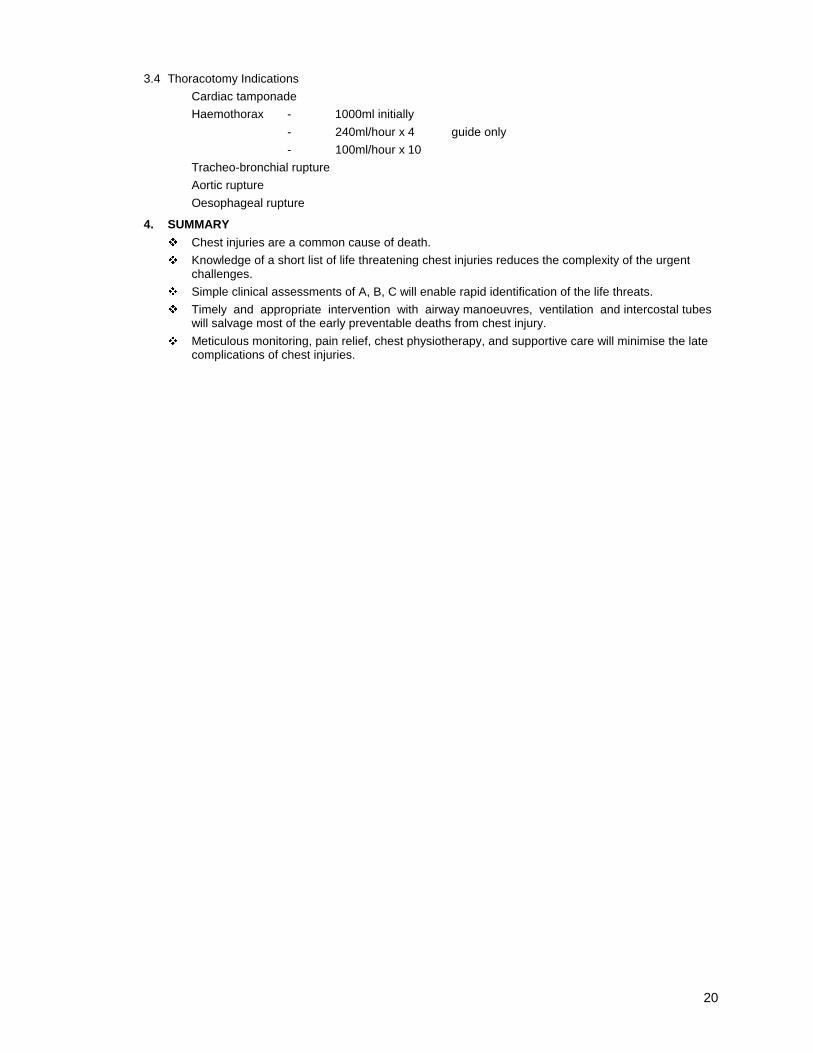
� Aortic rupture
� Tracheobronchial rupture
� Diaphragmatic rupture
� Oesophageal rupture
� Pulmonary contusion
� Myocardial contusion
Note that all of these are definitive anatomic diagnoses requiring some investigation, beyond clinical observations, to give rise to their suspicion, confirm their presence and dictate the appropriate interventions.
1.3 Other Injuries
� Pneumothorax
� Haemothorax
� Fractured ribs - upper, middle, lower
� Sternum
� Thoracic spine
The importance of rib fractures can be easily underestimated. The general clinical issues relate to pain and its management, underlying pulmonary contusion, and associated injuries.
Special care must be taken in evaluating and managing patients with fractured ribs who are elderly, very young, or have co-existing respiratory disease.
The importance of sternal fractures depends upon injury mechanism and fracture displacement.
Two further CXR manifestations require clear clinical strategies:
� subcutaneous emphysema
� mediastinal emphysema
1.4 Late Consequences/Complications of chest injuries and their treatment
� Atelectasis
19
� Aspiration pneumonitis
� Pulmonary consolidation/pneumonia
� Adult Respiratory Distress Syndrome (ARDS)
� Pulmonary oedema
� Pulmonary embolism
� Fat embolism
� Empyema
These events usually include very significant preventable causative events an< emphasise the need for meticulous total patient care, attentive monitoring and grea vigilance.
2. ASSESSMENT OF CHEST INJURIES
2.1 Primary Survey Assessment of Airway and Breathing
This is rapid and is not an exhaustive respiratory system examination.
Cyanosis Poor chest expansion
Tachypnoea Asymmetric chest expansion
Voice Hyperinflation (unilateral)
Stridor Hyperresonance (unilateral)
Confusion Breath sounds
"Respiratory distress" Tracheal shift
Air movement Diaphragmatic breathing
2.2 Primary Survey Assessment of Circulation
See the notes on "Shock".
The clinical assessments in 2.1 and 2.2 will permit the immediate diagnosis of all of th« injuries in 1.1.
2.3 Early Aids to Clinical Assessment/Monitoring
Oxymetry, CXR, ABG's, ECG, Pericardiocentesis, F.A.S.T. 2.4 Other Aids to Definitive Assessment
� Knowledge of a short list of life threatening chest injuries reduces the complexity of the urgent challenges.
� Simple clinical assessments of A, B, C will enable rapid identification of the life threats.
� Timely and appropriate intervention with airway manoeuvres, ventilation and intercostal tubes will salvage most of the early preventable deaths from chest injury.
� Meticulous monitoring, pain relief, chest physiotherapy, and supportive care will minimise the late complications of chest injuries.
21
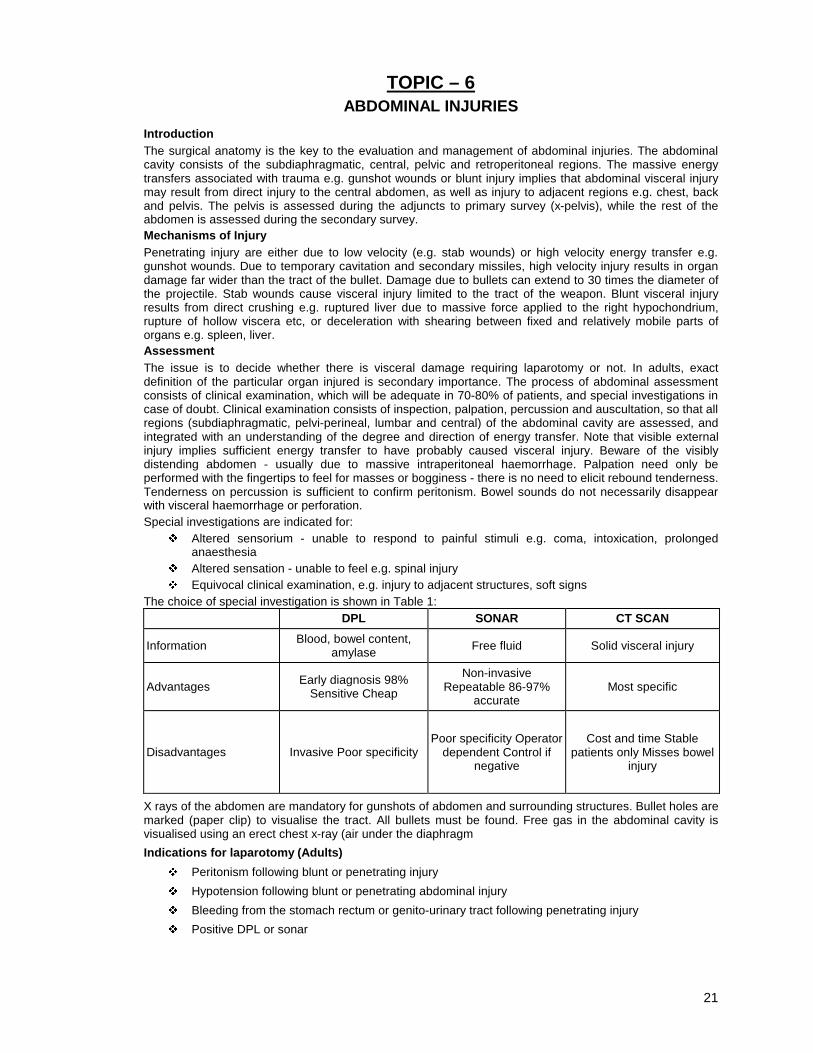
TOPIC – 6 ABDOMINAL INJURIES
Introduction The surgical anatomy is the key to the evaluation and management of abdominal injuries. The abdominal cavity consists of the subdiaphragmatic, central, pelvic and retroperitoneal regions. The massive energy transfers associated with trauma e.g. gunshot wounds or blunt injury implies that abdominal visceral injury may result from direct injury to the central abdomen, as well as injury to adjacent regions e.g. chest, back and pelvis. The pelvis is assessed during the adjuncts to primary survey (x-pelvis), while the rest of the abdomen is assessed during the secondary survey. Mechanisms of Injury Penetrating injury are either due to low velocity (e.g. stab wounds) or high velocity energy transfer e.g. gunshot wounds. Due to temporary cavitation and secondary missiles, high velocity injury results in organ damage far wider than the tract of the bullet. Damage due to bullets can extend to 30 times the diameter of the projectile. Stab wounds cause visceral injury limited to the tract of the weapon. Blunt visceral injury results from direct crushing e.g. ruptured liver due to massive force applied to the right hypochondrium, rupture of hollow viscera etc, or deceleration with shearing between fixed and relatively mobile parts of organs e.g. spleen, liver. Assessment The issue is to decide whether there is visceral damage requiring laparotomy or not. In adults, exact definition of the particular organ injured is secondary importance. The process of abdominal assessment consists of clinical examination, which will be adequate in 70-80% of patients, and special investigations in case of doubt. Clinical examination consists of inspection, palpation, percussion and auscultation, so that all regions (subdiaphragmatic, pelvi-perineal, lumbar and central) of the abdominal cavity are assessed, and integrated with an understanding of the degree and direction of energy transfer. Note that visible external injury implies sufficient energy transfer to have probably caused visceral injury. Beware of the visibly distending abdomen - usually due to massive intraperitoneal haemorrhage. Palpation need only be performed with the fingertips to feel for masses or bogginess - there is no need to elicit rebound tenderness. Tenderness on percussion is sufficient to confirm peritonism. Bowel sounds do not necessarily disappear with visceral haemorrhage or perforation. Special investigations are indicated for:
� Altered sensorium - unable to respond to painful stimuli e.g. coma, intoxication, prolonged anaesthesia
� Altered sensation - unable to feel e.g. spinal injury � Equivocal clinical examination, e.g. injury to adjacent structures, soft signs
The choice of special investigation is shown in Table 1: DPL SONAR CT SCAN
Information Blood, bowel content, amylase Free fluid Solid visceral injury
X rays of the abdomen are mandatory for gunshots of abdomen and surrounding structures. Bullet holes are marked (paper clip) to visualise the tract. All bullets must be found. Free gas in the abdominal cavity is visualised using an erect chest x-ray (air under the diaphragm
Indications for laparotomy (Adults)
� Peritonism following blunt or penetrating injury
� Hypotension following blunt or penetrating abdominal injury
� Bleeding from the stomach rectum or genito-urinary tract following penetrating injury
� Positive DPL or sonar
22
Genito- Urinary injury Post- traumatic haematuria is the cardinal sign of genito-urinary injury. Any form of genitourinary injury of any degree may present with microscopic haematuria. As a rule, only macroscopic haematuria is investigated, and investigation is considered from below upward depending on the level of injury, i.e.: 1. Blood in the urethral meatus or perineal injury requires a retrograde urethrogram 2. A clinically normal urethra allows passage of a urinary catheter. Clinical suspicion of local injury
indicates a cystogram. 3. Intravenous pyelography is the baseline investigation for haematuria and a clinically normal urethra and
bladder. Contrasted CT scanning is the most specific investigation to delineate the degree of renal injury.
Damage Control Surgery Indicated for hypothermia, acidosis and coagulopathy, and considered when massive blood transfusion or inotropes are required. The aim is to immediately stop haemorrhage and contamination without wasting time to effect anatomical repair. Examples include: ligating anatomically named vessels, packing the rest, stapling or tying damaged bowel, placing heparinised intraluminal shunts in major vessels, applying an external fixator to the pelvis, packing the pelvis, lavaging contaminated areas and closure of skin only. The patient is aggressively resuscitated in the intensive care unit, and returned to theatre for definitive repair as soon as resuscitation is completed. Summary
� Examination depends on consideration of the effects of energy transmitted from adjacent areas e.g. pelvis, chest, and back.
� The aim is to decide on the need for laparotomy rather than exact anatomical definition of injury. � Simple clinical examination suffices for the majority of abdominal injury. Diagnostic peritoneal
lavage is the default in case of doubt. � The urological tract is investigated from below upward, depending on presentation. � Damage control surgery is indicated for hypothermia, acidosis, or coagulopathy.
Key references 1. American College of Surgeons, Committee on Trauma. Advanced Trauma Life Support Program for
Physicians. American College of Surgeons, 1997. 2. Boone DC, Peitzman AB. Abdominal injury. The Trauma Manual. Lippincot-Raven, 1998.
23
TOPIC – 7 HEAD INJURY
Emergency Department Management Because hypoxia and hypotension interfere with cerebral oxygenation, complete and rapic physiologic resuscitation is the first priority for patients with head injuries. A large study frorr the Traumatic Coma Data Bank demonstrated that a single observation of systolic blooc pressure below 90 mm Hg in the field, or hypoxia (arterial oxygen tension PaOa below 6C mm Hg) was a major predictor of poor outcome. A multidisciplinary team should provide the patient with an adequate airway and ventilation and restore and maintain haemodynamk stability (with adequate fluid replacement and detection and treatment of any bleeding), al according to the principles developed by the Advanced Trauma Life Support system. The ABCs of emergency care (airway, breathing, and circulation) take precedence irrespective of neurologic injuries. The initial neurologic assessment, which does not take more than 10 seconds, consists of rating the patient on the Glasgow Coma Scale (GCS [see Page 3] and assessing the width and reactivity of the pupils. Although the sarm assessment is made after resuscitation as a guide for prognosis and therapy, it should alsc be made (and recorded) before resuscitation to permit evaluation of the effect o resuscitative measures and differentiation between primary and secondary neurologic injury Early establishment of a definitive airway and ventilation, if not performed in the field, ii recommended for patients with a GCS score of 8 or lower. Indications for intubation befon transport are deteriorating consciousness (even if the patient is not in a coma), bilatera fractured mandible, copious bleeding into the mouth (as occurs with fracture of the base c the skull), and seizures. An intubated patient must also be ventilated (PaCC-2 >35 mm Hg). Fluid replacement should be performed with isotonic solutions such as lactated Ringer' solution, normal saline, or packed red blood cells when appropriate. The patient should bi examined rapidly and thoroughly for any concomitant life-threatening injuries. Patients with spinal cord injury above T5 and vasogenic spinal shock may have sever hypotension, which should be treated vigorously. Intracranial hypertension should be suspected if there is rapid neurologic deterioratior Clinical evidence of intracranial hypertension, manifest by signs of herniation, include unilateral or bilateral dilatation of the pupils, asymmetrical pupillary reactivity, and mote posturing, Intracranial hypertension should be treated aggressively. Hyperventilation, whic does not interfere with volume resuscitation and results in rapid reduction of intracrani. pressure (ICP), should be established immediately in cases of pupillary abnormalities. Recent research has shown that unilateral or bilateral pupillary abnormalities do not result from compression of the third cranial nerves, as previously thought, but from compression of the brain stem, with resulting brain stem ischaemia. Therefore, administration of mannitol is effective because it not only decreases ICP but also increases cerebral blood flow (CBF) through modulation of viscosity. Because mannitol is not used to dehydrate the body, all fluid losses through diuresis must be replaced immediately or even preventively, especially in patients suffering shock as a result of blood loss. Although arterial hypertension occurring after a severe head injury may reflect intracranial hypertension (Cushing's phenomenon), especially when accompanied by bradycardia, it should not be treated, because it may be the sole mechanism permitting the brain to maintain perfusion despite increasing intracranial pressure. In the absence of signs of herniation, sedation should be used when indicated for safe and efficient transport of the patient. Transport of the patient should be kept to a minimum because it is often accompanied by secondary insults (e.g., hypoxia or hypotension). Pharmacologic paralysis, which interferes with neurologic examination, should be used only if sedation alone is inadequate for safe and effective transport and resuscitation of the patient. When pharmacologic paralysis is used, short-acting agents are preferred. Prophylactic hyperventilation, which may exacerbate early ischaemia, is not recommended for these patients. Minimal radiological evaluation consists of a lateral cervical spine film or a swimmer's view [see Spinal Cord Injury, below]. After haemodynamic stability is achieved, unenhanced CT of the head should be used for all patients with persistent impairment of consciousness. ICU Management A GCS score of 8 or lower after resuscitation is an indication for admission to a ICU if available. Although the focus of ICU management is prevention of secondary injury and maintenance of adequate cerebral oxygenation, admission to an ICU does not eliminate the occurrence of secondary insults. Management of Cerebral Perfusion Pressure The rationale behind CPP therapy is expressed in Poiseuille's law. The effect of CPP therapy has not been investigated in a randomised, controlled clinical trial, several studies suggest that a CPP of 70 to 80 mm Hg may be the clinical threshold below which mortality and morbidity increase. CPP therapy involves manipulation of both arterial BP and ICP, but its objective is the reduction of ICP.
24
Management of Intracranial Pressure Because ICP is a determinant of CPP, treatment of ICP inevitably affects CPP. Because the goal is maintenance or improvement of CBF, measures for treating ICP should be evaluated in the light of their effect on CBF. It is not possible to establish an arbitrary threshold for treatment of elevated ICP that would be applicable in all situations. Any interpretation of ICP must be combined with assessment of clinical features and evaluation of CT scan findings because it is possible to have transtentorial herniation with an ICP of 20 mm Hg in the presence of a mass lesion. Conversely, with diffuse brain swelling, adequate CPP can be maintained despite an ICP as high as 30 mm Hg. As a general rule, ICP values between 20 and 25 mm Hg indicate that therapy should be initiated. Mannitol is usually administered in I.V. boluses of 0.25 to 1 g/kg over 10 to 15 minutes until ICP is controlled or serum osmolality reaches 320 mOsm/L. Because volume depletion is an important side effect of mannitol therapy, urine losses should be replaced. Hyperventilation reduces ICP (by vasoconstriction) and CBF, which may be at ischaemic levels in certain parts of the brain. Therefore, hyperventilation (PaCOa < 30) should not be instituted prophylactically. If PaCC-2 must be reduced to extremely low levels, hyperventilation can be combined with mannitol, improving CBF by reducing blood viscosity. Haemoglobin, haematocrit, and Blood Viscosity To ensure optimal cerebral oxygenation, CPP, haemoglobin concentration, and oxygen saturation should be optimised; vessel diameter should be maximised; and viscosity should be in the low range. The haematocrit and viscosity are inversely related, and a balance must be established to ensure optimal oxygenation. If the haematocrit is too high, viscosity increases; if the haematocrit is too low, the oxygen-carrying capacity of blood decreases. Maintaining the haematocrit between 30% and 35% is recommended: below 30%, the oxygen-carrying capacity falls without a significant change in viscosity, and above 35%, the viscosity increases out of proportion to the oxygen-carrying capacity. Cerebral Metabolism At 1,200 to 1,400 g, the brain accounts for only 2% to 3% of total body weight and does not do any mechanical work; yet it receives 15% to 20% of all cardiac output to meet its high metabolic demands. Of the total energy generated, 50% is used for interneuronal communication and the generation, release, and reuptake of neurotransmitters (synaptic activity), 25% is used for maintenance and restoration of ion gradients across the cell membrane, and the remaining 25% is used for molecular transport, biosynthesis, and other, as yet unidentified, processes. In the absence of oxygen, anaerobic glycolysis can proceed, but energy production is much less efficient. Two molecules of ATP and two molecules of lactate are generated for each molecule of glucose: 1 glucose + 2 ADP + 2 Pi ® 2 lactate + 2 ATP Regulation of Blood Flow Because the reserves of glucose and glycogen within the astrocytes of the brain are limited and there is no significant storage capacity for oxygen, the brain depends on blood to supply the oxygen and glucose it requires. More specifically, substrate availability is determined by its concentration in blood, flow volume, and the rate of passage across the blood-brain barrier. Under normal circumstances and with certain physiologic alterations, an adequate supply of substrates can be maintained by regulation of CBF. CBF increases with vasodilatation and decreases with vasoconstriction. Calibre changes take place mainly in cerebral resistance vessels. Control of CBF by influencing vessel calibre is commonly referred to as autoregulation of blood flow. Metabolic autoregulation CBF is functionally coupled to cerebral metabolism, changing proportionally with increasing or decreasing regional or global metabolic demand. Thus, the brain precisely matches local CBF to local metabolic needs Raised intracranial pressure According to the Monro-Kellie doctrine, ICP is governed by three factors within the confines of the skull:
� Brain parenchyma plus cytotoxic oedema � CSF plus vasogenic oedema � Cerebral blood volume
When the volume in one compartment increases, ICP increases unless there is a compensatory decrease in volume in the other compartments.
25
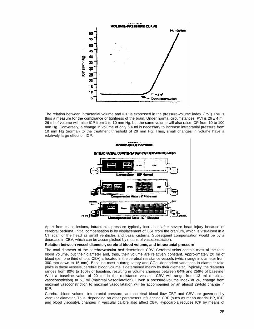
The relation between intracranial volume and ICP is expressed in the pressure-volume index. (PVI). PVI is thus a measure for the compliance or tightness of the brain. Under normal circumstances, PVI is 26 ± 4 ml; 26 ml of volume will raise ICP from 1 to 10 mm Hg, but the same volume will also raise ICP from 10 to 100 mm Hg. Conversely, a change in volume of only 6.4 ml is necessary to increase intracranial pressure from 10 mm Hg (normal) to the treatment threshold of 20 mm Hg. Thus, small changes in volume have a relatively large effect on ICP.
Apart from mass lesions, intracranial pressure typically increases after severe head injury because of cerebral oedema. Initial compensation is by displacement of CSF from the cranium, which is visualised in a CT scan of the head as small ventricles and basal cisterns. Subsequent compensation would be by a decrease in CBV, which can be accomplished by means of vasoconstriction. Relation between vessel diameter, cerebral blood volume, and intracranial pressure The total diameter of the cerebrovascular bed determines CBV. Cerebral veins contain most of the total blood volume, but their diameter and, thus, their volume are relatively constant. Approximately 20 ml of blood (i.e., one third of total CBV) is located in the cerebral resistance vessels (which range in diameter from 300 mm down to 15 mm). Because most autoregulatory and COa -dependent variations in diameter take place in these vessels, cerebral blood volume is determined mainly by their diameter. Typically, the diameter ranges from 80% to 160% of baseline, resulting in volume changes between 64% and 256% of baseline. With a baseline value of 20 ml in the resistance vessels, CBV will range from 13 ml (maximal vasoconstriction) to 51 ml (maximal vasodilatation). Given a pressure-volume index of 26, change from maximal vasoconstriction to maximal vasodilatation will be accompanied by an almost 29-fold change in ICP. Cerebral blood volume, intracranial pressure, and cerebral blood flow CBF and CBV are governed by vascular diameter. Thus, depending on other parameters influencing CBF (such as mean arterial BP, ICP, and blood viscosity), changes in vascular calibre also affect CBF. Hypocarbia reduces ICP by means of
26
vasoconstriction, consequently improving CPP. However, net CBF is decreased because in Poiseuille's equation, vessel diameter is carried to the fourth power.. There are two methods of reducing ICP by means of vasoconstriction without affecting CBF. The first is to reduce blood viscosity. Decreasing the blood viscosity will, by itself, lead to vasoconstriction, provided that viscosity autoregulation is intact. With impaired autoregulation, decreased viscosity will result in an increase in CBF but no decrease in ICP. However, this effect can be used to maintain CBF under vasoconstriction with hypocarbia. The effect of mannitol on ICP is thought to be mediated in part by lowering blood viscosity. The second method of reducing ICP without affecting CBF is to increase CPP, which can be done by raising blood pressure. Again, with intact autoregulation, an increase in CPP will lead to vasoconstriction, with net CBF remaining constant. With impaired autoregulation, CBF will follow CPP passively, and maintenance of normal blood pressure may be indicated in these cases. More important, however, is the avoidance of hypotension under these circumstances; the effect of CPP therapy may be attributable in part simply to prevention of hypotension. Cerebral ischaemia Cerebral ischaemia, defined as CBF that is inadequate to meet the metabolic demands of the brain, is an important mechanism of secondary injury in patients with severe head injury, and the adequacy of CBF has been associated with neurologic outcome. MANAGEMENT OF MAJOR FACIAL INJURIES AND BLEEDING. The management of facial trauma forms the majority of the clinical workload of academic maxillo-facial and oral surgeons. Over a fifteen year period in our department, in excess of 30 000 facial fractures have been treated. Although a large percentage of these are the result of fist fights these being minor in nature, a large proportion are as a result of motor vehicle accidents, pedestrian accidents and handgun injuries. These latter groups results in major facial injuries, usually to the middle third of the facial skeleton. The presenting symptoms of patients with major injuries are usually severe swelling with possible airway compromise in the unconscious patient, accompanied by haemorrhage to a greater or lesser degree. The principles of treatment of these patients revolve around airway management, maintenance of breathing and control of haemorrhage. In all unconscious patients establishment of a definitive airway is important and this should be secured as early in the management as possible. In some cases a tracheotomy is indicated. It is a given fact that unless severe soft tissue injury or haemorrhage is present, the definitive management of facial injuries must be delayed until more pressing life threatening injuries have received attention. Haemorrhage resulting from facial injuries can be either external or arising from the oral or nasal cavities. The haemorrhage may also be a capillary ooze or a bleed from a more substantial vessel such as facial artery, inferior alveolar vessels or infraorbital vessels. In some cases haemorrhage from the maxillary artery may occur. Basic principles of control of haemorrhage namely application of pressure, ligating smaller vessels are the most commonly used methods of haemorrhage control. In some cases exposure and ligation of the both superficial temporal artery as well as the posterior auricular artery in the retromandibular fossa has proved to be beneficial. Definitive management of major facial fractures make use of the principles of reduction, immobilisation and control of sepsis.
APPROACH TO EXAMINING HEAD INJURIES Initial Assessment: Primary Survey Airway: Compromise due to unstable facial fractures, bilateral # jaw, and GCS <8/15
Breathing: Compromise due to aspiration (coma, gastric dilatation). Hypoxia due to any cause aggravates secondary brain injury
Circulation: Scalp wounds are a potent cause of hemorrhage and hypotension. Hypotension decreases cerebral perfusion and greatly aggravates secondary brain injury.
Disability: Assess pupil size, reaction, AVPU or Glasgow Coma Scale, and movement of all 4 limbs
Exposure: Maintain normothermia
Adjuncts to Primary Survey Adequate monitoring ensures adequate cerebral oxygenation and perfusion
Secondary Survey Examination of the head, face, throat and neck. Glasgow Coma Scale Detailed neurological examination
Examination of the Head, Face and Neck Start at one point. Inspection and palpation in circular saggital direction back to the same point.
� Eyebrows and forehead: Lacerations in eyelids easily missed � Scalp: Palpated, cannot be visualised through hair. Palpate lacerations, feel for # skull
27
� External Ears & Canal:Inspect for CSF, blood, haemotympanum, perforation. � Mastoids: Inspect/ palpate for bruising � Cervical spine: palpate for steps, bogginess � Throat:Inspect and palpate for lacerations, especially penetrating platysma. Palpate for surgical
emphysema. Palpate larynx for #. � Jaw: Inspect for occlusion (canines), trismus, steps, alveolar haematoma. Palpate for #. � Mouth:Inspect for lacerations, damaged teeth. � Facial bones:Gently move alveolus, mid-face for stability. Cheeks: Palpate for sensation (Blow-out
# orbit) � Nose:lnspect for alignment, palpate for tenderness � Face: Inspect for enophthalmos, sunken cheek, malalignment of nose � Eyes:Examine from outside inward. Remove contact lenses. Inspect for diplopia, visual acuity,
visual fields
28
TOPIC – 8
SPINAL CORD INJURY Introduction
Spinal cord injury is a devastating injury due to disability, the cost of rehabilitation and the cost of chronic care of the spinally injured patient. Quality of care of the spinally injured can be measured by the incidence of incomplete spinal lesions emanating from a centre. It may be a measure of the degree to which no further harm (secondary injury) is caused to the spinal cord after injury. An estimated 47 % of spinally injured patients suffer associated injuries, involving head (26%), chest (24%) or long bones (23%). These injuries may affect spinal cord perfusion, oxygenation and limit rehabilitation.
Patho-anatomy
The intervertebral joints carry most of the axial load of the spinal column, the facet joints are the pivots, and the ligaments are the passive restraints. To cause instability, high degrees of energy transfer are required. A 3- column model of stability divides the spine into the anterior 2/3 of the vertebral body, the posterior 1/3 and the posterior longitudinal ligament, and the remaining posterior elements. Injury to 2 out of 3 columns produces instability, and the risk for spinal cord injury.
Blunt injuries occur at the most mobile parts of the spine i.e. cervical (55%) thoraco-lumbar(30%) and lumbar (15%). Penetrating injuries may cause damage by direct transection, but also by secondary energy transfer causing secondary injury. The latter may present days after the primary injury.
Spinal cord injuries affect the crossed cortico-spinal (motor) spinothalamic (pain, temperature) the uncrossed dorsal (position, vibration) and the autonomic tracts. Damage to the latter leads to loss of vasomotor (and visceral reflexes) immediately after the injury, called neurogenic shock. Neurogenic shock manifests as hypotension and bradycardia and occurs after cervical or upper thoracic injuries.
Due to the secondary events immediately after spinal cord injury, all function is often lost immediately after injury. The patient may give the appearance of complete injury and may improve afterward. Massive neuronal depolarisation immediately after injury leads to flaccid paralysis below the level of the lesion. This is called spinal shock.
Pathophysiology Primary injury to the spinal cord rarely occurs due to total transection of the cord. Most of the damage to the cord occurs secondarily due to haemorrhage, massive depolarisation, uncontrolled influx of calcium into neurones, vasospasm, uncoupling of oxidative phosphorylation, activation of calcium - dependent phospholipase-A, and free radical mobilisation induced membrane damage. Proper management of spinally injured patients (immobilisation, perfusion, and oxygenation) may limit secondary injury to the spinal cord. Clinical Syndromes Tracts of the spinal cord probably only require 5% of fibres to function. Once the secondary injury and spinal shock settles down, a true clinical picture will emerge over the next 6 months. These include: 1. Complete spinal cord injury is identified by the absence of motor or sensory function below the level of
injury. Incomplete spinal cord injury is defined as any motor or sensory function below the level of injury, and may manifest as the
2. Anterior cord syndrome occurs due to thrombosis of the anterior spinal artery, which supplies the cortico- spinal and spinothalamic tracts. Posterior columns are left intact
3. Central cord syndrome occurs due to injury to central fibres in the cervical area. Due to the layering of fibres from below upward, lower limb function is preserved. Upper limb motor function and to a lesser extent sensory function are lost
4. Brown-Sequard lesion occur due to partial lacerations of the cord, probable associated with limited secondary injury. The crossed spinothalamic fibres (pain, temperature), the uncrossed ascending posterior columns (position, vibration) and the crossed corticospinal (motor) fibres are damaged
5. Involvement of the cauda equina (L1 and below) results in asymmetric partial loss of distal limb sensory and/or motor function. Characteristically, radicular pain is present, but bowel and bladder function is preserved
6. Injuries to the conus medullaris also cause distal incomplete spinal cord syndromes, without radicular pain and commonly affect bowel and or bladder function
Management Primary Survey ¾ Airway and cervical spine control: Spinal cord injuries are unlikely to cause airway compromise
other than by vomiting and aspiration due to gastroparesis. Patients suffering high velocity blunt injury, loss or decreased level of consciousness or injury above the clavicle have cervical spinal cord injury till proven otherwise by adequate x-rays. These include AP, lateral and open mouth views of the cervical spine, and AP and lateral views of the thoracic and lumbar spine. The entire
29
spinal column should remain fully immobilised (head blocks, spider harness, spine board) and the spine moved as a unit (logrolled) till these x-rays are completed
¾ Breathing: In addition to ventilatory failure due to paralysis of the respiratory muscles, associated thoracic injuries may cause a severe VQ defect affecting ventilation.
¾ Circulation: Neurogenic shock manifests as hypotension and bradycardia. The principal cause of hypotension remains haemorrhagic shock due to associated injuries. Neurogenic shock may require treatment (inotropes, atropine) in order to maintain perfusion of the spinal cord.
¾ Disability: See head injuries ¾ Exposure: Due to transection of autonomic fibres, temperature control below the level of the lesion.
Quadriplegics are poikilothermic and rapidly become hypothermic if exposed. Adjuncts to Primary Survey Monitoring of oxygenation and perfusion is crucial, as in any patient suffering major injury (pulse rate, blood pressure, urine output, oxygen saturation). High spinal cord injured patients are especially prone to massive gastric dilatation causing vomiting and aspiration. Early nasogastric drainage is essential. During logrolling, the entire spinal column is palpated for bruising, tenderness or steps. Penetrating injury in the proximity of the spinal column may cause spinal cord injury. Immobilisation can be terminated in an alert, neurologically normal patient without local signs. X-rays of the cervical spine are part of the adjuncts to the primary survey. X-rays of the remainder of the spinal column are taken after the secondary survey, if indicated. Secondary Survey Secondary survey includes a detailed neurological examination (motor, pain and temperature, vibration and position, and reflexes. Since the clinical picture may develop further (pathophysiology) accurate documentation and repeated examination is essential. Neurological deficit may preclude assessment of the abdomen and DPI or sonar would be required to exclude intra-abdominal injury after secondary survey. Definitive Care High-dose methylprednisolone initiated within 3-8 hours after injury is of proven benefit to limit secondary spinal cord injury and improve neurological levels. Due to increased septic complications, it is contra-indicated for multiply injured patients or penetrating injuries. Epidemiological evidence suggests increased incidence of incomplete spinal cord injury where patients are rapidly referred to specialised centres. The patient must be transferred while fully immobilised, but with adequate protection of the skin, e.g. hourly logrolling (1/4 turns on spine board) and gentle padding of the spine board. Due to the effect of associated injuries on spinal cord function, early definitive care of all injuries is essential. Key References 1. American College of Surgeons Committee on Trauma. Advanced Trauma Life Support Program for
Physicians. American College of Surgeons, 1997. 2. Przybylski GJ, Marion DW. Injury to the vertebrae and spinal cord. In: Trauma. Feliciano DV, Moore EE,
Mattox KL, Eds. 1996.
30
TOPIC – 9
EXTREMITY INJURY
1. URGENCY AND IMPORTANCE 1.1 Where does it fit in initial Assessment ? Extremity injuries often look dramatic. Extremity injuries should NEVER be addressed first! Extremity injuries usually receive attention during secondary survey...
but Extremity injuries must be considered during primary survey at:
C-CIRCULATION they are potentially represented in three of the "Big 5" causes of haemorrhage.
¾ External - lacerations, major amputations ¾ Pelvis ¾ Long Bones - especially multiple fractures
Remember the additional fluid loss into large wounds ! 1.2 Time Dependent Conditions USE YOUR PATIENT'S TIME WELL!
(a) Bleeding (b) Ischaemia (c) Wound contamination (d) Compartment syndrome (e) Tissue pressure (skin, nerves) Note: (a) is immediately life threatening. (b), (c), (d) are life threatening if treatment is delayed (b), (c), (d), (e) are all limb threatening 1.3 Secondary Survey Systematic examination of extremities as part of the whole secondary survey
� LOOK � FEEL � MOVE
¾ Actively ¾ Passively
2. STOPPING THE BLEEDING � Local pressure * � Traction splintage * � Haemostats - exceptional circumstances � Tourniquets - deliberately planned and monitored � Definitive care
3. SPLINTAGE OF FRACTURES (especially traction splintage) � Relieves pain/assist patient movement � Restores circulation � Minimises soft tissue damage � reduces bleeding � Protects nerves � Note : Splintage should be achieved before patient transport � traditionally, we are not good at this in hospitals � it requires agreed and enforced protocols � assess distal vascular and neurological function � before splintage, after splintage, continuing
4. HISTORY Important features include :
� Mechanism - magnitude of forces; likelihood of other injuries
31
� TIME OF INJURY ! � ENVIRONMENTAL EXPOSURE - PRESSURE, HEAT, COLD, WATER, ETC � Blood at scene � Observations at scene - vascular, neurological (TIME !) � Interventions - manipulations, analgesia
5. INJURY TYPES and IMPLICATIONS (i.e. during initial Assessment) 5.1 Fractures
� will not be discussed in any detail 5.2 Dislocations
� associations with vascular injury, nerve injury, skin ischaemia � reduction early (expertise preferable) � recheck perfusion after reduction
5.3 Pelvic Fractures � can cause major bleeding (included in the "Big 5")
¾ especially if posterior disruption ¾ open book ¾ vertical shear
� liberal use of PXR in shocked patients with high energy blunt impact � limited attempts to diagnose clinically � importance of local protocols with respect to use of DPL, ext fixation, angiographic
embolisation, laparotomy, open reduction/fixation � association with bladder and urethral injuries � infrequent complex, open pelvic fractures
¾ association with urogenital, rectal, vascular, lumbosacral plexus injuries.
• What are the common causes of severe pelvic fracture in your community ? Motorcycles ? Pedestrians ? Fall from height ?
• What will be your plan/protocol for these injuries in your hospital ? Who will be included in you multidisciplinary team response ?
5.4 Vascular injuries Bleeding/expanding haematoma/distal ischaemia � importance of physical examination � importance of reduction of dislocations, splintage of fracture � role of Doppler signals/pressures � role of angiography (ED, Radiology, OT ?)
5.5 Amputations � Major
o above ankle or wrist � Digit
o role of replantations ? o care of the amputated part ? o transport with patient
5.6 Crush Syndrome � Rhabdomyolysis
o myoglobinuria, hyperkalaemia, acidosis, DIG � role of IV fluids, osmotic diuretics, alkalinisation of urine
Remember: Unconscious Patients can have compartment syndrome ! Unconscious patients don't complain of pain, sensation!
5.6 Contaminated Wounds � Aggressive, sharp debridement � Pulsatile saline irrigation � Avoid antiseptic solutions � This is an URGENT surgical problem � Polaroid/digital photographs
o Tetanus immunization 5.7 Nerve Injuries (esp dislocation)
� Restore bone alignment as soon as possible � Remember a goal of good trauma care
MINIMISE TIME from INJURY
to DEFINITIVE CARE ! � Surgery for vascular occlusion - within 6 hours FROM INJURY! � Fasciotomy for compartment syndrome - within 2 hours FROM DIAGNOSIS! � Debridement of contaminated wound - within 8 hours FROM INJURY!
7. TERTIARY SURVEY � repeat of Primary, Secondary Survey; review of x-rays � Performed on "The Morning After" � Frequently identifies lesser, occult musculoskeletal injuries � often injuries which have little impact on survival but significant impact on mobility and dexterity.
8. SUMMARY Question : How do blunt and penetrating limb injuries differ ? (e.g. soft tissue damage, vascular damage, sepsis risk, imaging strategies, definitive care strategies) � Prmary survey takes priority over dramatic appearances. � Life threateing conditions are TIME DEPENDENT � Early splintage should be achieved � Ischaemic tissue requires urgent revascularisation or excision � Contaminated wounds require urgent debridement � Tertiary survey is an important trauma team behaviour
33
TOPIC – 10 PAEDIATRIC INJURY
Aims • What makes the child unique? • Anatomy • Physiology • Psychology • Long term effects • Different skills • Different injury patterns • Remember the numbers! Anatomical Differences • Size and shape • Large head • Vulnerable trunkal structures • Soft skeleton • Large surface area : mass Physiological Differences
Normal Values
Age Group Wt (Kg) Heart Rate Syst. B.P Resp. Rate Urinary Output
Physiological Differences • Different signs of hypovolaemia • Primarily tachycardia • Remain normotensive until late • Precipitous deterioration • Prone to hypothermia • Metabolically brittle • Rapid "deteriorators and recoverers"
• 6 hours in child = a day in adult
Psychology • Child • Terrified and in pain • Separated from parents and familiar environment • Doctor and staff • Child is an innocent victim • Under stress to succeed • Crying!!!
• Unfamiliarity with doses, sizes etc. Long Term Effects • Growth • Development • Intellectual • Physical • Schooling • Family • Disfigurement
• Disability
Injury Patterns • Different injuries compared to adults by organ system • Blunt abdominal trauma • Thoracic injury • Head injury • Spinal injury • Orthopaedic injury
• Different combinations of injuries
Skills – Airway • Short • Correct placement • Correctly secured • Position of child for intubation • Importance of cricoid pressure • Equipment • Straight (Miller) blade
Intraosseous Access • Sites • Medial tibia proximally • Anterior femur distally • Uninjured and unpunctured bone • All resuscitation fluids • Temporizing manoeuvre • Relatively low flow rates
Evolving Injury Patterns • Lap belt complex • Pedestrian Vehicle Accident (PVA) complex • Forward facing infant complex • Airbag Injuries • The common cycle scenarios
• Non-accidental injury
Head Injury • Common (large vulnerable) • Better prognosis for recovery • Open fontanelles in infant • Large amount of blood loss intracranially • Window for assessment • Tendency for malignant oedema • Lower incidence of surgically drainable lesions Spinal Injury • Flexible ligaments and joints • Pseudosubluxation • Spinal Cord Injury Without Radiological Abnormality (SCIWORA) • Growth plates • Atlanto-occipital dislocation • Relatively commoner in children • Difficult to diagnose with certainty • Steroids for blunt injury
Analgesia • Morphine 0.1mg/kg ivi 4 hrly or when required • Safe • Humane • Facilitates resuscitation • Does not mask signs Summary • Priorities of management same • Be aware of important differences • Early involvement of Paediatric Surgeon if available
35
TOPIC – 11 DAMAGE CONTROL
The concept of "Damage Control," - also known as "Staged Laparotomy" has as its objective the delay in imposition of additional surgical stress at a moment of physiological frailty. The concept is not new, and livers were packed as much as 90 years ago, but with a failure to understand the underlying rationale, the results were disastrous. The concept was reviewed and the technique of initial abortion of laparotomy, establishment if intra-abdominal pack tamponade, and then completion of the procedure once coagulation has returned to an acceptable level proved to be life-saving. The concept of staging applies both to routine, and to emergency procedures, and can apply equally well in the chest, pelvis and neck as in the abdomen.
Stage 1: Patient selection:
The indications for Damage Control generally can be divided into the following:
a) Inability to achieve haemostasis
b) Inaccessible major venous injury e.g. Retrohepatic vena cava
c) Anticipated need for a time consuming procedure
d) Demand for non operative control of other injuries e.g. fractured pelvis.
e) Inability to approximate the abdominal incision
f) Desire to reassess the intra-abdominal contents (Directed relook)
Irrespective of setting, a coagulopathy is the single most common reason for abortion of a planned procedure, or the curtailment of definitive surgery. It is important to abort the surgery before the coagulopathy becomes obvious.
The technical aspects of the surgery are dictated by the injury pattern:
The primary objectives are as follows:
1) Arrest bleeding, and the resulting (causative) coagulopathy. Procedures for haemorrhage control include:
a) Repair or ligation of accessible blood vessels
b) Occlusion of inflow into the bleeding organ (e.g. Pringle's Manoeuvre for bleeding liver).
c) Tamponade using wraps or packs.
d) Intra-operative or post-operative embolisation.
e) Intravascular shunting.
2) Limit contamination and the sequelae thereof.
a) Ligation or stapling of bowel.
b) Resection of damaged segment with clips, clamps, or staples
3) Close the abdomen to limit heat and fluid loss, and to protect viscera.
This depends on whether the abdomen can be approximated to achieve closure. If not:
� Towel clips
� Bogota Bag - (Temporary silo)
� Opsite "Sandwich" Stage 2: Intra-operative reassessment for haemorrhage control
The timing of the transfer of the patient from the Operating Theatre to the ICU is critical. Prompt transfer is cost-effective; Premature transfer is counter-productive. In addition, once haemostasis has been properly achieved, it may not be necessary to abort the procedure in the same fashion. Conversely, there are some patients with severe head injuries, where the coagulopathy is induced due to severe irreversible cerebral damage, and further surgical energy is futile.
In the Operating Theatre, efforts must be started to reverse all the associated adjuncts, such as acidosis, hypothermia, and hypoxia, and it may be possible to improve the coagulation status through these methods alone. Adequate time should still be allowed for this, following which reassessment of the abdominal injuries should take place, as it is not infrequent to discover further injuries, or ongoing bleeding.
Stage 3: Physiological restoration in the ICU
Priorities in the ICU are:
1) The restoration of body temperature.
i) Passive rewarming using warming blankets, warmed fluids, etc.
ii) Active rewarming with lavage of chest or abdomen.
2) Correction of clotting profiles.
36
i) Blood component repletion
3) The optimisation of oxygen delivery.
i) Volume loading
ii) Haemoglobin optimisation to a Hb. of 12 g /dl.
Stage 4: Return to the operating theatre for definitive surgery
The patient is returned to the operating theatre as soon as Stage 3 is achieved. The time is determined by:
i) The indication for damage control in the first place
ii) The injury pattern
iii) The physiological response
Patients with persistent bleeding despite correction of the other parameters merit early return.
Patients who develop major Abdominal Compartment Syndrome must be re-looked early, and any further underlying causes corrected.
Every effort must be made to return all patients to the Operating Theatre within 24 hours of their initial surgery. By leaving matters longer, other problems such as ARDS, SIRS and sepsis may intervene, (cause or effect) and may preclude further surgery.
Stage 5: Abdominal wall reconstruction if required
Once the patient has receive definitive surgery, and no further operations are contemplated, then the abdominal wall can be closed.
Methods involved include:
i) Primary closure
ii) Closure of the sheath, leaving the skin open,
iii) Silo Bag (Bogota Bag), with subsequent gradual closure
iv) Grafts with Vicryl® mesh, Gore-Tex® sheets, or other synthetic sheets
37
TOPIC – 12
THE ABDOMINAL COMPARTMENT SYNDROME (ACS) Introduction
The Abdominal Compartment Syndrome (ACS) has become a topic of interest to surgeons and intensivists in the last few years. This article will provide an overview of the ACS, its frequency and implications in the management of trauma patients. Definition
The concept of the ACS was first reported by Fiestman in 1989, in four patients bleeding following aortic surgery.
To make the diagnosis you need at least three of the following:
¾ Appropriate clinical scenario (liver packing or large pelvic haematoma)
¾ Increased IAP (usually >20mmHg - often >25mmHg)
¾ Increase in PaCo2> 45 mm. Hg.
¾ Decrease in tidal volume and rise in airway pressure
Pathophysiology
The incidence of increased IAP (which we take as >18mmHg) is 30% of post operative general surgery patients in ICU. After emergency surgery the incidence is even higher. The causes of acutely increased IAP are usually multifactorial. The first clinical postoperative reported cases of increased IAP were often after aortic surgery with postoperative haemorrhage from the graft suture line. In patients with peritonitis and intra-abdominal sepsis, tissue oedema and ileus is the predominant cause of increased IAP. Raised IAP in trauma patients is often due to a combination of both blood loss and tissue oedema. Trauma and the surgery of trauma are one of the commonest subsets of patients to develop intra-abdominal hypertension and the ACS.
The common causes of increased IAP are shown below. Causes of raised IAP
¾ Tissue oedema secondary to insults such as ischaemia and sepsis
¾ Ileus
¾ Intraperitoneal or retroperitoneal haematoma
¾ Ascites
¾ Pneumoperitoneum
Effect of raised IAP on organ function
RENAL
In 1945, Bradley in a study of 17 volunteers demonstrated that there was a reduction in renal plasma flow and glomerular filtration rate (GFR) in association with increased IAP. In 1982, Harman showed as IAP increased from 0 to 20 mm. Hg. in dogs, the GFR decreased by 25%. At 40 mm. Hg., the dogs were resuscitated and their cardiac output returned to normal. However their GFR and renal blood flow did not improve, indicating a local effect on renal blood flow. The situation in seriously ill patients may, however, be different and the exact cause of renal dysfunction in the ICU is not clear, due to the complexity of critically ill patients. In a recent study, we found that out of 20 patients with increased IAP and renal impairment, 13 already had impairment before the IAP increased.
The most likely direct effect of increased IAP is an increase in the renal vascular resistance, coupled with a moderate reduction in cardiac output. Pressure on the ureter has been ruled out as a cause, as investigators have placed ureteric stents with no improvement in function. Other factors which may contribute to renal dysfunction include humeral factors and intra-parenchymal renal pressures.
The absolute value of IAP that is required to cause renal impairment is probably in the region of 20mmHg. Maintaining adequate cardiovascular filling pressures in the presence of increased IAP also seems to be important.
CARDIOVASCULAR
Increased IAP reduces cardiac output as well as, increasing central venous pressure (CVP), systemic vascular resistance, pulmonary artery pressure and pulmonary artery wedge pressure. Cardiac output is affected mainly by a reduction in stroke volume, secondary to a reduction in preload and an increase in afterload. This is further aggravated by hypovolaemia. Paradoxically, in the presence of hypovolaemia an increase in IAP can be temporarily associated with an increase in cardiac output. It has been identified that venous stasis occurs in the legs of patients with abdominal pressures above 12mmHg. In addition, recent studies of patients undergoing laparoscopic cholecystectomy show up to a four-fold increase in renin and aldosterone levels.
38
RESPIRATORY
In association with increased IAP, there is diaphragmatic stenting, exerting a restrictive effect on the lungs with reduction in ventilation, decreased lung compliance, increase in airway pressures, and reduction in tidal volumes.
In critically ill ventilated patients the effect on the respiratory system can be significant, resulting in reduced lung volumes, impaired gas exchange and high ventilatory pressures. Hypercarbia can occur and the resulting acidosis can be exacerbated by simultaneous cardiovascular depression as a result of raised IAP. The effects of raised IAP on the respiratory system in ICU can sometimes be life-threatening, requiring urgent abdominal decompression. Patients with true ACS undergoing abdominal decompression demonstrate a remarkable change in their intra-operative vital signs.
VISCERAL PERFUSION
Interest in visceral perfusion has increased with the popularisation of gastric tonometry and there is an association between IAP and visceral perfusion as measured by gastric pH. This has been confirmed recently in 18 patients
undergoing laparoscopy where reduction of between 11 % and 54% in blood flow was seen in the duodenum and stomach respectively at an IAP of 15 mm. Hg. Animals studies suggest that reduction in visceral perfusion is selective, affecting intestinal blood flow before, for example, adrenal blood flow. We have demonstrated in a study of 73 post-laparotomy patients that IAP and pH are strongly associated, suggesting that early decreases in visceral perfusion is related to levels of IAP as low as 15 mm. Hg.
INTRACRANIAL CONTENTS
Raised IAP can have a marked effect on intracranial pathophysiology and cause severe rises in intracranial pressure (ICP).
How do you measure IAP
The gold standard for IAP measurement involves using a urinary catheter. The patient is positioned flat in the bed. A standard Foley catheter is used with a T piece bladder pressure device attached between the urinary catheter and the drainage tubing. This piece is then connected to a pressure transducer, on-line to the monitoring system. The pressure transducer is placed in the mid axillary line and the urinary tubing is clamped. Approximately 50 mis of isotonic saline is inserted into the bladder via a three way stopcock. After zeroing, the pressure on the monitor is recorded.
Tips for IAP Measurement
¾ A strict protocol and staff education on the technique and interpretation of IAP is essential.
¾ Very high pressure (especially unexpected ones) is usually caused by a blocked urinary catheter.
¾ The size of the urinary catheter does not matter
¾ The volume of saline instilled into the bladder is not critical.
¾ A central venous pressure (CVP) manometer system can be used but it is more cumbersome than on-line monitoring.
¾ Elevation of the catheter and measuring the urine column provides a rough guide and is simple to perform.
¾ If the patient is not lying flat, IAP can be measured from the pubic symphysis.
Treatment General Support
The precise management of IAP remains somewhat clouded by many published anecdotal reports and uncontrolled series. Aggressive non-operative intensive care support is critical to prevent the complications of ACS. This involves careful monitoring of the cardiorespiratory system and aggressive intravascular fluid replacement.
Reversible Factors
The second aspect of management is to correct any reversible cause of ACS, such as intra abdominal bleeding. Massive retroperitoneal haemorrhage is often associated with a fractured pelvis and consideration should be given to measures which would control haemorrhage such as pelvic fixation or vessel embolisation. In some cases, severe gaseous distension or acute colonic pseudo-obstruction can occur in ICU patients. This may respond to drugs such as neostigmine but if it is severe, surgical decompression may be necessary. A common cause of raised IAP in ICU is related to the ileus. There is little that can be actively done in these circumstances apart from optimising the patient's cardiorespiratory status and serum electrolytes.
Remember the ACS is often only a symptom of an underlying problem. In a prospective review of 88 post-laparotomy patients, we found those with an IAP of 18 mm. Hg. had odds increased ratio for intra-abdominal sepsis of 3.9 (95%CI 0.7-22.7). Abdominal evaluation for sepsis is a priority and this obviously should include a rectal examination as well as investigations such as ultrasound and CT scan. Surgery is the obviously mainstay of treatment in patients whose rise in IAP is due to postoperative bleeding.
39
Surgery for raised IAP
As yet, there are few guidelines for exactly when surgical decompression is required in the presence of raised IAP. Some studies have stated that abdominal decompression is the only treatment and that it should be performed early in order to prevent ACS. This is an overstatement and not supported by level I evidence. The indications for abdominal decompression are related to correcting pathophysiological abnormalities as much as achieving a precise and optimum IAP.
Wittman, in two separate studies in 1990 and 1994 prospectively evaluated outcomes in 117 and 95 patients respectively. A multi-institutional study of 95 patients concluded that a staged approach to abdominal repair, with Temporary Abdominal Closure (TAG) was superior to conventional techniques for dealing with intra-abdominal sepsis. Torrie and colleagues from Auckland reported their experience with 64 patients (median APACHE II a ore 21) undergoing TAG and foui. ' the mortality to be 49%.
Indications for performing TAG include:
1) Abdominal Decompression
2) Facilitate Re-exploration in abdominal sepsis
3) Inability to close the abdomen
4) Prevention of abdominal compartment syndrome
A large number of different techniques have been used to facilitate a temporary abdominal closure, including IV bags, velcro, silicone and zips. Whatever technique, it is important that effective decompression be achieved with adequate incisions.
Tips for surgical decompression of IAP
¾ Early investigation and correction of the cause of raised IAP
¾ Reduction in urinary output is a late sign of renal impairment. Gastric tonometry may provide earlier information on visceral perfusion.
¾ Abdominal decompression requires a full length abdominal incision
¾ The surgical dressing should be closed using a sandwich technique using 2 suction drains placed laterally to facilitate fluid removal from the wound
¾ If the abdomen is very tight, pre-closure with a silo should be considered
¾ Unfortunately, clinical infection is common in the open abdomen and the infection is usually polymicrobial. Particular care needs to be taken in patients post-aortic surgery as the aortic graft may become colonised. The mesh in this situation should be removed and the abdomen left open. It is desirable to close the abdominal defect as soon as possible. This is often not possible due to persistent tissue oedema.
Future
The concept of IAP measurement and its significance is increasingly important in the ICU and is rapidly becoming part of routine care. Patients with raised IAP require close and careful monitoring, aggressive resuscitation and a low index of suspicion for requirement of surgical abdominal decompression.
RECOMMENDED READING
1. Sugrue M, Buist MD, Hourihan F, Deane S, Bauman A, Hillman K (1995) Prospective study of intra-abdominal hypertension and renal function after laparotomy. Br J Surg 82:235.
2. Burch J, Moore E, Moore F, Franciose R (1996) The abdominal compartment syndrome. Surg Clin North Am 76:833-842.
3. Sugrue M, Jones F, Janjua et al (1998) Temporary Abdominal Closure J Trauma 1998;45:914-921.
40
TOPIC – 13
RADIOLOGY IN TRAUMA Diagnosis
(a) Digital versus Conventional Angiography
The advantages of Digital Angiography include smaller contrast doses per run, rapid frame rates and image acquisition times, immediate image playback with dynamic evaluation, rapid multipositional acquisition and an overall rapid examination time. The relative disadvantages include inferior resolution to plain film images and a large degree of operator dependence. However, the worldwide trend is towards Digital Angiography.
(b) Angiography versus Helical C.T. for Blunt Thoracic Injury
Helical C.T. in expert hands can be as sensitive (but not quite as specific) was angiography in the detection of aortic injuries. It shows not only mediastinal haematoma but also the aortic injury itself. Any equivocal result can be followed by angiography
(c) C.T. versus Ultrasound for Blunt Abdominal Trauma
C.T. is now the most widely accepted imaging modality of choice in blunt abdominal trauma. It allows multiple areas to be examined at the same sitting, rapid image acquisition with helical scanning, and provides excellent information about solid organs, hollow viscera, free fluid and air and mesenteric and bone injuries. Ultrasound, being highly operator dependent, has a lower sensitivity for detecting organ injury (between 41% and 93% reported). It can miss bowel or mesenteric or retroperitoneal injuries. It has the advantage of being portable and so is very useful in unstable cases prior to urgent surgery. In C.T. the role of the unenhanced scan is doubtful, and bowel contrast administration has not been shown to add to any significant morbidity.
(d) Optimal Angiography
In obtaining the best possible angiographic studies one should use a contralateral access for lower limb studies, vasodilatation (warmth or drugs), multiple projections, pain relief and adequate resuscitation. Alternative contrast agents in patients with iodine allergy or renal dysfunction include Carbon Dioxide and Gadolinium (both require DSA). Intervention
Interventional Radiology is assuming a greater role in the management of internal injuries as a more conservative approach is being adopted towards certain injuries.
Interventional techniques include :
1) Embolisation
a) bleeding vessel(s)
b) pseudoaneurysm
c) AV fistula (Temporary Balloon Occlusion where haemorrhage cannot be controlled)
2) Vascular damage management
a) PTA
b) Stents
c) Stent Grafts
3) Percutaneous Drainage
Recent reports have shown the increasing use of stents and stent-grafts in aortic and other major arterial injuries. As the natural history of both minimal wall and transmural injuries unpredictable then endovascular intervention may be considered as an alternative to surgical management.