Page 1
© 2016. Published by The Company of Biologists Ltd.
This is an Open Access article distributed under the terms of the Creative Commons Attribution License
(http://creativecommons.org/licenses/by/3.0), which permits unrestricted use, distribution and reproduction
in any medium provided that the original work is properly attributed.
Morphology and distribution of taste papillae and oral denticles in the developing
oropharyngeal cavity of the bamboo shark, Chiloscyllium punctatum
Carla J. L. Atkinson1, Kyle J. Martin2, Gareth J. Fraser2, and Shaun P. Collin1,3
1 School of Biomedical Sciences and the Queensland Brain Institute, The University of Queensland,
St Lucia, QLD 4072 Australia 2Department of Animal and Plant Sciences, University of Sheffield, Sheffield, S10 2TN, UK 3 The School of Animal Biology and the UWA Oceans Institute, The University of Western
Australia, Crawley, WA 6009 Australia
Abbreviated title: Taste papillae distribution in the shark
Keywords: elasmobranch, oral denticle, taste, taste buds, taste papillae
Acknowledgements
The authors would like to thank UnderWater World for donating animal tissue for this research, and
Serina Akhtar, Liam Rasch and Joshua Maher for histological and lab assistance (Sheffield). This
research was supported by the following research grants: Natural Environment Research Council
standard grant, NE/K014595/1 (to GJF) the Australian Research Council and the Western
Australian State Government (to SPC).
Bio
logy
Ope
n •
Adv
ance
art
icle
by guest on June 20, 2018http://bio.biologists.org/Downloaded from
Page 2
Abstract
Gustation in sharks is not well understood, especially within species that ingest food items using
suction. This study examines the morphological and immunohistochemical characterisation of taste
papillae and oral denticles in the oropharynx of the brown-banded bamboo shark Chiloscyllium
punctatum and compares their distribution during development. Taste papillae of the brown-banded
bamboo shark Chiloscyllium punctatum are located throughout the oropharyngeal region and are
most concentrated on the oral valves (2,125-3,483 per cm2 in embryos; 89-111 per cm2 in mature
adults) close to the tooth territories. Papillae appearance is comparable at all stages of development,
with the exception of the embryos (unhatched specimens), where no microvilli are present. Oral
valve papillae are comparable in structure to Type I taste buds of teleost fishes, whereas those of the
rest of the oropharyngeal region are comparable to Type II. Both types of papillae show
immunofluorescence for a number of markers of taste buds, including -Catenin and Sox2. Taste
papillae densities are highest in embryos with 420-941 per cm2 compared to 8-29 per cm2 in mature
adults. The total number of papillae remains around 1,900 for all stages of development. However,
the papillae increase in diameter from 72 ± 1μm in embryos to 310 ± 7μm in mature individuals.
Microvilli protrude in multiple patches at the apical tip of the papilla covering ~0.5% of the papillar
surface area. We further document the relationship between taste papillae and the closely associated
oral denticles within the shark orophayngeal cavity. Oral denticles first break through the
epithelium in the antero-central region of the dorsal oral cavity, shortly after the emergence of teeth,
around time of hatching. Denticles are located throughout the oropharyngeal epithelium of both
immature and mature stages, with the highest concentrations in the antero-dorsal oral cavity and the
central regions of the pharynx. These denticle-rich areas of the mouth and pharynx are therefore
thought to protect the epithelium, and importantly the taste papillae, from abrasion since they
correlate with regions where potential food items are processed or masticated for consumption.
Taste papillae and denticles are more dense in anterior oropharyngeal regions in close association
with the oral jaws and teeth, and in the juvenile/hatchling shark taste units are functional, and
innervated, allowing the shark to seek out food in utero, at birth or on emergence from the egg case.
Introduction
Taste buds are secondary sense organs of the gustatory chemosensory system involved in the
evaluation of food quality. Taste buds in teleost fish are more numerous than in any other animal
(Kasumyan and Døving, 2003), yet they are poorly understood. Within teleost fishes, they are
present on the surface of the skin, lips, fins and barbels as well as within the epithelia of the oral
cavity, pharynx, oesophagus and gills (Jakubowski and Whitear, 1990; Reutter et al., 2000). Their
broad distribution distinguishes teleosts from other gnathostomes, which only contain taste buds
within the oral cavity. An exception is the Amphibia, which possess taste buds on the skin of the
head at some developmental stages (Kasumyan and Døving, 2003). Gustation is a contact sense
and therefore aquatic organisms differ from terrestrial organisms, as the medium in which they live
is a constant vector of chemical stimuli. Three types of taste buds exist in teleosts; Type I protrude
the furthest above the surrounding epithelium and have a depression around their base, which is
lower than the surrounding epithelium. Type II are similar to Type I but lack the depression and
Type III occur within a pore on the flat cornified, desquamating epithelium (Reutter et al., 1974).
Taste buds are comprised of receptor cells, support cells and sometimes basal cells and are
innervated by branches of the VII (facial), IX (glossopharyngeal) and X (vagal) cranial nerves
(Reutter, 1992).
There is a great paucity of literature on elasmobranch gustation with no record of any ontogenetic
differences in either the density or distribution of taste papillae within the oropharyngeal cavity or
over associated structures. The external skin of elasmobranchs is covered with protective scales
known as placoid scales or denticles (Kemp, 1999), which are composed of a calcified base and Bio
logy
Ope
n •
Adv
ance
art
icle
by guest on June 20, 2018http://bio.biologists.org/Downloaded from
Page 3
dentine protrusion covered by an enamel cap (Granvendeel et al., 2002). Denticles are also present
in the oral mucosa (Hertwig, 1874; Steinhard, 1903; Imms, 1905) and appear to have evolved a
structure used for altering hydrodynamic flow over the gills during swimming (especially in those
species that are required to maintain forward movement or a method of breathing known as ram
ventilation) or for protection from abrasion (Atkinson and Collin, 2012). The density and
distribution of denticles appears to compromise that of the taste papillae as each compete for space
(Atkinson and Collin, 2012).
In this study, light microscopy, immunohistochemistry and both scanning and transmission electron
microscopy are used to characterize the different types of taste papillae and determine whether there
are ontogenetic changes in the structure and distribution of the taste papillae and oral denticles in
the brown-banded bamboo shark, Chiloscyllium punctatum (Muller & Henle, 1838). C. punctatum
is a relatively common benthic selachian found off the southeast coast of Queensland, Australia and
the subject of a captive breeding program at UnderWater World, on the Sunshine Coast. This access
to an important model species of elasmobranch provided a range of developmental stages of both
wild-caught and captive-bred individuals. C. punctatum is found in coral reefs, tidal pools, sea grass
beds and mangrove bays (Last and Stevens, 1994) and is a benthic suction feeder (Lowry and
Motta, 2007; Wilga and Sanford, 2008; Goto et al., 2013). This involves ingesting prey by moving
fluid rapidly into the oral cavity by increasing the pressure differential between the inside of the
mouth and the surrounding environment following buccal expansion. This has been found to be
variable in teleosts with the size of the mouth aperture, the morphology of the jaw and strike
behaviour all contributing to the effectiveness of this type of feeding (Lauder, 1980; Van Leeuwin,
1984; Wainwright et al., 2007). It feeds on, in descending order of preference, annelid polychaete
worms, crustaceans, teleost fishes and cephalopods. When comparing size classes, however, more
teleost fishes and fewer annelid polychaete worms are consumed with increased body size (1,000-
1,240mm total length (TL)) with annelid polychaete worms dominating the stomach analyses of
smaller individuals (400-740mm TL) (Stead, 2008).
Our findings reveal that taste papillae appear early in development, closely linked with the timing
of tooth development (Rasch et al. 2016) and we show that these taste buds are functional during
later stages of embryo development before hatching. Densities of taste papillae were greatest in oral
regions associated with the dentition and we suggest that there is a relationship between prey
contact (jaws and teeth) and taste papillae density. Oral denticles, however, develop later during
post-hatching ontogeny suggesting a shift toward active prey capture that includes both an indirect
and direct abrasive diet, providing protection to the sensitive taste papillae within the oropharyngeal
cavity of the bamboo shark.
Materials and Methods
Specimens of C. punctatum were obtained within Queensland, Australia, caught by long-lining in
Moreton Bay, or acquired through the captive breeding program at UnderWater World. All stages
were observed feeding at UnderWater World; hatchlings (n=15) were housed together in small
tanks not on display and immature juveniles (n=10) and adults (n=7) were observed in display
aquaria. The hatchlings and immature juveniles were fed by simply depositing pieces of prawns or
whitebait into the tanks, whereas adults would predominantly pick up fallen pieces of food from the
substrate during shark feeds by divers in the main predator tank, where other elasmobranch species
including larger carcharhinids and batoids were present. Animals searched for food under both
conditions and were not hand fed.
Animals were anaesthetised with MS 222 (tricaine-methane sulfonate salt 1:250, Sigma) and
immediately decapitated anterior to the pectoral fins by severing just posterior to the heart to
include all the oral epithelium but minimal oesophageal tissue. All procedures followed the Bio
logy
Ope
n •
Adv
ance
art
icle
by guest on June 20, 2018http://bio.biologists.org/Downloaded from
Page 4
guidelines of the University of Queensland Animal Ethics Committee (AEC Number:
ANAT/978/08/ARC (NF)). Heads were fixed in Karnovsky’s fixative (2% paraformaldehyde, 2.5%
glutaraldehyde, 2.2% sodium cacodylate, pH 7.4).
Embryos (n=4, TL 134-165mm), hatchlings (n=3, TL 175-267mm), immature juveniles (n=12, TL
426-844mm) and mature adult (n=3, TL 1,062-1,177mm) heads were cut into dorsal and ventral
parts and placed in Toluidine blue stain overnight to highlight the position of papillae, which stain a
darker blue making them more easily distinguished from the surrounding flat epithelia. Dorsal and
ventral mouth linings were then photographed with a Sony Cybershot DWC-W200 camera and
papillae were counted in various topographic plains using Image Processing and Analysis in Java
(ImageJ) (cell_counter.jar plugin) before the densities of papillae were calculated. Denticles were
counted with the aid of a Nikon SMZ445 dissection microscope and a 0.5cm2 grid dropped
randomly five times in each oropharyngeal region. This grid size was chosen, as a limited number
of denticles would appear in the window helping to avoid any double counting. The mean for each
of these counts (papillae and denticles) was then calculated for each region ± standard errors (S.E.).
ANCOVA and Tukey’s pairwise comparison tests were used to determine any significant
differences, at the 5% significance level, in papillae and denticle densities and size (papillae
diameter and surface area, and maximal width and length dimensions as well as surface area of
denticles) during ontogeny.
Pieces of oropharyngeal epithelium were dissected from a range of different-sized individuals of C.
punctatum (TL 113mm, 116mm, 192mm, 484mm, 594mm, 635mm, 861mm, 1,103mm) and
processed in a Biowave® (PELCO International CA USA) for SEM. Processing involved rinsing
in 0.1M sodium cacodylate buffer in a vacuum at 80W for 40 seconds, and postfixing in 1%
osmium tetroxide in 0.1M cacodylate buffer in a vacuum at 80W for 2 minutes on, 2 minutes off,
repeated three times. Samples were then progressively dehydrated in an increasing gradient of
ethanols at 250W for 40 seconds each, and infiltrated with hexamethyldisilazane (HMDS) (1:1 with
100% ethanol, then twice in 100% HMDS) at 250W for 40 seconds each, then left to dry overnight.
Denticles were extracted from fixed epidermal tissue by macerating dissected epidermal pieces in
0.5M sodium hydroxide at 80˚C for a maximum of 30 minutes, periodically shaking until the
denticles were released. All samples were then platinum-coated (8nm) in an Eiko IB-5 Ion Coater
(Eiko Engineering Company, Japan) and examined using a JEOL JSM 6300F Scanning Electron
Microscope (JEOL LTD. Tokyo, Japan). Scanning electron micrographs were examined with
ImageJ in order to measure the diameters, surface areas and sensory areas of the papillae and
denticles. Please note surface areas are not corrected for three dimensions but instead are
representative of the space they occupy when viewed from a position directly overhead.
Tissue samples for light microscopy were embedded in paraffin wax. Processing involved removing
the tissue from the fixative and placing it under running water for 15 minutes. The tissue was then
progressively dehydrated in an increasing gradient of ethanols for 45 minutes each, twice in xylene
for 45 minutes each, wax at 60˚C for 45 minutes, wax at 60˚C in a vacuum at 4.08 atmospheres for
45 minutes (Thermo Scientific EC 350 Paraffin Embedding Centre, Thermo Fisher Scientific Inc.)
and then mounted in blocks. Serial sections (4μm thick) were collected using a Hyrax M25 Rotary
Microtome, Carl Zeiss MicroImaging GmbH, Germany, onto glass slides and stained with
Haemotoxylin and Eosin or Toluidine blue.
For molecular characterisation of taste papillae with immunofluorescence, prehatchling stage
embryos (n=3, TL 88-110mm) of C. punctatum were sourced from the Tropical Marine Centre,
Manchester, U.K and kept in a marine aquarium in the Department of Animal and Plant Sciences,
University of Sheffield, U.K. Animals were anesthetized with an overdose of MS222 and
decapitated anterior to the pectoral fins. Heads were bisected along the midline and fixation was
carried out in freshly prepared 4% paraformaldehyde (PFA) (Sigma) at 4°C in PBS (Phosphate Bio
logy
Ope
n •
Adv
ance
art
icle
by guest on June 20, 2018http://bio.biologists.org/Downloaded from
Page 5
Buffered Saline) pH 7.4 overnight. Samples were washed in PBS and progressively dehydrated in a
gradient of ethanol:PBS solutions and processed for embedding in paraffin wax at the Sheffield
University Medical School Department of Infection and Immunity by passing tissue through a
series of ethanol, chloroform, and paraffin wax baths according to standard protocols over a period
of 24 hours. Sections (14m) were cut in a sagittal plane using a Leica RM2145 microtome and
mounted on Superfrost Ultra Plus slides (Menzel-Gläser) and left to dry on a hotplate at 42°C
overnight. Samples were then baked at 58 °C for 2 hours. Slides were dewaxed in xylene and
rehydrated in a decreasing gradient of ethanol:TBS (Tri-buffered Saline) solutions.
Permeabilisation of cell and nuclear membranes was carried out by 2 x 10 minute washes in TBS-T
with 0.1% Triton X-100 (Sigma). Heat-mediated antigen retrieval was carried out in a buffer of
0.01M citric acid with 0.05% Tween-20 (Sigma) at pH 6.0 by preheating the buffer to boiling point,
followed by immersion of samples and microwaving for 10 minutes. Blocking was carried out for
1 hour at room temperature in a humidified chamber with 10% foetal goat serum (FGS), 1% bovine
serum albumin (BSA) and 0.05% Triton X-100 in TBS at pH 7.6. Primary antibodies were diluted
in 1%BSA/0.05% Triton X-100 in TBS pH 7.6 and incubated under a parafilm strip in a humidified
chamber overnight at 4°C. Primary antibodies and concentrations used were as follows: Anti-Sox2
(Abcam ab29) 1:250, anti-HNK-1 (DSHB 1C10-s) 1:50, anti-activated β–Catenin (anti-ABC)
(Millipore 8E7) 1:250. Samples were washed 2 x 10 minutes in TBS-T, and treated with secondary
antibodies (1:500 in TBS/1%BSA/0.05%Triton X-100) under parafilm coverslips in a humidified
chamber at room temperature for 1 hour. Secondary fluorescently conjugated antibodies used were
goat-anti-rabbit IgG (H+L) Alexa Fluor 647 (Thermofisher A-21245) for detection of Sox2 primary
antibody, goat-anti-mouse IgG (H+L) Alexa Fluor 488 (Thermofisher A-11001) for detection of β–
Catenin and goat-anti-mouse IgG-CFL 594 (Santa Cruz Biotechnology sc-395766) for detection of
HNK-1. Samples were subsequently washed, protected from light, counterstained with DAPI
(1:2000 in TBS pH 7.6) (Sigma D9542), postfixed 10 minutes in 4% PFA:TBS, rinsed in TBS and
mounted with ProLong gold antifade mountant (Thermofisher P36930). Images were taken on an
Olympus BX61 upright epifluorescence microscope with a Hamamatsu Orca monochrome camera
and post-processed with the software Volocity in the University of Sheffield Wolfson Light
Microscopy Facility.
Results
Feeding observations
Feeding observations of C. punctatum reveal that they are suction-feeders. Animals search for food
items with their heads down against the substrate working in a sweeping motion to cover a large
area. When a potential food item is found, the animal will inhale it head first and then rise up onto
its pectoral fins, thereby elevating the head. This type of benthic suction feeding has also been
observed in a number of other Chiloscyllium sp (Lowry and Motta, 2007; Wilga and Sanford,
2008), where the internal movement of parts of the cranium and pressure in the buccal, hyoid and
pharyngeal cavities generates a sequential change in suction pressure as prey is drawn into the
mouth (Wilga and Sanford, 2008). A chomping action then ensues and small pieces of tissue may
fall from the gill arches confirming the item is being crushed and possibly shredded. The jaws
remain closed during this process, which appears to take place in the pharynx. Once the item has
been consumed, the animal will remain still, whilst apparently ‘gasping’ excess amounts of water.
The movement is more exaggerated than the normal buccal pumping respiration that takes place
when not feeding and may help with the swallowing process, enabling the animal to open the
oesophagus for easier passage of food to the stomach. Small hatchlings (15) also ingest food in this
way, although due to the small size of their mouth items of food are more commonly spat out and
re-consumed multiple times (up to three times), where each food item appears to be progressively
more shredded. Food items considered too large to ingest whole are held in the jaws prior to a series
of head-shakes to break up the item into smaller pieces. Greater suction pressures may be expected
Bio
logy
Ope
n •
Adv
ance
art
icle
by guest on June 20, 2018http://bio.biologists.org/Downloaded from
Page 6
in larger individuals (as found for C. plagiosum, Lowry and Motta, 2008) but suction feeding
occurs throughout ontogeny, where larger individuals would consume larger prey items.
Taste papillae distribution
Taste papillae were distributed throughout the oropharyngeal epithelium with individual papillae
oriented towards the centre of the oral and pharyngeal cavities. Papillae were only rarely found in
the spiracles or on the receding gill arch epithelia and so these regions were discounted from the
statistical analyses.
Data were square-root and log transformed before analysis, in order to achieve approximate
normality and homogeneity of variances. An ANCOVA was carried out to determine taste bud
density differences between regions as the categorical variable with the developmental stage of the
animal used as a covariate. Normality of the residuals was checked using a normal quantile-
quantile plot and homogeneity of variances was checked by examining a plot of the residuals versus
the fitted values from the model. No significant difference among slopes was found (F11,216 = 0.723,
P = 0.867), but there were significant differences between taste papillae densities in different
regions (F11,216 = 42.150, P = <0.001). Tukey’s pairwise comparisons were then performed to
determine where these significant differences were located. Any differences mentioned are
significant at the 5% significance level. Please refer to Figures 1-4 for the mean densities ± S.E. of
papillae from each developmental stage as they are omitted from this text for clarity.
The density of taste buds in the maxillary and mandibulary valves were not different, but each has
more taste buds than all other regions. Taste bud densities in central regions show no differences
between each other or most side regions with the exception of the ventral central pharynx which has
fewer taste buds than the sides of the oral cavity and the dorsal central pharynx which has fewer
taste buds than the sides of the dorsal cavity. Central regions also have fewer taste buds than the
anterior regions of the oral cavity. Taste bud densities in side regions are not different from each
other with the exception of the dorsal oral cavity and pharynx, where the oral cavity has fewer taste
buds.
In summary, no significant differences were found between the total numbers of taste papillae at the
different stages of development or in individuals of different total length. The mean total number of
taste papillae in C. punctatum is 1,851 ± 86. For all ontogenetic stages, taste bud densities in
anterior regions are not different to the densities along the sides of the oral cavity but the anterior
regions do have more taste buds than all central regions and the sides of the pharynx. The oral
valves have the highest taste bud densities followed by the anterior regions of the oral cavity. Few
significant differences are seen between the other regions.
Taste papillae size
As the total length of the animal increases, so does the diameter of the taste papillae (Figure 5). The
smallest papillae measured in the present study were from an embryo (TL 116mm, n=142) with a
mean diameter of 72 ± 1μm. The largest papillae were from a mature individual (TL 1,103mm,
n=27) with a mean diameter of 310 ± 7μm.
Light microscopy and scanning electron microscopy of taste papillae
The oral cavity is comprised of a mosaic-like pavement of pentagonal and hexagonal stratified
squamous epithelial cells of around 10µm diameter. The surface structure of these cells was
constructed of a dense pattern of microvilli and some microplicae or ridge-like folds of the surface
of the epithelial cells, (Andrews, 1976; Collin and Collin, 2000). Both maxillary and mandibulary
valves are crescent-shaped, have an undulating surface, and taper at their edges. The mandibulary
valve (Figure 6b, d and f) has a less prominent margin to that of the maxillary valve (Figure 6a, c
and e), which is covered in projections. Observations at the level of the scanning electron Bio
logy
Ope
n •
Adv
ance
art
icle
by guest on June 20, 2018http://bio.biologists.org/Downloaded from
Page 7
microscope suggest these undulations and their associated projections are taste papillae, which
appear as a succession of mounds, not always clearly distinguishable from each other. Throughout
the oral cavity and pharynx, individual mounds (Figure 7a), which protrude a small distance above
the surrounding flat epithelium, are evenly distributed. In all stages of development, these mounds
are more numerous on the oral valves than on any other region of the oral cavity or pharynx. The
papillae over the maxillary valve protrude a larger distance above the surface of the surrounding
epithelia than those of the mandibulary valve, and often occur in rows of finger-like projections,
unlike the random distribution seen on the mandibulary valve.
Microvilli (Figure 7b) protrude above the surface of the epithelium at the apical tip of the papilla in
patches that cover ~0.5% of the surface area. Some papillae also have microvilli located within
depressions in the apical surface (Figure 8). Both groups of microvilli (those in depressions and
those that protrude above the epithelial surface) may be found on papillae of the oral cavity and
pharynx, and may even occur on the same individual papilla. Although this was seen for numerous
papillae, it should not be discounted as an artefact of tissue preparation. The appearance of the
papillae at all stages of development was comparable with the exception of the embryo stage in
which no microvilli could be clearly identified on the papillae (Figure 9). It is, however, important
to note that even in mature individuals, not every papilla viewed using scanning electron
microscopy would have such easily defined microvilli as those pictured (Figures 7 and 8), which
may be the result of tissue preparation or reflect various stages of taste bud development, possibly
with the depressions/pits representing degenerated taste cells (Beidler and Smallman, 1965).
It is important to note that serial sections (4μm thick) of the oral papillae revealed slightly lighter
stained cells in the central region but they did not reveal any microvillus cells protruding above the
surface, nor did they form the stereotypical pear-shaped structure that has been previously reported.
However, this issue has been previously noted for elasmobranch species (Reutter, 1993) and our
molecular characterisation analyses confirm these structures are taste buds.
Molecular characterization of the embryonic taste bud papillae
In order to determine the molecular characteristics of the shark taste papillae and to confidently
identify these structural units as functional ‘taste buds’, we investigated the molecular composition
of the developing taste bud papillae with range of immunohistochemistry assays. In the embryonic
stages of C. punctatum we found that Sox2 strongly labels cells within the epithelial core of the
differentiating taste bud papilla (Figure 10). Within the intervening basal oral epithelium adjacent
to taste papilla, Sox2 immunolocalisation was low or undetectable. Cells within the developing
taste bud ‘bulb’ comprising the presumptive sensory, support and basal cells (Figure 10 a and b) in
particular show high levels of Sox2 immunostaining, while expression was notably absent from the
overlying stratified squamous epithelial cells. Sox2 was present in all taste buds observed within the
oropharyngeal cavity, including the maxillary valves (Figure 10h), where it distinctly labels
multiple taste bud primordia developing on the anterior (labial) surface as well as on the distal tip.
However, on the mandibular valve of C. punctatum only one focus of Sox2 immunofluorescence,
consistent with taste bud primordia development, could be detected at the stage investigated, on the
distal tip.
We found that the cell surface antigen HNK-1 immunolocalisation marked the mesenchyme
directly underlying the taste unit, axons of afferent nerves innervating the developing taste buds,
and a collection of 1-4 presumptive sensory cells within the taste bud bulb (Figure 10). HNK-1-
positive axons enter and terminate within the core of the epithelial bulb of differentiating taste cells,
suggesting that already at this stage several taste buds were innervated and may be functional soon
after development, or even during the hatching period (Figure 10). This suggests that sharks may
have the capacity to taste even while still in the egg case (at least in oviparous species). In addition,
HNK-1 also labelled a number of prospective sensory cells within the taste bud, as well as Bio
logy
Ope
n •
Adv
ance
art
icle
by guest on June 20, 2018http://bio.biologists.org/Downloaded from
Page 8
underlying mesenchymal cells. In C. puntatum, β–Catenin accumulates at high levels in all cells of
the differentiating taste bud ‘bulb’ as well as within the marginal cells and throughout the basal oral
epithelium, albeit at lower levels than within taste bud papillae (Figure 10). We also noted
expression of β–Catenin in a subset of squamous epithelial cells directly overlying the taste bud
bulb, in the region of the prospective taste pore opening (Figure 10).
We present a model summarizing the expression of these immunohistochemical markers of taste
papillae development and differentiation (Figure 11). A number of other immunofluorescent
markers, i.e. β–Catenin, label the developing taste bud primordial and the supporting immediately
adjacent epithelial cells (Figure 10). β–Catenin expression overlaps with the expression of Sox2
(Figure 10) in the terminal epithelial cells, which contain the sensory cells of the taste units
Scanning electron microscopy of oral denticles
Scanning electron microscopy also revealed denticles, like those of the external shark skin (Figure
12a), within the oropharyngeal cavity. In embryos less than 134mm TL, denticles were seen on the
external skin but both teeth and oral denticles were absent from the mouth. Embryos between
144mm and 170mm TL had some teeth that had broken through the epidermal surface but no oral
denticles with the exception of one individual 165mm TL, which did possess some oral denticles.
Statistical analyses of the oral denticle size from all regions of the oropharyngeal cavity revealed
that denticle size is highly variable within the one individual. We suggest this large range in size is
the result of younger (smaller) denticles replacing older (larger) ones that are being shed. However,
larger individuals possess larger denticles. Denticles of the central dorsal oral cavity are more
rounded with a greater crown surface area ranging from 15,998 ± 986μm2 (maximal length and
width dimensions = 181 ± 3μm and 135 ± 7μm, TL 503mm, n=5) to 190,485 ± 10,007μm2
(maximal length and width dimensions = 457 ± 14μm and 539 ± 18μm, TL 1,103mm, n=19) to
those of the rest of the oropharyngeal cavity. The smallest denticles are generally found on the sides
of the pharynx ranging in crown surface area from 12,218 ± 1,137μm2 (maximal length and width
dimensions = 197 ± 10μm and 102 ± 7μm, TL 503mm, n=3) to 26,668 ± 2,626μm2 (maximal length
and width dimensions = 218 ± 18μm and 195 ± 10μm, TL 844mm, n=3).
The oral denticles in the smallest individuals are square/diamond-shaped and have an elevated spine
protruding into the oral cavity (Figure 12c and d). In one hatchling of 192mm total length, the
crown surface area of the denticles was 5,187 ± 186μm2 (maximal length and width dimensions =
86 ± 7μm and 112 ± 6μm, n=5). These denticles are similar in shape to those seen on the sides of
the oropharyngeal regions of immature and mature individuals (Figure 13e and f). Denticles in the
central dorsal oral cavity are circular, large, plate-like structures (Figure 13a and b), whereas those
of the pharyngeal region are similar in silhouette but instead possess a point on their surface, which
protrudes into the oropharyngeal cavity like those on the side regions (Figure 13c and d).
Distribution of oral denticles
Denticles were absent in all but one embryo (TL 165mm), where just 28 were located in the central
region of the dorsal oral cavity within an area of 0.229cm2. They were only present in the dorsal
oral cavity of hatchlings in the central (250 ± 63 per cm2), anterior (325 per cm2) and side (201 ± 19
per cm2) regions (Figure 12b) and the central region of the ventral oral cavity (75 ± 51 per cm2).
Denticles covered most of the oropharyngeal epithelium of immature juveniles and mature adults
and so only the immature and mature developmental stages were analysed statistically.
Data were square-root and log transformed before analysis, in order to achieve approximate
normality and homogeneity of variances. An ANCOVA was then carried out to determine denticle
density differences between regions as the categorical variable and developmental stage of the
animal as a covariate. Normality of the residuals was checked using a normal quantile-quantile plot
and homogeneity of variances was checked by examining a plot of the residuals versus the fitted Bio
logy
Ope
n •
Adv
ance
art
icle
by guest on June 20, 2018http://bio.biologists.org/Downloaded from
Page 9
values from the model. No significant difference among slopes was found (F9,130 = 0.987, P =
0.454), but there were significant differences between denticle densities of different regions
(F9,130=6.412, P=<0.001). Tukey’s pairwise comparisons were then performed to determine
significant differences. Any differences mentioned are significant at the 5% significance level.
The anterior region of the dorsal oral cavity has more denticles (419 ± 37 per cm2) than all regions
except for the central region of the dorsal pharynx (408 ± 35 per cm2). The central region of the
ventral pharynx has more denticles (352 ± 23 per cm2) than the ventral oral cavity, and less than the
central region of the dorsal pharynx. As no other significant differences are noted, data were pooled
to derive a mean density of 300 ± 11 per cm2 for the rest of the oropharyngeal regions.
Discussion
Taste papillae
Taste is a vital sense for the survival of all vertebrates. In contrast to other vertebrates, taste
primordia of fishes including elasmobranchs are located throughout the oropharyngeal cavity from
the jaws throughout the pharyngeal cavity to the foregut (Barlow and Klein, 2014). This is in stark
contrast to mammals, where taste buds are distinctly localised to specific pockets of epithelia
mainly on the tongue. The microvilli, which protrude in separate groups over the apical tip of the
papilla, are protrusions of gustatory receptor cells separated by non-microvillus support cells. In
Chiloscyllium punctatum, papillae diameter increases as the animal grows, as does the sensory area
of microvilli. This suggests that the taste buds are becoming larger with more receptor and support
cells fusing to form the bud. The non-microvillus papillae observed in some specimens may be the
result of tissue preparation, damage or represent damaged or aged taste buds that are degenerating,
as the cells have a limited lifespan (Beider and Smallman, 1965; Jakubowski and Źuwala, 2001).
Papillae without microvilli can also be affected by pollutants prior to capture (Brown et al., 1982;
Klaprat et al., 1992). However, gustatory sensitivity compromised by environmental factors is
known to reverse and taste responses return back to normal (pre-contaminant) levels (Kasumyan,
1997), which may be due to growth of new taste cells or the regeneration of damaged cells.
Taste buds in a range of teleost fishes are concentrated in areas of food mastication (Fishelson et al.,
2004; Linser et al., 1998) and the same is true for C. punctatum. When the animals feed they
sometimes hold large items in their jaws whilst shaking their head from side to side. While this
helps to break up the item, the highest densities of papillae located near the jaws on the oral valves
and in the anterior regions of the oral cavity, would also enable taste assessment as the item is held,
manipulated and ‘processed’. Animals may often hold an item in the pharynx before it is swallowed
so the taste buds of the pharyngeal cavity likely provide the final positive stimulus to ingest the
item. This is comparable to teleost fishes, where taste buds are concentrated on ridges and around
teeth within the oropharyngeal cavity (Kiyohara et al., 1980; Marui et al., 1983; Komada, 1993;
Hara et al., 1994; Linser et al. 1998). As in teleost fishes, the distribution of papillae in C.
punctatum does not alter with size and subsequent age, although the total number of taste buds
present in teleost fishes increases as the animal grows (Gomahr et al., 1992; Komada, 1993;
Fishelson and Delarea, 2004). This contrasts the situation in C. punctatum, where the total number
of papillae is constant (~1,900) and independent of total length and stage of development. However,
the total number of papillae in C. punctatum is relatively low in comparison with teleost fishes,
which can attain totals of between 6,600 in the minnow (Kiyohara et al., 1980) and 24,600 in some
cardinal fishes (Fishelson et al., 2004).
As the total number of papillae remains constant for C. punctatum, the densities within different
regions of the mouth decrease as the animal grows. Comparisons with previous studies are therefore
difficult as results are dependent on the size of the animal. For C. punctatum, the lowest densities
range from 420 ± 131 per cm2 in central regions of the oral cavity in embryos to 8 ± 2 per cm2 in Bio
logy
Ope
n •
Adv
ance
art
icle
by guest on June 20, 2018http://bio.biologists.org/Downloaded from
Page 10
the central regions of the pharynx in mature adults. Highest densities found in the anterior regions
of the oral cavity range from 941 ± 98 per cm2 in embryos to 29 ± 6 per cm2 in mature adults.
Considerably higher densities occur on the maxillary (3483 ± 286 per cm2 for embryos to 111 ± 16
per cm2 for mature individuals) and mandibulary (2125 ± 267 per cm2 for embryos to 89 ± 10 per
cm2 for mature individuals) valves. The densities recorded for mature C. punctatum are remarkably
low compared to teleost species, although this difference may primarily be due to the large size they
attain, as hatchlings and embryos have comparable densities. The highest densities of taste papillae
recorded for C. punctatum are comparable to char, Salvelinus sp. (2000-4000 per cm2, Hara et al.,
1993), rainbow trout, Oncorhynchus mykiss, which has densities as high as 3000 per cm2 adjacent
to the teeth and central ridge of the palate, and 800-1100 per cm2 in other areas of the mouth (Marui
et al., 1983) and the lowest densities observed in the oral cavity of the minnow, Pseudorasbora
parva (approx. 1500 per cm2, Kiyohara et al., 1980) and catfish, Ictalurus natalis (300-500 per cm2,
Atema 1971). Some teleost species however, have considerably higher densities of taste buds, for
example the tench, Tinca tinca (17,000 per cm2, Fishelson et al., 2004) and some cyprinids
(~30,000 taste buds per cm2, Gomahr et al., 1992).
The transcription factor Sox2 is a marker of taste buds throughout all stages of development, and is
expressed in both progenitor and mature supporting and receptor cells (Okubo et al., 2006; 2009;
Figure 10 a-f) in a variety of vertebrates, including the mouse (Okubo et al., 2006) and zebrafish
(Germana et al., 2009). In mouse, it has been shown that Sox2 expression levels in marginal
progenitor cells influences the determination of taste bud sensory versus keratinized papillary cell
fates (Okubo et al., 2009). The cell surface antigen HNK-1 is a marker of neural crest and neuronal
cells in vertebrates (Bronner-Fraser, 1985). HNK-1 immunolocalisation in C. puntatum is
consistent with observations in teleost fish (Linser et al., 1998), and rodents, which exhibit HNK-1
immunoreactivity in a stage specific manner in a subset of taste bud cells (Nolte and Martini, 1992).
Taste bud sensory cells have a number of properties of neuronal cells, which are unusual for
epithelial cells. To untangle potential species-specific differences in taste bud development, which
may add to uncertainty about their fundamental origins, further studies in model sharks such as C.
punctatum will be invaluable as chondrichthyans occupy a basal position compared with other well-
studied vertebrates, i.e. mammals, and may help reveal ancestral versus derived mechanisms of
taste bud development.
In mouse, activation of the Wnt/β–Catenin signalling pathway is both necessary and sufficient to
initiate taste bud formation (Liu et al., 2007) and also plays a role in subsequent differentiation of
taste bud primordia (Iwatsuki et al., 2007). Accumulation of β–Catenin to high levels as we
observe here is a strong indication that Wnt signalling is active and plays a role in the
differentiation and morphogenesis of the taste bud and enveloping papilla in C. punctatum.
Importantly, we also note that at this stage β–Catenin overlaps all regions of Sox2 expression within
the progenitors of the taste bud bulb and marginal cells. The activation of the Wnt/ β–Catenin
pathway has been shown to be genetically upstream of Sox2 expression in taste buds (Okubo et al.,
2006) and it is therefore likely that in C. punctatum the Wnt/ β–Catenin pathway also has a role to
play in regulating Sox2 and therefore determination of taste versus keratinocyte fates. We suggest
that taste bud development and patterning in sharks is similar and highly conserved among
vertebrates, both in terms of the unit development and molecular characterisation. Our observations
of the molecular characterisation of the shark taste papillae allow us to develop a model of taste bud
development (Figure 11) suggesting a highly conserved structural and functional unit that has
remained developmentally similar throughout vertebrate evolution. This model will be useful for
future studies on the molecular genetic composition of taste in vertebrates. These data highlight
remarkable conservation of the mechanism of gustatory development among vertebrates, including
sharks.
Bio
logy
Ope
n •
Adv
ance
art
icle
by guest on June 20, 2018http://bio.biologists.org/Downloaded from
Page 11
Oral denticles
Oral denticles initially protrude through the epidermis of the central region of the dorsal oral cavity
around the time of hatching. They then appear to protrude through the epidermis in the central,
anterior and side regions of the dorsal oral cavity and the central region of the ventral oral cavity.
Immature juveniles and mature adults possess oral denticles throughout the oral and pharyngeal
epithelia with the highest concentrations located in the anterior dorsal oral cavity and central
regions of the pharynx. When observed feeding, animals appeared to crush prey in their pharynx
and, if the items were too large, would hold them in the jaws and shake their heads from side to
side. The position of the larger, circular denticles of the central dorsal oral cavity, and the regions of
higher density observed in the central pharynx and anterior, dorsal oral cavity, correspond with the
parts of the mouth and pharynx that come into contact with the prey most during feeding. As the
denticles are oriented so that their spines are facing posteriorly into the rear of the oropharyngeal
cavity, it is likely that they help to grip food items during ingestion and direct them posteriorly. We
propose that the larger, flatter central denticles are used to prevent abrasion of the mouth lining
during food manipulation and consumption, as they provide hard plate-like protection as described
for other cartilaginous fishes (Raschi and Tabit 1992). These suggestions correspond with the
findings of Stead (2008), where smaller individuals of C. punctatum (<400mm TL), in which we
found no oral denticles, predominantly consumed soft bodied annelid polychaete worms, whereas
larger individuals preyed more on teleost fishes presenting the animals with spines and bones and
hence a greater need for protection. It is also possible that the ridges on the denticles direct water
flow as they do on the external epidermal surface of some elasmobranchs (Reif 1978; Raschi and
Musick, 1984), although it is unknown whether they direct water-flow over papillae to aid in
gustatory sensitivity or whether they aid in directing water flow over the gills.
Co-localisation of oral denticles and taste buds is a common feature of elasmobranchs (Atkinson
and Collin, 2012). These developmentally linked structures must be initiated from the same stock of
oral epithelial cells and so must share some elements of a common patterning mechanism. Here, we
have shown immuno-localisation of a number of markers of taste bud development and function
within the developing taste papillae in C. punctatum that co-develops with oral denticles in the
oropharyngeal cavity. The co-localisation of these distinct structures in the oropharyngeal cavity
supports the study by Atkinson and Colin (2012), which proposes that there is likely restricted
territory for taste units. These data therefore offer intriguing evidence that sharks taste prey on
biting (‘taste bites’) and, given the concentrated regions of taste papillae associated with the toothed
jaws, are able to determine the palatability of the potential prey item from this initial interaction. An
important aspect to the maintenance of taste papillae is the ability for regeneration, allowing a
specific taste site to remain functional, especially given our observation that taste number changes
little across ontogenetic time. Taste papillae are highly regenerative structures that renew
throughout the life of the animal (Barlow and Klein, 2014; Perea-Martinez, 2013). Our data suggest
that taste papillae develop early during development of the shark, and are functional prior to
hatching. This early developing and functional gustatory system, coupled with the development of
the teeth and protection from the oral denticles, allow the emerging juvenile shark to immediately
seek out food in utero, at birth or on emergence from the egg case.
Bio
logy
Ope
n •
Adv
ance
art
icle
by guest on June 20, 2018http://bio.biologists.org/Downloaded from
Page 12
References
Andrews PM. 1976. Microplicae: Characteristic ridge-like folds of the plasmalemma. J Cell Biol
68: 420-429.
Angers S, Moon RT. 2009. Proximal events in Wnt signal transduction. Nat Rev Mol Cell Bio
10:468–477.
Atema J. 1971. Structures and functions of the sense of taste in catfish (Ictalurus natalis). Brain
Behav Evol 4(4):273-294.
Atkinson CJL and Collin SP (2012). Structure and topographic distribution of oral denticles in
elasmobranch fishes. Biol Bull 222(1): 26-34.
Bardach JE, Atema J. 1971. The sense of taste in fishes. In: Beidler LM, Editor. Handbook of
Sensory Physiology 4, Chemical Senses 2, Taste. Heidelberg: Springer. p 293-336.
Bardach JE, Todd JH, Crickmer R. 1967. Orientation by taste in fish of genus Ictalurus. Science
155(3767):1276-1278.
Barlow L. A. and Klein O. D. 2014. Developing and regenerating a sense of taste. Curr. Top. In
Dev. Biol. Volume 111. Chapter 12. In. Trainor P, Editor. Neural Crest and Placodes.
Barlow LA, Northcutt RG. 1995. Embryonic origin of amphibian taste buds. Dev Biol 169:273–
285.
Bateson W. 1890. The sense-organs and perceptions of fishes; with remarks on the supply of bait. J
Mar Biol Assoc UK 1:225-256.
Beider LM, Smallman R. L. 1965. Renewal of cells within taste buds. J Cell Biol 27(2):263-272.
Bronner-Fraser M. 1985. Alterations in neural crest migration by a monoclonal antibody that affects
cell adhesion. J Cell Biol 101:610–617.
Brown SB, Evans RE, Thompson BE, Hara TJ. 1982. Chemoreception and aquatic pollutants. In:
Hara TJ, Editor. Chemoreception in Fishes. Amsterdam: Elsevier. p. 363-393.
Caprio J. 1988. Peripheral filters and chemoreceptor cells in fishes. In: Atema J, Fay RR, Popper
AN, Tavolga WN, Editors. Sensory Biology in Aquatic Animals. Berlin: Springer. p. 313-338.
Collin HB, Collin, S. P. 2000. The corneal surface structure in aquatic vertebrates: microprojections
with optical and nutritional function? Phil Trans Roy Soc B. 355: 1171-1176.
Cook MH, Neal HV. 1921. Are the taste-buds of elasmobranchs endodermal in origin? J Comp
Neurol 33(1):45-63.
Daniel JF. 1922. The elasmobranch fishes. University of California Press, Berkeley, California.
Ehrman L a, Mu X, Waclaw RR, Yoshida Y, Vorhees C V, Klein WH, Campbell K. 2013. The LIM
homeobox gene Isl1 is required for the correct development of the striatonigral pathway in the
mouse. P Natl Acad Sci USA 110:E4026–35.
Bio
logy
Ope
n •
Adv
ance
art
icle
by guest on June 20, 2018http://bio.biologists.org/Downloaded from
Page 13
Fahrenholz C. 1915. Über die Verbreitung von Zahnbildungen und Sinnesorganen im Vorderdarm
der Selachier und ihre phylogenetische Beurteilung. Jen Z Natur 53:389-444.
Fishelson L, Delarea Y. 2004. Taste buds on the lips and mouth of some blenniid and gobiid fishes:
comparative distribution and morphology. J Fish Biol 65:651-665.
Fishelson L, Delarea Y, Zverdling A. 2004. Taste bud form and distribution on lips and in the
oropharyngeal cavity of cardinal fish species (Apogonidae, Telesostei), with remarks on their
dentition. J Morphol 259:316-327.
Germana A, Montalbano G, Guerrera MC, Laura R, Levanti M, Abbate F, de Carlos F, Vega JA,
Ciriaco E. 2009. Sox-2 in taste bud and lateral line system of zebrafish during development.
Neurosci Lett 467(1) 36-39.
Gomahr A, Palzenberger M, Kotrschal K. 1992. Density and distribution of external taste buds in
cyprinids. Environ Biol Fish 33:125-134.
Gon O, Fishelson L, Delarea Y. 2007. Comparative morphology of the oropharyngeal cavity of
clinid fish (Perciformes: Clinidae), with particular attention to the form, number and distribution of
taste buds, and dentition. African J Mar Sci 26(2):283-298.
Goto T, Shiba Y, Shibagaki K, Nakaya K. 2013. Morphology and ventilatory function of gills in the
carpet shark family Parascyllidae (Elasmobranchii, Orectolobiformes). Zool Sci 30:461-468.
Gravendeel R, Neer WV, Brinkhuizen D. 2002. An identification key for dermal denticles of
Rajidae from the North Sea. Int J Osteoarchaeol 12:420-441.
Hara TJ, Sveinsson T, Evans RE, Klaprat DA. 2003. Morphological and functional characteristics
of the olfactory and gustatory organs of three Salvelinus species. Can J Zool 71:414-423.
Hara TJ, Kitada Y, Evans RE. 1994. Distribution patterns of palatal taste buds and their responses
to amino acids in salmonids. J Fish Biol 45:453-465.
Harahush BK. 2009. Ontogenetic changes in the visual system of the brown-banded bamboo shark,
Chiloscyllium punctatum (Elasmobranchii), with special reference to husbandry and breeding. PhD
Thesis. The University of Queensland.
Hertwig O. 1874. Ueber den bau der placoidschuppen und der zähne der selachier. Jen Z Natur
8:331-404.
Imms AD. 1905. On the oral and pharyngeal denticles of elasmobranch fishes. Proc Zool Soc Lond
1(IV):41-49.
Iwatsuki K, Liu H, Gronder A, Singer MA, Lane TF, Grosschedl R, Mistretta CM, Margolskee RF.
2007. Wnt signaling interacts with Shh to regulate taste papilla development. P Natl Acad Sci
USA.104 (7):2253–2258.
Jakubowski M, Źuwala K. 2001. Taste organs in lower vertebrates: morphology of the gustatory
organs in fishes. In: Dutta HM, Munshi JSD, Editors. Vertebrate functional morphology: horizon of
research in the 21st Century. Enfield: Science Publishers Inc. p. 159-172.
Kapoor BG, Evans HE, Pevzner RA. 1975. The gustatory system in fish. Adv Mar Biol 13:53-108. Bio
logy
Ope
n •
Adv
ance
art
icle
by guest on June 20, 2018http://bio.biologists.org/Downloaded from
Page 14
Kasumyan AO. 1997. Gustatory reception and feeding behaviour in fish. J Ichthyol 37:72-86.
Kasumyan AO, Døving KB. 2003. Taste preferences in fishes. Fish Fisher 4:289-347.
Kemp NE. 1999. Integumentary system and teeth. In: Hamlett WC, Editor. Sharks, Skates and
Rays. The Biology of Elasmobranch Fishes. Baltimore: John Hopkins University Press. p. 43–68.
Kiyohara S, Yamashita S, Kitoh J. 1980. Distribution of taste buds on the lips and inside the mouth
in the minnow, Pseudorasbora parva. Physiol Behav 24:1143-1147.
Klaprat DA, Evans RE, Hara TJ. 1992. Environmental contaminants and chemoreception in fishes.
In: Hara TJ, Editor. Fish Chemoreception. London: Chapman and Hall. p. 321-341.
Komada N. 1993. Distribution of taste buds in the oropharyngeal cavity of fry and fingerling amago
salmon, Oncorhynchus rhodurus. Jap J Ichthyol 40:110-116.
Last PR, Stevens JD. 1994. Longtail Carpet Sharks. In: Veroni M, Editor. Sharks and Rays of
Australia. Australia: CSIRO.
Lauder GV. 1980. The suction feeding mechanism in sunfishes (Lepomis): an experimental
analysis. J Exp Biol 88: 49-72.
Linser PJ, Carr ES, Cate S, Derby CD, Netherton JC. 1998. Functional Significance of the Co-
Localization of Taste Buds and Teeth in the Pharyngeal Jaws of the Largemouth Bass, Micropterus
salmoides. Biol Bull 195(3):273–281.
Liu F, Thirumangalathu S, Gallant NM, Yang SH, Stoick-Cooper CL, Reddy ST, Andl T, Taketo
MM, Dlugosz AA, Moon RT, Barlow LA, Millar SE. 20072007. Wnt-beta-catenin signalling
initiates taste papilla development. Nat Genet 39:106–112.
Lowry D, Motta PJ. 2007. Relative importance of growth and behaviour to elasmobranch suction-
feeding performance over early ontogeny. J Roy Soc Interface 5: 641-652.
Marui T, Evans RE, Zielinski B, Hara TJ. 1983. Gustatory responses of the rainbow trout (Salmo
gairdneri) palate to amino acids and derivatives. J Comp Physiol 153:423-433.
Nishida Y, Yoshie S, Fujita T. 2000. Oral sensory papillae, chemo- and mechano-receptors, in the
snake, Elaphe quadrivirgata. A light and electron microscopic study. Arch Histol Cytol 63(1):55-
70.
Nolte C, Martini R. 1992. Immunocytochemical localization of the L1 and N-CAM cell adhesion
molecules and their shared carbohydrate epitope L2/HNK-1 in the developing and differentiated
gustatory papillae of the mouse tongue. J Neurocytol 21:19–33.
Okubo T, Pevny LH, Hogan BLM. 2006. Sox2 is required for development of taste bud sensory
cells. Gene Dev. 20 (19), 2654–2659.
Okubo T, Clark C, Hogan BLM. 2009. Cell lineage mapping of taste bud cells and keratinocytes in
the mouse tongue and soft palate. Stem Cells. 27:442– 450
Bio
logy
Ope
n •
Adv
ance
art
icle
by guest on June 20, 2018http://bio.biologists.org/Downloaded from
Page 15
Perea-Martinez I, Nagai T, Chaudhari N. 2013 Functional cell types in taste buds have distinct
longevities. PLoS ONE 8(1): e53399.
Reif WE. 1978. Protective and hydrodynamic function of the dermal skeleton of elasmobranchs.
Neues Jahrb Geol Palaontol Abh 157:133-141.
Rasch LJ, Martin KJ, Cooper R, Metscher B, Underwood C, and Fraser GJ. 2016. An ancient dental
gene set governs development and continuous regeneration of teeth in sharks. Dev Biol 415(2):
347-370.
Raschi WG, Musick JA. 1984. Hydrodynamic aspects of shark scales. Special Report in Applied
Marine Science and Ocean Engineering No. 272. 82pp.
Raschi W, Tabit C. 1992. Functional aspects of placoid scales: a review and update. Aust J Mar
Freshw Res 43:123-147.
Reutter K. 1992. Structure of the peripheral gustatory organ, represented by the siluroid fish,
Plotosus lineatus (Thunberg). In: Hara TJ, Editor. Fish Chemoreception. London: Chapman and
Hall. p. 60-78.
Reutter K. 1993. Ultrastructure of taste buds in the spotted dogfish Scyliorhinus caniculus
(Selachii). In: Kurihara K, Suzuki N, Ogawa H, Editors. Olfaction and Taste XI. Springer. p. 754.
Reutter K, Breipohl W, Bijvank GJ. 1974. Taste bud types in fishes. Cell Tiss Res 153:151-165.
Stead J. 2008. The biology and ecology of the brown-banded bamboo shark, Chiloscyllium
punctatum and wobbegong sharks (Genus Orectolobus) in Southeast Queensland, Australia. PhD
Thesis. The University of Queensland.
Steinhard OI. 1903. Über placoidschuppen in der mund- und rachenhhle der plagiostomen. Archiv
Nat 69:1-46.
Thirumangalathu S, Harlow DE, Driskell AL, Krimm RF, Barlow LA. 2009. Fate mapping of
mammalian embryonic taste bud progenitors. Development 136:1519–1528.
Todaro F. 1872. Die geschmacksorgane der rochen. Cent Med Wiss 15:227-229.
Van Leeuwin JL. 1984. A quantitative study of flow in prey capture by rainbow trout, with general
consideration of the actinopterygian feeding mechanism. T. Zool Soc London 37: 171-227.
Wainwright P, Carroll AM, Collar DC, Day SW, Higham TE and Holzman RA. 2007. Suction
feeding mechanics, performance, and diversity of fishes. Integr Comp Biol 47: 96-106.
Whitear M, Moate RM. 1994. Microanatomy of taste buds in the dogfish, Scyliorhinus canicula. J
Submicrosc Cytol Pathol 26(3):357-367.
Wilga CD, Sanford CP. 2008. Suction generation in white-spotted bamboo sharks Chiloscyllium
plagiosum. J Exp Biol 211:3128-3138.
Bio
logy
Ope
n •
Adv
ance
art
icle
by guest on June 20, 2018http://bio.biologists.org/Downloaded from
Page 16
Figures
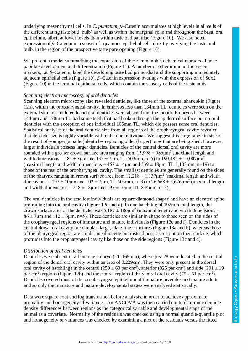
Figure 1 Mean taste papillae densities ± S.E. per cm2 found in various regions of the
oropharyngeal cavity of embryo Chiloscyllium punctatum (n=4, TL 134-165mm): Max 3483 ± 286;
DOCA 1040 ± 215; DOCC 420 ± 131; DOCS 711 ± 114; DPC 491 ± 85; DPS 608 ± 111; Man
2125 ± 267; VOCA 941 ± 98; VOCC 608 ± 24; VOCS 731 ± 174; VPC 459 ± 43; VPS 731 ± 174.
Maxillary valve (Max), mandibulary valve (Man), dorsal (D), ventral (V), oral cavity (OC),
pharynx (P), anterior (A), side (S), centre (C). TL of individual in the drawing is 146mm. Scale bar
3cm.
Bio
logy
Ope
n •
Adv
ance
art
icle
by guest on June 20, 2018http://bio.biologists.org/Downloaded from
Page 17
Figure 2 Mean taste papillae densities ± S.E. per cm2 found in various regions of the
oropharyngeal cavity of hatchling Chiloscyllium punctatum (n=3, TL 175-267mm): Max 2052 ±
437; DOCA 843 ± 76; DOCC 516 ± 90; DOCS 652 ± 115; DPC 438 ± 54; DPS 677 ± 104; Man
1565 ± 217; VOCA 801 ± 68; VOCC 385 ± 111; VOCS 559 ± 76; VPC 264 ± 20; VPS 500 ± 101.
Maxillary valve (Max), mandibulary valve (Man), dorsal (D), ventral (V), oral cavity (OC),
pharynx (P), anterior (A), side (S), centre (C). TL of individual in the drawing is 192mm. Half of
the pharynx is not included in the drawing. Scale bar 1mm.
Bio
logy
Ope
n •
Adv
ance
art
icle
by guest on June 20, 2018http://bio.biologists.org/Downloaded from
Page 18
Figure 3 Mean taste papillae densities ± S.E. per cm2 found in various regions of the
oropharyngeal cavity of immature juvenile Chiloscyllium punctatum (n=12, TL 426-844mm): Max
373 ± 62; DOCA 79 ± 15; DOCC 34 ± 7; DOCS 45 ± 10; DPC 30 ± 7; DPS 32 ± 10; Man 225 ±
26; VOCA 82 ± 23; VOCC 33 ± 6; VOCS 52 ± 15; VPC 27 ± 9; VPS 43 ± 10. Maxillary valve
(Max), mandibulary valve (Man), dorsal (D), ventral (V), oral cavity (OC), pharynx (P), anterior
(A), side (S), centre (C). TL of individual in the drawing is 521mm. Scale bar 0.5 cm.
Bio
logy
Ope
n •
Adv
ance
art
icle
by guest on June 20, 2018http://bio.biologists.org/Downloaded from
Page 19
Figure 4 Mean taste papillae densities ± S.E. per cm2 found in various regions of the
oropharyngeal cavity of mature adult Chiloscyllium punctatum (n=3, TL 1,062-1,177mm): Max 111
± 16; DOCA 27 ± 4; DOCC 12 ± 2; DOCS 20 ± 2; DPC 11 ± 3; DPS 10 ± 2; Man 89 ± 10; VOCA
29 ± 6; VOCC 10 ± 2; VOCS 29 ± 6; VPC 8 ± 2; VPS 12 ± 2. Maxillary valve (Max), mandibulary
valve (Man), dorsal (D), ventral (V), oral cavity (OC), pharynx (P), anterior (A), side (S), centre
(C). TL of individual in the drawing is 1,062mm. Scale bar 1 cm.
Bio
logy
Ope
n •
Adv
ance
art
icle
by guest on June 20, 2018http://bio.biologists.org/Downloaded from
Page 20
Figure 5 Taste papillae diameters (± standard error) for various individuals of Chiloscyllium
punctatum with different total lengths.
Bio
logy
Ope
n •
Adv
ance
art
icle
by guest on June 20, 2018http://bio.biologists.org/Downloaded from
Page 21
Figure 6 Scanning electron micrographs of taste papillae (P) on the a) maxillary and b)
mandibulary valves of an embryo (TL 116mm; note no teeth protruding), c) maxillary and d)
mandibulary valves of a hatchling (TL 192mm), and e) maxillary and f) mandibulary valves of an
immature (TL 484mm) Chiloscyllium punctatum. Nare (N), teeth (T).
Bio
logy
Ope
n •
Adv
ance
art
icle
by guest on June 20, 2018http://bio.biologists.org/Downloaded from
Page 22
Figure 7 Scanning electron micrographs of a) a taste papilla from the anterior dorsal oral
cavity of Chiloscyllium punctatum (TL 635mm), and b) a magnified view of the microvilli (arrow)
protruding from the apical surface of a papilla.
Bio
logy
Ope
n •
Adv
ance
art
icle
by guest on June 20, 2018http://bio.biologists.org/Downloaded from
Page 23
Figure 8 Scanning electron micrographs of a) a taste papilla situated within the ventral oral
cavity of Chiloscyllium punctatum (TL 635mm) with both protruding microvilli as well as b)
microvilli located within a pore.
Bio
logy
Ope
n •
Adv
ance
art
icle
by guest on June 20, 2018http://bio.biologists.org/Downloaded from
Page 24
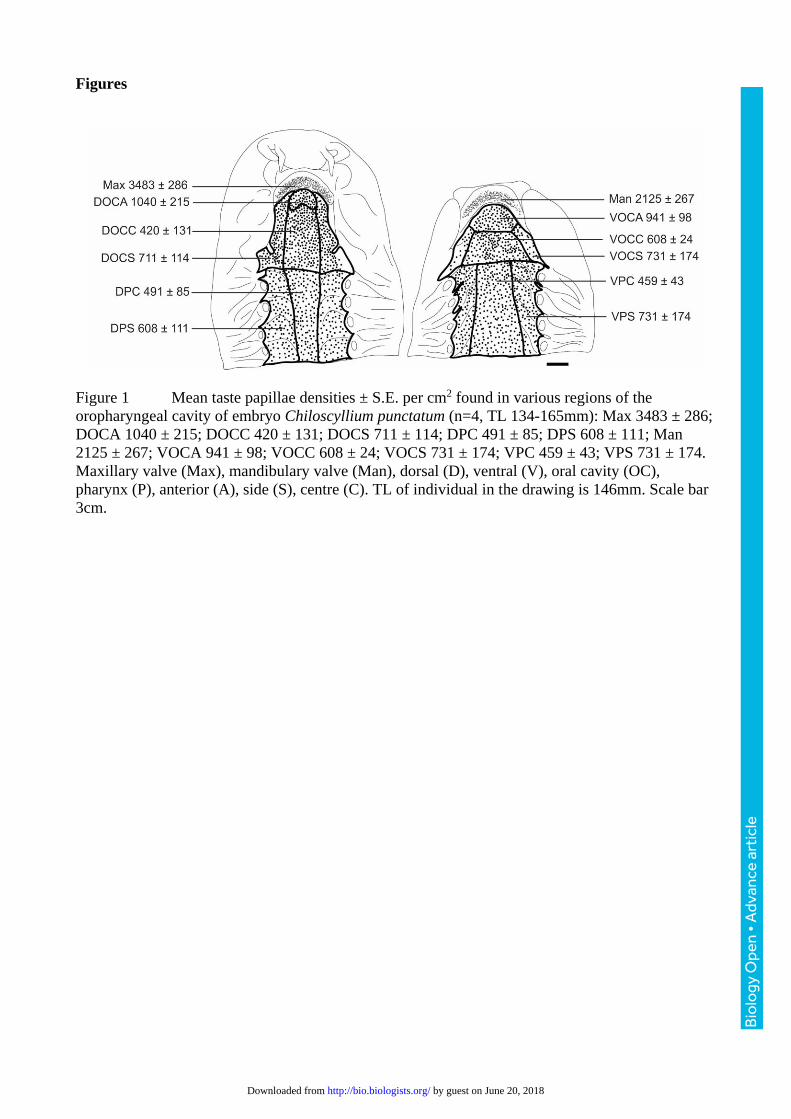
Figure 9 Scanning electron micrographs of a) taste papillae from an embryo Chiloscyllium
punctatum (TL 116mm). b) Higher magnification of an individual papilla with no noticeable
differentiated cells or microvilli protruding from the tip.
Bio
logy
Ope
n •
Adv
ance
art
icle
by guest on June 20, 2018http://bio.biologists.org/Downloaded from
Page 25
Figure 10 Immunofluorescent detection of markers of taste bud differentiation in a
Chiloscyllium punctatum prehatchling stage embryo (TL 103mm). A), B) β –Catenin is
concentrated in the cytoplasm and membrane and overlaps the nucleus of all cells of the primordial
Bio
logy
Ope
n •
Adv
ance
art
icle
by guest on June 20, 2018http://bio.biologists.org/Downloaded from
Page 26
taste bud ‘bulb’ (closed white arrow), marginal cells (white arrowhead) extending into the basal
epithelium of the papilla, as well as on superficial squamous epithelial cells at the apex of the
papilla (open white arrow). C), D) Strong HNK-1 immunoreactivity is detected the length of the
afferent nerve fibre innervating the taste papilla (white arrowhead) and within projections extending
into the taste bud, and is also detected on cell bodies in the mesenchyme directly underlying the
taste bud ‘bulb’ (open white arrow), and on cell bodies within the taste bud bulb (1-3 per section)
(closed white arrow). E-H) Nuclear Sox2 expression is seen in all cells of the taste bud ‘bulb’
including presumptive prospective sensory, support and basal cells (open white arrow) as well as
within marginal cells (white arrowhead). Sox2 is detected in all definitive taste papillae (white
asterisks in G) on the maxillary valve (3-5 per section). DNA is stained with DAPI and shown in
grey in all merged images. The borders of the epithelium with the underlying mesenchyme and the
oral cavity are delineated with yellow dotted lines in the single colour images. Scale bars denote
50μm. With the exception of the maxillary (G and H) valves all taste papillae imaged were in a
region of the lower jaw between the mandibular valve and the dental lamina.
Bio
logy
Ope
n •
Adv
ance
art
icle
by guest on June 20, 2018http://bio.biologists.org/Downloaded from
Page 27
Figure 11 Model of developing taste bud papilla in prehatchling stage Chiloscyllium punctatum
embryos. Cells expressing each of the four markers (Sox2, β –Catenin, HNK-1) of taste papilla
development studied are schematically arranged in a model papilla at mid-morphogenesis stage,
prior to final differentiation of sensory cells, and opening of the apical pore, but after innervation
has occurred. Epithelial contributions to the taste papillae from overlying squamous epithelium
(light grey background), and the basal columnar epithelium (light brown background) are denoted
as distinct from epithelium derived cells of the progenitors within the taste bud ‘bulb’ (light orange
background), and marginal cells (dark brown background).
Bio
logy
Ope
n •
Adv
ance
art
icle
by guest on June 20, 2018http://bio.biologists.org/Downloaded from
Page 28
Figure 12 Scanning electron micrographs of a hatchling Chiloscyllium punctatum (TL
192mm). a) External denticles of the rostrum, b) dorsal oral cavity showing denticles restricted to
the anterior (black arrow) and central oral cavity (white arrow), c) higher magnification of these
denticles and d) higher magnification of an individual denticle. Nares (N), spiracle (Sp), taste
papilla (P), teeth (T), oral denticle (D).
Bio
logy
Ope
n •
Adv
ance
art
icle
by guest on June 20, 2018http://bio.biologists.org/Downloaded from
Page 29
Figure 13 Scanning electron micrographs of the predominant oral denticles found in the a)
central dorsal oral cavity, which are circular and flat, c) the central pharynx, which are circular with
a raised point on their surface (arrow) and e) the sides of the oropharyngeal cavity with
square/diamond shaped denticles with prominent points on their surface (arrow) of Chiloscyllium
punctatum. b), d) and f) show the extracted denticles from these regions, respectively. Papilla (P).
(TLs for individuals used; a) 861mm, b) 1,117mm, c) 1,103mm, d) 601mm, e) 635mm, f) 503mm).
Bio
logy
Ope
n •
Adv
ance
art
icle
by guest on June 20, 2018http://bio.biologists.org/Downloaded from
Page 30
Graphical abstract
Using light microscopy, both scanning and transmission electron microscopy and
immunofluorescence the authors describe the ultrastructure, distribution and molecular
characterisation of taste buds in the developing bamboo shark, Chiloscyllium punctatum. Taste
papillae are functional, innervated units, allowing the shark to seek out food in utero, at birth or on
hatching.
Conflict of interest statement
All authors have no known or potential conflict of interest including any financial, personal or other
relationships with other people or organizations within three years of beginning the submitted work
that could inappropriately influence, or be perceived to influence, their work.
Role of authors
All authors had full access to all the data in the study and take responsibility for the integrity of the
data and the accuracy of the data analysis. Study concept and design: CJLA, KJM, GJF, SPC.
Acquisition of data: CJLA, KJM, GJF. Analysis and interpretation of data: CJLA, KJM, GJF, SPC.
Drafting of the manuscript: CJLA, KJM, GJF. Critical revision of the manuscript for important
intellectual content: SPC. Statistical analysis: CJLA. Obtained funding: GJF, SPC. Administrative,
technical, and material support: CJLA, KJM, GJF, SPC. Study supervision: GJF, SPC.
Bio
logy
Ope
n •
Adv
ance
art
icle
by guest on June 20, 2018http://bio.biologists.org/Downloaded from