Page 1
Review article DOI 10.1002/bies.200800207
Totally tubular: the mystery behind functionand origin of the brain ventricular systemLaura Anne Lowery1,2,3 and Hazel Sive1,2*
1 Whitehead Institute for Biomedical Research, Nine Cambridge Center, Cambridge, MA2Massachusetts Institute of Technology, Cambridge, MA, USA3Present address: Harvard Medical School, 240 Longwood Ave, Boston, MA, USA
A unique feature of the vertebrate brain is the ventricularsystem, a series of connected cavities which are filledwith cerebrospinal fluid (CSF) and surrounded by neu-roepithelium. While CSF is critical for both adult brainfunction and embryonic brain development, neitherdevelopment nor function of the brain ventricular systemis fully understood. In this review, we discuss the mys-tery of why vertebrate brains have ventricles, andwhence they originate. The brain ventricular systemdevelops from the lumen of the neural tube, as theneuroepithelium undergoes morphogenesis. The mole-cular mechanisms underlying this ontogeny aredescribed. We discuss possible functions of both adultand embryonic brain ventricles, as well as major braindefects that are associated with CSF and brain ventri-cular abnormalities. We conclude that vertebrates havetaken advantage of their neural tube to form the essentialbrain ventricular system.
Keywords: brain ventricle; CSF; morphogenesis; neural
tube; neural tube defects
What is the brain ventricular system?
The vertebrate brain has a characteristic and complex three-
dimensional structure. One highly conserved aspect of brain
structure is the brain ventricular system, a series of connected
cavities lying deep within the brain, filled with cerebrospinal
fluid (CSF) (Fig. 1).(1) The ventricles and the CSF they
contain, together with the surrounding neuroepithelium and
associated secretory structures, form the brain ventricular
system. The brain ventricles were first described over 2000
years ago, when it was believed that higher mental functioning
resided within them (reviewed in Ref.(2)). While this belief was
incorrect, the function of the brain ventricles is, indeed, very
important and under intense study.
In the adult human brain, there are four connected
ventricles: two lateral ventricles within the cerebrum, a third
ventricle within the diencephalon, and a fourth ventricle lying
*Correspondence to: H. Sive, Whitehead Institute for Biomedical Research,
Nine Cambridge Center, Cambridge, MA 02142, USA.
E-mail: [email protected]
446
between the cerebellum and pons (Fig. 1).(3) The lateral
ventricles are connected to the third ventricle, which is linked
to the fourth ventricle via the cerebral aqueduct. In turn, the
fourth ventricle joins to the spinal cord canal and the
subarachnoid space that envelops the brain. The adult
human brain contains about 140 mL of CSF, of which
approximately 20mL iswithin the ventricles and the remainder
is surrounding the brain.(1) Adult CSF is produced mainly by
the choroid plexuses, highly vascular structures located within
the ventricles (Fig. 1),(4) and some CSF may be produced by
cells lining the ventricles.(5) The choroid plexuses produce
about 500 mL/day, suggesting that the fluid is exchanged
3–4 times each day.(1) CSF flow forms a circulatory system
and is believed to flow from the lateral ventricles to the third
and fourth (Fig. 1), and then out into the subarachnoid space
where it is absorbed into the hematopoietic circulatory and
lymphatic systems.(1) Control of CSF flow is thought to
originate in pressure gradients produced by secretion and by
beating cilia with uniform orientation located on the
ependymal epithelium that lines the ventricles.(6)
What does the brain ventricular system do? The notion of a
circulatory system deep within the brain suggests functions
analogous to the hematopoietic circulatory system, including
transport of nutrients and wastes, and these functions have
been attributed to the adult CSF. In addition, CSF protects the
adult brain from physical trauma.(7) As we will discuss later in
this review, recent data suggest that in both the embryonic
and adult brains, CSF may additionally carry signaling
molecules that regulate neurogenesis and survival.(8,9)
Conservation of embryonic and adultbrain ventricular systems
The vertebrate embryonic brain originates from a columnar
epithelium that comprises the neural plate.(10) In humans, the
neural plate developsearly in the fourthweekafter fertilization,
and, later that week, completes neurulation to form the neural
tube.(11) The fundamental mechanisms of neurulation appear
to be largely conserved throughout vertebrates.(12) Subse-
quently, the ends (neuropores) of the tube close, the anterior
portion of the tube becomes the brain and the posterior
BioEssays 31:446–458, � 2009 Wiley Periodicals, Inc.
Page 2
Figure 1. The adult brain ventricular system. Cartoon representation of adult human brain ventricles. Blue represents brain tissue and yellow
shows brain ventricles. Choroid plexuses are in red, blue arrows designate direction of CSF flow. LV, lateral ventricle, 3V, third ventricle, 4V, fourth
ventricle.
L. A. Lowery and H. Sive Review article
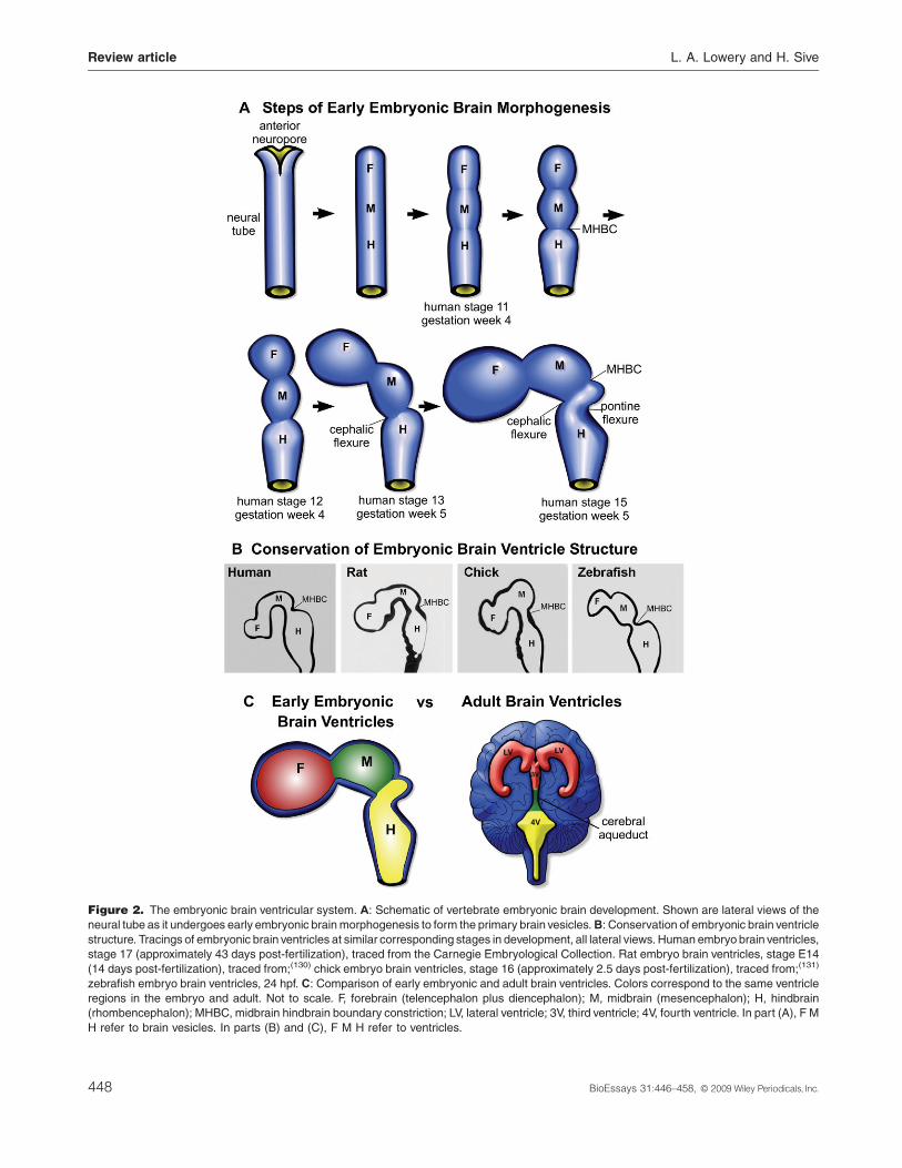
becomes the spinal cord. In most animals, toward the end of
neurulation, the future brain begins to undergo a series of
stereotypical constrictions, bends, and expansions, to sub-
divide into the primary embryonic ‘‘brain vesicles,’’ forming the
future forebrain, midbrain, and hindbrain (Fig. 2A and B).(10) In
teleosts, however, shaping of the brain occurs after neurula-
tion,(13) and thus themorphogenetic events of neurulation and
brain shaping are separable.
Thecavitiesof thebrainvesiclesarefilledwithCSFand form
the embryonic forebrain, midbrain, and hindbrain ventricles
(Fig. 2C). Following early brain ventricle shaping and initial
inflation, the ventricles undergo massive expansion, with
ventricle volume increasing significantly faster than brain
tissue growth.(14) In mammals and chicks, spinal cord
occlusion transiently seals off the brain ventricular space
directly preceding this expansion period,(15,16) whichmay then
allow intraluminalpressure topromoteventricleenlargement. It
is not knownwhether this process occurs in other vertebrates.
In most vertebrates, the embryonic forebrain ventricle splits
into the two lateral ventricles and the third ventricle. The
midbrain ventricle becomes the narrow cerebral aqueduct
which connects the third and fourth ventricles, and the
hindbrain ventricle becomes the fourth ventricle (Fig. 2C).(10)
While the gross anatomical development of the ventricles
is well documented, the molecular mechanisms underlying
this development remain poorly understood. However, several
vertebrate systems have been useful in understanding
development and function of the brain ventricles, including
chicken and rat. Recent work has demonstrated that the
zebrafish is a valuable system to study formation of the
embryonic brain ventricles, due to the ability to image live
embryos at single cell resolution, and through isolation of
genetic mutants.(13,17–19)
Molecular and cellular mechanisms of embryonic
brain ventricular system development
Crucial to ventricle formation is the neuroepithelium that
surrounds the brain ventricles. The embryonic brain ventri-
BioEssays 31:446–458, � 2009 Wiley Periodicals, Inc.
cular space directly reflects the position and shape of the
surrounding neuroepithelium, and thus, development of the
brain ventricles depends upon coordination of several aspects
of neuroepithelial development. First, the neuroepithelium is
patterned along the anteroposterior and dorsoventral axes,
which allows correct positioning of the ventricles and directs
downstream morphogenesis of the brain tissue. Second, the
neuroepithelium undergoes stereotypical and conserved
morphogenesis, regulated cell proliferation, and cell death,
in order to shape the brain and the ventricular cavities. Finally,
the neuroepithelium secretes the initial embryonic CSF
(eCSF) to inflate the ventricles. These processes are
described in more detail below.
Positioning the brain ventricles
Initial brain patterning along the anteroposterior and dorso-
ventral axes occurs before and during neurulation, such that
by the neural tube stage, embryonic brain tissue is subdivided
into distinct gene expression domains.(20) Thus, patterning
genes regulate the precise positioning of the brain ventricles,
including the characteristic and conserved constrictions and
bends within each brain region (Fig. 2B). Patterning genes
may play a proximal role in neuroepithelial morphogenesis
(Fig. 3), by directly controlling cytoskeletal machinery.
Conversely, patterning genes may play a distal role, required
for early tissue specification, which later leads to downstream
changes in the neuroepithelium, and, thus, brain ventricle
morphology.
Multiple publications document changes in patterning
gene expression that result in changes in brain morphology,
but it is generally unclear whether any of these genes directly
regulate brain morphogenesis. For example, zebrafish ace
embryos, that are mutant for fgf8 function, show severely
abnormal midbrain morphology, including incorrect shaping of
the brain ventricles and absence of the midbrain–hindbrain
boundary (MHB).(21) However, it is uncertain whether Fgf
signaling acts at the time ofmorphogenesis, or whether earlier
patterning defines cell types that later undergo morphogen-
447
Page 3
Figure 2. The embryonic brain ventricular system. A: Schematic of vertebrate embryonic brain development. Shown are lateral views of the
neural tube as it undergoes early embryonic brainmorphogenesis to form the primary brain vesicles.B: Conservation of embryonic brain ventricle
structure. Tracings of embryonic brain ventricles at similar corresponding stages in development, all lateral views. Human embryo brain ventricles,
stage 17 (approximately 43 days post-fertilization), traced from the Carnegie Embryological Collection. Rat embryo brain ventricles, stage E14
(14 days post-fertilization), traced from;(130) chick embryo brain ventricles, stage 16 (approximately 2.5 days post-fertilization), traced from;(131)
zebrafish embryo brain ventricles, 24 hpf. C: Comparison of early embryonic and adult brain ventricles. Colors correspond to the same ventricle
regions in the embryo and adult. Not to scale. F, forebrain (telencephalon plus diencephalon); M, midbrain (mesencephalon); H, hindbrain
(rhombencephalon); MHBC, midbrain hindbrain boundary constriction; LV, lateral ventricle; 3V, third ventricle; 4V, fourth ventricle. In part (A), F M
H refer to brain vesicles. In parts (B) and (C), F M H refer to ventricles.
Review article L. A. Lowery and H. Sive
448 BioEssays 31:446–458, � 2009 Wiley Periodicals, Inc.
Page 4
Figure 3. Neuroepithelial morphogenesis during brain ventricle development.A: Schematic showing zebrafish neuroepithelium as it opens into
the brain ventricles. After neurulation, the zebrafish neuroepithelium is a closed neural tube (i) connected by apical actin junctions and surrounded
by a basement membrane (i,iii). As the brain ventricles open, the neuroepithelium bends in locations of apical constriction (white asterisks) and
basal constriction at MHB (ii,iii). B: Schematic depicting stages of neurulation in mammals, beginning with the columnar epithelium of the neural
plate (i). Neurulation and hinge-point formation occur concurrently (ii), resulting in an open neural tube with hinge-points already formed. The
lumen remains open and expands after neurulation is complete (iii).C: Schematic depicting stages of neurulation in zebrafish, beginning with the
columnar neural plate (i). Neurulation progresses through a ‘‘neural keel’’ stage (ii) and ends with a closed neural tube (iii). Subsequently, the
neural tube opens and forms hinge-points to shape the ventricles (iv). D: Cartoons of transverse sections of the midbrain ventricle depicting
several phenotypes observed when neuroepithelium morphogenesis does not occur normally in zebrafish. When junctions are completely
disrupted (i), neurulation does not proceed and ventricle formation is impossible. When themidline does not form correctly (ii), the midline cannot
separate to form the ventricles. Somemutants show normal midline formation, but still do not separate at themidline and form hinge-points (iii). F,
Forebrain ventricle; M, midbrain ventricle; H, hindbrain ventricle; MHB, midbrain–hindbrain boundary.
L. A. Lowery and H. Sive Review article
BioEssays 31:446–458, � 2009 Wiley Periodicals, Inc. 449
Page 5
Review article L. A. Lowery and H. Sive
esis. A similar example involves zic1 and zic4, transcription
factors which regulate both cell fate specification and
proliferation in the zebrafish hindbrain and control hindbrain
ventricle formation, but at a time that is not yet known.(22) The
ventral neural signaling morphogen Sonic Hedgehog (Shh)
may have a proximal role in chick brain ventricle expansion.(23)
Shh is secreted from the notochord, which underlies the
midline of the neural tube, and later, from the floorplate.
Separating the notochord from the brain, after initial
patterning events have occurred, prevents brain ventricle
expansion, reduces cell proliferation, and increases cell
death,(23) suggesting that Shh may play a role in ventricle
formation. Shh signaling from the notochord is also required
for the regulation of neuroepithelial shape in mice, affecting
the location of dorsoventral hinge-points.(24) These limited
data indicate that a significant future challenge is to connect
patterning mechanisms with neuroepithelial morphogenesis.
Morphogenesis of the brain epithelium
In order for brain morphogenesis and associated ventricle
formation to occur, an intact, cohesive neuroepithelium must
form, with the appropriate junctions. The neuroepithelium
must be correctly shaped, requiring a functional cytoskeleton
and extracellular matrix (ECM). Additionally, regionally
restricted cell proliferation, and perhaps cell death, may
further modify the shape of the brain ventricles.
Forming the epithelium
The embryonic vertebrate neuroepithelium comprises a sheet
of cells that become connected by apically localized adherens
and tight junctions.(25–27) These junctions hold the cells
together to form a functional unit and also form a barrier
between the inside and outside of the neural tube. After the
neural tube has formed, the apical surface of the neuroe-
pithelium faces the lumen (the ventricles), and the basal
surface is on the outside of the tube (Fig. 3A). In most
vertebrates, shaping of the brain tube begins during
neurulation, as the lateral hinge-points form (Fig. 3B). These
hinge-points persist in the later embryonic brain and help
shape the ventricles. In the zebrafish neural tube, brain
ventricle morphogenesis occurs after neurulation is complete
(Fig. 3C). Thus, the fish model, unlike other vertebrate
models, offers the potential to separate processes controlling
neurulation from those controlling later brain ventricle
morphogenesis.
Information from zebrafish mutants has been highly
informative with regard to the role of specific junctional
components during brain development. In the N-cadherin
mutant, parachute, junctions fail to form and the neuroe-
pithelium falls apart prior to brain ventricle development
(Fig. 3Di).(28,29) Consistent data have been obtained from the
chick.(30) As N-cadherin is an integral member of neuroe-
450
pithelial junctions and is expressed throughout brain mor-
phogenesis,(31) it may be required at all stages of brain
ventricle morphogenesis.
Mutation in the apical adherens junction-associated
component, Mpp5, a MAGUK protein,(32) leads to a different
neuroepithelial phenotype. Zebrafish nagie oko mutants,
which lack Mpp5 function, form an intact neural tube, but
sections through the tube show that apical junctions are
disorganized (Fig. 3Dii).(13) The neuroepithelial midline does
not form or subsequently separate normally, leading to an
absence of brain ventricles (Fig. 3Dii). Additionally, apically
located hinge-points within the ventricles do not form.(13)
Further experiments will be able to address whether Mpp5 is
required both during neurulation and later during ventricle
morphogenesis.
In the zebrafish heart and soul mutant, which has a null
mutation in prkci (corresponding to protein kinase C iota),(33)
neuroepithelial junctions appear to be normal, but brain
ventricle inflation does not occur uniformly throughout the
tube (Fig. 3Diii).(18) These data suggest that some other
aspect of neuroepithelial function is abnormal in prkimutants,
perhaps cell–cell coordination, junction barrier formation, or
cytoskeletal remodeling.
Shaping the neuroepithelium
The neuroepithelium undergoes stereotypical constrictions
and bends that shape the brain ventricles. Mechanisms
required for shaping the neuroepithelium include midline
separation, cytoskeletal shape changes, and ECM function.
Midline separation In zebrafish, after neurulation, the
neural tube is closed, without a luminal space or morpholo-
gically visible midline(29) (Fig. 3Cii). Soon after, a midline
separating the left and right sides of the tube appears, with
apposition of apical surfaces on either side (Fig. 3Ciii). The
absence of a visible luminal space upon neural tube closure is
not unique to zebrafish, as transient occlusion occurs in other
model systems as well. For example in Xenopus, after neural
tube closure, the lumen disappears, to reappear as cells are
rearranged,(34) while in the chick spinal cord, the lumen is
occluded concomitant with ventricular expansion.(35) Subse-
quently, the tissue at the midline separates to form the
ventricular spaces (Fig. 3Civ). Part of this opening is due to
secretion of eCSF, however, analysis of zebrafish mutants
suggests that initial ventricle inflation requires some additional
process that results in separation of the left and right sides of
the tube.(18) We have called this latter process ‘‘midline
separation.’’(18) Several brain mutants that correspond to
abnormal apicobasal polarity/junction proteins (prkci/heart
and soul, crb2/oko meduzy, epb41l5/mosaic eyes) show a
morphologically distinct midline, based on actin staining, but
the ventricles only inflate partially, and in places appear to
remain shut.(18) These data indicate that correct apicobasal
polarity, junctions, and a functional epithelium are required for
BioEssays 31:446–458, � 2009 Wiley Periodicals, Inc.
Page 6
L. A. Lowery and H. Sive Review article
midline separation. One possible reason for this defect is that
the apical surfaces of the mutant neuroepithelia are unusually
adhesive, while another may be that mutant neuroepithelial
cells are less able than wild-type cells to move or change
shape.
Cytoskeleton Shaping the neuroepithelium and brain
ventricles requiresconstrictionsandbends inspecific locations
(Fig. 3Aii,iii), often involving individual cell shape changes and
cytoskeletal rearrangements.(36,37) For example, formation of
basally constricted cells and subsequent neuroepithelial
bending occurs at the MHB constriction (Fig. 3Aii).(19) Several
studies have looked at the cytoskeleton during neurulation and
hinge-point formation in frog and chick models. The hinge-
points that form during neurulation in these animals persist
later to shape the brain ventricles. The actin binding protein
Shroom, which influences both actin polymerization and
microtubule behavior, is required for apical constriction at
hinge-points in Xenopus and mice.(38,39) Members of the Ena/
VASP family coordinate cytoskeletal dynamics during Xeno-
pus neurulation, including apical constriction within the plate,
cell elongation, and cell–cell adhesion.(40)
Extracellular matrix Another component of the neuroe-
pithelium that is required for brain shaping is the ECM, located
at both apical and basal sides of the neuroepithelium. The
ECM may play a mechanical role by providing structural
support, allowing a changing epithelium to bend and hold its
shape, as a unit. The ECM may also play a crucial signaling
role, interacting with apical and basolateral junctions and the
cytoskeletal machinery to change the shape of cells.(41,42) A
recent study from our laboratory has shown that formation of
the zebrafishMHB constriction is caused by basal constriction
of neuroepithelial cells and is dependent on laminin in the
basement membrane.(19) Fibronectin is also required for
zebrafish brain ventricle expansion, perhaps by stabilizing
neuroepithelial structure.(18) The roles that laminin and
fibronectin play during zebrafish brain ventricle morphogen-
esis are consistent with the requirement for ECM in epithelial
morphogenesis during rat neurulation,(43) chick otic placode
invagination(44) and chick lens vesicle formation.(45)
ECM components at the apical surface of the neuroe-
pithelium may also be crucial. Chick and rat brain ventricles
contain an apical ECM rich in chondroitin sulfate, hyaluronic
acid, and other proteoglycans, and these may play a role in
brain ventricle formation by promoting neuroepithelial integrity
and cell shape changes as well as regulating the eCSF
osmolality and intraluminal pressure during brain ventricle
inflation.(43,46–50)
Regional cell proliferation and cell death
Another neuroepithelial process that may regulate brain
morphogenesis and ventricle development is cell proliferation,
and it has been suggested that brain ventricle shaping
depends upon localized cell proliferation throughout the
BioEssays 31:446–458, � 2009 Wiley Periodicals, Inc.
neural tube.(10,14,51) Consistent with this idea, regions of
constriction between the quail forebrain and midbrain, as well
as between the telencephalic ventricles, have significantly
higher numbers of post-mitotic cells than the surrounding
tissue.(52) Additionally, the MHB region in the zebrafish shows
about two-fold less proliferation than surrounding tissues.(13)
The MHB does not open to form a ventricular space (Fig. 3A),
but it is not known whether the lower rate of cell proliferation
regulates ventricle opening in this region. Addition of a DNA
synthesis inhibitor to zebrafish just before the ventricles open
results in smaller but normally shaped brain ventricles,(13)
indicating a requirement for cell proliferation in ventricle
development but not necessarily in neuroepithelial shaping.
This result is consistent with previous studies in Xenopus.(53)
Multiple genes are known to regulate region-specific
proliferation in the brain. For example, the transcription factor
Bf-1 is required for proliferation of telencephalic cells and is
essential for normal morphogenesis of the telencephalon in the
rat.(54) The zebrafish zic2a and zic5 transcription factors,
required for cell proliferation in the midbrain, and zic1 and zic4,
required for cell proliferation in the hindbrain, have been
implicated in formation of normal midbrain and hindbrain
ventricles,respectively.(22,55)Despitethesedata,themechanism
by which proliferation regulates ventricle formation is not clear.
Spatially regulated cell death (apoptosis) may also
contribute to brain shaping.(56,57) Blocking programmed cell
death in mice, by loss of caspase function, causes an
overgrowth of brain tissue and obstructed brain ventricles.(58)
Conversely, mouse mutants which show more cell death than
wild type also show reductions in the amount of brain tissue
and overexpansion of the ventricles.(59) However, localized
cell death is not apparent during initial shaping of the zebrafish
brain ventricles,(13) suggesting that apoptosis may not play a
role at these stages.
Inflation of the brain ventricles
A key process in brain ventricle formation is secretion of
embryonic CSF (eCSF) into the ventricular lumen (Fig. 4). In
zebrafish, formation of eCSF requires the NaþKþATPase ion
pump.(13) Zebrafish embryos lacking NaþKþATPase activity
fail to inflate the brain ventricles,(13) and this pump likely forms
an osmotic gradient required for fluid movement into the
ventricle lumen.(60,61) Studies in chick and rat embryos show
that proteoglycans in the eCSF, secreted by the neuroepithe-
lium, also regulate fluid movement into, and size of, the brain
ventricles (Fig. 4).(46,48,49)
Classic studies in chick embryos have suggested that
intraluminal pressure resulting from the accumulation of eCSF
is necessary for normal brain development, and consistently,
intubation of the chick embryonic hindbrain ventricle results in
a collapse of the ventricles.(62–64) While the choroid plexus
plays amajor role in CSF production in the adult, its role during
451
Page 7
igure 4. eCSF formation and function during brain ventricle infla-
on. Cartoon depicting eCSF secretion and function. Inset: dorsal
iew of embryonic brain, after initial lumen inflation, with enlarged
rea (hindbrain) boxed. Ion pumps and proteoglycan secretion are
ought to form an osmotic gradient regulating fluid flow. Signaling
nd growth factors are also secreted. Both fluid pressure and growth
ctors stimulate cell proliferation and gene expression within the
urrounding neuroepithelium. Not drawn to scale. PG: proteoglycans.
ircular cells at ventricular surface are mitotic cells.
Review article L. A. Lowery and H. Sive
Fti
v
a
th
a
fa
s
C
embryonic brain ventricle development is less clear. However,
when the brain ventricles initially fill with fluid and later expand,
the choroid plexus has not yet formed. In humans, brain
ventricles inflate several weeks prior to choroid plexus
formation.(14) In zebrafish, the ventricles begin inflating at 19
hours post-fertilization (hpf) and the choroid plexus is not
formed until approximately 48 hpf.(65) Thus, there must be
some other source of eCSF besides the choroid plexus during
embryonic CSF production, and the neuroepithelial tissue
surrounding the ventricles may play a large role.
Brain ventricle function
In this section, we review the functions of CSF in the adult and,
particularly, in the embryo (Figs. 4 and 5A).
Adult functions
The functions of CSF in the adult include protection, nutrient
transport, and waste removal. These functions were first
attributed over a 100 years ago, and there is undoubtedly
452
more to learn about themechanisms underlying each of them,
as well as their importance for adult brain function. Several
unstudied aspects of the ventricles and CSF remain. For
example, a circumventricular system of neurons sends
dendrites and axons into the ventricular space.(66) The
function of these neurons is unknown, but it seems reason-
able to suggest they may sense factors in the CSF or secrete
neurotransmitters into the CSF. More recently, it has been
suggested that the brain ventricles play a role in controlling
homeostatic, hormonal, and signaling mechanisms involved
in brain function.(8,9) Significant evidence indicates that
growth factors and other signals circulate within the CSF
and have an effect on brain function.(4,67) Thus, there is strong
evidence that gonadotropin-releasing hormone released into
CSF directly affects sexual behavior in sheep.(68) Recent work
has shown that cilia-mediated CSF flow in the lateral
ventricles directs migration of developing neurons in the
adult rat brain,(69) indicating that fluid flow may be a crucial
function of the brain ventricles. Overall, these data indicate
that the CSF within adult brain ventricles plays complex roles
in brain function.
Embryonic functions
Requirement for eCSF in neuroepithelial survival and
proliferation
A role for the eCSF has been considered only recently, but
significant data indicate that this fluid regulates neuronal
proliferation and determination. In humans, for several weeks
after neural tube closure, the embryonic brain primarily
comprises proliferating pluripotent neuroepithelial cells,
considered to be the first neural stem cells.(14) Neuroepithelial
proliferation occurs almost exclusively at the ventricular
surface,(70) and contact with eCSF and the factors it contains
may be a prerequisite for production of early, pluripotent
neuroepithelial cells(14) (Figs. 4 and 5A). Only a few neuronal
progenitor populations undergo mitosis distal to the ventricles
(e.g., Ref.(71)), and these cells are fate-restricted and
generated late in development. Moreover, there is a striking
correlation between brain ventricle size and amount of
neuronal cell proliferation within the corresponding periven-
tricular region(14)—thus, the bigger the ventricle, the greater
the amount of subsequent cell proliferation. Consistent with
these observations, drainage of eCSF leads to reduced cell
proliferation and increased apoptosis in the developing chick
brain,(72) indicating that eCSF is necessary for normal
neuronal development.
Role of fluid pressure on brain development
One mechanism by which eCSF may regulate brain
development is through creating pressure within the brain
ventricles (Fig. 4).(73) Desmond and colleagues observed a
BioEssays 31:446–458, � 2009 Wiley Periodicals, Inc.
Page 8
Figure 5. Summary of developmental mechanisms underlying ventricle formation, function of the brain ventricular system, and associated
abnormalities. See text for details.
L. A. Lowery and H. Sive Review article
50-fold increase in intraluminal pressure during chick brain
ventricle expansion and showed that artificially increasing
pressure via saline injection increases neuroepithelial
mitoses.(73) Although the mechanism by which fluid pressure
affects brain development is not understood, these data are
consistent with the development of other organ systems. For
example, in the zebrafish heart, blood flow modifies the
morphology of the atrial and ventricular lumens(74) and
stimulates valve morphogenesis,(75) and in tissue culture, cell
stretching increases cell proliferation.(76)
Factors contained in eCSF
In addition to fluid pressure, significant data indicate that
factors within the eCSF are pivotal in brain ventricle
development and function. Embryonic CSF has a complex
protein composition that differs substantially from adult CSF.
While adult CSF has only trace amounts of protein, with
detectable levels usually indicating infection, damage, or
other pathology,(1) eCSF is protein-rich, with a changing
composition during development and between ventricles.(77–79)
Proteomic analyses of human, rat, mouse, and chick eCSF
have identified approximately 200 different proteins, including
signaling and growth factors, ECM proteins, transport and
carrier proteins, and enzymes and proteases.(77–79)
Consistent with a role for factors in eCSF in promoting
neuroepithelial growth, isolated chick and rat embryonic brain
cells are not able to replicate or undergo neurogenesis in
definedmedium, but addition of eCSF to the cultures promotes
cell survival, proliferation, and neurogenesis.(80,81) Immuno-
BioEssays 31:446–458, � 2009 Wiley Periodicals, Inc.
depleting the chick ventricles of Fgf2, a component of eCSF
which contributes to regulating neurogenesis, reduces cell
proliferation by 50%.(82–84) Recent neuroepithelial explant
studies indicate roles for eCSF low-density lipoprotein and
retinoic acid in promoting neurogenesis in chicks, and this is
supported by knockout studies in mice.(85,86) Parada and
colleagues have shown that explants of chick midbrain tissue
cultured with basal medium do not express the midbrain
markers otx2 and fgf8. However, when cultured with eCSF-
supplemented medium the tissues maintain normal gene
expression patterns,(87) suggesting that eCSF may regulate
this early aspect of brain development. Finally, rat neural cells
behavedifferently inculturedependingontheembryonicageof
eCSF applied to the cells.(81) Prenatal E21 eCSF can support
proliferationofcorticalcells fromE19,butnotE17,embryos,(81)
suggesting that components within the eCSF regulate
neurogenesis in a developmental stage-dependent manner.
Together, thesestudies indicatethateCSFplaysacrucial role
in normal development, and understanding its function is likely
critical for the success of neural stem cell technology.(77,88)
Brain defects connected to ventricleabnormalities
Abnormalities in brain ventricle structure and CSF regulation
are associated with several common birth defects and often
have severe consequences for brain function. These defects
highlight the importance of the tubular nervous system, and
453
Page 9
Review article L. A. Lowery and H. Sive
most appear to relate to an absence of CSF, too much CSF, or
CSF of incorrect composition. These abnormalities include
cranial neural tube closure defects, hydrocephalus, and
neurodevelopmental mental health disorders (Fig. 5B). Sev-
eral of these disorders can also present for the first time in the
adult and may be due to long-term accumulation of mild early
defects or a sudden late causal event.
Anencephaly
Neural tube defects that result in failure of the tube to close
have devastating consequences for brain development and
mental function. When the anterior neuropore fails to close,
that is, the tube remains open at the front of the brain, a defect
called anencephaly results (Fig. 5B). Anencephaly leads to a
failure of forebrain development, and human fetuses with this
condition are either stillborn or die within 24 hours after birth.
In this condition, the brain ventricular system is open, eCSF
can escape, and the brain is exposed to the outside
environment. The outcome of this disorder indicates the
importance of the normal brain ventricular environment for
brain development. However, it is not known whether the
surrounding foreign fluid actively destroys the forebrain,(89) or
whether the forebrain fails to develop due to absence of
factors normally found in the eCSF that are required for
neuronal specification and survival.
Schizencephaly
In schizencephaly, a slit or cleft within the brain tissue allows
CSF to leave the brain ventricular system(90) (Fig. 5B). The
cleft can be bilateral (on both sides of the brain) or unilateral,
and the walls of the cleft can be apposed (closed lip) or
separated (open lip). Schizencephaly results in numerous
neurological problems, including seizures, developmental
delays, difficulties with language and motor skills, and death.
Severity of the problems depends on the size of the defect. For
example, open lip bilateral schizencephaly is more severe
than the closed lip unilateral form. It is thought that
schizencephaly arises during early brain development, and
some data suggest that the malformation is due to impaired
neuronal migration,(90) but the pathology and etiology of
schizencephaly are unclear. Although CSF escapes abnor-
mally from the forebrain in both schizencephaly and
anencephaly, schizencephaly is by far the less severe of
these disorders, suggesting that it arises after the telence-
phalon is established, and/or that the loss of CSF is not as
great as in anencephaly.
Hydrocephalus
Hydrocephalus is characterized by an excess of CSF and
overdilation of the brain ventricles (Fig. 5B). It is one of the
454
most common birth defects, occurring in up to 1/500 births
(congenital hydrocephalus),(91–93) but it can also arise in
children and adults (acquired hydrocephalus). Congenital
hydrocephalus often results in severe disruption of brain
development, including decreased neurogenesis,(93–95)
whereas acquired hydrocephalus damages brain tissue that
is already formed. One form of acquired hydrocephalus
prevalent in geriatric patients is ‘‘normal pressure hydro-
cephalus.’’ This late onset form leads to progressive mental
impairment and gait disturbances, but the underlying cause of
the disorder is often unknown andmay not be the same as the
embryonic form.
It has been suggested that hydrocephalus may result from
too much CSF production, too little CSF absorption, impaired
CSF flow, and/or abnormal brain shaping leading to blockages
of narrow canals that connect the ventricles, especially the
cerebral aqueduct.(93,95,96) All of these cases would lead to
excessive CSF within the brain ventricles and increased
intracranial pressure. These abnormal events may be due to
environmental insults, such as brain injury, or to genetic
defects (believed to account for at least half of all cases(93)).
Multiple genes/loci have been implicated in mammalian
models of hydrocephalus, many of which correspond to
proteins involved in neural development (for example, otx2,
rfx4, alpha-SNAP).(93) However, only one gene, the neural cell
adhesion molecule L1CAM, has been clearly linked to human
hydrocephalus, but the mechanism by which the L1CAM
mutation leads to hydrocephalus is unknown. Intriguingly,
recent studies have shown that mutations in genes required
for development of cilia (that line the brain ventricles) can
cause hydrocephalus in both mice(97) and zebrafish.(98,99)
This is likely because loss of cilia-induced fluid flow leads to
fluid accumulation within the ventricles.
Previously, it was assumed that the increased intracranial
pressure that results from hydrocephalus was the main
damaging force to the brain. However, in cases of early onset
hydrocephalus where shunts are used to relieve pressure
prior to tissue damage, brain development is still abnor-
mal.(94,100) Consistently, direct evidence indicates that
abnormal factors within eCSF may be responsible for
pathology in certain cases of hydrocephalus. While normal
eCSF promotes cell proliferation, eCSF obtained from the
enlarged ventricles of the hydrocephalic rat model inhibits cell
proliferation in culture.(101) Furthermore, abnormalities in
eCSF protein content, including reduced proteoglycans, are
detectable prior to any morphological brain defects in the rat
hydrocephalic model.(95) Finally, while cortical periventricular
cells in the hydrocephalic rat brain do not divide, they
proliferate normally once they are removed from the in vivo
environment and cultured in vitro with wild-type eCSF.(101)
These results all suggest that abnormal regulation of eCSF
factors, rather than increased fluid pressure, may be an
underlying cause of hydrocephalus-related brain damage.
BioEssays 31:446–458, � 2009 Wiley Periodicals, Inc.
Page 10
L. A. Lowery and H. Sive Review article
Neurodevelopmental disorders with altered brain
ventricle structure
In addition to neural tube defects and hydrocephalus, a wide
range of neurodevelopmental disorders have been correlated
with more subtle abnormalities in brain ventricle size and
shape (Fig. 5B). These include schizophrenia, autism,
idiopathic and syndromal mental retardation, fragile X
syndrome, Down’s syndrome, attention-deficit-hyperactivity
disorder, and other learning disorders.(102–111) Even mild
ventricle enlargements are associated with developmental
abnormalities, including motor and language delays, in the
first 2 years of life.(112–117) In addition, ventricular enlargement
is one of the earliest and most consistently reported structural
brain abnormalities found in schizophrenia.(103,110,118–122)
Polymorphisms of themed12 (mediator of RNA polymerase II
transcription, subunit 12 homolog) gene in humans are
associated with an increased risk for schizophrenia.(123)
Interestingly, zebrafish mutants which lack med12 function
show early brain ventricle structure defects, as well as loss of
specific neuronal classes.(18)
It is not obvious how brain ventricle abnormalities and
these mental health disorders are correlated. Does loss of
neural tissue lead to ventricle enlargement and altered
structure, or is ventricle enlargement a proximal cause? In
several disorders, brain ventricle abnormalities arise during
early stages of development,(102,120) suggesting that further
study of the mechanisms involved in brain ventricle formation
may shed light on brain ventricle abnormalities and how to
prevent them.
Conclusion
Origin of the brain ventricles
The conservation of the vertebrate neural tube raises the
question of its origin. In the mid-1800s, it was recognized that
tunicate larvae (urochordates) form a tubular nervous system
similar to that of vertebrates (discussed in Ref.(124)).
Subsequent analyses of morphology and gene expression
patterns suggest homology between the neural tube in non-
vertebrate chordates and the vertebrates.(125,126) In larvae of
the urochordate Ciona, a neural tube forms with a very tiny
lumen, which later expands anteriorly to form the prosence-
phalic ventricle.(124,127) As in vertebrates, the lumen of the
Ciona neural tube is lined with ciliated cells, perhaps
indicating a shared function with the vertebrate ventricular
system.(128) In the cephalochordate Amphioxus, the brain is
also tubular and forms a fluid-filled, anterior cerebral
vesicle(126,129) that may be homologous with the vertebrate
brain ventricles. Although the function of the urochordate and
cephalochordate tubes have been little considered, it is
BioEssays 31:446–458, � 2009 Wiley Periodicals, Inc.
reasonable to suggest that vertebrate ventricular function
may have originated in ancestral lineages.
Importance of the vertebrate brain ventricular
system, and what’s next?
Neither development nor function of the vertebrate brain
ventricular system is fully understood in any animal system,
and a long list of unanswered questions remains. Underlying
these is theassumption that theventricularsystemcarriesouta
common set of functions in all vertebrates. Part of this
assumption stems from the conserved tubular nervous
system, the similarity in embryonic brain morphology across
vertebrate groups, and the homologous set of genes known to
be required for development and function of the brain in model
vertebrates. One significant future challenge is to understand
the molecular connection between brain patterning and brain
morphogenesis, including ventricle shaping. The precise role
of epithelial junctional complexes and the ECM during brain
morphogenesis and ventricle formation remain unclear. The
connection between cell proliferation and brain morphogen-
esis is also not understood. The extent to which eCSF governs
neuroepithelial fate remainsanareaof key interest.What is the
roleof eCSFflowandpressure?What are the rolesof themany
factors in the eCSF? Does the eCSF primarily govern cell
division/proliferation in the brain, or is its primary role to direct
formation of specific neuronal or glial subtypes? What is the
molecular basis for anencephaly, schizencephaly, and hydro-
cephalus? Is the connectionbetweenventricular abnormalities
andmental health disorders causal? In sum, over the next few
years, collecting these data will help piece together the set of
mechanisms by which the vertebrate brain ventricular system
forms and the functional significance of the tubular nervous
system.
Acknowledgments: We thank members of the Sive Lab for
helpful comments. Supported by NIH MH70926 and
MH59942 to H.L.S., and an NRSA fellowship to L.A.L.
References
1. Davson, H. and Segal, M. B., Physiology of the CSF and blood-brain
barriers. Boca Raton, CRC Press, 1996.
2. Tascioglu, A. O. and Tascioglu, A. B., Ventricular anatomy: illustrations
and concepts from antiquity to Renaissance. Neuroanatomy 2005. 4: 57–
83.
3. Millen, J. W. and Woollam, D. H. M., The anatomy of the cerebrospinal
fluid. London, Oxford University Press, 1962.
4. Emerich, D. F., Skinner, S. J., Borlongan, C. V., Vasconcellos, A. V.
and Thanos, C. G., The choroid plexus in the rise, fall and repair of the
brain. Bioessays 2005. 27: 262–274.
5. Vigh, B., Manzano e Silva, M. J., Frank, C. L., Vincze, C., Czirok, S. J.,
et al. The system of cerebrospinal fluid-contacting neurons. Its supposed
role in the nonsynaptic signal transmission of the brain. Histol Histopathol
2004. 19: 607–628.
455
Page 11
Review article L. A. Lowery and H. Sive
6. Nicholson, C., Signals that go with the flow. Trends Neurosci 1999. 22:
143–145.
7. Segal, M. B., Transport of nutrients across the choroid plexus. Microsc
Res Tech 2001. 52: 38–48.
8. Miyan, J. A., Nabiyouni, M. and Zendah, M., Development of the brain:
a vital role for cerebrospinal fluid. Can J Physiol Pharmacol 2003. 81:
317–328.
9. Johanson, C. E. Duncan, J. A., III, Klinge, P. M., Brinker, T., Stopa,
E. G., et al. Multiplicity of cerebrospinal fluid functions: new challenges in
health and disease. Cerebrospinal Fluid Res 2008. 5: 10.
10. Gray, H. and Clemente, C. D., Anatomy of the human body. 30th
American Ed. Philadelphia, Lea & Febiger, 1985.
11. O’Rahilly, R. and Muller, F., Neurulation in the normal human embryo.
Ciba Found Symp 1994. 181: 70–82; discussion 82–89.
12. Lowery, L. A. and Sive, H., Strategies of vertebrate neurulation and a re-
evaluation of teleost neural tube formation. Mech Dev 2004. 121: 1189–
1197.
13. Lowery, L. A. and Sive, H., Initial formation of zebrafish brain ventricles
occurs independently of circulation and requires the nagie oko and
snakehead/atp1a1a.1 gene products. Development 2005. 132: 2057–
2067.
14. Bayer, S. A. and Altman, J., The human brain during the early first
trimester. Atlas of human central nervous system development, Boca
Raton, FL; London, CRC, 2008.
15. Schoenwolf, G. C. and Desmond, M. E., Neural tube occlusion pre-
cedes rapid brain enlargement. J Exp Zool 1984. 230: 405–407.
16. Desmond, M. E., Description of the occlusion of the spinal cord lumen in
early human embryos. Anat Rec 1982. 204: 89–93.
17. Schier, A. F., Neuhauss, S. C., Harvey, M., Malicki, J., Solnica-Krezel,
L., et al. Mutations affecting the development of the embryonic zebrafish
brain. Development 1996. 123: 165–178.
18. Lowery, L. A., De Rienzo, G., Gutzman, J. and Sive, H., Characteriza-
tion and classification of zebrafish brain mutants. Anat Rec Dec 2008.
292: 94–106.
19. Gutzman, J. H., Graeden, E. G., Lowery, L. A., Holley, H. and Sive, H.,
Formation of the zebrafish midbrain-hindbrain boundary constriction
requires laminin-dependent basal constriction. Mech Dev 125: 974–
983; DOI: 10.1016/j.mod.2008.07.004.
20. Lumsden, A. and Krumlauf, R., Patterning the vertebrate neuraxis.
Science 1996. 274: 1109–1115.
21. Reifers, F., Bohli, H.,Walsh, E. C., Crossley, P. H., Stainier, D. Y., et al.
Fgf8 is mutated in zebrafish acerebellar (ace) mutants and is required for
maintenance of midbrain-hindbrain boundary development and somito-
genesis. Development 1998. 125: 2381–2395.
22. Elsen, G. E., Choi, L. Y., Millen, K. J., Grinblat, Y. and Prince, V. E.,
Zic1 and Zic4 regulate zebrafish roof plate specification and hindbrain
ventricle morphogenesis. Dev Biol 2008. 314: 376–392.
23. Britto, J., Tannahill, D. and Keynes, R., A critical role for sonic hedge-
hog signaling in the early expansion of the developing brain. Nat
Neurosci 2002. 5: 103–110.
24. Ybot-Gonzalez, P., Gaston-Massuet, C., Girdler, G., Klingensmith, J.,
Arkell, R., et al. Neural plate morphogenesis during mouse neurulation is
regulated by antagonism of Bmp signalling. Development 2007. 134:
3203–3211.
25. Aaku-Saraste, E., Hellwig, A. and Huttner, W. B., Loss of occludin and
functional tight junctions, but not ZO-1, during neural tube closure–
remodeling of the neuroepithelium prior to neurogenesis. Dev Biol
1996. 180: 664–679.
26. Chenn, A., Zhang, Y. A., Chang, B. T. and McConnell, S. K., Intrinsic
polarity of mammalian neuroepithelial cells. Mol Cell Neurosci 1998. 11:
183–193.
27. Geldmacher-Voss, B., Reugels, A. M., Pauls, S. and Campos-Ortega,
J. A., A 90-degree rotation of the mitotic spindle changes the orientation
of mitoses of zebrafish neuroepithelial cells. Development 2003. 130:
3767–3780.
28. Lele, Z., Folchert, A., Concha, M., Rauch, G. J., Geisler, R., et al.
Parachute/n-cadherin is required for morphogenesis and maintained
integrity of the zebrafish neural tube. Development 2002. 129: 3281–
3294.
456
29. Hong, E. and Brewster, R., N-cadherin is required for the polarized cell
behaviors that drive neurulation in the zebrafish.Development 2006. 133:
3895–3905.
30. Ganzler-Odenthal, S. I. and Redies, C., Blocking N-cadherin function
disrupts the epithelial structure of differentiating neural tissue in the
embryonic chicken brain. J Neurosci 1998. 18: 5415–5425.
31. Suzuki, S. C. and Takeichi, M., Cadherins in neuronal morphogenesis
and function. Dev Growth Differ 2008. 50: S119–S130.
32. Wei, X. and Malicki, J., nagie oko, encoding a MAGUK-family protein, is
essential for cellular patterning of the retina. Nat Genet 2002. 31: 150–
157.
33. Horne-Badovinac, S., Lin, D.,Waldron, S., Schwarz, M., Mbamalu, G.,
et al. Positional cloning of heart and soul reveals multiple roles for
PKC lambda in zebrafish organogenesis. Curr Biol 2001. 11:
1492–1502.
34. Davidson, L. A. and Keller, R. E., Neural tube closure in Xenopus laevis
involves medial migration, directed protrusive activity, cell intercalation
and convergent extension. Development 1999. 126: 4547–4556.
35. Schoenwolf, G. C. and Desmond, M. E., Descriptive studies of occlu-
sion and reopening of the spinal canal of the early chick embryo. Anat
Rec 1984. 209: 251–263.
36. Lecuit, T. and Lenne, P. F., Cell surface mechanics and the control of
cell shape, tissue patterns and morphogenesis. Nat Rev Mol Cell Biol
2007. 8: 633–644.
37. Fristrom, D., The cellular basis of epithelial morphogenesis. A review.
Tissue Cell 1988. 20: 645–690.
38. Lee, C., Scherr, H. M. and Wallingford, J. B., Shroom family proteins
regulate gamma-tubulin distribution and microtubule architecture during
epithelial cell shape change. Development 2007. 134: 1431–1441.
39. Hildebrand, J. D. and Soriano, P., Shroom, a PDZ domain-containing
actin-binding protein, is required for neural tube morphogenesis in mice.
Cell 1999. 99: 485–497.
40. Roffers-Agarwal, J., Xanthos, J. B., Kragtorp, K. A. and Miller, J. R.,
Enabled (Xena) regulates neural plate morphogenesis, apical constric-
tion, and cellular adhesion required for neural tube closure in Xenopus.
Dev Biol 2008. 314: 393–403.
41. Larsen, M., Artym, V. V., Green, J. A. and Yamada, K. M., The matrix
reorganized: extracellular matrix remodeling and integrin signaling. Curr
Opin Cell Biol 2006. 18: 463–471.
42. Sechler, J. L., Corbett, S. A., Wenk, M. B. and Schwarzbauer, J. E.,
Modulation of cell-extracellular matrix interactions. Ann N Y Acad Sci
1998. 857: 143–154.
43. Tuckett, F. and Morriss-Kay, G. M., Heparitinase treatment of rat
embryos during cranial neurulation. Anat Embryol (Berl) 1989. 180:
393–400.
44. Moro-Balbas, J. A., Gato, A., Alonso, M. I., Martin, P. and de la Mano,
A., Basal lamina heparan sulphate proteoglycan is involved in otic
placode invagination in chick embryos. Anat Embryol (Berl) 2000.
202: 333–343.
45. Gato, A., Martin, C., Alonso, M. I., Martinez-Alvarez, C. and Moro,
J. A., Chondroitin sulphate proteoglycan is involved in lens vesicle
morphogenesis in chick embryos. Exp Eye Res 2001. 73: 469–478.
46. Gato, A., Moro, J. A., Alonso, M. I., Pastor, J. F., Represa, J. J., et al.
Chondroitin sulphate proteoglycan and embryonic brain enlargement in
the chick. Anat Embryol (Berl) 1993. 188: 101–106.
47. Ojeda, J. L. and Piedra, S., Evidence of a new transitory extracellular
structure within the developing rhombencephalic cavity. An ultrastruc-
tural and immunoelectron-microscopic study in the chick. Anat Embryol
(Berl) 2000. 202: 257–264.
48. Alonso, M. I., Gato, A., Moro, J. A., Martin, P. and Barbosa, E.,
Involvement of sulfated proteoglycans in embryonic brain expansion
at earliest stages of development in rat embryos. Cells Tissues Organs
1999. 165: 1–9.
49. Alonso, M. I., Gato, A., Moro, J. A. and Barbosa, E., Disruption of
proteoglycans in neural tube fluid by beta-D-xyloside alters brain enlar-
gement in chick embryos. Anat Rec 1998. 252: 499–508.
50. Yip, G. W., Ferretti, P. and Copp, A. J., Heparan sulphate proteogly-
cans and spinal neurulation in the mouse embryo. Development 2002.
129: 2109–2119.
BioEssays 31:446–458, � 2009 Wiley Periodicals, Inc.
Page 12
L. A. Lowery and H. Sive Review article
51. Bergquist, H. and Kallen, B., On the development of neuromeres to
migration areas in the vertebrate cerebral tube. Acta Anat (Basel) 1953.
18: 65–73.
52. Kahane, N. and Kalcheim, C., Identification of early postmitotic cells in
distinct embryonic sites and their possible roles in morphogenesis. Cell
Tissue Res 1998. 294: 297–307.
53. Harris, W. A. and Hartenstein, V., Neuronal determination without cell
division in Xenopus embryos. Neuron 1991. 6: 499–515.
54. Xuan, S., Baptista, C. A., Balas, G., Tao, W., Soares, V. C., et al.
Winged helix transcription factor BF-1 is essential for the development of
the cerebral hemispheres. Neuron 1995. 14: 1141–1152.
55. Nyholm, M. K., Wu, S. F., Dorsky, R. I. and Grinblat, Y., The zebrafish
zic2a-zic5 gene pair acts downstream of canonical Wnt signaling to
control cell proliferation in the developing tectum. Development 2007.
134: 735–746.
56. Glucksmann, A., Cell deaths in normal vertebrate ontogeny. Biol Rev
1951. 26: 59–86.
57. Kallen, B., Cell degeneration during normal ontogenesis of the rabbit
brain. J Anat 1955. 89: 153–161.
58. Kuida, K., Zheng, T. S., Na, S., Kuan, C., Yang, D., et al. Decreased
apoptosis in the brain and premature lethality in CPP32-deficient mice.
Nature 1996. 384: 368–372.
59. Keino, H., Masaki, S., Kawarada, Y. and Naruse, I., Apoptotic degen-
eration in the arhinencephalic brain of the mouse mutant Pdn/Pdn. Brain
Res Dev Brain Res 1994. 78: 161–168.
60. Lehmann, G. L., Gradilone, S. A. and Marinelli, R. A., Aquaporin water
channels in central nervous system. Curr Neurovasc Res 2004. 1: 293–
303.
61. Brown, P. D., Davies, S. L., Speake, T. and Millar, I. D., Molecular
mechanisms of cerebrospinal fluid production. Neuroscience 2004. 129:
957–970.
62. Coulombre, A. J. and Coulombre, J. L., The role of mechanical factors
in the brain morphogenesis. Anat Rec 1958. 130: 289–290.
63. Jelinek, R. and Pexieder, T., Pressure of the CSF and the morphogen-
esis of the CNS. I. Chick embryo. Folia Morphol (Praha) 1970. 18: 102–
110.
64. Pexieder, T. and Jelinek, R., Pressure of the CSF and the morphogen-
esis of the CNS. II. Pressure necessary for normal development of brain
vesicles. Folia Morphol (Praha) 1970. 18: 181–192.
65. Garcia-Lecea, M., Kondrychyn, I., Fong, S. H., Ye, Z. R. and Korzh, V.,
In vivo analysis of choroid plexus morphogenesis in zebrafish. PLoSONE
2008. 3: e3090.
66. Vigh, B. and Vigh-Teichmann, I., Actual problems of the cerebrospinal
fluid-contacting neurons. Microsc Res Tech 1998. 41: 57–83.
67. Chodobski, A. and Szmydynger-Chodobska, J., Choroid plexus: tar-
get for polypeptides and site of their synthesis. Microsc Res Tech 2001.
52: 65–82.
68. Skinner, D. C. and Caraty, A., Measurement and possible function of
GnRH in cerebrospinal fluid in ewes. Reprod Suppl 2002. 59: 25–39.
69. Sawamoto, K., Wichterle, H., Gonzalez-Perez, O., Cholfin, J. A.,
Yamada, M., et al. New neurons follow the flow of cerebrospinal fluid
in the adult brain. Science 2006. 311: 629–632.
70. Sauer, F. C., The interkinetic migration of embryonic epithelial nuclei.
J Morphology 1936. 60: 1–11.
71. Altman, J. and Bayer, S. A., Morphological development of the rat
cerebellum and some of its mechanisms. In: Chan-Palay, V. editor. The
cerebellum: new vistas. Berlin, Springer-Verlag, 1982.
72. Desmond, M. E. and Jacobson, A. G., Embryonic brain enlargement
requires cerebrospinal fluid pressure. Dev Biol 1977. 57: 188–198.
73. Desmond, M. E., Levitan, M. L. and Haas, A. R., Internal luminal
pressure during early chick embryonic brain growth: descriptive and
empirical observations. Anat Rec A Discov Mol Cell Evol Biol 2005. 285:
737–747.
74. Berdougo, E., Coleman, H., Lee, D. H., Stainier, D. Y. and Yelon, D.,
Mutation of weak atrium/atrial myosin heavy chain disrupts atrial function
and influences ventricular morphogenesis in zebrafish. Development
2003. 130: 6121–6129.
75. Hove, J. R., Koster, R. W., Forouhar, A. S., Acevedo-Bolton, G.,
Fraser, S. E., et al. Intracardiac fluid forces are an essential epigenetic
factor for embryonic cardiogenesis. Nature 2003. 421: 172–177.
BioEssays 31:446–458, � 2009 Wiley Periodicals, Inc.
76. Tanner, G. A., McQuillan, P. F., Maxwell, M. R., Keck, J. K. and
McAteer, J. A., An in vitro test of the cell stretch-proliferation hypothesis
of renal cyst enlargement. J Am Soc Nephrol 1995. 6: 1230–1241.
77. Zappaterra, M. D., Lisgo, S. N., Lindsay, S., Gygi, S. P., Walsh, C. A.,
et al. A comparative proteomic analysis of human and rat embryonic
cerebrospinal fluid. J Proteome Res 2007. 6: 3537–3548.
78. Parada, C., Gato, A., Aparicio, M. and Bueno, D., Proteome analysis of
chick embryonic cerebrospinal fluid. Proteomics 2006. 6: 312–320.
79. Parada, C., Gato, A. and Bueno, D., Mammalian embryonic cerebrosp-
inal fluid proteome has greater apolipoprotein and enzyme pattern
complexity than the avian proteome. J Proteome Res 2005. 4: 2420–
2428.
80. Gato, A., Moro, J. A., Alonso, M. I., Bueno, D., De La Mano, A., et al.
Embryonic cerebrospinal fluid regulates neuroepithelial survival, prolif-
eration, and neurogenesis in chick embryos. Anat Rec A Discov Mol Cell
Evol Biol 2005. 284: 475–484.
81. Miyan, J. A., Zendah, M., Mashayekhi, F. and Owen-Lynch, P. J.,
Cerebrospinal fluid supports viability and proliferation of cortical cells
in vitro, mirroring in vivo development. Cerebrospinal Fluid Res 2006. 3:
2.
82. Martin, C., Bueno, D., Alonso, M. I., Moro, J. A., Callejo, S., et al. FGF2
plays a key role in embryonic cerebrospinal fluid trophic properties over
chick embryo neuroepithelial stem cells. Dev Biol 2006. 297: 402–416.
83. Panchision, D. M. and McKay, R. D., The control of neural stem cells by
morphogenic signals. Curr Opin Genet Dev 2002. 12: 478–487.
84. Tao, Y., Black, I. B. and DiCicco-Bloom, E., In vivo neurogenesis is
inhibited by neutralizing antibodies to basic fibroblast growth factor.
J Neurobiol 1997. 33: 289–296.
85. Parada, C., Gato, A. and Bueno, D., All-trans retinol and retinol-binding
protein from embryonic cerebrospinal fluid exhibit dynamic behaviour
during early central nervous system development. Neuroreport 2008. 19:
945–950.
86. Parada, C., Escola-Gil, J. C. and Bueno, D., Low-density lipoproteins
from embryonic cerebrospinal fluid are required for neural differentiation.
J Neurosci Res 2008. 86: 2674–2684.
87. Parada, C., Martin, C., Alonso, M. I., Moro, J. A., Bueno, D., et al.
Embryonic cerebrospinal fluid collaborates with the isthmic organizer to
regulate mesencephalic gene expression. J Neurosci Res 2005. 82: 333–
345.
88. Cottingham, K., The complex composition of embryonic CSF.
J Proteome Res 2007. 6: 3366.
89. Moore, C. A. Classification of neural tube defects. In: Wyszynski, D. F.
editor. Neural tube defects: from origin to treatment. Oxford; New
York, NY, Oxford University Press, 2006.
90. Granata, T., Freri, E., Caccia, C., Setola, V., Taroni, F., et al. Schi-
zencephaly: clinical spectrum, epilepsy, and pathogenesis. J Child
Neurol 2005. 20: 313–318.
91. http://www.ninds.nih.gov/disorders/hydrocephalus/detail_hydrocephalus.
htm.
92. Dirks, P. Genetics of hydrocephalus. In: Sainte-Rose, C. editor. Pediatric
hydrocephalus. Milano, Springer-Verlag, 2004.
93. Zhang, J., Williams, M. A. and Rigamonti, D., Genetics of human
hydrocephalus. J Neurol 2006. 253: 1255–1266.
94. Mashayekhi, F., Draper, C. E., Bannister, C. M., Pourghasem, M.,
Owen-Lynch, P. J., et al. Deficient cortical development in the hydro-
cephalic Texas (H-Tx) rat: a role for CSF. Brain 2002. 125: 1859–1874.
95. Pourghasem, M., Mashayekhi, F., Bannister, C. M. and Miyan, J.,
Changes in the CSF fluid pathways in the developing rat fetus with early
onset hydrocephalus. Eur J Pediatr Surg 2001. 11: S10–S13.
96. Ibanez-Tallon, I., Pagenstecher, A., Fliegauf, M., Olbrich, H., Kispert,
A., et al. Dysfunction of axonemal dynein heavy chain Mdnah5 inhibits
ependymal flow and reveals a novel mechanism for hydrocephalus
formation. Hum Mol Genet 2004. 13: 2133–2141.
97. Town, T., Breunig, J. J., Sarkisian, M. R., Spilianakis, C., Ayoub,
A. E., et al. The stumpy gene is required for mammalian ciliogenesis.
Proc Natl Acad Sci USA 2008. 105: 2853–2858.
98. Kramer-Zucker, A. G., Olale, F., Haycraft, C. J., Yoder, B. K., Schier,
A. F., et al. Cilia-driven fluid flow in the zebrafish pronephros, brain and
Kupffer’s vesicle is required for normal organogenesis. Development
2005. 132: 1907–1921.
457
Page 13
Review article L. A. Lowery and H. Sive
99. Pathak, N., Obara, T., Mangos, S., Liu, Y. and Drummond, I. A., The
zebrafish fleer gene encodes an essential regulator of cilia tubulin
polyglutamylation. Mol Biol Cell 2007. 18: 4353–4364.
100. McAllister, J. P., II and Chovan, P., Neonatal hydrocephalus. Mechan-
isms and consequences. Neurosurg Clin N Am 1998. 9: 73–93.
101. Owen-Lynch, P. J., Draper, C. E., Mashayekhi, F., Bannister, C. M.
andMiyan, J. A., Defective cell cycle control underlies abnormal cortical
development in the hydrocephalic Texas rat. Brain 2003. 126: 623–631.
102. Gilmore, J. H., van Tol, J. J., Lewis Streicher, H., Williamson, K.,
Cohen, S. B., et al. Outcome in children with fetal mild ventriculomegaly:
a case series. Schizophr Res 2001. 48: 219–226.
103. Wright, I. C., Rabe-Hesketh, S., Woodruff, P. W., David, A. S., Murray,
R. M., et al. Meta-analysis of regional brain volumes in schizophrenia.
Am J Psychiatry 2000. 157: 16–25.
104. Piven, J., Arndt, S., Bailey, J., Havercamp, S., Andreasen, N. C., et al.
An MRI study of brain size in autism. Am J Psychiatry 1995. 152: 1145–
1149.
105. Reiss, A. L., Abrams, M. T., Greenlaw, R., Freund, L. and Denckla,
M. B., Neurodevelopmental effects of the FMR-1 full mutation in humans.
Nat Med 1995. 1: 159–167.
106. Frangou, S., Aylward, E., Warren, A., Sharma, T., Barta, P., et al. Small
planum temporale volume in Down’s syndrome: a volumetric MRI study.
Am J Psychiatry 1997. 154: 1424–1429.
107. Castellanos, F. X., Giedd, J. N., Marsh, W. L., Hamburger, S. D.,
Vaituzis, A. C., et al. Quantitative brain magnetic resonance imaging in
attention-deficit hyperactivity disorder. Arch Gen Psychiatry 1996. 53:
607–616.
108. Shenton, M. E., Dickey, C. C., Frumin, M. and McCarley, R. W.,
A review of MRI findings in schizophrenia. Schizophr Res 2001. 49:
1–52.
109. Rehn, A. E. and Rees, S. M., Investigating the neurodevelopmental
hypothesis of schizophrenia. Clin Exp Pharmacol Physiol 2005. 32: 687–
696.
110. Kurokawa, K., Nakamura, K., Sumiyoshi, T., Hagino, H., Yotsutsuji,
T., et al. Ventricular enlargement in schizophrenia spectrum patients with
prodromal symptoms of obsessive-compulsive disorder. Psychiatry Res
2000. 99: 83–91.
111. Hardan, A. Y., Minshew, N. J., Mallikarjuhn, M. and Keshavan, M. S.,
Brain volume in autism. J Child Neurol 2001. 16: 421–424.
112. Bromley, B., Frigoletto, F. D., Jr. and Benacerraf, B. R., Mild fetal
lateral cerebral ventriculomegaly: clinical course and outcome. Am J
Obstet Gynecol 1991. 164: 863–867.
113. Bloom, S. L., Bloom, D. D., DellaNebbia, C., Martin, L. B., Lucas, M. J.,
et al. The developmental outcome of children with antenatal mild isolated
ventriculomegaly. Obstet Gynecol 1997. 90: 93–97.
114. Vergani, P., Locatelli, A., Strobelt, N., Cavallone, M., Ceruti, P., et al.
Clinical outcome of mild fetal ventriculomegaly. Am J Obstet Gynecol
1998. 178: 218–222.
115. Pilu, G., Falco, P., Gabrielli, S., Perolo, A., Sandri, F., et al. The clinical
significance of fetal isolated cerebral borderline ventriculomegaly: report
458
of 31 cases and review of the literature.UltrasoundObstet Gynecol 1999.
14: 320–326.
116. Patel, M. D., Filly, A. L., Hersh, D. R. andGoldstein, R. B., Isolated mild
fetal cerebral ventriculomegaly: clinical course and outcome. Radiology
1994. 192: 759–764.
117. Whitaker, A. H., Van Rossem, R., Feldman, J. F., Schonfeld, I. S.,
Pinto-Martin, J. A., et al. Psychiatric outcomes in low-birth-weight
children at age 6 years: relation to neonatal cranial ultrasound abnorm-
alities. Arch Gen Psychiatry 1997. 54: 847–856.
118. Suddath, R. L., Christison, G. W., Torrey, E. F., Casanova, M. F. and
Weinberger, D. R., Anatomical abnormalities in the brains of monozy-
gotic twins discordant for schizophrenia. N Engl J Med 1990. 322: 789–
794.
119. Marsh, L., Suddath, R. L., Higgins, N. and Weinberger, D. R., Medial
temporal lobe structures in schizophrenia: relationship of size to duration
of illness. Schizophr Res 1994. 11: 225–238.
120. Malla, A. K., Mittal, C., Lee, M., Scholten, D. J., Assis, L., et al.
Computed tomography of the brain morphology of patients with first-
episode schizophrenic psychosis. J Psychiatry Neurosci 2002. 27: 350–
358.
121. Crespo-Facorro, B., Barbadillo, L., Pelayo-Teran, J. M. and Rodri-
guez-Sanchez, J. M., Neuropsychological functioning and brain struc-
ture in schizophrenia. Int Rev Psychiatry 2007. 19: 325–336.
122. Antonova, E., Sharma, T., Morris, R. and Kumari, V., The relationship
between brain structure and neurocognition in schizophrenia: a selective
review. Schizophr Res 2004. 70: 117–145.
123. Philibert, R. A., Bohle, P., Secrest, D., Deaderick, J., Sandhu, H., et al.
The association of the HOPA(12bp) polymorphism with schizophrenia in
the NIMH genetics initiative for schizophrenia sample.AmJMedGenet B
Neuropsychiatr Genet 2007. 144: 743–747.
124. Katz, M. J., Comparative anatomy of the tunicate tadpole, Ciona intes-
tinalis. Biol Bull 1983. 164: 1–27.
125. Nielsen, C., Origin of the chordate central nervous system—and the
origin of chordates. Dev Genes Evol 1999. 209: 198–205.
126. Lacalli, T. C., Basic features of the ancestral chordate brain: a proto-
chordate perspective. Brain Res Bull 2008. 75: 319–323.
127. Nicol, D. and Meinertzhagen, I. A., Development of the central nervous
system of the larva of the ascidian, Ciona intestinalis L. II. Neural plate
morphogenesis and cell lineages during neurulation. Dev Biol 1988. 130:
737–766.
128. Meinertzhagen, I. A. and Okamura, Y., The larval ascidian nervous
system: the chordate brain from its small beginnings. Trends Neurosci
2001. 24: 401–410.
129. Mazet, F. and Shimeld, S. M., The evolution of chordate neural seg-
mentation. Dev Biol 2002. 251: 258–270.
130. Altman, J. and Bayer, S. A., Atlas of prenatal rat brain development.
Boca Raton, FL, CRC Press, 1995.
131. Pacheco, M. A., Marks, R.W., Schoenwolf, G. C. and Desmond, M. E.,
Quantification of the initial phases of rapid brain enlargement in the chick
embryo. Am J Anat 1986. 175: 403–411.
BioEssays 31:446–458, � 2009 Wiley Periodicals, Inc.