Toward genomic identification of �-barrel membraneproteins: Composition and architecture ofknown structures
WILLIAM C. WIMLEYDepartment of Biochemistry SL43, Tulane University Health Sciences Center, New Orleans, Louisiana 70112-2699
(RECEIVED July 18, 2001; FINAL REVISION October 29, 2001; ACCEPTED November 1, 2001)
Abstract
The amino acid composition and architecture of all �-barrel membrane proteins of known three-dimensionalstructure have been examined to generate information that will be useful in identifying �-barrels in genomedatabases. The database consists of 15 nonredundant structures, including several novel, recent structures.Known structures include monomeric, dimeric, and trimeric �-barrels with between 8 and 22 membrane-spanning �-strands each. For this analysis the membrane-interacting surfaces of the �-barrels were identifiedwith an experimentally derived, whole-residue hydrophobicity scale, and then the barrels were alignednormal to the bilayer and the position of the bilayer midplane was determined for each protein from thehydrophobicity profile. The abundance of each amino acid, relative to the genomic abundance, was calcu-lated for the barrel exterior and interior. The architecture and diversity of known �-barrels was alsoexamined. For example, the distribution of rise-per-residue values perpendicular to the bilayer plane wasfound to be 2.7 ± 0.25 Å per residue, or about 10 ± 1 residues across the membrane. Also, as noted by otherauthors, nearly every known membrane-spanning �-barrel strand was found to have a short loop of sevenresidues or less connecting it to at least one adjacent strand. Using this information we have begun togenerate rapid screening algorithms for the identification of �-barrel membrane proteins in genomic data-bases. Application of one algorithm to the genomes of Escherichia coli and Pseudomonas aeruginosaconfirms its ability to identify �-barrels, and reveals dozens of unidentified open reading frames thatpotentially code for �-barrel outer membrane proteins.
Supplemental material: See www.proteinscience.org.
The �-barrel is one of two known structural motifs formembrane-spanning proteins. As many as several hundred�-barrel species can be found in the outer membrane ofGram-negative bacteria (Schulz 2000; Alm et al. 2000; Mol-loy et al. 2000), and they also occur in the outer membranesof mitochondria (Benz 1994) and chloroplasts (Fischer et al.1994). In addition to these native proteins, the �-barrel mo-tif is also used by a large, diverse set of secreted membrane
permeabilizing protein toxins and antibiotics that assembleinto �-barrels on exogenous membranes (Saier 2000). In arecent review, Schulz (2000) summarized the main struc-tural features shared by all known �-barrel membrane pro-teins in a list of 10 explicit rules: in summary, known �-bar-rels are composed of an even number of membrane-span-ning �-strands with an antiparallel �-meander topology.Neighboring strands in the barrel are connected by alternat-ing long and short loops. The lipid-interacting outer sur-faces of all �-barrels are hydrophobic, and have a band ofaromatics near the bilayer interfaces, while the internal resi-dues have an intermediate polarity. Known structures con-tain between 8 and 22 strands and include monomeric, di-
Reprint requests to: William C. Wimley, Department of BiochemistrySL43, Tulane University Health Sciences Center, New Orleans, LA 70112-2699; e-mail: [email protected]; fax: (504) 584-2739.
Article and publication are at http://www.proteinscience.org/cgi/doi/10.1110/ps.29402.
meric, and trimeric �-barrels. Many of these features areapparent in the structure of the dimeric �-barrel phospho-lipase, OmpLA, which is shown in Figure 1.
One might assume that knowing these explicit ruleswould make the prediction of �-barrel structure and topol-ogy and the identification of �-barrels in genome databasesreadily solvable problems. In fact, several different types ofstructure prediction algorithms have been applied withmixed success (Schirmer and Cowan 1993; Fischbarg et al.1995; von Heijne 1996), and recent structure prediction al-gorithms based on neural networks have been able to makereasonably accurate predictions of �-barrel structure andtopology (Gromiha et al. 1997; Jacoboni et al. 2001). Butthese predictions were made for proteins already known tobe �-barrel membrane proteins by other means. A moredifficult part of the problem, and one that has not yet beensolved, is the accurate identification of �-barrel membraneproteins in genome databases from physical principles. Cur-rently, �-barrels are identified in genome annotationsmainly by their homology to known �-barrels. Each Gram-negative bacterial genome has hundreds of “putative” and“probable” outer membrane proteins identified in this way.It would also be useful to able to identify them through theirfundamental physical properties so that novel classes of�-barrels can be identified, and so that the homology-basedannotation can be verified. Because each bacterial genomehas as many as 1000 hypothetical or unknown proteinsthat have not been classified at all, there are undoubtedlymany �-barrel membrane proteins that have not yet beenidentified.
We are broadly interested in understanding �-barrelmembrane proteins through a knowledge of their composi-tion and physical properties and through parallel studies of
how model �-sheets assemble in membranes (Bishop et al.2001). In theory, a thorough understanding of the funda-mental physical principles should contain sufficient infor-mation to allow researchers to determine if an unknownprotein sequence is a �-barrel membrane protein. For �-he-lical bundle membrane proteins this idea is a proven one;prediction algorithms based on the physical principle thatmembrane-spanning helices will have a contiguous stretchof 19 or more hydrophobic residues, have very high accu-racy (Rost et al. 1995; Casadio et al. 1996; Krogh et al.2001), exceeding 99% in recent applications (S. Jayasinghe,K. Hristova, and S.H. White, 2001). However, �-barrelmembrane proteins have been more difficult to identifyfrom physical principles for several reasons. First, their hy-drophobic, membrane-interacting residues are cryptic, hid-den in the alternating inside-outside (dyad repeat) motif.Second, compared to helical membrane proteins, there aremany fewer membrane-interacting residues on each strand,and this reduces the uniqueness of the membrane-spanningsequences. And third, some �-sheets in soluble proteinshave, superficially, many of the same physical properties,such as similar strand length and amphipathicity as the�-sheets of �-barrel membrane proteins. In this work we setout to analyze the composition and architecture of all �-bar-rel membrane proteins of known structure, including manynew structures, and to generate a body of data that will bea useful starting point in the rapid identification of �-barrelmembrane proteins in genome databases.
Results
The �-barrel database
All of the initial �-barrel structures published in the early1990s belong to the closely related class of trimeric porinsof 16 or 18 membrane-spanning � strands. The architectureof this class of porins has been discussed in the literature(Seshadri et al. 1998). In the last few years, the total numberof known �-barrel membrane proteins has nearly doubled,and the architectural diversity of known structures has in-creased significantly with the addition of new �-barrelmembrane proteins having different functions, topology,and architecture. For example, three-dimensional structuresare now known for the monomeric, TonB-dependent trans-port proteins FepA (Buchanan et al. 1999) and FhuA(Locher et al. 1998), which have 22 �-strands each and forthe trimeric, single-barrel transporter TolC (Koronakis et al.2000) in which each monomer contributes four �-strands toa 12-stranded barrel. New additions also include the firstknown dimeric �-barrel, OmpLA (Snijder et al. 1999),shown in Figure 1, and the adhesion protein OmpX (Vogtand Schulz 1999), a monomeric eight-stranded �-barrel.
For this work we identified all �-barrel membrane pro-teins in the Protein Data Bank (Berman et al. 2000) and useda BLAST (Altschul et al. 1990) sequence alignment to
Fig. 1. Molecular graphics image of a �-barrel outer membrane protein,the dimeric phospholipase OmpLA (Snijder et al. 1999). In this image weshow the interfacial aromatic residues tryptophan and tyrosine in green andexternal charged residues in blue. These residues were used to orient thedimer in the bilayer plane (see text). The grid superimposed over thestructure shows the protein in the bilayer-coordinate system that it wastransformed to by the procedures described in the text.
Wimley
302 Protein Science, vol. 11
screen each sequence against all other sequences in thePDB. For closely homologous or identical sequences (i.e.,those with more than 70% conserved residues) we elimi-nated all but one member. The �-barrel database that weused in the calculations is described in detail in Table 1. Ithas 15 diverse members comprising a total of 210 mem-brane-spanning �-strands with more than 2000 amino acidsin the membrane-spanning segments.
Identification of membrane-spanning segments
Three features, which are present in all �-barrel structures,were used to align the XY plane of each protein’s Cartesiancoordinates with the putative plane of the bilayer: the bandof aromatics that lies in the bilayer interfacial region(Schiffer et al. 1992; von Heijne 1994; Yau et al. 1998), theband of charged residues just outside of the aromatics, andthe band of aliphatic residues that interact with the hydro-carbon core of the bilayer (see Fig. 1 for an example).Structure coordinates were transformed as described in Ma-terials and Methods so that the three bands of residuesaround each �-barrel (aromatic, aliphatic, and charged) werealigned with the XY plane of the new coordinate system.
After aligning the structures along the bilayer normal, weidentified all �-strands in each structure using the annota-tion in the PDB datafile, and we identified the �-strands thatspan the membrane by inspection of molecular graphics
images. One additional residue beyond the designated mem-brane-spanning �-sheet was also included in each strandsegment. Residues in a membrane-spanning strand weredesignated as either exposed, internal, or involved in pro-tein–protein interfaces. Exposed residues were those whoseC� to C� vector extended away from the axis of the barreland whose side chain was more than 50% “solvent” exposedon the barrel surface. Internal residues were those whose C�
to C� vector pointed towards the interior of the barrel. Thegeometry of �-sheet secondary structure places side chainson alternating inner and outer surfaces of the �-sheet so thisdistinction is unambiguous. We classified the numerous gly-cine residues in the �-barrel database by the orientation of theirC�-H vectors and the exposure of the � carbon. We did notdifferentiate between internal residues that were exposed towater within an aqueous pore or those that were buried in theprotein. Residues in protein–protein contacts were those resi-dues whose C� to C� vector was oriented out from the barrelaxis, but whose side chain was not exposed in the multimerstructure because of protein–protein contacts. Because we aretrying to characterize and exploit the unique physical proper-ties of the membrane-interacting surfaces of these proteins, wehave excluded the residues in protein–protein contacts fromthe database. The properties and composition of these residues,which are similar to protein–protein interfaces in soluble pro-teins, have been discussed (Seshadri et al. 1998).
Table 1. The �-barrel database
Protein OrganismPDBcodea Architecture Strands Reference
Porin Rhodobactercapsulatus
2POR trimer 16 Weiss and Schulz 1992
Pho E Escherichia coli 1PHO trimer 16 Cowan et al. 1992Porin Rhodobacter
blastica1PRN trimer 16 Kreusch and Schulz 1994
Omp F Escherichia coli 1OPF trimer 16 Cowan et al. 1995� hemolysin Staphylococcus
aureus1AHL heptameric
single barrelb2 Song et al. 1996
Maltoporin SalmonellaTyphimurium
2MPR trimer 18 Meyer et al. 1997
Omp A Escherichia coli 1BXW monomer 8 Pautsch and Schulz 1998Sucrose
FepA Escherichia coli 1FEP monomer 22 Buchanan et al. 1999OmpLA Escherichia coli 1QD6 dimer 12 Snijder et al. 1999Omp X Escherichia coli 1QJ9 monomer 8 Vogt and Schulz 1999Tol C Escherichia coli 1EK9 trimeric
single barrelc4 Koronakis et al. 2000
Omp 32 Comamonasacidovorans
1E54 trimer 16 Zeth et al. 2000
a Accession number for the structure in the protein data bank (Berman et al. 2000).b Each monomer contributes two �-strands to the 14 stranded barrel.c Each monomer contributes four �-strands to the 12 stranded barrel.
Identification of �-barrel membrane proteins
www.proteinscience.org 303
Identification of the bilayer midplane withhydrophobicity profiles
Hydrophobicity profiles for the external and internal resi-dues for all XY-aligned structures were calculated by sum-ming the hydrophobicity of all �-strand residues within a5-Å sliding window that was moved along the axis of thebilayer normal. Examples of hydrophobicity profiles for ex-ternal residues are shown in Figure 2A and B. For thisanalysis we used an experimentally derived hydrophobicityscale measured for peptides partitioning into bulk octanol(Wimley et al. 1996). This scale is “absolute” in the sensethat it is a whole-residue hydrophobicity scale that includescontributions from both the side chains and the polypeptidebackbone. Thus, negative ��G values indicate a net pref-erence of the polypeptide in the window for an octanolphase relative to water. For all the �-barrel structures ex-amined, the hydrophobicity profile of the external surfaces
was very similar to the examples shown in Figure 2A and B,with a band of negative ��G 27-Å wide (average:26.5 ± 0.7 SD Å) flanked by regions of large positive ��G.The 27-Å band corresponds to the width of the bacterialouter membrane. The crossover points signify the edges ofthe hydrophobic membrane phase.
The midpoint of the negative ��G band, as delineated bythe crossover points, was taken to be the midpoint of thebilayer. We transformed the coordinates of the �-barrelstructures so that the bilayer midplane for all structures wasset to z � 0. This places all of the proteins in the databaseon a universal “bilayer” coordinate system. The transbilayerprofiles for all of the �-barrel proteins in the database (e.g.,Fig. 2A,B) were remarkably similar. Composite profilescalculated from the sum of all the �-barrels are shown inFigure 3A and B. There are several universal features of the
Fig. 2. Examples of external hydrophobicity profiles for two �-barrels. (A)The trimeric 18-stranded sucrose porin from Salmonella typhimurium(Table 1). (B) The monomeric 22-stranded iron transport protein fepA fromEscherichia coli (Table 1). A 5-Å sliding window was used to generatehydrophobicity profiles for exposed barrel residues that were identified andcentered on the bilayer midplane as described in the text. The hydropho-bicity scale used was an experimentally determined scale based on parti-tioning of model peptides into octanol. Negative numbers on the X-axissignify residues closer to the periplasmic space. Negative numbers of theY-axis signify residues that are more hydrophobic.
Fig. 3. Composite transbilayer profiles for all �-barrel membrane proteinsof known structure. (A) Fractional abundance of external aromatic andionized residues summed over a 5-Å sliding window. The abundance isdivided by the total number of external residues within the window. (B)Composite hydrophobicity of internal and exposed amino acids in the�-barrel membrane proteins of known structure (Table 1). The hydropho-bicity scale is an absolute scale based on octanol partitioning of modelpeptides (Wimley et al. 1996), and was calculated using a 5-Å slidingwindow. Negative numbers on the X-axis signify residues closer to theperiplasmic space, and negative numbers on the Y-axis of (B) signifygreater hydrophobicity. The hydrophobic thickness of the membrane, 27 Å,is centered on X � 0 Å, and is shown as a gray box. Note that the hydro-phobicity scale is an absolute scale that has not been normalized. The factthat the natural zero level of the octanol scale corresponds exactly to theactual membrane-spanning segments has been noted elsewhere for helicalbundle membrane proteins applications (S. Jayasinghe, K. Hristova, andS.H. White 2001).
Wimley
304 Protein Science, vol. 11
hydrophobicity profiles that may be important for genomicidentification of �-barrel membrane proteins. The 27-Ånegative ��G band, the pronounced peaks in the distribu-tion of external aromatic residues at ±10 Å, and the peaks inthe abundance of external charged residues at ±15 Å. InFigure 3B we also show the hydrophobicity profile of theinternal �-barrel residues, which have a featureless broadhydrophilic character across the membrane.
Composition of �-barrels
The �-barrel database contains 1592 amino acids in mem-brane-spanning �-barrels that are either exposed or internaland about 400 additional residues that are found at protein–protein interfaces. Raw abundance (Fig. 4) was determinedfor residues within the 27 Å width of the bilayer, or ±13.5Å from the bilayer midplane and also for interfacial andhydrocarbon core regions of the bilayer separately. The bi-layer thickness was subdivided, following structural models
of bilayers (Wiener and White 1992), into a hydrocarboncore region ±6.5 Å from the midplane and an interfacialregion between 6.5 and 13.5 Å from the midplane. Interiorresidues had similar abundances in both regions of the bi-layer, as shown in Figure 4B and listed in Table 2. However,some external residues had very distinct abundance differ-ences between the hydrocarbon core and the interface. Forexample, tyrosine is about twofold more abundant in theinterface than the core, and tryptophan is about sixfold moreabundant in the interface, while leucine and alanine areabout half as abundant in the interface as in the hydrocarboncore. Abundance data are given in Table 2, and are availableas electronic supplementary material.
The information content of an amino acid abundancemeasurement such as those shown in Figure 4A and B doesnot reside in the raw abundance values but instead in thedeviation of the observed abundance from the expected ge-nomic abundance. We, therefore, calculated the expectedabundance of each amino acid in the database, fx, using aweighted average of genomic abundances, f i
x, using
fx = �i
wi f xi
where the relative weight, wi, is for each organism, i.Weights were calculated by
wi =ni
ntotal,
where ni is the number of amino acids in the database thatare from each organism, i, and ntotal is the total number ofamino acids in the database. Relative �-barrel abundancevalues (Table 2) were calculated by dividing raw abundanceby the weighted expectation values, fx. Relative abundancesare plotted in Figure 5A and B and are listed in Table 2. Thedotted line in the relative abundance plots (Fig. 5A,B),shows the value of 1 expected from the genomic abundance.Deviations from 1 are a measure of the information contentof each amino acid (Seshadri et al. 1998). Note that the mostabundant external �-barrel residues leucine and valine (Fig.4A), have a smaller information content in the relative scale(Fig. 5A) because of their high natural abundance, while thearomatics have a high information content.
Architecture of �-barrels
The goal of this work is to obtain information from known�-barrels that will be useful in characterizing unknown se-quences in genome databases. Thus, we also need to explorethe architecture and architectural diversity of known struc-tures. The most relevant architectural variable is the rise perresidue of the �-strands along the direction normal to the
Fig. 4. Raw amino acid abundance for the external and internal aminoacids in the database of all known �-barrel membrane proteins. (A) Exter-nal residues. (B) Internal residues. Raw abundance values are the totalnumber of each amino acid divided by the total number of amino acids inthat structural subclass. In addition to the abundance across the wholebilayer, we also show the abundance for each of two bilayer regimes, thehydrocarbon core ±6.5 Å from the bilayer midplane and the bilayer inter-face between 6.5 and 13.5 Å from the midplane. Abundance values areranked, left to right, by the value for the whole bilayer.
Identification of �-barrel membrane proteins
www.proteinscience.org 305
bilayer plane. Simulations have shown that the shear num-ber and tilt angle of �-barrels can vary within certainbounds (Murzin et al. 1994; Sansom and Kerr 1995), asreflected in the known structures. Although the maximumpossible rise per residue is about 3.6 Å for a �-strand per-pendicular to the bilayer, known structures (Schulz 2000)and theory (Sansom and Kerr 1995) suggest that tiltedstrands are energetically preferred. We determined the dis-tribution of �-barrel rise per residue values at the bilayermidplane by calculating the value, over the three residuesclosest to the midplane, for each membrane-spanningstrand. The results, shown in Figure 6, demonstrate the nar-row range of variation in known structures. The rise perresidue in the database is 2.7 ± 0.25 Å per residue, or about10 ± 1 residues across the membrane.
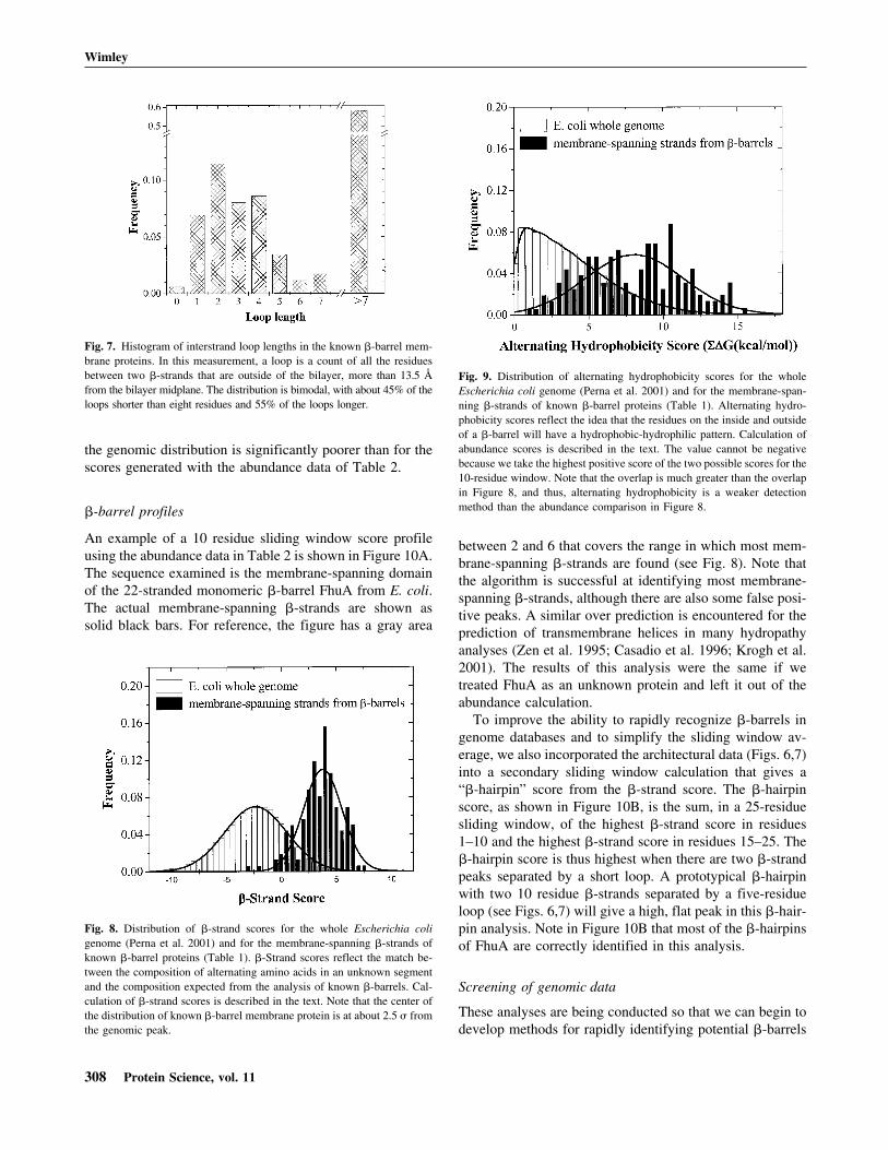
We also calculated the distribution of loop length in the�-barrels in the database. These data are shown in Figure 7.In this work, loops are defined as segments between mem-brane-spanning �-strands that are outside the thickness ofthe membrane. In other words, more than 13.5 Å from thebilayer midplane. Note that about half of the loops areshorter than six residues, indicating that most membrane-
spanning �-strands are connected to at least one other strandby a short loop. This suggests that the �-hairpin is the basicstructural building block of �-barrel membrane proteins. Asapparent in the example shown in Figure 1 and in Figure 2Aand B, the short and long loops of �-barrel membrane pro-teins are generally segregated onto opposite sides of themembrane.
Discussion
Uniqueness of membrane �-barrel dyad repeats
Membrane-spanning �-strands, like all �-sheets, have adyad repeat topology in which alternating residues are ori-ented toward alternating faces of the sheet. In �-barrelmembrane proteins about half of the membrane-spanningresidues are hydrophobic residues that are oriented towardthe membrane lipids, while the other half are more hydro-philic residues that are oriented towards the interior of thebarrel. Several �-barrel identification algorithms have beendeveloped, in part, on the idea that membrane �-barrelscould be recognizable through the dyad repeat of hydropho-
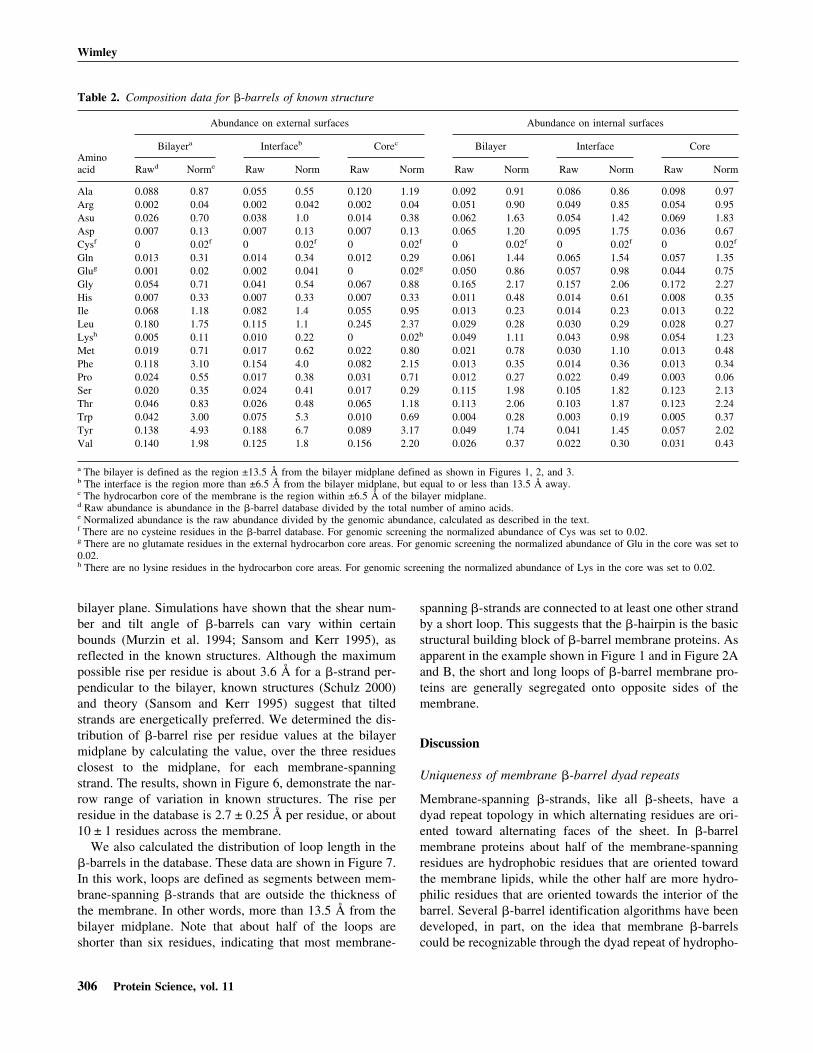
Table 2. Composition data for �-barrels of known structure
Aminoacid
Abundance on external surfaces Abundance on internal surfaces
Bilayera Interfaceb Corec Bilayer Interface Core
Rawd Norme Raw Norm Raw Norm Raw Norm Raw Norm Raw Norm
a The bilayer is defined as the region ±13.5 Å from the bilayer midplane defined as shown in Figures 1, 2, and 3.b The interface is the region more than ±6.5 Å from the bilayer midplane, but equal to or less than 13.5 Å away.c The hydrocarbon core of the membrane is the region within ±6.5 Å of the bilayer midplane.d Raw abundance is abundance in the �-barrel database divided by the total number of amino acids.e Normalized abundance is the raw abundance divided by the genomic abundance, calculated as described in the text.f There are no cysteine residues in the �-barrel database. For genomic screening the normalized abundance of Cys was set to 0.02.g There are no glutamate residues in the external hydrocarbon core areas. For genomic screening the normalized abundance of Glu in the core was set to0.02.h There are no lysine residues in the hydrocarbon core areas. For genomic screening the normalized abundance of Lys in the core was set to 0.02.
Wimley
306 Protein Science, vol. 11
bic (external) and hydrophilic (internal) residues (e.g.,Fischbarg et al. 1995). However, difficulties arise whengenome databases are screened for �-barrel membrane pro-teins using this simple idea because the interior of mem-brane-spanning �-barrels are not necessarily very hydro-philic, and because many soluble �-sheets also have a simi-lar dyad repeat motif in which one hydrophobic face of asheet is buried and one hydrophilic face is more exposed tothe aqueous phase. Our goal in this work was to use theknown �-barrels to generate a data set based on the ob-served abundance of the amino acids and the architecture of�-barrel membrane proteins that will further help to differ-entiate �-barrel membrane proteins from the abundant am-phipathic �-sheets of soluble proteins.
From the strand length distribution shown in Figure 6 weconcluded that a search for a membrane-spanning segmentof 10 residues will be able to identify most transmembrane�-strands. We performed a 10-residue sliding window
analysis for each protein examined. For each 10-residuesliding window in a protein’s amino acid sequence we cal-culated a “�-strand score” based on the two abundance datasets (interior and exposed) determined for �-barrel mem-brane proteins (shown in Fig. 5A,B, and listed in Table 2)using
� − Strand Score = �i=1
10
��AinXi�for i = 1,3,5,7,9�;
AoutXi �for i = 2,4,6,8,10��
or
� − Strand Score = �i=1
10
�AoutX1 �for i = 1,3,5,7,9�;
�AinXi �for i = 2,4,6,8,10��
whichever is highest, where AXlin and AXl
out are ln (relativeabundance) values for interior (in) and exterior (out) resi-dues (Table 2) for the ith amino acid in the sliding window.A comparison between the �-strand scores for the mem-brane-spanning �-strands of �-barrel membrane proteinsand the whole E. coli genome (Perna et al. 2001) is shownin Figure 8. The peak for the �-barrel strands is at approxi-mately 2.5 � from the center of the genome distribution.This is a good starting point for the distinction of mem-brane-spanning �-strands in genome databases. We alsomade the same calculations using a simple dyad repeat ofalternating octanol hydrophobicity (Wimley et al. 1996).The results of this comparison, shown in Figure 9, show thatthe distinction between membrane-spanning �-strands and
Fig. 6. Histogram of the rise per residue in �-barrel membrane proteins ofknown structure. For each lipid-exposed �-strand in our database we cal-culated the rise per residue from the three residues closest to the bilayermidplane. The scale at the top shows a conversion to the number of resi-dues required to span the 27-Å thickness of the membrane.
Fig. 5. Normalized amino acid abundance for the external and internalamino acids in the database of all known �-barrel membrane proteins. (A)External residues. (B) Internal residues. Normalized abundance values arethe raw abundance (Fig. 4, Table 2) divided by the weighted genomicabundance of each amino acid (see text). In addition to the abundanceacross the whole bilayer, we also show the abundance for each of twobilayer regimes: the hydrocarbon core ±6.5 Å from the bilayer midplaneand the bilayer interface between 6.5 and 13.5 Å from the midplane. Theline at 1.0 is the expectation value for residues whose abundance equals theexpected genomic abundance. Abundance values are ranked, left to right,by the value for the whole bilayer.
Identification of �-barrel membrane proteins
www.proteinscience.org 307
the genomic distribution is significantly poorer than for thescores generated with the abundance data of Table 2.
�-barrel profiles
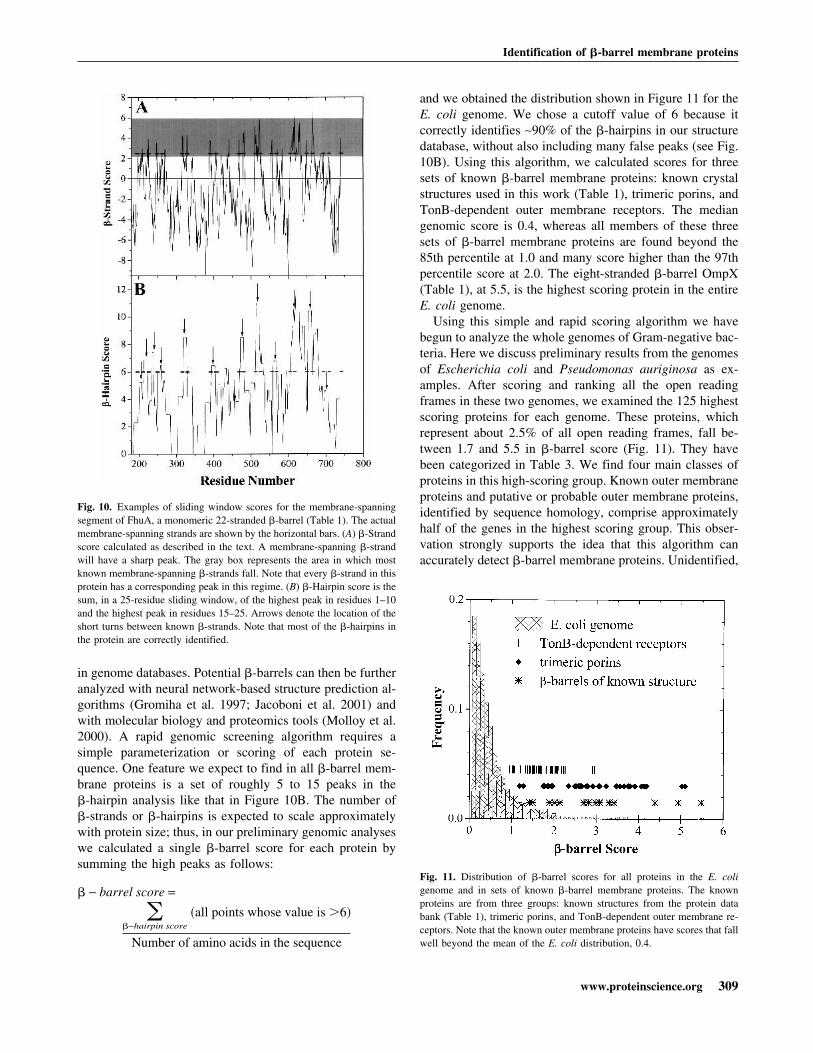
An example of a 10 residue sliding window score profileusing the abundance data in Table 2 is shown in Figure 10A.The sequence examined is the membrane-spanning domainof the 22-stranded monomeric �-barrel FhuA from E. coli.The actual membrane-spanning �-strands are shown assolid black bars. For reference, the figure has a gray area
between 2 and 6 that covers the range in which most mem-brane-spanning �-strands are found (see Fig. 8). Note thatthe algorithm is successful at identifying most membrane-spanning �-strands, although there are also some false posi-tive peaks. A similar over prediction is encountered for theprediction of transmembrane helices in many hydropathyanalyses (Zen et al. 1995; Casadio et al. 1996; Krogh et al.2001). The results of this analysis were the same if wetreated FhuA as an unknown protein and left it out of theabundance calculation.
To improve the ability to rapidly recognize �-barrels ingenome databases and to simplify the sliding window av-erage, we also incorporated the architectural data (Figs. 6,7)into a secondary sliding window calculation that gives a“�-hairpin” score from the �-strand score. The �-hairpinscore, as shown in Figure 10B, is the sum, in a 25-residuesliding window, of the highest �-strand score in residues1–10 and the highest �-strand score in residues 15–25. The�-hairpin score is thus highest when there are two �-strandpeaks separated by a short loop. A prototypical �-hairpinwith two 10 residue �-strands separated by a five-residueloop (see Figs. 6,7) will give a high, flat peak in this �-hair-pin analysis. Note in Figure 10B that most of the �-hairpinsof FhuA are correctly identified in this analysis.
Screening of genomic data
These analyses are being conducted so that we can begin todevelop methods for rapidly identifying potential �-barrels
Fig. 7. Histogram of interstrand loop lengths in the known �-barrel mem-brane proteins. In this measurement, a loop is a count of all the residuesbetween two �-strands that are outside of the bilayer, more than 13.5 Åfrom the bilayer midplane. The distribution is bimodal, with about 45% of theloops shorter than eight residues and 55% of the loops longer.
Fig. 8. Distribution of �-strand scores for the whole Escherichia coligenome (Perna et al. 2001) and for the membrane-spanning �-strands ofknown �-barrel proteins (Table 1). �-Strand scores reflect the match be-tween the composition of alternating amino acids in an unknown segmentand the composition expected from the analysis of known �-barrels. Cal-culation of �-strand scores is described in the text. Note that the center ofthe distribution of known �-barrel membrane protein is at about 2.5 � fromthe genomic peak.
Fig. 9. Distribution of alternating hydrophobicity scores for the wholeEscherichia coli genome (Perna et al. 2001) and for the membrane-span-ning �-strands of known �-barrel proteins (Table 1). Alternating hydro-phobicity scores reflect the idea that the residues on the inside and outsideof a �-barrel will have a hydrophobic-hydrophilic pattern. Calculation ofabundance scores is described in the text. The value cannot be negativebecause we take the highest positive score of the two possible scores for the10-residue window. Note that the overlap is much greater than the overlapin Figure 8, and thus, alternating hydrophobicity is a weaker detectionmethod than the abundance comparison in Figure 8.
Wimley
308 Protein Science, vol. 11
in genome databases. Potential �-barrels can then be furtheranalyzed with neural network-based structure prediction al-gorithms (Gromiha et al. 1997; Jacoboni et al. 2001) andwith molecular biology and proteomics tools (Molloy et al.2000). A rapid genomic screening algorithm requires asimple parameterization or scoring of each protein se-quence. One feature we expect to find in all �-barrel mem-brane proteins is a set of roughly 5 to 15 peaks in the�-hairpin analysis like that in Figure 10B. The number of�-strands or �-hairpins is expected to scale approximatelywith protein size; thus, in our preliminary genomic analyseswe calculated a single �-barrel score for each protein bysumming the high peaks as follows:
� − barrel score =
��−hairpin score
�all points whose value is �6�
Number of amino acids in the sequence
and we obtained the distribution shown in Figure 11 for theE. coli genome. We chose a cutoff value of 6 because itcorrectly identifies ∼90% of the �-hairpins in our structuredatabase, without also including many false peaks (see Fig.10B). Using this algorithm, we calculated scores for threesets of known �-barrel membrane proteins: known crystalstructures used in this work (Table 1), trimeric porins, andTonB-dependent outer membrane receptors. The mediangenomic score is 0.4, whereas all members of these threesets of �-barrel membrane proteins are found beyond the85th percentile at 1.0 and many score higher than the 97thpercentile score at 2.0. The eight-stranded �-barrel OmpX(Table 1), at 5.5, is the highest scoring protein in the entireE. coli genome.
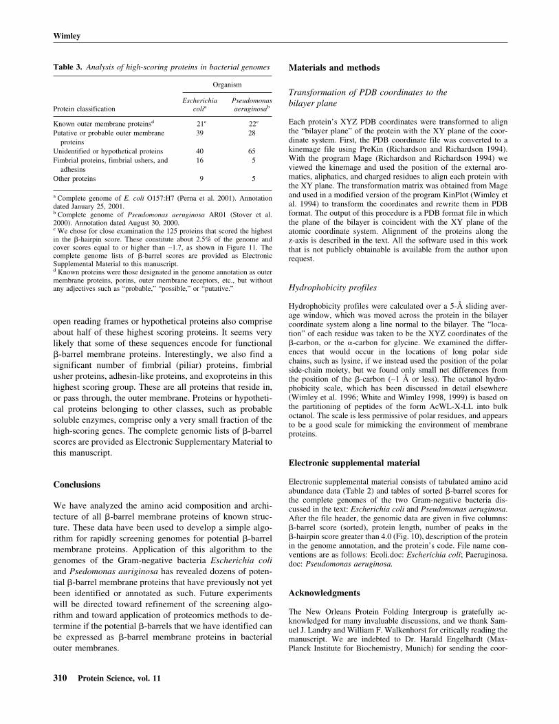
Using this simple and rapid scoring algorithm we havebegun to analyze the whole genomes of Gram-negative bac-teria. Here we discuss preliminary results from the genomesof Escherichia coli and Pseudomonas auriginosa as ex-amples. After scoring and ranking all the open readingframes in these two genomes, we examined the 125 highestscoring proteins for each genome. These proteins, whichrepresent about 2.5% of all open reading frames, fall be-tween 1.7 and 5.5 in �-barrel score (Fig. 11). They havebeen categorized in Table 3. We find four main classes ofproteins in this high-scoring group. Known outer membraneproteins and putative or probable outer membrane proteins,identified by sequence homology, comprise approximatelyhalf of the genes in the highest scoring group. This obser-vation strongly supports the idea that this algorithm canaccurately detect �-barrel membrane proteins. Unidentified,
Fig. 10. Examples of sliding window scores for the membrane-spanningsegment of FhuA, a monomeric 22-stranded �-barrel (Table 1). The actualmembrane-spanning strands are shown by the horizontal bars. (A) �-Strandscore calculated as described in the text. A membrane-spanning �-strandwill have a sharp peak. The gray box represents the area in which mostknown membrane-spanning �-strands fall. Note that every �-strand in thisprotein has a corresponding peak in this regime. (B) �-Hairpin score is thesum, in a 25-residue sliding window, of the highest peak in residues 1–10and the highest peak in residues 15–25. Arrows denote the location of theshort turns between known �-strands. Note that most of the �-hairpins inthe protein are correctly identified.
Fig. 11. Distribution of �-barrel scores for all proteins in the E. coligenome and in sets of known �-barrel membrane proteins. The knownproteins are from three groups: known structures from the protein databank (Table 1), trimeric porins, and TonB-dependent outer membrane re-ceptors. Note that the known outer membrane proteins have scores that fallwell beyond the mean of the E. coli distribution, 0.4.
Identification of �-barrel membrane proteins
www.proteinscience.org 309
open reading frames or hypothetical proteins also compriseabout half of these highest scoring proteins. It seems verylikely that some of these sequences encode for functional�-barrel membrane proteins. Interestingly, we also find asignificant number of fimbrial (piliar) proteins, fimbrialusher proteins, adhesin-like proteins, and exoproteins in thishighest scoring group. These are all proteins that reside in,or pass through, the outer membrane. Proteins or hypotheti-cal proteins belonging to other classes, such as probablesoluble enzymes, comprise only a very small fraction of thehigh-scoring genes. The complete genomic lists of �-barrelscores are provided as Electronic Supplementary Material tothis manuscript.
Conclusions
We have analyzed the amino acid composition and archi-tecture of all �-barrel membrane proteins of known struc-ture. These data have been used to develop a simple algo-rithm for rapidly screening genomes for potential �-barrelmembrane proteins. Application of this algorithm to thegenomes of the Gram-negative bacteria Escherichia coliand Psedomonas auriginosa has revealed dozens of poten-tial �-barrel membrane proteins that have previously not yetbeen identified or annotated as such. Future experimentswill be directed toward refinement of the screening algo-rithm and toward application of proteomics methods to de-termine if the potential �-barrels that we have identified canbe expressed as �-barrel membrane proteins in bacterialouter membranes.
Materials and methods
Transformation of PDB coordinates to thebilayer plane
Each protein’s XYZ PDB coordinates were transformed to alignthe “bilayer plane” of the protein with the XY plane of the coor-dinate system. First, the PDB coordinate file was converted to akinemage file using PreKin (Richardson and Richardson 1994).With the program Mage (Richardson and Richardson 1994) weviewed the kinemage and used the position of the external aro-matics, aliphatics, and charged residues to align each protein withthe XY plane. The transformation matrix was obtained from Mageand used in a modified version of the program KinPlot (Wimley etal. 1994) to transform the coordinates and rewrite them in PDBformat. The output of this procedure is a PDB format file in whichthe plane of the bilayer is coincident with the XY plane of theatomic coordinate system. Alignment of the proteins along thez-axis is described in the text. All the software used in this workthat is not publicly obtainable is available from the author uponrequest.
Hydrophobicity profiles
Hydrophobicity profiles were calculated over a 5-Å sliding aver-age window, which was moved across the protein in the bilayercoordinate system along a line normal to the bilayer. The “loca-tion” of each residue was taken to be the XYZ coordinates of the�-carbon, or the �-carbon for glycine. We examined the differ-ences that would occur in the locations of long polar sidechains, such as lysine, if we instead used the position of the polarside-chain moiety, but we found only small net differences fromthe position of the �-carbon (∼1 Å or less). The octanol hydro-phobicity scale, which has been discussed in detail elsewhere(Wimley et al. 1996; White and Wimley 1998, 1999) is based onthe partitioning of peptides of the form AcWL-X-LL into bulkoctanol. The scale is less permissive of polar residues, and appearsto be a good scale for mimicking the environment of membraneproteins.
Electronic supplemental material
Electronic supplemental material consists of tabulated amino acidabundance data (Table 2) and tables of sorted �-barrel scores forthe complete genomes of the two Gram-negative bacteria dis-cussed in the text: Escherichia coli and Pseudomonas aeruginosa.After the file header, the genomic data are given in five columns:�-barrel score (sorted), protein length, number of peaks in the�-hairpin score greater than 4.0 (Fig. 10), description of the proteinin the genome annotation, and the protein’s code. File name con-ventions are as follows: Ecoli.doc: Escherichia coli; Paeruginosa.doc: Pseudomonas aeruginosa.
Acknowledgments
The New Orleans Protein Folding Intergroup is gratefully ac-knowledged for many invaluable discussions, and we thank Sam-uel J. Landry and William F. Walkenhorst for critically reading themanuscript. We are indebted to Dr. Harald Engelhardt (Max-Planck Institute for Biochemistry, Munich) for sending the coor-
Table 3. Analysis of high-scoring proteins in bacterial genomes
Protein classification
Organism
Escherichiacolia
Pseudomonasaeruginosab
Known outer membrane proteinsd 21c 22c
Putative or probable outer membraneproteins
39 28
Unidentified or hypothetical proteins 40 65Fimbrial proteins, fimbrial ushers, and
adhesins16 5
Other proteins 9 5
a Complete genome of E. coli O157:H7 (Perna et al. 2001). Annotationdated January 25, 2001.b Complete genome of Pseudomonas aeruginosa AR01 (Stover et al.2000). Annotation dated August 30, 2000.c We chose for close examination the 125 proteins that scored the highestin the �-hairpin score. These constitute about 2.5% of the genome andcover scores equal to or higher than ∼1.7, as shown in Figure 11. Thecomplete genome lists of �-barrel scores are provided as ElectronicSupplemental Material to this manuscript.d Known proteins were those designated in the genome annotation as outermembrane proteins, porins, outer membrane receptors, etc., but withoutany adjectives such as “probable,” “possible,” or “putative.”
Wimley
310 Protein Science, vol. 11
dinates of Omp32 before their release from the PDB. Funded byNIH (GM60000) and the Louisiana Board of Regents SupportFund 1999-02-RD-A-43.
The publication costs of this article were defrayed in part bypayment of page charges. This article must therefore be herebymarked “advertisement” in accordance with 18 USC section 1734solely to indicate this fact.
References
Alm, R.A., Bina, J., Andrews, B.M., Doig, P., Hancock, R.E., and Trust, T.J.2000. Comparative genomics of Helicobacter pylori: Analysis of the outermembrane protein families. Infect. Immun. 68: 4155–4168.
Altschul, S.F., Gish, W., Miller, W., Myers, E.W., and Lipman, D.J. 1990. Basiclocal alignment search tool. J. Mol. Biol. 215: 403–410.
Benz, R. 1994. Permeation of hydrophilic solutes through mitochondrial outermembranes: Review on mitochondrial porins. Biochim. Biophys. Acta 1197:167–196.
Berman, H.M., Westbrook, J., Feng, Z., Gilliland, G., Bhat, T.N., Weissig, H.,Shindyalov, I.N., and Bourne, P.E. 2000. The Protein Data Bank. NucleicAcids Res. 28: 235–242.
Bishop, C.M., Walkenhorst, W.F., and Wimley, W.C. 2001. Folding of �-sheetmembrane proteins: Specificity and promiscuity in peptide model systems.J. Mol. Biol. 309: 975–988.
Buchanan, S.K., Smith, B.S., Venkatramani, L., Xia, D., Esser, L., Palnitkar,M., Chakraborty, R., van der Helm, D., and Deisenhofer, J. 1999. Crystalstructure of the outer membrane active transporter FepA from Escherichiacoli. Nat. Struct. Biol. 6: 56–63.
Casadio, R., Fariselli, P., Taroni, C., and Compiani, M. 1996. A predictor oftransmembrane �-helix domains of proteins based on neural networks. Eur.Biophys. J. 24: 165–178.
Cowan, S.W., Garavito, R.M., Jansonius, J.N., Jenkins, J.A., Karlsson, R.,Koenig, N., Pai, E.F., Pauptit, R.A., Rizkallah, P.J., Rosenbusch, J.P., Rum-mel, G., and Schirmer, T. 1995. The structure of OmpF porin in a tetragonalcrystal form. Structure 3: 1041–1050.
Cowan, S.W., Schirmer, T., Rummel, G., Steiert, M., Ghosh, R., Pauptit, R.A.,Jansonius, J.N., and Rosenbusch, J.P. 1992. Crystal structures explain func-tional properties of two E. coli porins. Nature 358: 727–733.
Dutzler, R., Rummel, G., Alberti, S., Hernandez-Alles, S., Phale, P., Rosen-busch, J., Benedi, V., and Schirmer, T. 1999. Crystal structure and func-tional characterization of OmpK36, the osmoporin of Klebsiella pneu-moniae. Struct. Fold. Design 7: 425–434.
Fischbarg, J., Li, J., Cheung, M., Czegledy, F., Iserovich, P., and Kuang, K.1995. Predictive evidence for a porin-type �-barrel fold in CHIP28 andother members of the MIP family. A restricted-pore model common to waterchannels and facilitators. J. Membr. Biol. 143: 177–188.
Fischer, K., Weber, A., Brink, S., Arbinger, B., Schunemann, D., Borchert, S.,Heldt, H.W., Popp, B., Benz, R., and Link, T.A. 1994. Porins from plants.Molecular cloning and functional characterization of two new members ofthe porin family. J. Biol. Chem. 269: 25754–25760.
Forst, D., Welte, W., Wacker, T., and Diederichs, K. 1998. Structure of thesucrose-specific porin ScrY from Salmonella typhimurium and its complexwith sucrose. Nat. Struct. Biol. 5: 37–46.
Gromiha, M.M., Majumdar, R., and Ponnuswamy, P.K. 1997. Identification ofmembrane spanning �-strands in bacterial porins. Protein Eng. 10: 497–500.
Jacoboni, I., Martelli, P.L., Fariselli, P., De, P.V., and Casadio, R. 2001. Pre-diction of the transmembrane regions of �-barrel membrane proteins with aneural network-based predictor. Protein Sci. 10: 779–787.
Jayasinghe, S., Hristova, K., and White, S.H. 2001. Energetics, stability, andprediction of transmembrane helices. J. Mol. Biol. 312: 927–934.
Koronakis, V., Sharff, A., Koronakis, E., Luisi, B., and Hughes, C. 2000.Crystal structure of the bacterial membrane protein TolC central to multi-drug efflux and protein export. Nature 405: 914–919.
Kreusch, A. and Schulz, G.E. 1994. Refined structure of the porin from Rho-dopseudomonas blastica. Comparison with the porin from Rhodobactercapsulatus. J. Mol. Biol. 243: 891–905.
Krogh, A., Larsson, B., von Heijne, G., and Sonnhammer, E.L. 2001. Predictingtransmembrane protein topology with a hidden Markov model: Applicationto complete genomes. J. Mol. Biol. 305: 567–580.
Locher, K.P., Rees, B., Koebnik, R., Mitschler, A., Moulinier, L., Rosenbusch,J.P., and Moras, D. 1998. Transmembrane signaling across the ligand-gatedFhuA receptor: Crystal structures of free and ferrichrome-bound states re-veal allosteric changes. Cell 95: 771–778.
Meyer, J.E.W., Hofnung, M., and Schulz, G.E. 1997. Structure of maltoporinfrom Salmonella typhimurium ligated with a nitrophenyl-maltotrioside. J.Mol. Biol. 266: 761–775.
Molloy, M.P., Herbert, B.R., Slade, M.B., Rabilloud, T., Nouwens, A.S., Willi-ams, K.L., and Gooley, A.A. 2000. Proteomic analysis of the Escherichiacoli outer membrane. Eur. J. Biochem. 267:2871–2881.
Murzin, A.G., Lesk, A.M., and Chothia, C. 1994. Principles determining thestructure of �-sheet barrels in proteins: I. A theoretical analysis. J. Mol.Biol. 236: 1369–1381.
Pautsch, A. and Schulz, G.E. 1998. Structure of the outer membrane protein Atransmembrane domain. Nat. Struct. Biol. 5: 1013–1017.
Sansom, M.S.P. and Kerr, I.D. 1995. Transbilayer pores formed by �-barrels:Molecular modeling of pore structures and properties. Biophys. J. 69: 1334–1343.
Schiffer, M., Chang, C.H., and Stevens, F.J. 1992. The functions of tryptophanresidues in membrane proteins. Protein Eng. 5: 213–214.
Schirmer, T. and Cowan, S.W. 1993. Prediction of membrane-spanning�-strands and its application to maltoporin. Protein Sci. 2: 1361–1363.
Seshadri, K., Garemyr, R., Wallin, E., von Heijne, G., and Elofsson, A. 1998.Architecture of �-barrel membrane proteins: Analysis of trimeric porins.Protein Sci. 7: 2026–2032.
Snijder, H.J., Ubarretxena-Belandia, I., Blaauw, M., Kalk, K.H., Verheij, H.M.,Egmond, M.R., Dekker, N., and Dijkstra, B.W. 1999. Structural evidencefor dimerization-regulated activation of an integral membrane phospholi-pase. Nature 401: 717–721.
Song, L., Hobaugh, M.R., Shustak, C., Cheley, S., Bayley, H., and Gouaux, J.E.1996. Structure of staphylococcal �-hemolysin, a heptameric transmem-brane pore. Science 274: 1859–1866.
Stover, C.K., Pham, X.Q., Erwin, A.L., Mizoguchi, S.D., Warrener, P., Hickey,M.J., Brinkman, F.S., Hufnagle, W.O., Kowalik, D.J., Lagrou, M., Garber,R.L., Goltry, L., Tolentino, E., Westbrock-Wadman, S., Yuan, Y., Brody,L.L., Coulter, S.N., Folger, K.R., Kas, A., Larbig, K., Lim, R., Smith, K.,Spencer, D., Wong, G.K., Wu, Z., and Paulsen, I.T. 2000. Complete genomesequence of Pseudomonas aeruginosa PA01, an opportunistic pathogen.Nature 406: 959–964.
Vogt, J. and Schulz, G.E. 1999. The structure of the outer membrane proteinOmpX from Escherichia coli reveals possible mechanisms of virulence.Struct. Fold. Design 7: 1301–1309.
von Heijne, G. 1994. Membrane proteins: From sequence to structure. Annu.Rev. Biophys. Biomol. Struct. 23: 167–192.
von Heijne, G. 1996. Prediction of transmembrane protein topology. In Proteinstructure prediction (eds. M.J.E. Sternberg), pp. 101–110. Oxford Univer-sity Press, Oxford.
Weiss, M.S. and Schulz, G.E. 1992. Structure of porin refined at 1.8 Å resolu-tion. J. Mol. Biol. 227: 493–509.
White, S.H. and Wimley, W.C. 1998. Hydrophobic interactions of peptides withmembrane interfaces. Biochim. Biophys. Acta 1376: 339–352.
———. 1999. Membrane protein folding and stability: Physical principles.Annu. Rev. Biophys. Biomol. Struct. 28: 319–365.
Identification of �-barrel membrane proteins
www.proteinscience.org 311
Wiener, M.C. and White, S.H. 1992. Structure of a fluid dioleoylphosphatidyl-choline bilayer determined by joint refinement of X-ray and neutron dif-fraction data. III. Complete structure. Biophys. J. 61: 434–447.
Wimley, W.C., Creamer, T.P., and White, S.H. 1996. Solvation energies ofamino acid sidechains and backbone in a family of host–guest pentapep-tides. Biochemistry 35: 5109–5124.
Wimley, W.C., Selsted, M.E., and White, S.H. 1994. Interactions between hu-man defensins and lipid bilayers: Evidence for the formation of multimericpores. Protein Sci. 3: 1362–1373.
Yau, W.M., Wimley, W.C., Gawrisch, K., and White, S.H. 1998. The preferenceof tryptophan for membrane interfaces. Biochemistry 37: 14713–14718.
Zen, K.H., Consler, T.G., and Kaback, H.R. 1995. Insertion of the polytopicmembrane protein lactose permease occurs by multiple mechanisms. Bio-chemistry 34: 3430–3437.
Zeth, K., Diederichs, K., Welte, W., and Engelhardtm H. 2000. Crystal structureof Omp32, the anion-selective porin from Comamonas acidovorans, incomplex with a periplasmic peptide at 2.1 A resolution. Struct. Fold. Design8: 981–992.