REVIEWS Drug Discovery Today Volume 17, Numbers 1/2 January 2012 Toward in silico structure-based ADMET prediction in drug discovery Gautier Moroy 1 , Virginie Y. Martiny 1 , Philippe Vayer 2 , Bruno O. Villoutreix 1 and Maria A. Miteva 1 1 Universite ´ Paris Diderot, Sorbonne Paris Cite ´, Mole ´ cules The ´ rapeutiques In Silico, Inserm UMR-S 973, 35 rue Helene Brion, 75013 Paris, France 2 BioInformatic Modelling Department, Technologie Servier, 45007 Orle ´ ans Cedex 1, France Quantitative structure–activity relationship (QSAR) methods and related approaches have been used to investigate the molecular features that influence the absorption, distribution, metabolism, excretion and toxicity (ADMET) of drugs. As the three-dimensional structures of several major ADMET proteins become available, structure-based (docking-scoring) computations can be carried out to complement or to go beyond QSAR studies. Applying docking-scoring methods to ADMET proteins is a challenging process because they usually have a large and flexible binding cavity; however, promising results relating to metabolizing enzymes have been reported. After reviewing current trends in the field we applied structure-based methods in the context of receptor flexibility in a case study involving the phase II metabolizing sulfotransferases. Overall, the explored concepts and results suggested that structure-based ADMET profiling will probably join the mainstream during the coming years. Introduction The success of a drug is determined not only by good efficacy but also by an acceptable ADMET profile. Although a large variety of medium- and high-throughput in vitro ADMET screens are avail- able, being able to predict some of these properties in silico is valuable. Today, it is recognized that employing computational ADMET, in combination with in vivo and in vitro predictions as early as possible in the drug discovery process, helps to reduce the number of safety issues [1]. Moreover, there is a pressure to reduce the number of animal experiments (e.g. the REACH project). Traditionally, data modeling methods, such as expert systems and quantitative structure–activity (property) relationships (QSARs/QSPRs) [2,3], have been used to investigate ADMET prop- erties. These methods use statistical and learning approaches, molecular descriptors and experimental data to model complex biological processes (e.g. oral bioavailability, intestinal absorption, permeability and mutagenicity [2,4]). The rules for drug-likeness or lead-likeness or metabolite-likeness [5,6] relying on simple physicochemical properties are also well-known and implemented in commercially and freely available packages [4,7,8]. However, limitations of all these approaches come from the fact that high quality experimental data are seldom available [9], and that the approaches tend to neglect direct structural information about the ADMET proteins. In silico approaches based on the 3D structures of these proteins could therefore be an attractive alternative or could complement ADMET data-modeling techniques [10]. The first attempt to predict ADMET taking into account the protein structures at the atomic level started about ten years ago with the early homology models of human cytochrome P450 (CYP) [11,12]. Several new studies have recently been reported that exploit the 3D structures of ADMET proteins, molecular docking and different strategies for taking into account protein flexibility during the process. They all highlight that these proteins are difficult to investigate – in part because of the presence of large and flexible ligand-binding cavities that can interact with diverse ligands. Most of these investigations focus on phase I metabolizing enzymes such as CYP (for recent key reviews, see Refs [10,13,14]). To date, predictions of interactions between drug candidates and phase II metabolizing enzymes based on 3D protein structures are still essentially missing. Reviews INFORMATICS Corresponding author:. Miteva, M.A. ([email protected]) 44 www.drugdiscoverytoday.com 1359-6446/06/$ - see front matter ß 2011 Elsevier Ltd. All rights reserved. doi:10.1016/j.drudis.2011.10.023
Transcript
Review
s�IN
FORMATICS
REVIEWS Drug Discovery Today � Volume 17, Numbers 1/2 � January 2012
Toward in silico structure-based ADMETprediction in drug discovery
Gautier Moroy1, Virginie Y. Martiny1, Philippe Vayer2, Bruno O. Villoutreix1 andMaria A. Miteva1
1Universite Paris Diderot, Sorbonne Paris Cite, Molecules Therapeutiques In Silico, Inserm UMR-S 973, 35 rue Helene Brion, 75013 Paris, France2BioInformatic Modelling Department, Technologie Servier, 45007 Orleans Cedex 1, France
Quantitative structure–activity relationship (QSAR) methods and related approaches have been used to
investigate the molecular features that influence the absorption, distribution, metabolism, excretion
and toxicity (ADMET) of drugs. As the three-dimensional structures of several major ADMET proteins
become available, structure-based (docking-scoring) computations can be carried out to complement or
to go beyond QSAR studies. Applying docking-scoring methods to ADMET proteins is a challenging
process because they usually have a large and flexible binding cavity; however, promising results relating
to metabolizing enzymes have been reported. After reviewing current trends in the field we applied
structure-based methods in the context of receptor flexibility in a case study involving the phase II
metabolizing sulfotransferases. Overall, the explored concepts and results suggested that structure-based
ADMET profiling will probably join the mainstream during the coming years.
IntroductionThe success of a drug is determined not only by good efficacy but
also by an acceptable ADMET profile. Although a large variety of
medium- and high-throughput in vitro ADMET screens are avail-
able, being able to predict some of these properties in silico is
valuable. Today, it is recognized that employing computational
ADMET, in combination with in vivo and in vitro predictions as
early as possible in the drug discovery process, helps to reduce the
number of safety issues [1]. Moreover, there is a pressure to reduce
the number of animal experiments (e.g. the REACH project).
Traditionally, data modeling methods, such as expert systems
and quantitative structure–activity (property) relationships
(QSARs/QSPRs) [2,3], have been used to investigate ADMET prop-
erties. These methods use statistical and learning approaches,
molecular descriptors and experimental data to model complex
REVIEWS Drug Discovery Today � Volume 17, Numbers 1/2 � January 2012
CYP 11A2 4% 1B1
2HI4 3PM0
CYP 5151A1
3JUS, 3JUV, 3LD6
CYP 4646A1
2F9Q, 2Q9G, 3MDM, 3MDR, 3MDT, 3MDV
CYP 2727
1MFX (model)
CYP 2121A2
2GEG (model)
CYP 1919A13EQM
CYP 1717A1
2C17 (model)
CYP 1111A13NAO
8A121AG, 3B6H
7A13DAX
3A4 50%1TQN, 1W0E, 1W0F, 1W0G, 2J0D,
2V0M, 3NXU
2A6 2%1Z10, 1Z11, 2FDU, 2FDV,
2FDW, 2FDY, 3EBS
2A132P85, 2PG5, 2PG6,
2PG72B6 3%
3IBD
2C81PQ2, 2NNH, 2NNI,
2NNJ, 2VN0
2C9 10%1OG2, 1OG5,
1R9O
2C182H6P
2D6 30%2F9Q2E1 2%
3E4E, 3E6I, 3GPH, 3KOH, 3LC4
2R13C6G, 3CZH, 3DL9
CYP 2 CYP 3
CYP 7 CYP 8
Human CYP450
SULT6SULT6B1
SULT4SULT4A1
1ZD1
SULT2SULT2A1
3F3Y 2QP3 2QP4 1OV4
1J99 1EFH
SULT2B1a1Q1Q
SULT2B1b1Q1Z 1Q10 1Q22
SULT1SULT1A1
2D06 1LS6
SULT1A1*3 1Z28
SULT1A21Z29
SULT1A32A3R 1CJM
SULT1A4
SULT1B13CKL 2Z5F 1XV1
SULT1C13BFX
SULT1C2 2AD1 2GWH
SULT1C32REO 2H8K
SULT1E1 1G3M 1HY3
Human sulfotransferases (b)
(a)
Drug Discovery Today
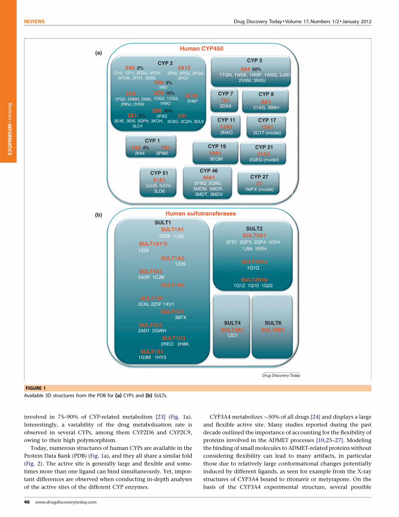
FIGURE 1
Available 3D structures from the PDB for (a) CYPs and (b) SULTs.
Review
s�IN
FORMATICS
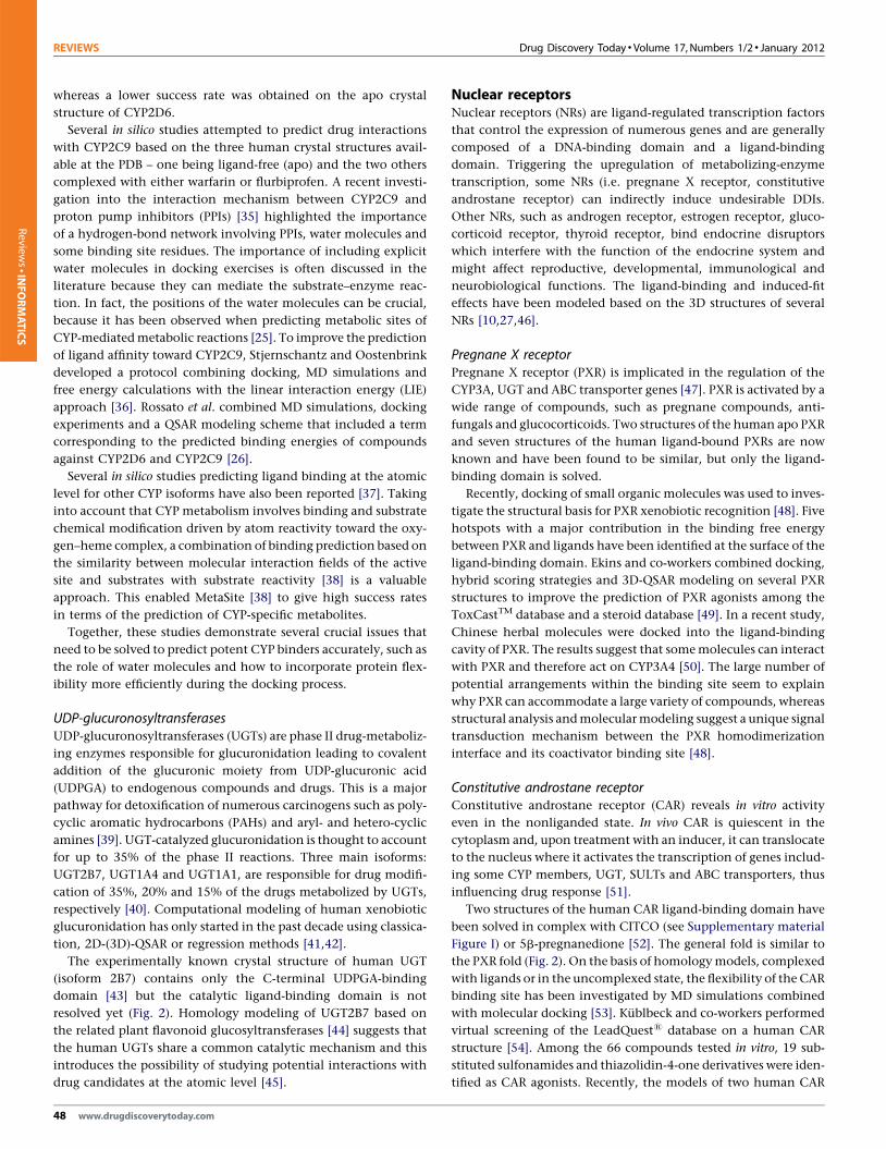
involved in 75–90% of CYP-related metabolism [23] (Fig. 1a).
Interestingly, a variability of the drug metabolization rate is
observed in several CYPs, among them CYP2D6 and CYP2C9,
owing to their high polymorphism.
Today, numerous structures of human CYPs are available in the
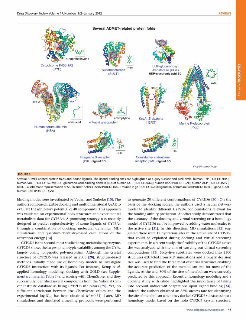
Protein Data Bank (PDB) (Fig. 1a), and they all share a similar fold
(Fig. 2). The active site is generally large and flexible and some-
times more than one ligand can bind simultaneously. Yet, impor-
tant differences are observed when conducting in-depth analyses
of the active sites of the different CYP enzymes.
46 www.drugdiscoverytoday.com
CYP3A4 metabolizes �50% of all drugs [24] and displays a large
and flexible active site. Many studies reported during the past
decade outlined the importance of accounting for the flexibility of
proteins involved in the ADMET processes [10,25–27]. Modeling
the binding of small molecules to ADMET-related proteins without
considering flexibility can lead to many artifacts, in particular
those due to relatively large conformational changes potentially
induced by different ligands, as seen for example from the X-ray
structures of CYP3A4 bound to ritonavir or metyrapone. On the
basis of the CYP3A4 experimental structure, several possible
Drug Discovery Today � Volume 17, Numbers 1/2 � January 2012 REVIEWS
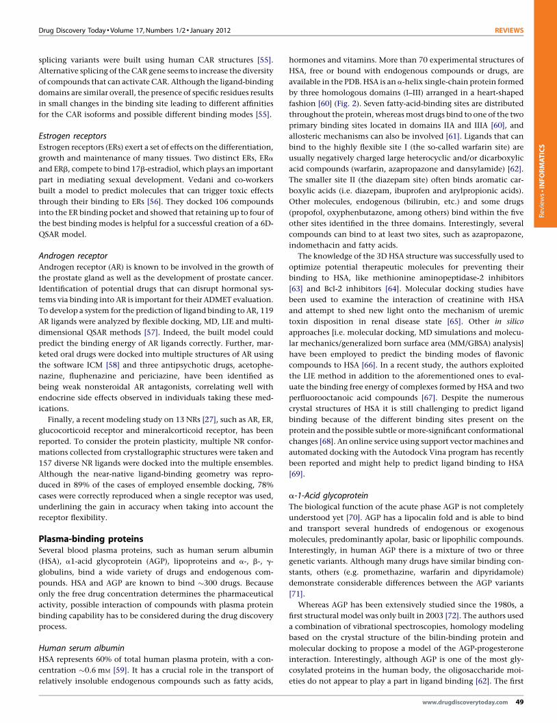
Several ADMET-related protein folds
Cytochrome P450 1A2(CYP)
α-naphthoflavone
UDP-glucuronosy l-transferase (UGT)
UDP-glucuronic acid BDSulfotransferase
(SULT)
OJZ
PCQ
site 2 site 1
S6
S5
P
Human serum albumin(HSA)
KcsA, S. lividans(hERG)oleic acid Murine P-glycoprotein
(P-gp)
α1-acid glycoproteinamitriptyline
Constitutive androstanereceptor (CAR) ligand BD
Pregnane X receptor(PXR) ligand BD
SRLCI2
Drug Discovery Today
AA2
aaphthoflavone
UDP-glucuronosyl-transferase (UGT)
UDP-glucuronic acid B DSulfotransferas e
(SULT)
OJZ
PCQ
site 1
S6
S5
P
KcsA, S. lividans(hERG)eeic acid Murine P-glycoprotein
(P-gp)
α1-acid glycoproteinamitriptyline
CI2
FIGURE 2
Several ADMET-related protein folds and bound ligands. The ligand-binding sites are highlighted as a grey surface and pink circle: human CYP (PDB ID: 2HI4);
human SULT (PDB ID: 1G3M); UDP-glucuronic acid binding domain (BD) of human UGT (PDB ID: 2O6L); human HSA (PDB ID: 1GNI); human AGP (PDB ID: 3APV);
hERG – a schematic representation of S5, S6 and P helices (KcsA; PDB ID: 1K4C); murine P-gp (PDB ID: 3G60); ligand BD of human PXR (PDB ID: 1NRL); ligand BD of
human CAR (PDB ID: 1XV9).
Reviews�INFORMATICS
binding modes were investigated by Vedani and Smiesko [10]. The
authors combined flexible docking and multidimensional QSAR to
evaluate the inhibitory potential of 48 compounds. This approach
was validated on experimental holo structures and experimental
metabolism data for CYP3A4. A promising strategy was recently
designed to predict regioselectivity of some ligands of CYP3A4
through a combination of docking, molecular dynamics (MD)
simulations and quantum-chemistry-based calculations of the
activation energy [14].
CYP2D6 is the second-most studied drug-metabolizing enzyme.
CYP2D6 shows the largest phenotypic variability among the CYPs,
largely owing to genetic polymorphism. Although the crystal
structure of CYP2D6 was released in 2006 [28], structure-based
methods initially made use of homology models to investigate
CYP2D6 interaction with its ligands. For instance, Kemp et al.
applied homology modeling, docking with GOLD (see Supple-
mentary material Table I) and scoring with ChemScore, and they
successfully identified several compounds from the National Can-
cer Institute database as being CYP2D6 inhibitors [29]. Yet, no
sufficient correlation between the ChemScore values and the
experimental log IC50 has been obtained (r2 = 0.61). Later, MD
simulations and simulated annealing protocols were performed
to generate 20 different conformations of CYP2D6 [30]. On the
basis of the docking scores, the authors used a neural network
model to identify different CYP2D6 conformations relevant for
the binding affinity prediction. Another study demonstrated that
the accuracy of the docking and virtual screening on a homology
model of CYP2D6 can be improved by adding water molecules to
the active site [31]. In this direction, MD simulations [32] sug-
gested there were 12 hydration sites in the active site of CYP2D6
that could be exploited during docking and virtual screening
experiments. In a recent study, the flexibility of the CYP2D6 active
site was analyzed with the aim of carrying out virtual screening
computations [33]. Sixty-five substrates were docked into 2500
structures extracted from MD simulations and a binary decision
tree was used to find the three most essential structures enabling
the accurate prediction of the metabolism site for most of the
ligands. At the end, 80% of the sites of metabolism were correctly
predicted by this approach. Recently, homology modeling and a
docking study with Glide highlighted the importance of taking
into account induced-fit adaptations upon ligand binding [34].
Indeed, the authors obtained an 85% success rate for identifying
the site of metabolism when they docked CYP2D6 substrates into a
homology model based on the holo CYP2C5 crystal structure,
www.drugdiscoverytoday.com 47
REVIEWS Drug Discovery Today � Volume 17, Numbers 1/2 � January 2012
Review
s�IN
FORMATICS
whereas a lower success rate was obtained on the apo crystal
structure of CYP2D6.
Several in silico studies attempted to predict drug interactions
with CYP2C9 based on the three human crystal structures avail-
able at the PDB – one being ligand-free (apo) and the two others
complexed with either warfarin or flurbiprofen. A recent investi-
gation into the interaction mechanism between CYP2C9 and
proton pump inhibitors (PPIs) [35] highlighted the importance
of a hydrogen-bond network involving PPIs, water molecules and
some binding site residues. The importance of including explicit
water molecules in docking exercises is often discussed in the
literature because they can mediate the substrate–enzyme reac-
tion. In fact, the positions of the water molecules can be crucial,
because it has been observed when predicting metabolic sites of
CYP-mediated metabolic reactions [25]. To improve the prediction
of ligand affinity toward CYP2C9, Stjernschantz and Oostenbrink
developed a protocol combining docking, MD simulations and
free energy calculations with the linear interaction energy (LIE)
approach [36]. Rossato et al. combined MD simulations, docking
experiments and a QSAR modeling scheme that included a term
corresponding to the predicted binding energies of compounds
against CYP2D6 and CYP2C9 [26].
Several in silico studies predicting ligand binding at the atomic
level for other CYP isoforms have also been reported [37]. Taking
into account that CYP metabolism involves binding and substrate
chemical modification driven by atom reactivity toward the oxy-
gen–heme complex, a combination of binding prediction based on
the similarity between molecular interaction fields of the active
site and substrates with substrate reactivity [38] is a valuable
approach. This enabled MetaSite [38] to give high success rates
in terms of the prediction of CYP-specific metabolites.
Together, these studies demonstrate several crucial issues that
need to be solved to predict potent CYP binders accurately, such as
the role of water molecules and how to incorporate protein flex-
ibility more efficiently during the docking process.
UDP-glucuronosyltransferasesUDP-glucuronosyltransferases (UGTs) are phase II drug-metaboliz-
ing enzymes responsible for glucuronidation leading to covalent
addition of the glucuronic moiety from UDP-glucuronic acid
(UDPGA) to endogenous compounds and drugs. This is a major
pathway for detoxification of numerous carcinogens such as poly-
cyclic aromatic hydrocarbons (PAHs) and aryl- and hetero-cyclic
amines [39]. UGT-catalyzed glucuronidation is thought to account
for up to 35% of the phase II reactions. Three main isoforms:
UGT2B7, UGT1A4 and UGT1A1, are responsible for drug modifi-
cation of 35%, 20% and 15% of the drugs metabolized by UGTs,
respectively [40]. Computational modeling of human xenobiotic
glucuronidation has only started in the past decade using classica-
tion, 2D-(3D)-QSAR or regression methods [41,42].
The experimentally known crystal structure of human UGT
(isoform 2B7) contains only the C-terminal UDPGA-binding
domain [43] but the catalytic ligand-binding domain is not
resolved yet (Fig. 2). Homology modeling of UGT2B7 based on
the related plant flavonoid glucosyltransferases [44] suggests that
the human UGTs share a common catalytic mechanism and this
introduces the possibility of studying potential interactions with
drug candidates at the atomic level [45].
48 www.drugdiscoverytoday.com
Nuclear receptorsNuclear receptors (NRs) are ligand-regulated transcription factors
that control the expression of numerous genes and are generally
composed of a DNA-binding domain and a ligand-binding
domain. Triggering the upregulation of metabolizing-enzyme
transcription, some NRs (i.e. pregnane X receptor, constitutive
androstane receptor) can indirectly induce undesirable DDIs.
Other NRs, such as androgen receptor, estrogen receptor, gluco-
The median structures from the six obtained clusters were
chosen to define a representative set of protein conformations.
For the virtual screening experiments we collected 157 known
substrates of SULT1A1 ([99]; databases: BRENDA, Aureus Sciences).
We clustered the active molecules using the fingerprint FCFP_4
available in Pipeline Pilot v.7.5 (SciTegic, Inc/Accelrys). As decoys
we took the diverse ChemBridgeTM PremiumSetTM and May-
bridge1 HitFinderTM sets. All actives and decoys were filtered with
a soft drug-like filter using the FAF-Drugs 2 server [8]. Finally, we
performed virtual screening experiments with Vina 1.0 [103] on
the representative MD structure set and on the X-ray structure of
SULT1A1 and the 60 diverse actives were merged with the 49,496
putative decoys from the ChemBridgeTM collection or with 13,088
molecules from the Maybridge1 collection.
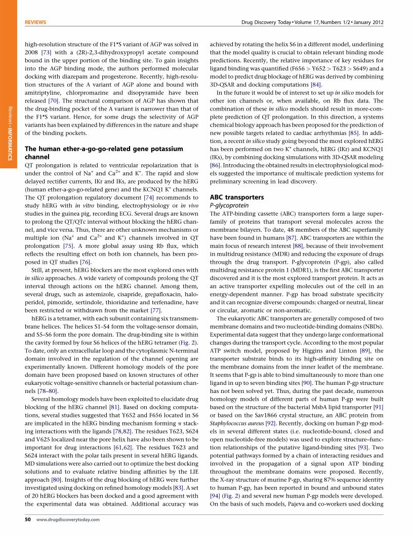
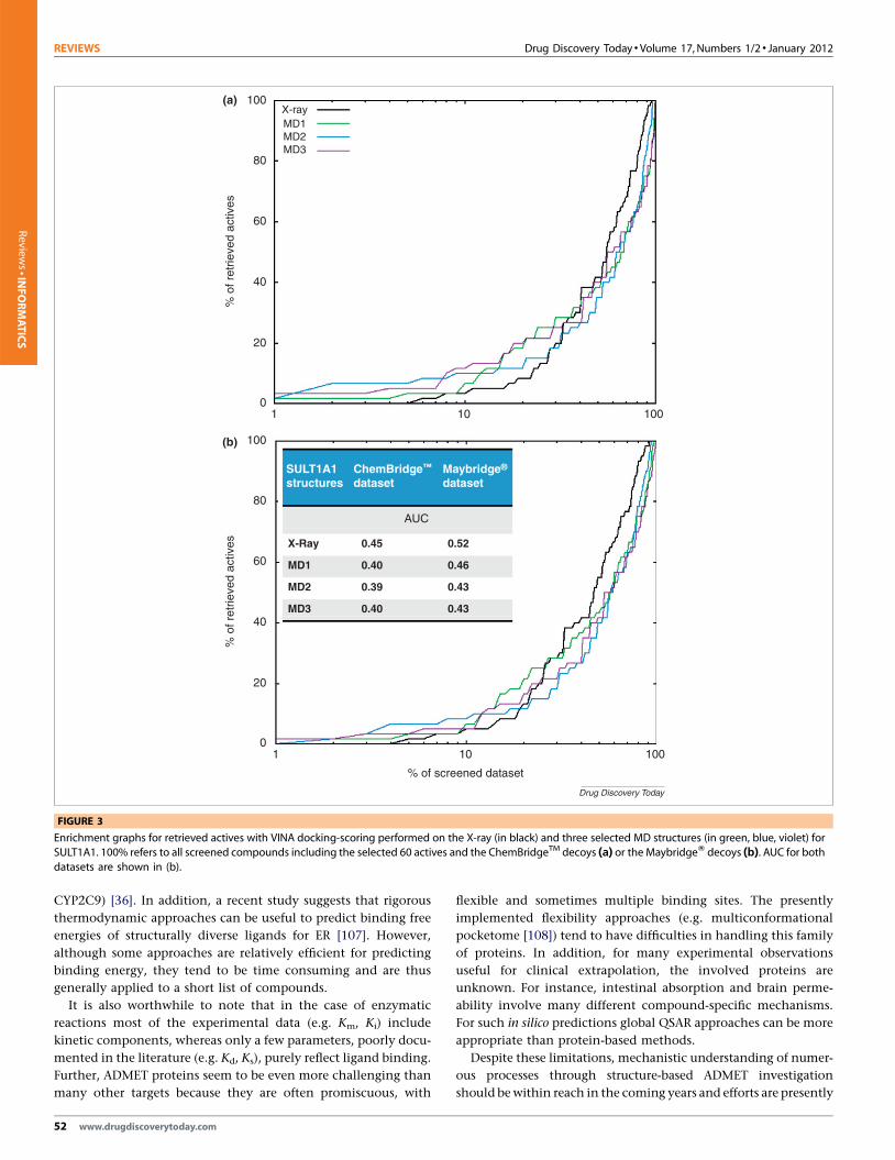
Figure 3 represents the enrichment graphs obtained for the X-
ray and MD protein conformations. Although the AUC (area under
curve) for the ROC (receiver operating characteristic) curves (not
shown) are better for the virtual screening experiments performed
on the X-ray SULT1A1 than on the selected MD structures, early
enrichment is better on some of the MD structures (up to 30% for
ChemBridgeTM, and up to 50% for Maybridge1). Obtaining earlier
enrichments with some MD extracted structures suggests that it is
important to take into account the flexibility of the binding site of
SULTs. However, further improvements could be achieved for
instance by employing induced-fit approximations, development
of tuned scoring functions and/or the use of interaction finger-
prints. For difficult proteins, combination of docking-scoring,
QSAR and network pharmacology or related approaches [104]
would seem valuable.
Future trends and conclusionsCurrent in silico ADMET predictions cannot fully replace well-
established in vitro cell-based approaches or in vivo assays but they
can provide significant insights. QSAR ADMET models are widely
used but are limited within the training set chemical space.
Regarding ADMET predictions based on the 3D structures of the
relevant proteins, improvements are still required owing to ambi-
guities in experimental structures and in the biological data used
for validation and inaccuracies in several force-field parameters
and terms. Obviously, the known limitations of docking-scoring
methods are also valid for ADMET proteins. For instance, the
difficulty in taking the contribution of water molecules into
account accurately [25], and known problems with docking-scor-
ing algorithms, and more specifically with scoring functions.
Although it is a common practice to select the docked poses
and to rank compounds using simple scoring functions [29,31],
insufficient correlation between the docked scores and experimen-
tal binding energies are generally observed [29], although some
promising results have also been reported [105,106]. In fact,
different protocols to improve scoring or to compute the free
energy of binding have been investigated and compared to experi-
mental binding data for structurally similar ligands (e.g. for
www.drugdiscoverytoday.com 51
REVIEWS Drug Discovery Today � Volume 17, Numbers 1/2 � January 2012
X-ray100
80
60
40
20
0
100
80
60
40
20
0
1 10 100
1 10 100
MD1MD2MD3
(a)
% o
f ret
rieve
d ac
tives
SULT1A1structures
ChemBridge™
datas etMaybrid ge®
datas et
AUC
(b)
X-Ray 0.520.45
0.460.40MD1
0.430.39MD2
0.430.40MD3
% o
f ret
rieve
d ac
tives
% of screened d ataset
Drug Discovery Today
FIGURE 3
Enrichment graphs for retrieved actives with VINA docking-scoring performed on the X-ray (in black) and three selected MD structures (in green, blue, violet) forSULT1A1. 100% refers to all screened compounds including the selected 60 actives and the ChemBridgeTM decoys (a) or the MaybridgeW decoys (b). AUC for both
datasets are shown in (b).Review
s�IN
FORMATICS
CYP2C9) [36]. In addition, a recent study suggests that rigorous
thermodynamic approaches can be useful to predict binding free
energies of structurally diverse ligands for ER [107]. However,
although some approaches are relatively efficient for predicting
binding energy, they tend to be time consuming and are thus
generally applied to a short list of compounds.
It is also worthwhile to note that in the case of enzymatic
reactions most of the experimental data (e.g. Km, Ki) include
kinetic components, whereas only a few parameters, poorly docu-
mented in the literature (e.g. Kd, Ks), purely reflect ligand binding.
Further, ADMET proteins seem to be even more challenging than
many other targets because they are often promiscuous, with
52 www.drugdiscoverytoday.com
flexible and sometimes multiple binding sites. The presently


![hybrids: Exploring their in silico ADMET, ergosterol ... · 1 Design, synthesis, and antimicrobial evaluation of 1,4-dihydroindeno[1,2-c]pyrazole tethered carbohydrazide hybrids:](https://static.documents.pub/doc/80x56/5fbcccd16dfee23f0408ffb6/hybrids-exploring-their-in-silico-admet-ergosterol-1-design-synthesis-and.jpg)


