69
TOWARDS BIOSENSORS IN FOOD PACKAGING

TOWARDS BIOSENSORS IN FOOD
PACKAGING

IMMOBILIZATION AND CHARACTERIZATION OF
FLEXIBLE DNAzyme-BASED BIOSENSORS FOR ON-THE-
SHELF FOOD MONITORING
By
HANIE YOUSEFI, B.Eng.
A Thesis Submitted to the School of Graduate Studies
in Partial Fulfilment of the Requirements for the Degree
Master of Science
McMaster University
© Copyright by Hanie Yousefi, September 2017
ii
McMaster University MASTER OF Applied SCIENCE (2017) Hamilton,
Ontario (Chemcial Engineering)
TITLE: Immobilization and Characterization of
DNAzyme-based Biosensors for on-the-shelf
Food Monitoring
AUTHOR: Hanie Yousefi
SUPERVISORS: Dr. Carlos D.M. Filipe
Dr. Tohid F. Didar
NUMBER OF PAGES:
xiii, 55

iii
Lay Abstract
Microbial pathogens can grow in food following packaging and preceding consumption.
Current biosensors are not efficient for post-packaging real-time food monitoring without
separating the sample from the stock. Packaged food such as meat and juice are directly in
touch with the surface of their containers or covers. Therefore, real-time sensing
mechanisms, installed inside the food packaging, tracing the presence of pathogens, are
much useful to ensure the food safety. Here we report on developing thin, transparent,
flexible and durable sensing surfaces using DNA biosensors, which generate a fluorescence
signal in the presence of a target bacterium in food or water samples. The covalently-
attached DNA probes can detect as low as 103 CFU/mL of Escherichia coli in meat, sliced
apple and apple juice. The fabricated sensing surfaces remained stable up to several days
under varying pH conditions (pH 5 to 9). In addition to pathogen monitoring in packaged
food or drinking bottles, these surfaces are promising for a variety of other applications in
health care settings, environmental monitoring, and biomaterials like wound dressing.

iv
Abstract
While the Canadian food supply is among the healthiest in the world, almost 4 million (1
in 8) Canadians are affected by food-borne illnesses, resulting in 11,600 hospitalizations
and 238 deaths per year. Microbial pathogens are one of the major causes of foodborne
sicknesses that can grow in food before or following packaging. Food distribution is an
important part of the food processing chain, in which food supplies are at a higher risk of
contamination due to lack of proper monitoring. Among myriad of research around
biosensors, current devices focusing on packaged food monitoring, such as leakage
indicators or time temperature sensors are not efficient for real-time food monitoring
without separating the sample from the stock. Packaged food such as meat and juice are
directly in touch with the surface of their containers or covers. Therefore, real-time sensing
mechanisms, installed inside the food packaging and capable of tracing the presence of
pathogens, are of great interest to ensure food safety. This work involves developing thin,
transparent, flexible and durable sensing surfaces using DNA biosensors, which report the
presence of a target bacterium in food or water samples by generating a fluorescence signal
that can be detected by simple fluorescence detecting devices. The covalently-attached
DNA probes generate the signal upon contact with the target bacteria with as low as 103
CFU/mL of Escherichia coli in meat and apple juice. The fabricated sensing surfaces
remained stable up to several days under varying pH conditions (pH 5 to 9). In addition to
detecting pathogens on packaged food or drinking bottles, these surfaces have the potential
to be used for a variety of other applications in health care settings, environmental
monitoring, food production chain, and biomaterials like wound dressing.

v
In Memory of My Dearest Cousin
Reyhaneh
vi
Acknowledgements
Firstly, I would like to express my deepest appreciation to my supervisor Dr. Carlos Filipe who has shown the attitude and substance of an incredible mentor. He conveyed the spirit of enthusiasm and encouragement in regard to assisting me with my research. It is because of Dr. Filipe’s priceless expertise and guidance in a technical and laboratory setting, as well as his genuine excitement towards teaching with a powerful and positive approach, that I have been able to develop as a motivated student and researcher. I am forever honored to have had the opportunity to work with him. I am also greatly indebted to my co-supervisor, Dr. Tohid Didar for his invaluable guidance, support, and mentorship. Dr. Didar went above and beyond to ensure a perfect balance between technical mentorship, invaluable guidance, moral support, and freedom of research. He constantly encouraged and challenged me to explore beyond my set criteria. His unfailing support and understanding was truly influential and impactful throughout my graduate studies and my research project success. I would also like to thank Dr. Ali Monsur for his excellent guidance and continual helps in the lab. He introduced me to the molecular biology field and was always ready to help me to promote my knowledge in the field as well as providing me with trainings and guidance required for the fulfillment of my experiments. I would like to thank Mr. Doug Keller for his great helps by providing me with all the materials I needed. Also thanks to bio-interface institute technicians, namely, Dr. Marta Princz, at McMaster University for providing me with continuous guidance on using the facilities. Furthermore, I am extremely thankful to our lab’s undergrad summer student, Mr. Hsuan-Ming Su as many of the experiments would not have been completed as easily without his hard work and contributions. I also want to thank all my colleagues, collaborators, and group mates, especially, Dr. Sana Jahanshahi, Mr. Vincent Leung, Ms. Azadeh Peivandi, Mr. Mathew Osborne Ms. Sara Jahromi, Ms. Sara Imani, Mr. Martin Villegas and Mr. Zachary Cetinic for their continual discussions, debates and support. I would like to express my appreciations to my friend, Dr. Maryam Aramesh for her constant moral and technical support throughout my studies. Finally, I wish to express my love and gratitude to my parents, Mohaddese and Hamed for their endless love and support throughout my life. I would also like to thank my beloved partner and best friend, Siavash: Thank you for your love, tolerance and passion. Nothing would have been possible without you. I also thank my cat, Cotton, for being amazing and bringing fun to my student life.

vii
TableofContents
LayAbstract................................................................................................................iii
Abstract......................................................................................................................iv
Acknowledgements.....................................................................................................vi
Abbreviations............................................................................................................xiii
1. Introduction..........................................................................................................1
1.1. Importanceofmonitoringfoodcontamination........................................................2
Foodcontaminants...........................................................................................................................2
Post-processingfoodcontamination................................................................................................3
1.2. Monitoringcontaminationinpackagedfood............................................................4
Biosensorsinfoodpackaging............................................................................................................5
Deoxyribozymes(DNAzyme)asbacterialdetectionprobes.............................................................6
1.3. Immobilizationofbioreceptorsonsensinginterfaces...............................................7
Surfaceimmobilizationofbiomoleculesforfoodpackaging............................................................8
1.4. Objectivesandthesisoutline...................................................................................9
1.5. References.............................................................................................................11
2. Investigationoffunctionalizedsurfacestodevelopstableandreloadable
biosensorsforfoodpackaging....................................................................................15
Abstract...........................................................................................................................................15
Introduction....................................................................................................................................16
Resultsanddiscussion....................................................................................................................18
Materialsandmethods:..................................................................................................................25

viii
References......................................................................................................................................29
3. Sentinelwraps;smartbiosensorsinfoodpackagingforreal-timeon-the-shelf
pathogendetection....................................................................................................33
Abstract...........................................................................................................................................33
Introduction....................................................................................................................................34
Resultsanddiscussion....................................................................................................................37
Conclusion.......................................................................................................................................43
Materialsandmethods...................................................................................................................44
References......................................................................................................................................48
4. Conclusionsandfutureworks.............................................................................54
Conclusions.....................................................................................................................................54
FutureWorks..................................................................................................................................55
ix
ListofFigures
FIGURE1.2CURRENTFOODPACKAGINGMONITORINGAPPLICATIONS.A)FISHSPOILAGEINDICATOR
INSTALLEDINSIDEFISHPACKAGING.THEMIDDLESECTIONCHANGESITSCOLORINCASEOF
PRODUCTSPOILAGE.B)FRESHNESSINDICATORFORGUAVA’SPACKAGING.DEPENDINGON
RIPENESSOFTHEFRUIT,THESENSORSHOWSDIFFERENTCOLORS.....................................................5
FIGURE1.3DNAZYME-BASEDFLUORESCENTBIOSENSORS.DNAZYMEPROBESAREATTACHEDTOA)GOLD
NANOPARTICLES(AUNPS),B)GOLDNANORODS(GNRS),C)CARBONNANOTUBES(CNTS)ADAPTED
FROMREF.(GONGETAL.,2015)...........................................................................................................7
FIGURE2.1COVALENTATTACHMENTOFTHEPROBESTOTHESELECTEDSURFACES.A)REPRESENTATIVE
FLUORESCENCEIMAGESOFDNA-PRINTEDSURFACES.(THEDISTANCEBETWEENEACHTWOPRINTED
AREIN100µM).B)REPRESENTATIVEFLUORESCENCEIMAGESOFDNA-PRINTEDSURFACESAFTER12
HOURSOFINCUBATIONANDWASHING.C)RELATIVEFLUORESCENCEINTENSITIESOFTHESURFACES
AFTERTHEWASHINGSTEP,COMPARINGTHEINTENSITYINCOVALENTANDNON-COVALENT
ATTACHMENTS.REDBARSREFERTOPLASTICSUBSTRATESANDBLUEBARSTOGLASSSUBSTRATES.
.............................................................................................................................................................19
FIGURE2.2SURFACECHARACTERIZATIONOFTHEFUNCTIONALIZEDSUBSTRATES.A)CONTACTANGLE
MEASUREMENTSOFTHESURFACESMODIFIEDWITHDNA.CONTACTANGLEOFTHESURFACES
WEREMEASUREDBEFOREANDAFTERDNATREATMENT.EPOXYSURFACESSHOWEDTHEHIGHEST
HYDROPHOBICITYANDCARBOXYLSLIDESSHOWEDTHEHIGHESTHYDROPHILICITY.B)XPSRESULTS
FORNITROGENELEMENTONAMINE-DNAANDCONTROLDNATREATEDSURFACES.NITROGEN
INCREASEDAFTERCOVALENTATTACHMENT,INDICATINGTHEPRESENCEOFDNAONTHE
SURFACES.RESULTSSHOWEDTHATEPOXYSURFACESHAVETHELARGESTCAPACITYTO
ACCOMMODATETHEHIGHESTCONCENTRATIONOFDNAPROBESONTHEM...................................20
FIGURE2.3COVALENTATTACHMENTREACTIONEFFICIENCYAFTERIMMOBILIZATION.DNAIMMOBILIZED
SURFACESWEREINCUBATEDINDIFFERENTPHCONDITIONS(PH=6,7.5,9)TOSIMULATETHEFOOD

x
CONDITIONFOR24HOURS.EPOXY-COATEDCOPFILMSWERETHEONLYGROUPOFCHEMISTRIES
THATSHOWEDAHIGHSTABILITYUNDERDIFFERENTPHCONDITIONS..............................................22
FIGURE2.4SEQUENTIALDNAHYBRIDIZATIONSTEPSONEPOXYSURFACES.A)FLUORESCENCEIMAGING
OFTHESLIDESAFTEREACHHYBRIDIZATIONSTEPSHOWSTHECONSISTENTDNADENSITYAND
SUCCESSFULRE-HYBRIDIZATION.SCALEBAR:200µM.B)FLUORESCENCEINTENSITYMEASUREMENTS
OFTHEAREASPRINTEDWITHDNAAFTERHYBRIDIZATIONWITHFLUORESCENTLYLABELLED
COMPLEMENTARYPROBE.ALTHOUGHTHEREWASADECREASEINFLUORESCENCEINTENSITYIN
FIRSTFEWSTEPS,ITSHOWEDACONSTANTVALUEAFTERWARD......................................................23
FIGURE3.1ILLUSTRATIONOFHIGHLYSENSITIVEDNAZYMESENSORSCLEAVINGINPRESENCEOFLIVEE.
COLICELLS.AMINE TERMINATED DNAZYME PROBES WERE COVALENTLY ATTACHED
TO FLEXIBLE, TRANSPARENT EPOXY FILMS. IN PRESENCE OF BACTERIA, RNA
CLEAVING SECTION IS DETACHED, CONSEQUENTLY, THE FLUORESCENCE INTENSITY
IS INCREASED..................................................................................................................................37
FIGURE3.2DNAZYMEBASEDSURFACESCHARACTERIZATIONANDSTABILITYASSAY:A)AMINE
TERMINATEDDNAZYMEANDAMINEFREEDNAPROBESWEREMIXEDWITHREACTIONBUFFERAND
PRINTEDWITHPICOLITERSIZEDDROPLETS,ONTRANSPARENTANDEPOXYFUNCTIONALIZED
FLEXIBLECOPOLYMERS.AMINETERMINATEDDNAZYMEPROBESWERECOVALENTLYATTACHEDTO
THEEPOXYSLIDESANDWERETHENCLEAVEDBYNAOHSOLUTION.SLIDESWEREWASHED
THOROUGHLYWITHWATERANDPBSBUFFER.DNAPROBESWITHOUTAMINEATTHEENDHADNO
NON-SPECIFICATTACHMENTTOTHEEPOXYSURFACE.B)DNAZYMESENSORS’STABILITYUNDER
DIFFERENTPHCONDITIONS.DNAZYMESLIDESWEREINCUBATEDUNDERDIFFERENTRANGESOFPH
FOR10DAYSTOMONITORTHEIRSTABILITY.BOTHCOVALENTATTACHMENTANDDNAZYME
FUNCTIONWERESTABLEAFTERTHEINCUBATIONPERIOD.DNAZYMESDIDNOTLOSETHEIR
ACTIVITYAFTERTHEINCUBATIONPERIOD.C)UPPERSECTIONOFSENSORSWASINCUBATEDWITH

xi
LIVEE.COLICELLSANDTHEBOTTOMSECTIONWEREINCUBATEDINREACTIONBUFFER.AFTER
INCUBATION,THEUPPERSIDESHOWEDASIGNIFICANTLYHIGHERFLUORESCENCEINTENSITY.......39
FIGURE3.3RESPONSEOFDNAZYMEBIOSENSORSTOBACTERIAINCUBATION.A)RESULTSOF
EXPERIMENTSSHOWTHATBACTERIAPRESENCECANLEADTOAHIGHFLUORESCENCEINCREASEIN
DNAZYMESENSORSWHICHWASMEASUREDAS7TIMESHIGHERFLUORESCENCEAFTERONLYTWO
HOURS.B)SPECIFICITYTEST.E.COLICELLSANDTWOGRAMNEGATIVEBACTERIAANDTWOGRAM
POSITIVEBACTERIAWERETESTEDWITHDNAZYMESLIDESTOSHOWTHESPECIFICATTACHMENTOF
DNAZYMEPROBESTOE.COLICELLS...................................................................................................40
FIGURE3.4LIMITOFDETECTIONOFDNAZYMEBIOSENSORS.E.COLICELLSWITHDIFFERENTINITIAL
CONCENTRATIONSWEREINCUBATEDWITHDNAZYMESLIDESINREACTIONBUFFERFORFOUR
HOURS(NAVYCOLUMNS)ANDFOURTEENHOURS(REDCOLUMNS).RESULTSSHOWTHATWITHTHE
OVERNIGHTINCUBATIONTHESENSORCANDETECTCONCENTRATIONSASLOWAS103CFU/ML.
FOUR-HOURINCUBATIONOFLIVECELLSANDDNAZYMELEDTOADETECTIONLIMITOF104CFU/ML.
THEDOTTEDLINEINDICATESNOFLUORESCENCEDIFFERENCEBETWEENBUFFERINCUBATEANDE.
COLIINCUBATEDSLIDES(RF=1)...........................................................................................................41
FIGURE3.5BIOSENSORS’APPLICATIONWITHFOODMATERIALANDBACTERIA.A)DNAZYMESLIDESWERE
INTRODUCEDTOE.COLIINFECTEDMEATANDAPPLE.B)BIOSENSORSWEREINCUBATEDWITHE.
COLIINFECTEDAPPLEJUICE,MEATANDSLICEDAPPLEFORFOURHOURS.FOODSUPPLYWITHOUTE.
COLI,NAOHBUFFERANDREACTIONBUFFERWEREUSEDASCONTROLS.RESULTSSHOWTHE
RESPONSEOFSENSORSWHICHPROVESTHEFUNCTIONALITYOFTHEMUNDERDIFFERENT
ENVIRONMENTALCONDITIONS.C)DNAZYMEBIOSENSORSWEREINCUBATEDINRAWMEAT,SLICED
APPLE,APPLEJUICE,1MNAOHANDREACTIONBUFFERFORTENDAYS.DNAZYMESENSORS
SHOWEDHIGHSTABILITYUNDERDIFFERENTENVIRONMENTALPHANDTHECOVALENT
ATTACHMENTANDQUENCHERATTACHMENTWERENOTAFFECTEDAFTERTENDAYS.THEDOTTED

xii
LINEINDICATESNOFLUORESCENCEDIFFERENCEBETWEENBUFFERINCUBATEANDE.COLI
INCUBATEDSLIDES(RF=1)...................................................................................................................42
List of Tables
TABLE3.1SYNTHESIZEDOLIGONUCLEOTIDES(5ʹ-3ʹ)USEDTOPREPARETHEBIOSENSINGAGENT
(DNAZYME)..........................................................................................................................................45

xiii
Abbreviations
AX Achromobacter Xylosoxidans
BS Bacillus Subtilis
CEM Crude Extracellular Mixtures
CIM Crude Intracellular Mixtures
COP Cyclo Olefin Copolymer
DNA Deoxyribonucleic Acid
DNAzyme Deoxyribozyme
E. coli Escherichia Coli
LOD Limit of Detection
MW Molecular Weight
PBS Phosphate Buffer Solution
PA Pediococcus Acidilactici
RB Reaction Buffer
RFD RNA-cleaving Fluorescent DNAzyme
SDS Sodium Dodecyl Sulfate
TSB Tryptic Soy Broth
XPS X-ray Photoelectron Spectroscopy
YR Yersinia Ruckeri

Master’s Thesis - H.Yousefi McMaster University - Chemical Engineering
1
1. Introduction
While the Canadian food supply is among the healthiest in the world, almost 4 million (1
in 8) Canadians are affected by food-borne illnesses, resulting in 11,600 hospitalizations
and 238 deaths per year (Bélanger, Tanguay, Hamel, & Phypers, 2015). Due to the various
storage conditions of food supplies during their shelf lives, expiration dates cannot
accurately detect food health at the time of usage. On the other hand, conservative
expiration dates lead to the mass wasting of on-the-shelf food that were otherwise still in
good condition. Therefore, food quality needs to be monitored as accurately as possible
during shelf life.
A few successful applications of sensors in food packaging are fruit freshness indicators,
time temperature sensors, fish spoilage sensors and leakage indicators. Figure 1.1 shows
some examples of final applications of these sensors. The significance of food health
monitoring underlines the need to improve the reliability of current methods such as
available sensors for food packaging. Biosensors have the potential to provide high
accuracy, processing speeds, and specificity. With recent advances in developing
innovative biosensing platforms, viable products have been introduced for real-time
monitoring, such as food processing, quality control, and the detection of specific elements
or contaminants (Mutlu, 2016; Thakur & Ragavan, 2013; Viswanathan, Radecka, &
Radecki, 2009).
In this work, we focused on developing specific, sensitive, reusable and stable biosensors
for real-time, and hands-free monitoring for packaged food. This chapter discusses recent
advances in biosensing for food monitoring and introduces DNAzyme-based sensors as

Master’s Thesis - H.Yousefi McMaster University - Chemical Engineering
2
reliable probes in biosensing devices for bacterial detection. In the second chapter, we focus
on developing and optimizing surfaces suitable for designing biosensors in food packaging.
In addition, we demonstrate possibility of developing reloadable biosensors that can be re-
used multiple times for detecting different target bacteria. In the third chapter, we introduce
thin, flexible and transparent DNAzyme-based biosensors for detecting bacteria in food
packaging. These physical characteristics, combined with the high stability and specificity
of the biosensors, could provide food suppliers or consumers with the ability to perform
real-time health monitoring of packaged food.
1.1. Importance of monitoring food contamination
Food contaminants
According to the World Health Organization’s (WHO) 2015 report, food supplies can be
contaminated with 31 infectious agents or chemicals (Kirk, Angulo, Havelaar, & Black,
2017). Food contaminants are a wide range of bacteria, viruses, parasites, prions, toxins
and chemicals (Dougherty et al., 2000). Biological contamination is when biological
hazards (biohazards) contaminate food. This is a common cause of food poisoning and food
spoilage. Among all biohazards, harmful bacteria (also called pathogens) are the main
source of foodborne diseases (Scallan et al., 2011), and may occur during any of the steps
in the farm-to-table period causing foodborne illnesses (Yang, Lin, Aljuffali, & Fang,
2017). Bacteria are small microorganisms that replicate very quickly. If one single-cell
bacterium enters a food supply, it can multiply and make the food prone to cause foodborne
illnesses in just a few hours (Zwietering, De Koos, Hasenack, De Witt, & Van't Riet, 1991).
Hence, fast, specific and accurate detection of bacteria is crucial in food health monitoring.
Master’s Thesis - H.Yousefi McMaster University - Chemical Engineering
3
Post-processing food contamination
The food production chain (food system) consists of several processes, usually starting
from the farm or fishery and ending at the consumers’ dining tables. The food production
chain includes 4 major categories (Control & Prevention, 2015):
• Production (farm or fishery)
• Processing (preparations, packaging)
• Distribution (transportation)
• Storage (retail)
Although contaminations can occur at any point along the food production chain (Roday,
1998), distribution and storage are two critical steps in which food products are at risk of
contamination (BRACKETT, 1992; Bryan, 1990; Food & Administration, 2010; Kennedy
et al., 2005; Lianou & Sofos, 2007). This is because of:
• unsuitable distribution (or inappropriate transportation)
• Incorrect refrigeration (or temperature control) of food products
• Lack of monitoring systems to provide proper hazards identification
• High chances of contamination while bringing the food supplies to the shelves
• The shelf storage period and potential contacts of the food with consumers or
workers
As discussed above, the lack of monitoring systems in stored food both in distribution
and shelf storage, may prevent on-time food recall and cause foodborne illnesses once
spoiled food is distributed to consumers. Therefore, the development of monitoring
Master’s Thesis - H.Yousefi McMaster University - Chemical Engineering
4
systems suitable for the storage period is fundamental for the future of food health
identification technology.
1.2. Monitoring contamination in packaged food
Food contamination detection methods can be categorized as slow (such as culture and
colony counting methods (Hill, Payne, & Aulisio, 1983) and immunology-based methods
(Lazcka, Del Campo, & Munoz, 2007)) and rapid (culture independent methods (Y. Xu et
al., 2015) such as time temperature sensors (Ahvenainen, 2003) and bacteria detecting
biosensors (Han, Bae, Magda, & Baek, 2001)). With respect to on-the-shelf food
monitoring needs, conventional methods are not acceptable to be used since they are not
integrated in food packaging and require several sample handling steps. Biosensors are the
new generation of rapid detection methods that combine a bioreceptor (or biochemical
recognition element) with a transducer (or detector) to capture and report the presence of a
specific target (Han et al., 2001). Biosensors are being increasingly used for medical
applications and environmental tests. Biosensors have shown great potential for microbial
pathogen detection in the food production chain (Rasooly & Herold, 2006) and are
continuously leading to reliable and promising advances in food pathogen detection
(Lazcka et al., 2007; Mutlu, 2016; Srinivasan, Umesh, Murali, Asokan, & Siva Gorthi,
2017; Thakur & Ragavan, 2013). Even so, there are still many challenges, such as
biosensors’ dependency on large accessories or electronic supports, sample handing and
lack of stability; this leads to many opportunities to improve current technologies and make
them practical and reliable choices (Nugen & Baeumner, 2008; Velusamy, Arshak,
Korostynska, Oliwa, & Adley, 2010). The ideal characteristics for the development of

Master’s Thesis - H.Yousefi McMaster University - Chemical Engineering
5
biosensors in resource limited settings are defined by the World Health Organization, as:
affordability (feasible to be used in a monitoring system), high sensitivity (able to detect
the lowest amount of pathogens capable of causing illness), user friendliness, rapidity (fast
response), equipment-free (no need for high end facilities), and deliverability (portable or
hand-held) (Wu & Zaman, 2012).
Figure1.1Currentfoodpackagingmonitoringapplications.a)Fishspoilageindicatorinstalledinsidefishpackaging.Themiddlesectionchangesitscolorincaseofproductspoilage.b)FreshnessindicatorforGuava’spackaging.Dependingonripenessofthefruit,thesensorshowsdifferentcolors.
Biosensors in food packaging
Recent advances in food processing technology have resulted in an increasing utilization
of biosensors in food preparations and analytical measurements related to food processing
(Mello & Kubota, 2002; Patel, 2002; Prodromidis & Karayannis, 2002). Considering the
recent improvements in biosensors over the last decade, current technologies need to be
enhanced in three major criteria so that biosensors are suitable for food packaging purposes
(Vanderroost, Ragaert, Devlieghere, & De Meulenaer, 2014):
• Self-reliance: Self-reliance of the sensors makes them independent from other
devices, accessories or complicated steps (ideally, hands-free applications).
a) b)

Master’s Thesis - H.Yousefi McMaster University - Chemical Engineering
6
• Stability: Stability helps the sensors to endure their shelf life and prevents the
bioreceptor from being released in to the food source.
• Reloadability: Reloadability makes replacing bioreceptors easy and having
biosensors with different functionalities possible.
Deoxyribozymes (DNAzyme) as bacterial detection probes
Synthetic catalytic DNA molecules (DNAzymes) are synthetic single-stranded DNA
molecules that have a catalytic ability or capable of performing a specific reaction (Breaker,
1997; Breaker & Joyce, 1994). The first generation of developed DNAzymes were able to
detect metal ions such as pb2+ with high specificity (Lan, Furuya, & Lu, 2010). Among
different DNAzyme types, the RNA-cleaving variety have become useful for developing
detection methods for a wide variety of targets (Schubert et al., 2003; D. Y. Wang & Sen,
2001). Recently, RNA-cleaving fluorescent DNAzymes (RFD) were generated by in vitro
selection for specific bacteria and optimized for real-time bacterial detection purposes
(Sergio D Aguirre, Ali, Kanda, & Li, 2012; Li, 2011; Zhang, Feng, Chang, Tram, & Li,
2016). These DNAzymes cleave a fluorogenic DNA substrate at a single ribonucleotide
embedded in the substrate. The cleavage section is contained by a fluorophore molecule
and a quencher, thus the substrate before cleavage reaction possesses minimal fluorescence
signal (meaning no bacteria is in contact with DNAzyme). When the substrate is cleaved
by the DNAzyme in the presence of the target bacterium, the fluorophore and the quencher
separates away from each other, which leads to a significant increase in fluorescence
intensity. High sensitivity and selectivity of these DNAzyme probes combined with their
facile real-time behavior in bacterial detection (S. D. Aguirre, Ali, Salena, & Li, 2013) and

Master’s Thesis - H.Yousefi McMaster University - Chemical Engineering
7
higher stability make them an ideal candidate for contamination monitoring in food
packaging (Gong et al., 2015). DNAzymes were previously optimized in liquid phase as
pathogen-sensing agents on magnetic beads (H. Zhang et al., 2016), metal organic
frameworks (MOF) (Chen et al., 2017), gold nanoparticles (J. Liu & Lu, 2004; Yin, Zuo,
Huo, Zhong, & Ye, 2010), carbon nanotubes (Lu & Liu, 2006), and with liquid crystals
(Liao et al., 2016). Figure 1.2 provides examples of DNAzyme immobilized on different
surfaces. However, so far there has not been a report to attached DNAzymes to surfaces in
a suitable manner for food packaging applications. In addition, these DNAzyme sensors
were only shown to respond to the crude extracellular mixtures (CEM) (Ali, Aguirre,
Lazim, & Li, 2011) and crude intracellular mixtures (CIM) (S. D. Aguirre et al., 2013) of
specific bacteria; however, their ability to detect live bacteria has not been demonstrated so
far.
Figure1.2DNAzyme-basedfluorescentbiosensors.DNAzymeprobesareattachedtoa)goldnanoparticles(AuNPs),b)goldnanorods(GNRs),c)carbonnanotubes(CNTs)adaptedfromRef.(Gongetal.,2015)
1.3. Immobilization of bioreceptors on sensing interfaces
Immobilization can be defined as the attachment of molecules to a surface, resulting in
reduction or loss of mobility (Nimse, Song, Sonawane, Sayyed, & Kim, 2014). One major
requirement for a biosensor is that the bioreceptor molecule has to be immobilized in the
Master’s Thesis - H.Yousefi McMaster University - Chemical Engineering
8
biosensor system (Prieto-Simon, Campas, & Marty, 2008; Sassolas, Blum, & Leca-
Bouvier, 2012). The probe may be immobilized by entrapment (immobilization in
matrices), adsorption (onto solid supports such as MOFs), cross-linking (covalently binding
the biomolecule with other biomaterials such as glutaraldehyde), covalent immobilization
(covalently coupling the biomolecule to a functionalized structure), affinity (biomolecule
is specifically oriented by having an activated support and a specific segment of the
biomolecule protein sequence) (Sassolas et al., 2012).
Surface immobilization of biomolecules for food packaging
Considering that most of the aforementioned immobilization methods do not show
adequate stability under different environmental conditions such as ionic strength, pH,
humidity and temperature, they may cause desorption of the biomolecules to the food
source. Sensing molecules should be properly bound to the surface; therefore, covalent
coupling is the most promising method to immobilize biomolecules for food packaging
purposes (Williams & Blanch, 1994). Generally, the choice of a suitable immobilization
strategy is determined by the physicochemical properties of both surface and
biomolecule probes. However, in specific applications such as food packaging, many of
the current methods turn out to be not appropriate in either stability or require physical
characteristics for packaging.
Several methods have been developed for fabricating biomolecular patterns, particularly,
DNA patterns, including contact and noncontact printing of DNA onto substrates, and in
situ synthesis of microarrays using electrochemistry (Egeland & Southern, 2005) and
photolithography (Barbulovic-Nad et al., 2006). On the other hand, there are several

Master’s Thesis - H.Yousefi McMaster University - Chemical Engineering
9
recommended chemistries to functionalize the surfaces and immobilize DNA through
them. The most well-known functional groups for covalent immobilization of biomolecules
are the following:
• Aldehyde
• Epoxy
• Amine
• Carboxyl
• N-Hydroxysuccinimide (NHS)
Choosing the appropriate functional group requires an in-depth understanding of the
physical and chemical interactions involved (Gibbs & Kennebunk, 2001). Therefore, there
is a need to investigate and optimize the most suitable chemistry among these functional
groups for developing stable biosensors for food packaging.
1.4. Objectives and thesis outline
The main objective of this work is to develop flexible biosensors suitable for food
packaging. In particular, these devices will perform real-time and easy-to-use bacteria
monitoring without the need for sample handling, accessories and complex procedures.
More detailed objectives are the following:
- To investigate critical parameters in order to choose the best surface chemistry
among several options based on physical characteristics, stability and reusability
(chapter 2)
- To test the reusability of the developed substrates for several repeated detection
steps (Chapter 2)

Master’s Thesis - H.Yousefi McMaster University - Chemical Engineering
10
- To demonstrate stability and performance of the chosen substrate and chemistry
(Chapter 2 and 3)
- To develop the biosensors on thin, flexible and transparent polymer substrates
(Chapters 3)
- To introduce real-time bacteria monitoring systems that can report the presence of
bacteria shortly after it is introduced (Chapter 3)

Master’s Thesis - H.Yousefi McMaster University - Chemical Engineering
11
1.5. References
1. Bélanger,P.;Tanguay,F.;Hamel,M.;Phypers,M.,AnoverviewoffoodborneoutbreaksinCanadareportedthroughOutbreakSummaries:2008-2014.CanadaCommunicableDiseaseReport2015,41(11),254.2. Kirk,M.D.;Angulo,F.J.;Havelaar,A.H.;Black,R.E.,Diarrhoealdiseaseinchildrenduetocontaminatedfood.BulletinoftheWorldHealthOrganization2017,95(3),233.3. Dougherty,C.P.;Holtz,S.H.;Reinert,J.C.;Panyacosit,L.;Axelrad,D.A.;Woodruff,T.J.,DietaryexposurestofoodcontaminantsacrosstheUnitedStates.EnvironmentalResearch2000,84(2),170-185.4. Scallan,E.;Hoekstra,R.M.;Angulo,F.J.;Tauxe,R.V.;Widdowson,M.-A.;Roy,S.L.;Jones,J.L.;Griffin,P.M.,FoodborneillnessacquiredintheUnitedStates—majorpathogens.Emerginginfectiousdiseases2011,17(1),7.5. Yang,S.C.;Lin,C.H.;Aljuffali,I.A.;Fang,J.Y.,CurrentpathogenicEscherichiacolifoodborneoutbreakcasesandtherapydevelopment.Archivesofmicrobiology2017.6. Zwietering,M.;DeKoos,J.;Hasenack,B.;DeWitt,J.;Van'tRiet,K.,Modelingofbacterialgrowthasafunctionoftemperature.AppliedandEnvironmentalMicrobiology1991,57(4),1094-1101.7. Control,C.f.D.;Prevention,TheFoodProductionChain—HowFoodGetsContaminated.FoodborneOutbreaks,InvestigatingOutbreaks2015.8. Roday,S.,Foodhygieneandsanitation.TataMcGraw-HillEducation:1998.9. Food,U.;Administration,D.,GuidanceforIndustry:SanitaryTransportationofFood.2010.10. Bryan,F.L.,Hazardanalysiscriticalcontrolpoint(HACCP)systemsforretailfoodandrestaurantoperations.Journaloffoodprotection1990,53(11),978-983.11. Lianou,A.;Sofos,J.N.,AreviewoftheincidenceandtransmissionofListeriamonocytogenesinready-to-eatproductsinretailandfoodserviceenvironments.JournalofFoodProtection2007,70(9),2172-2198.12. BRACKETT,R.E.,Shelfstabilityandsafetyoffreshproduceasinfluencedbysanitationanddisinfection.JournalofFoodProtection1992,55(10),808-814.13. Kennedy,J.;Jackson,V.;Blair,I.;McDowell,D.;Cowan,C.;Bolton,D.,Foodsafetyknowledgeofconsumersandthemicrobiologicalandtemperaturestatusoftheirrefrigerators.Journaloffoodprotection2005,68(7),1421-1430.14. Han,I.S.;Bae,Y.H.;Magda,J.J.;Baek,S.G.,Biosensor.GooglePatents:2001.15. Rasooly,A.;Herold,K.E.,Biosensorsfortheanalysisoffood-andwaterbornepathogensandtheirtoxins.JournalofAOACInternational2006,89(3),873-883.16. Lazcka,O.;DelCampo,F.J.;Munoz,F.X.,Pathogendetection:Aperspectiveoftraditionalmethodsandbiosensors.Biosensorsandbioelectronics2007,22(7),1205-1217.

Master’s Thesis - H.Yousefi McMaster University - Chemical Engineering
12
17. Nugen,S.;Baeumner,A.,Trendsandopportunitiesinfoodpathogendetection.Analyticalandbioanalyticalchemistry2008,391(2),451.18. Velusamy,V.;Arshak,K.;Korostynska,O.;Oliwa,K.;Adley,C.,Anoverviewoffoodbornepathogendetection:Intheperspectiveofbiosensors.Biotechnologyadvances2010,28(2),232-254.19. Wu,G.;Zaman,M.H.,Low-costtoolsfordiagnosingandmonitoringHIVinfectioninlow-resourcesettings.BulletinoftheWorldHealthOrganization2012,90(12),914-920.20. Selke,S.E.,Nanotechnologyandagrifoodpackaging:applicationsandissues.2008.21. Mello,L.D.;Kubota,L.T.,Reviewoftheuseofbiosensorsasanalyticaltoolsinthefoodanddrinkindustries.Foodchemistry2002,77(2),237-256.22. Patel,P.,(Bio)sensorsformeasurementofanalytesimplicatedinfoodsafety:areview.TrACTrendsinAnalyticalChemistry2002,21(2),96-115.23. Prodromidis,M.I.;Karayannis,M.I.,Enzymebasedamperometricbiosensorsforfoodanalysis.Electroanalysis2002,14(4),241.24. Vanderroost,M.;Ragaert,P.;Devlieghere,F.;DeMeulenaer,B.,Intelligentfoodpackaging:Thenextgeneration.TrendsinFoodScience&Technology2014,39(1),47-62.25. Gibbs,J.;Kennebunk,M.,ImmobilizationPrinciples–SelectingtheSurface.ELISATechnicalBulletin2001,1,1-8.26. Breaker,R.R.;Joyce,G.F.,ADNAenzymethatcleavesRNA.Chemistry&biology1994,1(4),223-229.27. Breaker,R.R.,DNAenzymes.Naturebiotechnology1997,15(5),427-431.28. Lan,T.;Furuya,K.;Lu,Y.,AhighlyselectiveleadsensorbasedonaclassicleadDNAzyme.ChemicalCommunications2010,46(22),3896-3898.29. Wang,D.Y.;Sen,D.,AnovelmodeofregulationofanRNA-cleavingDNAzymebyeffectorsthatbindtobothenzymeandsubstrate.Journalofmolecularbiology2001,310(4),723-734.30. Schubert,S.;GuÈl,D.C.;Grunert,H.P.;Zeichhardt,H.;Erdmann,V.A.;Kurreck,J.,RNAcleaving‘10-23’DNAzymeswithenhancedstabilityandactivity.Nucleicacidsresearch2003,31(20),5982-5992.31. Aguirre,S.D.;Ali,M.M.;Kanda,P.;Li,Y.,DetectionofbacteriausingfluorogenicDNAzymes.JoVE(JournalofVisualizedExperiments)2012,(63),e3961-e3961.32. Li,Y.,AdvancementsinusingreporterDNAzymesforidentifyingpathogenicbacteriaatspeedandwithconvenience.Futuremicrobiology2011,6(9),973-976.33. Zhang,W.;Feng,Q.;Chang,D.;Tram,K.;Li,Y.,InvitroselectionofRNA-cleavingDNAzymesforbacterialdetection.Methods2016,106,66-75.34. Aguirre,S.D.;Ali,M.M.;Salena,B.J.;Li,Y.,AsensitiveDNAenzyme-basedfluorescentassayforbacterialdetection.Biomolecules2013,3(3),563-77.

Master’s Thesis - H.Yousefi McMaster University - Chemical Engineering
13
35. Gong,L.;Zhao,Z.;Lv,Y.-F.;Huan,S.-Y.;Fu,T.;Zhang,X.-B.;Shen,G.-L.;Yu,R.-Q.,DNAzyme-basedbiosensorsandnanodevices.ChemicalCommunications2015,51(6),979-995.36. Zhang,H.;Lin,L.;Zeng,X.;Ruan,Y.;Wu,Y.;Lin,M.;He,Y.;Fu,F.,Magneticbeads-basedDNAzymerecognitionandAuNPs-basedenzymaticcatalysisamplificationforvisualdetectionoftraceuranylioninaqueousenvironment.BiosensBioelectron2016,78,73-9.37. Chen,M.;Gan,N.;Zhou,Y.;Li,T.;Xu,Q.;Cao,Y.;Chen,Y.,Anovelaptamer-metalions-nanoscaleMOFbasedelectrochemicalbiocodesformultipleantibioticsdetectionandsignalamplification.SensorsandActuatorsB:Chemical2017,242,1201-1209.38. Liu,J.;Lu,Y.,ColorimetricbiosensorsbasedonDNAzyme-assembledgoldnanoparticles.JournalofFluorescence2004,14(4),343-354.39. Yin,B.-C.;Zuo,P.;Huo,H.;Zhong,X.;Ye,B.-C.,DNAzymeself-assembledgoldnanoparticlesfordeterminationofmetalionsusingfluorescenceanisotropyassay.Analyticalbiochemistry2010,401(1),47-52.40. Lu,Y.;Liu,J.,FunctionalDNAnanotechnology:emergingapplicationsofDNAzymesandaptamers.CurrentopinioninBiotechnology2006,17(6),580-588.41. Liao,S.;Ding,H.;Wu,Y.;Wu,Z.;Shen,G.;Yu,R.,Label-freeliquidcrystalbiosensorforL-histidine:ADNAzyme-basedplatformforsmallmoleculeassay.BiosensorsandBioelectronics2016,79,650-655.42. Ali,M.M.;Aguirre,S.D.;Lazim,H.;Li,Y.,FluorogenicDNAzymeprobesasbacterialindicators.AngewChemIntEdEngl2011,50(16),3751-4.43. Sassolas,A.;Blum,L.J.;Leca-Bouvier,B.D.,Immobilizationstrategiestodevelopenzymaticbiosensors.Biotechnologyadvances2012,30(3),489-511.44. Prieto-Simon,B.;Campas,M.;Marty,J.-L.,Biomoleculeimmobilizationinbiosensordevelopment:tailoredstrategiesbasedonaffinityinteractions.Proteinandpeptideletters2008,15(8),757-763.45. Egeland,R.D.;Southern,E.M.,ElectrochemicallydirectedsynthesisofoligonucleotidesforDNAmicroarrayfabrication.Nucleicacidsresearch2005,33(14),e125-e125.46. Barbulovic-Nad,I.;Lucente,M.;Sun,Y.;Zhang,M.;Wheeler,A.R.;Bussmann,M.,Bio-microarrayfabricationtechniques—areview.Criticalreviewsinbiotechnology2006,26(4),237-259.

Master’s Thesis - H.Yousefi McMaster University - Chemical Engineering
14
Chapter 2
Investigation of functionalized surfaces to develop
stable and reloadable biosensors for food
packaging
Control strand
NH2
Overnight Reaction
Wash w/ water NH2
Aminated strand
Aminated strand
Aminated strand
Control strand
Control strand
Aldehyde Amine Carboxyl Epoxy NHS-1 NHS-2
a)
b)
In chapter 2, all the experiments were conducted by myself and Hsuan-Ming Su who worked with me as undergraduate student. My advisors (Prof. Filipe and Prof. Didar) gave me many helpful suggestions in both experiments and data analysis. Dr. Ali Monsur helped me with data analysis. I wrote the first draft of the paper with help of Hsuan-Ming Su. Prof. Didar, Dr. Monsur and Prof. Filipe helped me in revising the draft to final version.
Master’s Thesis - H.Yousefi McMaster University - Chemical Engineering
15
2. Investigation of functionalized surfaces to develop stable and
reloadable biosensors for food packaging
Hanie Yousefia, Hsuan-Ming Sub, M. Monsur Alic, Carlos D.M. Filipea, Tohid F.
Didar*d,e,f
a Department of Chemical Engineering, McMaster University, Hamilton, Ontario, Canada b Faculty of Health Sciences, McMaster University, Hamilton, Ontario, Canada c Biointerface Institute, McMaster University, Hamilton, Ontario, Canada d Institute for Infectious Disease Research (IIDR), McMaster University, Hamilton, Ontario, Canada e Mechanical Engineering Department, McMaster University, Hamilton, Ontario, Canada, Canada f School of Biomedical Engineering. McMaster University, Hamilton, Ontario, Canada Corresponding author: Tohid Didar, Email: [email protected]
Abstract
Real-time monitoring of food quality is a trending topic in response to the high prevalence
of food contamination due to poor storage of fresh food products. Despite the development
of biosensors in the food packaging industry, certain characteristics such as stability,
specificity, real-time sample free monitoring, and reusability have not yet been properly
addressed; these are important qualities needed in an effective biosensor for monitoring
food contamination. In this work, we performed a comparative study on several plastic and
glass based substrates with different surface chemistries to address the viability of these
sensors in detecting food-borne pathogens. We conducted various experiments on these
substrates to further evaluate their characteristics and effectiveness in food packaging
applications. Through our investigation on the durability and reproducibility of different
substrates and chemistries, we concluded that epoxy-coated cyclo olefin copolymer (COP)
films are the best candidates for the creation of bio-sensing wraps in food packaging.

Master’s Thesis - H.Yousefi McMaster University - Chemical Engineering
16
Multiple rounds (up to 8) of hybridization and de-hybridization experiments on a DNA-
treated surface showed stable fluorescence intensities over time, demonstrating the
reusability of the developed biosensors.
Introduction.
Food contamination represents one of the most prevalent biosafety hazards in the world,
resulting in over 600 million illnesses and 420,000 deaths every year (Organization, 2015).
Although the responsibility of producing safe consumables lies within the mandate of the
food and packaging industry, food sources can become contaminated in the distribution and
storage process due to poor handling, improper refrigeration and lack of monitoring
(BRACKETT, 1992; Bryan, 1990; Food & Administration, 2010; Kennedy et al., 2005;
Lazcka et al., 2007; Lianou & Sofos, 2007). This highlights the need for real-time
monitoring of food safety during the critical time period between packaging and
consumption. While the unsafe food handling processes associated with the packaging
systems remain an area of continual development, biosensors are currently the most
promising technologies in detecting contamination within food packaging (Brockgreitens
& Abbas, 2016).
Among the myriad of biosensors currently in development, surface-based biosensors have
shown promising results in food packaging, pharmaceutical chemistry, and environmental
analysis (Baeumner, 2003; Bejjani & Shaffer, 2006; Lee, Harbers, Grainger, Gamble, &
Castner, 2007; Scott, 1998). Choosing the appropriate surface and biomolecule requires an
in-depth understanding of the physical and chemical interactions involved (Gibbs &
Master’s Thesis - H.Yousefi McMaster University - Chemical Engineering
17
Kennebunk, 2001). The need for several operations, such as packaging, storage and re-
usability require that the biosensors have long-term storage stability and high
reproducibility. These biosensors usually have specific types of biomolecules that must
remain bonded to the surface and maintain their structure, function, and biological activity
after immobilization. Although efforts have been made to develop successful
immobilization strategies in order to assure greater sensitivity and selectivity (Sassolas et
al., 2012), stability still remains a concern that needs to be addressed.
While the research on DNA-based biosensors has mostly been performed on glass
substrates, other biosensors have also been developed using non-glass substrates like
polymers, which have different physical and chemical properties(Karamessini, Poyer,
Charles, & Lutz, 2017; Y. Liu & Rauch, 2003; Pu, Oyesanya, Thompson, Liu, & Alvarez,
2007). The importance of a substrate’s physical properties in food packaging has inspired
us to perform this study on both glass- and polymer-based surfaces. We chose five different
chemistries that are considered suitable for covalent DNA immobilization and created our
DNA microarrays on both glass and plastic substrates.
In this work, complementary surface characterization techniques, including X-ray
photoelectron spectroscopy (XPS), fluorescence scanning, and hydrophobicity (contact
angle measurements) were used to study DNA immobilization efficiency and its effect on
the physical properties of each surface. The combination of these results with stability
testing has led us to consider one substrate as the strongest candidate. We were then able
to compare the hybridization efficiency of the amine-terminated single-stranded DNA
(ssDNA) probes on the selected substrate for 8 rounds of hybridization and dehybridization.
Master’s Thesis - H.Yousefi McMaster University - Chemical Engineering
18
We demonstrated that thin, flexible, and transparent epoxy-coated COP films show other
favorable and important aspects for food packaging biosensors in terms of stability and
efficiency. Amongst all of the selected surfaces and chemistries, epoxy coated COP films
showed considerable stability through the hybridization steps, which makes them great
candidates for the creation of reusable biosensors assays.
Results and discussion
Investigating concentration of immobilized DNA probes on different chemistries
Amine-terminated DNA probes were printed onto the functionalized surfaces along the
control strands, which did not contain a terminal amine group. Printing was done with an
inkjet printer with droplet sizes of 450 picoliters. Details of the printing procedures are
provided in materials section. Fluorescence intensities across the substrates were measured
and quantified using a fluorescence microscope and a fluorescence scanner in order to
determine the most effective chemistry for immobilizing amine-terminated DNA.
Figure 1.2a,b shows images of each substrate before and after rinsing with water. As
shown across all chemistries, the amine terminated DNA has a significantly higher binding
affinity to the functionalized surfaces than the control DNA strand. Figure 2.1c shows the
average fluorescence intensity of the immobilized DNA on each substrate. The results have
been categorized according to the type of substrate material; the epoxy and carboxyl
surfaces (red bar plot) were plastic-based, while NHS, amine, and aldehyde (blue) were
made of glass. To better present the florescence imaging results, we chose to calculate the
relative fluorescence as the ratio of the immobilized amine terminated DNA signal to the

Master’s Thesis - H.Yousefi McMaster University - Chemical Engineering
19
control (DNA with no amine groups). As shown in Figure 2.1c the epoxy-functionalized
substrates emitted the highest relative fluorescence signal (13 times higher than that of the
background), suggesting that epoxy is the most effective functional group for immobilizing
amine-terminated DNA. In contrast, DNA immobilized onto carboxyl-functionalized slides
showed the lowest relative fluorescence.
Figure2.1Covalentattachmentoftheprobestotheselectedsurfaces.a)RepresentativefluorescenceimagesofDNA-printedsurfaces.(Thedistancebetweeneachtwoprintedarein100µm).b)RepresentativefluorescenceimagesofDNA-printedsurfacesafter12hoursofincubationandwashing.c)Relativefluorescenceintensitiesofthesurfacesafterthewashingstep,comparingtheintensityincovalentandnon-covalentattachments.Redbarsrefertoplasticsubstratesandbluebarstoglasssubstrates.
Surface characterization of the functionalized surfaces with DNA probes
Depending on the substrate’s material and its surface coating chemistry, a sensor’s
hydrophobicity may differ. Hydrophobicity can directly affect the DNA probe density in
covalent attachment protocols. In addition, DNA probe concentration and surface hydration
can conversely change the properties of the surfaces. Therefore, we measured contact
angles of the developed surfaces to investigate their hydrophobicity. Furthermore, to
Washing Amine
Carboxyl
Aldehyde
a) c)
Epoxy
NHS
b)
0
2
4
6
8
10
12
14
16
Aldehyde Amine NHS Carboxyl Epoxy
Rel
ativ
e Fl
uore
scen
t Int
ensi
ty
Functionalized Surfaces
Relative Fluorescent Intensity of DNA Functionalized Surfaces
WaterPBS at pH=7.5
Amine DNA Control ControlAmine DNA
Plastic SubstrateGlass Substrate

Master’s Thesis - H.Yousefi McMaster University - Chemical Engineering
20
confirm covalent attachment of the probes, we used X-ray photoelectron spectroscopy
(XPS) to investigate the chemical composition of the functionalized surfaces.
Contact angle measurements using water droplets were performed on the functionalized
slides before and after DNA immobilization in order to identify and compare the
differences in hydrophobicity of the surfaces Figure 2.2a. These results describe that
epoxy-functionalized substrates are the most hydrophobic, while carboxyl substrates are
the most hydrophilic. DNA has been previously shown to decrease the contact angle after
surface immobilization due to the hydrophilic hydroxyl groups on its ribose and phosphate
backbone (Chrisey, Lee, & O'Ferrall, 1996) and our findings confirm this (Liechti,
Schnapp, & Swadener, 1997; Metwalli, Haines, Becker, Conzone, & Pantano, 2006). Other
side reactions, such as hydrolysis, can escalate the effect of DNA immobilization on
0
10
20
30
40
50
60
70
80
90
Epoxy Aldehyde Amine NHS Carboxyl
Co
nta
ct A
ng
le (
De
gre
es
)
Functional Surfaces
Before Immobilization After DNA Immobilization
a) b)
0
1
2
3
4
5
6
Aldehyde Amine Carboxyl Epoxy NHS
Nit
rog
en
Pe
rce
nta
ge
Functional Surfaces
No DNA DNA Without Amine DNA with Amine
Figure2.2SurfaceCharacterizationofthefunctionalizedsubstrates.a)ContactanglemeasurementsofthesurfacesmodifiedwithDNA.ContactangleofthesurfacesweremeasuredbeforeandafterDNAtreatment.Epoxysurfacesshowedthehighesthydrophobicityandcarboxylslidesshowedthehighesthydrophilicity.b)XPSresultsfornitrogenelementonamine-DNAandcontrolDNAtreatedsurfaces.Nitrogenincreasedaftercovalentattachment,indicatingthepresenceofDNAonthesurfaces.ResultsshowedthatepoxysurfaceshavethelargestcapacitytoaccommodatethehighestconcentrationofDNAprobesonthem.
Master’s Thesis - H.Yousefi McMaster University - Chemical Engineering
21
hydrophobicity on some surfaces (Hermanson, 2008; Wong, 1991) mostly on NHS and
carboxyl surfaces.
Since nitrogen is unique to DNA in most of the surfaces due to its nitrogenous bases, its
surface composition percentage can therefore be an indicator for the relative presence of
DNA. For each substrate, measurements were taken from areas covered with amine-
terminated DNA, control DNA (without terminal functional group), and only the surface
without DNA immobilization. The results are summarized Figure 2.2b. A consistent trend
across all substrates was found. Areas with amine-terminated DNA showed the highest
nitrogen composition, followed by areas with control DNA, with the areas without any
DNA showing the least amount of nitrogen. Reported nitrogen percentages are evident of
the presence of this element at the site of immobilization alongside nitrogenous bases on
DNA with terminal amine. Different functionalized surfaces also showed varying percent
composition of nitrogen, with amine being the highest due to the presence of nitrogen in its
structure. Therefore, in order to conduct a precise calculation of the DNA covalently
attached to the surfaces, changes in the nitrogen percentage must be monitored. As depicted
in Figure 2.2b, nitrogen has the highest increase in epoxy based substrates compared to the
control surfaces presenting epoxy as the best candidate for covalent DNA immobilization.
Stability assay under varying pH
Immobilization chemistry, printing buffer, pH, probe concentration, incubation
temperature, and reaction time are all factors that may influence the fabrication of DNA
biosensors (Taylor, Smith, Windle, & Guiseppi-Elie, 2003). Stability plays a crucial role in
Master’s Thesis - H.Yousefi McMaster University - Chemical Engineering
22
withstanding long shelf lives since food storage can provide various ranges of humidity and
pH for the food packaging biosensors. Although the chemistries that we selected for our
work have been widely studied, there is limited research on post immobilization stability,
which is a crucial requirement for food packaging. In order to study these properties, DNA-
printed slides were incubated under different pH conditions for 24 hours. Fluorescence
intensities of the slides were measured before and after the incubation in order to identify
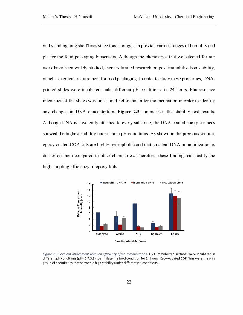
any changes in DNA concentration. Figure 2.3 summarizes the stability test results.
Although DNA is covalently attached to every substrate, the DNA-coated epoxy surfaces
showed the highest stability under harsh pH conditions. As shown in the previous section,
epoxy-coated COP foils are highly hydrophobic and that covalent DNA immobilization is
denser on them compared to other chemistries. Therefore, these findings can justify the
high coupling efficiency of epoxy foils.
Figure2.3Covalentattachmentreactionefficiencyafterimmobilization.DNAimmobilizedsurfaceswereincubatedindifferentpHconditions(pH=6,7.5,9)tosimulatethefoodconditionfor24hours.Epoxy-coatedCOPfilmsweretheonlygroupofchemistriesthatshowedahighstabilityunderdifferentpHconditions.
0
2
4
6
8
10
12
14
16
Aldehyde Amine NHS Carboxyl Epoxy
Rel
ativ
e Fl
uore
scen
t In
tens
ity (a
.u.)
Functionalized Surfaces
Incubation pH=7.5 Incubation pH=6 Incubation pH=9

Master’s Thesis - H.Yousefi McMaster University - Chemical Engineering
23
Evaluation of reusability through fluorescent probe hybridization.
Reproducibility is one of the most important characteristics of biosensors used for
monitoring food quality as most of the current biosensors need to be replaced with new
biomolecules after each detection. To study the reproducibility of our biosensors, amine-
terminated DNA probes were immobilized onto epoxy slides, followed by hybridization of
fluorescent complementary DNA probes as explained in methods. We performed this
hybridization and de-hybridization reaction on the same epoxy surfaces for up to 8 times
using complementary DNA strand containing a fluorescent tag. Hybridization results were
assessed using a fluorescence scanner to provide information regarding the relative density
and homogeneity of the immobilized and the complementary fluorescent probes. As shown
in Figure 2.4a, the presence of fluorescent DNA after 8 hybridization cycles showed that
FirstHybridization SecondHybridization
SixthHybridization
ThirdHybridization
FifthHybridization
ForthHybridization
SeventhHybridization EighthHybridization
a)
b)
0
5
10
15
20
1st 2nd 3rd 4th 5th 6th 7th 8th
Rela
tive F
luor
esce
nt
Inte
nsity
Number of Hybridization Cycles
Figure2.4SequentialDNAhybridizationstepsonepoxysurfaces.a)FluorescenceimagingoftheslidesaftereachhybridizationstepshowstheconsistentDNAdensityandsuccessfulre-hybridization.Scalebar:200µm.b)FluorescenceintensitymeasurementsoftheareasprintedwithDNAafterhybridizationwithfluorescentlylabelledcomplementaryprobe.Althoughtherewasadecreaseinfluorescenceintensityinfirstfewsteps,itshowedaconstantvalueafterward.

Master’s Thesis - H.Yousefi McMaster University - Chemical Engineering
24
the DNA probes remain functional and that the washing and heating procedures in the de-
hybridization process did not affect covalent attachment of DNA on the epoxy substrate.
Figure 2.4b shows the fluorescence intensity measurements of the complimentary DNA
probe after each hybridization reaction. It is seen that there is a slight drop in fluorescence
intensity over the first three measurements, followed by a stable and consistent reading for
all of the remaining cycles. This initial drop in fluorescence intensity can be attributed to
the removal of nonspecifically attached DNA from the surface. The stability of the
immobilized DNA allows for the creation of a reloadable biosensor for detecting food-
borne pathogens. This easy to use, reusable and stable platform would enable both
consumables and store owners to reload and create their personalized biosensors based on
the need (e.g. when there is an outbreak of a specific pathogen).
Conclusions
We investigated several substrate and surface chemistry options to be used as food
packaging biosensors. Although other substrates contain useful properties such as shorter
reaction time for covalent attachment, we demonstrated that overall, epoxy coated slides
are the best candidates for the producing DNA-based biosensors. These epoxy surfaces
showed promising performances for covalent immobilization, binding strength, stability,
durability, and low non-specific immobilization. We also showed that these slides are
suitable substrates for reloadable biosensors in food packaging because of their consistent
efficiency after several hybridization processes. Finally, COP slides can be transformed
from thick slides to thin films to be used inside food packaging wraps.

Master’s Thesis - H.Yousefi McMaster University - Chemical Engineering
25
Materials and methods:
Chemicals
Epoxy-coated plastic and carboxyl plastic slides were purchased from AutoMate Scientific
Inc. Aldehyde and amine glass slides were purchased from Arrayit. N-Hydroxysuccinimide
(NHS) glass slides were purchased from MicroSurfaces Inc. Phosphate-buffered saline was
purchased from BioShop. N-(3-Dimethylaminopropyl)-N′-ethylcarbodiimide (EDC), 2-
(N-Morpholino) ethanesulfonic acid (MES), Sodium dodecyl sulfate (SDS) and N-
Hydroxysuccinimide (NHS) was purchased from Sigma-Aldrich. Sodium Phosphate
Monobasic was purchased from EMD. Sodium Phosphate Dibasic and 50% Glutaraldehyde
Solution were purchased from Fisher Scientific. All synthetic oligonucleotides were
obtained from Integrated DNA Technologies and were purified using denaturing
polyacrylamide gel electrophoresis (dPAGE). 5’-aminated DNA probe bearing a 3’-FAM
label [5’-/5AmMC12/TTT TTC ACG GAT CCT GAC AAG GAT/36-FAM/-3’], 5’-
aminated DNA probe [5’-/5AmC12/ TTT TTT TTT TAG GAA GAA GTT TCA AGG
AAA GGA-3’], and a FAM labeled probe without terminal amine and was complement to
the aminated probe [5’-/56-FAM/TCC TTT CCT TGA AAC TTC TTC CT-3’] were used
in this work.
DNA immobilization on selected surfaces
Five immobilization chemistries that are commonly used in biosensors, namely epoxide,
carboxyl, amine, aldehyde, and N-hydroxysuccinimide (NHS) reactive ester, were selected
for this work (Ramakrishnan et al., 2002). Epoxy and carboxyl functionalized surfaces

Master’s Thesis - H.Yousefi McMaster University - Chemical Engineering
26
utilized a cyclo olefin copolymer (COP) substrate, while NHS, amine, and aldehyde slides
were made of glass.
In this study, Scienion SciFlexArrayer, a pico liter sized droplet-dispensing non-contact
printer, was used to print solutions containing DNA probes onto the different surfaces.
Using the Scienion printer, we were able to print droplets as small as 500 picoliter, which
produced DNA microarrays. Following printing, the DNA was rehydrated through
incubation in 75% relative humidity at room temperature overnight. The humidity chamber
used in this work was prepared by placing a 100% sodium chloride solution in a sealed box.
Humidity inside the box was monitored by a humidity meter, which was also installed
inside the box.
Aldehyde Slides: a 5µM single stranded DNA in 0.3M sodium phosphate buffer at pH 9.0
were added onto the functionalized surface. Following the overnight reaction in the humid
chamber, the samples were washed once with 0.1% SDS, twice with Milli-Q water, then
incubated in sodium borohydride solution containing 2.5mg of NaBH4, 750µL of PBS, and
250µL of 100% ethanol for 2 hours under agitation for reduction of Schiff base. Amine
Slides: the functionalized surfaces were activated through incubation in solution containing
2.5% glutaraldehyde in 0.1M sodium phosphate buffer pH 7.0 for 2 hours. The slides were
then rinsed in sodium phosphate buffer. Following the activation, 5µM of single-stranded
DNA in 0.1M sodium phosphate buffer at pH 7.0 was added onto functionalized surface.
Carboxyl Slides: the substrates were treated in a CO2 plasma cleaner for 2 minutes prior to
immobilization in order to induce carboxyl functional groups on the surface. A 5µM single-
stranded DNA in 0.1M MES buffer, with 25mM of EDC, and a 25mM NHS at pH 4.3 were

Master’s Thesis - H.Yousefi McMaster University - Chemical Engineering
27
then added onto the functionalized surface. Epoxy Slides: a 5µM single stranded DNA in
0.1M sodium phosphate buffer at pH 7.5 was added onto the functionalized surface. NHS
Slides: a 5µM single-stranded DNA in 0.1M phosphate buffer solution at pH 8.3 was added
onto the functionalized surface.
After the immobilization of DNA probes, the substrates were rinsed for 30 seconds with
Milli-Q water and imaged at pH 7.5 using the ChemiDoc and fluorescence microscope.
Oligonucleotides lacking amine functional groups can also attach to surfaces via
physisorption (e.g., combinations of hydrogen bonding, acid-base, hydrophobic,
electrostatic interactions).
Surface characterization
Contact angle measurement. Contact angles of water droplets on the substrates were
measured by Future Digital Scientific Corp contact angle measurement system
(Biointerface Institute, McMaster University). A micro-needle was used to dispense 2 µl
droplets of deionized (dI) H2O on all substrates before and after the DNA immobilization.
X-ray Photoelectron Spectroscopy (XPS). XPS measurements were performed using a
Physical Electronics (PHI) Quantera II spectrometer equipped with an Al anode source for
X-ray generation and a quartz crystal monochromator was used to focus the generated X-
rays (Biointerface Institute, McMaster University). For XPS measurements, DNA was
hand printed to cover a large surface area allowing proper analysis. A minimum of 3 areas
containing DNA were analyzed on each substrate.
Stability test
Master’s Thesis - H.Yousefi McMaster University - Chemical Engineering
28
Incubation buffers at different acidity were prepared, including PBS buffer at pH 6, sodium
phosphate buffer at pH 7.5, and carbonate buffer at pH 9. After DNA immobilization, slides
were incubated in each buffer for 24 hours and imaged using a fluorescence microscope
and ChemiDoc.
Hybridization and de-hybridization cycle using complementary probes
In order to determine the stability and reproducibility of the immobilized DNA
strand on the surface, we conducted 8 rounds of hybridization and de-hybridization
using complimentary fluorescent DNA strand and compared the fluorescence
intensity after each cycle. After initial immobilization of amine-terminated DNA
probes on the epoxy slides, fluorescent complimentary strand was incubated on the
surface in 1x SDS buffer for 2 hours. Following the reaction, the substrates were
rinsed with water and imaged at pH 7.5 using fluorescence scanner. De-
hybridization of the complimentary fluorescent DNA strand involved incubating the
substrates in 4M Urea solution at 70°C for 1 hour.
Master’s Thesis - H.Yousefi McMaster University - Chemical Engineering
29
References
1. Organization, W. H., WHO’s first ever global estimates of foodborne diseases
find children under 5 account for almost third of deaths. 2015.
2. Brockgreitens, J.; Abbas, A., Responsive food packaging: recent progress and
technological prospects. Comprehensive Reviews in Food Science and Food Safety 2016,
15 (1), 3-15.
3. Baeumner, A. J., Biosensors for environmental pollutants and food contaminants.
Analytical and bioanalytical chemistry 2003, 377 (3), 434-445.
4. Scott, A. O., Biosensors for food analysis. Elsevier: 1998.
5. Bejjani, B. A.; Shaffer, L. G., Application of array-based comparative genomic
hybridization to clinical diagnostics. The Journal of Molecular Diagnostics 2006, 8 (5),
528-533.
6. Lee, C.-Y.; Harbers, G. M.; Grainger, D. W.; Gamble, L. J.; Castner, D. G.,
Fluorescence, XPS, and TOF-SIMS surface chemical state image analysis of DNA
microarrays. Journal of the American Chemical Society 2007, 129 (30), 9429-9438.
7. Gibbs, J.; Kennebunk, M., Immobilization Principles–Selecting the Surface.
ELISA Technical Bulletin 2001, 1, 1-8.
8. Sassolas, A.; Blum, L. J.; Leca-Bouvier, B. D., Immobilization strategies to
develop enzymatic biosensors. Biotechnology advances 2012, 30 (3), 489-511.

Master’s Thesis - H.Yousefi McMaster University - Chemical Engineering
30
9. Karamessini, D.; Poyer, S.; Charles, L.; Lutz, J. F., 2D Sequence-Coded
Oligourethane Barcodes for Plastic Materials Labeling. Macromolecular Rapid
Communications 2017.
10. Liu, Y.; Rauch, C. B., DNA probe attachment on plastic surfaces and microfluidic
hybridization array channel devices with sample oscillation. Analytical biochemistry
2003, 317 (1), 76-84.
11. Pu, Q.; Oyesanya, O.; Thompson, B.; Liu, S.; Alvarez, J. C., On-chip
micropatterning of plastic (cylic olefin copolymer, COC) microfluidic channels for the
fabrication of biomolecule microarrays using photografting methods. Langmuir 2007, 23
(3), 1577-1583.
12. Bain, C. D.; Troughton, E. B.; Tao, Y. T.; Evall, J.; Whitesides, G. M.; Nuzzo, R.
G., Formation of monolayer films by the spontaneous assembly of organic thiols from
solution onto gold. Journal of the American Chemical Society 1989, 111 (1), 321-335.
13. Chrisey, L. A.; Lee, G. U.; O'Ferrall, C. E., Covalent attachment of synthetic
DNA to self-assembled monolayer films. Nucleic acids research 1996, 24 (15), 3031-
3039.
14. Liechti, K.; Schnapp, S.; Swadener, J., Contact angle and contact mechanics of a
glass/epoxy interface. International journal of fracture 1997, 86 (4), 361-374.
15. Metwalli, E.; Haines, D.; Becker, O.; Conzone, S.; Pantano, C., Surface
characterizations of mono-, di-, and tri-aminosilane treated glass substrates. Journal of
colloid and interface science 2006, 298 (2), 825-831.

Master’s Thesis - H.Yousefi McMaster University - Chemical Engineering
31
16. Hermanson, G. T., Bioconjugate techniques. 2nd ed.; Elsevier Academic Press:
Amsterdam ; Boston, 2008; pp. 1 online resource (xxx, 1202 pages).
17. Wong, S. S., Chemistry of protein conjugation and cross-linking. CRC press:
1991.
18. Nimse, S. B.; Song, K.; Sonawane, M. D.; Sayyed, D. R.; Kim, T., Immobilization
techniques for microarray: challenges and applications. Sensors (Basel) 2014, 14 (12),
22208-29.
19. Taylor, S.; Smith, S.; Windle, B.; Guiseppi-Elie, A., Impact of surface chemistry
and blocking strategies on DNA microarrays. Nucleic Acids Research 2003, 31 (16), e87-
e87.
20. Ramakrishnan, R.; Dorris, D.; Lublinsky, A.; Nguyen, A.; Domanus, M.;
Prokhorova, A.; Gieser, L.; Touma, E.; Lockner, R.; Tata, M.; Zhu, X.; Patterson, M.;
Shippy, R.; Sendera, T. J.; Mazumder, A., An assessment of Motorola CodeLink
microarray performance for gene expression profiling applications. Nucleic Acids Res
2002, 30 (7), e30.
21. Castner, D. G.; Ratner, B. D., Biomedical surface science: Foundations to
frontiers. Surface Science 2002, 500 (1), 28-60.

Master’s Thesis - H.Yousefi McMaster University - Chemical Engineering
32
Chapter 3
Sentinel wraps; smart biosensors in food
packaging for real-time on-the-shelf pathogen
detection
In chapter 3, all experiments were done by myself. Dr. Sana Jahanshahi Anbuhi, a postdoc fellow of prof. Filipe, helped me with data analysis. Dr. Ali Monsur helped me with DNAzyme designing and preparations as well as experiments planning. Prof. Filipe and Prof. Didar gave many helpful suggestions with experimental deigns and data analysis. I started writing the paper draft. Dr. Monsur, Prof. Filipe and Prof Didar helped me revise the draft and prepare the final version.

Master’s Thesis - H.Yousefi McMaster University - Chemical Engineering
33
3. Sentinel wraps; smart biosensors in food packaging for real-time on-
the-shelf pathogen detection
Hanie Yousefia, M. Monsur Alib, Sana Jahanshahi-Anbuhia, Carlos D.M. Filipea, Tohid F.
Didar*c,d,e
a Department of Chemical Engineering, McMaster University, Hamilton, Ontario, Canada b Biointerface institute, McMaster University, Hamilton, Ontario c Institute for Infectious Disease Research (IIDR), McMaster University, Hamilton, Ontario, Canada dMechanical Engineering Department, McMaster University, Hamilton, Ontario, Canada, Canada e School of Biomedical Engineering. McMaster University Corresponding author: Tohid Didar, Email: [email protected]
Abstract
Microbial pathogens can grow in food at any point in food processing chain, causing
foodborne illnesses. Biosensors, developed based on liquid phase sensors or lab-on-a-chip
devices cannot easily be used for real-time food examinations after packaging without
taking the sample out of the stock. Packaged food such as meat, apple and juice are directly
in touch with the surface of their containers or covers. Therefore, real-time on surface
sensing mechanisms, installed inside the food packaging, tracing the presence of pathogens
inside the packaged food are much needed to examine food safety. Here we report on
developing thin, transparent, flexible and durable sensing surfaces based on DNAzyme
biosensors, that generate fluorescent signal in the presence of a target non-pathogenic
bacteria in food or water samples. The covalently attached DNAzyme probe glowed upon

Master’s Thesis - H.Yousefi McMaster University - Chemical Engineering
34
contact with the packaged food (meat, sliced apple and apple juice) contaminated with
target bacterium (Escherichia coli). We were able to detect Escherichia coli in food
packaging with concentrations as low as 103 CFUs/mL. The developed sensing surfaces
remained stable up to 10 days under varying pH conditions (pH 5 to 9). In addition to
detecting pathogens on packaged food or drinking bottles, the developed sensing surfaces
has the potential to be applied for a variety of other applications such as health care settings,
environmental monitoring, food production chain, and biomaterials like wound dressing.
Introduction
Microbial growth in food products, derived from packaging deficiencies or
incorrect manipulation by consumers during distribution and storage period, results in an
increased prospect of consuming contaminated food and large-scale outbreaks. This threat
assigns shelf storage period a paramount importance throughout the whole food supply
chain. On-the-shelf, real-time, precise and simple tracing of pathogens can provide a
powerful tool in addressing this issue. Traditional microbiological identification methods
for pathogens in food, are well known to be prolonged and challenging (Nicholson et al.,
1998; R.-F. Wang, Cao, & Cerniglia, 1996). These methods are progressively being
recognized as insufficient to meet the requirements of real-time response and remote
analysis in food storing conditions.
In addition, an important aspect of reliable sensors that makes them suitable for real-time
measurements, is their stability, which is normally expected to be longer than a few hours,
preferably days or weeks (Wilson & Gifford, 2005). Therefore, designing innovative
Master’s Thesis - H.Yousefi McMaster University - Chemical Engineering
35
sensing devices which can be used in food packaging and kept in storage conditions to
evaluate real-time freshness of food products are highly desired. During the past several
years, researchers have designed and constructed a number of sensors for on-the-shelf
detection of pathogens using magnetic nanobeads (L. Xu et al., 2017), specific food
targeted sensors based on polyaniline (Kuswandi, Restyana, Abdullah, Heng, & Ahmad,
2012) and humidity based wireless sensors (Tan, Ng, Shao, Pereles, & Ong, 2007) in order
to indicate food rotting or infection in a real-time manner. However, these sensors do not
meet the main requirements for packaged food monitoring, which are mainly stability and
self-reliance (L. Xu et al., 2017). Therefore, it demands appropriate solutions for smart
packaging in food controlling. Thus, there is a greater need for developing faster, more
sensitive, and more stable food monitoring instruments that can be located inside the food
packaging during the storage time (Yoo & Lee, 2016).
Synthetic catalytic DNA molecules (DNAzymes) as functional nucleic acids, are artificial
single-stranded DNA molecules that have a catalytic ability (Breaker & Joyce, 1994; Gong
et al., 2015; Lu & Liu, 2006). Among the DNAzymes, RNA-cleaving ones have become
attractive particularly in developing detection methods for a wide variety of targets (Pun et
al., 2004; Schubert et al., 2003). Recently RNA-cleaving fluorescent DNAzymes (RFD)
were generated by in vitro selection for specific bacteria and optimized for real-time
bacterial detection purposes (Sergio D Aguirre et al., 2012; Li, 2011; W. Zhang et al.,
2016). These DNAzymes cleave a fluorogenic DNA substrate at a single ribonucleotide
embedded in the substrate. The cleavage junction is surrounded by a fluorophore and a
quencher so that the intact substrate prior to cleavage reaction possesses minimal

Master’s Thesis - H.Yousefi McMaster University - Chemical Engineering
36
fluorescence signal. When the substrate is cleaved by the DNAzyme in the presence of the
target bacterium, the fluorophore and the quencher separates away from each other and
enhances the fluorescence signal. High sensitivity and selectivity of these DNAzyme
probes combined by their facile real-time behavior in bacterial detection (S. D. Aguirre et
al., 2013), and, higher stability, make them a great candidate for food packaging sensors.
DNAzymes were previously optimized in liquid phase as pathogen sensing agents, on
magnetic beads (H. Zhang et al., 2016), on metal organic frameworks (MOF) (Chen et al.,
2017) and with liquid crystals (Liao et al., 2016). However, there is no report on attaching
DNAzymes to flexible polymer based surfaces so far. In addition, these DNAzyme sensors
were shown to respond to crude extracellular mixtures (CEM) (Ali et al., 2011) and crude
intracellular mixtures (CIM) (S. D. Aguirre et al., 2013) of specific bacteria but their
application for detecting live cells of pathogens has not been demonstrated so far.
Here we demonstrate, for the first time, employing DNAzyme biosensors on flexible
surfaces, for detecting bacteria inside packaged food. Thin, transparent and flexible COP
(cyclo olefin copolymer) films functionalized with epoxy, were used as the substrate to
immobilize DNAzyme probes. This work is the first microarray production of DNAzyme
probes on customary surfaces which can provide the food industry with on the package
sensing and tracking opportunities. In addition, our novel DNAzyme based sensors showed
a high range of stability which proved them as a reliable candidate for on-the-shelf bacterial
detection. In this work, DNAzyme based surfaces showed a sensitive feature to detect live
bacterial cells in both liquid (juice) and solid (meat and apple) food supplies without a need
for high-level monitoring system. Furthermore, the developed biosensors are able to detect

Master’s Thesis - H.Yousefi McMaster University - Chemical Engineering
37
live bacterial cells eliminating the need to lyse the bacterial cells in order to detect them.
Moreover, their high stability, in different environments with varying pH, makes them a
perfect candidate to be used in different food packaging.
Figure3.1 IllustrationofhighlysensitiveDNAzymesensorscleaving inpresenceof liveE.colicells.Amine terminated DNAzyme probes were covalently attached to flexible, transparent epoxy films. In presence of bacteria, RNA cleaving section is detached, consequently, the fluorescence intensity is increased.
Results and discussion
Sensors fabrication, characterization and stability assays
As mentioned above, long-term stability is an important issue in sensing devices that are
designed to be used in consumer packages (Scott, 1998). We used a previously reported
DNAzyme that cleaves a fluorogenic DNA substrate in the presence of CEM or CIM of E.
coli. The fluorogenic substrate consists of the three parts: fluorophore, the quencher
molecules and the cleavage junction. The substrate is located at the 3’-end of the
DNAzyme. Therefore, the DNAzyme was synthesized with an amine group at the 5’-end,
so that, after cleavage reaction, the fluorophore remains bounded on the surface losing the
quencher to increase the fluorescence. To minimize the waste of DNAzyme reagent and

Master’s Thesis - H.Yousefi McMaster University - Chemical Engineering
38
achieve mass production with ease, we used a picoliter inkjet printer which allows us to
rapidly produce DNA microarrays. The amine group of the printed DNAzyme reacts with
the epoxy group on the surface of COP foil forming covalent bond (Figure 3.1). The
covalent attachment after printing was investigated by alkaline treatment (see methods). It
is assumed that the alkali labile ribonucleotide in the DNAzyme substrate is hydrolyzed
and removes the quencher fragment resulting in enhancement of fluorescence signal on the
printed spot (Figure 3.2a, left side), shows the cleaved DNAzyme surface. In addition, a
control experiment without amine modified DNAzyme was carried out. After washing with
alkaline solution, no fluorescence signal was observed (Figure 3.2a, right side) indicating
the complete wash off of the DNAzyme with no amine groups. DNAzyme probes were also
printed on non-functionalized COP foils to further confirm the covalent attachment. Results
confirmed successful attachment of DNAzyme probes when conjugated with amine groups.
Since packaged food reside under different pH conditions, we tested the surface attached
DNAzyme reaction efficiency and stability at different pH conditions. Our experimental
results indicated that the DNAzyme is stable under a broad range of pH conditions
(Figure 3.2b). The relative fluorescence is the ratio of the immobilized amine terminated
DNA signal to the control. The fluorescence imaging results are reported in relative
fluorescence format to emphasize on detectability of the signals.
To evaluate the activity of DNAzyme probes after the attachment to the plastic surfaces in
the presence of the target bacteria, DNAzyme-COP surfaces were incubated with live E.
coli cells in reaction buffer for 4 h. A negative control was also conducted wherein the
DNAzyme-COP surfaces were incubated in the reaction buffer without E. coli. After

Master’s Thesis - H.Yousefi McMaster University - Chemical Engineering
39
removing the substrates from the reaction tube the surfaces were washed and imaged using
a fluorescence imager (ChemiDocTM, Bio-Rad). Figure 3.2c shows the fluorescence
intensity difference between E. coli and buffer incubated COP surfaces. These results
indicate that the coupling process (printing, incubation and washing) do not affect
DNAzyme functionality producing a reliable sensing surface similar to the solution phase.
Figure3.2DNAzymebasedsurfacescharacterizationandstabilityassay:a)AmineterminatedDNAzymeandaminefreeDNA probes weremixed with reaction buffer and printed with picoliter sized droplets, on transparent and epoxyfunctionalizedflexiblecopolymers.AmineterminatedDNAzymeprobeswerecovalentlyattachedtotheepoxyslidesandwere then cleavedbyNaOH solution. Slideswerewashed thoroughlywithwater and PBS buffer.DNAprobeswithoutamineattheendhadnonon-specificattachmenttotheepoxysurface.b)DNAzymesensors’stabilityunderdifferentpHconditions.DNAzymeslideswere incubatedunderdifferent rangesofpH for10days tomonitor theirstability.BothcovalentattachmentandDNAzymefunctionwerestableaftertheincubationperiod.DNAzymesdidnotlosetheiractivityaftertheincubationperiod.c)UppersectionofsensorswasincubatedwithliveE.colicellsandthebottom section were incubated in reaction buffer. After incubation, the upper side showed a significantly higherfluorescenceintensity.
Real-time fluorescence assay
Another key consideration for the real-time on-site detection, is the rate of interaction
between the developed sensor and bacteria. The real-time activity of DNAzyme sensors
was investigated and measured by introducing the sensing surfaces to E. coli cells and
0.5
2
3.5
5
6.5
8
9.5
pH=3 pH=6 pH=9 Reaction buffer
NaOH
RF
(a. u
.) Day 10
E.coli Live Cells
pH 7.5
a)
b)
c)
500 μm
200 μm200 μm

Master’s Thesis - H.Yousefi McMaster University - Chemical Engineering
40
collecting the fluorescence signal at different time points see experimental section for detail
procedure). NaOH and the reaction buffer without adding cells were used as a positive and
negative controls respectively. The results indicated that the fluorescence intensity
increased by 7 folds in less than 2 hours with the bacterial sample (Figure 3.3a).
Specificity
Although the DNAzyme, RFD-EC1, has been reported to be specific against E. coli in
the previously published reports, we were further interested to investigate specificity with
its surface immobilized form. In order to examine specificity gram-positive bacteria
(Pediococcus acidilactici, Bacillus subtilis) and gram-negative bacteria (Yersinia ruckeri
and Achromobacter xylosoxidans) were used. All bacteria samples were incubated
overnight in TSB growth media and prepared as described in methods. DNAzyme slides
were immerged into the cell suspension and incubated for 2 hours. The fluorescence
Figure 3.3 Response of DNAzyme biosensors to bacteria incubation. a) Results of experiments show that bacteriapresence can lead to a high fluorescence increase in DNAzyme sensors which was measured as 7 times higherfluorescenceafteronlytwohours.b)Specificitytest.E.colicellsandtwogramnegativebacteriaandtwogrampositivebacteriaweretestedwithDNAzymeslidestoshowthespecificattachmentofDNAzymeprobestoE.colicells.
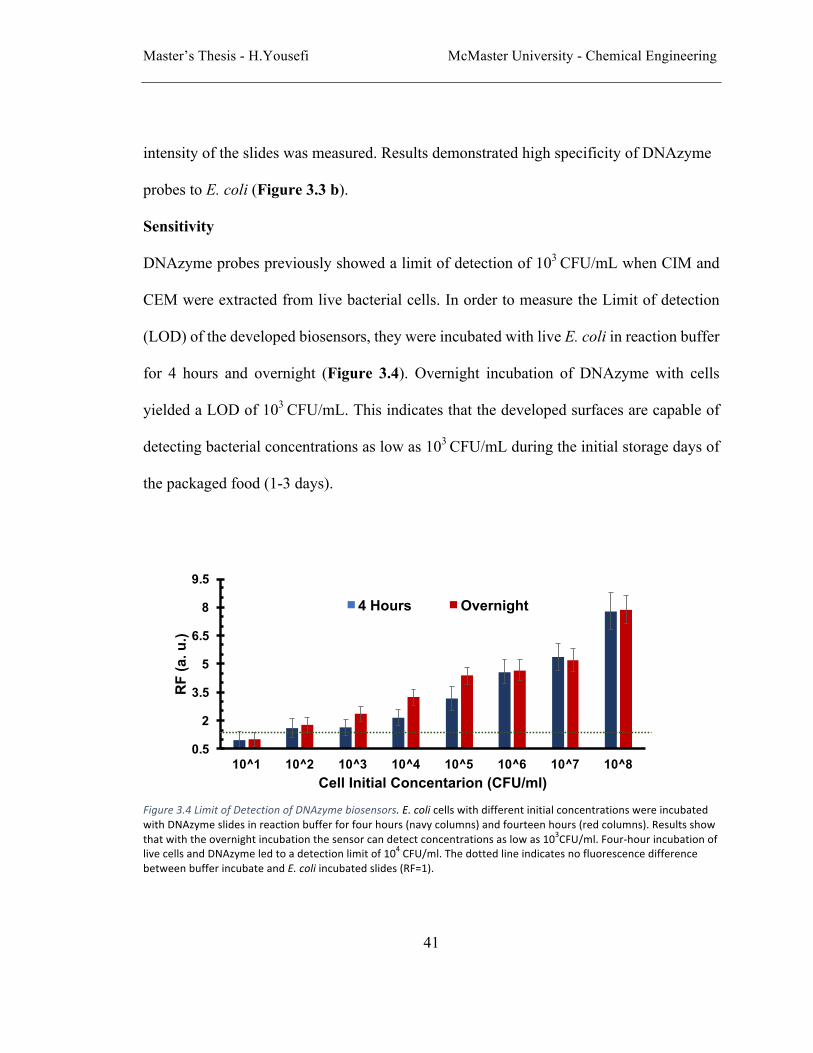
Master’s Thesis - H.Yousefi McMaster University - Chemical Engineering
41
intensity of the slides was measured. Results demonstrated high specificity of DNAzyme
probes to E. coli (Figure 3.3 b).
Sensitivity
DNAzyme probes previously showed a limit of detection of 103 CFU/mL when CIM and
CEM were extracted from live bacterial cells. In order to measure the Limit of detection
(LOD) of the developed biosensors, they were incubated with live E. coli in reaction buffer
for 4 hours and overnight (Figure 3.4). Overnight incubation of DNAzyme with cells
yielded a LOD of 103 CFU/mL. This indicates that the developed surfaces are capable of
detecting bacterial concentrations as low as 103 CFU/mL during the initial storage days of
the packaged food (1-3 days).
Figure3.4LimitofDetectionofDNAzymebiosensors.E.colicellswithdifferentinitialconcentrationswereincubatedwithDNAzymeslidesinreactionbufferforfourhours(navycolumns)andfourteenhours(redcolumns).Resultsshowthatwiththeovernightincubationthesensorcandetectconcentrationsaslowas103CFU/ml.Four-hourincubationoflivecellsandDNAzymeledtoadetectionlimitof104CFU/ml.ThedottedlineindicatesnofluorescencedifferencebetweenbufferincubateandE.coliincubatedslides(RF=1).
0.5
2
3.5
5
6.5
8
9.5
10^1 10^2 10^3 10^4 10^5 10^6 10^7 10^8
RF
(a. u
.)
Cell Initial Concentarion (CFU/ml)
4 Hours Overnight
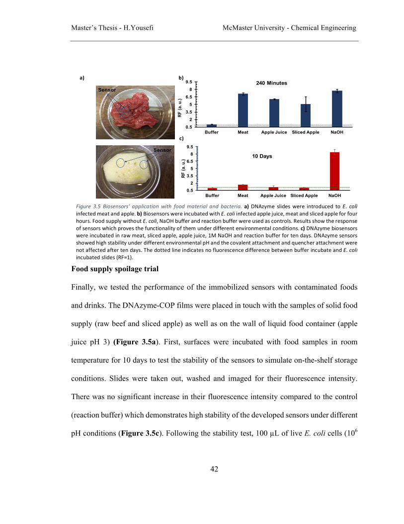
Master’s Thesis - H.Yousefi McMaster University - Chemical Engineering
42
Food supply spoilage trial
Finally, we tested the performance of the immobilized sensors with contaminated foods
and drinks. The DNAzyme-COP films were placed in touch with the samples of solid food
supply (raw beef and sliced apple) as well as on the wall of liquid food container (apple
juice pH 3) (Figure 3.5a). First, surfaces were incubated with food samples in room
temperature for 10 days to test the stability of the sensors to simulate on-the-shelf storage
conditions. Slides were taken out, washed and imaged for their fluorescence intensity.
There was no significant increase in their fluorescence intensity compared to the control
(reaction buffer) which demonstrates high stability of the developed sensors under different
pH conditions (Figure 3.5c). Following the stability test, 100 µL of live E. coli cells (106
Figure 3.5 Biosensors’ applicationwith foodmaterial and bacteria. a)DNAzyme slideswere introduced toE. coliinfectedmeatandapple.b)BiosensorswereincubatedwithE.coliinfectedapplejuice,meatandslicedappleforfourhours.FoodsupplywithoutE.coli,NaOHbufferandreactionbufferwereusedascontrols.Resultsshowtheresponseofsensorswhichprovesthefunctionalityofthemunderdifferentenvironmentalconditions.c)DNAzymebiosensorswereincubatedinrawmeat,slicedapple,applejuice,1MNaOHandreactionbufferfortendays.DNAzymesensorsshowedhighstabilityunderdifferentenvironmentalpHandthecovalentattachmentandquencherattachmentwerenotaffectedaftertendays.ThedottedlineindicatesnofluorescencedifferencebetweenbufferincubateandE.coliincubatedslides(RF=1).
Sensor
a) b)
c)
0.52
3.55
6.58
9.5
Buffer Meat Apple Juice Sliced Apple NaOH
RF (a
. u.)
240 Minutes
0.52
3.55
6.58
9.5
Buffer Meat Apple Juice Sliced Apple NaOH
RF (a
. u.)
10 DaysSensor

Master’s Thesis - H.Yousefi McMaster University - Chemical Engineering
43
CFU/mL) were spiked with the food samples, incubated for 4 h and tested for fluorescence
signals. Each COP slide had and area of 10-5 m2. Although the total number of cells added
to each food sample were 105 colonies, the sensors were in touch with a small part of the
food sample (approximately 3%) which means not all the bacteria were needed to reach the
slides and activate the DNAzyme on them. Fluorescence intensity of the sensors was
compared to the controls and results showed significant increase (up to 7 time) in
fluorescence intensity of the sensors indicating successful detection of pathogens in the
food samples (Figure 3.5b). These properties make the sensors a great candidate for smart
packaging applications.
Conclusion
The DNAzyme-based sensing surfaces described here appear to have several promising
features for on-the-shelf health monitoring in food supply such as: (1) no need to lyse the
cells (2) no need for liquid handling, pipetting, flow or external accessories, (3) real-time
response to bacterial growth (4) sensitivity (LOD of 103 CFU/mL) and (5) high stability in
food storage conditions. On the other hand, fluorescence sensing software are being
developed to enable the cellphones to detect fluorescence signals. This gives us the hope
for real usage of our sensors in food packaging as smart packaging. The cleavage-based
RNA detection presented here is suitable for use with diverse bacterial targets, because the
modified DNAzyme probes can be designed to target different pathogens due to their high
specificity to each RNA of bacteria.

Master’s Thesis - H.Yousefi McMaster University - Chemical Engineering
44
Materials and methods
Chemicals
The amine modified DNAzyme, ligation template (NH-EC1 and LT in Table 3.1)
oligonucleotides were purchased from Integrated DNA Technologies (IDT). The
fluorogenic substrate (FS1) was purchased from Yale University (Sequence is provided in
Table 3.1). Epoxy coated COP foils were purchased from PolyAn molecular surface
engineering. NaCl, MgCl2, Tween 20, Na2PO4, NaHPO4, Tryptic Soy Broth (TSB), KCl,
Na2CO3 (99.99%), NaHO3, NaOH and HEPES (4-(2-hydroxyethyl)-1-
piperazineethanesulfonic acid) were purchased from Sigma Aldrich. ATP, PEG4000, T4
DNA ligase and polynucleotide kinase (PNK) and their respective buffers were purchased
from Thermo Scientific, Canada. E. coli K12 strains are regularly maintained in our
laboratory. Other bacteria types (Pediococcus acidilactici, Bacillus subtilis, Yersinia
ruckeri and Achromobacter xylosoxidans) were donated by Dr. Yingfu Li laboratory at
McMaster University. ChemiDoc imaging system, Zeiss and Olympus inverted
microscopes were used to image DNAzyme slides. Scienion FLEXARRAYER was used
to print DNAzyme on epoxy slides.
Preparation of RFD-EC1
NH-EC1 was enzymatically ligated to FS1 as follows: 500 pmol of FS1 was
phosphorylated in 100 µL volume containing 1x PNK buffer A for 35 min at 37 C. The
enzyme was inactivated by heating at 90 C for 5 min and cooled down to room temperature
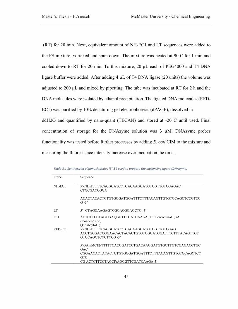
Master’s Thesis - H.Yousefi McMaster University - Chemical Engineering
45
(RT) for 20 min. Next, equivalent amount of NH-EC1 and LT sequences were added to
the FS mixture, vortexed and spun down. The mixture was heated at 90 C for 1 min and
cooled down to RT for 20 min. To this mixture, 20 µL each of PEG4000 and T4 DNA
ligase buffer were added. After adding 4 µL of T4 DNA ligase (20 units) the volume was
adjusted to 200 µL and mixed by pipetting. The tube was incubated at RT for 2 h and the
DNA molecules were isolated by ethanol precipitation. The ligated DNA molecules (RFD-
EC1) was purified by 10% denaturing gel electrophoresis (dPAGE), dissolved in
ddH2O and quantified by nano-quant (TECAN) and stored at -20 C until used. Final
concentration of storage for the DNAzyme solution was 3 µM. DNAzyme probes
functionality was tested before further processes by adding E. coli CIM to the mixture and
measuring the fluorescence intensity increase over incubation the time.
Table3.1Synthesizedoligonucleotides(5ʹ-3ʹ)usedtopreparethebiosensingagent(DNAzyme)
Probe Sequence
NH-EC1 5’-NH2TTTTTCACGGATCCTGACAAGGATGTGGTTGTCGAGAC CTGCGACCGGA ACACTACACTGTGTGGGATGGATTTCTTTACAGTTGTGTGCAGCTCCGTCCG -3’
LT 5’- CTAGGAAGAGTCGGACGGAGCTG -3’
FS1 ACTCTTCCTAGCFrAQGGTTCGATCAAGA (F: fluorescein-dT, rA: riboadenosine, Q: dabcyl-dT)
RFD-EC1
5’-NH2TTTTTCACGGATCCTGACAAGGATGTGGTTGTCGAG ACCTGCGACCGGAACACTACACTGTGTGGGATGGATTTCTTTACAGTTGTGTGCAGCTCCGTCCG -3’ 5’/5AmMC12/TTTTTCACGGATCCTGACAAGGATGTGGTTGTCGAGACCTGCGAC CGGAACACTACACTGTGTGGGATGGATTTCTTTACAGTTGTGTGCAGCTCCGTC CG ACTCTTCCTAGCFrAQGGTTCGATCAAGA-3’

Master’s Thesis - H.Yousefi McMaster University - Chemical Engineering
46
Bacteria preparation
E. coli K12, bacillus subtilis (BS), yersinia ruckeri (YR), pediococcus acidilactici (PA)
and achromobacter xyloxsoxidans (AX), were cultured overnight (14 hours at 37 °C with
shaking at 250 rpm) in TSB culture media. In order to measure the colony formation unit
(CFU/ml) of E. coli cells, the cells were grown in TSB media overnight and a fresh culture
was conducted until the OD600 reached to ~1. Next, a serial dilution (10 fold) was
conducted with 1 mL volume. 100 µL from dilution tube 8 was spread onto a TSA (tryptic
soy agar) plate and incubated at 37 C overnight. This was done in triplicate samples. The
CFUs were counted and averaged to obtain the number of CFUs. E. coli cells concentration
was calculated to be 7.7 *108 CFU/mL in the culture. Other bacteria colonies were plated
onto a TSA plate and grown for 14 h at 37 °C. A single colony was taken and inoculated
into 2 mL of TSB and grown for 14 h at 37 °C with shaking at 250 rpm. The final
concentration of all the bacteria were adjusted on OD600 of ≈ 1. Live cells were collected
by centrifuge at 5000 rpm for 5 minutes and added to reaction buffer (1× RB; 100 mM
HEPES, 300 mM NaCl, 30 mM MgCl2, 0.1% Tween 20, pH 7.5) to obtain the same
concentration and ready to use.
Covalent immobilization of RFD-EC1 onto the surfaces
A 5 µL of DNAzyme probes were mixed with 5µL of 2x printing buffer (autoclaved sodium
phosphate buffer at pH 7.5). Scienion printer was used to print the reaction solution onto
epoxy coated slides following by overnight incubation (14 hours) at room temperature and
75% relative humidity. Then, slides were washed thoroughly to make sure unreacted

Master’s Thesis - H.Yousefi McMaster University - Chemical Engineering
47
DNAzymes are washed out. Washing process was two minutes rinsing by autoclaved Milli-
Q water and one minute rinsing by PBS buffer at pH 7.5.
Master’s Thesis - H. Yousefi McMaster University - Chemical Engineering
48
References
Aguirre,S.D.,Ali,M.M.,Kanda,P.,&Li,Y.(2012).DetectionofbacteriausingfluorogenicDNAzymes.JoVE(JournalofVisualizedExperiments)(63),e3961-e3961.
Aguirre,S.D.,Ali,M.M.,Salena,B.J.,&Li,Y.(2013).AsensitiveDNAenzyme-basedfluorescentassayforbacterialdetection.Biomolecules,3(3),563-577.doi:10.3390/biom3030563
Ahvenainen,R.(2003).Novelfoodpackagingtechniques:Elsevier.Ali,M.M.,Aguirre,S.D.,Lazim,H.,&Li,Y.(2011).FluorogenicDNAzymeprobesas
bacterialindicators.AngewChemIntEdEngl,50(16),3751-3754.doi:10.1002/anie.201100477
Baeumner,A.J.(2003).Biosensorsforenvironmentalpollutantsandfoodcontaminants.Analyticalandbioanalyticalchemistry,377(3),434-445.
Barbulovic-Nad,I.,Lucente,M.,Sun,Y.,Zhang,M.,Wheeler,A.R.,&Bussmann,M.(2006).Bio-microarrayfabricationtechniques—areview.Criticalreviewsinbiotechnology,26(4),237-259.
Bejjani,B.A.,&Shaffer,L.G.(2006).Applicationofarray-basedcomparativegenomichybridizationtoclinicaldiagnostics.TheJournalofMolecularDiagnostics,8(5),528-533.
Bélanger,P.,Tanguay,F.,Hamel,M.,&Phypers,M.(2015).AnoverviewoffoodborneoutbreaksinCanadareportedthroughOutbreakSummaries:2008-2014.CanadaCommunicableDiseaseReport,41(11),254.
BRACKETT,R.E.(1992).Shelfstabilityandsafetyoffreshproduceasinfluencedbysanitationanddisinfection.JournalofFoodProtection,55(10),808-814.
Breaker,R.R.(1997).DNAenzymes.Naturebiotechnology,15(5),427-431.Breaker,R.R.,&Joyce,G.F.(1994).ADNAenzymethatcleavesRNA.Chemistry&
biology,1(4),223-229.Brockgreitens,J.,&Abbas,A.(2016).Responsivefoodpackaging:recentprogressand
technologicalprospects.ComprehensiveReviewsinFoodScienceandFoodSafety,15(1),3-15.
Bryan,F.L.(1990).Hazardanalysiscriticalcontrolpoint(HACCP)systemsforretailfoodandrestaurantoperations.JournalofFoodProtection,53(11),978-983.
Chen,M.,Gan,N.,Zhou,Y.,Li,T.,Xu,Q.,Cao,Y.,&Chen,Y.(2017).Anovelaptamer-metalions-nanoscaleMOFbasedelectrochemicalbiocodesformultipleantibioticsdetectionandsignalamplification.SensorsandActuatorsB:Chemical,242,1201-1209.
Chrisey,L.A.,Lee,G.U.,&O'Ferrall,C.E.(1996).CovalentattachmentofsyntheticDNAtoself-assembledmonolayerfilms.NucleicAcidsResearch,24(15),3031-3039.

Master’s Thesis - H. Yousefi McMaster University - Chemical Engineering
49
Control,C.f.D.,&Prevention.(2015).TheFoodProductionChain—HowFoodGetsContaminated.FoodborneOutbreaks,InvestigatingOutbreaks.
Dougherty,C.P.,Holtz,S.H.,Reinert,J.C.,Panyacosit,L.,Axelrad,D.A.,&Woodruff,T.J.(2000).DietaryexposurestofoodcontaminantsacrosstheUnitedStates.EnvironmentalResearch,84(2),170-185.
Egeland,R.D.,&Southern,E.M.(2005).ElectrochemicallydirectedsynthesisofoligonucleotidesforDNAmicroarrayfabrication.NucleicAcidsResearch,33(14),e125-e125.
Food,U.,&Administration,D.(2010).GuidanceforIndustry:SanitaryTransportationofFood.
Gibbs,J.,&Kennebunk,M.(2001).ImmobilizationPrinciples–SelectingtheSurface.ELISATechnicalBulletin,1,1-8.
Gong,L.,Zhao,Z.,Lv,Y.-F.,Huan,S.-Y.,Fu,T.,Zhang,X.-B.,...Yu,R.-Q.(2015).DNAzyme-basedbiosensorsandnanodevices.ChemicalCommunications,51(6),979-995.
Han,I.S.,Bae,Y.H.,Magda,J.J.,&Baek,S.G.(2001).Biosensor:GooglePatents.Hermanson,G.T.(2008).Bioconjugatetechniques(pp.1onlineresource(xxx,1202
pages)).Retrievedfromhttp://libaccess.mcmaster.ca/login?url=http://MCMU.eblib.com/patron/FullRecord.aspx?p=349307
Hill,W.,Payne,W.,&Aulisio,C.(1983).DetectionandenumerationofvirulentYersiniaenterocoliticainfoodbyDNAcolonyhybridization.AppliedandEnvironmentalMicrobiology,46(3),636-641.
Karamessini,D.,Poyer,S.,Charles,L.,&Lutz,J.F.(2017).2DSequence-CodedOligourethaneBarcodesforPlasticMaterialsLabeling.MacromolecularRapidCommunications.
Kennedy,J.,Jackson,V.,Blair,I.,McDowell,D.,Cowan,C.,&Bolton,D.(2005).Foodsafetyknowledgeofconsumersandthemicrobiologicalandtemperaturestatusoftheirrefrigerators.JournalofFoodProtection,68(7),1421-1430.
Kirk,M.D.,Angulo,F.J.,Havelaar,A.H.,&Black,R.E.(2017).Diarrhoealdiseaseinchildrenduetocontaminatedfood.BulletinoftheWorldHealthOrganization,95(3),233.
Kuswandi,B.,Restyana,A.,Abdullah,A.,Heng,L.Y.,&Ahmad,M.(2012).Anovelcolorimetricfoodpackagelabelforfishspoilagebasedonpolyanilinefilm.FoodControl,25(1),184-189.
Lan,T.,Furuya,K.,&Lu,Y.(2010).AhighlyselectiveleadsensorbasedonaclassicleadDNAzyme.ChemicalCommunications,46(22),3896-3898.
Lazcka,O.,DelCampo,F.J.,&Munoz,F.X.(2007).Pathogendetection:Aperspectiveoftraditionalmethodsandbiosensors.BiosensorsandBioelectronics,22(7),1205-1217.
Master’s Thesis - H. Yousefi McMaster University - Chemical Engineering
50
Lee,C.-Y.,Harbers,G.M.,Grainger,D.W.,Gamble,L.J.,&Castner,D.G.(2007).Fluorescence,XPS,andTOF-SIMSsurfacechemicalstateimageanalysisofDNAmicroarrays.JournaloftheAmericanChemicalSociety,129(30),9429-9438.
Li,Y.(2011).AdvancementsinusingreporterDNAzymesforidentifyingpathogenicbacteriaatspeedandwithconvenience.Futuremicrobiology,6(9),973-976.
Lianou,A.,&Sofos,J.N.(2007).AreviewoftheincidenceandtransmissionofListeriamonocytogenesinready-to-eatproductsinretailandfoodserviceenvironments.JournalofFoodProtection,70(9),2172-2198.
Liao,S.,Ding,H.,Wu,Y.,Wu,Z.,Shen,G.,&Yu,R.(2016).Label-freeliquidcrystalbiosensorforL-histidine:ADNAzyme-basedplatformforsmallmoleculeassay.BiosensorsandBioelectronics,79,650-655.
Liechti,K.,Schnapp,S.,&Swadener,J.(1997).Contactangleandcontactmechanicsofaglass/epoxyinterface.Internationaljournaloffracture,86(4),361-374.
Liu,J.,&Lu,Y.(2004).ColorimetricbiosensorsbasedonDNAzyme-assembledgoldnanoparticles.JournalofFluorescence,14(4),343-354.
Liu,Y.,&Rauch,C.B.(2003).DNAprobeattachmentonplasticsurfacesandmicrofluidichybridizationarraychanneldeviceswithsampleoscillation.Analyticalbiochemistry,317(1),76-84.
Lu,Y.,&Liu,J.(2006).FunctionalDNAnanotechnology:emergingapplicationsofDNAzymesandaptamers.CurrentOpinioninBiotechnology,17(6),580-588.
Mello,L.D.,&Kubota,L.T.(2002).Reviewoftheuseofbiosensorsasanalyticaltoolsinthefoodanddrinkindustries.Foodchemistry,77(2),237-256.
Metwalli,E.,Haines,D.,Becker,O.,Conzone,S.,&Pantano,C.(2006).Surfacecharacterizationsofmono-,di-,andtri-aminosilanetreatedglasssubstrates.Journalofcolloidandinterfacescience,298(2),825-831.
Mutlu,M.(2016).Biosensorsinfoodprocessing,safety,andqualitycontrol:CRCPress.Nicholson,P.,Simpson,D.,Weston,G.,Rezanoor,H.,Lees,A.,Parry,D.,&Joyce,D.
(1998).DetectionandquantificationofFusariumculmorumandFusariumgraminearumincerealsusingPCRassays.PhysiologicalandMolecularPlantPathology,53(1),17-37.
Nimse,S.B.,Song,K.,Sonawane,M.D.,Sayyed,D.R.,&Kim,T.(2014).Immobilizationtechniquesformicroarray:challengesandapplications.Sensors(Basel),14(12),22208-22229.doi:10.3390/s141222208
Nugen,S.,&Baeumner,A.(2008).Trendsandopportunitiesinfoodpathogendetection.Analyticalandbioanalyticalchemistry,391(2),451.
Organization,W.H.(2015).WHO’sfirsteverglobalestimatesoffoodbornediseasesfindchildrenunder5accountforalmostthirdofdeaths.
Patel,P.(2002).(Bio)sensorsformeasurementofanalytesimplicatedinfoodsafety:areview.TrACTrendsinAnalyticalChemistry,21(2),96-115.
Master’s Thesis - H. Yousefi McMaster University - Chemical Engineering
51
Prieto-Simon,B.,Campas,M.,&Marty,J.-L.(2008).Biomoleculeimmobilizationinbiosensordevelopment:tailoredstrategiesbasedonaffinityinteractions.Proteinandpeptideletters,15(8),757-763.
Prodromidis,M.I.,&Karayannis,M.I.(2002).Enzymebasedamperometricbiosensorsforfoodanalysis.Electroanalysis,14(4),241.
Pu,Q.,Oyesanya,O.,Thompson,B.,Liu,S.,&Alvarez,J.C.(2007).On-chipmicropatterningofplastic(cylicolefincopolymer,COC)microfluidicchannelsforthefabricationofbiomoleculemicroarraysusingphotograftingmethods.Langmuir,23(3),1577-1583.
Pun,S.H.,Tack,F.,Bellocq,N.C.,Cheng,J.,Grubbs,B.H.,Jensen,G.S.,...Janssens,B.(2004).TargeteddeliveryofRNA-cleavingDNAenzyme(DNAzyme)totumortissuebytransferrin-modified,cyclodextrin-basedparticles.Cancerbiology&therapy,3(7),641-650.
Ramakrishnan,R.,Dorris,D.,Lublinsky,A.,Nguyen,A.,Domanus,M.,Prokhorova,A.,...Mazumder,A.(2002).AnassessmentofMotorolaCodeLinkmicroarrayperformanceforgeneexpressionprofilingapplications.NucleicAcidsRes,30(7),e30.
Rasooly,A.,&Herold,K.E.(2006).Biosensorsfortheanalysisoffood-andwaterbornepathogensandtheirtoxins.JournalofAOACInternational,89(3),873-883.
Roday,S.(1998).Foodhygieneandsanitation:TataMcGraw-HillEducation.Sassolas,A.,Blum,L.J.,&Leca-Bouvier,B.D.(2012).Immobilizationstrategiesto
developenzymaticbiosensors.Biotechnologyadvances,30(3),489-511.Scallan,E.,Hoekstra,R.M.,Angulo,F.J.,Tauxe,R.V.,Widdowson,M.-A.,Roy,S.L.,...
Griffin,P.M.(2011).FoodborneillnessacquiredintheUnitedStates—majorpathogens.Emerginginfectiousdiseases,17(1),7.
Schubert,S.,GuÈl,D.C.,Grunert,H.P.,Zeichhardt,H.,Erdmann,V.A.,&Kurreck,J.(2003).RNAcleaving‘10-23’DNAzymeswithenhancedstabilityandactivity.NucleicAcidsResearch,31(20),5982-5992.
Scott,A.O.(1998).Biosensorsforfoodanalysis:Elsevier.Srinivasan,R.,Umesh,S.,Murali,S.,Asokan,S.,&SivaGorthi,S.(2017).BarefiberBragg
gratingimmunosensorforreal-timedetectionofEscherichiacolibacteria.JBiophotonics,10(2),224-230.doi:10.1002/jbio.201500208
Tan,E.L.,Ng,W.N.,Shao,R.,Pereles,B.D.,&Ong,K.G.(2007).Awireless,passivesensorforquantifyingpackagedfoodquality.Sensors,7(9),1747-1756.
Taylor,S.,Smith,S.,Windle,B.,&Guiseppi-Elie,A.(2003).ImpactofsurfacechemistryandblockingstrategiesonDNAmicroarrays.NucleicAcidsResearch,31(16),e87-e87.
Thakur,M.,&Ragavan,K.(2013).Biosensorsinfoodprocessing.Journaloffoodscienceandtechnology,50(4),625-641.

Master’s Thesis - H. Yousefi McMaster University - Chemical Engineering
52
Vanderroost,M.,Ragaert,P.,Devlieghere,F.,&DeMeulenaer,B.(2014).Intelligentfoodpackaging:Thenextgeneration.TrendsinFoodScience&Technology,39(1),47-62.
Velusamy,V.,Arshak,K.,Korostynska,O.,Oliwa,K.,&Adley,C.(2010).Anoverviewoffoodbornepathogendetection:Intheperspectiveofbiosensors.Biotechnologyadvances,28(2),232-254.
Viswanathan,S.,Radecka,H.,&Radecki,J.(2009).Electrochemicalbiosensorsforfoodanalysis.MonatsheftefürChemie-ChemicalMonthly,140(8),891.
Wang,D.Y.,&Sen,D.(2001).AnovelmodeofregulationofanRNA-cleavingDNAzymebyeffectorsthatbindtobothenzymeandsubstrate.Journalofmolecularbiology,310(4),723-734.
Wang,R.-F.,Cao,W.-W.,&Cerniglia,C.E.(1996).PCRdetectionandquantitationofpredominantanaerobicbacteriainhumanandanimalfecalsamples.AppliedandEnvironmentalMicrobiology,62(4),1242-1247.
Williams,R.,&Blanch,H.(1994).Covalentimmobilizationofproteinmonolayersforbiosensorapplications.BiosensorsandBioelectronics,9(2),159-167.
Wilson,G.S.,&Gifford,R.(2005).Biosensorsforreal-timeinvivomeasurements.BiosensorsandBioelectronics,20(12),2388-2403.
Wong,S.S.(1991).Chemistryofproteinconjugationandcross-linking:CRCpress.Wu,G.,&Zaman,M.H.(2012).Low-costtoolsfordiagnosingandmonitoringHIV
infectioninlow-resourcesettings.BulletinoftheWorldHealthOrganization,90(12),914-920.
Xu,L.,Lu,Z.,Cao,L.,Pang,H.,Zhang,Q.,Fu,Y.,...Wang,J.(2017).In-fielddetectionofmultiplepathogenicbacteriainfoodproductsusingaportablefluorescentbiosensingsystem.FoodControl,75,21-28.
Xu,Y.,Zhou,W.,Zhou,M.,Xiang,Y.,Yuan,R.,&Chai,Y.(2015).Toeholdstranddisplacement-drivenassemblyofG-quadruplexDNAforenzyme-freeandnon-labelsensitivefluorescentdetectionofthrombin.BiosensBioelectron,64,306-310.doi:10.1016/j.bios.2014.09.018
Yang,S.C.,Lin,C.H.,Aljuffali,I.A.,&Fang,J.Y.(2017).CurrentpathogenicEscherichiacolifoodborneoutbreakcasesandtherapydevelopment.Archivesofmicrobiology.
Yin,B.-C.,Zuo,P.,Huo,H.,Zhong,X.,&Ye,B.-C.(2010).DNAzymeself-assembledgoldnanoparticlesfordeterminationofmetalionsusingfluorescenceanisotropyassay.Analyticalbiochemistry,401(1),47-52.
Yoo,S.M.,&Lee,S.Y.(2016).OpticalBiosensorsfortheDetectionofPathogenicMicroorganisms.TrendsBiotechnol,34(1),7-25.doi:10.1016/j.tibtech.2015.09.012
Zhang,H.,Lin,L.,Zeng,X.,Ruan,Y.,Wu,Y.,Lin,M.,...Fu,F.(2016).Magneticbeads-basedDNAzymerecognitionandAuNPs-basedenzymaticcatalysisamplification

Master’s Thesis - H. Yousefi McMaster University - Chemical Engineering
53
forvisualdetectionoftraceuranylioninaqueousenvironment.BiosensBioelectron,78,73-79.doi:10.1016/j.bios.2015.11.024
Zhang,W.,Feng,Q.,Chang,D.,Tram,K.,&Li,Y.(2016).InvitroselectionofRNA-cleavingDNAzymesforbacterialdetection.Methods,106,66-75.
Zwietering,M.,DeKoos,J.,Hasenack,B.,DeWitt,J.,&Van'tRiet,K.(1991).Modelingofbacterialgrowthasafunctionoftemperature.AppliedandEnvironmentalMicrobiology,57(4),1094-1101.

Master’s Thesis - H. Yousefi McMaster University - Chemical Engineering
54
4. Conclusions and future works
Conclusions
The major contributions of this work are given as follows:
In chapter 2, we demonstrated that epoxy coated polymer substrates are the best candidates
for producing DNA-based biosensors amongst the current available functionalized
surfaces. The following is to support this claim based on the experimental results:
• Epoxy coated surfaces have easy DNA immobilization protocol.
• DNA immobilized epoxy surfaces showed the highest stability under different
environmental conditions.
• Epoxy coated surfaces showed the highest binding strength without influencing
DNA probes functionality, which led to consistent efficiency in several
hybridizations and dehybridizations cycles on the same substrates (up to 8).
• Reloadable sensing surfaces makes them suitable for monitoring outbreaks by being
able to change their functionality.
• COP substrates can easily be made of thin films which makes them ideal for food
packaging.
In chapter 3, we developed DNAzyme-based sensing devices as potential candidates for
on-the-shelf monitoring of contamination in food packaging. The following is to support
this claim based on the experimental results:
• Surface based DNAzyme biosensors are established by covalently immobilizing
DNAzyme probes on the surfaces.
Master’s Thesis - H. Yousefi McMaster University - Chemical Engineering
55
• Surfaces are made of polymer-based, thin, flexible and transparent functionalized
substrates. These physical properties make the substrates ideal for food packaging
purposes.
• Prepared biosensors perform an easy and fast E. coli detection that can be expanded
to other bacteria and pathogens.
• Prepared biosensors are applied for real-time detection of bacteria.
• DNAzyme biosensors are stable both in ambient condition and in contact with food
supply which makes them suitable for on-the-shelf storage.
Future Works
The results and findings in this thesis present a great potential to bring the thin sensing
films to the real applications in biosensors. In order to do so, the following research
suggestions can play a vital role:
• Developing pathogen specific DNAzyme probes and implementing them in the
developed platform to target desired pathogens in food packaging.
• Developing reloadable DNAzyme biosensors that can be switched depending on
the need in order to monitor different pathogens. This can be a step forward towards
both prevention and monitoring of outbreaks in food industry.
• Developing multiplex microarrays of various sensing agents on the presented thin
films in order to provide a multiplex high-throughput pathogen monitoring in
packaged food.


