Page 1/18 The role of Garlic and Honey on Nicotine-Induced Toxicity on the Cerebellum of adult Wistar Rats Ibegbu O Augustine Alex Ekwueme Federal University Ndufu-Alike Okechukwu N. Gertrude Alex Ukwueme, Federal University, Ndufu-Alike Ikwo, Ebonyi Anosike N. Loveth Alex Ekwueme Federal University Ndufu-Alike Sarah Al-Rashed King Saudi University, Riyadh Uchewa O. Obinna Alex Ekwueme Federal University Ndufu-Alike Ezemagu K. Uchenna Alex Ekwueme Federal University Ndufu-Alike IBE MICHAEL USMAN ( [email protected]) Kampala International University - Western Campus https://orcid.org/0000-0001-6624-1286 Egwu A. Ogugua Alex Ekwueme Federal University Ndufu-Alike Gaber El-Saber Batiha Damanhour University Faculty of Veterinary Medicine Research article Keywords: Nicotine, Cerebellum, Honey, Garlic, Wistar rats, neurotoxicity Posted Date: October 5th, 2020 DOI: https://doi.org/10.21203/rs.3.rs-70046/v1 License: This work is licensed under a Creative Commons Attribution 4.0 International License. Read Full License
Transcript
Page 1/18
The role of Garlic and Honey on Nicotine-InducedToxicity on the Cerebellum of adult Wistar RatsIbegbu O Augustine
Alex Ekwueme Federal University Ndufu-AlikeOkechukwu N. Gertrude
Alex Ukwueme, Federal University, Ndufu-Alike Ikwo, EbonyiAnosike N. Loveth
Alex Ekwueme Federal University Ndufu-AlikeSarah Al-Rashed
King Saudi University, RiyadhUchewa O. Obinna
Alex Ekwueme Federal University Ndufu-AlikeEzemagu K. Uchenna
Alex Ekwueme Federal University Ndufu-AlikeIBE MICHAEL USMAN ( [email protected] )
Kampala International University - Western Campus https://orcid.org/0000-0001-6624-1286Egwu A. Ogugua
Alex Ekwueme Federal University Ndufu-AlikeGaber El-Saber Batiha
Damanhour University Faculty of Veterinary Medicine
BackgroundNicotine form the major constituent of consumed tobacco and cigarettes throughout the World resultingin oxidative stress while garlic and honey have been shown to contain numerous natural antioxidants. Weinvestigated the role of combined effects of garlic and honey on nicotine induced Cerebellar andbehavioral toxicity in Wistar rats.
Method: Forty rats were divided into eight groups of �ve rats each. Group 1 received water only, Group 2 and 3received 50 mg/kg of nicotine with garlic and honey at (300 mg/kg and 1000 mg/kg) and (600 mg/kgand 2000 mg/kg) respectively. Group 4 received 50 mg/kg of nicotine only, Group 5 received garlic300 mg/kg and honey 1000 mg/kg before 50 mg/kg nicotine, Group 6 received garlic 600 mg/kg andhoney 2000 mg/kg before 50 mg/kg nicotine. Group 7 and 8 received 50 mg/kg nicotine + 450 mg/kggarlic only and 1500 mg/kg honey only respectively. The administration was through oral route andlasted for 21 days respectively. Limb impairment and grip were conducted. After the administration, bloodand cerebellum were harvested for lactate dehydrogenase assay, oxidative stress and histopathologicalanalyses.
ResultThere was signi�cant decrease in body weight, grip strength and limb impairment in Group 4 withincrease in Group 2 compared to the Control (p < 0.001). There was signi�cant increase in LDH in Groups2, 4, 7 and 8 with signi�cant decrease in Group 3 compared to Group 1. The result showed signi�cantdecrease in SOD and CAT activities in all the groups compared to Group 1 (p < 0.001). Groups 1, 7 and 8showed normal histology of the cerebellum while Group 4 showed areas of neuronal degeneration withdistortions in histology and Groups 3, 5 and 6 showed few neural changes.
ConclusionThe study showed that garlic and honey may be useful in ameliorating nicotine toxicity dose-dependentlywhich may be due to their antioxidant activity.
1.0 BackgroundThe cerebellum is the central part of the major circuitry that links sensory areas to the motor areas ofbrain and functions in coordination of �ne movements (1). In health, it provides connections duringmovement which is the basis for precision and accuracy and it is initially involved in motor learning and
Page 3/18
re�ex modi�cation according to Fine et al. (3). It is particularly one of the most vulnerable parts of thebrain to neurotoxins and poisoning (1) and it forms the main target of drug exposure, drug abuse andenvironmental toxins (2). Researchers have shown that exposure of organs to tobacco and cigarettes area major risk factor to various clinical conditions determining their functionality (4, 5) and one of thesingle greatest preventable cause of death in the world (6). Nicotine is one of the major constituents oftobacco which has been implicated to cause postural imbalance in smokers (7). Studies have shown thattobacco addiction and pattern of its usage is in�uenced by its nicotine content which is also used to aidsmoking cessation pharmacologically (8). When the health impact is made known, most users desire toquit but it becomes di�cult due to the addictiveness of nicotine (9). Nicotine replacement therapy is totemporarily replace it in tobacco consumption or to reduce tobacco intake, and thus ease transition fromsmoking cigarette to complete abstinence (10). Various alternative nicotine sources such as gum,transdermal patch, nasal spray, inhaler and sublingual tablets or lozenges incorporated into tobaccocessation programs and some of the side effects caused by nicotine are common to these products.Serious risks include continued addiction and nicotine poisoning (11). Adulthood exposure has beenimplicated in neurotoxicity and this is due to the ability of nicotine to generation of free radical in the cell(12).
A good diet is one of the best ways of promoting good health while a poor diet could cause variousailments (13). Despite cultural differences, there are some shared characteristics of healthy dietarypatterns and natural medicinal products have been used for millennia in the treatment of numerousdiseases (14) and of them, garlic and honey is one of the widely used natural products. A recent studyhas shown that combined effect of garlic and honey helps in the treatment of respiratory infections (13).Garlic (Allium sativum) as a phytomedicine is a specie in the onion genus and allium is one of the mostresearched plants, with a long history of medicinal uses (15). According to El Demerdash et al. (16), garliccontains sulphur, phosphorus, potassium, zinc ions, moderate amounts of selenium, vitamin A, vitamin Cand small amounts of calcium, magnesium, sodium, iron, vitamin B complex and allicin. Recent studieshave shown that allicin contain anti-mutagenic, anti-oxidative, anti-in�ammatory and anti-carcinogeniceffects (17). Aqueous garlic extracts exerts its antioxidant action by enzymes, superoxidase dismutase,catalase and glutathione peroxidase and inhibits lipid peroxidation, which causes oxidative stress (18).However, honey has been considered to share some similar properties with garlic (13). Honey is the onlyinsect-derived natural product, and it has nutritional, cosmetic, therapeutic and industrial values (19).Honey consists mainly of sugars and water with fructose as the most abundant sugar as well asvitamins – B complex, vitamin C, ascorbic acid, pantothenic acid, niacin and ribo�avin; minerals includecalcium, copper, manganese, iron, potassium, phosphorus and zinc (20, 21), amino acids, antibiotic-richinhibine, proteins and phenol antioxidants (22, 23). It also contains other bioactive substances such asphenolic constituents, �avonoids, organic acids, carotenoid-derived compounds and nitric oxidemetabolites (24). Studies have shown that the presence of many �avonoids, phenolic acids, ascorbicacid, tocophrenols, catalase, superoxidase dismutase, reduced glutathione, and peptides, work together toprovide a synergist antioxidant effect (25, 26, 27). Research suggests that the polyphenol constituents ofhoney can quench biological reactive oxygen species and counter oxidative stress while restoring the
Page 4/18
cellular antioxidant defense system (28). The present study was designed to examine the effects of garlicand honey on nicotine induced toxicity on behavior and cerebellum of adult Wistar rats.
2.0 Methods
2.1 Collection and Extraction of garlicThe garlic cloves used were procured, peeled, washed and dried under room temperature. The garliccloves were grinded into �ne powder and the powder was soaked in water for 72hours in the ratio of100 g to 50 ml of water and was stirred every 12 hours. The mixture was �ltered to separate the residuefrom the �ltrate �rst with a sieve and further using No 1 Whitman �lter paper. The extract was dried usingwater bath at a temperature of 40oC till it was concentrated and was preserved in a refrigerator at 4oCuntil required. The aqueous garlic extract was obtained following the method described by Senapati et al.(29)
2.2 Honey and NicotineHoney was purchased from Shine foods Inc. and was stored in a cool dry place. Nicotine tablets of 50 mgeach was bought from Royal Pharmacy, with brand name Nicotinic Acid with code numberPB/DRUGS/1608-B.
2.3 Experimental Animal Care and ManagementEthical clearance was sort and obtained from the Faculty of Basic Medical Sciences Ethics Committee.Forty (40) male Wistar rats of average weight of 170 g were purchased and housed in cages understandardized conditions and allowed free access to food and water ad-libitum. The procedures for takingcare of research animals were according to the University guidelines. The animals were randomlyassigned into eight (8) groups of �ve (5) rats each after a period of two (2) weeks of acclimatization.
2.4 Experimental ProcedureGroup 1 (Control) received water only, Group 2 received nicotine at 50 mg/kg body weight for 2 weeks,garlic at 300 mg/kg body weight and honey at 1000 mg/kg body weight for 1 week. Group 3 receivednicotine at 50 mg/kg body weight for 2 weeks, then garlic at 600 mg/kg body weight and honey at2000 mg/kg body weight for 1 week. Group 4 received nicotine at 50 mg/kg body weight for 2 weeks.Group 5 received garlic 300 mg/kg body weight and honey 1000 mg/kg body weight for 1 week andnicotine at 50 mg/kg body weight for 2 weeks. Group 6 received garlic at 600 mg/kg body weight andhoney at 2000 mg/kg body weight for 1 week and nicotine at 50 mg/kg body weight for 2 weeks. Group 7received nicotine at 50 mg/kg body weight for 2 weeks and garlic at 450 mg/kg body weight for 1 week.Group 8 received nicotine at 50 mg/kg body weight for 2 weeks and honey at 1500 mg/kg body weightfor 1 week. The administrations were carried out using oral gavage, once daily for 21 days. The animalswere observed daily and humanely sacri�ced after the last administration.
2.5 Neurobehavioral Study
Page 5/18
The behavior of the experimental animals relative to the cerebellum was studied using the string testmethod. The method was used to measure grip strength and limb impairment of the animals. Theanimals were trained before the administration called pretest and the post test was done after theadministration before the sacri�ce.
2.6 Behavioral studies
2.6.1 Assessment of Grip StrengthGrip strength was assessed using a steel wire, 2 mm diameter and 60 cm length and the rats wereallowed to hold-on the wire with their fore paws and were placed at a height of 50 cm over a cushionsupport. The time taken (latency) by each rat to release the paws and fall was recorded with a cut-offtime of 180 s. The latency for the loss of grip was considered as a measure of grip strength according tothe method of Tariq et al. (30).
2.6.2 Assessment of Limb ImpairmentLimb impairment was assessed according to the Method of Yi et al. (31), where rats were scored 3 forgripping the wire with both hind paws, 2 for gripping the wire with one hind paw, and 0 for not grippingthe wire with either of the hind paws. The results were expressed as the mean of the total scores foranimals per group.
2.7 Animal Sacri�ceThe animals were sacri�ced by cervical dislocation after 24 hours of fasting and blood samples werecollected from the apex of the heart for biochemical analysis. The animals were decapitated, skinned andthe skull �xed in Bouin’s �uid. After 48 hours of �xation, the skull was opened and the cerebellumharvested and further re-�xed in 10% formal saline for histological studies. The tissues were processed,sectioned and stained using H&E and Cresyl fast violet methods.
2.8 Biochemical AnalysisBlood samples collected were centrifuged at 1000rmp for 10 min and the sera were collected and usedfor the assessment of the following biochemical parameters;
2.8.1 Assessment of Catalase (CAT) activityThe activity of catalase was assayed by the method of Sinha (32). Serum was pipetted into a tube to0.9 ml of phosphate, 0.1 ml of plasma and 0.4 ml of H2O2 added. The reaction was stopped after 15, 30,45 and 60 seconds by adding 2 ml of dichromate acetic acid mixture. The tubes were kept in a boilingwater bath for 10 minutes, cooled and the colour developed was read at 530 nm. The activity of Catalasewas expressed as U/ml of plasma.
2.8.2 Assessment of Superoxide Dismutase (SOD) Activity
Page 6/18
Superoxide dismutase (SOD) activity was measured using the method of Fredovich (33). Adrenalin(0.01 g) was dissolved in 17 ml of distilled water and 0.1 ml of serum and 0.9 ml of phosphate buffer (pH7.8) were taken in triplicates in 2.5 ml buffer. A volume, (0.3 ml) adrenaline solution was added andmixed inside the cuvette. The absorbance was taken at 480 nm at 30 seconds interval for �ve times. Thechanging rate of absorbance was used to determine superoxide dismutase activity and was expressed asunits/mg protein.
2.8.3 Assessment of Lactate Dehydrogenase Activity (LDH)Lactate dehydrogenase is an oxido-reductase which catalyzes the interconversion of lactate andpyruvate. The protocol measures the reduction of a yellow tetrazolium salt by NADH into a red, watersoluble formazan-class dye by absorbance at 492 nm. The amount of formazan is directly proportionalto the amount of LDH in the serum. LDH activity was expressed as units/liter (U/l) (34).
2.9 Statistical AnalysisData obtained were analyzed using statistical Package for Social Sciences (SPSS) version 20 and resultswere expressed as mean ± SEM and the presence of signi�cant difference between the means weredetermined using one way analysis of variance (ANOVA). The level of signi�cance was established at p ≤ 0.05.
3.0 Results
3.1 body weight changeThe results of the effect of garlic and honey on nicotine induced toxicity on the body weight of the ratsare shown in Table 1. The Control group had 8.30% body weight change at the end of the experiment withsigni�cant decrease in percentage weight change in Groups 2, 4 and 5 compared to the Control (p < 0.05).Animals in Groups 3, 6, 7 and 8 showed signi�cant increase in percentage weight change when comparedto Group 1 (p < 0.05). However, there was a signi�cant decrease in the percentage weight gain in Groups 2and 5 compared to Groups 1 and 4 as shown in Table 1.
Page 7/18
Table 1The effect of nicotine induced toxicity on body weight change of adult Wistar rats
Groups Final Weight Initial Weight Weight Change % Weight gain
* = p < 0.05, ** = p < 0.01, when compared to Group 1 and Group 2
3.2 Neurobehavioural StudyThe behavior of the animals was assessed using the latency of the grip strength and limb impairmentwas assessed by the string test method. The results of grip strength and limb impairment are shown inTables 2 and 3. The result showed a signi�cant decrease in grip strength in the test compared to the pre-test in Groups 4 and 8 (p ≤ 0.05). The result of grip strength showed signi�cant decrease in all testGroups compared to animals in Group 2 (P ≤ 0.05) as in Table 2.
Table 2The grip strength of the animals during the pre-test and test periodGroups Pre-test (s) Test (s)
1 34.07 ± 7.80 17.60 ± 7.80*
2 44.40 ± 12.85 61.00 ± 40.49**
3 39.73 ± 8.19 11.00 ± 1.58*
4 68.93 ± 16.19 53.60 ± 32.34*
5 96.92 ± 21.17 63.50 ± 39.05*
6 51.33 ± 17.72 11.25 ± 4.42**
7 34.53 ± 4.16 17.20 ± 5.15*
8 49.93 ± 9.37 23.40 ± 17.19*
* = p < 0.05, ** = p < 0.01, when compared to Group 1 and Group 2
Page 8/18
Table 3Measure of limb impairment of the animals during the pre-test and
test periodGroups Pre-test
(s)
Test
(s)
1 1.67 ± 0.19 1.00 ± 0.00*
2 2.07 ± 0.22 2.00 ± 0.57**
3 1.92 ± 0.21 1.40 ± 0.24*
4 2.27 ± 0.18 1.60 ± 0.40*
5 2.17 ± 0.17 1.50 ± 0.50**
6 1.58 ± 0.26 1.00 ± 0.00*
7 2.00 ± 0.17 1.80 ± 0.37*
8 1.93 ± 0.21 1.40 ± 0.40*
* = p < 0.05, ** = p < 0.01, when compared to Group 1 and Group 2
The result of assessment of limb impairment showed signi�cant decrease in the test compared to thepre-test in Groups 4 and 8 (p ≤ 0.05). The result of limb impairment test showed a signi�cant decrease inall the Groups in the test compared to Group 2 (p ≤ 0.05) as shown in Table 3.
Biochemical studies
The result of biochemical analyses of lactate dehydrogenase (LDH) activity, Superoxide dismutaseactivity (SOD) and catalase (CAT) activity are shown in Table 4. The result showed that LDHconcentration was increased in all the Groups compared to Group 1 but the LDH concentration in Groups6 and 8 was signi�cantly increased compared to Group 2 (p ≤ 0.005). The result of SOD analysesshowed a signi�cant decrease in SOD concentration in Group 2 compared to all the Groups (p < 0.05).The result of CAT analyses showed a signi�cant decrease in CAT concentration in Group 2 comparedwith all the Groups (p < 0.05) while CAT concentration was signi�cantly increased in all the groupscompared to Group 2 animals (p < 0.05) as shown in Table 4.
Page 9/18
Table 4Garlic and honey on nicotine induced changes in biochemical
parametersGroups LDH(U/l) SOD(U/mg) CAT(U/mg)
1 177.98 ± 6.59 11.49 ± 0.01 18.25 ± 0.32aaa
2 198.14 ± 20.16 11.21 ± 0.05* 8.09 ± 0.03***aa
3 141.17 ± 8.21a 11.11 ± 0.03** 11.87 ± 0.18***
4 203.01 ± 5.25* 11.09 ± 0.05** 12.07 ± 0.45***
5 178.55 ± 12.63 11.48 ± 0.04 8.63 ± 0.29***aa
6 237.31 ± 12.63** 11.13 ± 0.07** 13.98 ± 0.78**
7 193.10 ± 10.14 11.46 ± 0.04 12.58 ± 0.53***
8 208.44 ± 9.11** 11.44 ± 0.06 10.96 ± 0.03***
* = p < 0.05, ** = p < 0.01, *** = p < 0.001 when compared to Group 1
a = p < 0.05, aa = p < 0.01, aaa = p < 0.001 when compared to Group 4
3.5 Histological studiesThe result of histological analyses showed normal architecture of the cerebellum in Group 1 while Group2 showed degeneration of Purkinje cells resulting in the reduction of the Purkinje cells in the layer asshown in Figs. 1&2 (A&B) when compared to the histology of the cerebellum in the other treated groupswhich showed reduced degeneration of the purkinje cells due to the administration of garlic and honeyresulting in slight distortion of the histology of the cerebellum in the rats as shown in Figs. 3, 4, 5 ,6,7 &8respectively.
4.0 DiscussionNicotine has been reported in previous studies to cause a reduction in body weight (35) and the resultfrom the present study showed decrease in body weight in the treated groups compared to the Controlwhich may be due to the reduction in food intake. This agrees with the previous studies of Ijomone et al.(35); Omotoso and Babalola, (36). Our results also showed that there was increase in the body weight inGroups 7 and 8 compared to Group 2. This agrees with the works of Farnaz et al. (37) and Mohammed etal. (38) which showed that garlic and honey increases body weight in experimental animals whileanimals in Group 5 showed decrease in body weight which suggests that low doses of garlic and honeywere not su�cient enough to increase the body weight before the decrease associated with nicotine. Theresult also showed increase in the body weight of Group 6 which suggests that the high doses of garlicand honey were su�cient enough to increase body weight in the animals.
Page 10/18
The string test method can be used to measure grip strength and as a traction apparatus for assessinglimb impairment (35). Reduced latency in grip loss was indicative of compromised muscle strength andability to grasp and hold onto objects (39), while the reduced scores on limb impairment indicatecompromised limb function and strength of the animals. The present study showed that there wasreduced latency to grip loss in Group 1 and reduced scores on limb impairment. These values suggestthat some certain factors could cause the loss of grip and limb impairment in Group 1 and animals inGroup 4 showed signi�cant decrease in grip strength and reduced scores on limb impairment. Thissuggests that there was compromised muscle strength and ability to grasp and hold things in theanimals. The loss in grip was also con�rmed by the increased concentration of LDH and the neuronaldegeneration which showed neurodegeneration in the experimental groups which was characterized bydecreased population of Purkinje cells with Purkinje cells undergoing pyknosis and necrosis and loss ofCerebellar white mater. This disagrees with the previous study by Ijonome et al. (35) which showed thatnicotine produced no major de�cit in the motor function and coordination as in animals of Group 2 withincreased latency to grip and a slight reduction in the score on limb impairment. These suggest that thelow doses of garlic and honey administered after nicotine administration had increased the latency togrip loss. This restoration was con�rmed by the increased population of the Purkinje cell layer. Theresults showed increase in grip loss and a reduced score in limb impairment which suggests that therewas a greater compromised muscle strength and ability to grasp and hold. This is con�rmed in a sectionof hippocampus of animals in Group 3 which showed decreased population of the Purkinje cell layer,decreased thickness of the granular layer and necrosis of Purkinje cells while animals in Groups 5 and 6showed a greater increase in grip loss and reduced score of limb impairment compared to Group 4 whichsuggests that the administration of low and high doses of garlic and honey before nicotineadministration was not protective to counter the toxicity associated with nicotine. The animals in Groups7 and 8 showed signi�cant increase in grip loss while Group 7 showed slight reduction in limbimpairment and Group 8 showed decreased limb impairment. This suggests that the administration ofmedium dose of garlic had a possible effect of improving limb functionality which can be aligned to theproliferation of Purkinje cells in Group 7 animals. Previous study by Thompson et al., (40) revealed thatgarlic administration had a reducing effect on the levels of LDH. The animals in Groups 5 and 6 treatedwith low and high doses of garlic and honey respectively showed a decrease and increase in LDHconcentration which suggests that high doses of garlic and honey caused a further increase in cellulardamage, probably because the increased concentration of honey could have triggered a reaction whichcaused further cellular damage as seen in the group treated with honey alone after nicotine intoxicationwhich showed increase in the level of LDH concentration.
SOD concentration were increased in Groups 5 and 6 which suggests that low doses of garlic and honeywas more effective in increasing SOD concentration while the activity of CAT was not effective by lowdoses of garlic and honey as there was signi�cant decrease as the administration of high doses of garlicand honey was more effective. According to previous study by Auza et al. (41) which reported thatnicotine caused oxidative stress by decreasing the concentration of SOD and CAT in animals. Oxidativestress is a common pathology caused due to the imbalance between production and detoxi�cation of
Page 11/18
reactive oxygen species (ROS) and has been implicated in many neurodegenerative diseases (42, 43).Oxidative stress has been implicated in mechanisms leading to neuronal cell injury in various states ofthe brain. Because of its high oxygen demand as the brain is the most susceptible organ to oxidativedamage (44). Cellular damage increases the level of lactate dehydrogenase (LDH) in the blood and thepresent study showed increase in the concentration of LDH, SOD and CAT activities were shown to bedecreased with consequent Cerebellar toxicity in Group 4 compared to Group 1. This toxicity wascon�rmed in the histological sections which showed necrotic Purkinje cells, nuclear pyknosis of Purkinjecells and a decreased in the thickness of granular layer. The present study thus agreed with the previousstudy by Auza et al., (41) on the biochemical alterations in the cerebellum brought about by nicotineadministration. The harmful effect of nicotine administration has been established to cause a distortionin the histoarchitecture of the cerebellum (41, 45).
5.0 ConclusionThe present study con�rmed that nicotine causes neurotoxicity in the cerebellum and the toxicities areseen in reduction in the body weight, reduced latency of grip loss and reduced scores in limb impairmentindicating compromised muscle strength and ability to grasp, increased concentration in LDH andreduction in antioxidant activities of SOD and CAT and the distortion of Cerebellar histoarchitecture withdecreased Purkinje cell population, granular layer and loss of white mater. While the administration ofgarlic and honey in combined and single doses had effect in ameliorating the toxicity associated withnicotine by reducing the oxidative stress and causing proliferation of Purkinje cells. These effects areshown to be dose-dependent, as the low doses of garlic and honey administered before nicotine offered abetter curative effect to nicotine toxicity.
Ethical clearance was sort and obtained from the Faculty of Basic Medical Sciences Ethics Committee.The procedures for taking care of research animals were according to the University guidelines
Consent for publication
Not required
Availability of data and materials
Data set are available at https://�gshare.com/s/e6d145ceacabe737b23d
Competing Interests
The authors declare no con�icts of interest.
FundingThe authors extend their appreciation to the researchers supporting project number (RSP-2020/200), KingSaud University, Riyadh, Saudi Arabia for sponsoring this study
Author contributionsIOA, and ONG conceptualized the study; IOA, ONG, ANL designed the study; IOA, ONG, and ANL collectedthe data; SA, UOO, EKU, IMU, EAO and GEB conducted statistical analysis, SA, UOO, EKU, IMU, EAO andGEB conducted data interpretation. IOA, ONG and ANL drafted the initial manuscript while IOA, ONG, ANL,SA, UOO, EKU, IMU, EAO and GEB reviewed it for intellectual content. All authors approved the �nal versionfor publication and remain in agreement to ensure that questions related to the integrity of any part of thework are resolved.
AcknowledgmentsNot applicable
References1. Manto, M. Toxic agents causing cerebellar ataxias. Handbook of Clinical Neurology; (2012), 103:201-
13
2. Fine, E.J., Ionita, C.C. and Lohr, L. The history of the development of the cerebellar examination.Seminars in Neurology; (2002), 22(4): 375-84.
3. Manto, M. Cerebellotoxic agents. In: Manto M., Schmahmann, J. D., Rossi, F., Gruol, D.L., Koibuchi, N.(eds) Handbook of the Cerebellum and Cerebellar Disorders. (2013), 2079-2117.
Page 13/18
4. Jill, S., Halterman, J.S., Fagnano, M., Conn, K.M., Szilagyi, P.G. Do parents of urban children withpersistent asthma ban smoking in their homes and cars? Ambulatory Pediatrics; (2006), 6(2):115-119.
5. Henderson, A.J. The effects of tobacco smoke exposure on respiratory health in school-agedchildren. Paediatric Respiratory Reviews; (2008), 9 (1): 21-28
�. Alshishtawy, M.M. Tobacco Smoking: Fact and action. Sultan Qaboos University Medical Journal;(2013), 13(3):341-344.
7. Pereira, M., Strupp, T. and Holzleitner, T. Smoking correlation of nicotine induced nystagmus andpostural disbalances. Neuroreport; (2008), 12(6):1223–1226.
�. Benowitz, N.L., Hukkanen, J. and Jacob, P. Nicotine Chemistry, Metabolism, Kinetics and Biomarkers.Handbook Experimental Pharmacology; (2009), 192: 29–60.
9. Wadgave, U., and Nagesh, L. Nicotine Replacement Therapy: An overview. International Journal ofHealth Sciences; (2016), 10(3):425-435.
10. Henning�eld J.E. Nicotine medications for smoking cessation. New England Journal of Medicine;(1995), 333(18): 1196-203.
11. Wilder, N., Daley, C., Sugerman, J. and Partridge, J. (2016). Nicotine without smoke: Tobacco harmreduction.
12. Tewari, A., Hasan, M.,Sahai, A., Sharma, P.K., Rani, A. and Agarwal, A. K. White Core of Cerebellum inNicotine Treated Rats - A Histological Study. Journal of Anatomical Society India; (2010), 59(2):150-153.
13. Muley, A., Adugna, S. and Bayu, E. Antibacterial properties of mixture of Honey and Garlic (Alliumsativum) extracts against respiratory tract infection causing bacteria. Journal of Pharmacovigilance;(2018), 6(4): 267.
14. Manyi-Loh, C.E., Clarke, A.M., Ndip, R.N. An overview of honey: Therapeutic properties andcontribution in nutrition and human health. African Journal of Microbiology Research; (2011),5(8):844-852.
15. Omotoso, G., Oyewopo, A., Kadir, Olawuyi, S. and Jimoh, A. Effects of aquoeus extract (Alliumsativum) on semen parameters in Wistar Rats. The Internet Journal of Urology, (2009), 7 (2).
1�. El- Demerdash, F.M., Yousef, M.I. and El-Naga, N.I. Biochemical study on onion and garlic in alloxaninduced diabetic rats. Food and Chemical Toxicology; (2005), 43: 57-63.
17. Rana, S.V., Pal, R., Vaiphei, K., Sharma, K.S. and Ola, R.P. Garlic in health and disease. NutritionResearch Reviews; (2011), 24:60-71.
1�. Ataei, M.L. and Alireza, E.B. The effects of nano silver and garlic administration during pregnancy onneuron apoptosis in rat offspring’s hippocampus. Iranian Journal of Basic Medical Sciences; (2014),17(6): 411-418.
19. Bansal, V., Medhi, B. and Pandhi, P. Honey - A remedy rediscovered and its therapeutic utility.Kathmandu University Medical Journal; (2005), 3: 305-309.
Page 14/18
20. Lachman, J., Kolihová, D., Miholová, D., Košata, J. and Titera, D. Analysis of minority honeycomponents: Possible use for the evaluation of honey quality. Food Chemistry; (2007), 101: 973-979.
21. Ajibola, A., Chamunorwa, J.P., Erlwanger, K.H. Nutraceutical values of natural honey and itscontribution to human health and wealth. Nutrition &Metababolism (London); (2012), 9: 61.
22. Beretta, G., Gelmini, F., Lodi, V., Piazzalunga, A. and MaffeiFacino, R. Pro�le of nitric oxide (NO)metabolites (nitrate, nitrite and N-nitroso groups) in honeys of different botanical origin: nitrateaccumulation as index of origin, quality and of therapeutic opportunities. Journal of Pharmaceuticaland Biomedical Analysis; (2010), 53: 343-349.
23. Arriaga, E., Navarro-Calvo, A.L. and Díaz-Carbajal, E. Botanical characterization of Mexican honeysfrom a subtropical region (Oaxaca) based on pollen analysis. Grana (2011), 50: 40-45.
24. Gheldof, N., Wang, X.H. and Engeseth, N.J. Identi�cation and quanti�cation of antioxidantcomponents of honeys from various �oral sources. Journal of Agriculture and Food Chemistry;(2002), 50: 5870-5877.
25. Al-Mamary, M., Al-Meeri, A., Al-Habori, M. Antioxidant activities and total phenolics of different typesof honey. Nutrition Research; (2002), 22: 1041-1047.
2�. Gheldof, N. and Engeseth, N.J. Antioxidant capacity of honeys from various �oral sources based onthe determination of oxygen radical absorbance capacity and inhibition of in vitro lipoproteinoxidation in human serum samples. Journal of Agriculture and Food Chemistry; (2002), 50: 3050-3055.
27. Rakha, M.K., Nabil, Z.I. and Hussein, A.A. Cardioactive and vasoactive effects of natural wild honeyagainst cardiac malperformance induced by hyperadrenergic activity. Journal of Medicinal Food;(2008), 11: 91-98.
2�. Mohammad, M.R., Siew, H. G. and Md. Ibrahim, K. Neurological Effects of Honey: Current and FutureProspects. Evidence-Based Complementary and Alternative Medicine; (2014), 958721.
29. Senapati, S.K. Dey, S. And Dwivedi, S.K. The defects of garlic (Allium sativum) extract on tissue leadlevel in rats. Journal of Ethnopharmacology; (2000), 76: 229-232.
30. Tariq M, H.A. Khan, S. Al Deeb, K. Al Moutaery, Neuroprotective effect of nicotine against 3-nitropropionic acid (3-NP)-induced experimental Huntington’s disease in rats, Brain Res. Bull. 67(2005) 161–168.
31. Yi, P.L., Tsai, C.H., Lu, M.K. Interleukin-1β mediates sleep alteration in rats with Rotenone-inducedParkinsonism. Sleep; (2007), 30:413-425.
32. Sinha, K. A. Colorimetric Assay of Catalase. Annals of Biochemistry, (1972), 47: 389 – 394.
33. Fredovich I. Superoxide dismutase. Journal of Biology and Chemistry; (1989), 264:7761-4
34. Kumar, P., Nagarajan, A. and Uchil, P.D. Analysis of cell viability by the Lactate dehydrogenase assay.Cold Spring Harbor Laboratory Protocol; (2018), 6.doi:10.1101
35. Ijomone, O.M., Olaibe, O.K., Biose, I.J, Mba, C., Umoren, K.E and Nwoha, P.U. Performance of motorassociated behavioural tests following chronic nicotine administration. Annals of Neuroscience;
Page 15/18
(2014a), 21(2):42-46.
3�. Omotoso, G.O and Babalola, F.A. Histological changes in the cerebelli of adult Wistar rats exposed tocigarette smoke. Nigerian Journal of Physiological Sciences; (2014), 29: 043-046.
37. Farnaz, S., Qamar, M.Z., Karim, S. and Khurhsid. Effect of feeding garlic (Allium sativum) on bodyweight and serum cholesterol levels in rats. Pakistan Journal of Physiology; (2011), 7(1):17-19
3�. Asdaq, S.M. and Inamdar, M.N. Pharmacodynamic and pharmacokinetic interactions of propranololwith garlic (Allium sativum) in rats. Evidence-Based Complementary and Alternative Medicine.(2011), https://doi.org/10.1093/ecam/neq076
39. Weich, K.D., P�ster, J.A. and Lima, F.G. Effect of α7 nicotinic acetylcholine receptor agonists andantagonists on motor function in mice. Toxicology and Applied Pharmacology; (2013), 266:366-374
40. Thomson, M., Al-Qattan, K., Al-Sawan, S., Divya, J.S. and Muslim, A. In�uence of garlic and ginger onindicators of oxidative stress and diabetes in STZ-induced diabetic rats. Faseb Journal; (2011), 1302-04.
41. Auza, M.I., Ibegbu, A. and Danborno, B. Biochemical and Histological assessment of the cerebellumin adult wistar rats following the administration of Aqueous Extract of Nicotiana Tabacuum. AfricanJournal of Cellular Pathology; (2017), 9:28-35.
42. Gilgun-Sherki, Y., Melamed, E., and Offen, D. Oxidative stress induced neurodegenerative diseases:the need for antioxidants that penetrate the blood brain barrier. Neuropharmacology, (2001), 40(8),959-975.
43. Trushina, E., and McMurray, C. T. Oxidative stress and mitochondrial dysfunction inneurodegenerative diseases. Neuroscience; (2007), 145(4):1233-1248.
44. Uttara B, Singh AV, Zamboni P, Mahajan RT. Oxidative stress and neurodegenerative diseases: areview of upstream and downstream antioxidant therapeutic options. Curr Neuropharmacol.2009;7(1):65-74. doi:10.2174/157015909787602823
45. Aljohani, Z. M., Alharbi, A.A., Alsaedi, M. A., Alahmadi, A. F., Alahmadi, O. A., Alharbi, A. A. and Abo-Hade, H. M. Evaluation of potential bene�cial effect of thymoquinone against nicotine inducedtoxicity. International journal of pharmaceutical and clinical research; (2015), 7(6): 395-398.
Figures
Page 16/18
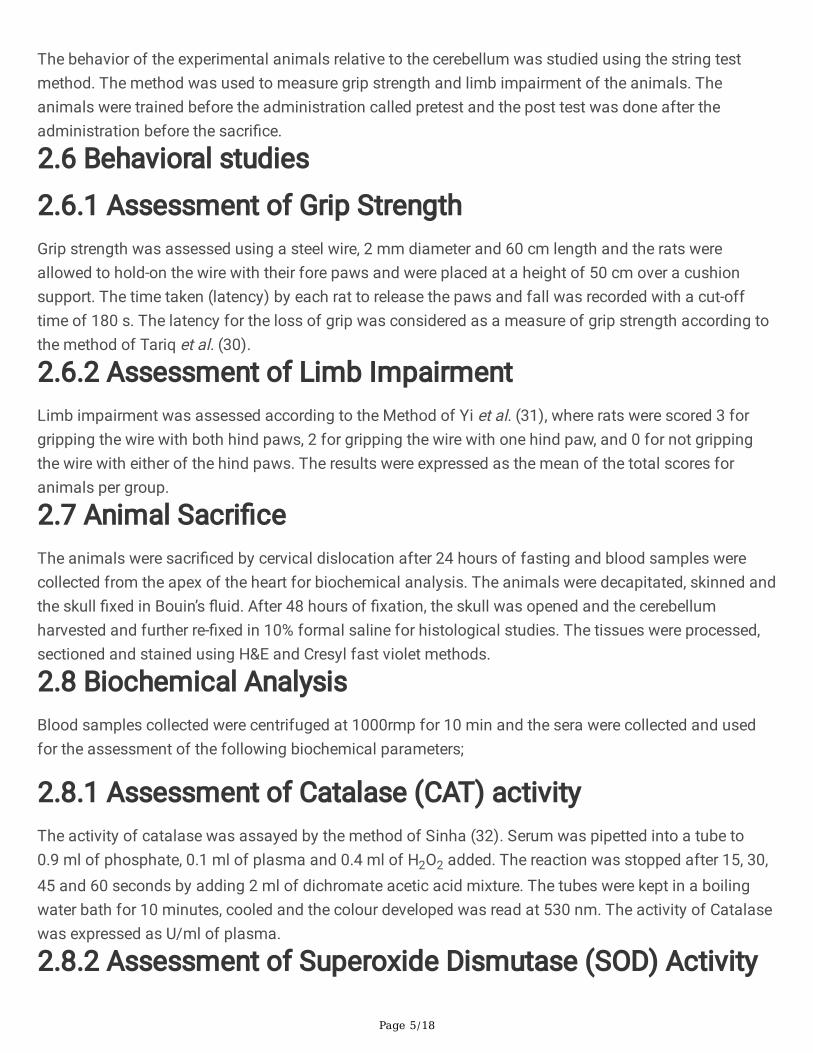
Figure 1
A section of the Cerebellar cortex of Rats in Group 1, showing normal Cerebellum with molecular layer(ML), the Purkinje cell layer (PCL) made up of a single layer of Purkinje cells, the granular layer (GL) andthe underlying Cerebellar white matter (WM). (H&E. Mag. X100 and X400; Scale Bar: 5µm =1mm).
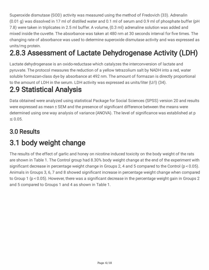
Figure 2
A section of the Cerebellar cortex of Rats in Group 2, showing Cerebellum with decreased Purkinje celllayer (DPPCL) and decreased thickness of the granular layer (DTGL), with increased in the Purkinje celllayer (IPPCL) and increased thickness of the granular layer (ITGL) with increased degeneration of thecerebellar neurons, Molecular layer (ML), and cerebellar white matter (WM). (H&E. Mag. 2A X100; 2BX400; Scale Bar: 5µm =1mm).
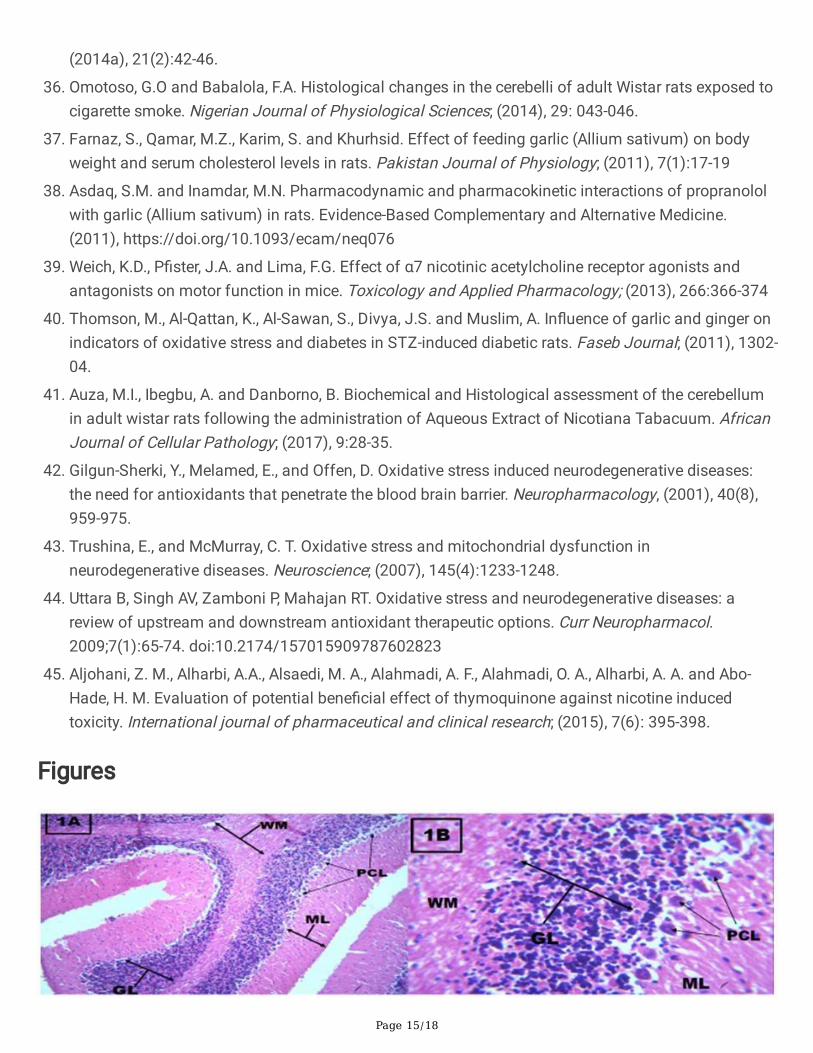
Figure 3
A section of the cerebellar cortex of animals in Group 3, showing altered cerebellar histoarchitecture withdegeneration of Purkinje cell layer (DPPCL), with decreased granular layer (DTGL) and necrosis of thePurkinje cell layer (NPCL). Molecular layer (ML), Cerebellar white matter (WM). (H&E. Mag. 3A X100 &3BX400; Scale Bar: 5µm =1mm).
Page 17/18
Figure 4
A section of cerebellar cortex of animals in Group 4, showing cerebellar histoarchitecture with decreasedpopulation of the Purkinje cell layer (DPPCL), decreased thickness of the granular layer (DTGL) and lossof cerebellar white matter (LWM). Molecular layer (ML) and granular layer (GL). (H&E. Mag.4A X100; 4BX400; Scale Bar: 5µm =1mm).
Figure 5
A Section of cerebellar cortex of animals in Group 5, showing cerebellar histoarchitecture with no majoralteration in the cerebellum. However, there was presence of few necrotic Purkinje cells (NPC), mild lossof cerebellar white matter (MLWM). Molecular layer (ML), granular layer (GL), Purkinje cell layer (PCL),blood vessel (BV). (H&E. Mag. 5A X100 5B X400; Scale Bar: 5µm =1mm).
Figure 6
Page 18/18
A Section of the cerebellar cortex of animals in Group 6, showing cerebellar histoarchitecture withPurkinje cells undergoing mild vacuolated cytoplasm (PCMVC), decreased thickness of the granular layer(DTGL) and presence of microcytic spaces (MS). Also present are the Molecular layer (ML), granular layer(GL) and Pia matter (PM). (H&E. Mag. 6A X100; 6B X400; Scale Bar: 5µm =1mm).
Figure 7
A Section of cerebellar cortex of animals in Group 7, showing cerebellar histoarchitecture with mildreduction in cellularity of the granular layer (MRCGL), and necrotic Purkinje cells (NPC), Purkinje cells withmild vacuolated cytoplasm (PCMVC), loss of cerebellar white matter (LWM) and Molecular layer (ML).(H&E. Mag. 7A X100; 7B X400; Scale Bar: 5µm =1mm).
Figure 8
A Section of cerebellar cortex of animals in Group 8, showing no major alteration in the cerebellarhistoarchitecture. However, there was presence of few necrotic Purkinje cells (NPC) and few Purkinje cellswith mild vacuolated cytoplasm (PCMVC). Also present are the molecular layer (ML), granular layer (GL)and cerebellar white matter (WM). (H&E. Mag. 8A X100; 8B X400; Scale Bar: 5µm =1mm).
Supplementary Files
This is a list of supplementary �les associated with this preprint. Click to download.


![Review Article Antioxidant Delivery Pathways in the Anterior EyeBioMed Research International other species, including Wistar rats [ ],lacktheenzymeL-gulonolactone oxidase necessary](https://static.documents.pub/doc/80x56/60ede369aa01591d0548db98/review-article-antioxidant-delivery-pathways-in-the-anterior-eye-biomed-research.jpg)


