EPA/635/R02/002 TOXICOLOGICAL REVIEW OF 1,1-DICHLOROETHYLENE (CAS No. 75-35-4) In Support of Summary Information on the Integrated Risk Information System (IRIS) June 2002 U.S. Environmental Protection Agency Washington, DC
Transcript
EPA/635/R02/002
TOXICOLOGICAL REVIEW
OF
1,1-DICHLOROETHYLENE(CAS No. 75-35-4)
In Support of Summary Information on the Integrated Risk Information System (IRIS)
June 2002
U.S. Environmental Protection AgencyWashington, DC
ii
DISCLAIMER
This document has been reviewed in accordance with U.S. Environmental Protection Agencypolicy and approved for publication. Mention of trade names or commercial products does notconstitute endorsement or recommendation for use. Note: This document may undergorevisions in the future. The most up-to-date version will be made available electronically via theIRIS Home Page at http://www.epa.gov/iris.
iii
CONTENTS —TOXICOLOGICAL REVIEW FOR 1,1-DICHLOROETHYLENE (CAS No. 75-35-4)
The purpose of this Toxicological Review is to provide scientific support and rationalefor the hazard and dose-response assessment in IRIS pertaining to chronic exposure to 1,1-dichloroethylene. It is not intended to be a comprehensive treatise on the chemical ortoxicological nature of 1,1-dichloroethylene.
In Section 6, EPA has characterized its overall confidence in the quantitative andqualitative aspects of hazard and dose response. Matters considered in this characterizationinclude knowledge gaps, uncertainties, quality of data, and scientific controversies. Thischaracterization is presented in an effort to make apparent the limitations of the assessment andto aid and guide the risk assessor in the ensuing steps of the risk assessment process.
For other general information about this assessment or other questions relating to IRIS,the reader is referred to EPA’s IRIS Hotline at 301-345-2870.
vi
AUTHORS, CONTRIBUTORS, AND REVIEWERS
U.S. EPA Region 8 and the Office of Solid Waste and Emergency Response (OSWER)were responsible for preparing the IRIS toxicological review and summary documents. Acomprehensive literature review was conducted in September 1999. The literature review wassupplemented with additional references until May 2002.
Chemical Manager/Author
Robert BensonMunicipal SystemsOffice of Regulatory AssistanceU.S. EPA Region 8, Denver, Colorado
Reviewers
This document and summary information on IRIS have received peer review both byEPA scientists and by independent scientists external to EPA. Subsequent to external reviewand incorporation of comments, this assessment has undergone an Agency-wide review processwhereby the IRIS Program Manager has achieved a consensus approval among the Office ofResearch and Development; Office of Air and Radiation; Office of Prevention, Pesticides, andToxic Substances; OSWER; Office of Water; Office of Policy, Planning, and Evaluation; and theRegional Offices.
Colorado Department of Public Health and EnvironmentDiane Niedzwiecki
External Peer Reviewers
Melvin E. Andersen James V. BrucknerColorado State University University of GeorgiaFt. Collins, Colorado Athens, Georgia
Poh-Gek Forkert Sam KacewQueen’s University University of OttawaKingston, Ontario, Canada Ottawa, Ontario, Canada
vii
Kannan KrishnanUniversity of MontrealMontreal, Quebec, Canada
A summary of the external peer reviewers’ comments and the disposition of theirrecommendations are in Appendix A.
1
1. INTRODUCTION
This document presents background and justification for the hazard and dose-responseassessment summaries in the U.S. Environmental Protection Agency’s (EPA’s) Integrated RiskInformation System (IRIS). IRIS summaries may include an oral reference dose (RfD), aninhalation reference concentration (RfC), and a carcinogenicity assessment.
The RfD and RfC provide quantitative information for noncancer dose-responseassessments. The RfD is based on the assumption that thresholds exist for certain toxic effects,such as cellular necrosis, but may not exist for other toxic effects, such as some carcinogenicresponses. It is expressed in units of mg/kg-day. In general, the RfD is an estimate (withuncertainty spanning perhaps an order of magnitude) of a daily exposure to the humanpopulation (including sensitive subgroups) that is likely to be without an appreciable risk ofdeleterious noncancer effects during a lifetime. The inhalation RfC is analogous to the oral RfD,but provides a continuous inhalation exposure estimate. The inhalation RfC considers toxiceffects for both the respiratory system (portal-of-entry) and for effects peripheral to therespiratory system (extrarespiratory or systemic effects). It is generally expressed in units ofmg/m3.
The carcinogenicity assessment provides information on the carcinogenic hazardpotential of the substance in question and quantitative estimates of risk from oral exposure andinhalation exposure. The information includes a weight-of-evidence judgment of the likelihoodthat the agent is a human carcinogen and the conditions under which the carcinogenic effectsmay be expressed. Quantitative risk estimates are presented in three ways. The slope factor isthe result of application of a low-dose extrapolation procedure and is presented as the risk permg/kg-day. The unit risk is the quantitative estimate in terms of either risk per :g/L drinkingwater or risk per :g/m3 air breathed. Another form in which risk is presented is a drinking wateror air concentration providing cancer risks of 1 in 10,000; 1 in 100,000; or 1 in 1,000,000.
Development of these hazard identification and dose-response assessments for 1,1-dichloroethylene (DCE) has followed the general guidelines for risk assessment as set forth bythe National Research Council (1983). EPA guidelines that were used in the development of thisassessment may include the following: the Guidelines for Carcinogen Risk Assessment (U.S.EPA, 1986a), Guidelines for the Health Risk Assessment of Chemical Mixtures (U.S. EPA,1986b), Guidelines for Mutagenicity Risk Assessment (U.S. EPA, 1986c), Guidelines forDevelopmental Toxicity Risk Assessment (U.S. EPA, 1991), Proposed Guidelines for CarcinogenRisk Assessment (U.S. EPA, 1996a), Guidelines for Reproductive Toxicity Risk Assessment (U.S.EPA, 1996b), and Guidelines for Neurotoxicity Risk Assessment (U.S. EPA, 1998a);Recommendations for and Documentation of Biological Values for Use in Risk Assessment (U.S.EPA, 1988); (proposed) Interim Policy for Particle Size and Limit Concentration Issues inInhalation Toxicity (U.S. EPA, 1994a); Methods for Derivation of Inhalation ReferenceConcentrations and Application of Inhalation Dosimetry (U.S. EPA, 1994b); Peer Review andPeer Involvement at the U.S. Environmental Protection Agency (U.S. EPA, 1994c); Use of theBenchmark Dose Approach in Health Risk Assessment (U.S. EPA, 1995); Draft Revised
2
Guidelines for Carcinogen Risk Assessment (U.S. EPA, 1999); Science Policy CouncilHandbook: Peer Review (U.S. EPA, 1998b, 2000a); Memorandum from EPA Administrator,Carol Browner, dated March 21, 1995, Policy for Risk Characterization; and Science PolicyCouncil Handbook, Risk Characterization (U.S. EPA, 2000b).
Literature search strategies employed for this compound were based on the CASRN andat least one common name. At a minimum, the following databases were searched: RTECS,HSDB, TSCATS, CCRIS, GENETOX, EMIC, EMICBACK, DART, ETICBACK, TOXLINE,CANCERLINE, MEDLINE, and MEDLINE backfiles. Any pertinent scientific informationsubmitted by the public to the IRIS Submission Desk was also considered in the development ofthis document.
EPA has previously reviewed 1,1-DCE (U.S. EPA, 1985a, b). This review replaces thoseassessments.
2. CHEMICAL AND PHYSICAL INFORMATION RELEVANT TO ASSESSMENTS
1,1-DCE does not occur naturally. It is produced commercially by thedehydrochlorination of 1,1,2-trichloroethane in the presence of excess base. 1,1-DCE is usedprincipally for the production of polyvinylidene chloride polymers (PVDC). PVDC is usedprincipally in the food packaging industry as cast and extruded film (Saran and Velon wraps)and as a barrier coating for paper, cellulose, polypropylene, and other plastics. Extrudedfilaments of PVDC are also used in the textile industry for furniture and automobile upholstery,drapery fabric, and outdoor furniture. 1,1-DCE enters in the environment though release duringits manufacture and use, from the breakdown of PVDC products, and from the biotic or abioticbreakdown of 1,1,1-trichloroethane, tetrachloroethylene, 1,1,2-trichoroethene, and 1,1-dichloroethane (ATSDR, 1994; IARC, 1999; and U.S. EPA, 1985 a, b).
The chemical and physical properties of 1,1-DCE (ATSDR, 1994; IARC, 1999) arepresented below.
Chemical structure: Cl2C=CH2Molecular weight: 96.94
3
Boiling point: 31.6 °C
Melting point: -122.5 °C
Specific gravity: 1.218
Vapor pressure: 67 kPa at 20 °C
Solubility: Practically insoluble in water; soluble in acetone, ethanol, andmany organic solvents; very soluble in diethyl ether.
Odor: Mild, sweet, resembling chloroform
Odor threshold: 500 ppm in air; no data in water
Partition coefficients:Log Kow 1.32Log Koc 1.81
Flash point: -19 °C, closed cup; -15 °C, open cup
Autoignition: 570 °C
Conversion factor: 1 ppm = 3.97 mg/m3
3. TOXICOKINETICS RELEVANT TO ASSESSMENTS
1,1-DCE is rapidly absorbed following inhalation and oral exposures. Because of its lowmolecular weight and hydrophobic nature, dermal absorption is also likely, but no relevant datawere found in the literature. In rats treated with 1,1-DCE by gavage in corn oil, completegastrointestinal absorption was found to occur at #350 mg/kg (Jones and Hathway, 1978a, b;Putcha et al., 1986). 1,1-DCE is easily transported across the alveolar membrane. At constant#750 ppm concentration in the air, equilibrium or near steady-state is reached in the blood in ratsin approximately 45 minutes (Dallas et al., 1983). Continued uptake in rats reflects to someextent continuing deposition in fatty tissues, but this is primarily a result of metabolism of 1,1-DCE.
The major route of excretion for unchanged 1,1-DCE is through the lung (Jones andHathway, 1978a). However, the majority of 1,1-DCE is rapidly metabolized to nonvolatilecompounds and covalently bound derivatives (McKenna et al., 1997, 1978a, b). Micemetabolize more 1,1-DCE than do rats. For example, when given 50 mg/kg by oral gavage incorn oil, mice excreted 6% and rats excreted 28% of the dose as unchanged 1,1-DCE through the
4
lungs (Jones and Hathway, 1978b). When exposed to 10 ppm for a single 6-hour episode, miceexcreted 0.65% and rats excreted 1.63% of the absorbed dose as unchanged 1,1-DCE through thelungs (McKenna et al., 1977). Intraperitoneal (i.p.) administration of 125 mg/kg 14C-1,1-DCE tomice resulted in the highest concentrations of covalent binding (based on protein content) in thekidney, lung, and liver (Okine et al., 1985; Okine and Gram, 1986a, b). The covalent bindingand cellular damage in kidney, lung, and liver correlated with the high concentration of CYP2E1in certain cell populations in these tissues.
The proposed metabolic pathways for 1,1-DCE are summarized in Figure 1. Thesepathways were determined from experimental studies in laboratory animals. It is not knownwhether the metabolism of 1,1-DCE is the same in humans, although in vitro microsomalpreparations from human liver and lung form the same initial products (Dowsley et al., 1999). Oxidation of 1,1-DCE by CYP2E1 should produce three metabolites: DCE epoxide, 2-chloroacetyl chloride, and 2,2-dichloroacetaldehyde. All of these metabolites react withglutathione (GSH) and/or water. In the kidney, further metabolism of S-(2,2-dichloro-1-hydroxy)ethylglutathione could form another toxic compound, dicholorothioketene. The GSHconjugates formed are catabolized in the kidney to a variety of urinary excretion products. Theepoxide, and perhaps to a lesser extent the chloroacetaldehyde, are believed to be associated withthe tissue reactivity and toxic effects in tissues that ensue after significant depletion of GSH.
The primary metabolites of 1,1-DCE formed in rat hepatic microsomal incubations areDCE epoxide, 2,2-dichloroacetaldehyde, and 2-chloroacetyl chloride (Liebler et al., 1985, 1988;Costa and Ivanetich, 1982). These metabolites were also identified from mouse microsomalincubations (Dowsley et al., 1995). All these electrophilic metabolites undergo secondaryreactions, including oxidation, conjugation with GSH, and hydrolysis. The major productsformed are GSH conjugates, 2-(S-glutathionyl)acetyl glutathione [B], and 2-S-glutathionylacetate [C], which are believed to be derived from the DCE epoxide (Fig. 1). S-(2,2-Dichloro-1-hydroxy ethyl glutathione [A], the GSH conjugate formed from reaction of GSH with 2,2-dichloroacetaldehyde, was not observed in rat liver microsomal incubations containing GSH(Dowsley et al., 1995). The acetal, together with chloroacetic acid and S-(2-chloroacetyl)-glutathione [D]—the hydrolysis and GSH-conjugated products of 2-chloroacetyl chloride,respectively—was detected at levels much lower than those for the DCE epoxide-derivedconjugates [B] and [C].
In human liver and lung microsomal incubations, the DCE epoxide-derived GSHconjugates [B] and [C] were the major metabolites detected (Dowsley et al., 1999). 2,2-Dichloroacetaldehyde was detected at low levels. Liver microsomes from three out of fivehuman samples metabolized 1,1-DCE to the epoxide-derived GSH conjugates at levels that were2.5- to 3-fold higher than in mouse liver microsomes, based on milligrams of microsomalprotein. These GSH conjugates were also the major products formed in lung microsomes fromeight human samples; only low levels of 2,2-dichloroacetaldehyde were formed. The mean level
5
Cl
Cl
Cl
Cl
O
Cl
O
Cl
Cl
O
OH Cl
O
SG
GS
O
Cl
GS
O
OHGS
O
SG
Cl
Cl
OCl
Cl
OH
OH
Cl
Cl
OH
SG
P450 P450
P450
GSH
GSH H20
H20
H20 GSH
2,2-Dichloroacetaldehyde
2-Chloroacetyl Chloride
DCE-Epoxide
H20
GSH
[A]
[D]
[B] [C]
1,1-DCE
Tissue Targets & Resultant Toxicity
Tissue Targets & Resultant Toxicity
Cl
Cl
S
[?]
Figure 1: Proposed pathways for 1,1-DCE metabolism and toxicity.
Source: Adapted from Forkert, 1999a, b
in lung microsomes from humans was about 50% of the amount formed in lung microsomesfrom mice. In both animal and human tissues, cytochrome P450 CYP2E1 catalyzes theformation of the DCE epoxide (Dowsley et al., 1996).
The significance of the metabolic pathway in the liver involving 2,2-dichloroacetaldehyde is unclear. Existing evidence, however, suggests that this pathway is ofminor toxicological importance. In addition to 2,2-dichloroacetaldehyde and the GSH conjugate,potential metabolites include the acetal (the hydration product of the aldehyde), dichloroaceticacid, and dichloroethanol. An initial study with rat liver microsomes found a trace level of 2,2-dichloroacetaldehyde but no detectable dichloroacetic acid (Costa and Ivanetich, 1982). A laterreport using isolated rat hepatocytes detected dichloroacetic acid and trace levels of 2,2-dichloroacetaldehyde, 2,2-dichloroethanol, and chloroacetic acid (Costa and Ivanetich, 1984). Forkert (1999a) and Forkert and Boyd (2001), using intact mice, found no acetal in liver cytosol;however, acetal was detected in the bile in the first study but was not mentioned as being foundin the bile in the second study. In early studies on the metabolism of 1,1-DCE, none of thepotential metabolites from this pathway were reported as being found in the urine of rodentsusing techniques that readily identified chloroacetic acid (Jones and Hathway, 1978a, b;McKenna et al., 1977, 1978a, b). A pharmacokinetic analysis showed that any dichloroaceticacid formed in the liver is rapidly metabolized in the liver to two carbon, nonchlorinatedchemicals and carbon dioxide (Merdink et al., 1998).
6
The oxidative metabolism of 1,1-DCE has been found to reach saturation in rats at anoral exposure of 10–50 mg/kg and an inhalation exposure of 200 ppm (794 mg/m3) (Andersen etal., 1979; D’Souza and Andersen, 1988; Dallas et al., 1983; McKenna et al., 1977).
Because 1,1-DCE is lipophilic and has a blood-to-air partition coefficient of 5 in rats(D’Souza and Andersen, 1988), any 1,1-DCE not metabolized following oral or inhalationexposure is rapidly exhaled unchanged when exposure is terminated. Because of its lowoctanol:water partition coefficient, 1,1-DCE will not bioaccumulate in tissues to a significantextent. The major metabolites found in urine of rodents include oxalic acid, thiodiglycolic acid,thioglycolic acid, dithioglycolic acid, N-acetyl-S-(2-carboxymethyl) cysteine, N-acetyl-S-(2-hydroxyethyl) cysteine, other –acetyl-S-cysteinyl derivatives, andmethylthioacetylaminoethanol.
D’Souza and Andersen (1988) developed physiologically based pharmacokinetic (PBPK)models for 1,1-DCE in the rat for both oral and inhalation exposure. No validated model isavailable for humans. D’Souza and Andersen (1988) used allometric scaling to estimatecomparative amounts of epoxide formed (mg/kg) in rats and humans. Cardiac output andpulmonary ventilation were scaled by (body weight)0.7, Vmax was scaled by (body weight)0.74,and body fat was estimated at 7% in the 200 g rat and 20% in the 70 kg human. When the oralexposure was less than 5 mg/kg, the estimated amount of epoxide formed was about the same inrats and humans. When the inhalation exposure was less than 100 ppm, the estimated amount ofepoxide formed was fivefold lower in humans than in rats.
El-Masri et al. (1996a, b) used a combination of gas uptake experiments in Sprague-Dawley rats and PBPK modeling to assess the potential for interaction between 1,1-DCE andtrichloroethylene. Both substrates are activated by CYP2E1. Thus, there is a potential forcompetitive inhibition when simultaneous exposure to both substrates occurs. The results of thegas uptake experiments confirmed a model based on competitive inhibition. There was,however, no evidence of competitive inhibition when exposure to both substrates was 100 ppmor less. As environmental exposures to these chemicals are expected to be less than 100 ppm,there is little potential for reduced toxicity from 1,1-DCE when individuals are also exposed totrichloroethylene.
4. HAZARD IDENTIFICATION
4.1. STUDIES IN HUMANS—EPIDEMIOLOGY
Ott et al. (1976) investigated the health records of 138 employees who wereoccupationally exposed to 1,1-DCE in processes not involving vinyl chloride. The individualsincluded in the study had worked in experimental or pilot plant polymerization operations, in amonomer production process as tankcar loaders, or in a production plant manufacturing amonofilament fiber. Time-weighted average concentrations (8 hours) of 1,1-DCE in theworkplace were estimated from job descriptions and the results of industrial hygiene sampling.
7
The subjects were grouped into three exposure categories: less than 10 ppm, 10–24 ppm, andgreater than 25 ppm. The researchers estimated career exposure by taking into account averageduration of employment. Results of the most recent health inventory for individuals in theexposed cohort were compared with findings for matched controls. An analysis of mortality inthe cohort indicated no statistically significant differences. Overall, there were no significantdifferences in hematology and clinical chemistry parameters between the exposed cohort and thecontrols.
Three reports suggest an association between exposure to dichloroethylenes and birthdefects. The California Department of Health Services (Swan et al., 1985) reported an increasein the number of cardiac congenital anomalies during 1980 and 1981 in an area served by apublic water supply contaminated with 1,1,1-trichloroethane and dichloroethylene. The publicwater supply also contained chlorinated disinfection by products. Goldberg et al. (1990)reported an increase in congenital cardiac malformations between 1969 and 1987 in an area ofArizona where the drinking water was contaminated with trichloroethylene and dichloroethylene(isomer not specified). The dichloroethylene concentration in the drinking water was usually 5%to 10% of the trichloroethylene concentration. The paper does not specify whether the drinkingwater was chlorinated. Finally, Bove et al. (1995) reported increased odds ratio for oral cleftdefects (1.71), for central nervous system defects (2.52), and for neural tube defects (2.60)associated with exposure to total dichloroethylenes of more than 2 :g/L from public drinkingwater supplies in an area of northern New Jersey. The period of time studied was 1985 to 1988. The drinking water also contained chlorinated disinfection by-products. It is not clear from thepaper whether there was also co-exposure to other chlorinated solvents also reported on,including trichloroethylene, tetrachloroethylene, 1,1,1-trichloroethane, carbon tetrachloride, and1,2-dichloroethane. As all of these situations involved exposure to multiple contaminates, acause-and-effect relationship between the reported birth defects and exposure to 1,1-DCE cannotbe established.
4.2. PRECHRONIC AND CHRONIC STUDIES AND CANCER BIOASSAYS INANIMALS—ORAL AND INHALATION
4.2.1. Acute Exposure
Mice are more sensitive than rats to acute toxicity from 1,1-DCE. The NationalToxicology Program (NTP) (NTP, 1982) conducted a study to determine lethality in five maleand five female F344 rats and five male and five female B6C3F1 mice (all animals 9 weeks old)after a single exposure to 1,1-DCE by gavage in corn oil at 0, 10, 50, 100, 500, or 1000 mg/kg.
By day 14 postexposure, mortality was 0/10, 1/10, 0/10, 0/10, 1/10, and 2/10 in the rats and 0/10,0/10, 1/10, 0/10, 8/10, and 10/10 in the mice, respectively. Other representative lethality data arepresented in Tables 1 and 2.
Table 1. Representative lethality (LD50) from oral exposure to 1,1-DCE
8
SpeciesDose
(mg/kg) Effect Reference
Rat male male male, adrenalectomized female
1550 1800
84 1500
LD50Jenkins et al., 1972Ponomarkov and Tomatis, 1980Jenkins et al., 1972Ponomarkov and Tomatis, 1980
Mouse male female
217 194
LD50Jones and Hathway, 1978b
Table 2. Representative lethality (LC50) or time for 50% lethality (LT50) frominhalation exposure to 1,1-DCE
SpeciesExposure
(ppm) Effect Reference
Rat male, fed
male, fasted
6350 for 4 hr
200 for 4.1 hr 400 for 3.6 hr 500 for 3.0 hr
1000 for 2.4 hr 2000 for 1.4 hr
LC50
LT50
Siegal et al., 1971
Andersen et al., 1979
Mouse male female
98 for 22–23 hr 105 for 22–23 hr
LC50Short et al, 1977a
9
Toxicity is enhanced by fasting (Andersen and Jenkins, 1977; Chieco et al., 1981; Jaegeret al., 1974, 1975, 1977a, b; McKenna et al., 1978a, b; Moslen et al., 1985), by GSH depletion(Andersen et al., 1980; Jaeger et al., 1974, 1977a, b; Kanz et al., 1988; Moussa and Forkert,1992), and by administration in oil vehicles compared to administration in aqueous Tween(Chieco et al., 1981). Toxicity is decreased by agents that decrease metabolism by the P450system (Andersen et al., 1978; Moslen et al., 1989) or by hypothyroidism, which increasesintracellular GSH (Kanz et al., 1991).
The target organs for toxicity after acute oral or inhalation exposure are the liver, thekidney, and the Clara cells of the lung. The effects in the liver include an increase in liverenzymes in the serum (Jenkins et al., 1972; Jaeger, 1977a, b; Short et al., 1977a; Jenkins andAndersen, 1978; Reynolds et al., 1980); severe histopathological damage, including disruption ofbile canaliculi, cytoplasmic vacuolization, and hemorrhagic necrosis (Short et al., 1977a; Kanzand Reynolds, 1986; Reynolds et al., 1984); an increase in covalent binding of 1,1-DCE (Forkertand Moussa, 1991, 1993; Jaeger et al., 1977a, b); and a decrease in GSH (Forkert and Moussa,1991, 1993; Kanz et al., 1988; Reichert et al., 1978, 1979) mediated by CYP2E1 metabolism of1,1-DCE to intermediates that react with GSH (Kainz et al., 1993; Lee and Forkert, 1994).
Several researchers have investigated the hepatotoxicity of 1,1-DCE. In a study byJenkins and Andersen (1978), four female Sprague-Dawley rats (body weight, 223 g) received asingle oral exposure by gavage in corn oil at 400 mg/kg. Four to 8 hours after exposure, therewas a significant increase in aspartate aminotransferase (approximately 75-fold), alanineaminotransferase (approximately 70-fold), lactate dehydrogenase (approximately 110-fold), andsorbitol dehydrogenase (approximately 320-fold). The serum enzymes returned toapproximately normal values within 82 hours after exposure.
Reynolds et al. (1984) administered a single oral exposure by gavage in mineral oil at200 mg/kg 1,1-DCE to fasted male Sprague-Dawley rats (body weight, 225–375 g). Within 2hours after exposure, the livers showed evidence of dilatation and disruption of bile canaliculi,plasma membrane invagination and loss of microvilli, cytoplasmic vacuolization, and loss ofdensity in mitochondrial matrices. One hour after a single inhalation exposure at 250 ppm 1,1-DCE for 4 hours, Sprague-Dawley rats showed a significant decrease (p<0.05) in GSHconcentration in the liver (Jaeger, 1977). Four hours after exposure there was an increase in theserum concentration of sorbitol dehydrogenase (approximately 230-fold) and ornithinecarbamoyl transferase (approximately 380-fold).
Short et al. (1977a) studied CD-1 male mice (Charles River) and CD male rats (CharlesRiver) exposed by inhalation for 22–23 hrs/day for 1–5 days at 0, 15, 30, or 60 ppm 1,1-DCE(mice) or for 1–3 days at 0 or 60 ppm (rats). In male mice exposed to $15 ppm, serum enzymes(alanine aminotransferase and aspartate aminotransferase) were significantly increased (four- tosixfold), and hepatocellular degeneration was observed in one of five mice after the firstexposure. In two of five male rats exposed to 60 ppm, mild centrilobular degeneration and/ornecrosis was observed after the first exposure, but serum enzymes (alanine aminotransferase and
10
aspartate aminotransferase) were not significantly increased (four- to sixfold) until after thesecond exposure.
Reynolds et al. (1980) found that after a single 4-hour exposure by inhalation at 200 ppm1,1-DCE, the liver of fasted male Sprague-Dawley rats (body weight, 150–200 g) showedcatastrophic morphological alterations of the parenchymal cells, including retraction and centralrarefaction of nuclei with peripheral displacement of chromatin to nuclear margins, progressingto frank hemorrhagic centrilobular necrosis. GSH concentrations were also depleted. After theextensive hepatocellular damage, cytochrome P450 and oxidative –demethylase weredeactivated.
Toxic effects of 1,1-DCE exposure in the kidney include increased kidney weight,increased blood urea nitrogen and creatinine (Jackson and Conolly, 1985; Jenkins and Andersen,1978), and histopathological changes, including vacuolization, tubular dilatation, and nephrosisand necrosis of the proximal tubules (Short et al., 1977a; Jackson and Conolly, 1985; Jenkinsand Andersen, 1978). These changes were correlated with metabolic activation of 1,1-DCE byCYP2E1 in the proximal tubules, decreased GSH concentration, increased covalent binding of1,1-DCE, and the presence of a relatively high concentration of $-lyase activity in rodent kidneytissue (Brittebo et al., 1993; Dekant et al., 1989; Dekant, 1996). In addition, renal toxicity canbe inhibited by pretreatment of mice and rats with aminooxyacetic acid, an inhibitor of renalcysteinyl-ß-lyase (Ban et al., 1995; Cavelier et al., 1996).
Jenkins and Andersen (1978) investigated the nephrotoxicity of 1,1-DCE in Sprague-Dawley rats after a single oral exposure by gavage in corn oil. Fasted male rats (two to six pergroup; body weight, 300 g) were administered 0, 50, 100, 200, 400, or 600 mg/kg. At 600mg/kg, there was a fivefold increase in blood urea nitrogen. Histopathological examination wasnot conducted in animals treated at 600 mg/kg. In male rats at 400 mg/kg, there was astatistically significant increase (p<0.05) in blood urea nitrogen (fourfold) and in creatinine(threefold). The increases became apparent 8 hours after exposure, reached a peak 24 hours afterexposure, and returned to normal 96–144 hours after exposure. In male rats at 400 mg/kg, therewas also a twofold increase in relative kidney weight 48 hours after exposure. The relativekidney weight had nearly returned to normal 144 hours after exposure. In female rats at 400mg/kg, there was no substantial increase in blood urea nitrogen, creatinine, or relative kidneyweight. Histopathological lesions (tubular dilatation and tubular necrosis) were observed in bothsexes at 400 mg/kg. No significant effects were seen at 200 mg/kg and below.
Short et al. (1977a) studied CD-1 male mice (Charles River) after inhalation exposure for22–23 hrs/day for 1–5 days at 0, 15, 30, or 60 ppm 1,1-DCE. Tubular nephrosis was observed at$15 ppm after the first exposure. Jackson and Conolly (1985) reported that in male Sprague-Dawley rats (body weight, 225–275 g) exposed continuously for 4 hours to 0, 200, 250, 300,375, or 400 ppm, mortality was 0/22, 1/4, 1/16, 3/14, 3/12, and 3/6, respectively. At $250 ppmthere were significant increases (p<0.05) in kidney-to-body weight ratios (approximately 1.4-fold), serum urea nitrogen (approximately fourfold) and creatinine (approximately threefold).
11
Histopathological examination revealed severe tubular necrosis with calcium deposits at $300ppm.
Using autoradiographic methods, Brittebo et al. (1993) investigated the mechanism ofnephrotoxicity in C57BL6 mice (body weight, 18–22 g) following i.p. injection of 0.4 mg/kg of14C-labeled 1,1-DCE. Selective covalent binding of radioactivity occurred in the proximaltubules, in the midzonal parts of the liver lobules, and in the mucosa of the upper and lowerrespiratory tract. Treatment with buthionine sulphoximine (BSO), an irreversible inhibitor of (-glutamylcysteine synthetase and a GSH-depleting agent, caused a threefold increase incovalent binding of 1,1-DCE. Histopathological examination of kidneys in BSO-pretreated malemice given single i.p. injections of 25 and 50 mg/kg 1,1-DCE showed necrosis in the proximaltubules (S1 and S2 segments). In mice given 1,1-DCE only, no significant lesions in the kidneyswere observed. The authors concluded that the severe renal toxicity of 1,1-DCE in BSO-pretreated mice is related to metabolic activation of 1,1-DCE in the proximal tubules, resulting inGSH depletion and covalent binding.
The effects in the Clara cells of the lung in mice include extensive histopathologicalchanges (Forkert and Reynolds, 1982; Forkert et al., 1985, 1990), repair of damage through cellproliferation (Forkert et al., 1985), depletion of GSH, and covalent binding of 1,1-DCE mediatedthrough the formation of DCE epoxide by CYP2E1 (Dowsley et al., 1996; Forkert and Mousa,1991; Forkert, 1999b; Lee and Forkert, 1994; Moussa and Forkert, 1992). No studies areavailable showing similar effects in the lungs of rats.
Forkert and Reynolds (1982) investigated the ability of 1,1-DCE administered orally toinduce pulmonary injury. Male C57BL6 mice (three to five per group) were administered asingle dose of 1,1-DCE by gavage in mineral oil at 0, 100, or 200 mg/kg. At 100 mg/kg, Claracells showed extensive dilatation of cisternae and degeneration of the endoplasmic reticulum.The bronchiolar epithelium showed a few vacuolated cells 12 hours after exposure. By 24 hoursthe Clara cells showed prominent cytoplasmic vacuoles, but ciliated cells were not affected. By48 hours, complete recovery had occurred. At 200 mg/kg, both ciliated and Clara cells showednecrosis of the bronchiolar epithelium. By 24 hours, the lesion had increased in severity andareas of bronchioles were denuded of epithelium. Peribronchial and perivascular edema,hemorrhage, and focal atelectasis were also present. Complete recovery occurred by 7 days.
A subsequent study (Forkert et al., 1985) examined regeneration of the damagedepithelium by cellular proliferation. Male C57BL6 mice were administered a single dose of 1,1-DCE by gavage in mineral oil at 200 mg/kg followed by a single pulse of 3H-thymidine. Changes in cellular proliferation were calculated from measurement of radioactivity incorporatedinto total pulmonary DNA. Incorporation of radioactivity was significantly inhibited 1 day aftertreatment and thereafter increased. The peak incorporation of radioactivity occurred between 3and 5 days after treatment and returned to baseline by day 7. The majority of the radioactivitywas taken up by the nonciliated bronchiolar epithelial cells.
4.2.2. Longer-Term Exposure
12
4.2.2.1. Oral
4.2.2.1.1. Rats. NTP (1982) conducted a 14-day study of 1,1-DCE in male and female F344 rats(five animals of each sex, 9 weeks old) by gavage in corn oil at 0, 10, 50, 100, 500, or 1,000mg/kg. Survival was 10/10, 10/10, 10/10, 10/10, 7/10, and 3/10 mg/kg, respectively. Meanbody weight was significantly depressed at $500 mg/kg. Hemorrhagic necrosis in the liver wasobserved in all of the rats that died at 500 and 1,000 mg/kg.
In the same study, male and female F344 rats (10 of each sex, 9 weeks old) wereadministered 1,1-DCE by gavage in corn oil at 0, 5, 15, 40, 100, or 250 mg/kg five times perweek for 13 weeks. Representative tissues from rats receiving 250 mg/kg and from control ratswere examined microscopically. Livers from all groups were examined. Three female ratsreceiving 250 mg/kg died during the first week of the study. No other rats died. Mean bodyweight was depressed 13% for male rats receiving 250 mg/kg as compared with controls. Meanbody weight in other groups was comparable. Only the liver showed effects attributed to 1,1-DCE. At 250 mg/kg, the three female rats that died showed severe centrilobular necrosis. Minimal to moderate hepatocytomegaly was seen in the rest of the rats at 250 mg/kg. Minimalto mild hepatocytomegaly was seen in 6/10 male rats and 3/10 female rats that received 100mg/kg. No biologically significant changes were observed in rats that received 40 mg/kg or less. The no-observed-adverse-effect level (NOAEL) in this study is 40 mg/kg (equivalent to 28.5mg/kg-day); the lowest-observed-adverse-effect level (LOAEL) is 100 mg/kg (equivalent to 71.4mg/kg-day).
4.2.2.1.2. Mice. NTP (1982) conducted a 14-day study in male and female B6C3F1 mice (fiveof each sex, 9 weeks old) administered 1,1-DCE by gavage in corn oil at 0, 10, 50, 100, 500, or1,000 mg/kg. Survival was 10/10 in all groups except the 1000 mg/kg group, where survival was0/10. Hemorrhagic necrosis in the liver was observed in all mice at 1,000 mg/kg.
In the same study, male and female B6C3F1 mice (10 of each sex, 9 weeks old) wereadministered 1,1-DCE by gavage in corn oil at 0, 5, 15, 40, 100, or 250 mg/kg five times perweek for 13 weeks. Representative tissues from mice receiving 100 and 250 mg/kg and fromcontrol mice were examined microscopically. Livers from all groups were also examined. Survival was 20/20, 19/20, 19/20, 19/20, 15/20, and 1/20 at 0, 5, 15, 40, 100, and 250 mg/kg,respectively. At 100 mg/kg, there was a decrease in mean body weight in males (14%) but not infemales. No change in mean body weight was observed at lower exposures. Only the livershowed effects attributed to 1,1-DCE. Centrilobular necrosis of the liver was observed in 5/10males and 5/10 females that received 250 mg/kg and 2/10 males and 2/10 females that received100 mg/kg. No biologically significant changes in the liver occurred in mice receiving 40 mg/kgor below. The NOAEL in this study is 40 mg/kg (adjusted to a continuous daily exposure of28.6 mg/kg-day); the LOAEL is 100 mg/kg (adjusted to a continuous daily exposure of 71.4mg/kg-day).
4.2.2.1.3. Dogs. Quast et al. (1983) conducted a study in beagle dogs (four per group, 8 monthsold) administered 1,1-DCE by gavage in peanut oil at 0, 6.25, 12.5, or 25 mg/kg-day for 97 days.
13
There were no significant differences among the groups in appearance and demeanor, mortality,body weight, food consumption, hematology, urinalysis, clinical chemistry determinations, organweights, and organ-to-body weight ratios. No exposure-related gross or histopathologicalchanges were present in tissues. There was no depletion of the nonprotein sulfhydryl levels inthe liver or kidneys. The NOAEL in this study is 25 mg/kg-day (the highest exposure tested).
4.2.2.2. Inhalation
Gage (1970) exposed four male and four female Alderly Park rats (body weight 200 g) to200 ppm or 500 ppm 1,1-DCE 6 hrs/day for 20 days. At 200 ppm there was slight nasalirritation (not further described). At necropsy all organs appeared normal. At 500 ppm therewas nasal irritation (not further specified), retarded weight gain (data not reported), and liver celldegeneration (not further defined).
Plummer et al. (1990) exposed black hooded Wistar rats to 50 ppm 1,1-DCE (18 malesand 18 females, age not specified) continuously for 4 weeks (except for two 1.5-hour periods perweek) or to 250 ppm (six males and six females, age not specified) for 6 hrs/day, 5 days/wk for 4weeks. The total exposure (concentration x time) was the same for the two profiles (33,533 ppm/hr for the continuous exposure and 32,200 ppm/hr for the intermittent exposure). Rats in the intermittent exposure group showed signs of early coagulative necrosis in the liver(incidence not reported). Eleven of the 12 rats in the continuous-exposure group showed lesssevere injury, including fatty changes in variable numbers of hepatocytes and only veryoccasional focal liver cell necrosis. The LOAEL in this study is 50 ppm.
Prendergast et al. (1967) evaluated the toxicity of 1,1-DCE in Long-Evans and Sprague-Dawley rats, Hartley guinea pigs, beagle dogs, New Zealand albino rabbits, and squirrelmonkeys. One set of test animals (15 rats/group, 15 guinea pigs/group, 3 rabbits/group, 2 dogs/group, or 3 monkeys/group) was exposed to 1,1-DCE vapors for 8 hrs/day, 5 days/wk, fora total of 30 exposures at 395 ± 32 mg/m3. The age of the animals was not specified. Theexposed animals were evaluated for visible signs of toxicity, mortality, and hematologic,biochemical, pathologic, and body weight changes. In this study there were no deaths, no visiblesigns of toxicity, and no histopathological changes. The NOAEL in this study is 395 mg/m3 (thehighest exposure tested), equivalent to an adjusted NOAEL based on continuous exposure of 94mg/m3.
Another set of test animals (15 rats/group, 15 guinea pigs/group, 3 rabbits/group, 2 dogs/group, or 3 or 9 monkeys/group) was exposed continuously for 90 days to 1,1-DCEvapors at 189 ± 6.2, 101 ± 4.4, 61 ± 5.7, or 20 ± 2.1 mg/m3. The concurrent controls included304 rats, 314 guinea pigs, 48 rabbits, 34 dogs, and 57 monkeys. The age of the animals was notspecified. The exposed animals were evaluated for visible signs of toxicity, mortality, andhematologic, biochemical, pathologic, and body weight changes. There was apparent exposure-related mortality in guinea pigs and monkeys. In the 0, 20, 61, 101, or 189 mg/m3 exposuregroups, guinea pig mortality was 2/314, 2/45, 3/15, 3/15, and 7/15, and monkey mortality was1/57, 1/21, 0/9, 2/3, and 3/9, respectively. The guinea pigs died between days 3 to 9 of exposure;
14
the monkeys died on days 26, 39, 47, 60, and 64 of exposure. There were no visible signs oftoxicity in any surviving animals. At the highest exposure in monkeys, but not in guinea pigs,there was some histopathological evidence of liver damage (see below). In guinea pigs at thehighest exposure, there was an increase in serum glutamic-pyruvic transaminase and liveralkaline transaminase (see below). Because visible signs of toxicity were not observed, and onlyminor liver damage was apparent in this study, the mortality data in guinea pigs and monkeys aregiven no weight.
Varying degrees of growth depression were found in all exposures, but were significantin all species only at 189 mg/m3. The test animals exhibited no significant hematologicalterations, and serum urea nitrogen levels were within control limits in all exposures in whichdeterminations were made. Significant elevations of serum glutamic-pyruvic transaminase andliver alkaline phosphatase activities were found in rats (a threefold and 1.75-fold increase,respectively) and guinea pigs (a sevenfold and 2.4-fold increase, respectively) exposed to 189mg/m3 (other species not tested) but not at 20 mg/m3 (enzyme levels at intermediate exposuresnot tested). Histopathological examination of liver from dogs, monkeys, and rats revealeddamage at 189 mg/m3 (other species not examined). The effects observed included fattymetamorphosis, focal necrosis, hemosiderosis deposition, lymphocytic infiltration, bile ductproliferation, and fibrosis. The changes were most severe in dogs. Sections of kidney from allrats showed nuclear hypertrophy of the tubular epithelium. No detectable liver or kidneydamage was observed in any species exposed to 101 mg/m3 or less. The NOAEL in this study is101 mg/m3 (equivalent to 25 ppm); the LOAEL is 189 mg/m3 (equivalent to 47 ppm).
4.2.3. Chronic Studies and Cancer Bioassays
4.2.3.1. Oral
4.2.3.1.1. Rats. Ponomarkov and Tomatis (1980) treated 24 female BD IV rats by gavage with1,1-DCE dissolved in olive oil (150 mg/kg body weight) on gestation day (GD) 17. Theiroffspring (81 males and 80 females) were treated weekly with 1,1-DCE at 50 mg/kg body weightby gavage in olive oil from the time of weaning for 120 weeks or until the animal was moribund. A control group of offspring (49 males and 47 females) received only olive oil. Liver andmeningeal tumors were more frequently observed in treated than in untreated animals, but thedifference was not statistically significant. The total number of tumor-bearing animals was notstatistically different between the treated and untreated groups.
NTP (1982) conducted chronic toxicity and carcinogenicity studies for 104 weeks inmale and female F344 rats (50 of each sex in each group, 9 weeks old) by gavage in corn oil at 0,1, or 5 mg 1,1-DCE/kg-day. There were no significant differences in survival, clinical signs, orbody weight between test animals and controls for any group, suggesting that the maximumtolerated dose was not achieved. The results of histopathological examination indicated chronicrenal inflammation in male rats (26/50, 24/48, 43/48) and female rats (3/49, 6/49, 9/44). Theincrease was statistically significant only in males at the highest exposure. As this lesioncommonly occurs in aged male albino rats (Kluwe et al., 1984, Kluwe, 1990), it is not
15
considered to be biologically significant in this study. All of the increased tumor incidences thatwere statistically significant by the Fisher exact test or by the Cochran-Armitage linear trend test(adrenal pheochromocytoma, pancreatic islet cell adenoma or carcinoma, and subcutaneousfibroma in males and pituitary adenoma in females) were not significant when life-table analyseswere used. This difference occurs because life table analyses adjust for intercurrent mortalityand thus minimize the impact of animals dying before the onset of late-appearing tumor. Thisadjustment was particularly critical for the analyses of tumor incidences in male rats because 12controls and 10 low-dose animals were accidentally killed during week 82 of the study. Accordingly, NTP concluded that no increased incidence of tumors was found at any site inthese bioassays. Under the conditions of this bioassay, 1,1-DCE administered by gavage was notcarcinogenic for F344 rats. The NOAEL in this study is 5 mg/kg-day (the highest exposuretested).
Quast et al. (1983) conducted a 2-year chronic toxicity and carcinogenicity study of 1,1-DCE in Sprague-Dawley rats (6–7 weeks old). There were 80 rats of each sex in the controlgroup and 48 rats of each sex in each exposed group. The 1,1-DCE was incorporated in thedrinking water of the rats at nominal concentrations of 0, 50, 100, or 200 ppm. The time-weighted average exposure over the 2-year period was 7, 10, or 20 mg/kg-day for males and 9,14, or 30 mg/kg-day for females. Rampy et al. (1977) also reported some of the data; Humistonet al. (1978) reported more detailed data. No significant differences were found between thegroups in appearance and demeanor, mortality, body weight, food consumption, waterconsumption, hematology, urinalysis, clinical chemistry determinations, organ weights, or organ-to-body weight ratios. After 1 year of study, no depletion of the nonprotein sulfhydryl levels inthe liver or the kidneys was observed (Rampy et al., 1977).
The only treatment-related effect observed was minimal hepatocellular midzonal fattychange and hepatocellular swelling. At the termination of the study, male rats showed anincreased incidence of minimal heptocellular fatty change (control, 14/80; 50 ppm, 5/48; 100ppm, 13/48; 200 ppm, 19/47) and minimal hepatocellular swelling (control, 0/80; 50 ppm, 1/48;100 ppm, 2/48; 200 ppm, 3/47). The changes were statistically significant (p<0.05) only in the200 ppm group. Female rats also showed an increased incidence of minimal hepatocellular fattychange (control, 10/80; 50 ppm, 12/48; 100 ppm, 14/48; 200 ppm, 22/48; statistically significant[p<0.05] at 100 and 200 ppm) and minimal hepatocellular swelling (control, 3/80; 50 ppm, 7/48;100 ppm, 11/48; 200 ppm, 20/48; statistically significant [p<0.05] in all groups). No exposure-related neoplastic changes occurred at any exposure. No hepatocellular necrosis was evident atany exposure.
On the basis of the minimal nature of the hepatocellular swelling reported by the authorsand no change in liver weight, no change in clinical chemistry measurements diagnostic for liverdamage, and no other indication of abnormal liver function, the hepatocellular swelling is notconsidered to be biologically significant or an adverse effect in this study. The statisticallysignificant hepatocellular midzonal fatty change, however, is considered a minimal adverseeffect in this study. Accordingly, the NOAEL in male rats is 10 mg/kg-day and the LOAEL is20 mg/kg-day; the NOAEL in female rats is 9 mg/kg-day and the LOAEL is 14 mg/kg-day. A
16
benchmark dose (BMD) analysis was conducted for the results in female rats (Appendix B). Infemale rats, the BMD10 (the dose that gives a 10% response) is 6.6 mg/kg-day and the BMDL10(the lower 95% confidence limit on the BMD10) is 4.6 mg/kg-day.
Maltoni et al. (1985) conducted a carcinogenicity and toxicity study of 1,1-DCE inSprague-Dawley rats. Animals (9 or 10 weeks old) were exposed by gavage in olive oil to 0,0.5, 5, 10, or 20 mg/kg, 4–5 days/wk for 52 weeks. There were two control groups, one with 150animals (75 of each sex) and the other with 200 animals (100 of each sex). The exposed groups had 100 animals (50 of each sex). Following the 52-week exposure, animals were observed untilspontaneous death (total duration 147 weeks). Body weight was measured every 2 weeks duringthe 52-week exposure and every 8 weeks thereafter. Full necropsy and histopathologicalexamination were performed. No biologically significant changes were observed in mortality orbody weight, and no biologically significant noncancer or cancer effects were found in anyorgan.
4.2.3.1.2. Mice. NTP (1982) conducted chronic toxicity and carcinogenicity studies for 104weeks of 1,1-DCE in male and female B6C3F1 mice (50 of each sex in each group, 9 weeks old)by gavage in corn oil at 0, 2, or 10 mg/kg. No significant differences were observed in survival,clinical signs, or body weight in any group, and there was evidence of only slight toxicity in theliver, suggesting that the maximum tolerated dose was not achieved. The only noncancer effectobserved by histopathological examination was necrosis of the liver (male: 1/46, 3/46, 7/49;female: 0/47, 4/49, 1/49). The effect was not statistically significant at either exposure (p = 0.6and 0.06 at the mid- and high-exposure levels in males using a two-tailed test, respectively). Theonly observed significant increase (p<0.05) in tumor incidence occurred in low-dose females forlymphoma (2/48, 9/49, 6/50) and for lymphoma or leukemia (7/48, 15/49, 7/50). These increaseswere not considered to be related to 1,1-DCE administration because similar effects were notfound in the high-dose females or in males. Under the conditions of this bioassay, 1,1-DCEadministered by gavage was not carcinogenic for B6C3F1 mice. In male and female mice theNOAEL is 10 mg/kg-day (the highest exposure tested). The BMD10 is 7.8 mg/kg-day and theBMDL10 is 4.1 mg/kg-day.
4.2.3.1.3. Trout. Hendricks et al. (1995) conducted an 18-month carcinogenicity study of 1,1-DCE in rainbow trout (8 weeks old) at 4 mg/kg-day. Tissues examined for neoplasms includedliver, kidney, spleen, gill, gonads, thymus, thyroid, heart, stomach, pyloric ceca, duodenum,rectum, pancreas, and swimbladder. 1,1-DCE produced no neoplasms at the exposure levelsused and no increase in liver weight. There was no evidence of any other chronic toxic effects.
17
4.2.3.2. Inhalation
4.2.3.2.1. Rats. Lee et al. (1977, 1978) exposed 2-month-old Charles River CD rats (36 malesand 35 females) to 55 ppm 1,1-DCE for 6 hrs/day, 5 days/wk for 12 months. No significantchanges were observed in survival, body weight, hematology, clinical blood chemistry,pulmonary macrophage count, cytogenetic analysis of bone marrow, x-ray examination ofextremities, collagen contents in liver and lung, serum aminolevulinic acid (ALA) synthetase,urinary ALA level, or serum alpha-fetoprotein. A mild to markedly severe focal, disseminatedvacuolization was observed in livers of most of the rats. No hemangiosarcomas were found inthe liver or lung. The incidence of hemangiosarcomas in mesenteric lymph node orsubcutaneous tissue was 2/36 in males and 0/35 in females.
Viola and Caputo (1977) exposed 2-month-old Sprague-Dawley rats (30 males and 30females per group) to 0, 75, or 100 ppm 1,1-DCE for 22–24 months (hours of daily exposure notreported). The incidence of tumors observed at necropsy (males and females combined) was 15/60, 10/36, and 20/60, respectively. The tumors observed were classified as subcutaneousfibromas or abdominal lymphomas. The histopathological results from this study have not beenpublished. No other data were reported.
In the same study, 2-month-old albino Wistar rats (37 males and 37 females) wereexposed to 1,1-DCE for 4 hrs/day, 5 days/wk for 12 months. Exposures were 200 ppm for thefirst 6 months and 100 ppm for the rest of the study. A control group of 30 males and 30 femalesreceived air only. The incidence of tumors (described as reticulum cell sarcomas of anonsincytial type, primarily in the abdominal cavity) was 15/60 and 17/74 in control andexposed rats, respectively. No other data were reported.
Hong et al. (1981) evaluated mortality and tumor incidence in groups of 2-month-old CDrats of both sexes exposed to 0 or 55 ppm 1,1-DCE 6 hrs/day, 4 days/wk for 1 month (4 of eachsex), 3 months (4 of each sex), 6 months (4 of each sex), or 10 months (16 of each sex). Following exposure, all animals were observed for an additional 12 months. In rats exposed for10 months, there was an increase in mortality following the 12-month observation period (67%in exposed; 41% in controls). There was no significant increase in tumors at any site for anyexposure period.
Maltoni et al. (1985) conducted a carcinogenicity and toxicity study of 1,1-DCE inSprague-Dawley rats. Animals (16 weeks old) were exposed by inhalation to 0, 10, 25, 50, 100,or 150 ppm for 4 hrs/day, 4–5 days/wk for 52 weeks. The control group had 200 animals (100 ofeach sex); the 10, 25, 50, and 100 ppm groups had 60 animals (30 of each sex), and the 150 ppmgroup had 120 animals (60 of each sex). Following the 52-week exposure, animals wereobserved until spontaneous death (total duration 137 weeks). Body weight was measured every2 weeks during the 52-week exposure and every 8 weeks thereafter. Full necropsy andhistopathological examination were performed. No biologically significant changes in mortalityor body weight were observed, and there were no biologically significant noncancer effects inany organ in either sex or an increase in tumors in males at any site. There was a statistically
18
significant increase (p<0.05) in each treatment group as compared with the control group in thenumber of females with mammary fibromas and fibroadenomas. The incidence was 44/56(78.6%), 24/24 (100%), 20/20 (100%), 21/22 (95.4%), 21/23 (91.3%), and 38/43 (88.4%) in thecontrol, 10, 25, 50, 100, and 150 ppm groups, respectively. The latency time and the number oftumors per tumor-bearing animal were similar among all groups. The incidence of mammarycarcinoma in the exposed groups was consistently less than that of controls—16/56 (28.6%),5/24 (20.8%), 4/20 (20%), 1/21 (4.5%), 3/21 (13.0%), and 9/38 (20.9%) in the control, 10, 25,50, 100, and 150 ppm groups, respectively. This study provides no evidence that 1,1-DCE iscarcinogenic in male and female Sprague-Dawley rats.
Quast et al. (1986) and Rampy et al. (1977) reported results from studies in which maleand female Sprague-Dawley rats (Spartan substrain, 86 animals/group) were exposed to 1,1-DCEby inhalation 6 hrs/day, 5 days/wk for up to 18 months. Interim sacrifices occurred at 1, 6, and12 months. Rats were exposed to 1,1-DCE concentrations of 10 ppm and 40 ppm for the first 5weeks of the study. Because of the absence of observable treatment-related effects among ratssacrificed after 1 month of exposure, the concentrations were increased to 25 and 75 ppm. Exposures were continued at these concentrations through the 18th month of the study. Thesurviving animals were then held without exposure to 1,1-DCE until 24 months. Cytogeneticevaluations were performed on a separate group of animals (four/sex) exposed to 0, 25, or 75ppm for 6 months.
A separate 90-day study using 20 rats/sex/treatment group was conducted at 0, 25, and 75ppm, with an interim sacrifice of 8 rats/group at 30 days. No exposure-related changes inmortality, appearance and demeanor, body weight, clinical chemistry determinations,hematologic evaluations, urinalysis, or cytogenetic evaluation of bone marrow preparations wereobserved. Minimal hepatocellular fatty change in the midzonal region of the hepatic lobule wasobserved in both male and female rats in the 25 ppm and 75 ppm groups at the 6-month interimsacrifice (male: control, 0/5; 25 ppm, 1/5; 75 ppm, 4/5; female: control, 0/5; 25 ppm, 2/5; 75ppm, 4/5). The fatty change was also observed at the 12-month sacrifice, but there was noindication of progression of severity (male: control, 0/5; 25 ppm, 3/5; 75 ppm, 5/5; female:control, 0/5; 25 ppm, 5/5; 75 ppm, 5/5). At the 18-month sacrifice the incidence of this changewas no longer increased in male rats (control, 0/27; 25 ppm, 0/25; 75 ppm, 1/27). However, thechange persisted in female rats (control, 0/16; 25 ppm, 6/29; 75 ppm, 7/20). In female rats thefatty change was statistically significant (p<0.05) only at the higher exposures. During the last 6months of the study, after exposure had been discontinued, this effect was no longer discernible(male: control, 0/46; 25 ppm, 1/47; 75 ppm, 0/51; female: control, 0/49; 25 ppm, 0/46; 75 ppm,1/48).
Although the incidence of several tumors and/or tumor types was found to be statisticallyincreased or decreased as compared to controls, none of these differences were judged to beattributable to 1,1-DCE. The tumor incidence data for both control and treated rats in this studywas comparable to historical control data for the Sprague-Dawley rats (Spartan substrain) usedby this laboratory for several studies of similar design and duration. Although the minimalhepatocellular midzonal fatty change is reversible, did not result in altered organ weight, clinical
19
chemistry changes diagnostic for liver damage, or any obvious decrement in liver function, thefatty change in liver is considered a minimal adverse effect. Accordingly, the NOAEL in malerats in this study is 75 ppm (the highest exposure tested). The NOAEL for female rats is 25ppm; the LOAEL is 75 ppm. A BMD analysis was conducted (Appendix B). In female rats theBMC10 (the concentration that gives a 10% response) is 15.1 ppm and the BMCL10 (the lower95% confidence limit on BMC10) is 9.8 ppm, equivalent to 1.8 ppm adjusted for continuousexposure (9.8 ppm × 6/24 × 5/7).
Cotti et al. (1988) exposed Sprague-Dawley rats to 1,1-DCE at 0 or 100 ppm for 4–7hrs/day, 5 days/wk. The exposures were to 13-week-old females for 104 weeks (60 controlanimals and 54 exposed animals) and to the offspring of pregnant rats exposed from GD 12 andfor 15 or 104 weeks after birth (158 males and 149 females as controls, 60 males and 60 femalesexposed for 15 weeks, and 62 males and 61 females exposed for 104 weeks). Animals wereobserved until spontaneous death. In males and females exposed for 104 weeks and in maleoffspring exposed for 15 weeks, a slight decrease in body weight was observed (data notreported). An increased percentage of rats bearing malignant tumors (30.9 vs. 17.3% in controls)and an increased number of malignant tumors per 100 animals (34.1 vs. 17.9% in controls) wereobserved in male and female offspring exposed for 104 weeks (statistical analysis not presented). An increase in leukemia that appeared to be related to length of exposure was also observed inoffspring (4.2% for controls, and 8.3% and 11.4% for exposure of 15 and 104 weeks,respectively). Tumors at other sites (total benign and malignant tumors, total benign andmalignant mammary tumors, malignant mammary tumors, pheochromocytomas) showed nochange or a decreased incidence. Data from this study are also reported in Maltoni et al. (1985).
4.2.3.2.2. Mice. Lee et al. (1977, 1978) exposed 2-month-old CD-1 mice (18 males and 18females) to 0 or 55 ppm 1,1-DCE for 6 hrs/day, 5 days/wk, for up to 12 months. No deathsoccurred in the control or exposed groups. Weight gain was comparable between groups. Nochanges in hematology, clinical blood chemistry, cytogenetic analysis of bone marrow, x-rayexamination of extremities, or serum alpha-fetoprotein were observed. The livers showed noincrease in mitotic figures using 14C-thymidine incorporation. Animals exposed for 6 to 12months had several changes in the liver, including enlarged and basophilic hepatocytes withenlarged nuclei, mitotic figures or polyploidy, microfoci of mononuclear cells, focaldegeneration, and necrosis. The incidence and severity of these lesions progressed with lengthof exposure (data not reported). The incidence of bronchioalveolar adenoma (males and femalescombined) for 1–3 months, 4–6 months, 7–9 months, and 10–12 months of exposure was 0/24,1/8, 2/10, and 3/28, respectively. The incidence of hemangiosarcomas in liver (males andfemales combined) for 6 months, 7–9 months, and 10–12 months of exposure was 0/16, 1/10,and 2/28, respectively. No hemangiosarcomas were found in other tissues.
Hong et al. (1981) evaluated mortality and tumor incidence rates in mice exposed to 1,1-DCE. Groups of 2-month-old albino CD-1 mice of both sexes were exposed to 0 or 55 ppm for 6hrs/day, 4 days/wk for 1 month (8 of each sex), 3 months (8 of each sex), or 6 months (12 ofeach sex). Following exposure, all animals were observed for an additional 12 months. In miceexposed for 6 months there was a slight increase in mortality following the 12-month
20
observation period (46% in exposed, 39% in controls). There was no significant increase intumors at any site for any exposure period.
Maltoni et al. (1985) conducted a carcinogenicity and toxicity study of 1,1-DCE in Swissmice. Animals (9 or 16 weeks old) were exposed by inhalation to 0, 10, or 25 ppm for 4 hrs/day,4–5 days/wk, for 52 weeks. Groups of animals exposed to $50 ppm showed extreme toxicityafter only a few exposures, causing termination of this portion of the bioassay. There were twocontrol groups, one with 180 animals (90 of each sex) and the other with 200 animals (100 ofeach sex). The 10 ppm group had 60 animals (30 of each sex). Two groups were exposed to 25 ppm: one group consisted of 60 animals (30 of each sex) and the other of 240 animals (120 ofeach sex). Following the 52-week exposure, animals were observed until spontaneous death(total duration 126 weeks). Body weight was measured every 2 weeks during the 52-weekexposure and every 8 weeks thereafter. Full necropsy and histopathological examination wereperformed.
No biologically significant changes in body weight were seen. The exposed animals hada somewhat higher survival than did controls. No biologically significant noncancer effectswhere observed in any organ, except for a marginal increase in regressive changes in the kidney(presumably necrosis and proliferation of the cortical tubules) and a marginal increase in kidneyabscesses and nephritis. In males the incidence of regressive changes was 103/190 (54%), 23/30(77%), and 102/150 (68%), and the incidence of kidney abscesses and nephritis was 45/190(24%), 13/30 (43%), and 58/150 (39%) in the control, 10 ppm, and 25 ppm exposure groups,respectively. The results in male mice were statistically significant (p<0.05) for both effects atboth exposures. In females the incidence of regressive changes was 93/190 (49%), 19/30 (63%),and 97/150 (65%), and the incidence of kidney abscesses and nephritis was 52/190 (27%), 8/30(27%), and 50/150 (33%) in the control, 10 ppm, and 25 ppm exposure groups, respectively. The results in female mice were statistically significant (p<0.05) only for regressive changes atthe higher exposure. There was a statistically significant increase (p<0.01) over controls inkidney adenocarcinomas in male mice at 25 ppm, but not in male mice at 10 ppm or in femalemice at either exposure. The incidence was 0/126 (0%), 0/25 (0%), and 28/119 (23.5%) in malemice in the combined control, 10 ppm, and combined 25 ppm groups, respectively.
A statistically significant increase (p<0.01) over controls was seen in mammarycarcinomas in female mice at both exposures, but there was no clear exposure-responserelationship. The incidence was 3/185 (1.6%), 6/30 (20%), and 16/148 (11%) in females in thecombined control, 10 ppm, and combined 25 ppm groups, respectively. There was also astatistically significant increase (p<0.01) over controls in pulmonary adenomas in both exposedgroups, but there was no clear exposure-response relationship. The incidence was 12/331(3.6%), 14/58 (24.1%), and 41/288 (14.2%) in male and female mice combined in the combinedcontrol, 10 ppm, and combined 25 ppm groups, respectively. No pulmonary carcinomas wereobserved in any mice. The incidence data are reported as the number of tumor-bearing animalscompared to the number of animals alive when the first tumor was observed in that organ(kidney adenocarcinoma, 55 weeks; mammary tumor, 27 weeks; pulmonary adenoma, 36weeks).
21
4.2.3.2.3. Hamsters. Maltoni et al. (1985) conducted a carcinogenicity and toxicity study of1,1-DCE in Chinese hamsters. Animals (28 weeks old) were exposed by inhalation to 0 or 25ppm for 4 hrs/day, 4–5 days/wk for 52 weeks. The control group had 35 animals (18 male and17 female); the 25-ppm group had 60 animals (30 of each sex). Following the 52-weekexposure, animals were observed until spontaneous death (total duration 157 weeks). Bodyweight was measured every 2 weeks during the 52-week exposure and every 8 weeks thereafter. Full necropsy and histopathological examination were performed. No biologically significantchanges were seen in mortality or body weight, and there were no biologically significantnoncancer or tumor effects in any organ.
4.2.3.3. Dermal
Van Duuren et al. (1979) evaluated the carcinogenicity of 1,1-DCE in male and femalenon-inbred Ha:ICR Swiss mice. Carcinogenicity was assessed in three types of tests: a dermalinitiation-promotion assay, a repeated dermal application assay, and a subcutaneous injection assay. Vehicle, no-treatment, and positive control groups were included in the tests. In theinitiation-promotion assay, 1,1-DCE was tested as a tumor-initiating agent with phorbolmyristate acetate as the promoter. Thirty female mice were treated with 121 mg 1,1-DCE. Asignificant increase (p<0.005) was observed in skin papillomas (nine in eight mice). In therepeated dermal application assay, exposures of 40 and 121 mg/mouse were used. 1,1-DCE wasapplied to the back of the shaved animals (30 females/dose). No sarcomas were observed at thesite of treatment. No statistically significant increase in tumors was observed at any site remotefrom the site of treatment. In the subcutaneous injection assay, the test animals were givenweekly injections of 2 mg of 1,1-DCE. After 548 days on test, none of the animals haddeveloped sarcomas at the injection site. 1,1-DCE showed initiating activity in the two-stagecarcinogenesis experiments but was inactive as a whole-mouse dermal carcinogen and aftersubcutaneous injection.
4.3. REPRODUCTIVE AND DEVELOPMENTAL STUDIES—ORAL ANDINHALATION
4.3.1. Direct Infusion
Dawson et al. (1990) conducted studies in Sprague-Dawley rats using direct infusion of asolution of 1.5 or 150 ppm 1,1-DCE to the gravid uterus during the period of organdifferentiation and development. The delivery rate of the test solution was 0.5 :L/hourbeginning at GD 7 and continuing for 2 weeks. On GD 22 the pregnant rats were killed and thegravid uterus was removed for examination. The only effect noted was an increase in a varietyof congenital heart changes (atrial septal, pulmonary valve, aortic valve, and membranousventricular septal changes). The incidence of total cardiac changes was 3% in the control groupand 12.5% and 21% in the 1.5 and 150 ppm groups, respectively. The increase was statisticallysignificant (p<0.05) at both exposures; however, the statistical analysis was based on totaloccurrence, not on numbers of litters affected or fetuses per litter affected.
22
Goldberg et al. (1992) conducted studies on chick embryos to determine whether 1,1-DCE was a cardiac teratogen. On day 3 of incubation, fertilized White Leghorn chick eggs (N =418) were inoculated just above the embryo with 30 :L of a test solution of 1,1-DCE in mineraloil at 5 :M (N = 76), 20 :M (N = 62), or 25 :M (N = 76). Two control groups were also testedusing normal saline (N = 96) or mineral oil (N = 108). Chicks were terminated on day 18 ofincubation. No change was seen in mortality among groups. Cardiac changes included atrialand ventricular septal changes, malformations of all valves, and great vessel changes. Cardiacand great vessel changes occurred in 4% of each of the two control groups and in 17, 19, and 2%of the low-, mid-, and high-dose groups, respectively.
4.3.2. Oral
Nitschke et al. (1983) evaluated the reproductive and developmental toxicity of 1,1-DCEin Sprague-Dawley rats. Three generations of the test animals were exposed to drinking watercontaining nominal 1,1-DCE concentrations of 0 (initially 15 males and 30 females), 50, 100, or200 ppm (initially 10 males and 20 females at each exposure). The authors provided noinformation on water consumption. This study was a companion study to Quast et al. (1983) andused the same concentrations of 1,1-DCE in drinking water. In the Quast et al. study the averageexposure to females was 9, 14, or 30 mg/kg-day. After 100 days of exposure, the rats weremated. In the Nitschke et al. three-generation study, there were no biologically significantchanges in fertility index, in average number of pups per litter, in average body weight of pups,or in pup survival at any exposure. Neonatal survival was decreased from concurrent controlvalues in the f2 and f3a litters of dams ingesting 1,1-DCE from drinking water. The survivalindices, however, were within the range of control values for this strain of rats in this laboratory. The authors attributed the decreased survival index in f2 to increased litter size at birth in damsexposed to 1,1-DCE. The apparent effect seen in the f3a litters was not repeated in subsequentmatings of the same adults to produce either the f3b or the f3c litters. The authors attributed thedecreased survival in the f3a litters as being due to chance.
Histopathological examination of tissues of rats exposed to 1,1-DCE in the drinkingwater in utero, during lactation, and postweaning revealed slight hepatocellular fatty change andan accentuated hepatic lobular pattern of a reversible nature in the adult rats (data not reported,but the observation is consistent with that reported by Quast et al. [1983] in a chronic bioassay). These effects were observed in the 100 and 200 ppm groups in the F1 generation and in allgroups of the F2 generation. The authors did not present incidence data and did not reportstatistical analysis. Exposure to 1,1-DCE in drinking water at concentrations causing mild, dose-related changes in the liver did not affect the reproductive capacity of rats through threegenerations that produced six sets of litters. The NOAEL for reproductive and developmentaltoxicity in this study is 200 ppm for exposure to 1,1-DCE in drinking water (the highestexposure tested and about 30 mg/kg-day).
Murray et al. (1979) evaluated the developmental toxicity of 1,1-DCE administered indrinking water at 0 (27 animals) or 200 ppm (26 animals) to pregnant Sprague-Dawley rats(body weight 250 g). Rats were exposed on GDs 6–15 at 40 mg/kg-day. Using standard
23
techniques for soft and hard tissue examination, no teratogenic effects were seen in the embryos,and there was no evidence of toxicity to the dams or their offspring. The NOAEL fordevelopmental toxicity in this study is 40 mg/kg-day (the highest exposure tested).
Dawson et al. (1993) evaluated the ability of 1,1-DCE administered in drinking water at110 ppm or 0.15 ppm to female Sprague-Dawley rats (body weight 250 g) to induce fetal cardiacchanges. Rats were administered 110 ppm 1,1-DCE for 61 days before mating or for 48 daysbefore mating and for 20 days during gestation. Other rats were administered 0.15 ppm 1,1-DCEfor 82 days before mating or for 56 days before mating and for 20 days during gestation. Thedams were killed on GD 22 and the gravid uterus was removed and examined. No effect wasseen on maternal weight gain, average resorption sites (sites where development began butresorption later occurred), or average implantation sites (sites that did not appear to developbeyond implantation and contained a metrial gland only). There was no increase in the incidenceof cardiac changes when dams were exposed only before mating. There was, however, astatistically significant increase (p<0.01) in the percent of fetuses with cardiac changes (atrialseptal, mitral valve, and aortic valve changes) when the dams were exposed before mating andduring gestation. The incidence was control, 7/232 (3%); 0.15 ppm, 14/121 (12%); and 110ppm, 24/184 (13%). This statistical analysis was based on total occurrence of affected fetuses. Because the exposure was to the dam and not to individual fetuses, a nested statistical analysis ispreferred. Such an analysis takes into account the correlation among fetuses within a litter andthe possible nesting of effects within litters. This analysis has not been conducted because allthe necessary data are not available.
The author provided additional data (letter from B. Dawson, University of Auckland,New Zealand, to R. Benson, U.S. EPA, January 24, 2001) to resolve typographical errors in theexposure information for each group and to clarify the number of affected litters and number offetuses per litter affected. The exposure to dams before and during pregnancy was 0, 0.02, or 18mg/kg-day in the control, 0.15 ppm, and 110 ppm groups, respectively. The number of affectedlitters was 5/21 (24%), 8/11 (73%), and 13/17 (76%). The mean number of affected fetuses perlitter for affected litters only was 1.40 (13% of the fetuses in the litter), 1.75 (16% of the fetusesin the litter), and 1.85 (17% of the fetuses in the litter). The mean number of affected fetuses perlitter all litters was 0.33 (3% of the fetuses in the litter), 1.27 (12% of the fetuses in the litter),and 1.41 (13% of the fetuses in the litter).
These investigators did a much more thorough evaluation of alterations in cardiacdevelopment than is done in standard developmental toxicity testing protocols. There is noexperience with the background rates or the functional significance of such alterations fromother studies or laboratories. The incidence of alterations in control fetuses (3% of all fetuses,24% of all litters, and 1.40 affected fetuses per affected litter) suggests a high backgroundincidence. The authors report that examinations were done blind to the treatment group, so thedata are presumed to be unaffected by observer bias.
No demonstrated exposure-response relationship was found in the Dawson et al. (1993)study. A 900-fold increase in exposure did not produce a significant increase in response in any
24
measure of effect. The observed cardiac changes are of questionable biological significance, asthere were no biologically significant effects reported on growth and survival in the three-generation study (Nitschke et al., 1983). No cardiac effects were reported in a prenataldevelopmental study (Murray et al., 1979); however, in this study exposure to 1,1-DCE did notoccur throughout pregnancy. The pharmacokinetics of 1,1-DCE make it biologically implausiblethat the observed cardiac changes were causally associated with exposure to 1,1-DCE. Theexposures used in Dawson et al. (1993) were below the level of saturation of CYP2E1 in the ratliver. Essentially all of the 1,1-DCE administered to the dams would have been metabolized inthe liver and would have reacted with GSH or macromolecules in the liver. See the discussionand references in section 3. Therefore, it is extremely unlikely that any significant amount of1,1-DCE or any toxic metabolite would have been present in the fetal compartment. CYP2E1 isnot expressed in fetal liver but begins to be expressed shortly after birth (Cresteil, 1998). EPA isnot aware of any information on the expression of CYP2E1 in fetal cardiac tissue. Cardiactissue, however, is not generally considered to be a tissue with significant potential formetabolism of xenobiotics. For these reasons EPA cannot conclude that the observed cardiacchanges were caused by exposure to 1,1-DCE.
4.3.3. Inhalation
Short et al. (1977b) evaluated developmental toxicity of 1,1-DCE administered byinhalation to pregnant CD-1 rats (Charles River). Animals were exposed to 0 (58 animals), 15ppm (18 animals), 57 ppm (20 animals), 300 ppm (18 animals), or 449 ppm (18 animals) for22–23 hrs/day on GDs 6–16. Dams were sacrificed on GD 20. Maternal toxicity was indicatedby severe maternal weight loss (> 28 g/dam) at $15 ppm and by maternal mortality at $57 ppm. There was a statistically significant increase in the mean number of fetuses per litter, withhydrocephalus at 15 and 57 ppm, malaligned sternebrae at 15 ppm, and unossified sternebrae at57 ppm. Because of the severe maternal toxicity at $15 ppm ($ 60 mg/m3), this study is notuseful for evaluating developmental toxicity.
In the same study, pregnant CD-1 mice (Charles River) were exposed by inhalation to1,1-DCE at 0 (65 animals), 15 ppm (23 animals), 30 ppm (19 animals), 57 ppm (21 animals), 144ppm (18 animals), or 300 ppm (15 animals) for 22–23 hrs/day on GDs 6–16. Dams weresacrificed on GD 17. Maternal toxicity occurred at $ 30 ppm, as shown by statisticallysignificant decreases in maternal weight gain. At 144 and 300 ppm there was an increase inmaternal mortality. At 30 ppm and higher there was severe fetal toxicity with complete earlyresorption of the litters. At 15 ppm there was no evidence of maternal toxicity, no decrease infetal body weight, and no decrease in the percentage of viable fetuses. At 15 ppm, there was anincrease in the mean number of fetuses per litter with hydrocephalus, occluded nasal passages,microphthalmia, cleft palate, small liver, and hydronephrosis. None of these changes, however,were statistically significant when compared to controls. Also at 15 ppm there was a statisticallysignificant increase in the mean number of fetuses with an unossified incus and withincompletely ossified sternebrae. This study provides evidence of fetal toxicity at 15 ppm, theonly exposure without significant maternal toxicity. In this study the LOAEL for developmentaltoxicity is 15 ppm (60 mg/m3), the lowest exposure tested.
25
Murray et al. (1979) evaluated developmental toxicity of 1,1-DCE administered byinhalation to pregnant Sprague-Dawley rats (body weight 250 g). Animals were exposed to 0(20 or 47 animals), 20 ppm (44 animals), 80 ppm (30 animals), or 160 ppm (30 animals) for 7hours/day on GDs 6–15. At 20 ppm there was no maternal toxicity and no effect on embryonalor fetal development. At 80 and 160 ppm, there was toxicity to the dams (statistically significantdepression in weight gain at GDs 6–9, more severe at 160 ppm). At 80 and 160 ppm, there wasalso a statistically significant increased incidence of wavy ribs and delayed ossification of theskull, which was regarded as an embryotoxic effect. Both effects were more severe at 160 ppm. No teratogenic effects were seen at any exposure. The NOAEL for developmental toxicity inthis study is 20 ppm (80 mg/m3); the LOAEL is 80 ppm (320 mg/m3). Under the guidelines fordevelopmental toxicity (U.S. EPA, 1991), these values are not adjusted to continuous exposure.
Murray et al. (1979) evaluated the developmental toxicity of 1,1-DCE administered byinhalation to New Zealand white rabbits (body weight 3.4–4.7 kg). Animals were exposed to 0(16 animals), 80 ppm (22 animals), or 160 ppm (18 animals) for 7 hrs/day on GDs 6–18. Nomaternal toxicity or effect on embryonal or fetal development was observed at 80 ppm. Toxicityto both the dams and their developing embryos was observed at 160 ppm, as indicated by amarked increase in the incidence of resorptions per litter (0.3 ± 0.6 vs. 2.7 ± 3.9) and asignificant change in the incidence of several minor skeletal variations in their offspring,including an increase in the occurrence of 13 pairs of ribs and an increased incidence of delayedossification of the fifth sternebra (data not reported). No teratogenic effects were seen at anyexposure. The NOAEL for developmental toxicity in this study is 80 ppm (320 mg/m3); theLOAEL is 160 ppm (640 mg/m3). Under the guidelines for developmental toxicity (U.S. EPA,1991), these values are not adjusted to continuous exposure.
4.4. OTHER STUDIES
4.4.1. Developmental Neurotoxicity
Short et al. (1977b) evaluated developmental neurotoxicity of 1,1-DCE administered byinhalation to CD-1 rats (Charles River). Pregnant rats were exposed to 0 (24 animals), 56 ppm(20 animals), or 283 ppm (19 animals) for 22–23 hrs/day on GDs 8–20. Maternal toxicity wasseen at both exposures, as shown by weight loss of 7 g per dam at 56 ppm and 15 g per dam at283 ppm. There was complete resorption of three litters at 283 ppm. A statistically significantdecrease in average pup weight as compared to controls was noted at both exposures on post-natal day 1. The difference in pup weight between control and exposed groups decreased withtime and disappeared by postnatal day 21. There was no evidence of developmentalneurotoxicity at either exposure in pups tested at various times from postnatal day 1 to day 21 ina battery of behavioral tasks, including surface righting, pivoting, auditory startle, bar holding,righting in air, visual placing, swimming ability, physical maturation, and activity. This studyshowed evidence of maternal and fetal toxicity at both exposures, but no evidence ofdevelopmental neurotoxicity at either exposure. Accordingly, the NOAEL for developmentalneurotoxicity in this study is 283 ppm (1124 mg/m3), the highest exposure tested.
26
4.4.2. Cardiac Sensitization
Siletchnik and Carlson (1974) investigated the effects of epinephrine on cardiacsensitization by exposure to 1,1-DCE in male albino rats. The test animals (body weight,250–400 g) were exposed to 1,1-DCE at 0 or 25,600 ± 600 ppm and the dose of epinephrine wastitrated to determine the minimum concentration needed to produce arrhythmias. A dose of 4 :g/kg of epinephrine failed to induce cardiac arrhythmias in air-exposed animals. However,the dose necessary to produce life-threatening arrhythmias was 2.0 :g/kg following 58 to 61minutes of exposure to 1,1-DCE, 1.0 :g/kg following 64 minutes of exposure to 1,1-DCE, and0.5 :g/kg following 67 to 80 minutes of exposure. The cardiac sensitization was found to becompletely reversible upon discontinuance of exposure.
4.4.3. Species Specificity
Speerschneider and Dekant (1995) investigated the metabolic basis for the species- andsex-specific nephrotoxicity and tumorigenicity of 1,1-DCE. In kidney microsomes from Swiss-Webster male mice, the rate of oxidation of 1,1-DCE depended on the hormonal status of theanimals. Oxidation of 1,1-DCE was decreased by castration and restored when the castrate wassupplemented with exogenous testosterone. In kidney microsomes from naive female mice, therate of oxidation of 1,1-DCE was significantly lower than in males but could be increased byadministration of exogenous testosterone. Using an antibody to rat liver CYP2E1, theresearchers showed expression of a cross-reacting protein in male mouse kidney microsomes thatwas regulated by testosterone and correlated with the ability to oxidize 1,1-DCE and othersubstrates for CYP2E1 (e.g., p-nitrophenol and chlorozoxazone). The researchers also showedthat different strains of mice express different levels of CYP2E1. The strains most sensitive tothe effects of 1,1-DCE expressed greater levels of CYP2E1. Nephrotoxicity in Swiss-Webstermice after inhalation of 1,1-DCE was observed in males and in females treated with exogenoustestosterone, but not in naive females. In kidney microsomes obtained from both sexes of ratsand in six samples of human kidney from male donors, no p-nitrophenol oxidase activity wasdetected. Other research groups have also reported the absence of detectable CYP2E1 in humankidney tissue (Amet et al., 1997; Cummings et al., 2000).
4.4.4. Genetic Toxicity
Reitz et al. (1980) investigated the ability of 1,1-DCE to cause DNA alkylation, DNArepair, DNA replication, and tissue damage in liver and kidney of rats and mice. Male Sprague-Dawley rats (body weight, 200–250 g) and male CD-1 mice (body weight, 18–20 g) wereexposed by inhalation for 6 hours. Rats were exposed to 0 or 10 ppm; mice were exposed to 0,10, or 50 ppm. In rats at 10 ppm, there was only a minimal increase in DNA alkylation and asmall increase in DNA replication (twofold increase in 3H-thymidine incorporation) in thekidney but no increase in liver. In mice at 10 and 50 ppm, there was only a minimal increase inDNA alkylation. In mice DNA repair was not increased in liver or kidney at 10 ppm or in liver at
27
50 ppm, but was increased in kidney at 50 ppm. In kidney of mice, there was an eightfoldincrease in DNA replication at 10 ppm and a 25-fold increase at 50 ppm, as measured by 3H-thymidine incorporation. There was a corresponding increase in mitotic figures. Nohistopathological damage or increased DNA replication in the liver of mice was observed at 10or 50 ppm. In mice at 10 ppm, there was slight dilation and swelling and variable amounts ofnephrosis in the kidney, but no effect in the liver. At 50 ppm, mice showed toxic nephrosis inthe kidney and slight centrilobular swelling in the liver.
1,1-DCE induced mutations in Salmonella typhimurium and Escherichia coli in thepresence of an exogenous metabolic system. In Saccharomyces cerevisiae, 1,1-DCE inducedreverse mutation and mitotic gene conversion in vitro and in a host-mediated assay in mice. In asingle study in Saccharomyces cerevisiae, it induced aneuploidy in the presence and absence ofmetabolic activation. In vitro, gene mutations were increased in mouse lymphoma cells but notin Chinese hamster lung cells, with or without an exogenous metabolic system. In a singlestudy, 1,1-DCE induced sister chromatid exchanges in Chinese hamster lung cells in thepresence of an exogenous metabolic system but not in its absence. In single studies in vivo, 1,1-DCE did not induce micronuclei or chromosomal aberrations in bone marrow or in fetalerythrocytes of mice or dominant lethal mutations in mice or rats.
1,1-DCE causes gene mutations in microorganisms in the presence of an exogenousactivation system. Although most tests with mammalian cells show no evidence of genetictoxicity, the test battery is incomplete, as it lacks an in vivo assessment of chromosomal damagein the mouse lymphoma assay, a test EPA considers an important component of a genotoxicitybattery. Data on the genetic and related effects of 1,1-DCE are summarized in Table 3.
4.5. SYNTHESIS AND EVALUATION OF MAJOR NONCANCER EFFECTS ANDMODE OF ACTION
There are no useful epidemiological studies or case reports in humans characterizing thenoncancer health effects of 1,1-DCE.
Table 3. Genetic and related effects of 1,1-DCE
Test system
Resulta
Doseb
(LED/HID) ReferenceWithout With
S. typhimurium BA13/BAL13, forwardmutation
– + 500 Roldan-Arjona et al.,1991
Test system
Resulta
Doseb
(LED/HID) ReferenceWithout With
28
S. typhimurium TA 100,reverse mutation
NT + 2% in air Malaveille et al.,1997
NT + 5% in air Jones and Hathway,1978c
– + 5% in air Simmon and Tardiff,1978
+ + 5% in air Waskell, 1978
NT + 2% in air Bartsch et al., 1979
– + 375 ppm in air Oesch et al., 1983
(+) + 125 Strobel and Grummt,1987
S. typhimurium TA 104,reverse mutation
– – 500 Strobel and Grummt,1987
S. typhimurium TA 1535,reverse mutation
– + 3% in air Baden et al., 1977
NT + 5% in air Jones and Hathway,1978c
– + 375 ppm in air Oesch et al., 1983
S. typhimurium TA 1537,reverse mutation
– (+) 375 ppm in air Oesch et al., 1983
S. typhimurium TA 98,reverse mutation
– + 375 ppm in air Oesch et al., 1983
– (+) 125 Strobel and Grummt,1987
S. typhimurium TA 92,reverse mutation
– (+) 375 ppm in air Oesch et al., 1983
S. typhimurium TA 97,reverse mutation
– + 5 Strobel and Grummt,1987
E. coli K12, forward orreverse mutation
– (+) 242 Oesch et al., 1983
Table 3. Genetic and related effects of 1,1-DCE (continued)
Test system
Resulta
Doseb
(LED/HID) ReferenceWithout With
29
E. coli WP2 uvrA, reversemutation
– + 375 ppm in air Oesch et al., 1983
S. cerevisiae D7, geneconversion
– + 2910 Bronzetti et al., 1983
S. cerevisiae D7, mitoticgene conversion
+c – 7300 Koch et al., 1988
S. cerevisiae D7, reversemutation
– + 2910 Bronzetti et al., 1983
+c + 4876 Koch et al., 1988
S. cerevisiae D61.M,aneuploidy
+ + 2435 Koch et al., 1988
Gene mutation, Chinesehamster lung V79 cells, hprtlocus in vitro
– – 10% in air Drevon and Kuroki,1979
Gene mutation, Chinesehamster lung V79 cells,ouabain resistance in vitro
– – 10% in air Drevon and Kuroki,1979
Gene mutation, mouselymphoma L5178Y cells, tklocus in vitro
a +, positive; (+), weak positive; –, negative; NT, not tested; ?, inconclusive. b LED, lowest effective dose; HID, highest ineffective dose. In vitro tests, :g/ml; in vivo tests, mg/kg body weight; po, orally; inh, inhalation c Positive in cells grown in logarithmic phase
In laboratory animals 1,1-DCE is rapidly absorbed following oral and inhalationexposure. Most of the free 1,1-DCE, its metabolites, and covalently bound derivatives are foundin the liver and kidney. 1,1-DCE is rapidly oxidized by CYP2E1 to 1,1-DCE epoxide, which canbe transformed to 2-chloroacetyl-chloride and 2,2-dichloroacetaldehyde (Figure 1). It is notknown whether the metabolism of 1,1-DCE is the same in humans, although in vitro microsomalpreparations from human liver and lung form the same initial products.
31
Following acute exposure by the oral or the inhalation route, the target organs are theliver, the kidney, and the Clara cells of the lung.
Following longer term and chronic exposure at less than an acutely toxic exposure, theliver is the major target in rats following oral or inhalation exposure. The minimal fatty changeobserved in the liver of rats following long-term exposure—the critical effect—occurs primarilyin mid-zonal hepatocytes, but the change is not restricted to the centrilobular region. Theminimal fatty change in the liver also occurs in the absence of significant depletion of cellularGSH. It is not known whether this reversible effect is the consequence of covalent binding of1,1-DCE derivatives formed in situ by CYP2E1 or of disruption of phospholipid synthesis in thecells. Although the minimal fatty change might not be considered adverse—as there is noevidence of a functional change in the liver in rats exposed at this level, and GSH levels are notreduced—it is defined as the critical effect from both oral and inhalation exposure becauselimiting exposure to this level will protect the liver from more serious damage (for example,fatty liver or necrosis) that could compromise liver function.
The kidney is the major target organ in mice following inhalation exposure. The effectsin the kidney appear to be related to a gender-specific expression of CYP2E1 in male mice, thepresence of higher amount of $-lyase in kidney tissue of mice relative to other species, and thegeneral pharmacokinetic principle that more 1,1-DCE will be delivered to the kidneys followinginhalation exposure relative to oral exposure.
There is no evidence that toxicity occurs in the respiratory tract following exposure to1,1-DCE at levels that cause minimal toxicity in the liver of rats and in the kidney of mice. However, regional responses in olfactory epithelium or bronchiolar changes in Clara cells mighthave been missed by the methods used in the toxicological studies to evaluate these regions.
As shown in a three-generation study, there is no evidence that reproductive toxicity is acritical effect for 1,1-DCE. No reproductive or developmental toxicity was observed at anexposure that caused minimal toxicity in the liver of the dams. There is also no evidence thatteratogenicity is a critical effect. Some evidence was found of developmental variations in theheart following direct infusion of 1,1-DCE into the uterus of pregnant rats and fertilized chickeneggs and ingestion of 1,1-DCE by pregnant rats from drinking water, but it is not clear whetherthese effects were directly caused by exposure to 1,1-DCE. The biological significance of thesecardiac structural variations is unclear. There is no indication that the structural variations havefunctional consequences in the animals. However, animals known to have the structuralvariations have not been tested under conditions of stress.
There are no focused studies on neurotoxicity, but no indication from chronic,reproductive, and developmental bioassays in rats and mice by oral or inhalation exposure thatneurotoxicity is an important toxic endpoint. No long-term studies have evaluatedimmunotoxicity in laboratory animals by any route of exposure; however, the existing bioassaysprovide no suggestion that immunotoxicity is a critical effect.
32
These various observations on toxicity and metabolism of 1,1-DCE indicate that cytotoxicity is associated with cytochrome P450-catalyzed metabolic activation of 1,1-DCE toreactive intermediates that bind covalently to cellular macromolecules. The extent of binding isinversely related to loss of GSH, so that severities of tissue damage parallel the decline in GSH(Forkert and Moussa, 1991; Moussa and Forkert, 1992). Hepatotoxicity is also exacerbated bytreatments that diminish GSH (McKenna et al., 1978b; Andersen et al., 1980; Jaeger et al., 1973,1974). Thus, the responses to 1,1-DCE at low doses, which cause little depletion of GSH, areexpected to be very different from the responses at high doses, which cause substantial GSHdepletion. The targets of toxicity are centrilobular hepatocytes and bronchiolar Clara cells(Forkert et al., 1986), cell types that are rich in CYP2E1 (Forkert et al., 1991; Forkert, 1995). Immunohistochemical studies showed formation of DCE epoxide-cysteine protein adductswithin the centrilobular hepatocytes and Clara cells (Forkert et al., 1999a, b). In combination,these findings indicate that DCE-induced toxicity is associated with formation and reactivity ofthe DCE epoxide within the target centrilobular hepatocytes and Clara cells.
In the absence of specific information on the toxicity of 1,1-DCE in humans, the mostscientifically appropriate way of conducting a risk assessment would be to use a PBPK model tocalculate the concentration of the toxic metabolite in the target tissue. The model wouldincorporate the appropriate physiological variables for laboratory animals and humans and whatis known about the mode of action for 1,1-DCE. As discussed above, the toxicity of 1,1-DCE isattributed to its metabolites, not to the parent compound. Intracellular GSH provides amechanism for detoxification of the metabolites. Toxicity is attributed to the amount ofmetabolite that escapes conjugation in the liver. The model would thus also incorporateinformation on the rate of metabolism of 1,1-DCE in the liver, the initial amount of GSH in theliver, the rate of conjugation of GSH with the reactive metabolites, and the rate of regenerationof GSH.
Such a model is available for vinyl chloride (Clewell et al., 1999a, b). In fact, the vinylchloride model was developed using a simpler model developed by D’Souza and Andersen(1988) for 1,1-DCE. The vinyl chloride model has been validated for humans by successfullypredicting the concentration of vinyl chloride in volunteers. Application of the model for oralexposure to vinyl chloride shows that the human equivalent dose is equal to the dose to ratsdivided by approximately 1.4; similarly, for inhalation exposure the human equivalentconcentration is equal to the inhalation exposure to rats divided by approximately 5. Thesefactors are not significantly different from those determined for 1,1-DCE in the original modeldeveloped by D’Souza and Andersen (1988).
EPA does not believe that it is appropriate to apply the vinyl chloride model to thisassessment for 1,1-DCE at this time. For dose estimates in liver, the original 1,1-DCE modelneeds to be updated to include the more current understanding of the metabolism of 1,1-DCE. Inaddition, it also appears necessary for the model to estimate dose in other target tissues, namely,the lung and kidney. For EPA to provide such analysis is beyond the scope of effort for anassessment for the IRIS program. EPA also does not believe that there is adequate informationto apply the vinyl chloride model to 1,1-DCE using a simpler parallelogram approach (Jarabek et
33
al., 1994; Williams et al., 1996). EPA may, however, modify this assessment when a morecomplete PBPK model is available.
In the absence of a suitable PBPK model, EPA used its default procedure to determinethe RfD and the default procedure for a category 3 gas to determine the RfC. EPA recognizesthe scientific limitations of this approach for determining the RfC. The default procedure for acategory 3 gas was developed with the assumption that the parent gas, not a metabolite, is thetoxic substance. As discussed above, that is not the case for 1,1-DCE. At an exposure much lessthan the point of saturation of the oxidative pathway for 1,1-DCE (approximately 200 ppm),essentially all of the absorbed 1,1-DCE is metabolized in the liver. Under these conditions therewill be a constant ratio between the concentration of 1,1-DCE in the ambient air and theconcentration of the toxic metabolite in the liver. Therefore, the category 3 gas defaultprocedure will provide a reasonable approximation of the exposure-response relationship.
4.6. WEIGHT-OF-EVIDENCE EVALUATION AND CANCERCHARACTERIZATION
Under the 1986 cancer guidelines (U.S. EPA, 1986a), 1,1-DCE is assigned to Group C,possible human carcinogen.
Under the draft revised guidelines for carcinogen risk assessment (U.S. EPA, 1999), EPAconcludes 1,1-DCE exhibits suggestive evidence of carcinogenicity but not sufficient evidence toassess human carcinogenic potential following inhalation exposure in studies in rodents. Malemice developed kidney tumors at one exposure in a lifetime bioassay, a finding tempered by theabsence of similar results in female mice or male or female rats and by the enzymatic differences(i.e., CYP2E1) between male mice and female mice, male and female rats, and human kidneycells. Limited evidence of genotoxicity has been reported in bacterial systems with metabolicactivation. The data for 1,1-DCE are inadequate for an assessment of human carcinogenicpotential by the oral route, based on the absence of statistically or biologically significant tumorsin limited bioassays in rats and mice balanced against the suggestive evidence in male mice in asingle bioassay by inhalation and the limited evidence of genotoxicity. The humanepidemiological results on the carcinogenicity of 1,1-DCE are too limited to draw usefulconclusions. EPA concludes that the results of kidney tumors in one sex and one exposure in asingle species of rodents are too limited to support an exposure-response assessment.
Bioassays for cancer by the oral route of exposure have been conducted in rats (Maltoniet al., 1985; NTP, 1982; Ponomarkov and Tomatis, 1980; Quast et al., 1983), mice (NTP, 1982),and trout (Hendricks et al., 1995). Some of these bioassays were conducted at an exposurebelow the maximum tolerated dose. The bioassay conducted by Maltoni et al. (1985) exposedthe animals for only 1 year. The bioassay conducted in rats by Quast et al. (1983) and thebioassay conducted in mice by NTP (1982) were well conducted, and both showed some toxicityin the liver at the highest exposure. Neither of these bioassays provides any significant evidencethat 1,1-DCE is a carcinogen by the oral route of exposure. The genotoxicity studies areincomplete, but most studies in mammalian cells indicate a lack of genotoxicity.
34
This assessment of carcinogenicity by the oral route of exposure under the draft revisedguidelines for carcinogen risk assessment (U.S. EPA, 1999) differs from the previous EPAevaluation (U.S. EPA, 1985a, b, 1987). The previous evaluation derived an oral slope factorfrom the highest of four slope factors calculated from two studies (NTP, 1982; Quast et al.,1983) that did not show statistically significant increases in tumor incidence attributable to oralexposure. The highest slope factor was based on the adrenal pheochromocytomas in male rats(NTP, 1982). Following the 1999 draft revised guidelines for carcinogen risk assessment, EPAemphasizes the importance of using data that show a statistically significant increase in tumorincidence for calculating a slope factor. As there is no statistically or biologically significantincrease in tumor incidence at any site in the relevant oral bioassays, the present evaluationcharacterizes the weight of evidence as inadequate and, accordingly, does not derive an oralslope factor. This conclusion is consistent with the evaluation by the International Agency forResearch on Cancer (IARC, 1999).
Bioassays for cancer by the inhalation route of exposure have been conducted in rats (Leeet al., 1977, 1978; Viola and Caputo, 1977; Hong et al., 1981; Maltoni et al., 1985; Quast et al.,1986; Cotti et al., 1988), mice (Lee et al., 1977, 1978; Hong et al., 1981; Maltoni et al., 1985),and hamsters (Maltoni et al., 1985). None of these bioassays was conducted by a protocol thatmeets current standards. The major defects in most of these bioassays include exposure of theanimals for 1 year and exposure at less than the maximum tolerated dose. The only bioassayshowing some evidence of carcinogenicity was the study in Swiss-Webster mice (Maltoni et al.,1985). This study was conducted at or near the maximum tolerated dose, as animals exposed at50 ppm died after a few exposures. Although the animals were exposed for only 1 year and thenobserved until natural death, this study showed an increased incidence of kidneyadenocarcinomas in male mice at 25 ppm but not at 10 ppm. The incidence of mammarycarcinomas in female mice and pulmonary adenomas in male and female mice did not increasewith increased exposure. The responses were actually lower at 25 ppm than at 10 ppm, butsurvival and other toxicities were comparable.
There is evidence that the induction of kidney adenocarcinomas is a sex- and species-specific response related to the expression of CYP2E1 in the kidney of male mice(Speerschneider and Dekant, 1995; Amet et al., 1997; Cummings et al., 2000). However, thedata presented by these researchers are not sufficient to justify a conclusion that the kidneytumors in male mice have no relevance for a human health risk assessment. This conclusion ismade with the knowledge that compounds similar in structure to 1,1-DCE (e.g.,tetrachloroethylene, trichloroethylene, and 1,2-dichloroethylene) produce varying degrees ofkidney tumors in animal bioassays. The genotoxicity studies are incomplete, but most studies inmammalian cells indicate a lack of genotoxicity. Accordingly, EPA concludes that the data onthe increased incidence of kidney adenocarcinomas in male mice (Maltoni et al., 1985) providesuggestive evidence of carcinogenicity by the inhalation route of exposure. EPA also concludes,considering the evidence of a potential sex- and species-specific response, that the results of thisbioassay showing an increase in tumors in one sex and one exposure in a single species ofrodents are too limited to support an exposure-response assessment.
35
This assessment of carcinogenicity by the inhalation route of exposure under the draftrevised guidelines for carcinogen risk assessment (U.S. EPA, 1999) differs from the previousEPA evaluation (U.S. EPA, 1985a, b, 1987). EPA’s previous evaluation considered theincidence of kidney adenocarcinomas (Maltoni et al., 1985) as providing sufficient evidence ofcarcinogenicity to justify deriving an inhalation unit risk for quantifying the potential humancancer risk. As noted in the paragraph above and in Section 4.4.3, the new data suggesting thatthe kidney adenocarcinomas could be a sex- and species-specific response reduce the weight ofevidence for carcinogenicity by the inhalation route of exposure. Accordingly, the presentevaluation does not derive an inhalation unit risk. This conclusion is consistent with theevaluation by IARC (1999).
1,1-DCE causes gene mutations in microorganisms in the presence of an exogenousactivation system. Although most tests with mammalian cells have shown no evidence ofgenetic toxicity, the test battery is incomplete because it lacks an in vivo test for chromosomaldamage in the mouse lymphoma system.
A number of uncertainties exist in the assessment of the carcinogenicity of 1,1-DCE. Asnoted above, many of the bioassays by the inhalation route of exposure were not conducted atthe maximum tolerated dose or for the full lifetime of the animals. EPA has acknowledged thisuncertainty in the weight of evidence classification. In addition, our knowledge of the metabolicpathways for 1,1-DCE in the human is incomplete. Although it is likely that the initial oxidationof 1,1-DCE in humans occurs via CYP2E1, there could be other CYP isoforms that couldactivate 1,1-DCE. Thus, there is some potential for a species-specific carcinogenic response inhumans similar to the apparent sex- and species-specific response observed by Maltoni et al.(1985) in the kidney of male mice.
4.7. SUSCEPTIBLE POPULATIONS
There are no adequate epidemiological studies or case reports in humans directlydemonstrating a susceptible human population. However, because of the role of CYP2E1 andGSH in the expression of toxicity of 1,1-DCE, individuals with high levels of CYP2E1 (e.g.,abusers of ethanol and individuals routinely exposed to ketones and heterocyclic compounds andother inducers of CYP2E1) could be more sensitive to the adverse effects of 1,1-DCE. There issome evidence, however, that the rate of hepatic blood flow is an important limiting factor in themetabolism of 1,1-DCE (Kedderis, 1997). This effect would reduce the importance of thevariability among individuals in concentration of CYP2E1 in the liver as a determinant ofsusceptibility to the adverse effects of 1,1-DCE. Individuals at risk from exposure to 1,1-DCEwould also include those who have an extremely low level of GSH, for example, individuals whoare malnourished or fasting or who are poisoned from acetaminophen (Wright and Moore, 1991).
4.7.1. Possible Childhood Susceptibility
Although there are many drugs that exhibit a higher systemic clearance in children thanin adults, no studies in laboratory animals or epidemiological studies or case reports in humans
36
have demonstrated increased susceptibility (i.e., greater response at the same exposure) ofchildren to 1,1-DCE. The major determinants of the liver toxicity of 1,1-DCE are the CYP2E1and the GSH content of the liver, cardiac output, and liver volume. CYP2E1 was not detectablein fetal liver samples from humans, but it increased dramatically within hours after birth andnearly reached levels found in adults by 1 year of age (Cresteil, 1998). Fetal CYP2E1 may beinducible by exposure to CYP2E1 substrates such as ethanol (Carpenter et al., 1996). Nosignificant difference between children and adults was found in the activity of CYP2E1 using anonspecific substrate, ethoxycoumarin (Blanco et al., 1999). On the basis of these observations,it does not seem likely that children will exhibit increased susceptibility to the adverse livereffects of 1,1-DCE. The variability between children and adults in the GSH content of the liver,cardiac output, and liver volume are likely to be within the intraspecies uncertainty factor (UF)of 10 used to derive the RfD and the RfC.
As noted in section 4.3.2, some data in laboratory animals studies suggest an increasedincidence of cardiac changes following exposure to 1,1-DCE (Dawson et al., 1990; Goldberg etal., 1992; Dawson et al., 1993). It would be helpful if more definitive studies with a greaterrange of exposures were conducted to determine the cause and biological significance of thecardiac changes apparently associated with exposure to 1,1-DCE during the period of cardiacorganogenesis.
4.7.2. Possible Gender Differences
Some data suggest that nephrotoxicity might be a specific response in male mice, andthere is some indication that female rats might be more sensitive than male rats to hepatotoxicity,as the fatty change—the critical effect—appeared at a slightly lower exposure in female rats ascompared to male rats. There are no epidemiological studies or case reports in humanssuggesting gender specificity for any target tissue.
5. DOSE-RESPONSE ASSESSMENTS
5.1. ORAL REFERENCE DOSE (RfD)
5.1.1. Choice of Principal Study and Critical Effect
The candidate oral chronic or long-term studies for deriving the RfD included the studiesby Maltoni et al. (1985) and Quast et al. (1983) in Sprague-Dawley rats, the three-generationreproduction study by Nitschke et al. (1983) in Sprague-Dawley rats, the study by NTP (1982) inF344 rats, and the study by NTP (1982) in C57Bl6 mice. The Maltoni et al. (1985) study wasrejected because the animals were exposed for only 1 year and there was no evaluation ofendpoints at the termination of exposure. The Nitschke et al. (1983) study provides evidence ofminimal liver toxicity at exposures comparable to those reported in Quast et al. (1983); however,Nitschke et al. did not provide information on the actual exposure of the animals the number ofanimals responding at each exposure or a statistical analysis of the results. The NTP (1982)
37
study in rats did not show any toxicity at the highest exposure tested (5 mg/kg-day) but did showa nonstatistically significant increase in liver necrosis in male mice at 10 mg/kg-day followinggavage dosing. This study was not used because the gavage route of exposure affects thepharmacokinetics of 1,1-DCE and the exposure-response relationship.
The Quast et al. (1983) study exposed animals for 2 years to 1,1-DCE in drinking waterand provided exposure-response data for minimal toxicity in the liver (hepatocellular midzonalfatty change and hepatocellular swelling). These data were used to derive the RfD. Theincidence of hepatocellular swelling was statistically significant at all exposures in female rats. On the basis of the minimal nature of this effect, as reported by the authors, this response is notconsidered to be biologically significant in this study. Nonetheless, BMD modeling wasconducted on the exposure-response data for this effect and revealed a BMD10 of 7.7 mg/kg-dayand a BMDL10 of 4.7 mg/kg-day (analysis not presented). The critical effect is hepatocellularmidzonal fatty change in female rats. The NOAEL for this effect is 9 mg/kg-day, the LOAEL is14 mg/kg-day, the BMD10 is 6.6 mg/kg-day, and the BMDL10 is 4.6 mg/kg-day (Appendix B). Although this minimal effect might not be considered adverse—as there is no evidence of afunctional change in the liver in rats exposed at this level, and GSH levels are not reduced—theBMDL10 is used to derive the RfD, as limiting exposure to the BMDL10 will protect the liverfrom more serious damage (for example, fatty liver or necrosis) that could compromise liverfunction.
5.1.2. Methods of Analysis
BMD modeling using data from Quast et al. (1983) for the critical effect was used todetermine the BMD10 of 6.6 mg/kg-day and the BMDL10 of 4.6 mg/kg-day.
As discussed in section 4.5, no validated pharmacokinetic model was available for thisassessment. Accordingly, EPA used its default procedure for determining the RfD.
5.1.3. RfD Derivation
The RfD of 0.05 mg/kg-day was calculated from the BMDL10 of 4.6 mg/kg-day and atotal UF of 100 and a modifying factor (MF) of 1 (4.6 mg/kg-day × 1/100 = 0.046, rounded to0.05 mg/kg-day). Individual UFs of 10 each were used for interspecies extrapolation andintraspecies variability because there were no applicable data to justify departing from thedefault values. Derivation of the RfD from the BMDL10 for the minimal fatty change in the liverdoes not require an effect-level extrapolation. This conclusion is based on the minimal nature ofthe fatty change and its questionable biological significance because of the absence of anyobservable functional deficit in the liver. A subchronic-to-chronic extrapolation factor was notapplied because the Quast et al. (1983) study exposed the animals for 2 years. A database UFwas not applied because the database is considered complete.
A number of long-term bioassays in rodents exposed by the oral or inhalation route showthat liver toxicity is the critical effect. There is no chronic bioassay in a nonrodent mammal;
38
however, 90-day bioassays in several species (rats, mice, dogs, guinea pigs, rabbits, andmonkeys) suggest similar exposure-response relationships across species. Therefore, the lack ofa chronic bioassay in a nonrodent mammal is not considered a data gap. No focused studies of90 days or longer exist for evaluating neurotoxicity or immunotoxicity. EPA does not considerthese data gaps compelling enough to require application of a database UF.
This RfD differs from the previous EPA value of 0.009 mg/kg-day. The previous EPAevaluation used the same study but considered the lowest exposure of 9 mg/kg-day in female ratsas a LOAEL for minimal hepatocellular fatty change and minimal hepatocellular swelling andapplied a total UF of 1000 (10 for LOAEL-to-NOAEL extrapolation, 10 for interspeciesextrapolation, and 10 for human variability). As noted above, EPA no longer considershepatocellular swelling in the absence of other effects, such as increased liver enzymes in theserum, as biologically significant in this bioassay. The increased incidence of midzonal fattychange at 9 mg/kg-day in female rats is not statistically significant. The NOAEL in this bioassayin 9 mg/kg-day. In addition, the present evaluation uses BMD methodology and calculatesBMDL10 for midzonal fatty change in female rats.
5.2. INHALATION REFERENCE CONCENTRATION (RfC)
5.2.1. Choice of Principal Study and Critical Effect
The candidate studies for deriving the RfC included the studies by Maltoni et al. (1985)in Sprague-Dawley rats and Swiss-Webster mice and by Quast et al. (1986) in Sprague-Dawleyrats. The Maltoni et al. (1985) study was rejected because the animals were exposed for only 1year, and there was no evaluation of endpoints at the termination of exposure. Thus the trueincidence of the effect due to exposure to 1,1-DCE cannot be determined. The Quast et al.(1986) study exposed the animals for 18 months and provided exposure-response information forminimal toxicity in the liver. The critical effect is minimal hepatocellular midzonal fatty changein female Sprague-Dawley rats. The NOAEL for this effect in female Sprague-Dawley rats is 25ppm, the LOAEL is 75 ppm, the BMC10 is 15.1 ppm, and the BMCL10 is 9.8 ppm (Appendix B). The BMCL10 adjusted to continuous exposure (BMCLADJ) is 1.75 ppm (6.9 mg/m3) (9.8 ppm x6/24 x 5/7 = 1.75 ppm, 1.75 ppm x 3.97 mg/m3 per ppm = 6.9 mg/m3). Although this minimaleffect might not be considered adverse—as there is no evidence of a functional change in theliver in rats exposed at this level and GSH levels are not reduced—the BMCL10 is used to derivethe RfC, as limiting exposure to the BMCL10 will protect the liver from more serious damage(fatty liver or necrosis) that could compromise liver function.
5.2.2. Methods of Analysis
As discussed in section 4.5, no validated PBPK model is available for interspeciesextrapolation. Accordingly, EPA used its default procedure for a category 3 gas (a gas that isrelatively insoluble and unreactive in the extrathoracic and tracheobroncial liquid and tissue[U.S. EPA, 1994b]) to determine the RfC. BMD analysis was used to determine a BMC10 of15.1 ppm and a BMCL10 of 9.8 ppm. The BMCLADJ is 6.9 mg/m3. The human equivalent
39
concentration for the BMCL10 (BMCLHEC) is calculated using inhalation dosimetry for a category3 gas:
BMCLHEC = BMCLADJ x (Hb/g)A/(Hb/g)H
The blood:air partition coefficient in rats [Hb/g)A] is 5 (D’Souza and Andersen, 1988). Nopublished data are available to determine the blood:air partition coefficient in humans [(Hb/g)H)]. Unpublished data from a single measurement in one person (verbal statement by M. Andersen,Colorado State University, to R. Benson, U.S. EPA, Aug. 7, 2001) suggest a value for theblood:air partition coefficient of 1.75. EPA does not consider this observation sufficiently robustfor deriving the RfC. In addition, EPA has made a policy decision that a ratio for the blood:airpartition coefficient greater than 1 will not be used to derive the RfC (U.S. EPA, 1994b). Therefore, the default value of 1 is used for (Hb/g)A/(Hb/g)H. The BMCLHEC is 6.9 mg/m3.
5.2.3. RfC Derivation
The RfC of 0.2 mg/m3 is calculated from the BMCLHEC of 6.9 mg/m3 in a chronicbioassay using a total UF of 30 and an MF of 1 (6.9 mg/m3 × 1/30 = 0.23, rounded to 0.2 mg/m3). A UF of 3 is used for interspecies extrapolation because a dosimetric adjustment was used. There is some suggestion that effects in the kidney of mice may occur at an exposure lower thanthe level that causes effects in the liver of rats. Thus, there is some uncertainty as to whether themost sensitive species has been used to derive the RfC. As noted above, however, the long-termstudy in mice (Maltoni et al., 1985) is not suitable for deriving the RfC. A UF of 10 is used forintraspecies variability because there were no applicable data to justify departure from thedefault value. Derivation of the RfC from the BMCL10 for the minimal fatty change in the liverdoes not require an effect-level extrapolation. This conclusion is based on the minimal nature ofthe fatty change and its questionable biological significance because of the absence of anyobservable functional deficit in the liver. Although the rats in Quast et al. (1986) were exposedfor 18 months rather than for their full lifetime, there was no indication that the fatty change wasprogressing. In contrast, the evidence indicated that the fatty change was decreasing inincidence with continued exposure. EPA, therefore, did not apply a subchronic-to-chronicextrapolation factor. A database UF was not applied because the database is consideredcomplete.
A number of long-term bioassays in rodents exposed by the oral or inhalation route showthat liver toxicity is the critical effect. There is no chronic bioassay in a nonrodent mammal;however, 90-day bioassays in several species (rats, mice, dogs, guinea pigs, rabbits, andmonkeys) suggest similar exposure-response relationships across species. Therefore, the lack ofa chronic bioassay in a nonrodent mammal is not considered a data gap. No studies of 90 daysor longer exist for evaluating neurotoxicity or immunotoxicity. EPA does not consider thesedata gaps compelling enough to require application of a database UF.
The previous EPA evaluation did not derive an RfC.
40
5.3. CANCER ASSESSMENT
None of the bioassays by the oral route of exposure provide sufficient evidence that 1,1-DCE is a carcinogen. Accordingly, EPA did not derive an oral slope factor. This differs fromEPA’s previous evaluation (U.S. EPA, 1987), which relied on studies that did not show astatistically significant increase in tumor incidence attributable to oral exposure to 1,1-DCE.
One bioassay by the inhalation route of exposure showed suggestive evidence ofcarcinogenicity for humans. There is evidence suggesting that the tumor response in male miceis a sex- and species-specific response. While the previous EPA evaluation relied on these data,EPA does not currently believe that the suggestive evidence of a tumor response providessufficient weight of evidence to justify deriving an inhalation unit risk.
6. MAJOR CONCLUSIONS IN THE CHARACTERIZATION OF HAZARD AND DOSE RESPONSE
6.1. HUMAN HAZARD POTENTIAL
1,1-DCE does not occur naturally. It is used mainly in the production of PVDC, which isused principally in food packaging. 1,1-DCE can be found in the environment from releaseduring its manufacture and use, from the breakdown of products containing its polymers, andfrom breakdown of other chlorinated ethanes and ethenes.
1,1-DCE is rapidly absorbed following oral and inhalation exposure. It is rapidlyoxidized by CYP2E1 to reactive intermediates that bind covalently with tissue macromolecules,or it can be conjugated with tissue GSH. The GSH status of the exposed animal is a majordeterminant in the expression of cellular toxicity. In addition, the presence of renal CYP2E1 andrenal $-lyase activity seem to be major determinants in the expression of nephrotoxicity in mice. As there is evidence that human kidney does not contain CYP2E1, the kidney is unlikely to be atarget tissue in humans.
There are no useful epidemiological studies or case reports in humans showing adversehealth effects. The target organs for noncancer effects in laboratory animals are the liver, thekidney, and the Clara cells of the lung. A number of bioassays show that 1,1-DCE is a notcarcinogen by the oral or dermal route of exposure. One bioassay in male mice showssuggestive evidence that 1,1-DCE is a carcinogen by the inhalation route of exposure. However,the weight of evidence is not sufficient to conclude that carcinogenesis is the critical effect bythe inhalation route of exposure. No useful epidemiological studies or case reports exist thatdirectly demonstrate a susceptible human population or increased susceptibility of children to theadverse effects of 1,1-DCE. Some data demonstrate gender specificity in mice to the increasedincidence of renal adenocarcinomas, but no useful epidemiological studies or case reports inhumans suggest gender specificity for any target tissue.
41
6.2. DOSE RESPONSE
The RfD of 0.05 mg/kg-day was calculated from the BMDL10 for liver toxicity of 4.6mg/kg-day in a chronic bioassay in rats using a total UF of 100 and an MF of 1. Individual UFsof 10 each were used for interspecies extrapolation and intraspecies variability.
The RfC of 0.2 mg/m3 was calculated from the BMCLHEC for liver toxicity of 6.9 mg/m3
in a chronic bioassay in rats using a total UF of 30 and an MF of 1. A UF of 3 was used forinterspecies extrapolation, as a dosimetric adjustment was used. A UF of 10 was used forintraspecies variability.
Data showing equivocal carcinogenicity by the oral route of exposure are not sufficient tojustify calculating an oral slope factor under the draft revised cancer guidelines (U.S. EPA,1999). The suggestive data showing carcinogenicity by the inhalation route of exposure are notconsidered of sufficient weight to justify calculating an inhalation unit risk.
Andersen, ME; Jenkins, LJ, Jr. (1977) Oral toxicity of 1,1-dichloroethylene in the rat: effects ofsex, age, and fasting. Environ Health Perspect 21:157–163.
Andersen, ME; Jones, RA; Jenkins, LJ, Jr. (1978) The acute toxicity of single, oral doses of1,1-dichloroethylene in the fasted male rat: effect of induction and inhibition of microsomalenzyme activities on mortality. Toxicol Appl Pharmacol 46:227–234.
Andersen, ME; French, JE; Gargas, ML; et al. (1979) Saturable metabolism and the acutetoxicity of 1,1-dichloroethylene. Toxicol Appl Pharmacol 47:385–394.
Andersen, ME; Thomas, OE; Gargas, ML, et al. (1980) The significance of multipledetoxification pathways for reactive metabolites in the toxicity of 1,1-dichloroethylene. ToxicolAppl Pharmacol 52:422–432.
Anderson, D; Hodge, MCE; Purchase, IFH. (1977) Dominant lethal studies with thehalogenated olefins vinyl chloride and vinylidene chloride in male CD-1 mice. Environ HealthPerspect 21:71–78.
ATSDR (Agency for Toxic Substances and Disease Registry). (1994) Toxicological profile for1,1-dichloroethene. ATSDR, Atlanta, GA.
Baden, JM; Kelley, M; Wharton, RS; et al. (1977) Mutagenicity of halogenated etheranesthetics. Anesthesiology 46:346–350.
Ban, M; Hettich, D; Cavelier, L. (1995) Nephrotoxicity mechanism of 1,1-dichloroethylene inmice. Toxicol Lett 78:87–92.
Bartsch, H; Malaveille, C; Barbin, A; et al. (1979) Mutagenic and alkylating metabolites ofhalo-ethylenes, chlorobutadienes, and dichlorobutenes produced by rodent or human livertissues. Evidence for oxirane formation by P450-linked microsomal mono-oxygenases. ArchToxicol 41:249–277.
Blanco, JG; Harrison, PL; Evans, WE; et al. (1999) Human cytochrome P450 maximal activitiesin pediatric versus adult liver. Drug Metab. Disp. 28:379–382.
Bove, F; Fulcomer, M; Klotz, J; et al. 1995. Public drinking water contamination and birthoutcomes. Am J Epidemiol 141: 850–862.
43
Brittebo, EB; Darnerud, PO; Eriksson, C; et al. (1993) Nephrotoxicity and covalent binding of1,1-dichloroethylene in buthione sulphoximine-treated mice. Arch Toxicol 67:605–612.
Bronzetti, G; Bauer, C; Corsi, C; et al. (1981) Genetic activity of vinylidene chloride in yeast. Mutat Res 89:179–185.
Bronzetti, G; Bauer, C; Corsi, C; et al. (1983) Comparison of genetic and biochemical effects ofhalogenated olefins (Abstract No. 24). Mutat Res 113:236–237.
Carpenter, SP; Lasker, JM; Raucy, JL. (1996) Expression, induction, and catalytic activity ofethanol-inducible cytochrome P-450 (CYP2E1) in human fetal liver and hepatocytes. MolecularPharmacol 40:260–268.
Cavelier, L; Bonnet, P; Morel, G; et al. (1996) Role of cysteine conjugation in vinylidenechoride-induced nephrotoxicity and hepatotoxicity in fasted rats. J Appl Toxicol 16:109–113.
Chieco, P; Moslen, MT; Reynolds, ES. (1981) Effect of administration vehicle on oral 1,1-dichloroethylene toxicity. Toxicol Appl Pharmacol 57:146–155.
Clewell, HJ; Covington, TR; Crump, KS; et al. (1995a) The application of a physiologicallybased pharmacokinetic model for vinyl chloride in a noncancer risk assessment. Prepared byICF Kaiser/Clement Associates for the National Center for Environmental Assessment, U. S.Environmental Protection Agency, Washington, DC, under EPA contract number 68-D2-0129.
Clewell, HJ; Gentry, PR; Gearhart, JM, et al. (1995b) Considering pharmacokinetic andmechanistic information in cancer risk assessments for environmental contaminants: exampleswith vinyl chloride and trichloroethylene. Chemosphere 31:2651–2578.
Costa AK; Ivanetich KM. (1982) Vinylidine chloride: its metabolism by hepatic microsomalcytochrome P-450 in vitro. Biochem Pharmacol 31:2093–2102.
Costa AK; Ivanetich KM. (1984) Chlorinated ethylenes: their metabolism and effect on DNArepair in hepatocytes. Carcinogenesis 5:1629–1636.
Cotti, G; Maltoni, C; Lefemine, G. (1988) Long-term carcinogenicity bioassay on vinylidenechloride administered by inhalation to Sprague-Dawley rats. New results. Ann N Y Acad Sci534:160–168.
Cresteil, T. (1998) Onset of xenobiotic metabolism in children: toxicological implications. Food Additives and Contam 15 (supplement):45–51.
Cummings, BS; Lasker, JM; Lash, LH. (2000) Expression of glutathione-dependent enzymesand cytochrome P450s in freshly isolated and primary cultures of proximal tubular cells fromhuman kidney. J Pharmacol Exp Ther 293:677–685.
44
D’Souza, RW; Andersen, ME. (1988) Physiologically based pharmacokinetic model forvinylidene chloride. Toxicol Appl Pharmacol 95:230–240.
Dallas, CE; Weir, FW; Feldman, S; et al. (1983) The uptake and disposition of 1,1-dichloroethylene in rats during inhalation exposure. Toxicol Appl Pharmacol 68:140–151.
Dawson, BV; Johnson, PD; Goldberg, SJ; et al. (1990) Cardiac teratogenesis of trichloro-ethylene and dichloroethylene in a mammalian model. J Am Coll Cardiol 16:1304–1309.
Dawson, BV; Johnson, PD; Goldberg, SJ; et al. (1993) Cardiac teratogenesis of halogenatedhydrocarbon-contaminated drinking water. J Am Coll Cardiol 21:1466–1472.
Dekant, W. (1996) Biotransformation and renal processing of nephrotoxic agents. Arch Toxicol (Suppl.) 18:163–172.
Dekant,W; Vamvakas, S; Anders, MW. (1989) Bioactivation of nephrotoxic haloalkenes byglutathione conjugation: formation of toxic and mutagenic intermediates by cysteine conjugate ß-lyase. Drug Metab Rev 20:43–83.
Dowsley, TF; Forkert, P-G; Benesch, LA; et al. (1995) Reaction of glutathione with theelectrophilic metabolities of 1,1-dichloroethylene. Chem-Biol Interact 95:227–244.
Dowsley, TF; Ulreich, JB; Bolton, JL; et al. (1996) CYP2E1-dependent bioactivation of 1,1-dichloroethylene in murine lung: formation of reactive intermediates and glutathione conjugates. Toxicol Appl Pharmacol 139:42–48.
Dowsley, TF; Reid, K; Petsikas, D; et al. (1999) Cytochrome P-450-dependent bioactivation of1,1-dichloroethylene to a reactive epoxide in human lung and liver microsomes. J PharmacolExp Ther 289:641–648.
Drevon, C, Kuroki, T. (1979) Mutagenicity of vinyl chloride, vinylidene chloride and chloro-prene in V79 Chinese hamster cells. Mutat Res 67:173–182.
El-Masri HA; Tessari JD; Yang SH. (1996a) Exploration of an interaction threshold for the jointtoxicity of trichloroethylene and 1,1-dichloroethylene: utilization of a PBPK model. ArchToxicol 70:527–539.
El-Masri HA; Constan AA; Ramsdell HS; Yang SH. (1996b) Physiologically basedpharmacodynamic modeling of an interaction threshold between trichloroethylene and 1,1-dichloroethylene in Fischer 344 rats. Toxicol Appl Pharmacol 141:124–132.
Forkert P-G. (1995) CYP2E1 is preferentially expressed in Clara cells of murine lung:localization by in situ and immunohistochemical methods. Am J Respir Cell Mol Biol12:589–596.
45
Forkert P-G. (1999a) In vivo formation and localization of 1,1-dichloroethylene epoxide inmurine liver: identification of its glutathione conjugate 2-S-glutathionyl acetate. J PharmacolExp Ther 290:1299–1306.
Forkert, P-G. (1999b) 1,1-Dichloroethylene-induced Clara cell damage is associated with insitu formation of the reactive epoxide. Immunohistochemical detection of its glutathioneconjugate. Am J Respir Cell Mol Biol 20:1310–1318.
Forkert, P-G; Boyd, SM. (2001) Differential metabolism of 1,1-dichloroethylene in livers ofA/J, CD-1, and C57BL/6 mice. Drug Metab Disp 29:1396–1402.
Forkert, P-G; Moussa, M. (1991) 1,1-Dichloroethylene elicits dose-dependent alterations incovalent binding and glutathione in murine liver. Drug Metab Dispos 19:580–586.
Forkert, P-G; Moussa, M. (1993) Temporal effects of 1,1-dichloroethylene on nonproteinsulfhydryl content in murine lung and liver. Drug Metab Disposition 21:770–776.
Forkert, P-G; Forkert, L; Farooqui, M; et al. (1985) Lung injury and repair: DNA synthesisfollowing 1,1-dichloroethylene. Toxicol 36:199–214.
Forkert P-G; Stringer V; Troughton KM. (1986) Pulmonary toxicity of 1,1-dichloroethylene:correlation of early changes with covalent binding. Can J Physiol Pharmacol 64:112–121.
Forkert, P-G; Geddes, BA; Birch, DW; et al. (1990) Morphologic changes and covalent bindingof 1,1-dichloroethylene in Clara and alveolar type II cells isolated from lungs of mice followingin vivo administration. Drug Metab Dispos 18:534–539.
Forkert P-G; Massey TE; Park SS; et al. (1991) Distribution of cytochrome P450IIE1 in murineliver after ethanol and acetone administration. Carcinogenesis 12:2259–2268.
Forkert, P-G; Dowsley, TF; Lee, RP; et al. (1996) Differential formation of 1,1-dichloro-ethylene-metabolites in the lungs of adult and weanling male and female mice: correlation withseverities of bronchiolar cytotoxicity. J Pharmacol Exp Ther 279:1484–1490.
Gage, JC. (1970) The subacute inhalation toxicity of 109 industrial chemicals. Br J Ind Med27:1–18.
Goldberg, S.J.; Lebowitz, MD; Graver, EJ. (1990) An association of human congenital cardiacmalformations and drinking water contaminants. J Am Coll of Cardiol 16:155–164.
46
Goldberg, SJ; Dawson, BV; Johnson, PD; et al. (1992) Cardiac teratogenicity ofdichloroethylene in a chick model. Pediatr Res 32:23–26.
Hendricks, JD; Shelton, DW; Loveland, PM; et al. (1995) Carcinogenicity of dietary dimethyl-nitrosomorpholine, –methyl-N'-nitro-–nitrosoguanidine, and dibromoethane in rainbow trout.Toxicol Pathol 23:447–457.
Hong, CB; Winston, JM; Thornburg, LP; et al. (1981) Follow-up study on the carcinogenicityof vinyl chloride and vinylidene chloride in rats and mice; tumor incidence and mortalitysubsequent to exposure. J Toxicol Environ Health 7:909–924.
Humiston, CG; Quast, JF; Wade, CE; et al. (1978) Results of a two-year toxicity andoncogenicity study with vinylidene chloride incorporated in the drinking water of rats. Toxicology Research Laboratory, Health and Environmental Research, Dow Chemical USA,Midland, MI.
IARC (International Agency for Research on Cancer). (1999) IARC monographs on theevaluation of carcinogenic risks to humans. Volume 71: re-evaluation of some organicchemicals, hydrazine, and hydrogen peroxide (part 3). Lyon, France, pp. 1163–1180.
Ishidate, M (ed.) (1983) The data book of chromosomal aberration tests in vitro on 587chemical substances using a Chinese hamster fibroblast cell line (CHL Cell). Tokyo: Realize Inc.
Jackson, NM; Conolly, RB. (1985) Acute nephrotoxicity of 1,1-dichloroethylene in the rat afterinhalation exposure. Toxicol Lett 29:191–200.
Jaeger, RJ. (1977) Effect of 1,1-dichloroethylene exposure on hepatic mitochrondria. ResCommun Chem Pathol Pharmacol 18:83–94.
Jaeger RJ; Connolly RB; Murphy SD. (1973) Diurnal variation of hepatic glutathioneconcentration and its correlation with 1,1-dichloroethylene inhalation toxicity in rats. ResCommun Chem Pathol Pharmacol 6:465–471.
Jaeger, RJ; Conolly, RB; Murphy, SD. (1974) Effect of 18-hr fast and glutathione depletion of1,1-dichloroethylene-induced hepatotoxicity and lethality in rats. Exp Mol Pathol 20:187–198.
Jaeger, RJ; Conolly, RB; Murphy, SD. (1975) Short-term inhalation toxicity of halogenatedhydrocarbons: effects on fasting rats. Arch Environ Health 30:26–31.
Jaeger, RJ, Shoner, LG, Coffman, L. (1977a) 1,1-Dichloroethylene hepatotoxicity: proposedmechanism of action and distribution and binding of carbon-14 radioactivity followinginhalation exposure in rats. Environ Health Perspect 21:113–119.
47
Jaeger, RJ; Szabo, S; Coffman, LJ. (1977b) 1,1-Dichloroethylene hepatotoxicity: effect ofaltered thyroid function and evidence for the subcellular site of injury. J Toxicol Environ Health3:545–556.
Jarabek, AM; Fisher JW; Rubenstein, R et al. (1994) Mechanistic insights aid the search forCFC substitutes: risk assessment of HCFC-123 as an example. Risk Analysis 14:231–250.
Jenkins, LJ, Jr; Andersen, ME. (1978) 1,1-Dichloroethylene nephrotoxicity in the rat. ToxicolAppl Pharmacol 46:131–142.
Jenkins, LJ, Jr; Trabulus, MJ; Murphy, SD. (1972) Biochemical effects of 1,1-dichloroethylenein rats: comparison with carbon tetrachloride and 1,2-dichloroethylene. Toxicol Appl Pharmacol23:501–510.
Jones, BK; Hathway, DE. (1978a) The biological fate of vinylidene chloride in rats. Chem-Biol Interact 20:27–41.
Jones, BK; Hathway, DE. (1978b) Differences in metabolism of vinylidene chloride betweenrats and mice. Br J Cancer 37:411–417.
Jones, BK; Hathway, DE. (1978c) Tissue-mediated mutagenicity of vinylidene chloride inSalmonella typhimurium TA 1535. Cancer Lett 29:191–199.
Kainz, A; Cross, H; Freeman, S; et al. (1993) Effects of 1,1-dichloroethene and of some of itsmetabolites on the functional viability of mouse hepatocytes. Fundam Appl Toxicol 140–148.
Kanz, MF; Reynold, ES. (1986) Early effects of 1,1-dichloroethylene on canalicular and plasmamembranes: ultrastructure and stereology. Exp Mol Pathol 44:93–110.
Kanz, MF; Whitehead, RF; Ferguson, AE; et al. (1988) Potentiation of 1,1-dichloroethylenehepatotoxicity: comparative effects of hyperthyroidism and fasting. Toxicol Appl Pharmacol95:93–103.
Kanz, MF; Taj, Z; Moslen, MT. (1991) 1,1-Dichloroethylene hepatotoxicity: hyperthyroidismdecreases metabolism and covalent binding but not injury in the rat. Toxicology 70:213–229.
Kedderis, GL. (1997) Extrapolation of in vitro enzyme induction data to humans in vivo. Chem-Biol Interact 107:109–121.
Kluwe, WM. (1990) Chronic chemical injury to the kidney. In: Goldstein, RS; Hewitt, WR;Hook, JB, eds. Toxic Interactions. San Diego, CA: Academic Press, pp. 367–406.
48
Kluwe, WM; Abdo, KM; Huff, J. (1984) Chronic kidney disease and organic chemicalexposures: evaluations of causal relationships in humans and experimental animals. FundamAppl Toxicol 4:899–901.
Koch, R; Schlegelmilch, R; Wolf, HU. (1988) Genetic effects of chlorinated ethylenes in theyeast Saccharomyces cerevisiae. Mutat Res 206:209–216.
Lee, RP; Forkert, P-G. (1994) In vitro biotransformation of 1,1-dichloroethylene by hepaticcytochrome P4502E1 in mice. J Pharmacol Exp Therap 270:371–376.
Lee, CC; Bhandari, JC; Winston, JM; et al. (1977) Inhalation toxicity of vinyl chloride andvinylidene chloride. Environ Health Perspect 21:25–32.
Lee, CC; Bhandari, JC; Winston, JM; et al. (1978) Carcinogenicity of vinyl chloride andvinylidene chloride. J Toxicol Environ Health 24:15–30.
Liebler DC; Meredith MJ; Guengerich FP. (1985) Formation of glutathione conjugates byreactive metabolites of vinylidene chloride in microsomes and isolated hepatocytes. Cancer Res45:186–193.
Liebler DC; Latwesen DG; Reeder TC. (1988) S(2-Chloroacetyl)glutathione, a glutathione thiolester and putative metabolite of 1,1-dichloroethylene. Biochemistry 27: 3652–3657.
Malaveille, C; Plaanche, G; Bartsch, H. (1997) Factors for efficiency of theSalmonella/microsome mutagenicity assay. Chem-Biol Interact 17:129–136.
Maltoni, C; Lefemine, G; Cotti, G; et al. (1985) Experimental research on vinylidene chloridecarcinogenesis. In: Maltoni, C; Mehlman, MA, eds. Archives of Research on IndustrialCarcinogenesis, Volume III. Princeton, NJ: Princeton Scientific.
McKenna, MJ; Watanabe, PG; Gehring, PJ. (1977) Pharmacokinetics of vinylidene chloride inthe rat. Environ Health Perspect 21:99–105.
McKenna, MJ; Zemple, JA; Madrid, EO; et al. (1978a) The pharmacokinetics of [14C]-vinylidene chloride in rats following inhalation exposures. Toxicol Appl Pharmacol45:599–610.
McKenna, MJ; Zemple, JA; Madrid, EO; et al. (1978b) Metabolism and pharmacokinetic profileof vinylidene chloride in rats following oral administration. Toxicol Appl Pharmacol45:821–835.
McGregor, D; Brown, AG; Cattanach, P; et al. (1991) Responses of the L5178Y mouselymphoma forward mutation assay: V. Gases and vapors. Environ Mol Mutagen 17:122–129.
49
Merdink, JL; Gonzalez-Leon, A, Bull, RJ, et al. (1998) The extent of dichloroacetate formationfrom trichloroethylene, chloral hydrate, trichloracetate, and trichloroethanol in B6C3F1 mice. Toxicol Sci 45:33–41.
Moslen, MT; Poisson, LR; Reynolds, ES. (1985) Cholestasis and increased biliary excretion ofinulin in rats given 1,1-dichloroethylene. Toxicology 34:201–209.
Moslen, MT; Whitehead, RF; Ferguson, AE; et al. (1989) Protection by L-2-oxothiazolidine-4-carboxylate, a cysteine prodrug, against 1,1-dichloroethylene hepatotoxicity in rats is associatedwith decrease in toxin metabolism and cytochrome P-450. J Pharmacol Exp Therap248:157–163.
Moussa, MT; Forkert, P-G. (1992) 1,1-Dichloroethylene-induced alterations in glutathione andcovalent binding in murine lung: morphological, histochemical, and biochemical studies. JPathol 166:199–207.
Murray, FJ; Nitschke, KD; Rampy, LW; et al. (1979) Embryotoxicity and fetotoxicity of inhaledor ingested vinylidene chloride in rats and rabbits. Toxicol Appl Pharmacol 49:189–202.
National Research Council. (1983) Risk assessment in the Federal Government: managing theprocess. Washington, DC: National Academy Press.
Nitschke, KD; Smith, FA; Quast, JF; et al. (1983). A three-generation rat reproductive toxicitystudy of vinylidene chloride in the drinking water. Fundam Appl Toxicol 3:75–79.
NTP (National Toxicology Program). (1982) Carcinogenesis bioassay of vinylidene chloride inF344 rats and B6C3F1mice (gavage study). National Toxicology Program Technical ReportSeries No. 228.
Oesch, F; Protic-Sablijic, M; Friedberg, T; et al. (1983) Vinylidene chloride: changes in drug-metabolizing enzymes, mutagenicity and relation to its targets for carcinogenesis. Carcinogenesis 4:1031–1038.
Okine, LK; Gram, TE. (1986a) In vitro studies on the metabolism and covalent binding of[14C]1,1-dichloroethylene by mouse liver, kidney, and lung. Biochem Pharmacol35:2789–2795.
Okine, LK; Gram, TE. (1986b) Tissue distribution and covalent binding of [14C] 1,1-dichloroethylene in mice: in vivo and in vitro studies. Adv Exp Med Biol 197:903–910.
Okine, LK; Gochee, JM; Gram, TE. (1985) Studies on the distribution and covalent binding of[14C] 1,1-dichloroethylene in the mouse. Biochem Pharmacol 34:4051–4057.
50
Ott, MG; Fishbeck, WA; Townsend, JC; et al. (1976) A health study of employees exposed tovinylidene chloride. J Occup Med 18:735–738.
Plummer, JL; Hall, P; Iisley, AH; et al. (1990) Influence of enzyme induction and exposureprofile on liver injury due to chlorinated hydrocarbon inhalation. Pharmacol Toxicol67:329–335.
Ponomarkov, V; Tomatis, L. (1980) Long-term testing of vinylidene chloride and chloroprenefor carcinogenicity in rats. Oncology 37:136–141.
Prendergast, JA; Jones, RA; Jenkins, JR, Jr; et al. (1967) Effects on experimental animals oflong-term inhalation of trichloroethylene, carbon tetrachloride, 1,1,1-trichloroethane,dichlorodifluoromethane, and 1,1-dichloroethylene. Toxicol Appl Pharmacol 10:270–289.
Putcha, L; Bruckner, JV; D’Souza, R et al. (1986) Toxicokinetics and bioavailability of oral andintravenous 1,1-dichloroethylene. Fundam Appl Toxicol 6:240–250.
Quast, JF; Humiston, CG; Wade, CE; et al. (1983) A chronic toxicity and oncogenicity study inrats and subchronic toxicity study in dogs on ingested vinylidene chloride. Fundam ApplToxicol 3:55–62.
Quast, JF; McKenna, MJ; Rampy, LW; et al. (1986) Chronic toxicity and oncogenicity study oninhaled vinylidene chloride in rats. Fundam Appl Toxicol 6:105–144.
Rampy, LW; Quast, JF; Humiston, CG; et al. (1977) Interim results of two-year toxicologicalstudies in rats of vinylidene chloride incorporated in the drinking water or administered byrepeated inhalation. Environ Health Perspect 21:33–43.
Reichert, D; Werner, HW; Henschler, D. (1978) Role of liver glutathione in 1,1-dichloroethylene metabolism and hepatotoxicity in intact rats and isolated perfused rat liver. Arch Toxicol 41:169–178.
Reichert, D; Werner, HW; Metzler, M; et al. (1979) Molecular mechanism of 1,1-dichloroethylene toxicity: excreted metabolites reveal different pathways of reactiveintermediates. Arch Toxicol 42:159–169.
Reitz, RH; Watanabe, PG; McKenna, MJ; et al. (1980) Effects of vinylidene chloride and DNAsynthesis and DNA repair in the rat and mouse: a comparative study with dimethylnitrosamine. Toxicol Appl Pharmacol 52:357–370.
Reynolds, ES; Moslen, MT; Boor, JP; et al. (1980) 1,1-dichloroethylene hepatoxicity. Timecourse of GSH changes and biochemical aberrations. Am J Pathol 101:331–342.
51
Reynolds, ES; Kanz, MF; Chieco, P; et al. (1984) 1,1-dichloroethylene: an apoptic hepatoxin? Environ Health Perspect 57:313–320.
Roldan-Arjona, T; Garcia-Pedrajas, D; Luque-Romero, L; et al. (1991) An association betweenmutagenicity of the Ara test of Salmonella typhimurium and carcinogenicity in rodents for 16halogenated aliphatic hydrocarbons. Mutagenesis 6:199–205.
Sasaki, M; Sugimura, K; Yoshida, MA; et al. (1980) Cytogenic effects of 60 chemicals oncultured human and Chinese hamster cells. Kromosoma II 20:574–584.
Sawada, M; Sofuni, T; Ishidate, M, Jr. (1987) Cytogenic studies on 1,1-dichloroethylene and itstwo isomers in mammalian cells in vitro and in vivo. Mutat Res 187:157–163.
Short, RD; Winston, JM; Minor, JL; et al. (1977a) Toxicity of vinylidene chloride in mice andrats and its alteration by various treatments. J Toxicol Environ Health 3:913–921.
Short, RD; Minor, JL; Winston, JM; et al. (1977b) Toxicity studies of selected chemicals taskII: the developmental toxicity of vinylidene chloride inhaled by rats and mice during gestation. U.S. Environmental Protection Agency, EPA-560/6-77-022.
Short, RD; Minor, JL; Winston, JM; et al. (1977c) A dominant lethal study in male rats afterrepeated exposures to vinyl chloride or vinylidene chloride. J Toxicol Environ Health3:965–968.
Siegel, J; Jones, RA; Coon, A. (1971) Effects on experimental animals of acute, repeated andcontinuous inhalation exposures to dichloroacetylene mixtures. Toxicol Appl Pharmacol18:168–174.
Siletchnik, LM; Carlson, GP. (1974) Cardiac sensitizing effects of 1,1-dichloroethylene:enhancement by phenobarbital pretreatment. Arch Int Pharmacodyn Ther 210:359–364.
Simmon, VF; Tardiff, RG. (1978) The mutagenic activity of halogenated compounds found inchlorinated drinking water. In: Jolley, RL; Gorchev, H; Hamilton, DH, Jr., eds. WaterChlorination. Environmental Impact and Health Effects, Vol. 2. Ann Arbor, MI: Ann ArborScience, pp. 417–431.
Speerschneider, P; Dekant, W. (1995) Renal tumorigenicity of 1,1-dichloroethene in mice: therole of male-specific expression of cytochrome P450 2E1 in the renal bioactivation of 1,1-dichloroethene. Toxicol Appl Pharmacol 130:48–56.
Strobel, K; Grummt, T. (1987) Aliphatic and aromatic hydrocarbons as potential mutagens indrinking water. III. Halogenated ethanes and ethenes. Toxicol Environ Chem 15:101–128.
52
Swan, S; Deane, M; Harris, J; et al. (1985) Pregnancy outcomes in relation to watercontamination, 1980-1981, CA. In: Pregnancy Outcomes in Santa Clara County 1980-1982:Reports of Two Epidemiological Studies. San Jose, CA: California Department of HealthServices.
U.S. EPA (U.S. Environmental Protection Agency). (1985a) Health assessment document forvinylidene chloride. Office of Health and Environmental Assessment, Environmental Criteriaand Assessment Office, Research Triangle Park, NC. EPA/600/8-83-031F.
U.S. EPA. (1985b) Drinking water criteria document for 1,1-dichloroethylene (vinylidenechloride). Office of Health and Environmental Assessment, Environmental Criteria andAssessment Office, Cincinnati, OH.
U.S. EPA. (1986a) Guidelines for carcinogen risk assessment. Federal Register51(185):33992–34003.
U.S. EPA. (1986b) Guidelines for the health risk assessment of chemical mixtures. FederalRegister 51(185):34014–34025.
U.S. EPA. (1986c) Guidelines for mutagenicity risk assessment. Federal Register51(185):34006–34012.
U.S. EPA. (1987) IRIS entry for 1,1-dichloroethylene. Verification date January 7, 1987.
U.S. EPA. (1988) Recommendations for and documentation of biological values for use in riskassessment. EPA/600/6-87/008, NTIS PB88-179874/AS, February 1988.
U.S. EPA. (1991) Guidelines for developmental toxicity risk assessment. Federal Register56(234):63798–63826.
U.S. EPA. (1994a) Interim policy for particle size and limit concentration issues in inhalationtoxicity: notice of availability. Federal Register 59(206):53799.
U.S. EPA. (1994b) Methods for derivation of inhalation reference concentrations andapplication of inhalation dosimetry. Office of Research and Development, Research TrianglePark, NC. EPA/600/8-90/066F.
U.S. EPA. (1994c) Peer review and peer involvement at the U.S. Environmental ProtectionAgency. Signed by the U.S. EPA Administrator, Carol M. Browner, dated June 7, 1994.
U.S. EPA. (1995) Use of the benchmark dose approach in health risk assessment. Office ofResearch and Development, Washington, DC. EPA/630/R-94/007.
53
U.S. EPA. (1996a) Proposed guidelines for carcinogen risk assessment. Federal Register61(79):17960–18011.
U.S. EPA. (1996b) Guidelines for reproductive toxicity risk assessment. Federal Register61(212):56274–56322.
U.S. EPA. (1998a) Guidelines for neurotoxicity risk assessment. Federal Register63(93):26926–26954.
U.S. EPA. (1998b) Science Policy Council handbook: peer review. Prepared by the Office ofScience Policy, Office of Research and Development, Washington, DC. EPA/100-B-98-001.
U.S. EPA. (1999) Guidelines for carcinogen risk assessment. Review Draft, NCEA-F-0644, July1999. Risk Assessment Forum. Office of Research and Development, Washington, DC.
U.S. EPA. (2000a) Science Policy Council handbook: peer review. 2nd edition. Prepared by theOffice of Science Policy, Office of Research and Development, Washington, DC. EPA/100-B-00-001.
U.S. EPA. (2000b) Science Policy Council handbook: risk characterization. Prepared by theOffice of Science Policy, Office of Research and Development, Washington, DC. EPA 100-B-00-002.
Van Duuren, B; Goldschmidt, BM; Loewengert, G; et al. (1979) Carcinogenicity of halogenatedolefinic and aliphatic hydrocarbons in mice. J Natl Cancer Inst 63:1433–1439.
Viola, PL; Caputo, A. (1977) Carcinogenicity studies on vinylidene chloride. Environ HealthPerspect 21:45–47.
Waskell, L. (1978) A study of the mutagenicity of anesthetics and their metabolism. Mutat Res57:141–153.
Williams RJ; Vinegar, A; McDougal, AM et al. (1996) Rat to human extrapolation of HCFC-123 kinetics deduced from halothane kinetics: a corollary approach to PBPK modeling. FundamAppl Toxicol 30:55–66.
Wright, PB; Moore, L. (1991) Potentiation of the toxicity of model hepatotoxicants byacetominophen. Toxicol Appl Pharmacol 109:327–335.
54
APPENDIX A. SUMMARY OF EXTERNAL PEER REVIEW COMMENTS AND DISPOSITION
At the Peer Review Workshop held on August 7, 2001, in Washington, DC, the PeerReview Panel addressed each of the General Questions and Chemical-Specific Questions in itscharge. The questions and a summary of the Panel’s responses follow. EPA also receivedscientific comments from the public. These comments are included in a separate section. EPAprovides a response only if the recommendation differs significantly from what EPA included inthe final assessment or if additional explanation was necessary.
Scientific Comments from the Peer Review Panel
General Question 1: Are you aware of any other data/studies that are relevant (i.e., usefulfor the hazard identification or dose-response assessment) for the assessment of the adversehealth effects, both cancer and noncancer, of this chemical?
The Panel was not aware of any other primary toxicity studies with 1,1-DCE that need tobe considered. One panelist provided copies of recent mechanistic studies completed by thepanelist’s research group on the relationship between metabolism and toxicity in mice. Acontinuing theme throughout the comments from several panelists was the strongrecommendation to emphasize the very-well-developed understanding of the mode of action of1,1-DCE, including the mechanistic basis of tissue toxicity caused by 1,1-DCE metabolites inlung, liver, and kidney in rodents.
General Question 2: (a) For the RfD and the RfC, has the most appropriate critical effectbeen chosen (i.e., that adverse effect appearing first in a dose-response continuum)? (b) Forthe cancer assessment, are the tumors observed biologically significant? Are the tumorsobserved relevant to human health? Points relevant to this determination include whetherthe choice follows from the dose-response assessment, whether the effect is consideredadverse, and whether the effect (including tumors observed in the cancer assessment) andthe species in which it is observed is a valid model for humans.
The Panel agreed that the liver fatty changes in Quast et al. (1983, 1986) were theappropriate critical effects, although the Panel was divided on the question of whether theseminimal, reversible fatty changes were adverse. Two panelists called the response adverse. Because these alterations appeared to have no impact on organ function or the health of theanimals, other panelists believed that they should be regarded as adaptive rather than adversechanges.
In the 1986 IRIS documentation for 1,1-DCE, the exposure of 9 mg/kg-day from the oralstudy (Quast et al., 1983) was considered the LOAEL and was used as the point of departure fordetermining the RfD. The 2001 documents regard this same exposure from Quast et al. (1983)as a NOAEL and calculate a BMD (BMDL10) as the point of departure for subsequent analysis. EPA should include a justification explaining why 9 mg/kg-day was previously considered a
55
LOAEL and is currently considered a NOAEL. At least one of the panelists was opposed tousing BMD methods when good quality data were available to estimate NOAELs.
The Panel regarded the kidney tumors in Maltoni et al. (1985) as biologically significant,that is, they were directly related to the 1,1-DCE exposures and were increased in incidencesignificantly as compared to controls at the 25 ppm exposure concentration. However, thisincreased incidence was found only in a single study, it was found only at the highestconcentration, and it was species-, strain- and gender-specific. The tumors might have relevanceas indicators of potential carcinogenic responses for humans at very high exposures. However,the mode of action, including metabolism by an enzyme (CYP2E1) present in the mouse kidneyat very much higher activity than in the human kidney, and the intrinsic nephrotoxicity of1,1-DCE are not expected to lead to carcinogenic potential at much lower environmentalexposures in humans. The enzyme required to bioactivate the S-(2,2-dichloro-1-hydroxy)ethylglutathione directly in kidney, that is, cysteine-beta-lyase, is also present at muchlower activities in human compared to mouse kidneys. Thus, the high-exposure carcinogenicresponses in mice were not considered a relevant model for human cancer risk at environmentalexposures. Again, one panelist indicated that these differences in bioactivating enzyme activitiesbetween mice and humans were so large that 1,1-DCE should be not be regarded as a potentialhuman carcinogen at all.
EPA has added wording to indicate that the fatty change might not be consideredadverse, but it is being used to derive the RfD and RfC, as limiting exposure to this level willprotect the liver from more serious damage that might compromise liver function.
EPA believes that BMD analysis has several advantages because it uses more of theexperimental data from the study and allows comparison of different studies using the sameincidence for the effect. For example, in Quast et al. (1983) the response at the lowest exposureis not statistically different from that of controls, but the response is still elevated (i.e., 25% vs.13%) and is not much different from the response at the mid-exposure level (29%) that wasstatistically different from the control. Similarly the response for necrosis in male mice (NTP,1982) at the highest exposure was 14%, but this change was not statistically significant (p=0.06)when a two-tailed test was used.
General Question 3: For the RfD and the RfC, have the appropriate studies been chosen as“principal”? The principal study should present the critical effect in the clearest dose-response relationship. If not, what other study (or studies) should be chosen and why?
The Panel unanimously agreed that Quast et al. (1983, 1986) were the appropriate studiesfor the RfC and RfD evaluations. The Panel also discussed the Dawson et al. (1993)developmental study, which suggested an increased incidence of cardiac malformations inneonatal rats after exposure of dams to 1,1-DCE in drinking water before mating and throughoutgestation. This study was discussed both to assert why the Quast et al. (1983, 1986) studies wereused and why the panel did not recommend use of the Dawson et al. (1993) developmental studyas the principal study.
56
Although their reasons differed, the panelists unanimously believed that the Dawson etal. (1993) developmental toxicity study should not be considered as the principal study orconsidered to represent a potential developmental hazard from 1,1-DCE exposure. The reasonsincluded concerns for the high positive responses on a litter basis in the controls, the lack ofincreased response between the two exposures that varied by 900-fold, and quality control issuesidentified in a 1996 Agency for Toxic Substances and Disease Registry review of otherdevelopmental toxicity studies with trichloroethylene (TCE) conducted by these investigators. Quality control issues, including lack of analytical confirmation of the concentrations in thedrinking water in the TCE studies, were brought to the attention of the Panel by one panelist onthe basis of his participation in an earlier review of these studies. Finally, other studies by Fisheret al., 2001 were cited as failing to replicate developmental cardiac changes with TCE.
Before the discussion of the deficiencies in the developmental toxicity drinking waterstudies, no panel member felt that Dawson et al. (1993) study should be used as the principalstudy. Interestingly, the panelists were against using the Dawson et al. (1993) study because itdoes not provide confidence that the effects were exposure-related and associated with DCEexposures, not because the changes were variations in cardiac morphology.
General Question 4: Studies included in the RfD and RfC under the heading“Supporting/Additional Studies” are meant to lend scientific justification for thedesignation of critical effect by including any relevant pathogenesis in humans, anyapplicable mechanistic information, and any evidence corroborative of the critical effect orto establish the comprehensiveness of the database with respect to various endpoints (suchas reproductive/developmental toxicity studies). Should other studies be included underthe “Supporting/Additional” category? Should some studies be removed?
In terms of supporting/additional studies, the Panel once more stressed the need toinclude (1) information on mode of action of 1,1-DCE regarding its metabolism to toxicintermediates, (2) the role of GSH in limiting the reactivity of the metabolites with tissue targets,and (3) the nephrotoxicity of 1,1-DCE as a precursor in development of the mouse tumors. Thetoxicity of 1,1-DCE increases markedly in fasted rats and in rats treated to deplete GSH stores. The protective role of GSH also means that responses at high exposures, where GSH issignificantly depleted, cannot be extrapolated directly to lower exposure levels withoutconsidering the role of GSH in detoxifying metabolites before they react to initiate toxicity. Some early quantitative attempts were made to assess the protection afforded by GSH againstliver toxicity in rats (Andersen et al., 1980). In general, the Panel noted that quantitativeinformation related to pharmacokinetics and metabolism of 1,1-DCE has not been optimallyutilized for evaluating the RfC and RfD for this compound.
One panelist suggested including in the IRIS Summary the Short et al. (1977c) paper ondominant lethality in male rats after vinyl chloride or vinylidene chloride exposures. TheToxicological Review includes this reference. Suggestions were also make to include specificreferences on susceptible populations and children’s health issues—two concerns that need to beaddressed in evaluating risks posed by 1,1-DCE to diverse human populations.
57
General Question 5: (a) Are there other data that should be considered in developing theUFs or the MF? (b) Do you consider that the data support use of different values than thoseproposed?
The Panel agreed that the UFs applied to derive the RfD were acceptable. EPA based theRfD on conventional risk assessment methods and, as a result, developed a conservative estimateof toxicity. EPA should include text about mode of action data and the rationale for the use of aBMD. The Panel agreed that the UFs applied to derive the RfC were acceptable. However, thepanelists suggested that EPA justify the use of the default values in deriving the RfC and expandthe discussions of mode-of-action data. One panelist also suggested that EPA include textdescribing how the RfC would ideally be derived if appropriate mode of action data wereavailable. This panelist also suggested that EPA consider using the pharmacokinetic modelsdeveloped for vinyl chloride or chloroform in the 1,1-DCE assessment. Language clarifying thetext in Section 5.1.2 of the Toxicological Review was also necessary.
General Question 6: Do the confidence statements and weight-of-evidence statementspresent a clear rationale and accurately reflect the utility of the principal study, therelevancy of the critical effect to humans, and the comprehensiveness of the database? Dothese statements make sufficiently apparent all the underlying assumptions and limitationsof these assessments? If not, what needs to be added?
The Panel agreed that the document was well organized and clearly stated the reasons forselecting the two major studies for the exposure-response analysis. The weight of evidence forcarcinogenic potential was also clearly articulated. The rationale for using the factor of 10 forintra-individual differences, which would include both knowledge of metabolic parameters andthe role of these parameters together with blood flow in controlling amount metabolized, needsto be strengthened. In addition, the decision for not using Dawson et al. (1993) as a criticalstudy needs to be strengthened in relation to the quality and reproducibility of the study ratherthan questioning the nature of the changes as normal variation or potentially adverse alterationsin structure. In regard to the carcinogenic response in the high-concentration male mice in thesingle positive study, both the mode-of-action discussion and the nephrotoxicity of 1,1-DCEdeserve to be more heavily emphasized.
Chemical-Specific Question 1: Do you agree that the minimal hepatocellular swelling(Quast et al., 1983) is not an adverse response but that the minimal hepatocellular fattychange in the midzonal region (Quast et al., 1983, 1986) is adverse response?
The Panel unanimously agreed that the hepatocellular swelling is not an adverseresponse. The Panel also unanimously agreed that the fatty changes in the oral and inhalationstudies should be used for the exposure-response assessment, although the Panel was notunanimous in calling these changes adverse. Several panelists believed that these changes aretransient adaptive responses that clarify upon cessation of exposure. However, the Panel did notbelieve that calling the responses adverse was imperative for using them for the exposure-response analysis.
58
EPA has added wording to indicate that the fatty change might not be consideredadverse, but it is being used to derive the RfD and RfC, as limiting exposure to this level willprotect the liver from more serious damage that might compromise liver function.
Chemical-Specific Question 2: Do you agree that the cardiac changes (Dawson et al., 1993)are properly characterized as variations in cardiac morphology and should not beconsidered adverse effects?
The Panel’s determination that the Dawson et al. (1993) study was unusable was notbecause the changes were acceptable variations in cardiac morphology. Instead, the unanimousopinion of the Panel was that the study does not provide confidence that the effects wereexposure-related and causally associated with DCE exposures. The Panel did not formallyaddress the question of whether these valvular and septal changes should be regarded as simplyvariations in cardiac morphology. However, several panelists stated that these changes would beconsidered adverse and suitable for a exposure-response assessment if they were actually relatedto 1,1-DCE exposures. Due to the concerns with the study noted in General Question 3, thePanel unanimously believed that this study should not be used for exposure-response assessmentand believed that there was no convincing evidence that these changes were actually related to1,1-DCE in the drinking water.
Chemical-Specific Question 3: Is the weight of evidence for cancer from both oral andinhalation exposure assigned at the appropriate level?
The Panel agreed that the weight of evidence assigned for oral exposures is appropriate,that is, the available data do not indicate any cancer risks via this route of administration. Forinhalation exposures, the weight of evidence under the new cancer guidelines (U.S. EPA, 1999)should be used. The Panel felt that the renal tumors observed in the Maltoni et al. (1985) studyare biologically significant, that is, tumor incidence is causally related to exposure to 1,1-DCE. However, the renal tumors were observed only at the highest exposure level of 25 ppm in malemice. Such tumors were not observed in any other study regardless of the species, strain,exposure, or exposure route. On the basis of published studies on the lack of activity of CYP2E1in human kidneys and the much lower activity of beta-lyase in human kidney, the Panel felt thatthe renal tumors observed in the mouse study are of questionable relevance to humans exposed atenvironmental levels. Further, the Panel agreed that 1,1-DCE is likely to be carcinogenic only atexposures at which GSH is depleted and cytotoxicity is expressed. One panelist believed that1,1-DCE should be regarded as not likely to be carcinogenic to humans. Without dissent, thePanel unanimously agreed that derivation of an inhalation unit risk (IUR) from the renal tumorincidence in mice was inappropriate.
The Panel recommended that EPA include the following narrative (from the 1999 draftEPA guidance document for cancer risk assessment) for 1,1-DCE cancer risk assessment:
Suggestive evidence of carcinogenicity but not sufficient to assess humancarcinogenic potential: This descriptor is applied when carcinogenicity data are
59
suggestive but inconclusive. For example, this descriptor would be applicable insituations where increased tumor incidence is marginal or is observed only in asingle study. According to EPA guidelines, a cancer dose-response assessment isnot indicated for chemicals with this descriptor.
On the basis of the above narrative, neither a quantitative cancer assessment nor an IURderivation is warranted for 1,1-DCE.
Chemical-Specific Question 4: Do you agree that an inhalation unit risk should not bederived from the data on kidney adenocarcinomas in Swiss mice (Maltoni et al., 1985)?
The Panel agreed unanimously that it was inappropriate to use the Maltoni et al. (1985)study with the high-exposure-level increased incidence of adenocarcinoma in male mice toderive an IUR. However, differences of opinion existed about whether the cancer endpointshould be evaluated using a margin-of-exposure approach from the proposed revisions to theEPA carcinogen risk assessment guidelines.
The Panel noted in Chemical-Specific Question 3 that the 1999 EPA guidelines wouldnot pursue a cancer risk assessment for the descriptor: Suggestive evidence of carcinogenicitybut not sufficient to assess human carcinogenic potential.
However, additional discussions among the panelists led to the question of the benefit ofconducting an nonlinear cancer risk assessment for 1,1-DCE using a margin-of-exposureapproach with appropriate UFs. Three panelists felt that, based on the in-depth knowledge of themode of action, the weight of evidence for 1,1-DCE carcinogenicity, and the toxicity of 1,1-DCEto mouse kidneys, the use of a margin-of-exposure approach for 1,1-DCE is not warranted at thistime. However, two panelists felt that the Swiss mice data on renal tumor incidence should beanalyzed according to the margin-of-exposure approach, as available quantitative data on 1,1-DCE metabolism (the relative enzyme levels and GSH levels) in Swiss mice and human kidneysare not sufficient to ignore the concern of possible renal tumor incidence in humans at highexposures. One panelist felt that a margin-of-exposure cancer risk approach could be used,although the UFs applied should be no larger than those recommended for the RfC derivationwith the critical effects in the liver.
EPA does not believe that a margin-of-exposure approach for a cancer risk assessmentfor 1,1-DCE is warranted.
Additional Comments
The Panel also provided other comments to improve the scientific quality of thedocument. The Panel emphasized adding materials on susceptibility, interactions with othercompounds, specific risks to children and neonates, and influences of lifestyle such as smokingin altering susceptibility or risk. The Panel strongly suggested a revision of Figure 1 and carefuldevelopment of mode-of-action arguments in the text. The Panel provided a suggested revision
60
to Figure 1 and a rewrite of the mode-of-action section based on current knowledge of thetoxicology of 1,1-DCE.
Scientific Comments from the Public
One public commentor wanted EPA to include additional references to LC50 studies.
EPA has incorporated several references to LC50 studies. Several references to LC50studies listed by the commentor were not included, as they are unpublished reports from BASFand are not available to EPA.
One public commentor thought that the RfC should be lower than the value selected byEPA because 1,1-DCE shows acute and developmental toxicity in the 10–25 ppm range, onlyslightly higher than the BMCL10 calculated from the results of Quast et al. (1986). Thecommentor was concerned that EPA had not used the most sensitive studies and endpoints.
EPA has reevaluated the results of the studies cited. The acute study by Reitz et al.(1980) shows evidence of slight damage to the kidney of mice at 10 ppm following a single 6-hour exposure. The exposure is equivalent to a continuous exposure of 2.5 ppm. Thedevelopmental study of Short et al. (1977b) in mice shows effects at 15 ppm, the only exposurenot showing severe maternal toxicity. Exposure in this study was for 22-23 hours/day on GDs 6to 16. The effects in these studies occurred at an exposure higher than the 1.8 ppm calculatedcontinuous exposure in Quast et al. (1986) and are not used to derive the RfC. The long-termstudy by Maltoni et al. (1985) in male mice showed effects in the kidney at 10 ppm. Exposure inthis study was for 4 hrs/day, 4.5 days/wk, for 52 weeks, which is equivalent to a continuousexposure of 1.1 ppm. Animals were then held without exposure until spontaneous death (totalduration 126 weeks). Because there was no investigation of the effects at the termination of the 1-year exposure, this study cannot be used to derive the RfC. EPA has used the defaultinterspecies UF of 3 because there is some concern that the most sensitive species was not usedto derive the RfC.
One public commentor, although concurring with EPA’s decision not to use the cardiacchanges observed by Dawson et al., (1993) to derive the RfD, wanted a more extensivediscussion of the uncertainty raised by this observation. The commentor suggested that thisuncertainty does not support EPA’s decision to assign the database to the “medium” confidencecategory in Section I.A.5 of the IRIS summary. The commentor was concerned about the reportsof cardiac abnormalities associated with exposure to chlorinated solvents from drinking water inhuman epidemiological studies. The commentor was also concerned that, because there is notest of cardiac function in stressed animals, the functional consequences of the morphologicalchanges might not be observable in the developmental studies that have been conducted. Thecommentor cited the results of Siletchnik and Carlson (1974) on cardiac sensitization in supportof that latter concern.
61
Public commentors were concerned about EPA’s withdrawal of the IUR for 1,1-DCE. These commentors wanted a stronger justification for EPA’s position that the IUR should not bederived from Maltoni et al. (1985) and a more extensive discussion of the uncertainties and datagaps. One commentor expressed the view that a full discussion of the uncertainties and datagaps should lead EPA to a different conclusion on the need for providing an IUR in thisassessment. The commentors advanced a number of scientific reasons for their concern aboutthe lack of an IUR in this assessment. The most important reasons included (1) the poor qualityof the bioassays on 1,1-DCE and the suspicious results in some of these bioassays; (2) thestructural correlation between 1,1-DCE and vinyl chloride, the fact that vinyl chloride seems toexpress its full carcinogenic potential from short-term exposure to young animals, and the lackof comparable testing on 1,1-DCE; (3) limitations in the range of metabolites investigated bySpeerschneider and Dekant (1995) that argue against using this study as the primary rationale fornot quantifying the cancer risk; (4) the lack of detailed knowledge about the metabolism of 1,1-DCE by humans; (5) the possibility that CYP isoforms other than CYP2E1 could activate 1,1-DCE in humans; and (6) EPA’s unstated assumption of concordance in tumor site between miceand humans.
EPA has added additional discussion of the uncertainties in the cancer assessment toSection 4.6 of the Toxicological Review. However, EPA does not believe it is appropriate toincrease the weight of evidence in the cancer assessment based on uncertainty. With regard tothe testing of 1,1-DCE for carcinogenicity in immature animals, the only relevant bioassay wasreported by Cotti et al. (1988). This bioassay exposed pregnant Sprague-Dawley rats from GD12 to parturition and the subsequent offspring for 13 or 104 weeks. The results of this bioassayprovide no convincing evidence that 1,1-DCE is carcinogenic.
Additional References
EPA added the following references to the toxicological review: Blanco et al. (1999);Clewell et al. (1995a); Costa and Ivanetich (1984); Cresteil (1998); El-Masri et al. (1996a); El-Masri et al. (1996b); Fisher et al. (2001); Forkert (1995); Forkert (1999a); Forkert et al. (1986);Forkert et al. (1991); Jaeger et al. (1973); Jarabek et al. (1994); Liebler et al. (1985); Liebler etal. (1988); Short et al. (1977a); Short et al. (1977c); Williams et al. (1996); and Wright andMoore (1991).
62
APPENDIX B. BENCHMARK DOSE (BMD) ANALYSIS
B.1. ORALData on fatty change in the liver from Quast et al. (1983) were analyzed using EPA’s
BMD software. Each of the seven models gave an adequate fit (p>0.2). The gamma, logistic,multistage, quanta-linear, and Waybill models showed the best visual fit to the data points. Thegamma, multistage, quanta-linear, and Waybill models showed identical Acacia’s InformationCriterion (AID) values and identical BMD and BMDLs. The results from the gamma model arepresented.
Asymptotic correlation matrix of parameter estimates:
(*** The model parameter(s) -Power have been estimated at a boundary point or havebeen specified by the user and do not appear in the correlation matrix)
Background SlopeBackground 1 -0.54Slope -0.54 1
Parameter estimates:
Variable Estimate SEBackground 0.125627 0.0350171Slope 0.0158781 0.00405428Power 1 NA
NA indicates that this parameter has hit a bound implied by some inequality constraintand thus has no standard error.
63
0.1
0.2
0.3
0.4
0.5
0.6
0 5 10 15 20 25 30
Frac
tion
Affe
cted
dose
Gamma Multi-Hit Model with 0.95 Confidence Level
09:28 04/25 2001
BMDL BMD
Gamma Multi-Hit
Analysis of deviance table:
Model Log(likelihood) Deviance Test DF p-valueFull model -119.212Fitted model -119.229 0.0326243 2 0.9838Reduced model -128.113 17.8011 3 0.0004834
Specified effect = 0.1Risk Type = Extra risk Confidence level = 0.95BMD = 6.63557BMDL = 4.61215
64
B.2 INHALATIONData on fatty change in the liver from Quast et al. (1986) were analyzed using EPA’s
BMD software. The gamma, multistage, and quantal-linear models gave an adequate fit (p>0.2). These models also gave an adequate visual fit to the data points. The quantal-linear model gavethe lowest AIC value. The results from this model are presented.
Asymptotic correlation matrix of parameter estimates:
(*** The model parameter(s) -Background -Power have been estimated at a boundarypoint or have been specified by the user and do not appear in the correlation matrix)
Slope = 1
Parameter estimates:
Variable Estimate SE Background 0 NASlope 0.00697979 0.00194885
NA indicates that this parameter has hit a bound implied by some inequality constraintand thus has no standard error.
Analysis of deviance table:
Model Log(likelihood) Deviance Test DF p-valueFull model -27.7336Fitted model -28.0929 0.718624 2 0.6982Reduced model -32.5262 9.58514 2 0.008291


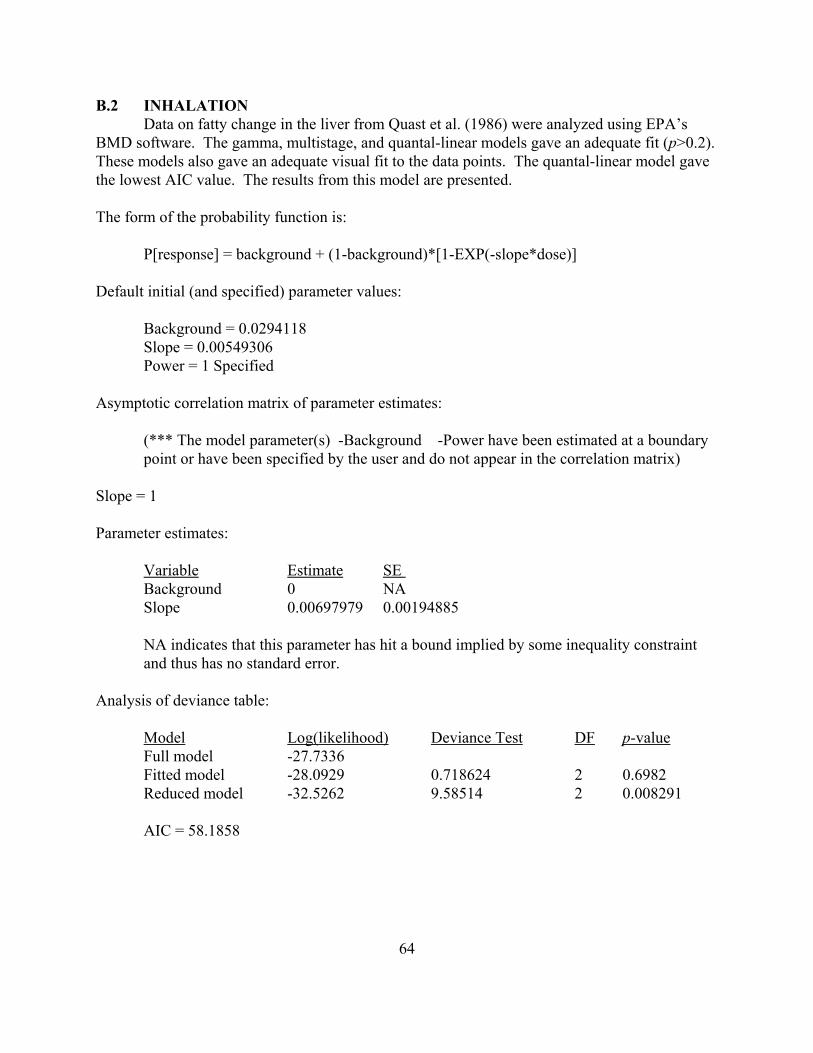


![1,1 DICHLOROETHYLENE (DCE) - California Department … · 1,1 Dichloroethylene (DCE) ... bKd = [(Koc) × (fraction organic matter)], a site and soil zone specific parameter eestimated](https://static.documents.pub/doc/80x56/5b44b7d67f8b9a02148b7ca3/11-dichloroethylene-dce-california-department-11-dichloroethylene-dce.jpg)


![684.8 [1,1]](https://static.documents.pub/doc/80x56/5681611b550346895dd07429/6848-11.jpg)


