170
The 15th International Symposium on Trace Elements in Man and Animals (TEMA 15) June 22-26, 2014 The B Resort | Orlando, Florida USA Hosted by:
| Date post: | 14-Mar-2018 |
| Category: |
Documents |
| Upload: | phamnguyet |
| View: | 220 times |
| Download: | 5 times |

The 15th International Symposium on Trace Elements in Man and Animals (TEMA 15)
June 22-26, 2014
The B Resort | Orlando, Florida USA
Hosted by:

June 22-26, 2014 Orlando, Florida
i
Welcome to the 15th International Symposium on Trace Elements in Man and Animals (TEMA 15)
Dear Fellow Trace Element Researchers: On behalf of the Local Organizing Committee, we welcome you to Orlando and the 15th International Symposium on Trace Elements in Man and Animals (TEMA 15). TEMA 15 has been designed to model the 1st meeting, held in Aberdeen, Scotland in 1969, which was widely considered a success. Our intent was thus to provide a premier opportunity for attendees to learn about cutting-edge research on integrative aspects of the trace elements. The plenary sessions and symposia were planned to provide a broad array of trace mineral-related topics to ensure that all of those in attendance find something of interest to them. We have also attempted to involve many younger investigators in the program, as speakers at oral sessions. The venue of Orlando, Florida was chosen since it is one of the world’s most popular tourist destinations, it is easily accessible by air and it is recognized as a major conference site with many opportunities for entertainment and relaxation. The weather in June is warm with frequent afternoon showers, but the B-Resort Hotel is fully air-conditioned and the pool is available for all TEMA participants. We will provide heavy hors d’oeuvres at the poster sessions in the late afternoon, which then frees up the evenings for attendees to gather at nearby restaurants or to visit the numerous attractions that the Orlando area has to offer. We anticipate that TEMA 15 will carry on the tradition of excellent science and fellowship in another unique environment. Thanks for your attendance and please feel free to see one of us if you have questions or concerns. Bob Cousins, Principal Organizer
John Arthington, Jamie Collins, Mitch Knutson, Co-Organizers

The 15th International Symposium on Trace Elements in Man and Animals (TEMA 15)
ii

June 22-26, 2014 Orlando, Florida
iii
Table of Contents
Welcome Letter .............................................................................................. i
Sponsor Recognition ...................................................................................... v
Local Organizing Committee ......................................................................... vi
TEMA Parent Committee ............................................................................. vi
Plenary Speakers .......................................................................................... vii
General Symposium Information ................................................................ xiii
Detailed Program Agenda ............................................................................ xv
Poster Directory .......................................................................................... xxi
Abstracts ........................................................................................................ 1
Author Index ..............................................................................................133
Map of Meeting Place ................................................................................138
Notes ..........................................................................................................139

The 15th International Symposium on Trace Elements in Man and Animals (TEMA 15)
iv

June 22-26, 2014 Orlando, Florida
v
A Special Thank You to our Symposium Sponsors
Gold
Albion Human Nutrition
HarvestPlus
Mead Johnson Pediatric Nutrition Institute
National Institute of Diabetes and Digestive and Kidney Diseases Funding for this conference was made possible (in part) by 1 R13 DK102349-0l from NIDDK. The views expressed in written conference materials or publications and by speakers and moderators do not necessarily reflect the official policies of the Department of Health and Human Services; nor does mention by trade names, commercial practices, or organiza-tions imply endorsement by the U.S. Government
National Institute of Health, Office of Dietary Supplements
Nutreco
Zinpro
Silver
Kelatron
Micronutrients
University of Florida, Office of Research
Bronze
Animine
NOVUS
Friends of TEMA
Mutimin 90
Disney’s Animals, Science and Environment

The 15th International Symposium on Trace Elements in Man and Animals (TEMA 15)
vi
Local Organizing Committee (University of Florida)
Robert J. Cousins College of Agricultural and Life Sciences, College of Medicine
John Arthington Center Director and Professor Beef Cattle Management Range Cattle Research & Education Center
James F. Collins College of Agricultural and Life Sciences
Mitchell D. Knutson College of Agricultural and Life Sciences
TEMA Parent Committee
Mary L’Abbe, Chair
University of Toronto, Canada
Harry McArdle, Secretary/Treasurer Rowett Institute of Nutrition and Health, UK
Magdalena Araya Universidad de Chile, Chile
Bob Cousins University of Florida, USA
Xingen Lei Cornell University, USA
Bo Lonnerdal University of California-Davis, USA
Lothar Rink RWTH Aachen University Hospital, Germany

June 22-26, 2014 Orlando, Florida
vii
Plenary Speakers
Greg Anderson
Greg Anderson is Deputy Director of the QIMR Berghofer Medical Research Institute in Brisbane, Australia, and is Head of the Iron Metabolism Laboratory at the Institute, as well as a Senior Research Fellow of the National Health and Medical Research Council of Australia. He has adjunct appointments in the School of Chemistry and Molecular Biosciences, and the School of Medicine at the University of Queensland, and at Griffith University. After a PhD at the University of Queensland on mechanisms of intestinal iron absorption, he conducted postdoctoral work on the genetics of iron uptake in yeast at the US National Institutes of Health. He has worked in the area of iron homeostasis for 30 years, and studies both basic iron homeostasis and human disorders of iron metabolism. Particular research interests include understanding the mechanisms and regulation of intestinal iron absorption, the pathogenesis of iron loading diseases (particularly HFE-related haemochromatosis and beta-thalassaemia), the role of iron in the pathogenesis of bacterial infections (specifically in cystic fibrosis), and iron homeostasis in early postnatal life. He is currently the President of the International BioIron Society and serves on the Board of the International Biometals Society.
Michael Aschner
Dr. Aschner serves as the Harold and Muriel Block Chair in Molecular Pharmacology at Albert Einstein College of Medicine. He served on numerous toxicology panels (Institute of Medicine, US Environmental Protection Agency, Center for Disease Control), and is a member of the Neurotoxicology and Alcohol study section (NIH). Research in our lab focuses on the following topics: (1) Modulation of C. elegans genes (aat, skn-1, daf-16) that are homologous to mammalian regulators of MeHg uptake and cellular resistance will modify dopaminergic neurodegeneration in response to MeHg exposure. (2) Under conditions of MeHg-induced oxidative stress, Nrf2 (a master regulator of antioxidant responses) coordinates the upregulation of cytoprotective genes that combat MeHg-induced oxidative injury, and that genetic and biochemical changes that negatively impact upon Nrf2 function increase MeHg’s neurotoxicity. (3) PARK2, a strong PD genetic risk factor, alters neuronal vulnerability to modifiers of cellular Mn status, particularly at the level of mitochondrial dysfunction and oxidative stress. Our studies are designed to (1) shed novel mechanistic insight into metal-induced neurodegeneration; (2) identify targets for genetic or pharmacologic modulation of neurodegenerative disorders; (3) increase knowledge of the pathway involved in oxidative stress; (4) develop improved research models for human disease using knowledge of environmental sciences.

The 15th International Symposium on Trace Elements in Man and Animals (TEMA 15)
viii
Fabrice Chimienti
Fabrice Chimienti received his PhD from University Rene Descartes, Paris, where he studied the molecular mechanisms involved in zinc signaling. In the process of identifying new zinc transporters in Prof Favier’s lab, he discovered ZnT8, a key regulator of insulin secretion in human pancreatic beta cells. Later on ZnT8 was shown to be a risk locus for type 2 diabetes. In subsequent years he published several papers on the role of zinc in beta cell function. Following this discovery he co-funded Mellitech, a biotech company dedicated to identify and develop new drugs against diabetes. At Mellitech Fabrice served as a Chief Scientific Officer and successfully setup a cell-based bioassay to detect fluctuations in intracellular zinc levels. Using this assay to screen a small molecule library led to the identification of innovative compounds targeting ZnT8, active both in vitro and in vivo on blood glucose control. Fabrice then led the project through lead optimization and development to late preclinical stage. Fabrice also actively participates in the design of an innovative, ZnT8-specific imaging approach dedicated to non-invasive in vivo evaluation of beta cell mass/function. Fabrice then moved to AstraZeneca in the field of beta cell research.
Pamela Fraker
Pam Fraker is a University Distinguished Professor in the Department of Biochemistry and Molecular Biology at Michigan State University. She received a BS from Purdue University and a Ph.D in Biochemistry from the University of Illinois. She has had a lifetime interest in the impact of changes in nutritional stature that can markedly change immune defense to include the neuroendocrine factors that facilitate these changes.
Our lab is interested in the interplay between nutritional status, immune defense, and the neuroendocrine system. We demonstrated that single element deficiencies in zinc or multi-component deficiencies in protein-calories caused a rapid decline in antibody and cell mediated responses. We showed that such deficiencies activate the stress axis causing chronic production of glucocorticoids which rapidly reduced lymphopoiesis by accelerating apoptosis among pre T- and pre B-cells. We recently discovered a compensatory mechanism whereby the elevated glucocorticoids promote myelopoiesis. Thus, the phagocytic cells that are the first line of defense remain plentiful with retention of key functions, thereby providing interim protection. We also demonstrated similar outcomes for human bone marrow and neutrophils exposed to elevated endogenous glucocorticoids. We were among the first to demonstrate that FACS could be used to rapidly quantitate apoptotic cells in heterogeneous tissues, like the marrow. Currently, we are examining the changes in the immune system created by obesity using the morbidly obese undergoing gastric bypass surgery and the overfed mouse as models for study. Our goal is to better understand the neuroendocrine-metabolic and immunological changes caused by obesity. We have 30 years of experience integrating the cross-talk between the nutritional, neuroendocrine, and immune systems.

June 22-26, 2014 Orlando, Florida
ix
Vadim Gladyshev
Vadim Gladyshev received his B.S./M.S. degree in 1988 and Ph.D. degree in 1992 from Moscow State University, Russia, followed by postdoc training at NIH, and a faculty position at University of Nebraska. Since 2009, he has been a Professor of Medicine and Director of the Center for Redox Medicine at Brigham and Women’s Hospital, Harvard Medical School, and an Associate Member of the Broad Institute. Dr. Gladyshev has been working in the areas of selenium and redox biology as applied to aging and cancer, in which he has made contributions, such as characterization of the human selenoproteome (an entire set of selenium-containing proteins), methods to predict catalytic redox-active cysteines in proteins, and sequencing the genomes of exceptionally long-lived mammals. He has a long-term interest in the understanding of functions of trace elements and mechanisms of redox control. Dr. Gladyshev has published >250 articles, edited books on selenium and redox biochemistry, and was elected as an AAAS fellow. He also recently received an NIH Director’s Pioneer Award. Dr. Gladyshev participated in the last 5 TEMA meetings.
Hajo Haase
Hajo Haase received his diploma in chemistry in 1998 and his Ph.D. in natural sciences in 2001, both from the University of Bremen in Germany. From 2001 to 2003 he was a postdoctoral research fellow at the Center for Biochemical and Biophysical Sciences and Medicine at the Harvard Medical School in Boston. There, he worked in the group of Dr. Wolfgang Maret, where he investigated the role of zinc ions in insulin signaling and developed methods for analyzing the zinc load and oxidation state of metallothionein.
In 2003, Dr. Haase returned to Germany to become an assistant professor at the institute of immunology of the RWTH Aachen University Hospital. Here, Dr. Haase is currently teaching immunology and has co-authored an introductory textbook on this subject with Dr. Andrea Kruse and Dr. Lothar Rink. His research interests converge are at the interface of chemistry, molecular biology, immunology, and nutrition. The work in his lab focuses on the impact of essential trace elements and toxic metal ions on the immune system, with a special focus on zinc as an intracellular second messenger in immune cells.

The 15th International Symposium on Trace Elements in Man and Animals (TEMA 15)
x
Stephanie Hansen
Stephanie Hansen is an Assistant Professor in Beef Feedlot Nutrition at Iowa State University. She completed her B.S. in Animal Science at Iowa State and received her M.S. in Animal Science from North Carolina State University in 2005. She completed her Ph.D. at North Carolina State University in 2008 and has been a faculty member in the Department of Animal Science at Iowa State since August of 2009. Dr. Hansen teaches undergraduate and graduate classes in the areas of animal nutrition and vitamin and mineral metabolism. She has an active research program at Iowa State University which is focused on the influence of micronutrients on beef cattle performance, carcass quality and meat characteristics, improving feed efficiency of beef cattle, and finding ways for producers to safely increase the inclusion of high sulfur co-products of the ethanol industry to cattle diets. Dr. Hansen currently supervises 5 graduate students, a post-doctoral associate and several undergraduate researchers. In her free time Stephanie loves to travel, especially to see amazing wildlife and landscapes. She enjoys photography and hiking.
Xingen Lei
Xingen Lei received his B.S. and M.S. in China, and Ph.D. from Michigan State University. He was among the first to demonstrate nutritional and environmental values of supplemental phytase in improving feed phosphorus utilization. Lei developed a new generation of bacterial phytases that are used worldwide by the feed industry. Lei also pioneered the nutritional genomics of selenium in pigs and chicks that revealed dual roles of selenium in oxidative stress and in diabetes. Lei is an international leader for the global fight against mineral deficiencies in humans. Lei has advised 95 graduate students and postdoc fellows, and served on panels for NIH and USDA-NRI/NIFA, committees for the NRC report on Mineral Toxicity and CAST task force, and 8 Editorial Boards. Overall, Lei’s creative research on minerals has demonstrated clear benefits to animal agriculture and humankind.

June 22-26, 2014 Orlando, Florida
xi
Cathy Levenson
Dr. Levenson is a Professor of Biomedical Sciences and Neuroscience at the Florida State University College of Medicine where she serves as the Course Director for Medical Biochemistry & Genetics. After completing a BA in Neurobiology from the University of Virginia, Levenson received a master’s degree from Florida State University and a PhD from the University of Chicago. Before her faculty appointment at FSU, she was a research associate in the lab of Robert Cousins at the University of Florida. Her current research activities include understanding the cellular and molecular mechanisms responsible for the role of zinc in neuronal stem cell proliferation, differentiation and survival, and how this information can be used to treat the wide spread public health problems of depression and anxiety that are associated with traumatic brain injury.
Martina Muckenthaler
Martina Muckenthaler is professor of Molecular Medicine at the Department of Pediatric Hematology, Oncology and Immunology of the University of Heidelberg. She trained as a molecular biologist during her PhD at the University of Oxford (UK) and post-doctoral fellowship at the EMBL.
Martina’s longstanding research interest focuses on regulatory mechanisms maintaining iron homeostasis and their disturbance in hematological disease. She significantly contributes to the understanding of hepcidin regulation and the pathophysiological consequences of iron overload and iron deficiency.
Martina’s scientific performance at the interface between medicine and molecular biology led to her appointment as Principal Investigator in the prestigious Molecular Medicine Partnership Unit, a translational joint venture between the EMBL and the Medical Faculty of Heidelberg. As chairwoman of the postdoctoral program of the Medical Faculty, she leads the program that enables young MDs to receive top research training. She is a steering committee member of the Centre of Rare Diseases into which her research on rare anemia is integrated. She was elected as a scientific committee member of ASH and as a board member of EHA in 2013. In recognition of her engagement for women in science and keen interest in science communication, she was selected as one of ten female European researchers for the EU-funded Insight Lecture.

The 15th International Symposium on Trace Elements in Man and Animals (TEMA 15)
xii
Yatrik Shah
Dr. Yatrik Shah did his graduate work at the Medical College of Ohio obtaining a Ph.D in Biochemistry in 2005. He did his postdoctoral training at the NIH in the laboratory of Dr. Frank Gonzalez. In 2009 he started his independent lab at the University of Michigan, Department of Molecular & Integrative Physiology, where he is currently. His lab studies the role of the hypoxic response in the intestine. A major focus of his lab is understanding how the intestinal hypoxic response regulates the adaptive increase in iron absorption and how this pathway is dysregulated in certain congenital anemias such as beta–thalassemia and sickle cell. Recent work focuses on identifying therapeutics that prevent tissue damage due to iron toxicity in patients with congenital iron-loading anemias.
Eric Skarr
Dr. Skaar received his B.S. in Bacteriology from the University of Wisconsin at Madison and his Ph.D. and M.P.H. from Northwestern University in the laboratory of Dr. Hank Seifert. He performed postdoctoral training in the laboratory of Dr. Olaf Schneewind at the University of Chicago. He is currently an Ernest W. Goodpasture Associate Professor at Vanderbilt University where his laboratory focuses on the struggle for nutrients between bacterial pathogens and their vertebrate hosts. The Skaar laboratory investigates this topic through a number of projects that seek to understand (i) nutrient acquisition by bacterial pathogens, (ii) how vertebrate immune proteins sequester nutrients during the pathogenesis of bacterial infection, and (iii) changes in the host-pathogen interface during infection as investigated by multi-modality imaging. Dr. Skaar has been the recipient of numerous awards including the Pfizer Aspire Award, the Searle Scholars Award, the ICAAC/IDSA Young Investigator Award, the Chancellor’s award for Research and the Postdoctoral Mentor of the Year from Vanderbilt University, and he was named a Burroughs Wellcome Investigator in the Pathogenesis of Infectious Diseases. Dr. Skaar is currently a standing member on the Bacterial Pathogenesis NIH Study Section and he serves on numerous editorial boards.

June 22-26, 2014 Orlando, Florida
xiii
General Symposium Information Registration and Name Badges Symposium Attendees and accompanying Guests (must be registered and paid) will be issued a name badge upon arrival at the Registration Desk in Grand IV. Name badges must be worn daily and especially during the meal functions. Guests Guests are allowed in the evening functions only and must be 18 years or older due to the fact that alcohol is being served. Thank you for your cooperation in this. Currency Exchange: Downtown Disney Guest Services https://disneyworld.disney.go.com/guest-services/downtown-disney/ Nearest Hospital and Walgreens Drugstore To locate the nearest hospital or drugstore for personal items to purchase, please check out the following PDF to assist you: http://conference.ifas.ufl.edu/tema15/healthcare_info.pdf Please note that in an emergency, always call 911 first. Parking Parking at the B Resort for overnight guests attending TEMA 15 is complimentary for self-parking. You will be charged a fee if you choose to valet park. Meeting Venue The beautiful brand new B Resort in the Walt Disney World Resort has just been completed. A Hotel Fact Sheet and Directions can be found by going to their website: http://www.bhotelsandresorts.com/b-walt-disney-world/ Internet Access All attendees staying at the hotel will receive complimentary Wi-Fi throughout the hotel and guestrooms. If you should need assistance, please contact the Front Desk at the hotel. Presenters: Oral: Please provide your presentation one-day in advance of your talk on a USB drive or CD. If you have an afternoon talk, please submit your talk no later than the morning of your presentation. Please see Lisa Pennington, AV Coordinator, at the AV download station when you check-in at Registration. Please review the Speaker Instructions: http://conference.ifas.ufl.edu/TEMA15/speakers.pdf Posters: Poster Boards will be setup in Grand I. A limited supply of Velcro tape will be provided for mounting your poster. Poster Session setup and removal times are listed on the TEMA 15 website and on the Poster Instructions: http://conference.ifas.ufl.edu/TEMA15/posters.pdf POSTER BOARDS WILL BE REMOVED ON WEDNESDAY, JUNE 25 AT 10AM.

The 15th International Symposium on Trace Elements in Man and Animals (TEMA 15)
xiv
June 22-26, 2014 Orlando, Florida
xv
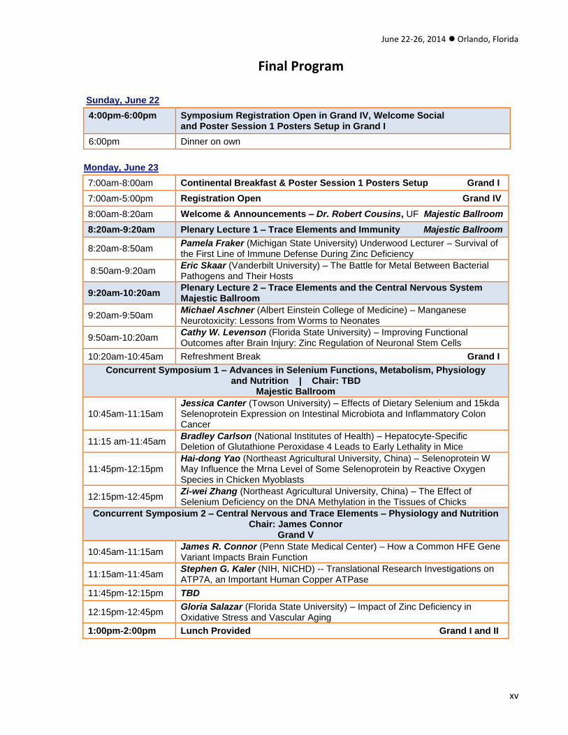
Final Program
Sunday, June 22
4:00pm-6:00pm Symposium Registration Open in Grand IV, Welcome Social and Poster Session 1 Posters Setup in Grand I
6:00pm Dinner on own
Monday, June 23
7:00am-8:00am Continental Breakfast & Poster Session 1 Posters Setup Grand I
7:00am-5:00pm Registration Open Grand IV
8:00am-8:20am Welcome & Announcements – Dr. Robert Cousins, UF Majestic Ballroom
8:20am-9:20am Plenary Lecture 1 – Trace Elements and Immunity Majestic Ballroom
8:20am-8:50am Pamela Fraker (Michigan State University) Underwood Lecturer – Survival of the First Line of Immune Defense During Zinc Deficiency
8:50am-9:20am Eric Skaar (Vanderbilt University) – The Battle for Metal Between Bacterial Pathogens and Their Hosts
9:20am-10:20am Plenary Lecture 2 – Trace Elements and the Central Nervous System Majestic Ballroom
9:20am-9:50am Michael Aschner (Albert Einstein College of Medicine) – Manganese Neurotoxicity: Lessons from Worms to Neonates
9:50am-10:20am Cathy W. Levenson (Florida State University) – Improving Functional Outcomes after Brain Injury: Zinc Regulation of Neuronal Stem Cells
10:20am-10:45am Refreshment Break Grand I
Concurrent Symposium 1 – Advances in Selenium Functions, Metabolism, Physiology and Nutrition | Chair: TBD
Majestic Ballroom
10:45am-11:15am Jessica Canter (Towson University) – Effects of Dietary Selenium and 15kda Selenoprotein Expression on Intestinal Microbiota and Inflammatory Colon Cancer
11:15 am-11:45am Bradley Carlson (National Institutes of Health) – Hepatocyte-Specific Deletion of Glutathione Peroxidase 4 Leads to Early Lethality in Mice
11:45pm-12:15pm Hai-dong Yao (Northeast Agricultural University, China) – Selenoprotein W May Influence the Mrna Level of Some Selenoprotein by Reactive Oxygen Species in Chicken Myoblasts
12:15pm-12:45pm Zi-wei Zhang (Northeast Agricultural University, China) – The Effect of Selenium Deficiency on the DNA Methylation in the Tissues of Chicks
Concurrent Symposium 2 – Central Nervous and Trace Elements – Physiology and Nutrition Chair: James Connor
Grand V
10:45am-11:15am James R. Connor (Penn State Medical Center) – How a Common HFE Gene Variant Impacts Brain Function
11:15am-11:45am Stephen G. Kaler (NIH, NICHD) -- Translational Research Investigations on ATP7A, an Important Human Copper ATPase
11:45pm-12:15pm TBD
12:15pm-12:45pm Gloria Salazar (Florida State University) – Impact of Zinc Deficiency in Oxidative Stress and Vascular Aging
1:00pm-2:00pm Lunch Provided Grand I and II

The 15th International Symposium on Trace Elements in Man and Animals (TEMA 15)
xvi
Monday, June 23 (CONTINUED)
Concurrent Symposium 3 – Global Trace Element Biofortification for Health Improvement of Populations (Sponsored by Harvest Plus) | Chair: Ross Welch
Majestic Ballroom
2:00pm-2:30pm Ross Welch (Cornell University) – Prosectives on Global Biofortification Efforts: Evidence Base and Future Directions
2:30pm-3:00pm Sarah Luna (Cornell University) – A Review of the Efficacy of Iron Biofortified Staple Food Crops
3:00pm-3:30pm Leland Miller (University of Colorado) – Bioavailability of Zinc from Zinc Biofortified Staples for Large Populations Across the Globe
3:30pm-4:00pm Xingen Lei (Cornell University) – Biofortification Initiatives in China and Latin America
Concurrent Symposium 4 – Advances in Zinc Functions, Metabolism, Physiology and Nutrition | Chair: Shannon Kelleher
Grand V
2:00pm-2:30pm Shannon Kelleher (Pennsylvania State University and Penn State Hershey College of Medicine) – Mammary Gland Zinc Transport: Implications For Breast Function And Disease
2:30pm-3:00pm Manuel Ruz (University of Chile) -- Zinc Participation In Insulin Secretion And Peripheral Action And Its Relevance For Diabetes Therapy
3:00pm-3:30pm Tolunay Beker Aydemir (University of Florida) – Zinc Transporter Zip14 Is Involved With Regulation Of Insulin And Glucose Homeostasis In Intestine, Pancreas And Liver
3:30pm-4:00pm Alvaro Perez (University of Chile) – Zinc Attenuates Insulin Secretion Stimulated By Interleukine 6 (IL-6) In The Pancreatic Cell Line RIN M5f
4:00pm-4:30pm Daren Knoell (Ohio State University) – Role Of Zip8 In Cadmium-Induced Lung Disease
Concurrent Symposium 5 – Trace Elements in Production, Companion & Exotic Animal Nutrition and Health | Chair: Joel Caton
Grand III
2:00pm-2:30pm Joel Caton (North Dakota State University) – Effects of Maternal Selenium Supply and Nutritional Plane on Offspring Intestinal Biology
2:30pm-3:00pm Zach Rambo (Zinpro Corporation) – Protein Accretion and Zinc Nutrition: Application and New Technologies in Animal Agriculture
3:00pm-3:30pm Karen Wedekind (Novus International) – The Link between Mineral Excesses and Chronic Diseases in Pets
3:30pm-4:00pm Gretchen Hill (Michigan State University) – Metallothionein in Swine Fed Different Amounts and Sources of Zinc
4:00pm-4:30pm Juxing Chen (Novus International Inc) – Effects of Trace Minerals on the Development of Bacterial Chondronecrosis with Osteomyelitis in Poultry
4:30pm-4:45pm Marielle van Zelst (Ghent University, Belgium) – In Vitro Selenium Bioaccessibility in Pet Foods
5:00pm-7:00pm Poster Session 1 and Reception Grand I
7:30pm Poster Session 1 Poster Removal
Dinner on own

June 22-26, 2014 Orlando, Florida
xvii
Tuesday, June 24
7:00am-8:00am Continental Breakfast and Poster Session 2 Posters Setup Grand I
7:00am-5:00pm Registration Open Grand IV
8:20am-9:20am Plenary Lecture 3 – Molecular Aspects of Iron Utilization Majestic Ballroom
8:20am-8:50am Yatrik Shah (University of Michigan) – Intestinal Hypoxia Signaling: A Critical Regulator of Systemic Iron Homeostasis and Iron Related Disorders
8:50am-9:20am Martina U. Muckenthaler (University of Heidelberg) – Regulation of Iron Homeostasis
9:20am-10:20am Plenary Lecture 4 – Genetic/Proteomic Approaches to Trace Elements Majestic Ballroom
9:20am-9:50am Vadim Gladyshev (Harvard University) – Comparative and Functional Genomics of Mammalian Selenoproteomes
9:50am-10:20am Greg Anderson (QIMR Berghofer Medical Research Institute) – Mutant Mouse Models to Relate Trace Elements to Human Disease
10:20am-10:45am Refreshment Break Grand I
Concurrent Symposium 6 – Trace Elements: Microbial Interactions and Immune Defenses Chair: Lothar Rink Majestic Ballroom
10:45am-11:15am Lothar Rink (RWTH Aachen University Hospital) -- Zinc Homeostasis as a Gatekeeper of the Immune Response
11:15am-11:45am Inga Wessels (University of Florida) -- Changes In Zinc Homeostasis During Different Stages Of Sepsis
11:45am-12:15pm Sandeep Prabhu (Pennsylvania State University) -- Selenoprotein-Induced Macrophage Phenotype Switching Is Imperative in Helminth Parasite Clearance
12:15pm-12:45pm Jadwiga Turlo (Medical University of Warsaw, Poland) -- Se-Enriched Polysaccharides –Structure and Biological Activity
Concurrent Symposium 7 – Advances in Iron Functions, Metabolism, Physiology and Nutrition | Chair: Mitchell Knutson
Grand V
10:45am-11:15am Mitchell D. Knutson (University of Florida) -- Ablation of the Metal-Ion Transporter Slc39a14 (Zip14) Prevents Hepatic Iron Overload in Hereditary Hemochromatosis
11:15 am-11:45am Paul Sharp (King’s College, London) -- Quercetin Decreases Iron Bioavailability Through Inhibition of Ferroportin Expression in Intestinal Epithelial Cells
11:45pm-12:15pm Rick Eisenstein (University of Wisconsin) -- The IRP1-HIF-2a Axis: Coordinating Iron and Oxygen Sensing with Erythropoiesis and Iron Absorption
12:15pm-12:45pm Kimberly O’Brien (Cornell University) -- Neonatal Iron Status Impacts Placental Iron Transporter Expression
12:45pm-1:00pm Kathryn Page (University of California-Berkeley) -- Iron Deficient Toxic Milk Leads to the Mask Phenotype in Hephaestin Knockout Mice
1:00pm-2:00pm Lunch Provided Grand I & II

The 15th International Symposium on Trace Elements in Man and Animals (TEMA 15)
xviii
Tuesday, June 24 (CONTINUED)
Concurrent Symposium 8 – Animal Models for Trace Element Research Relevant to Human Disease | Chair: Thomas Bartnikas
Majestic Ballroom
2:00pm-2:30pm Thomas Bartnikas (Brown University) -- Metal Homeostasis And Transferrin Deficiency
2:30pm-3:00pm Claudia Kiermayer (Helmholtz Zentrum Munchen, Germany) -- A Genetic Mouse Model for Selenoprotein Research: Heart-Specific Knockout of Thioredoxin Reductase 2
3:00pm-3:30pm Wen-Hsing Cheng (Mississippi State University) -- A Role of Selenium at Nutritional Levels of Intake in Mouse Aging
3:30pm-4:00pm Young Ah Seo (Harvard University School of Public Health, Boston) -- Manganese Deficiency in Flatiron Mice
4:00pm-4:30pm Catalina Troche (University of Florida) -- Zinc Mediation of Adipocyte Differentiation and Metabolism Through Zip14
Concurrent Symposium 9 – Biomarkers of Trace Element Status, Requirements and Bioavailability | Chair: Janet King
Grand V
2:00pm-2:30pm Janet C. King (Children’s Hospital of Oakland Research Institute, UCSF) --Zinc Biomarkers: Is There a Solution?
2:30pm-3:00pm Harry McArdle (Rowett Institute for Nutrition and Health,University of Aberdeen) -- Iron Metabolism in the Perinatal Period in the Rat – Defining the Hierarchy of Requirements
3:00pm-3:30pm
Berislav Momcilovic (Institute for Research and Development of the Sustainable Eco Systems, Zagreb, Croatia) -- A Novel Concept to Assess the Human Iodine and Selenium Nutritional Status by Analyzing Their Frequency Distribution Properties in the Hair
3:30pm-4:00pm Miriam M.J. van Riet (Institute for Agricultural and Fisheries Research, (ILVO) Ghent University, Belgium) -- Correlation Between Bone Mineralisation and Zinc Status Markers in Sows
4:00pm-4:30pm John Beattie (University of Aberdeen, UK) -- Human Biomarkers of Zinc Status
Concurrent Symposium 10 – Advances in Copper Functions, Metabolism, Physiology and Nutrition | Chair: James Collins
Grand III
2:00pm-2:30pm James F. Collins (University of Florida) -- Physiological Roles of Copper in Mammalian Iron Homeostasis
2:30pm-3:00pm Joseph Prohaska (University of Minnesota, Duluth) -- Anemia of Copper Deficiency and the Hepcidin Response
3:00pm-3:30pm Zhen Zhang (Regenerative Medicine Research Center, China) -- Copper-Dependent and –Independent Hypoxia-Inducible Factor-1 Regulation of Gene Expression
3:30pm-4:00pm Y. James Kang (Sichuan University, West China Hospital) -- Mechanistic Understanding for Copper-Induced Regression of Cardiac Hypertrophy
4:00pm-4:15pm Caglar Doguer (University of Florida) -- Caco-2 Cells Contain a Functional Ferroxidase that Is Not Hephaestin
4:30pm-5:00pm Lothar Rink – Special Presentation on Zinc-net
5:00pm-7:00pm Poster Session 2 and Reception Grand I
7:30pm Poster Session 2 Poster Removal
Dinner on own

June 22-26, 2014 Orlando, Florida
xix
Wednesday, June 25
Participants on own for the day. Because of the unusual variety of attractions in the Orlando area, the Local Organizing Committee did not wish to select just one. We do highly recommend Disney World, Universal Studios, MGM, and the Kennedy Space Center.
- See website for Area Information: www.conference.ifas.ufl.edu/TEMA15/area.htm
Thursday, June 26
7:00am-8:00am Continental Breakfast Grand I
7:00am-5:00pm Registration Open Grand IV
8:20am-9:20am Plenary Lecture 5 – Zinc and Biological Signal Processing Majestic Ballroom
8:20am-8:50am Hajo Haase (RWTH Aachen University Hospital) – Zinc Regulation of Cell Signaling Pathways
8:50am-9:20am Fabrice Chimienti (Mellitech, France) – Zinc, Pancreatic Islet Cell Function and Diabetes
9:20am-10:20am Plenary Lecture 6 – Role of Trace Elements in Animal Health Majestic Ballroom
9:20am-9:50am Xingen Lei (Cornell University) – Dual Implications of Selenium Genomics of Food Animals
9:50am-10:20am Stephanie Hansen (Iowa State University) – Interactions Between Trace Minerals and Production Practices in Beef Cattle
10:20am-10:45am Refreshment Break Grand I
Concurrent Symposium 11 – Trace Elements and Diabetes, Obesity, Sepsis, Oxidative Stress and Metabolic Disorders | Chair: Liping Huang
Majestic Ballroom
10:45am-11:15am Liping Huang (USDA, University of California-Davis) -- Linking Cellular Zinc Status to Body Weight and Fat Mass: Mapping Quantitative Trait Loci in Znt7 Knockout Mice
11:15 am-11:45am Juan Liuzzi (Florida International University) -- Autophagy Induced by Zinc Confers Protection Against Ethanol Toxicity
11:45pm-12:15pm In-Sook Kwun (Andong National University) -- Regulation of Vascular Smooth Muscle Cell Calcification and Apoptosis by Zinc Deficiency
12:15pm-12:45pm Shiwen Xu (Northeast Agricultural University) -- Selenium Deficiency Induced Higher Inflammation and Apoptosis in Chicken Liver
12:45pm-1:00pm Richard Coffey (University of Florida) -- Roles of Zip14 and Zip8 in Iron Uptake by Pancreatic Beta Cells
Concurrent Symposium 12 – Trace Elements in Cell Death, Tissue Regeneration, Epigenetics and Tumorigenesis | Chair: Louise Fong
Grand V
10:45am-11:15am Louise Y. Fong (Thomas Jefferson University) -- Dietary Zinc and Oral-Esophageal Cancer Development and Prevention
11:15am-11:45am Rui Li (Sichuan University, China) -- Vimentin Is Involved in Copper-Induced Regression of Cardiomyocyte Hypertrophy
11:45am-12:15pm Ryuta Tobe (National Institutes of Health) -- Differences in Redox Regulatory Systems in Human Lung and Liver Cancers Suggest Different Avenues for Therapy
1:00pm-2:00pm Lunch Provided Grand I & II

The 15th International Symposium on Trace Elements in Man and Animals (TEMA 15)
xx
Thursday, June 26 (CONTINUED)
Concurrent Symposium 13 – Metalloproteins, Metal Chaperones and Metal Transporters Chair: Wolfgang Maret
Majestic Ballroom
2:00pm-2:30pm Wolfgang Maret (King’s College London) -- Functions of Zinc Ions in Phosphorylation Signaling
2:30pm-3:00pm Young Rok Seo (Dongguk University, Seoul) -- Identification of Novel Targets and Molecular Signaling Networks af Selenomethionine-Mediated Chemoprevention in Colorectal Carcinoma Mouse Model
3:00pm-3:30pm Maria Linder (California State University) -- Delivery of Copper to Cells by Blood Plasma Ceruloplasmin
3:30pm-4:00pm Christer Hogstrand (King’s College, London) -- Zinc Transporters and Cell Signaling
4:00pm-4:15pm Wei Zhang (University of Florida) -- Zip8 Is Expressed and Regulated by Iron in the Placenta
Concurrent Symposium 14 – Trace Element Intervention and Food Fortification and Health Outcomes | Chair: Samir Samman
Grand V
2:00pm-2:30pm Samir Samman (University of Sydney, Australia) -- Effect of Vegetarian Diets on Zinc Status
2:30pm-3:00pm Anatoly Skalny (Russian Society of Trace Elements in Medicine, Russia and Institute for UNESCO, France) -- Trace Element Status of Population and Demography in Russia: Possible Linkage
3:00pm-3:30pm WQ Zhang (Tianjin Medical School, China) -- The Effect of Iodine Excess on Vulnerable Populations
3:30pm-4:00pm Margaret P. Rayman (University of Surrey, UK) -- Effect of Selenium on Markers of Risk of Pre-Eclampsia in UK Pregnant Women
4:00pm-4:30pm TBD
Concurrent Symposium 15 – Trace Element Supplementation in Animal Nutrition Chair: John Arthington
Grand III
2:00pm-2:30pm Paul Bikker (Wageningern UR Livestock Research, Wageningen, the Netherlands) -- Zinc Requirements of Young Growing Pigs
2:30pm-3:00pm John D. Arthington (University of Florida) -- Immune Response Augmentation in Cattle through Injectable Trace Minerals
3:00pm-3:30pm Terry Engle (Colorado State University) -- The Role of Trace Minerals in Lipid Metabolism and Immunity in Ruminants
3:30pm-4:00pm Stéphane Durosoy (Animine, Sillingy, France) -- High Dosed Zinc Oxide in Piglet Diets : Still Debated
4:00pm-4:30pm Hanne Damgaard Poulsen (Aarhus University, Denmark) -- Zinc - Nutrient or Pharmaceutical in Weaned Piglets?
4:30pm-4:45pm Olivia Genther (Iowa State University) -- Interaction Between A ß-Agonist and Supplemental Zinc in Cattle
5:00pm-5:30pm Plenary Closing Remarks Majestic Ballroom
7:00pm-9:00pm TEMA 15 Closing Banquet [Poolside] with Acoustic Guitarist Matt Collins

June 22-26, 2014 Orlando, Florida
xxi
Poster Directory Poster Session 1 - Monday, June 23 - 5:00pm-7:00pm
# Authors Affiliations Abstract Title
1 Paul Bikker, Jurgen van Baal, Hans van Diepen, Gisabeth Binnendijk, Linda Troquet and Age Jongbloed
Wageningen UR Livestock Research, Wageningen, the Netherlands
EFFECT OF LEVEL AND DURATION OF COPPER SUPPLEMENTATION OF PIG DIETS
2 Anna Błażewicz, Liao Kuan-Yung, Wojciech Dolliver, and Ryszard Kocjan
Department of Analytical Chemistry, Medical University of Lublin, Poland
ION CHROMATOGRAPHY METHOD FOR DETERMINATION OF TRACE ELEMENTS IN BIOLOGICAL FLUIDS AND TISSUES - A USEFUL TOOL FOR BETTER UNDERSTANDING DISEASES OF DIFFERENT ETIOLOGY
3 Penny D Colbourne1, David W Kinniburgh2 and Stacey C Hagen1
1Alberta Health Services, University of Alberta Hospital, Edmonton, Alberta, Canada 2Alberta Centre for Toxicology, University of Calgary, Calgary, Alberta, Canada
INVESTIGATING A POTENTIAL MANGANESE EXPOSURE IN A RURAL COMMUNITY
4 James F. Collins, Caglar Doguer, and Sukru Gulec
Food Science & Human Nutrition Department, University of Florida, Gainesville, FL, USA
SILENCING OF THE COPPER-TRANSPORTING ATPASE 1 (ATP7A) IN RAT IEC-6 CELLS INCREASES CYCLIN D1 PROTEIN EXPRESSION AND IMPAIRS CELL GROWTH
5 Luiz Fernando Costa e Silva 1,2, Terry E. Engle1, Polyana Pizzi Rotta 1,2, Sebastião de C. Valadares Filho 2, Flávia A. da Silva Sales 2, and Ana Clara M. Baião 2
1Animal Science Department, Colorado State University, Fort Collins, CO, USA; 2Animal Science Department, Universidade Federal de Viçosa, Viçosa, MG, Brazil
TRACE ELEMENT REQUIREMENTS FOR NELLORE CATTLE
6 Harry McArdle, Helen Hayes, Valerie Stevens, and Sarah Cottin
Rowett Institute of Nutrition and Health, Aberdeen, UK
THE EFFECT OF IRON DEFICIENCY ON VITAMIN A METABOLISM DURING PREGNANCY IN RATS
7 Roger Davin, Edgar Garcia Manzanilla and José Francisco Pérez
Departament de Ciència Animal i dels Aliments, Universitat Autònoma de Barcelona, Cerdanyola del Vallès, Barcelona, Spain
IN VITRO SOLUBILITY OF DIFFERENT ZN SOURCES USING PIG GIT CONTENT
8 Hari Hundal, Pete Taylor and Ailsa Downie
Division of Cell Signalling & Immunology, College of Life Sciences, University of Dundee, Dundee, UK
EFFECTS OF IRON AVAILABILITY ON AMINO ACID UPTAKE AND SIGNALLING EVENTS IN CACO-2 CELLS
9 Paolo Trevisi1, Stéphane Durosoy2, Yuri Gherpelli3, Vincenzo Motta1, Michela Colombo1 and Paolo Bosi1
1University of Bologna, DISTAL, Reggio Emilia, Italy; 2Animine, Sillingy, France; 3Istituto Zooprofilattico Sperimentale Bruno Ubertini, Reggio Emilia, Italy
EFFECT OF ZINC OXIDE ON GROWTH PERFORMANCE AND HEALTH OF E.COLI K88-CHALLENGED SUSCEPTIBLE WEANING PIGS
10 Tarek Elnimr and Reda Morsy Radioanalysis Research Laboratory, Physics Department, Faculty of Science, Tanta University, Tanta, Egypt
PROSPECTIVE STUDY ON THE ROLE OF TRACE ELEMENTS IN PSORIASIS
11 Tarek Elnimr and Reda Morsy Radioanalysis Research Laboratory, Physics Department, Faculty of Science, Tanta University, Tanta, Egypt
STUDY OF THE ROLE OF SOME TRACE ELEMENTS IN ACNE VULGARIS
12 Tarek Elnimr and Reda Morsy Radioanalysis Research Laboratory, Physics Department, Faculty of Science, Tanta University, Tanta, Egypt
TRANSMISSION BEHAVIOR OF SPECIFIC ELEMENTS OF FOOD IN HUMAN MILK ASSOCIATED WITH THE DIET OF THE EGYPTIAN MOTHER DURING LACTATION
13 Harry McArdle1, Cristina Campoy2,3, Maria-Teresa Miranda4, Jesus Florido5, Pilar Brandi3, Helen Hayes1 and Luz Garcia-Valdes1,3
1Rowett Institute of Nutrition and Health, University of Aberdeen, UK; 2Department of Paediatrics, School of Medicine, University of Granada, Spain; 3EURISTIKOS Excellence Centre for Paediatric Research, University of Granada, Spain; 4Department of Statistics, School of Medicine, University of Granada, Spain; 5Department of Obstetrics and Gynecology, University of Granada, Spain
IRON STATUS, HEPCIDIN AND PLACENTAL TRANSFERRIN RECEPTOR EXPRESSION IN OBESE PREGNANT WOMEN FROM THE PREOBE STUDY

The 15th International Symposium on Trace Elements in Man and Animals (TEMA 15)
xxii
# Authors Affiliations Abstract Title
14 Breanne N. Wright1, Joseph R. Prohaska2, Nana Gletsu Miller1
1Nutrition Science, Purdue University, West Lafayette, IN, USA; 2Biomedical Sciences, University of Minnesota, Medical School Duluth, Duluth, MN, USA
COPPER SUPPLEMENTATION IN MARGINAL AND DEFICIENT GASTRIC BYPASS PATIENTS
15 Ahmed Hammad, Toshimi Kaido, Kohei Ogawa, Yasuhiro Fujimoto, Koichiro Hata, Tadahiro Uemura, Koji Tomiyama, Akira Mori and Shinji Uemoto
Division of Hepato-Biliary-Pancreatic and Transplant Surgery, Department of Surgery, Graduate School of Medicine, Kyoto University, Kyoto, Japan
PREOPERATIVE NUTRITIONAL STATUS IN PATIENTS UNDERGOING LIVER TRANSPLANTATION
16 Jia-Qiang Huang1, Shi-Wen Xu2 , Fa-Zheng Ren1 and Xin Gen Lei1,3
1College of Food Science and Nutritional Engineering, China Agricultural University, Beijing, China; 2Department of Veterinary Medicine, Northeast Agricultural University, Harbin, China; 3Department of Animal Science, Cornell University, Ithaca, New York, USA
PREOPERATIVE NUTRITIONAL STATUS IN PATIENTS UNDERGOING LIVER TRANSPLANTATION
17 Yi Jia, Jun Zhou, Hongmei Liu, Kaixun Huang
Hubei Key Laboratory of Bioinorganic Chemistry & Materia Medica, Huazhong University of Science and Technology, China
SELENIUM/VITAMIN E DEFICIENCY DISEASE EXUDATIVE DIATHESIS IN CHICKS IS ASSOCIATED WITH THE FAK/AKT PATHWAY
18 Harry J McArdle1, Sophie E Moore2 and Modou Lamin Jobarteh1 2
1 Rowett Institute of Nutrition and Health, University of Aberdeen, Greenburn Road, Bucksburn, Aberdeen, UK; 2 MRC International Nutrition Group, MRC Keneba, MRC Unit The Gambia
THE EFFECT OF NUTRITIONAL SUPPLEMENTATION DURING PREGNANCY ON MATERNAL NUTRITIONAL STATUS AND GENES INVOLVED IN PLACENTAL NUTRIENT TRANSPORT
19 Mi-Hyun Kim1, Mi-Kyeong Choi2 and Yun-Jung Bae3
1Department of Food and Nutrition, Korea National University of Transportation, Jeungpyung, South Korea; 2Division of Food Science, Kongju National University, Yesan, South Korea; 3Department of Food and Nutrition, Hanbuk University, Kyonggi, South Korea
EFFECT OF SILICON SUPPLEMENTATION ON BALANCE OF CALCIUM AND SILICON IN GROWING FEMALE RATS FED WITH DIETS CONTAINING DIFFERENT CALCIUM LEVELS
20 Yang-Sub Shin1, Mi-Kyeong Choi2, Yun-Jung Bae3 and Mi-Hyun Kim4
1Department of Nutrition Education, Graduate School of Education, Kongju National University, Yesan, South Korea; 2Division of Food Science, Kongju National University, Yesan, South Korea ; 3Department of Food and Nutrition, Hanbuk University, Kyonggi, South Korea; 4Department of Food and Nutrition, Korea National University of Transportation, Jeungpyung, South Korea
SODIUM AND POTASSIUM INTAKES AND THEIR FOOD SOURCES OF ELEMENTARY SCHOOL CHILDREN IN SOUTH KOREA
21 Min-Hyun Kim, Tolunay Beker Aydemir, Catalina Troche, and Robert J. Cousins
Food Science and Human Nutrition Department, University of Florida, Gainesville, FL, USA
ZINC TRANSPORTER ZIP6 FUNCTIONS IN PANCREATIC INSULIN METABOLISM VIA INTERLEUKIN-6 DURING THE ACUTE-PHASE INFLAMMATORY RESPONSE
22 Marija Knez1, Robin D. Graham 1, Ross M. Welch2 and James C.R. Stangoulis 1
1School of Biological Sciences, Flinders University, Adelaide SA, Australia; 2USDA/ARS, Robert W. Holley Centre for Agriculture and Health, Cornell University, Ithaca, NY, USA
NEW PERSPECTIVES ON THE REGULATION OF IRON ABSORPTION VIA CELLULAR ZINC CONCENTRATIONS IN HUMANS
23 Maria C. Linder, Theodros Z. Kidane, Alica La, Jessica Morgan, Michael Doan, David Tu, and Bao Truong
Department of Chemsitry and Biochemistry, California State University, Fullerton, CA, USA
STEPS IN THE MOBILIZATION OF IRON STORED IN FERRITIN
24 Ryan Foster,1, Bradley A. Carlson 2, Chukwuka Onyewu1, Charlotte V. Saylor 1, Ryuta Tobe 2, Harold E. Seifried3, Vadim N. Gladyshev 4, Cindy D. Davis5, Dolph L. Hatfield2, and Petra A. Tsuji 1
1Department of Biological Sciences, Towson University, Towson, MD, US; 2Molecular Biology of Selenium Section, MCGP, NCI, NIH, Bethesda, MD, USA; 3Nutritional Science Research Group, NCI, NIH, Rockville, MD, USA; 4Brigham and Women’s Hospital, Harvard Medical School, Boston, MA, USA; 5Office of Dietary Supplements, NIH, Bethesda, MD, USA
POTENTIAL ROLE OF THE 15KDA SELENOPROTEIN IN COLORECTAL INFLAMMATION

June 22-26, 2014 Orlando, Florida
xxiii
Poster Session 2 - Tuesday, June 24 - 5:00pm-7:00pm
# Authors Affiliations Abstract Title
1 Jürgen Gropp,1 Christer Hogstrand,2 Noël Dierick,3 Gloria López-Gálvez4 and Claudia Roncancio-Peña4
1Universität Leipzig, Faculty of Veterinary Medicine, Leipzig, Germany; 2King's College London, London , United Kingdom; 3Ghent University, Faculty of Bioscience Engineering, Melle, Belgium; 4European Food Safety Authority, Parma, Italy
ZINC IN ANIMAL NUTRITION IN THE EU: EFSA PROPOSAL TO REDUCE MAXIMUM CONTENT IN FEED
2 Iuliia Liubyma1, Paul Thomas2, Volodymyr Kaplunenko3, Volodymyr Lynnyk4
1Scientific Collaboration with Foreign Partners and Standardization, Ukrainian State Scientific Research Institute of Nanobiotechnologies and Resource Preservation, Kyiv, Ukraine; 2Professor Emeritus, Ukrainian State Scientific Research Institute of Nanobiotechnologies and Resource Preservation, Kyiv, Ukraine; 3Deputy Director in Science, Ukrainian State Scientific Research Institute of Nanobiotechnologies and Resource Preservation, Kyiv, Ukraine; 4Director General, Ukrainian State Scientific Research Institute of Nanobiotechnologies and Resource Preservation, Kyiv, Ukraine
ESSENTIONAL TRACE ELEMENTS PRODUCED USING NANOTECHNOLOGY
3 Volodymyr Kaplunenko1, Larysa Kovalenko 2, Paul Thomas3, Volodymyr Lynnyk4, and Iuliia Liubyma5
1Deputy Director in Science, Ukrainian State Scientific Research Institute of Nanobiotechnologies and Resource Preservation, Kyiv, Ukraine; 2 Head of Chemical Biochemistry and Immunology Laboratory, Institute of Experimental and Clinical Veterinary Medicine, Kharkiv, Ukraine; 3Professor Emeritus, Ukrainian State Scientific Research Institute of Nanobiotechnologies and Resource Preservation, Kyiv, Ukraine; 4Director General, Ukrainian State Scientific Research Institute of Nanobiotechnologies and Resource Preservation, Kyiv, Ukraine; 5Scientific Collaboration with Foreign Partners and Standardization, Ukrainian State Scientific Research Institute of Nanobiotechnologies and Resource Preservation, Kyiv, Ukraine
METAL CITRATES IMPACT ON NONSPECIFIC IMMUNE REACTIVITY OF RAT’S ORGANISM
4 Inna Lubyanova1, Igor Voitovich2, Michael Primin2
1Institute for Occupational Health of the National Academy of Medical Sciences of Ukraine; 2Glushkov Institute of Cybernetics of the National Academy of Sciences of Ukraine
MAGNETOMETRIC NONCONTACT METHOD FOR DETERMINATION OF IRON ACCUMULATION IN RAT LIVER WITH LEAD INTOXICATION
5 Aphzal Mohammed1, Andrew Adogwa2 and Fayez Youssef3
1 Biosciences, The University of Trinidad and Tobago, Centeno Trinidad; 2School of Veterinary Medicine, Faculty of Medical Sciences U.W.I., Mt. Hope, Trinidad, West Indies; 3Department of Food Production, The University of the West Indies, St. Augustine Trinidad
THE PATHOLOGY OF SWAYBACK OF LAMBS AND KIDS FROM TRINIDAD
6 Berislav Momčilović Institute for Research and Development of the Sustainable Eco Systems, Zagreb, Croatia
ON VARIABILITY AND DISPERSION OF TRACE ELEMENT ANALYTICAL DATA RESULTS IN THE BIOLOGICAL INDICATOR TISSUE OF HAIR – THE POWER LAW DISGUISED IN THE ANALYTICAL ERROR
7 Deborah R Morris1, Elise C. Cope1, Lei Zhu3, and Cathy W. Levenson1,2
1Department of Biomedical Sciences, Florida State University College of Medicine, Tallahassee, FL, USA; 2Program in Neuroscience, Florida State University College of Medicine, Tallahassee, FL, USA; 3Department of Chemistry and Biochemistry, Florida State University, Tallahassee, Florida, USA
ZINC AND TRAUMATIC BRAIN INJURY: EFFECT OF ETHANOL INTAKE ON CHELATABLE ZINC AND BEHAVIORAL OUTCOMES

The 15th International Symposium on Trace Elements in Man and Animals (TEMA 15)
xxiv
# Authors Affiliations Abstract Title
8 Forrest H. Nielsen USDA, ARS, Grand Forks Human Nutrition Research Center, Grand Forks, ND, USA
BORON BENEFICIAL BIOACTIVITY MAY BE ASSOCIATED WITH CHANGES IN PLASMA HOMOCYSTEINE AND LIVER S-ADENOSYLMETHIONINE
9 Donald Oberleas Professor Emeritus, Texas Tech University Lubbock, Texas, USA
DIABETES, TYPE II WITH CHROMIUM+3 AS AN EFFECTIVE TREATMENT
10 Berislav Momčilović1, Juraj Prejac2, Vjeran Višnjević1, Andrey A Skalny3, Stipe Drmić4 and Ninoslav Mimica5
1Institute for Research and Development of the Sustainable Eco Systems, Zagreb, Croatia; 2Department of oncology, University Hospital Centre Zagreb, Zagreb, Croatia; 3ANO Center for Biotic Medicine, Moscow, Russia; 4Neuropsychiatric Hospital “Dr. Ivan Barbot”, Popovača, Croatia; 5University Psychiatric Hospital Vrapče, Zagreb, Croatia
HAIR AND WHOLE BLOOD ALUMINUM FOR ASSESSING THE HUMAN ALUMINUM EXPOSURE
11 Hiroshi Koyama1, Masahiko Kurabayashi2, Satomi Kameo1, Chiho Yamazaki1, Tatsuya Iso2, Mas Rizky A. A Syamsunarno2, Mirasari Putri1, 2
1Department of Public Health Gunma University Graduate School of Medicine, Gunma, Japan; 2Department of Medicine and Biological Science Gunma University Graduate School of Medicine, Maebashi, Gunma, Japan
NO INFLUENCE OF SODIUM SELENITE SUPPLEMENTATION ON THE FORMATION OF HIGH DENSITY LIPOPROTEIN IN HUMAN PRIMARY HEPATOCYTE CELL LINE
12 Lauren E. Rosso1, Sarah E. Galinn, Bradley A. Carlson2, Ryuta Tobe2, Salvador Naranjo-Suarez3, and Petra A. Tsuji1
1Department of Biological Sciences, Towson University, Towson, MD, USA; 2Molecular Biology of Selenium Section, MCGP, NCI, NIH, Bethesda, MD, USA;3Johns Hopkins University, Baltimore, MD, USA
EFFECT OF FLAVONOIDS ON THIOREDOXIN REDUCTASE 1 AND THE 15KDA SELENOPROTEIN IN COLON CANCER CELLS
13 Polyana Pizzi Rotta1,2, Terry E. Engle1, Luiz Fernando Costa e Silva1,2, Sebastião de Campos Valadares Filho2, Tathyane Ramalho Santos2, Marcos Inacio Marcondes2, Fernanda Samarini Machado3, Luis Henrique Rodrigues Silva2, Breno Castro Silva2, Marcos Pacheco Carneiro2, Flavia Adriane Sales Silva2
1Animal Science Department, Colorado State University, Fort Collins, CO, USA; 2Animal Science Department, Universidade Federal de Viçosa, Viçosa, MG, Brazil; 3EMBRAPA Dairy Cattle, Juiz de Fora, MG, Brazil
TRACE ELEMENT CONCENTRATIONS AND ACCRETION IN FETUSES FROM HOLSTEIN × GYR COWS
14 Harry J. McArdle, Christine Kennedy, Helen Hayes and Guenievre Roussel
Lifelong Health Department, Rowett Institute of Nutrition and Health, Aberdeen, Scotland
EFFECTS OF AMINO ACID STATUS ON IRON METABOLISM IN CACO-2 CELLS
15 G.O Latunde-Dada, X. Li, A. Parodi, C. Edwards, P. R. Ellis and P. A. Sharp
King's College London, Diabetes and Nutritional Sciences Division, School of Medicine, London, UK
MICRO-MILLING ENHANCES IRON BIOACCESSIBILITY FROM WHOLEGRAIN WHEAT
16 Margarita G. Skalnaya ANO Center for Biotic Medicine, Moscow, Russia
TRACE ELEMENT STATUS OF WOMEN SUFFERING OF ANDROGENETIC ALOPECIA
17 Eduardo V. Valdes1,2, Kathleen E. Sullivan1,3, Shana R. Lavin1,2, Shannon E. Livingston1, Mitchell D. Knutson3, and Lori K. Warren2
1Department of Animal Health, Disney’s Animal Kingdom, Lake Buena Vista, FL, USA; 2Department of Animal Science, University of Florida, Gainesville, FL, USA; 3Food Science & Human Nutrition Department, University of Florida, Gainesville, FL, USA
SAFETY OF A NOVEL IRON CHELATOR IN EQUINE AS A MODEL FOR BLACK RHINOCEROS
18 Celia Colli¹, Pryscila D S Teixeira1, Ana L C C Sales1, Alexandre R Lobo1*, Eduardo De Carli1*, Lilian R M Sá2, Ed W C O Santos3*, Primavera Borelli3, Liliam Takayama4, Rosa M R Pereira4, and Fabiana S Lima1
1Department of Food and Experimental Nutrition, FCF/USP;2Department of Pathology, FMVZ/USP;3Department of Clinical and Toxicological Analyses, FCF/USP;4Bone Metabolism Laboratory, Rheumatology Division, FM/USP
IRON STATUS OF RATS FED A MAGNESIUM-RESTRICTED HIGH-FAT DIET

June 22-26, 2014 Orlando, Florida
xxv
# Authors Affiliations Abstract Title
19 Miriam M.J. van Riet 1,2, Geert P.J. Janssens2, Elena Nalon1,4, Frank A.M. Tuyttens1 , Dominiek Maes4, Bart Ampe1, Pieter Cornillie5, Wim van den Broeck5 and Sam Millet1
1Animal Sciences Unit, Institute for Agricultural and Fisheries Research (ILVO), Melle, Belgium;
2Department of Nutrition, Genetics and Ethology, Ghent University, Merelbeke, Belgium; 3Institute of Animal Nutrition, Vetsuisse Faculty, University of Zurich, Zurich, Switzerland; 4Department of Obstetrics, Reproduction and Herd Health, Ghent University, Merelbeke, Belgium; 5Department of Morphology, Ghent University, Merelbeke, Belgium
THE EFFECT OF INORGANIC ZINC ON CLAW CONFORMATION IN PIGS
20 Miriam M.J. van Riet 1,2, Sam Millet1, Emilie-Julie Bos1,3, Elena Nalon3, Frank A.M. Tuyttens1, Dominiek Maes3 and Geert P.J. Janssens2
1Animal Sciences Unit, Institute for Agricultural and Fisheries Research (ILVO), Melle, Belgium;
2Department of Nutrition, Genetics and Ethology, Ghent University, Merelbeke, Belgium; 3Department of Obstetrics, Reproduction and Herd Health, Ghent University, Merelbeke, Belgium
INTERACTION BETWEEN PROTEIN AND ZINC SOURCE ON ZINC BIOAVAILABILITY
21 Tao Wang, Rui Li, Chen Lin, Miao Sun, Y. James Kang
Regenerative Medicine Research Center, West China Hospital, Sichuan University, Chengdu, China
COPPER SUPPRESSION OF VEGFR-2 PATHWAY
22 Beata Janasik1, Leszek Gruszczynski2 and Wojciech Wasowicz1
1Department of Toxicology and Carcinogenesis, Nofer Institute of Occupational Medicine, Lodz, Poland; 2KGHM Companies, Copper Health Center, Lubin, Poland
ENVIRONMENTAL LEAD EXPOSURE IN CHILDREN LIVING IN SOUTH-WEST PART OF POLAND.
23 Shiwen Xu, Haidong Yao, Ziwei Zhang, Wenchao Zhao, Shu Li
Department of Veterinary Medicine, Northeast Agricultural University, Harbin, P. R. China
SELENIUM DEFICIENCY INDUCED HIGHER INFLAMMATION AND APOPTOSIS IN CHICKEN LIVER
24 Ali Asci1,2, Aylin Balci1, Atanur Ersayin3, Unzile Yaman1,2, Pinar Erkekoglu1, Dilara N. Zeybek4, Ofcan Oflaz5, Belma Kocer-Gumusel1
1Hacettepe University, Faculty of Pharmacy, Department of Toxicology, Ankara, Turkey; 2Atatürk University, Faculty of Pharmacy, Department of Toxicology, Erzurum, Turkey; 3University Joseph Fourier, School of Biotechnology, Grenoble, France; 4Hacettepe University, Faculty of Medicine, Department of Histology and Embryology, Ankara, Turkey; 5Hacettepe University, Faculty of Science, Department of Biology, Ankara, Turkey
THE EFFECTS OF AROCLOR 1254 ON HEPATIC OXIDANT/ANTIOXIDANT STATUS OF SELENIUM DEFICIENT AND SELENIUM SUPPLEMENTED RATS

The 15th International Symposium on Trace Elements in Man and Animals (TEMA 15)
xxvi

June 22-26, 2014 Orlando, Florida
1
Abstracts

The 15th International Symposium on Trace Elements in Man and Animals (TEMA 15)
2

June 22-26, 2014 Orlando, Florida
3
MUTANT MOUSE MODELS TO RELATE TRACE ELEMENTS TO HUMAN DISEASE
Gregory J Anderson QIMR Berghofer Medical Research Institute, Brisbane, Queensland Australia
Disorders of trace element metabolism represent an important class of human diseases. The best known of these reflect abnormalities in iron, copper or zinc metabolism. These diseases can lead to a deficiency of the metal in the tissues (e.g. matriptase 2 deficiency for iron, Menkes disease for copper, acrodermatitis enteropathica for zinc) or an excess (e.g. haemochromatosis for iron, Wilson disease for copper). A wide range of mouse models exist that phenocopy these human disorders, either in part, or essentially completely. While there is an ongoing debate on the applicability of murine models as surrogates for human diseases, the way mice handle trace elements is, for the most part, very close to how they are handled in humans. The available murine resources range from naturally occurring or induced (radiation, chemical) mutants, to genetically engineered animals where the gene of interest is knocked out or overexpressed, or a mutant version of the gene is knocked in. Conditional knock outs or transgenics (e.g. tissue-specific knockouts), some of which may be inducible, have proved particularly valuable. As an example, there exists at least one genetically modified mouse model for every known step in the iron uptake and export pathways, as well the quite complex pathway that modulates the expression of the iron regulatory hormone hepcidin. Not only have these mouse models helped us greatly in understanding basic aspects of trace element homeostasis and how these processes are disturbed in disease states, they also serve as systems for testing novel therapies for these disorders. Contact Information: Greg Anderson, QIMR Berghofer Medical Research Institute, 300 Herston Road, Brisbane, Queensland 4006 Australia, Phone: +61-7-3362-0187; Email: [email protected]

The 15th International Symposium on Trace Elements in Man and Animals (TEMA 15)
4
IMMUNE RESPONSE AUGMENTATION IN CATTLE THROUGH INJECTABLE TRACE MINERALS
John Arthington University of Florida, Institute of Food and Agricultural Sciences, Range Cattle Research and Education Center, Ona, FL, USA
Supplementation of trace minerals may occur through a variety of means, including free-choice loose-mineral mixes, trace mineral blocks, and fortified energy/protein supplements. Injectable trace minerals (ITM), another method of supplementation, have received recent interest due to new technologies, targeted applications, and scientific assessment among cattle producers, veterinarians, and researchers. An advantage of ITM, compared with traditional oral supplementation methods is the targeted delivery of a known amount of trace minerals to individual animals. This removes the variability associated with individual and seasonal fluctuations in voluntary intake, which are common among cattle provided a free-choice mineral mix. Our interest in ITM investigation originated from research studies that reported increased mineral status and feed efficiency, reduced treatments for illness, and reduced morbidity treatment costs in stressed feeder calves. Two experiments were conducted with the specific aim of assessing measures of mineral status, performance, and immune competence in weaned beef calves (feeder steers and developing heifers, Exp. 1 and 2, respectively) receiving ITM or a Control injection of sterile saline. In Exp. 1, we evaluated a single 7 mL subcutaneous injection of ITM (MultiMin®; Fort Collins, CO) containing 15, 40, and 10 mg/mL of Cu, Zn, and Mn, as disodium EDTA chelates, and 5 mg/mL of Se, as Na selenite or 7 mL sterile saline (Control) administered to weaned steer calves concurrently with a single dose of a commercially-available modified live vaccine. All calves enrolled in the study were determined to be seronegative for the key viral pathogens targeted by the vaccine (BHV-1, BVDV-1, and BVDV-2). As a response variable, we measured serum neutralizing antibody titers following vaccination. On the day of vaccination and treatment administration, serum concentrations of Cu, Zn, Mn, and Se were similar (P > 0.30) among all steers and all values were within the sufficient range for cattle, suggesting that there were no pre-existing mineral deficiencies among the group of steers utilized in this study. By d 14 after treatment administration, steers receiving the saline control treatment experienced a decrease in serum Zn and Se concentrations and on that sampling day were less than steers receiving ITM. Neutralizing antibody concentrations to BVD-1 and 2 and BHV-1 (the primary causative pathogen for infectious bovine rhinotracheitis - IBR) increased in all steers following vaccination. Antibody titers against BHV-1 were greatest (P < 0.003) for steers receiving ITM vs. Control on d 14, 30, and 60 post-vaccination. In Exp. 2, 34 yearling heifers were randomly assigned to receive 4, 2.5 mL injections of ITM (MultiMin 90) or sterile saline (Control) on d 0, 51, 83, and 127 of the study. The ITM product used in Exp. 2 was similar to Exp. 1, except for increased Se (60 mg/mL). The heifers grazed winter, stockpiled limpograss pastures and were provided free-choice salt with no added trace minerals. On d 51, at the time of the second injection, all heifers were challenged with a 10-mL injection of a 25% porcine red blood cell (PRBC) solution as a novel pathogen exposure. Increased PRBC-titers were observed in both treatments on d 7 and 14 (P < 0.03) relative to pre-injection values (d 0), and the difference in magnitude of overall change in PRBC titer was greater (P < 0.05) in ITM- compared to saline-injected control heifers. At the conclusion of the study (d 177), heifers receiving ITM had a 21% greater average daily gain (P = 0.06) and 83% greater (P < 0.01) liver concentrations of Se compared to control heifers. Antibody production to vaccine appears to be heightened in calves receiving injectable trace minerals, even among calves exhibiting adequate trace mineral status. It is unclear, therefore, if these responses are a result of increased trace mineral status or a priming response to the humoral immune system. Nonetheless, this heightened immune response may be an important contributing factor to the improved measures of health and performance of ITM-treated beef calves in previous studies. Keywords: trace minerals, injection, cattle, antibody
Contact Information: John Arthington, University of Florida - RCREC, 3401 Experiment Station, Ona, FL 33865 United States, Phone: 863-735-1314, Email: [email protected]

June 22-26, 2014 Orlando, Florida
5
MANGANESE NEUROTOXICITY: LESSONS FROM WORMS TO NEONATES
Michael Aschner, Nathalie L. Maitre and Judy L Aschner Department of Pediatrics, Vanderbilt University, Nashville, TN, USA
Manganese is an essential mineral. Exposure to high Mn levels results in lesions in the basal ganglia, referred to as manganism. Combining genetics and biochemical assays, we established in the nematode (C. elegans) that dopamine (DA) is responsible for Mn-induced DAergic neurodegeneration and that this process (1) requires functional DA-reuptake transporter (DAT-1) and (2) is associated with oxidative stress and lifespan reduction. Additional studies tested if parenteral nutrition represent risk factors for increased Mn brain deposition. Brain Mn was assesed by T1 relaxation (T1R) times. Brain Mn and its relationship to total dietary Mn, total days on PN, conjugated bilirubin levels and blood Mn concentrations was determined. Dietary Mn exposure was be inversely associated with GP T1R. The results show that T1–weighted MRI can be used to screen infants on prolonged PN for increased brain Mn deposition. Hepatic cholestasis was also found to be a risk factor for increased brain Mn deposition in neonates receiving PN. These data suggest that PN may be a risk for Mn neurotoxicity Keywords: manganese, parenteral nutrition, MRI, dopamine Contact Information: Michael Aschner, Albert Einstein College of Medicine, 1300 Morris Park Avenue, Bronx, NY 10461 United States, Phone: 718-430-231, Email: [email protected]

The 15th International Symposium on Trace Elements in Man and Animals (TEMA 15)
6
METAL HOMEOSTASIS AND TRANSFERRIN DEFICIENCY
Tom Bartnikas, Carolina Herrera and Michael Pettiglio Department of Pathology and Laboratory Sciences, Brown University, Providence, RI, USA
An abundant serum iron-binding protein, transferrin is a critical factor in mammalian iron homeostasis. As evidenced by the anemia that develops in transferrin-deficient patients and mice, transferrin is essential for iron delivery to bone marrow for erythropoiesis. Paradoxically, transferrin-deficient patients and mice also develop systemic iron overload. Iron overload results from secondary deficiency in hepcidin, a peptide hormone secreted largely by the liver that inhibits dietary iron absorption and macrophage iron efflux. Using transferrin-deficient mice as our model system, we have shown that transferrin deficiency produces hepcidin deficiency for two reasons. First, the anemia and/or hypoxia that results from inadequate iron delivery to bone marrow actively suppresses hepcidin expression by the liver. Second, transferrin direct stimulates hepcidin expression by the liver independently of transferrin’s role in erythropoiesis. Here we present data examining the specificity of transferrin in metal distribution and the reversibility of iron loading in transferrin deficiency. Measurement of tissue metal levels in transferrin-deficient mice demonstrates progressive and systemic iron loading yet minimal to moderate changes in manganese levels. This suggests that, while transferrin has been shown to bind manganese in vivo, transferrin is not essential for manganese distribution. To examine the reversibility of iron loading in transferrin deficiency, we measured tissue iron levels in adult transferrin-deficient mice treated with exogenous transferrin. This resulted in near normalization of red cell parameters and hepcidin levels and a significant reduction in iron load in most organs. To explore this phenomenon further, we have employed metabolic labeling of iron-loaded cell lines and demonstrated that iron mobilization is transferrin-dependent, suggesting that transferrin is also required for efficient mobilization of tissue iron stores. Possible mechanisms and implications of these findings will be discussed. Contact information: Tom Bartnikas, Department of Pathology and Laboratory Sciences, Brown University, 70 Ship St., Providence, RI 02912, USA, Phone: 401-863-3478, Email: [email protected]
June 22-26, 2014 Orlando, Florida
7
HUMAN BIOMARKERS OF ZINC STATUS
John H. Beattie1, John Draper2,Manfred Beckmann2, Mandy Lloyd2, Shaobo Zhou3, Nancy F. Krebs4, Jamie Westcott4, Graham Horgan1, Claus-Dieter Mayer1, Shelley Rhodes1 and Eva Rosenkranz1
1Rowett Institute of Nutrition and Health, University of Aberdeen, Aberdeen, UK
2Institute of Biological, Environmental and Rural Sciences, Aberystwyth University, Aberystwyth, UK
3Department of Life Science, University of Bedfordshire, Luton, UK
4Department of Pediatrics, University of Colorado Denver, Aurora, Colorado, USA
The quest for reliable biomarkers of zinc status has a long history and most proposed candidates have proven to be inadequate in some respect, either because their use is impractical, their sensitivity is poor or they lack specificity. The need for a good biomarker is urgent because dietary zinc intake data suggest that particular population groups in both developing and developed countries are at risk of low zinc status, which may be directly deleterious to health and/or promote the development of chronic diseases. Inadequate zinc intake is usually accompanied by poor nutrition generally, and the challenge is to develop a biomarker, or combination of biomarkers, that will not only identify low zinc status specifically and unequivocally, but can be measured in basic clinical facilities. A piecemeal approach to biomarker discovery has proved unsatisfactory and a more integrated and comprehensive approach is required. The objective of the present study was to take a holistic approach to biomarker discovery utilising the power of new technologies to screen thousands of genes, proteins and metabolites for responsiveness to physiologically-relevant changes in zinc intake, including dietary zinc depletion. Eighty-four healthy adult male volunteers aged 18-45, BMI 16-30, were recruited for the 10-week intervention study. Volunteers were given either a complete 7-day rotating menu low zinc diet (LZD: 4.7 mg Zn/d) or high zinc diet (HZD: 18.1 mg Zn/d) during the 10-week period and, for the last 4 weeks, the LZD volunteers were divided into groups receiving daily zinc supplement (3, 6 or 9 mg Zn/d) or placebo (0 mg Zn/d) capsules. HZD volunteers received only placebo capsules for the last 4 weeks of the study. The supplementation phase of the study was double-blinded and the exchangeable zinc pool (EZP) size was measured before and after supplementation using stable isotope methodology. Samples of blood, plasma, serum, urine and saliva were taken at baseline (before consumption of the diets) and at 3, 6 (before supplementation) and 10 (after supplementation) weeks of the study. Urine and plasma were screened for all metabolites using positive and negative ion mode mass spectrometry-based metabolomic analysis and >1000 plasma proteins were analysed using a novel targeted proteomic technique. The transcript levels of all ~20,000 genes was analysed in RNA extracted from whole blood. In addition, currently used or previously proposed biomarkers such as plasma zinc, retinol binding protein, alkaline phosphatase and angiotensin converting enzyme were analysed individually. The systems biology approach indicated several potential biomarkers and, intriguingly, some were related to the size of the EZP. The depth of this study in terms of time-related global changes in whole-body metabolism in response to changing zinc intake has given valuable insights into the complexity of inter-individual variation in maintaining and measuring zinc status. We also confirmed that many of the currently used biomarkers are not useful for detecting low zinc status in individuals. Even EZP was not consistently affected by supplementation although both the mean plasma zinc and EZP size significantly increased in response to zinc supplementation. We conclude that the holistic approach used in this study has revealed potentially novel concepts for assessing low zinc status risk and has identified some novel candidate biomarkers. We gratefully acknowledge funding from Public Health England. Contact Information: John Beattie, Rowett Institute of Nutrition and Health, University of Aberdeen, Greenburn Road, Bucksburn, Aberdeen, UK, Phone: +44-1224-438631, Email: [email protected]

The 15th International Symposium on Trace Elements in Man and Animals (TEMA 15)
8
ZINC TRANSPORTER ZIP14 IS INVOLVED WITH REGULATION OF INSULIN AND GLUCOSE HOMEOSTASIS IN INTESTINE, PANCREAS AND LIVER
Tolunay Beker Aydemir, Greg Guthire, Catalina Troche and Robert J. Cousins Food Science & Human Nutrition Department, University of Florida, Gainesville, FL, USA
Zip14 is a SLC39 family member zinc transporter which traffics zinc into the cytoplasm. We have previously shown that Zip14 was the most up-regulated transporter in models of liver inflammation, endotoxemia and sepsis. ZIP14 is also up-regulated during the cytokine requiring initiation step of liver regeneration. Using a ZIP14 KO mouse model we further demonstrated that ZIP14 was essential for controlling signaling events for glucose homeostasis and inflammation. Phenotypically, ZIP14 KO mice display enlarged pancreatic islets with hyperinsulinemia and greater body fat, conditions which resemble diet induced diabetes (type 2 diabetes) and obesity. High fat diet and endotoxin infusion are known models of diet-induced diabetes and obesity. Metabolic endotoxemia is the common feature of these models and is primarily caused by impaired intestinal barrier function. Blood from ZIP14 KO contained significantly higher endotoxin due to a hyperpermeable intestine. Pancreas of ZIP14 KO reported higher zinc concentrations and greater insulin transcript levels through activation of MYD88 pathway. In the ZIP14 KO pancreas, activation of the MYD88 pathway, hyperzincemia and higher insulin transcript levels were returned to control level when mice were treated with antibiotics. Mice fed high zinc diets displayed enhanced activation of the MYD88 pathway in the pancreas of ZIP14 KO. Enhanced MYD88 activation indicated that zinc is the regulator of the MYD88 pathway in pancreas during metabolic endotoxemia. Within the liver, the ZIP14 KO phenotype included greater phosphorylation of the insulin receptor (IR) and increased glucose concentration. In an effort to explain increased liver glucose, we measured gluconeogenic activity and glucose transport capacity of the ZIP14 KO liver. Pyruvate tolerance tests revealed that gluconeogenesis was impaired in the liver of ZIP14 KO mice. Higher hepatic glucose uptake was detected in ZIP14 KO liver following a glucose tolerance test. Glucose transporter 2 (GLUT2) transcripts were higher and phosphoenolpyruvate carboxykinase (PEPCK) and Glucose 6-phosphatase (G6Pase) transcripts were lower in ZIP14 KO liver during glucose uptake. Immunoprecipitation experiments revealed higher glucose uptake was due to the increased association between GLUT2 and IR in ZIP14 KO mice compared with the WT. Our data show the differing roles of the zinc transporter ZIP14 in three tissues; intestine, pancreas and liver. ZIP14 ablation caused impaired barrier function in intestine, hyperinsulinemia and impaired gluconeogenesis, along with greater glucose uptake due to the increased association between GLUT2 and IR in liver. Supported by NIDDK Grant RO1 DK94244 to RJC. Contact Information: Tolunay Beker Aydemir, Food Science and Human Nutrition Department, University of Florida, 572 Newell Dr., PO Box 110370 Gainesville, FL 32611, USA, Phone: 352-392-1991 ext 271, Email: [email protected]

June 22-26, 2014 Orlando, Florida
9
EFFECT OF LEVEL AND DURATION OF COPPER SUPPLEMENTATION OF PIG DIETS
Paul Bikker, Jurgen van Baal, Hans van Diepen, Gisabeth Binnendijk, Linda Troquet and Age Jongbloed Wageningen UR Livestock Research, Wageningen, the Netherlands
Inclusion of pharmacological levels of copper (150 to 250 mg Cu/kg) in diets of weaned pigs generally increases feed intake and daily gain. The majority of this copper is excreted in the manure, thus contributing to accumulation in soil, ground and surface waters. Therefore, this study was conducted to determine the influence of level and duration of a growth promoting Cu-supplement within EC legal limits of 170 mg/kg as allowed until 12 weeks of age. The experiment comprised 7 treatments with 10 replicate pens of 10 weaned pigs (26 d of age), equal numbers of males and females each, as experimental unit. Pigs of treatments 1 to 4 received diets with 15, 80, 120 and 160 mg Cu from CuSO4 per kg during an experimental period of 8 weeks post weaning to determine the dose effect of Cu inclusion. Pigs of treatments 5 to 7 received the diet with 160 mg/kg added Cu during a period of 6, 4 and 2 weeks post weaning, respectively, and subsequently the diet with 15 mg/kg added Cu, to determine the effect of duration of the high Cu supplement. Feed intake per pen and individual BW were determined in 2-week periods and growth rate and gain:feed ratio (G/F) determined. Faecal consistency was determined twice a week as percentage of pigs with loose faeces. On Day 56 post weaning 2 pigs from 4 replicate pens of treatments 1, 2 and 4 were sacrificed and samples of blood and tissues from liver, kidneys, adrenal glands, and various segments of the small intestinal tract were collected to determine copper content and mRNA abundance of two members of the mammalian copper transporter family, CTR1 and CRT2, and the Cu-ATPases, ATP7A and ATP7B by quantitative PCR. Analysis of variance was used to determine treatment differences, including linear and quadratic effects of dietary Cu level (Treatments 1 to 4). Pig performance in 2-week periods and the full 8-week period responded linearly to incremental dietary Cu levels in treatments 1 to 4 (P<0.001), showing the growth promoting effect of pharmacological levels of Cu. Nonetheless, the difference in performance between the inclusion of 120 and 160 mg/kg was very small and statistically insignificant. The results of treatments 5 to 7 confirmed the effect of high Cu and indicated a significantly lower growth rate (P<0.05) during a 2-week period after withdrawal of the high Cu level. In addition, the percentage of pigs with loose faecal consistence increased from 6% when supplemented with 160 mg Cu/kg to more than 20% (P<0.05) after withdrawal of the Cu supplement in week 3-4 and 10-15% in week 5-8 (P<0.05). In the full 8-week period, performance of pigs receiving the Cu supplement of 160 mg/kg during a 4, 6 or 8-week period were only marginally different (P>0.05). In conclusion these results suggest that a reduction in Cu supplementation of pig diets to 120 mg/kg during 8 weeks or in duration of supplementation of 160 mg Cu to 4 weeks is possible without substantial loss in performance compared to a supply of 160 mg/kg during an 8-week period. The effect on faecal consistency requires further attention. Molecular analyses of copper transporters are being conducted. Contact Information: Paul Bikker, Wageningen UR Livestock Research, PO Box 338, 6700 AH Wageningen, The Netherlands, Phone +31-320-291272, Email [email protected]

The 15th International Symposium on Trace Elements in Man and Animals (TEMA 15)
10
ZINC REQUIREMENTS OF YOUNG GROWING PIGS
Paul Bikker, Rik Verheijen, Hans van Diepen, Gisabeth Binnendijk and Age Jongbloed Wageningen UR Livestock Research, Wageningen, the Netherlands
Zinc recommendations for growing pigs are poorly document by dose-response studies to determine zinc requirements. Therefore this study was conducted to determine the response to incremental dietary zinc levels and to derive zinc requirements in young growing pigs. In total 540 piglets, weaned at 26 d of age and 8.0 kg body weight (BW), received a prestarter diet with 10 mg/kg Zn from ZnSO4 in a two-week depletion period. Subsequently, 480 pigs, housed in pens with eight animals, females and castrates , received one of six treatments with 0, 15, 30, 45, 60, and 100 mg/kg Zn from ZnSO4, added to a basal starter diet with 33 mg/kg Zn, during a 6-week period. Feed intake per pen and individual BW were determined in 2-week periods and growth rate and gain:feed ratio (G/F) subsequently calculated. On Day 35 and 56 post weaning blood samples were taken from the Vena Jugularis of a selection of two pigs per pen from six replicate pens per treatment, and serum Zn, Cu and alkaline phosphatase (ALP) were determined. Liver and bone tissue are major sites of zinc accumulation in the body. Therefore, two piglets per pen from four replicate pens per treatment were sacrificed at the end of the experiment to determine Zn and Cu content with ICP-AES in fresh liver samples and in Metacarpus III and IV fat-free bone tissue. Analysis of variance was used to determine linear and quadratic effects of dietary zinc level. A broken-line regression model was used to determine zinc requirements based on different response parameters.
The mean final body weight at 12 weeks of age was 33 kg in pigs of Treatment 1 without added zinc and 40 kg for pigs of Treatments 2-6. Daily feed intake, live weight gain, and G/F increased quadratically (P<0.001) with increasing dietary Zn content. Using a broken-line model, a plateau in feed intake of 1.1 kg/d, in daily gain of 676 g/d and in G/F of 0.61 was reached at a total dietary zinc content of 47±1.5, 46±1.5 and 48±2.5 mg/kg, respectively.
At the lowest zinc level, piglets showed signs of parakeratosis as indication of a zinc deficiency. No signs of parakeratosis were observed in any other dietary treatment, indicating that the addition of 15 mg zinc per kg of diet was adequate to avoid any clinical signs of zinc deficiency. Dietary zinc content did not affect faecal consistency of the newly weaned pigs.
Serum zinc and ALP levels were used as indicator of the zinc status of the pigs. The levels increased quadratically (P<0.001) with increasing dietary zinc content. A plateau was reached at a total dietary zinc content of 63±2.6 and 70±2.6 mg/kg for serum zinc and ALP on Day 35 and 62±2.1 and 67±2.7 mg/kg for serum zinc and ALP on Day 56 of the experimental period. We did not observe differences in breakpoint between Day 35 and 56 post weaning, indicating similar zinc requirements of the pigs in these two periods. Liver zinc content increased quadratically (P<0.001) and copper content decreased quadratically (P<0.001) with increasing dietary zinc content. A plateau in zinc and copper content was reached at a dietary zinc level of 83±8.5 and 67±6.5 mg/kg, respectively. Bone zinc content accumulated to the highest intake level of zinc.
In conclusion, an addition of 15 mg zinc from zinc sulphate per kg basal diet with 33 mg zinc/kg was adequate for growth performance and to avoid clinical signs of zinc deficiency. An addition of 40 mg of zinc per kg basal diet was adequate to maximise serum zinc and ALP levels as commonly used indicators of zinc status. Taking into account a safety margin, a total dietary zinc content of 80 mg/kg of diet is adequate for growing pigs from 6-12 weeks of age (10-40 kg of body weight). These results allow a reduction of commonly used zinc inclusion levels without risk of detrimental effects on animal performance and health.
Contact Information: Paul Bikker, Wageningen UR Livestock Research, PO Box 338, 6700 AH Wageningen, The Netherlands, Phone +31-320-291272, Email [email protected]

June 22-26, 2014 Orlando, Florida
11
ION CHROMATOGRAPHY METHOD FOR DETERMINATION OF TRACE ELEMENTS IN BIOLOGICAL FLUIDS AND TISSUES - A USEFUL TOOL FOR BETTER UNDERSTANDING DISEASES OF DIFFERENT ETIOLOGY
Anna Błażewicz, Liao Kuan-Yung, Wojciech Dolliver, and Ryszard Kocjan Department of Analytical Chemistry, Medical University of Lublin, Poland
Understanding the role of trace elements (TE) in the body, their possible mutual interactions, and studies on their bioavailability resulted in development of various analytical techniques. The use of the accurate and sensitive analytical methods that are able to produce fast multi-element analysis of TE, sometimes within a large concentration range, is gaining increasing popularity and appreciation. In our studies High Performance Ion Chromatography (HPIC) is used to assess the content of trace elements such as, among others, iodine, zinc, copper, cadmium, nickel, iron, cobalt, manganese and lead in samples of various origin (e.g. hair, nail, biological fluids). The problems faced by the analyst before (e.g. sample collection and storage) and during the analysis of clinical samples as well as their influence on the interpretation of analytical data are discussed. The present study includes evaluation of the effects of a method for processing a complex sample matrix to a homogeneous solution injected into an ion chromatograph on the final result of the determination of selected TE by HPIC. One of our goals is to make a choice of the optimal parameters for the digestion method, to reduce the time of sample preparation step and number of required reagents. The aforementioned studies made it possible to apply the optimized digestion procedures and evaluate HPIC method with different detection systems for the determination of TE content in patients suffering from diseases of different etiology, and in healthy people. Contact Information: Anna Błażewicz, Department of Analytical Chemistry, Medical University of Lublin, Chodźki 4A Street , 20-093 Lublin, Poland, Email: [email protected]

The 15th International Symposium on Trace Elements in Man and Animals (TEMA 15)
12
EFFECTS OF DIETARY SELENIUM AND 15KDA SELENOPROTEIN EXPRESSION ON INTESTINAL MICROBIOTA AND INFLAMMATORY COLON CANCER
Jessica A. Canter1, Sarah E. Sheckells1, Charlotte V. Saylor1, Bradley A. Carlson2, Vadim N. Gladyshev3, Meghan May4, Cindy D. Davis5, Dolph L. Hatfield2, Petra A. Tsuji1
1Department of Biological Sciences, Towson University, Towson, MD, USA
2Molecular Biology of Selenium Section, MCGP, NCI, NIH, Bethesda, MD, USA
3Brigham and Women’s Hospital, Harvard Medical School, Boston, MA, USA
4Department of Biomedical Sciences, University of New England, ME, USA
5Office of Dietary Supplements, NIH, Bethesda, MD, USA
The intestinal microbiota in mammals is a community of microorganisms vital to many host physiological systems, including humans. These bacterial species are sensitive to environmental changes in the intestinal tract such as inflammation and levels of dietary trace elements, including selenium. Inflammation, suboptimal selenium levels and imbalances in the intestinal microbiota may leave the mammalian host susceptible to colon cancer, which remains the second leading cause of cancer related deaths in humans in the USA. Importantly, selenium exerts much of its mechanistic effects through its incorporation into selenium-containing proteins (selenoproteins). Many selenoproteins participate in redox-regulation in normal and cancer cells, and thus may contribute to cancer prevention as well as cancer promotion. One such selenoprotein that has been implicated in both protective and cancer promoting functions is the 15 kDa selenoprotein (Sep15). Sep15 is frequently mutated in human cancers, thus indicating a protective function, but mice lacking Sep15 are protected from chemically-induced colon cancer initiation, indicating a tumor promoting function of this metalloprotein. In our current study, Sep15 knockout mice and littermate controls were used as an in vivo model to investigate the effects of Sep15 expression on composition of the intestinal microbiota in a chemically-induced model of colon tumorigenesis, using the colon-specific carcinogen azoxymethane (AOM) followed by treatment with the inflammatory agent dextran sulfate sodium (DSS). Sep15 knockout mice and littermate controls were maintained on diets incorporating deficient, adequate or supra-nutritionally supplemented selenium levels (0.04, 0.1 or 2.0 ppm, respectively) for 20 weeks in conjunction with AOM injection/DSS treatment or control conditions (saline injection/water treatment). Currently, we are analyzing colon tumor pathology of these mice by quantifying colonic tumor mass and number, as well as the formation of aberrant crypt foci, which are thought to be pre-neoplastic lesions. Because inflammatory colitis is thought to induce expansion of microorganisms with genotoxic capabilities while decreasing overall bacterial diversity, we used barcoded PCR amplification of the bacterial 16S rRNA gene, extracted from mouse fecal samples in each treatment, to quantitate and categorize the gastrointestinal microbial composition. This study should provide insight into the possible mechanisms of Sep15 in colon cancer prevention and promotion, as well as the roles of inflammation and the diversity of the intestinal microbiota on colorectal tumor pathology. Contact Information: Jessica Canter, Department of Biological Sciences, Towson University, 8000 York Road, Towson, MD 21252 USA, Phone: 410-704-4177, Email: [email protected]; [email protected]

June 22-26, 2014 Orlando, Florida
13
HEPATOCYTE-SPECIFIC DELETION OF GLUTATHIONE PEROXIDASE 4 LEADS TO EARLY LETHALITY IN MICE
Bradley A. Carlson1, Ryuta Tobe1, Petra A. Tsuji2, Min-Hyuk Yoo1, Harold Seifried3, Victoria J. Hoffmann4, Marcus Conrad5, Ulrich Schweizer6, Vadim N. Gladyshev7 and Dolph L. Hatfield1
1 Molecular Biology of Selenium Section, Mouse Cancer Genetics Program, National Cancer Institute, National Institutes of
Health, Bethesda, MD, USA 2Department of Biological Sciences, Towson University, Towson, MD, USA
3Nutritional Science Research Group, National Cancer Institute, National Institutes of Health, Bethesda, MD, USA
4Office of the Director Diagnostic and Research Services Branch, National Institutes of Health, Bethesda, MD, USA
5Helmholtz Zentrum München, Institute for Developmental Genetics, Neuherberg, Germany
6Institut für Biochemie und Molekularbiologie, Rheinische Friedrich-Wilhelms-Universität Bonn, Bonn, Germany
7Division of Genetics, Department of Medicine, Brigham and Women's Hospital, Harvard Medical School, Boston, MA, USA
Glutathione peroxidase 4 (GPx4) is one of eight glutathione peroxidases found in mammals. It is an important selenoprotein, as the targeted removal of its gene (Gpx4) in mice is embryonic lethal. One of the primary functions of GPx4 is to protect cells against membrane lipid peroxidation. To study the effects of hepatic GPx4 loss in mice, a conditional knockout of Gpx4 in hepatocyes was generated. Previously generated thioredoxin reductase 1 (Txnrd1) and selenocysteine (Sec) tRNA (Trsp) hepatocyte knockout mice were used as controls. In contrast to TXNRD1- and Sec tRNA-deficient mice, Gpx4 knockout mice die within 48 hours after birth. GPx4-deficient mice were found to have extensive hepatocyte degeneration and necrosis. Cell and nuclear membranes in necrotic areas were indistinct, leaving amorphous cell material in necrotic zones. Similar to Txnrd1-deficient livers, microarray and real-time RT-PCR analyses showed that Gpx4-deficient livers show an increase in several NRF2-regulated genes. Interestingly, western blot analysis of newborn pups revealed that mice deficient in Trsp still produced appreciable levels of both GPx4 and TXNRD1 and had no observable phenotypes. Presumably, these enzymes contained other amino acids in place of Sec as they exhibited dramatically reduced enzymatic activities. However, these activities may be sufficient for survival, which may account for the lack of lethality in Trsp knockout pups. We further found that Gpx4 knockout pups born from mice that were fed a vitamin E-enriched diet (dl-α-tocopheryl acetate; 500 IU/g) did not experience early death, indicating that vitamin E can prevent Gpx4 knockout-induced cell death. This study demonstrates that GPx4 is critical for hepatocyte survival and proper liver function, and that vitamin E may compensate for its loss by protecting cells against lipid peroxidation. Contact Information: Bradley A. Carlson, Molecular Biology of Selenium Section, Mouse Cancer Genetics Program, National Cancer Institute, National Institutes of Health, 9000 Rockville Pike, Bethesda, MD 20878, USA, Phone: 301-402-6432, Email: [email protected]

The 15th International Symposium on Trace Elements in Man and Animals (TEMA 15)
14
EFFECTS OF MATERNAL SELENIUM SUPPLY AND NUTRITIONAL PLANE ON OFFSPRING INTESTINAL BIOLOGY
J. S. Caton1, A. M. Meyer2, R. D. Yunusova1, P. P. Borowicz1, L. P. Reynolds1, D. A. Redmer1, C. J. Hammer1, and K. A. Vonnahme1
1Center for Nutrition and Pregnancy, Department of Animal Sciences, North Dakota State University, Fargo, ND, USA
2Division of Animal Sciences, University of Missouri, Columbia MO, USA
Purpose, Scope, and Methods: The objectives were to investigate the effects of maternal dietary selenium (Se) supply and nutritional plane during gestation on intestinal biology in near adult (180 ± 2 d of age; Exp. 1) and neonatal (20.6 ± 0.9 d of age; Exp. 2) offspring. Ewes were individually housed and fed until parturition. In each experiment the treatment design was a 2 x 3 factorial arrangement with 2 levels of Se and 3 planes of maternal nutrition. Treatments included dietary Se (Exp. 1: adequate Se [ASe, 9.5 µg/kg BW] or high Se [HSe, 81.8 µg/kg BW]; Exp. 2: adequate Se [ASe,11.5 µg/kg BW] or high Se [HSe, 77.0 µg/kg BW]) initiated at breeding and nutritional plane (60% [restricted, RES], 100% [control, CON], and 140% [high, HI] of requirements) initiated at approximately d 45 of gestation. Therefore, the 6 treatments were: ASe-RES, ASe-CON, ASe-HI, HSe-RES, HSe-CON, and HSe-HI. At parturition, lambs were immediately removed from their dams and fed artificial colostrum for the first 20 h followed by ad libitum access to milk replacer. In Exp. 1, lambs were raised on milk replacer until weaning (57 d) and then on commercial diets until d 180. In Exp. 2, lambs were raised on milk replacer until necropsy at d 20. Results Exp. 1: Birth weight was affected by nutritional treatment (P < 0.001), and was decreased in RES and HI compared with CON. For offspring from CON, those from HSe ewes had greater (P < 0.02) intestinal mass compared with ASe ewes. For offspring from HSe ewes, both RES and HI had reduced (P ≤ 0.05) intestinal mass compared with CON. Jejunal capillary area density was greater (P = 0.08) in offspring from HSe compared with ASe ewes. In addition, area per capillary was greater (P ≤ 0.09) in offspring of CON compared with RES ewes. Expression of most mRNAs of angiogenic factors and their receptors was unaltered (P ≥ 0.13) by maternal treatment; however, expression of glucagon-like peptide-2 (GLP-2) was lower (P = 0.07) in offspring from RES compared with CON ewes. Offspring from HI ewes had increased (P = 0.08) jejunal mucosal maltase activity. Results Exp. 2: At birth, lambs born to RES ewes weighed less and had decreased curved crown-rump lengths (P = 0.08) compared with those from CON and HI ewes. Additionally, lambs from ASe-RES ewes were lighter (P < 0.08) than those from HSe-RES, ASe-CON, and ASe-HIH ewes. Lambs born to CON and HI ewes had greater (P < 0.04) gastrointestinal tract, stomach complex, and liver masses at necropsy than those from RES ewes. Nutritional plane and Se supply interacted to affect (P ≤ 0.003) jejunal percent proliferating and total proliferating cells, although jejunal crypt depth and villus length were not affected by maternal gestational treatment (P ≥ 0.17). Jejunal GLP-2 mRNA expression was greater (P ≤ 0.07) in lambs born to RES compared with CON and HI ewes. Jejunal capillary size was affected (P = 0.09) by the interaction of nutritional plane × Se supply. Lambs from CON had greater (P ≤ 0.04) jejunal capillary surface density than those from RES ewes. Nutritional plane and Se supply also interacted to affect (P = 0.07) jejunal soluble guanylate cyclase (sGC; NO receptor) mRNA expression. Conclusions and Recommendations: Maternal Se supply and nutritional plane during gestation altered offspring intestinal biology. Maternal Se-induced changes in neonatal offspring intestinal crypt cell proliferation and sGC expression were dependent on maternal plane of nutrition. Maternal Se supply increased capillary vascular area in 180-d-old offspring. Additional work is needed to confirm these findings and determine potential impacts on efficiency of intestinal nutrient use, nutrient transport, and tissue energetics. Contact Information: J. S. Caton, Department of Animal Science, NDSU Dept. 7630, PO Box 6050, Fargo ND. 58108-6050, USA, Phone: 701-231-7653, Email: [email protected]

June 22-26, 2014 Orlando, Florida
15
EFFECTS OF TRACE MINERALS ON THE DEVELOPMENT OF BACTERIAL CHONDRONECROSIS WITH OSTEOMYELITIS IN POULTRY
Juxing Chen1, James D. Richards1, Karen J. Wedekind1, Megharaja K. Manangi1, Terra D. Wineman1, Robert. F. Wideman Jr2, and Julia J. Dibner1
1Novus International Inc. 20 Research Park Drive, St Charles, Missouri, USA
2University of Arkansas, Division of Agriculture, Fayetteville, USA
Bacterial Chondronecrosis with Osteomyelitis (BCO) is one of the most common forms of lameness and is associated with opportunistic bacterial infections in the proximal long bones, often occurring in microfractures in the bone. Some of the bacterial species identified from BCO lesions originate in the gut, indicating that the disease is associated with gut barrier failure. Chelated trace minerals have been shown to increase bone strength and to reduce the incidence of other forms of lameness and structural defects. Therefore, we hypothesize that trace minerals can improve bone structural integrity and gut barrier function thereby decreasing bacteria leakage from the gut into the bone and reducing BCO lesions. A wire flooring model has been developed to reliably induce BCO and lameness in broilers. We used this model to test the efficacy of MINTREX®, the chelated trace minerals, in reducing BCO symptoms including femoral and tibial head necrosis in broilers. Ross 308 male broiler chicks were assigned to three treatments: (1) ITM (100 ppm ZnSO4, 125 ppm CuSO4, 90 ppm MnSO4), (2) low levels of MINTREX® (32 ppm Zn, 8 ppm Cu, 32 ppm Mn) and (3) moderate levels of MINTREX® (64 ppm Zn, 16 ppm Cu, 64 ppm Mn). There were 2 replicates per treatment with 70 chicks per pen. All chicks were reared on 5΄X10΄ wire flooring pens with waterers and feed on the opposite ends, and fed corn-soybean meal-based diet. Lame birds during day 14-55 and non-lame birds on day 14 and 55 were scored for femoral and tibial head lesions. Compared to ITM, low and moderate levels of MINTREX® reduced the incidence of tibial head necrosis in day 14 non-lame birds (p<0.05), and moderate levels of MINTREX® reduced the incidence of femoral lesions in lame birds (p<0.05) and day 55 non-lame birds (p<0.05). The impact of MINTREX® on BCO lesions was also tested in broilers reared in a commercial farm. In this trial, a total of 122,400 Hubbard x Cobb 500 chicks were assigned to two treatments: (1) ITM (100 ppm Zn, 100 ppm Mn, 125 ppm Cu); (2) MINTREX® (50 ppm Zn, 50 ppm Mn, 25 ppm Cu), and fed corn-soybean meal-based diet. There were 4 houses per treatment and 15,300 chicks per house. On day 53, birds were subjected to gait scoring; 8 gait-score-0 (non-lame) and 8 gait-score-1 (lame) male birds per house were scored for femoral and tibial head lesions. Lame birds exhibited a greater severity of femoral head lesions (p=0.039) and tibial head lesions (p=0.015) than non-lame birds. MINTREX® numerically reduced tibial lesion scores (p=0.087) and the incidence of the most severe form of femoral (p=0.11) and tibial head necrosis (p=0.12) compared to ITM. Taken together, MINTREX® improved femoral and/or tibial head lesions in broilers reared on wire flooring (research farm) and on litter (commercial farm). These results suggest that MINTREX® may be effective in reducing BCO in poultry. MINTREX® trace mineral is a trademark of Novus International, Inc. and is registered in the United States and other countries. Contact Information: Juxing Chen, Novus International Inc. 20 Research Park Drive, St Charles, Missouri 63304, USA. Phone: 636-926-7418, Email: [email protected]

The 15th International Symposium on Trace Elements in Man and Animals (TEMA 15)
16
A ROLE OF SELENIUM AT NUTRITIONAL LEVELS OF INTAKE IN MOUSE AGING
Wen-Hsing Cheng1,2, Ryan T. Wu2, Kenneth W. Witwer3, Jay Cao4, Huawei Zeng4, Gerald F. Combs, Jr4., and Lei Cao1
1Mississippi State University, Starkville, MS, USA
2University of Maryland, College Park, MD, USA
3Johns Hopkins University, Baltimore, MD, USA
4USDA Human Nutrition Center, Grand Forks, ND, USA
While recent research has demonstrated that mice unable to express selenoproteins in epidermal cells or in osteo-chondroprogenitor cells show an apparently aging phenotype characterized by alopecia and bone abnormality, a role of selenium (Se), particularly, at nutritional levels of intake, in aging is largely unknown. Clearly, what is lacking is an appropriate aging model of dietary Se deprivation relative to human physiology displaying features of normal aging and age-related degenerations. Telomere attrition provokes DNA damage response and, subsequently, replicative senescence. Because the chromosomes of mice carry longer telomeres than those of humans, we propose that lengthy telomeres preclude mice deprived of Se to display aging phenotypes and age-related disorders. To test this hypothesis, weanling late generation Terc-/- mice were fed a Se-deficient diet or the diet supplemented with selenate (0.15 ppm) throughout their life. As evidenced by changes in metabolic markers (body weight, glucose intolerance, insulin resistance and bone structure) and aging phenotypes (grey hair, alopecia and wound healing), our data strongly indicate health span deterioration by dietary Se deficiency in the short telomere mice. MicroRNAs (miRNA) have been proposed as biomarkers for a variety of diseases and physiological conditions, including aging. To identify the key circulatory mediators during the aging process, we used a high-throughput platform, TaqMan low density array to profile more than 800 miRNAs in plasma, and validated the expression of miRNAs using individual quantitative PCR. We have identified a couple of miRNAs whose expression were induced both by dietary Se deprivation and aging. Altogether, we have thus established a very interesting model of aging by deprivation of Se that displays many hallmarks of human aging and can reveal the roles of Se at nutritional levels, in contrast with previous approaches in which this effect may have been masked by lengthy telomeres. Contact Information: Wen-Hsing Cheng, Department of Food Science, Nutrition and Health Promotion, Mississippi State University, Mail Stop 9805, 208 Herzer Building, Mississippi State, MS 39762 USA, Phone: 662-325-4002, Fax: 662-325-8728, Email: [email protected]

June 22-26, 2014 Orlando, Florida
17
ZINC, PANCREATIC ISLET CELL FUNCTION AND DIABETES
Fabrice Chimienti AstraZeneca R&D | CVMD iMED, In vitro Islet Health,Mölndal, Sweden
Zinc (Zn) is an essential oligoelement for insulin synthesis, storage and secretion and thus, it is highly abundant within the pancreatic beta cell. Since the cloning of the first zinc transporter in the 1990s, we have seen a step change in our understanding of zinc homeostasis and the function of zinc transporters. For many zinc transporters the fundamental physiological roles have been validated, especially in developmental processes, neurology, cancer or metabolic diseases. Regarding diabetes, the islet cell restricted zinc transporter ZnT8 (SLC30A8) has recently encountered a lot of interest because it has been linked to type 2 diabetes and insulin secretion in a number of genetic and clinical studies. The key role of ZnT8 in beta cell function makes this protein an exciting therapeutic target for intervention in type 2 diabetes. The author’s laboratory previously characterized the role of ZnT8 during glucose-stimulated insulin secretion. Studies using KO mice or in cultured beta cells suggest that targeting ZnT8 may affect positively beta cell function and protects beta cells from cell death. To further highlight the links between Zn, pancreatic islet cells and diabetes, the role of other ‘zinc genes’ such as ZnT5 or ZnT3, and the effect of pancreatic metallothioneins will also be described. Contact Information: Fabrice CHIMIENTI, AstraZeneca, R&D CVMD iMED, Pepparedsleden 1, Mölndal, SE-431 83, Sweden, Phone: 46317762707, Email: [email protected]

The 15th International Symposium on Trace Elements in Man and Animals (TEMA 15)
18
ROLES OF ZIP14 AND ZIP8 IN IRON UPTAKE BY PANCREATIC BETA CELLS
Mitchell Knutson and Richard Coffey Food Science and Human Nutrition Department, University of Florida, Gainesville, FL, USA
The link between iron and beta cell dysfunction has long been recognized as individuals with iron overload display an increased incidence of diabetes. This development is attributed to the accumulation of excess iron in pancreatic beta cells leading to cellular damage and loss of function. However, the molecular mechanisms of beta cell iron transport remain unclear and study of beta cell iron accumulation is hindered by the absence of beta cell iron loading in rodent models of iron overload. The aim of this study was to determine the cellular expression of the metal-ion transporters ZIP14 and ZIP8 in mouse and human pancreas and to evaluate the contributions of these proteins to iron uptake by beta cells. Immunofluorescent staining of human pancreas indicates both ZIP14 and ZIP8 are expressed comparably in acinar and beta cells, whereas mice show notably lower expression of ZIP14 and ZIP8 in beta cells compared with that seen in acinar cells. To assess the role of ZIP14 and ZIP8 in beta cell iron uptake, we treated Blox5 cells, a cell line derived from human beta cells, with siRNA targeting ZIP14/ZIP8 and then measured the uptake of radiolabeled iron. We found that suppression of ZIP14 expression reduced cellular iron uptake by 50% whereas suppression of ZIP8 had no effect. We additionally found that that iron loading of Blox5 cells strongly induced ZIP14 protein expression. These findings suggest that ZIP14 contributes to beta cell iron loading in humans and may partially explain the observed differences in the pattern of pancreatic iron accumulation between humans and mice. Contact Information: Mitchell Knutson, Food Science and Human Nutrition Department, PO Box 110370, Gainesville, FL 32611, USA, Phone: 352-392-1991 X 204, Email: [email protected]

June 22-26, 2014 Orlando, Florida
19
INVESTIGATING A POTENTIAL MANGANESE EXPOSURE IN A RURAL COMMUNITY
Penny D Colbourne1, David W Kinniburgh2 and Stacey C Hagen1 1Alberta Health Services, University of Alberta Hospital, Edmonton, Alberta, Canada
2Alberta Centre for Toxicology, University of Calgary, Calgary, Alberta
Purpose: An investigation was undertaken to determine the reason for abnormally high urine manganese results in patients from a rural Alberta community. The high manganese results were often accompanied by high urine aluminum and high urine chromium levels. Scope: The laboratory investigation centered on analytical and pre-analytical factors. Analytical factors were investigated by having patients collected on multiple occasions, sending patient samples to two other laboratories for comparison of specimen results and determining potential interference with the analytical method by running in scan mode. Pre-analytical factors were investigated by asking collection facilities to provide examples of the collection and aliquot containers currently in use and sample processing procedures were queried to determine the fate of specimens from time of collection to receipt in the testing department. Methods: Blood and urine samples were diluted 1 in 20 with an internal standard diluent prior to analysis. Collection and aliquot containers were tested using both urine and water to determine the suitability of the containers being used. Water samples were assayed after the addition of internal standard and nitric acid. All samples were analyzed using a matrix matched calibration curve and a Perkin Elmer NexION 300 Inductively Coupled Plasma – Mass Spectrometer. Results: The cluster of 26 patients from the rural community in question had urine manganese levels ranging from 58 - 6577 nmol/24 hour (reference range 0 - 36 nmol/24 hour), urine aluminum levels ranging from 0.30 - 16.30 umol/24 hour (reference range 0.00 - 1.20 umol/24 hour) and urine chromium levels ranging from 9 - 100 nmol/24 hour (reference range 0 - 20 nmol/24 hour). Receipt of the collection and aliquot containers identified the use of a non-standard aliquot container. Due to internal processes, the use of the non-standard container was unbeknownst to the testing department. Testing of the aliquot container clearly demonstrated that the lid of the container dramatically increased the level of manganese, aluminum and chromium in both water and urine specimens. Conclusions: Pre-analytical factors play an important role in laboratory testing. The urinary levels of manganese, aluminum and chromium in patient samples were falsely elevated and were attributed to the use of an inappropriate aliquot container. A laboratory operational update was sent to all collection facilities stressing the importance of the use of appropriate collection and aliquot containers. Internal specimen processing procedures were also changed to ensure the original aliquot containers will be forwarded to the testing department. Referring physicians have requested their patients recollect a 24 hour urine sample to get a true reflection of trace element status. Recommendations: Appropriate collection procedures must be stressed to ensure accurate results and prevent unnecessary investigations. Contact Information: Penny Colbourne, University of Alberta Hospital, 8440-112 Street, 4B4.27 WMC, Edmonton, Alberta, Canada, Phone: 780-407-4729, Email: [email protected]

The 15th International Symposium on Trace Elements in Man and Animals (TEMA 15)
20
THE INFLUENCE OF COPPER ON INTESTINAL IRON HOMEOSTASIS
James F. Collins Food Science and Human Nutrition Department, University of Florida, Gainesville, FL, USA
Body iron levels are controlled by modulation of iron absorption in the proximal small bowel, as no regulated means to excrete iron exist. Moreover, iron deficiency results in adverse physiological outcomes, and as such, iron absorption is enhanced during states of deficiency. Upregulation of iron absorption occurs via transactivation of genes encoding iron transporters by a hypoxia-inducible transcription factor (Hif2α). Coincident with increased iron absorption, copper levels are elevated in the duodenal mucosa, liver and serum. Consistent with this, copper homeostasis-related genes are activated in duodenal enterocytes during iron deficiency, including those encoding intracellular copper binding proteins (metallothionein 1/2) and a copper export protein (Menke’s copper-transporting ATPase [Atp7a]). It was further noted that Atp7a gene transcription is co-regulated with iron transport-related genes by Hif2α, providing a mechanistic explanation for alterations in copper transport during iron deficiency. These discoveries illustrate novel iron-copper interactions, supporting the hypothesis that copper is critical for proper control of body iron metabolism. Contact information: James Collins, UF/IFAS, Food Science & Human Nutrition Department, FSN Building (#475), Room 441A, 572 Newell Dr., University of Florida, Gainesville, FL 32611, Phone: 352 392 1991 #289, Email: [email protected]

June 22-26, 2014 Orlando, Florida
21
SILENCING OF THE COPPER-TRANSPORTING ATPASE 1 (ATP7A) IN RAT IEC-6 CELLS INCREASES CYCLIN D1 PROTEIN EXPRESSION AND IMPAIRS CELL GROWTH
James F. Collins, Caglar Doguer, and Sukru Gulec Food Science & Human Nutrition Department, University of Florida, Gainesville, FL
Copper is an essential mineral that is involved in many critical physiologic processes including cell growth and proliferation, cardiac function, and connective tissue formation. Copper-transporting ATPase 1 (ATP7A) is a widely expressed copper transporter with dual functions; it transports copper into the trans-Golgi network to support cuproenzyme synthesis and under conditions of copper excess, it translocates to the plasma membrane and mediates copper efflux. Although ATP7A has traditionally been associated with copper homeostasis, we noted that ATP7A is strongly induced in the rodent duodenum during iron deprivation, suggesting that copper may influence iron homeostasis. To investigate the role of ATP7A in intestinal iron transport, ATP7A knockdown (KD) IEC-6 cells were thus generated using shRNA technology. Upon significant reduction of ATP7A protein expression (>80%), we observed two unexpected phenomena; first, the growth rate of ATP7A KD cells was slower (~40% less at 96 hours; p<0.0001) and the KD cells were bigger as compared to negative control shRNA transfected cells. Therefore, we sought to elucidate the molecular mechanisms by which ATP7A silencing affected cell cycle control. Since cyclin D1 (CD1) is involved in cell cycle arrest and senescence, its expression was quantified in ATP7A KD and control cells. CD1 protein expression was significantly increased in ATP7A KD cells, perhaps providing a mechanistic explanation for the altered growth phenotype of the KD cells. Furthermore, CD1 protein expression was unaltered by copper loading, demonstrating that induction of CD1 was independent of cellular copper content. Expression of cell senescence marker genes (p53, p21 & Ki67) was also investigated; expression of these genes was consistent with observed cell growth rates. These findings suggest that ATP7A may directly influence intestinal epithelial cell proliferation and differentiation independently of its influence on intracellular copper levels. Further experiments will be required to determine if ATP7A KD impairs synthesis of cuproenzymes involved in cell cycle control. Contact information: James Collins, UF/IFAS, Food Science & Human Nutrition Department, FSN Building (#475), Room 441A, 572 Newell Dr., University of Florida, Gainesville, FL 32611, Phone: 352 392 1991 #289, Email: [email protected]

The 15th International Symposium on Trace Elements in Man and Animals (TEMA 15)
22
HOW A COMMON HFE GENE VARIANT IMPACTS BRAIN FUNCTION
James Connor Department of Neurosurgery, Penn State University College of Medicine, Hershey, PA, USA
HFE polymorphisms are extremely common in the United States, with prevalence of 6.2% for C282Y and 15.1% for H63D in Caucasians. The HFE protein is an atypical class I MHC molecule that acts on iron
homeostasis through its interaction with 2 microglobulin (2m) and the transferrin receptors (I and II). Normal HFE function limits iron intake into the cell. The H63D polymorphism fails to participate in the interactions with the Tf-I receptor. In addition, HFE mutations also creates ER stress and activates the unfolded protein response (UPR) and increases the vulnerability to normally sublethal doses of cell stressors in cell culture models. In addition to cell culture models we developed a mouse model in which the wildtype HFE gene has been replaced with a H67D variant of the HFE gene (the mouse equivalent of the human H63D variant). We will discuss the results of two studies with this animal model. First, because approximately 30% of patients with amyotrophic lateral sclerosis (ALS) carry the H63D gene variant, we crossbred the H67D HFE mouse with the existing SOD1 mutant mouse model of ALS. We found the SOD1/H67D mice have a shorter survival and an accelerated disease progression. Increased L-ferritin and caspase-3 levels in SOD1/H67D mice suggest microglial activation. Increased hemeoxygeanse-1 and decreased nuclear factor E2-related factor 2 (Nrf2) levels in SOD1/H67D compared to SOD1 mice indicate increased oxidative stress. Although both SOD1 and SOD/H67D mice had increased GFAP expression, the magnitude of an increase was higher in SOD/H67D mice suggesting gliosis was more active in these mice. Our findings support the argument that H63D HFE is a disease modifier for ALS and suggest the pathway involves increased oxidative stress and microglial activation. Secondly, HFE gene variants occur at higher frequency in individuals with Alzheimer’s disease although these results vary suggesting a gene/environment interaction. The human data are more consistent when the HFE gene variant is combined with the ApoE4 allele; the onset of dementia is earlier. Thus we examined our animal model and found a genotype and age dependent decline in brain cholesterol in the H67D mice relative to WT-HFE controls. Moreover, there was a significant decrease in synapses, and cortical and hippocampal neurodegeneration. Performance on learning and memory tasks indicated that H67D-HFE mice had poorer cognitive function than WT-HFE mice. The cell culture data and the data from our novel animal model suggest that the HFE H63D gene variant establishes a permissive milieu in cells that supports the convergence of pathogenic mechanisms to accelerate cell death and impaired function. Because of the prevalence of these HFE variants, the development of models provides the opportunity to understand the risks and treatment considerations that apply to patients who carry variant HFE alleles. Contact Information: James R. Connor, Department of Neurosurgery, Penn State University College of Medicine, 500 University Drive, Hershey, PA 17033 USA. Phone: 717-531-4541, Fax: 717-531-0091, Email: [email protected].

June 22-26, 2014 Orlando, Florida
23
TRACE ELEMENT REQUIREMENTS FOR NELLORE CATTLE
Luiz Fernando Costa e Silva 1,2, Terry E. Engle1, Polyana Pizzi Rotta 1,2, Sebastião de C. Valadares Filho 2, Flávia A. da Silva Sales 2, and Ana Clara M. Baião 2
1Animal Science Department, Colorado State University, Fort Collins, CO, USA
2Animal Science Department, Universidade Federal de Viçosa, Viçosa, MG, Brazil
Two experiments were conducted to estimate the net requirements for maintenance and growth as well as retention coefficient of 7 trace elements (Co, Cr, Cu, Fe, Mn, Se, and Zn) for Nellore cattle. In the experiment I, thirty seven Nellore bulls (259 ± 25 kg) were used. Five bulls were used as a control reference group (REF) and slaughtered at the beginning of the experiment, four bulls were fed at maintenance (MAIN) and the remaining 28 bulls had ad libitum access to feed through the experiment (ADLIB). One MAIN bull and 7 ADLIB bulls were slaughtered at four different time points (42, 84, 126, and 168 d). All animals were fed a corn silage-concentrate based diet (55:45, respectively). In the experiment II, 18 Nellore steers (150 ± 44 kg) and 32 Nellore heifers (180 ± 41 kg) were used. Four animals from each gender were used as REF animals and slaughtered at the beginning of the experiment. Four animals from each gender were fed at MAIN and 10 steers and 24 heifers were assigned to the ADLIB treatments. All animals received a concentrate-sugarcane based diet (16% CP). Half of the ADLIB animals were slaughtered on d 50 and the remaining animals slaughtered on d 100. All of the MAIN animals were slaughtered on d 100. In both experiments, apparent digestibility and retention of minerals were determined over a 72 h period prior to slaughter. After slaughter, empty body weight (EBW) was obtained. The net requirements for maintenance and the retention coefficients for minerals were was estimated by linear regression between retained mineral in the body and intake. Equations were developed which the amount of each mineral was expressed as a function of EBW using an exponential model. The net requirements for growth for each mineral was achieved using the derivative of these equations. The dietary requirement was obtained by summing the net requirement for maintenance and for growth and dividing by the retention coefficient. The net requirement for maintenance and retention coefficient were 3.00 μg/kg BW and 16% for Cr; 1.30 μg/kg BW and 50% for Co; 0.03 μg/kg BW and 2% for Cu; 1,626 μg/kg BW and 43% for Fe; 14.2 μg/kg BW and 0.4% for Mn; 0.3 μg/kg BW and 15% for Se; 280 μg/kg BW and 6% and for Zn. Under the conditions of this experiment, the equations generated indicate that the dietary requirements for a 300 kg Nellore beef animal gaining 1.0 kg/d, are 48.9 mg/d for Cr; 26 mg/d for Co; 385 mg/d for Cu; 1,545 mg/d for Fe; 250 mg/d for Mn; 5.26 mg/d for Se; and 378 mg/d for Zn. Contact Information: Luiz Fernando Costa e Silva, Colorado State University, 5619 Quarry Street, Timnath, CO 80547 United States, Phone: 970-818-6713, Email: [email protected]

The 15th International Symposium on Trace Elements in Man and Animals (TEMA 15)
24
THE EFFECT OF IRON DEFICIENCY ON VITAMIN A METABOLISM DURING PREGNANCY IN RATS
Harry McArdle, Helen Hayes, Valerie Stevens, and Sarah Cottin Rowett Institute of Nutrition and Health, Aberdeen, UK
Vitamin A and iron deficiencies often coexist but very little is known on how one nutrient can influence the other. In rodents, iron deficiency leads to the accumulation of vitamin A in the liver, but the mechanisms are unknown and have not been investigated in pregnancy. We examined the impact of iron deficiency on vitamin A metabolism in pregnant rats. Rowett Hooded Lister rats (n=15) were fed either a control (50mg/kg) or low iron diet (7.5mg/kg) for 4 weeks prior to and during pregnancy. Maternal liver, placenta and fetal liver were collected at day 21 of gestation for analysis of iron content, total retinol, and expression of genes related to vitamin A metabolism: Transthyretin (TTR), Retinol binding protein (RBP4), Cellular retinol binding protein I (CRBPI), Stimulated by retinoic acid 6 (STRA6), Retinol binding protein receptor 2 (RBPR2), Lecithin retinol acyll transferase (LRAT). Total retinol increased by 48.9% in iron deficient maternal liver (P < 0.001) compared to control but remained unchanged in fetal liver and placenta. Iron deficiency decreased CRBP I and TTR expression in fetal liver by 25% and 18%, respectively (P < 0.05). Iron levels were inversely correlated to CRBPI (r = -0.598, P = 0.024) and TTR expression (r = -0.643, P = 0.013) in the fetal liver. No other correlation was observed between iron and gene expression in the 3 tissues analysed. Our results suggest that iron deficiency during pregnancy impairs vitamin A metabolism in the mother, but not in the fetus. How the fetus maintains normal levels of retinol remains unclear at present. Data from studies of identical design (n=32) are currently being analysed, which will increase the power of the present study and allow further correlation analysis. Contact Information: Dr. Sarah Cottin, Research Fellow, Micronutrients Group, Lifelong Health, Rowett Institute of Nutrition and Health, University of Aberdeen, Greenburn Road, Bucksburn, Aberdeen, AB21 9SB UK, Tel: +44(0) 1224 438719, Email: [email protected]

June 22-26, 2014 Orlando, Florida
25
IN VITRO SOLUBILITY OF DIFFERE NNT ZN SOURCES USING PIG GIT CONTENT
Roger Davin, Edgar Garcia Manzanilla and José Francisco Pérez
Departament de Ciència Animal i dels Aliments, Universitat Autònoma de Barcelona, Cerdanyola del Vallès, Barcelona, Spain
High in-feed levels of Zn (2500ppm) as ZnO are used to treat post-weaning diarrhea in early-weaned pigs although its mode of action is still hypothetical. New more soluble in-feed Zn products are sought to obtain the antidiarrheal effect, and to prevent negative effects on feed appetence and environmental contamination with a lower Zn dose. WHO recommends ZnSO4 tablets to prevent and treat childhood diarrhea. Other commercial Zn sources include chelates, chlorides, and nanoparticles, mainly claiming a better solubility and bioavailability in the gastrointestinal tract (GIT). We assessed the Zn concentration in the soluble fraction and calculated solubility of different commercial Zn products on GIT content (stomach and jejunum) from early-weaned pigs. Gastric and jejunal content was collected from 35 d old piglets fed a diet (based in cereals and soy, 5.26 g/kg phytate) with no added Zn. Incubation tubes were filled with 30 mL of gastric or jejunal content and the necessary amount of each Zn source was added to reach a final concentration of 150 ppm of Zn. Treatments were as follows: commercial ZnO (Apsamix®), nanoZnO (Sigma), ZnSO4 powder (Sigma), ZnSO4 tablets (Nutriset, France), Zn acetate (ZnA; Sigma), Zn chloride (ZnCl; Sigma), and Zn chelates. A negative (basal amount of Zn in stomach = 59.8 and in jejunum = 74.0 mg/kg) and a positive control (3000 ppm ZnO added) were also included. The procedure was performed in duplicate. All tubes were incubated at 37 °C, 60 rpm, for 30 minutes, and then 10 mL of content was centrifuged at 18,000 x g at 37 °C for 30 minutes to obtain the soluble fraction that were analyzed for Zn by ICP. Solubility of added Zn was calculated. Mean Zn concentration in the soluble fraction was two and a half times higher in stomach than in jejunum (29.8 vs. 11.8 mg/kg) except for 3000ZnO that was 7 times higher in stomach than in jejunum (212 vs. 30.3 mg/kg). Zn concentration in stomach soluble fraction was lower in ZnO (20.8 mg/kg) compared to ZnSO4 powder (32.4 mg/kg; P = 0.02), ZnCl (32.9 mg/kg; P = 0.01), Zn chelates (31.8 mg/kg; P = 0.01) and 3000ZnO (212 mg/kg; P < 0.001). 3000ZnO showed higher Zn concentration than all other treatments (P < 0.001). Zn concentration in soluble jejunum fraction was lower in ZnO treatment (5.35 mg/kg) than ZnSO4 powder (13.6; P = 0.07), ZnCl (13.3; P = 0.055), Zn chelates (12.6; P = 0.04) and 3000ZnO (30.3 mg/kg; P < 0.01). Mean solubility was two times higher in stomach than jejunum (13.3 vs 6.08 %) except for 3000ZnO that was 5 times higher in stomach than in jejunum (3.90 and 0.78%). Zn solubility in stomach was lower in ZnO (6.37 %) than ZnSO4 powder(10.8 %; P = 0.03), ZnCl (10.6 %; P = 0.03) and Zn chelates (10.6 %; P = 0.01). Solubility was not different for 3000ZnO (3.90 %) compared to ZnO. Zn solubility in jejunum was no different among different treatments, all treatments showed a solubility below 5%. 3000ZnO showed the lowest solubility both in stomach and jejunum. ZnO is less soluble in gastrointestinal content of piglets than the rest of the products here tested. Zn concentration in soluble fraction is higher when high levels of ZnO are included. Previous results obtained in our lab showed that a high in-feed level of ZnO was able to increase Zn concentrations in serum and liver of weaned piglets in contrast to more soluble sources. This can suggest that a high in-feed ZnO may be transitory needed to provide more Zn is available for the young piglets. Contact Information: Roger Davin, Departament de Ciència Animal i dels Aliments, Universitat Autònoma de Barcelona, 08193 Cerdanyola del Vallès, Barcelona, Spain, Phone: +34935811504, Fax: +34935811494, E-mail: [email protected]

The 15th International Symposium on Trace Elements in Man and Animals (TEMA 15)
26
CACO-2 CELLS CONTAIN A FUNCTIONAL FERROXIDASE THAT IS NOT HEPHAESTIN
James F. Collins, Sukru Gulec and Caglar Doguer Food Science & Human Nutrition Department, University of Florida, Gainesville, FL, USA
Absorption of non-heme iron in the mammalian duodenum requires an oxidase so that ferrous iron exported by ferroportin (FPN) can be converted to the ferric state to allow binding to transferrin in the interstitial fluids. Hephaestin (HEPH) represents the only intestinal ferroxidase (FOX) identified to date, but recent reports have suggested that additional FOXs exist. This is in fact consistent with the observation that mice that express a mutant form of HEPH (sla) or lack HEPH entirely (knockout) are not iron deficient as adults. We recently reported that enterocytes isolated from mice lacking HEPH contained notable membrane and cytosolic FOX activity, and hypothesized that novel FOXs complement HEPH activity. The current study was thus designed to directly test the hypothesis that HEPH is not the sole intestinal FOX. Accordingly, a HEPH knockdown (KD) Caco-2 cell line was developed by stable transfection of HEPH-specific or negative control, plasmid-derived shRNAs. KD cells had significantly reduced HEPH protein levels in cytosolic and membrane fractions. When cells were treated with an iron chelator (deferroxamine [DFO]), HEPH protein expression increased slightly in KD cells, but it was still much lower than in control cells. The relative purity of fractions was confirmed, as the Na+/K+/ATPase protein was only detected in the membrane fraction. FOX activity was then assessed using a well-established spectrophotometric transferrin-coupled assay. Unexpectedly, HEPH KD increased membrane and cytosolic FOX activity (>2-fold). Next, to determine whether iron transport was impaired by HEPH KD, 59Fe flux was measured in fully differentiated cells grown on collagen-coated, bicameral cell culture inserts. Transepithelial electrical resistance (TEER) of the monolayers was routinely >300 Ωcm2, indicative of fully formed tight junctions. Surprisingly, KD cells absorbed iron as efficiently as control cells. Moreover, after DFO treatment, HEPH KD cells transported significantly more iron than untreated and DFO-treated control cells (2-3-fold), including enhanced iron import and efflux. Membrane and cytosolic FOX activity also increased in iron-chelated KD cells as compared to controls. Further experiments were performed to determine whether changes in the expression of iron transport-related genes could provide clues as to the underlying mechanism(s) responsible for these observations. Expression of divalent metal-ion transporter 1 (DMT1; the brush-border membrane iron importer) and FPN mRNA increased in DFO-treated HEPH KD cells, consistent with increases in vectorial iron flux. From these experiments, several conclusions can be drawn: 1) HEPH is not required for Caco-2 cells to transport iron; 2) additional membrane and cytosolic FOXs exist in Caco-2 cells; 3) in the absence of HEPH, iron chelation can enhance iron transport; and 4) induction of DMT1 and FPN in the iron-deprived KD cells might provide a mechanistic explanation for enhanced iron transport. Further investigation is warranted to identify the novel FOXs in Caco-2 cells and to elucidate the molecular mechanism(s) underlying the induction of DMT1 and FPN in the HEPH KD cells. Contact Information: James Collins, UF/IFAS, Food Science & Human Nutrition Department, FSN Building (#475), Room 441A, 572 Newell Dr., University of Florida, Gainesville, FL 32611. Phone: 352 392 1991 #289, Email: [email protected]

June 22-26, 2014 Orlando, Florida
27
EFFECTS OF IRON AVAILABILITY ON AMINO ACID UPTAKE AND SIGNALLING EVENTS IN CACO-2 CELLS
Hari Hundal, Pete Taylor and Ailsa Downie Division of Cell Signalling & Immunology, College of Life Sciences, University of Dundee, Dundee, UK
Iron deficiency is one of the most common global nutritional deficiencies and is most prevalent in women of child-bearing age and children. Whilst only present in the diet in trace amounts, iron is known to play an integral role in the regulation of numerous cellular functions. It is essential for cell survival, growth and proliferation and depletion of iron has been shown to suppress cell growth both in vitro and in vivo. The signalling pathways responsible for this are poorly understood but recent studies indicate that iron depletion may modulate the Mammalian Target Of Rapamycin Complex 1 (mTORC1) – a major regulator of cell growth that is strongly modulated by nutrients such as amino acids. Consequently, we have hypothesised that modification of mTORC1 signalling under iron depleted conditions may reflect a role for iron in the regulation of amino acid transport. We utilised Caco-2 cells, an intestinal cell line derived from a human colorectal carcinoma that expresses many of the transporter proteins typical of absorptive enterocytes of the small intestine. Caco-2 cells were grown as monolayers either in their undifferentiated state on plastic or differentiated by growth as a polarised monolayer on Transwell® inserts that enable access to the basal or apical cell membrane. Cells were incubated overnight (16 h) in media containing or lacking 100 μM deferoxamine (DFO; a well established iron chelator) prior to (i) analysis of amino acid transport using radiolabelled amino acid tracers, (ii) RNA extraction for qPCR analyses to assess mRNA expression of amino acid transporters (specifically SNAT2 and LAT2) and (iii) immunoblotting studies to determine the signalling status of the mTORC1 pathway. SNAT2 and LAT2, which respectively mediate the uptake of short-chain neutral amino acids and the large neutral amino acids such as leucine (a potent mTORC1 activator) were differentially regulated by DFO-induced iron depletion. Whilst expression and function of SNAT2 was enhanced (2.5 fold) by DFO treatment in a transcriptionally-dependent manner, that of LAT2 was reduced by ~50%. The reduced leucine uptake was associated with a striking reduction in Thr389 phosphorylation of S6K1 and that of ribosomal S6 protein on Ser240/244, which serve as downstream readouts of mTORC1 signalling. The loss in S6 phosphorylation was reversed upon cellular resupplementation of iron. Intriguingly, the reduction in mTORC1 signalling was only induced when cells were subjected to DFO treatment on the apical and not basal membrane surface. The findings indicate that an iron-depleted diet may promote a loss in LAT2-mediated uptake of amino acids such as leucine - an essential dietary amino acid that exerts a potent stimulatory effect on mTORC1 nutrient signalling. Consequently, diminshed leucine uptake brought about by luminal (apical) iron depletion will profoundly restrict mTORC1 activation and thereby impact negatively on growth and function of the intestinal epithelium. This work was supported by The Scottish Government's Rural and Environment Science and Analytical Services Division (RESAS) Contact Information: Ailsa Downie, Division of Cell Signalling & Immunology, Sir James Black Centre, College of Life Sciences, University of Dundee, Dow Street, Dundee, DD1 5EH, UK, Phone: +44 (0) 1382 386279, Email: [email protected]

The 15th International Symposium on Trace Elements in Man and Animals (TEMA 15)
28
EFFECT OF ZINC OXIDE ON GROWTH PERFORMANCE AND HEALTH OF ESCHERICHIA COLI K88-CHALLENGED SUSCEPTIBLE WEANING PIGS
Paolo Trevisi1, Stéphane Durosoy2, Yuri Gherpelli3, Vincenzo Motta1, Michela Colombo1 and Paolo Bosi1 1University of Bologna, DISTAL, Reggio Emilia, Italy
2Animine, Sillingy, France
3Istituto Zooprofilattico Sperimentale Bruno Ubertini, Reggio Emilia, Italy
Introduction Colibacillosis by Escherichia coli K88 (ETEC) depends on inherited presence of receptors for adhesion to pig intestine. The effects of a potentiated zinc oxide form (HiZox®, Animine), already tested under ex vivo (1) and in vivo (2) conditions, are measured on ETEC-challenged piglets vs the pharmacological dosage of the regular ZnO. Material and Methods Thirty-six 24-days-old pigs were selected to be ETEC-susceptible by the gene marker Mucin 4, assigned to three different diets with 150mg (basal+ZnO, C); 300mg (basal+HiZox®, HZ); 2500mg/kg Zn (basal+ZnO, PC), orally challenged (107 CFU) on d7, and slaughtered on d14 or d15. Data were analyzed as factor design (diet, litter) and orthogonal contrasts calculated. Results and Conclusions For C, HZ and PC : 3, 4 and 3 pigs died, respectively. HZ and PC improved overall growth (P<0.05), reduced fecal score post-challenge (P<0.05) and days pigs had fecal score > 2 (P=0.078). Feed intake, total E. coli shedding (before and 3 day post challenge), and ETEC excretion post challenge were not affected. Final total IgA activity in blood serum tended to be higher in HZ and PC, than in C (P=0.09). The same response was measured with HiZox® supplemented at 1/10 dosage of the regular ZnO. (1) Vahjen et al. (2012) J. Anim. Sci., 90; 334-336 (2) Cho et al, Proceedings of ISAH Conference, 5-9 May 2013, Nanjing, pp 316-318 Contact Information: Paolo Bosi, University of Bologna, Dipartimento di Scienze e Tecnologie Agro-Alimentari, Via F.lli Rosselli, 107- Coviolo, Reggio Emilia, Italy, Email: [email protected]

June 22-26, 2014 Orlando, Florida
29
HIGH DOSED ZINC OXIDE IN PIGLET DIETS: STILL DEBATED
Stéphane Durosoy Animine, Sillingy, France
Almost all diets for farm animals are supplemented with zinc, at nutritional level. A special usage of zinc oxide at pharmacological dosage is restricted to weaned piglets. At weaning, pigs are exposed to exceptional stress following the switch from sow’s milk to solid feed and the change of housing. As a result, piglets face digestive disorders which may induce morbidity and mortality. To prevent diarrheas, animal nutritionists aim to formulate diets with high digestibility and they can also use a wide choice of feed additives. One of these dietary solutions is the zinc oxide supplementation of 2000-2500 mg/kg Zn in complete feeds, much higher than the animal requirements (80-100 mg/kg). Discovered since approximately 25 years, this practice is worldwide popular to improve growth performance and/or to prevent diarrheas. Even if the antibacterial effect of zinc and copper compounds is well recognized in animal feeding, the mechanism of high dosed zinc oxide on piglets is still debated. Doses and sources of zinc oxide can interfere with bacterial growth and adhesion to the intestinal cells. Recent studies have measured its effect on the piglet gut microbiota, analysed with the support of molecular techniques. Cost effective and easy to use, zinc oxide at pharmacological dosage is however under pressure. Some negative consequences are under scrutiny by regulatory bodies, including the risk of heavy metal contamination, the environment load of zinc, and the risk of development of microbial resistance. A potentiated form of zinc oxide (HiZox®, Animine) used at low supplementation level, is now available for animal nutritionists. Contact Information: Stéphane Durosoy, Animine, 335 chemin du noyer, 74330 Sillingy, France, Email: [email protected]

The 15th International Symposium on Trace Elements in Man and Animals (TEMA 15)
30
THE IRP1-HIF-2 AXIS: COORDINATING IRON AND OXYGEN SENSING WITH ERYTHROPOIESIS AND IRON ABSORPTION
Rick Eisenstein1, Sheila Anderson1, Chris Nizzi1, Yuan-I Chang2, Kathryn M. Deck1, Paul Schmidt5, Bruno Galy4, Aimee T. Broman3, Christina Kendziorski3, Matthias Hentze4, Mark D. Fleming5 and Jing Zhang2
1Depts. of Nutritional Sciences, Univ. of Wisconsin-Madison, Madison, WI, USA
2Depts. of Nutritional Sciences Oncology, Univ. of Wisconsin-Madison, Madison, WI, USA
3Depts. of Nutritional Sciences, Biostatistics, Univ. of Wisconsin-Madison, Madison, WI, USA
4European Molecular Biology Laboratories, Heidelberg, Germany
5Department of Pathology, Children's Hospital Boston and Harvard Medical School, Boston, MA, USA
Red blood cell production is a finely tuned process that requires coordinated oxygen- and iron-dependent regulation of erythroid cell differentiation and iron metabolism. Here we show that
translational regulation of HIF-2 by IRP1 is critical for controlling erythrocyte number. IRP1gt/gt mice display a profound developmentally regulated polycythemia primarily due to increased extramedullary
erythropoiesis. HIF-2 mRNA is derepressed in kidney of IRP1gt/gt but not IRP2 deficient mice leading to
increased renal erythropoietin (Epo) mRNA and inappropriately elevated serum Epo level. Expression of
iron transport proteins DCytb, DMT1 and ferroportin and other HIF-2 targets is enhanced in IRP1gt/gt
duodenum. Analysis of mRNA translation state in liver revealed IRP1-dependent dysregulation of HIF-2 mRNA while IRP2 deficiency derepressed all other mRNA whose translation is controlled by IRP. These results uncover novel separable roles of each IRP and identify IRP1 as a therapeutic target for coordinately manipulating Epo production and iron absorption in hematologic disorders. Contact Information: Rick Eisenstein, Dept. of Nutritional Sciences, University of Wisconsin, 1415 Linden Drive, Madison, WI 53706, Phone: (608) 262-5830, Fax: (608) 262-5860; email: [email protected]

June 22-26, 2014 Orlando, Florida
31
PROSPECTIVE STUDY ON THE ROLE OF TRACE ELEMENTS IN PSORIASIS
Tarek Elnimr and Reda Morsy Radioanalysis Research Laboratory, Physics Department, Faculty of Science, Tanta University, Tanta, Egypt
Background: Psoriasis is a chronic, recurrent skin disorder characterized histologically by cutaneous inflammation, increased epidermal proliferation, hyperkeratosis, angiogenesis, abnormal keratinization, shortened maturation time and parakeratosis. Data on the involvement of trace elements in the pathogenesis of psoriasis is limited. Objectives: To evaluate and provide an insight about the possible role of the trace elements in the etiopathogenesis and the extent of psoriasis. Methods: Serum levels of 21 trace elements namely Lithium, Boron, Rubidium, Manganese, Iron, Zinc, Copper, Selenium, Cadmium, Magnesium, Aluminum, Potassium, Chromium, Cobalt, Bromine, Tin, Mercury, Sodium, Calcium, Cesium, Iodine were analyzed in the serum samples of 30 patients with psoriasis patients and 10 healthy volunteers served as a control group. Patients were clinically evaluated by Psoriasis Area Severity Index (PASI) score of skin lesions presented at the time of blood collection. Trace element analysis was carried out by using an inductively coupled plasma optical emission spectrometry (ICP/ OES). Results: There were a highly significant decrease in serum level of lithium, boron, cadmium and iron, and there were a highly significant increase in serum level of rubidium, manganese, zinc, copper and selenium in patients in comparison to control group. And there was non significant difference in the remaining elements (magnesium, aluminum, potassium, chromium, cobalt, bromine, tin, mercury sodium, calcium, cesium, iodine) in patients compared to controls. Conclusions: There is a disturbance in the element contents and also element–element interdependency in psoriasis patient’s serum when compared to controls. These elements may serve as biomarkers for the disease as a prognostic tool and normalization of these elements may serves as a parameter of the efficacy of the treatment. Contact Information: T. Elnimr, Radioanalysis Research Laboratory, Faculty of Science, Tanta University, Tanta, EGYPT, Phone: 0020403344352, Fax: 002043350804, Email: [email protected]

The 15th International Symposium on Trace Elements in Man and Animals (TEMA 15)
32
STUDY OF THE ROLE OF SOME TRACE ELEMENTS IN ACNE VULGARIS
Tarek Elnimr and Reda Morsy Radioanalysis Research Laboratory, Physics Department, Faculty of Science, Tanta University, Tanta, Egypt
Background: Acne is a common disorder affecting the pilosebaceous unit, clinically characterized by the presence of comedones, inflammatory papules, pustules and sometimes, nodules and cysts arising commonly during adolescence and causing great psychosocial stress. Diet may be an important factor, particularly in mediating the inflammation and oxidative stress of the acne process. Objectives: To evaluate and provide an insight about the possible role of some trace elements in the etiopathogenesis and treatment of acne vulgaris. Patients & Methods: Serum and urine levels of some trace elements namely zinc, selenium, chromium, manganese, lithium and copper were analyzed in the serum and urine samples of 30 patients with acne vulgaris and 10 healthy volunteers served as a control group. Patients were clinically classified into 4 groups, using a simple grading system. Each group was subjected to treatment with zinc tablets for 4 weeks to assess the effect of the drug. Trace element analysis was carried out by using an inductively coupled plasma optical emission spectrometry (ICP/ OES). Results: There was a highly significant decrease in serum and urine level of zinc, lithium and in serum level of chromium. There was a highly significant increase in serum and urine level of manganese and copper in acne patients in comparison to control group. While, there was non significant difference in serum and urine level of selenium and urine level of chromium in patients compared to controls. Conclusion: There is a disturbance in the element contents and also element–element interdependency in acne patients. These elements may serve as biomarkers for the disease as a prognostic tool and normalization of these elements may serve as a parameter of the efficacy of the treatment. Contact Information: T. Elnimr, Radioanalysis Research Laboratory, Faculty of Science, Tanta University, Tanta, EGYPT, Phone: 0020403344352, Fax: 002043350804, Email: [email protected]
June 22-26, 2014 Orlando, Florida
33
TRANSMISSION BEHAVIOR OF SPECIFIC ELEMENTS OF FOOD IN HUMAN MILK ASSOCIATED WITH THE DIET OF THE EGYPTIAN MOTHER DURING LACTATION
Tarek Elnimr and Reda Morsy Radioanalysis Research Laboratory, Physics Department, Faculty of Science, Tanta University, Tanta, Egypt
Background: Breast milk has unique properties; one is the variability of the compounds in ratio according to the baby's needs for every lactation period. Information on the secretion of trace elements in human milk is needed, not only in order to estimate the intake by the breast fed infant, but also as a starting point for recommendations of intakes from other types of infant foods and minerals diets during lactation. Objectives: to evaluate and provide an insight about the possible role of the trace elements in human milk and it's correlation between the maternal nutritional status, quality of lactation and child growth, by measuring, the transfer factors of chromium, copper, iron, manganese, zinc and selenium from food to milk in nursing mothers. Methods: Food and milk samples from 120 mothers were taken daily over the first ten days. The samples were analyzed for element content after digestion with an inductively coupled plasma mass spectrometer. The transfer factor, or the portion of element intake passed on in the milk, was calculated as the element concentration in food (g/kg) divided by the element concentration in milk (g/L). Results: The calculated transfer factors were 11.85, 12.70 and 14.56 for chromium to lactating high-income, middle and low, respectively, 5.55, 6.02, 5.88 for copper, 102.90, 118.75, 121.50 for iron, 62.53, 56.72, 46.11 for manganese, 29, 34.76, 50.95 for zinc, 67.76, 50.96, 48.66 for selenium. Factors differed significantly across individuals and days after birth. Conclusion: These differences can be attributed to the fact that the different levels of elements in breast milk are the result of individual differences in milk production and factors other than the amount of any particular element absorbed by the body and that the nutritional status of the mother is more important during pregnancy than during lactation. Contact Information: T. Elnimr, Radioanalysis Research Laboratory, Faculty of Science, Tanta University, Tanta, EGYPT, Phone: 0020403344352, Fax: 002043350804, Email: [email protected]

The 15th International Symposium on Trace Elements in Man and Animals (TEMA 15)
34
THE ROLE OF TRACE MINERALS IN LIPID METABOLISM AND IMMUNITY IN RUMINANTS
Terry E. Engle and Karen Sellins Colorado State University, Fort Collins, CO, USA
Trace minerals have long been identified as essential components in the diets of domestic livestock species. Included in the category of essential trace minerals (or microminerals) are chromium, cobalt, copper, iodine, iron, manganese, molybdenum, nickel, selenium, and zinc. Numerous biochemical reactions have been identified that require trace minerals for proper function. It has been well documented that deficiencies of various trace minerals can result in metabolic diseases. However, the interactions between trace minerals and metabolic processes are extremely complex. Trace minerals have been identified as essential components for carbohydrate, lipid, protein, and vitamin metabolism, and have been shown to be involved in hormone production, immunity, and cellular homeostasis. Research suggests that both Cu and Zn are involved in various aspects of lipid metabolism in ruminants. Copper supplemented at physiological concentrations to beef cattle has been reported to be involved in cholesterol metabolism, ruminal biohydrogenation, catecholamine production, and lipid metabolism of subcutaneous adipose tissue. Zinc supplementation has been reported to alter subcutaneous and intramuscular adipose tissue metabolism in beef cattle. Additional impacts of Cu and Zn on lipid metabolism are currently being investigated. Despite the apparent involvement of certain trace minerals in animal production and disease resistance, deficiencies of trace minerals have not always increased the susceptibility of domesticated livestock species to natural or experimentally-induced infections or decreased performance. There are many factors that can affect an animal’s response to trace mineral supplementation such as the duration, concentration, and source of trace mineral supplementation, physiological status of an animal (i.e. pregnant vs. non pregnant), the absence or presence of dietary antagonists, environmental factors, and the influence of stress on trace mineral metabolism. Therefore, the intent of this review is to discuss the role of trace minerals in lipid metabolism and immunity in ruminants. Contact Information: Terry Engle, Department of Animal Sciences, Colorado State University, Fort Collins, CO 80523-1171, USA, Phone: 970-491-3597, Email: [email protected]

June 22-26, 2014 Orlando, Florida
35
DIETARY ZINC AND ORAL-ESOPHAGEAL CANCER DEVELOPMENT AND PREVENTION
Louise Y.Y. Fong Department of Pathology, Anatomy & Cell Biology, Thomas Jefferson University, Philadelphia, PA, USA
Oral-esophageal cancer, including esophageal squamous cell carcinoma (ESCC, predominant subtype of esophageal cancer) and tongue SCC (major site of oral cancer), is a major cause of cancer death worldwide. Because early symptoms are absent, ESCC is typically diagnosed at an advanced stage and has a 5-year survival of only 10%. Tongue SCC patients have a high mortality rate, because they frequently develop a second cancer in the esophagus due to field cancerization effects. Thus, clarification of the mechanisms underlying the pathogenesis of these deadly cancers and development of new prevention/therapeutic strategies are urgently needed. Dietary zinc [Zn] deficiency [ZD] increases the risk of oral-esophageal cancer. Global dietary ZD is estimated to affect 30% of the population (4-73% across subregions), with higher rates in developing countries. Our well-characterized ZD rat esophageal cancer model reproduces the biology of human oral-esophageal cancer. A ZD diet creates a precancerous condition in the tongue and esophagus by inducing cellular proliferation and
gene expression changes. Short-term ZD (5 weeks) in rats induces overexpression of proinflammation mediators, S100a8 and S100a9, associated with esophageal hyperplasia. Zn modulates the link between S100A8-RAGE interaction and downstream NF- -2 signaling, evidence that Zn regulates an
inflammatory pathway in early esophageal carcinogenesis. Prolonged ZD (21 weeks) induces a distinct inflammatory gene signature in the esophagus with upregulation of numerous cancer-related inflammation genes (CXC and CC chemokines, chemokine receptors, cytokines, and Cox-2) in addition to S100a8 and S100a9, providing a microenvironment conducive to ESCC development. Oral Zn replenishment reverses this inflammatory signature and prevents ESCC development. In addition, Zn-supplementation suppresses tongue SCC development in rats on a Zn-sufficient diet through attenuation of inflammation. Thus, Zn has chemopreventive effects even in Zn-sufficient subjects. Using the nanoStringTM technology, we show that ZD alters microRNA expression in a tissue-specific manner. The inflammatory ZD esophagus has a distinct microRNA gene signature with overexpression of oncogenic miR-31 and miR-21, resembling that in human ESCC/tongue SCC. In ZD rats, esophageal miR-31 and miR-21 levels are directly associated with the appearance of ESCC. In situ hybridization study of miR-31 and miR-21 expression in rat/human tongue SCC reveals that their upregulation is cell type-specific and is Zn-therapy responsive. Because chronic inflammation is a hallmark of many human cancers, including ESCC/tongue SCC, our findings provide a new understanding of the role of Zn in inflammation-associated cancer development and prevention. Contact Information: Louise Y. Y. Fong, Department of Pathology, Anatomy & Cell Biology, Thomas Jefferson University, Philadelphia, PA 19107, USA, Phone: 215-955-5616, Email: [email protected]

The 15th International Symposium on Trace Elements in Man and Animals (TEMA 15)
36
SURVIVAL OF THE FIRST LINE OF IMMUNE DEFENSE DURING ZINC DEFICIENCY
Pam Fraker, Louis King and Mark Trottier Department of Biochemistry & Molecular Biology, Michigan State University, East Lansing, MI, USA
Few investigators to include many immunologists realize that granulocytes (neutrophils, macrophages) are able to survive natural stresses that includes malnutrition. Our lab demonstrated that both moderate and severe zinc deficiency (ZD) in mice greatly reduced the number of lymphocytes in the circulation, nodes, etc. This along with thymic atrophy became the hallmarks of ZD as well as protein calorie deficiencies (PCD) in both mice and humankind. We realized it would also be important to examine the bone marrow for its capacity to carry out lymphopoiesis and myelopoiesis (includes production of neutrophils and macrophages). It was revealed that ZD substantially reduced lymphopoiesis and erythropoiesis while myelopoiesis was actually significantly increased for both moderate and severe deficiencies. Similar patterns were noted in PCD. Moreover, additional multimarker and DNA flow cytometric analysis showed that both developing T-cells in the thymus and B-cells in the marrow were dying rapidly by apoptotic mechanisms. Why under these circumstances would cells of the myeloid lineages in the marrow not only survive but thrive with enhanced production? Our lab and others had shown that ZD and PCM readily caused the chronic production of elevated glucocorticoids at levels known to induce cell death in immature T and B-cells .This explained in part the reduction of lymphopoiesis and thymopoiesis and the ongoing apoptotic deaths. Moreover survival of myeloid cells in this glucocorticoid rich environment fit with an early observation that to promote granulopoiesis in primary bone marrow cultures one must add small amounts of hydrocortisone. Plus normal, well fed mice with glucocorticoid implants mimicked all of the above noted changes in the thymus, bone marrow and external immune system. We know that the production of glucocorticoids makes significant metabolic changes to include promoting gluconeogenesis during malnutrition. If one considers the large size of the immune system and the obvious fact that it produces billions of new leucocytes each day, it must be a major user of nutrients. Perhaps an important additional role of the glucocorticoids is to down size the lymphocytic branch while preserving and promoting the ever active first line of immune defense as a form of temporal protection during limited nutriture. Thus, in the case of ZD and PCM the many key protective functions of neutrophils and macrophages could continue and provide considerable immune protection while nutrients are spared from producing lymphocytes. We argue that within these ideas may lay important principles of how the immune system might reprogram itself to good advantage during some forms of malnutrition. Contact Information: Pam Fraker, Michigan State University, 603 Wilson Ave, East Lansing, MI 48823 United States, Phone: 517-353-3513, Email: [email protected]

June 22-26, 2014 Orlando, Florida
37
IRON FORTIFICATION IN FOOD: AN INDUSTRY PERSPECTIVE
E. Habeych, V. von Kogelenberg, M. Michel and N. Galaffu Nestlé Research Center, Vers-Chez-Les-Blanc CH-1000 Lausanne 26, Switzerland
Food Fortification has been recognized as one of most cost-effective solutions to improve the micronutrient status of a population (Copenhagen Consensus, 2008). The main advantage of this approach is the high penetration in low socio-economic classes in developing countries due to the fact that the addition of mineral in food has little impact on dietary habits and it is potentially affordable. To help preventing micronutrient deficiencies among children and women of child bearing age, Nestlé committed to provide up to 200 billion fortified serving products by 2016. In view of the high figures of Iron Deficiency Anemia (20 % population, WHO 2008) Nestlé R&D is focusing on developing nutritionally relevant and sustainable iron fortified products. However, iron fortification with high bioavailable sources is challenging as compounds such as ferrous sulfate are the ones causing stability issues in food matrices i.e. color, odor, taste, and texture alteration. As an example, products containing fruits and vegetables rich in polyphenols (i.e. anthocyanins) are considered sensitive matrices, as little amounts of iron can cause color shifts and on the long term darkening due to oxidation. As consumers do not compromise on product features and especially color, ideal solutions should provide minimal impact on organoleptic properties but also maintain the benefits associated to fortification. Finally, under a food manufacturer point of view the formulation of fortificants into food carriers should be straightforward and not increase the cost of a product. The optimal selection of the iron fortificant will require a proper assessment of the presence of reactive species in the product, the processing that will be applied and the shelf life conditions. In this presentation we will share some of our knowledge in the fortification of sensitive matrices with commercial iron fortificants and potential processing adjustment that can be done in order to avoid organoleptic changes. Contact Information: Dr Nico Galaffu, Nestle Research Centre, Vers-Chez-Les-Blanc, Lausanne, CH-1000, Switzerland. Phone: +41 21 785 8742, E-mail: [email protected]

The 15th International Symposium on Trace Elements in Man and Animals (TEMA 15)
38
IRON STATUS, HEPCIDIN AND PLACENTAL TRANSFERRIN RECEPTOR EXPRESSION IN OBESE PREGNANT WOMEN FROM THE PREOBE STUDY
Harry McArdle1, Cristina Campoy2,3, Maria-Teresa Miranda4, Jesus Florido5, Pilar Brandi3, Helen Hayes1 and Luz Garcia-Valdes1,3
1Rowett Institute of Nutrition and Health, University of Aberdeen, UK
2Department of Paediatrics, School of Medicine, University of Granada, Spain
3EURISTIKOS Excellence Centre for Paediatric Research, University of Granada, Spain
4Department of Statistics, School of Medicine, University of Granada, Spain
5Department of Obstetrics and Gynecology, University of Granada, Spain
Background and objectives Obesity is associated with a poor iron status and increased hepcidin, a negative regulator of iron absorption. In pregnancy, iron deficiency (ID) has deleterious consequences both for the mother and the progeny. In animal models, we have shown that ID is compensated for by increased placental transferrin receptor (pTfR) expression, resulting in a rise in iron transfer to the fetus at the cost of maternal iron stores. We aim to verify the effect of obesity during pregnancy on maternal iron status and whether the placenta can compensate the decreased maternal iron stores by increasing transferrin receptor expression. Methods Iron status from pregnant women (normal BMI<25 kg/m2 and obese BMI≥30 kg/m2) was estimated by ferritin (Ftn) (Elecsys-2010-E170, Roche) and serum transferrin (sTfR) levels (ELISA R&D) in serum at 24, 34 weeks gestation and at delivery. Total body iron (TBI) was calculated using a formula by Cook et al*. Serum hepcidin was measured by ELISA, pTfR protein expression by Western blotting and pTfR RNA levels by real time RT-PCR. Statistical analysis was performed using IBM-SPSS software version 20.0. Results Iron status was lower in obese than controls at delivery (Ftn (µg/L): 16.31±6.64, n=17 vs 25.60±14.38, n=76; p=0.020; sTfR (nmol/L): 27.24±11.49, n=18 vs 21.83±6.71, n=67; p=0.022; TBI (mg/kg): 5.85±2.57, n=14 vs 7.90±3.14, n=63; p=0.048), with inverse correlation between pre-pregnancy-BMI and iron status parameters. Maternal hepcidin levels (ng/mL) were higher among obese at delivery (26.03±12.95, n=18 vs 18.00±10.77, n=65; p=0.018) and correlated with pre-pregnancy BMI (r=0.2, p=0.039). Maternal hepcidin was correlated with cord blood hepcidin levels (r=0.5-0.7, p<0.001, 24, 34 weeks and at delivery). Maternal and cord blood hepcidin were correlated with maternal, but not cord blood, iron status parameters. Significantly, pTfR expression (protein and RNA levels) and cord blood sTfR levels were inversely related to maternal iron status (p<0.05), irrespective of BMI. Conclusions Although causality cannot be demonstrated, these data imply altered regulation of iron metabolism in obese women. Importantly, these data also imply that iron levels, rather than other factors, modulate pTfR expression in the placenta. Further, the data suggest that pTfR does increase to compensate for maternal iron deficiency. Funding: Spanish Government (Preobe Excellence Project P06-CTS-02341), Scottish Government EU (EARNEST&NuGO) and Alfonso Martin Escudero Foundation (Spain). *Cook JD, Flowers CH & Skikne BS (2003) The quantitative assessment of body iron. Blood 101, 3359-3364. Contact Information: Luz Garcia Valdes, Rowett Institute of Nutrition and Health, University of Aberdeen, Greenburn Road, Bucksburn, Aberdeen, AB21 9SB, UK, Phone: 1224-438724, Email: [email protected]

June 22-26, 2014 Orlando, Florida
39
INTERACTION BETWEEN A Β-AGONIST AND SUPPLEMENTAL ZINC IN CATTLE
Stephanie L. Hansen and Olivia N. Genther Department of Animal Science and Interdepartmental Nutritional Sciences Program, Iowa State University, Ames, IA, USA
Previous swine research has indicated that there may be an additive benefit to growth performance when feeding the β-agonist ractopamine hydrochloride (RAC) in combination with supplemental Zn complex. However, this potential interaction has not been investigated in cattle. Forty-one yearling crossbred steers (380 ± 5.3 kg) were used in a 29 d study to determine the effect of supplementing a zinc (Zn) amino-acid complex (ZnC; provided as Availa-Zn) to steers fed diets with or without RAC on growth performance and blood markers of the immune and beta-adrenergic responses. All steers consumed a rolled corn-based diet supplemented with 60 mg Zn/kg DM as ZnSO4 (total dietary Zn = 94 mg/kg DM). Prior to the beginning of the experimental period, steers were assigned to one of four supplemental ZnC treatments for 86 d: Zn0: 0 mg Zn/kg DM (n = 6), Zn30: 30 mg Zn/kg DM (n = 12), Zn60: 60 mg Zn/kg DM (n = 12), and Zn90: 90 mg Zn/kg DM (n = 11). On d 86 one of the two pens of supplemental ZnC steers was randomly selected to receive RAC at 300 mg•steer–1•d–1 for 29 days before harvest, creating 7 final treatments: Con) 0 mg Zn/kg DM from ZnC, no RAC (n = 6); Zn30) 30 mg Zn/kg DM from ZnC, no RAC (n = 6); Zn30R) 30 mg Zn/kg DM from ZnC + RAC (n = 6); Zn60) 60 mg Zn/kg DM from ZnC, no RAC (n = 6); Zn60R) 60 mg Zn/kg DM from ZnC + RAC (n = 6); Zn90) 90 mg Zn/kg DM from ZnC, no RAC (n = 5); and Zn90R) 90 mg Zn/kg DM from ZnC + RAC (n = 6). Blood samples were taken on d -10 and d 24 of the experiment. Initial performance and blood values were used as covariates in analysis. Individual degree of freedom contrasts compared Con vs. ZnC supplementation, the effect of RAC supplementation, and the linear and quadratic effects of ZnC supplementation within either RAC or non-RAC supplemented treatments. As supplemental ZnC increased, there was a linear increase in average daily gain and feed efficiency (P < 0.05) within RAC steers. Plasma Cu increased (P < 0.01), and plasma Fe decreased (P = 0.04) in response to RAC supplementation. Supplementation of RAC also increased serum interleukin-8 concentration (P = 0.04), and total WBC counts (P < 0.05), specifically lymphocytes, monocytes, eosinophils, and basophils (P < 0.05). Within RAC-fed steers, increasing supplemental ZnC tended to linearly decrease serum haptoglobin concentrations (P = 0.07), as well as liver Cu and Zn concentrations (P < 0.04). ZnC supplementation without RAC tended to increase plasma cAMP compared with Con (P = 0.09). These results indicate that feeding ractopamine hydrochloride impacts trace mineral status and potentially elicits an inflammatory response, and the role of Zn as an anti-inflammatory agent may explain the additive effect on performance in RAC-fed steers. Further research is required to define this response. Contact Information: Stephanie Hansen, 313 Kildee Hall, Department of Animal Science, Iowa State University, Ames, IA, USA, Phone: 515-294-7326, Email: [email protected]

The 15th International Symposium on Trace Elements in Man and Animals (TEMA 15)
40
COMPARATIVE AND FUNCTIONAL GENOMICS OF MAMMALIAN SELENOPROTEOMES
Vadim N. Gladyshev Brigham and Women’s Hospital, Harvard Medical School, Boston, MA, USA
Selenium has both beneficial and toxic effects in human health. The importance of having adequate amounts of this micronutrient in the diet is primarily due to the fact that selenium is used in selenoproteins in the form of selenocysteine. In this presentation, discussion will be focused on evolution and function of selenium utilization in mammals. Comparative and functional genomics methods allow assessing the use of selenium at the levels of proteins, cells, organs and entire organisms. The most challenging is the functional analysis of about a half of mammalian selenoproteins, for which no function is currently known. New functions are also being discovered for previously characterized selenoproteins, such as reversible regulation of actin through methionine-R-sulfoxidation. More generally, selenoproteins with known functions are oxidoreductases, and the tight link between selenium and redox biology offers an opportunity to better understand selenoproteins and use this information to examine questions central to the redox control of cellular processes. Contact Information: Vadim N. Gladyshev, Brigham and Women’s Hospital, Harvard Medical School, Boston, MA 02115, USA, Phone: 617-525-5122, Email: [email protected]

June 22-26, 2014 Orlando, Florida
41
COPPER SUPPLEMENTATION IN MARGINAL AND DEFICIENT GASTRIC BYPASS PATIENTS
Breanne N. Wright1, Joseph R. Prohaska2, Nana Gletsu Miller1 1Nutrition Science, Purdue University, West Lafayette, IN, USA
2Biomedical Sciences, University of Minnesota, Medical School Duluth, Duluth, MN, USA
Purpose: Roux-en-y gastric bypass surgery (RYGB) is effective for weight loss, but may promote copper deficiency; our previous findings show an incidence of 19% following surgery. Scope: Early copper deficiency may be asymptomatic and occur unnoticed, ultimately causing irreversible clinical manifestations. Our objective was to evaluate the response of copper (Cu) supplementation in marginally deficient and deficient Cu RYGB patients. Methods used: In a pilot study, we randomized marginally deficient (plasma Cu concentration 0.80-1.0 μg/mL, N=3) and deficient (plasma Cu < 0.80 μg/mL, N=1) patients to double-blind administration of either 2 mg or 8 mg Cu daily; measurements were taken at baseline (before supplementation) and at 2, 4, and 8 weeks. We measured plasma Cu concentrations via atomic mass spectrometry and ceruloplasmin (Cp) activity using the oxidase method. The results: There was no change in plasma Cu concentrations or Cp activity with supplementation in
the marginally deficient group; means ± s.e. at baseline, 2, 4 and 8 weeks were 0.970.13, 0.980.11,
1.000.11 and 0.950.08 g/mL for plasma Cu and 82.037.05, 76.715.23, 74.014.89 and 74.487.37 units/L for Cp activity, respectively. We saw an increase in plasma Cu and Cp concentrations with supplementation in the deficient patient; data at baseline, 2, 4 and 8 weeks were 0.79, 0.95, 0.96 and
0.83 g/mL for plasma Cu and 54.9, 70.9, 70.9 and 44.4 units/L for Cp activity, respectively. Conclusions: Marginally Cu deficient patients may not respond to Cu therapy. Recommendations: Further studies using more sensitive biomarkers including erythrocyte superoxide dismutase and copper chaperone for superoxide dismutase may be needed to determine the efficacy of Cu supplementation in marginal Cu deficiency. Contact Information: Nana Gletsu Miller, Purdue University, 700 W State Street, West Lafayette, IN 47907 United States, Phone: 765-496-9462, Email: [email protected]

The 15th International Symposium on Trace Elements in Man and Animals (TEMA 15)
42
ZINC REGULATION OF CELL SIGNALING PATHWAYS
Hajo Haase Institute of Immunology, Medical Faculty, RWTH Aachen University, Aachen, Germany
During the last several decades, knowledge about the biochemical functions of zinc has been growing continually. Today, it is recognized that zinc is a structural component of several thousand proteins; these contain tightly protein bound zinc ions serving catalytic or structural functions. Among this plethora of different proteins, in particular enzymes and transcription factors, several serve functions in signal transduction. In addition, evidence has arisen for a regulatory role of free zinc ions in several cell signaling pathways. The data point to a function of the concentration of free or loosely bound zinc ions as an intracellular signal. Zinc signals are controlled by a network based on zinc transport proteins from the ZIP and ZnT families and the storage protein metallothionein. Together, they tightly regulate the intracellular availability of free zinc ions and their sub-cellular distribution. The zinc signals control the activity of several molecular targets including phosphatases, cyclic nucleotide phosphodiesterases, caspases, and kinases. These interactions make zinc a second messenger, regulating several important aspects of life, such as immune system, central nervous system, cell death, and energy metabolism. Contact Information: Hajo Haase, RWTH Aachen University Hospital, Pauwelsstrasse 30, Aachen 52074 Germany, Phone: 492418080205, Email: [email protected]

June 22-26, 2014 Orlando, Florida
43
PREOPERATIVE NUTRITIONAL STATUS IN PATIENTS UNDERGOING LIVER TRANSPLANTATION
Ahmed Hammad, Toshimi Kaido, Kohei Ogawa, Yasuhiro Fujimoto, Koichiro Hata, Tadahiro Uemura, Koji Tomiyama, Akira Mori and Shinji Uemoto
Division of Hepato-Biliary-Pancreatic and Transplant Surgery, Department of Surgery, Graduate School of Medicine, Kyoto University, Kyoto, Japan
Protein-energy malnutrition is common in patients requiring liver transplantation (LT). However, their nutritional parameters characteristics remain unclear. We examined various nutritional parameters upon admission in 226 consecutive adult patients who underwent LT between February 2008 and August 2012; correlations with Child-Pugh (CP) classification, original disease, skeletal muscle mass (SMM) and zinc (Zn) levels. CP-A patients had higher Zn, prealbumin (PA) and branched-chain amino acids (BCAAs)-to-tyrosine (TYR) ratio (BTR) than those with CP-B or CP-C. When patients were grouped whether or not they received BCAAs before admission, TYR levels were higher among those with CP-C than with CP-A/B (P=0.025) who did not receive BCAAs, but did not differ in those administered with BCAAs. BTR was lower in patients with CP-C than with CP-A/B in both groups (P=0.001 and P=0.010, respectively), whereas BCAAs did not differ between patients with CP-A/B and CP-C in either group.SMM correlated positively with TYR (r=0.435, P<0.001) and BCAAs (r=0.193, P=0.029) but negatively with BTR (r=0.272, P=0.002). Zn correlated positively with PA (r=0.457, P<0.001) and BTR (r=0.261, P<0.001) but negatively with ammonia (r=-0.172, P=0.014) and TYR (r=-0.197, P=0.005).Acute liver failure patients had the highest ammonia, Zn, TYR, lowest BTR with relatively low BCAAs and high PA while alcoholic cirrhosis patients had the highest BCAAs and TYR mean levels. Hepatocellular carcinoma patients had higher BCAAs than cholestatic diseases patients who had the lowest BCAAs and Zn mean levels. Pre-transplant disease, SMM and Zn play important roles in amino acid metabolism and nutrition. Evaluation of said parameters is recommended to tailor peri-LT nutritional regimens. Contact Information: Ahmed Hammad, Division of Hepato-Biliary-Pancreatic and Transplant Surgery, Department of Surgery, Graduate School of Medicine, Kyoto University ,54 Kawahara-cho, Shogoin, Sakyo-ku, Kyoto 606-8507, Japan , Phone & Fax: +81-75-751-4322, Email: [email protected]

The 15th International Symposium on Trace Elements in Man and Animals (TEMA 15)
44
INTERACTIONS BETWEEN TRACE MINERALS AND PRODUCTION PRACTICES IN BEEF CATTLE
Stephanie L Hansen and Olivia N Genther Department of Animal Science and Interdepartmental Nutritional Sciences, Iowa State University, Ames, IA, USA
Trace minerals are necessary to support optimum animal growth and health. Unfortunately, the trace mineral requirements of beef cattle to support ideal performance are not well defined. Additionally, the use of growth enhancing technologies or other nutritional practices may further complicate our understanding of trace mineral requirements of feedlot cattle. The trace minerals Cu, Zn, Mn and Se appear to influence intramuscular fat percentage (IMF) and ribeye area of feedlot cattle. When feedlot steers were deprived of supplemental Cu, Zn, Mn and Se, and supplemented with Fe and Mo as antagonists over a 90 day growing period, IMF was decreased compared to steers supplemented with dietary inorganic Cu, Zn, Mn and Se. Supplementing Cu, Zn, Mn and Se as an injectable mineral to feedlot cattle also improved IMF at harvest, while also increasing ribeye area. These data suggest that trace minerals, provided orally or as an injection, may be positively influencing carcass quality of feedlot cattle. In addition to genetic differences in cattle, some common feedlot nutritional practices such as the inclusion of high S corn co-products from ethanol production may be influencing trace mineral metabolism in the bovine. Feeding excess dietary S to ruminants has been found to decrease Cu absorption, but we have also found that feeding high S diets causes decreased retention of Mn and Zn in cattle fed high concentrate diets. Additionally, we have noted that high S diets decrease tissue and plasma concentrations of the critical antioxidant glutathione, particularly reduced glutathione. Beef steers fed high S diets appear to have decreased antioxidant capacity, which may relate to decreased glutathione as well as decreased availaibility of trace minerals critical in the antioxidant defense system. The use of growth-enhancing technologies is another common industry practice that may influence trace mineral metabolism. It is estimated that approximately 70% of feedlots in the United States utilize a ß-adrenergic agonist in the final 20-40 days prior to harvest. We have examined the impact of supplemental Zn, provided as a Zn amino acid complex (0, 30, 60, or 90 mg supplemental Zn/kg DM in addition to a basal diet containing 94 mg Zn/kg DM), when finishing steers are fed ractopamine hydrochloride (a Type I ß-agonist) for the final 29 days of the finishing period. Linear improvements in performance indices (average daily gain, feed efficiency) were noted as supplemental Zn amino acid complex increased during the ractopamine hydrochloride feeding period. This additive effect of Zn on ractopamine hydrochloride-responsive gain may relate to availability of Zn in support of rapid muscle growth during this feeding period, as there was no effect of added Zn on performance of steers during this period that were not supplemented with ractopamine hydrochloride. Interestingly, in this and another experiment we have noted an apparent inflammatory response to feeding ractopamine hydrochloride, as evidenced by increased plasma concentrations of Cu, and decreased plasma concentrations of Fe. In support of this we also noted increases in white blood cell components (lymphocytes, eosinophils and basophils) in steers fed ractopamine hydrochloride compared to unsupplemented steers. It seems likely that common dietary components or use of growth enhancing technologies influence trace mineral status of cattle and should be considered carefully when balancing cattle diets. Further work is needed to characterize the importance of this influence on trace mineral metabolism in beef cattle to optimize cattle performance and health and maximize producer profitability. Contact Information: Stephanie Hansen, Department of Animal Science, Iowa State University, Ames, IA 50011, USA, Phone: 515-294-7326, Email: [email protected]

June 22-26, 2014 Orlando, Florida
45
METALLOTHIONEIN IN SWINE FED DIFFERENT AMOUNTS AND SOURCES OF ZINC
Gretchen Myers Hill1, Donald C.Mahan2, and Patty S. D. Weber1
1Michigan State University, East Lansing, MI, USA
2The Ohio State University, Columbus, OH, USA
Determining the dietary Zn needs of pigs is confounded by Zn source, Zn status, age, and feedstuffs. Additionally, Zn is often used as a pharmacological agent in diets of young pigs when they are weaned. Because metallothionein (MT) is important in the biological management of Zn, we have determined the expression and molecular regulation of this important protein. Three pig studies were completed. Mucosal scrapings and hepatic tissue were snap-frozen in liquid N, cDNA was synthesized from RNA isolates and used for gene expression of MT1A by specific Taqman Expression Assays. Data was normalized using the geometric mean of GAPDH and SDHA by qbase software. We found that pigs at weaning (5 kg) have 2X as much hepatic MT protein as pigs fed 50 mg organic Zn/kg of diet or 75 mg inorganic Zn/kg diet at 10 d post weaning (1,116 vs. 560 and 550 μg/g tissue.) Zn supplementation increased hepatic MT gene expression irrespective of Zn source (P = 0.02). Duodenal MT was similar at 0 and 10 d post weaning for pigs fed 75 mg organic Zn/kg diet, and higher than pigs fed 25, 50, 75, or 100 mg/kg of inorganic Zn. Duodenal MT concentrations did not differ regardless of the amount of dietary Zn in their diet, but were higher than when no Zn was added to the diet (P ≤ 0.01). Jejunal MT concentrations did not differ regardless of age or concentration of dietary Zn. In grow-finish pigs (117 kg) fed 0, 25, 50 mg Zn /kg diet, both MT1A and Zn transporter -1 were expressed in duodenal mucosal cells , but there was no Zn dietary treatment effect. In this age pig, the MT protein concentrations in the duodenum and jejunum did not differ, but hepatic Mt was greatest in pigs fed 25 mg Zn/kg diet vs. 0 or 50 mg/kg Zn (P = 0.02). Results from these studies indicate that the swine industry will need to utilize additional indications of Zn status and management and molecular techniques to determine how to meet the Zn needs of pigs of various genetic backgrounds fed different Zn sources and feedstuffs. Contact Information: Dr. Gretchen Myers Hill, Professor, Department of Animal Science, Michigan State University, East Lansing, MI 48824. Phone: 517-355-9676, Fax: 517-432-0190, E-mail: [email protected]

The 15th International Symposium on Trace Elements in Man and Animals (TEMA 15)
46
ZINC TRANSPORTERS AND CELL SIGNALLING
Kathryn M.Taylor1, Wolfgang Maret2 and Christer Hogstrand2 1Breast Cancer Molecular Pharmacology Group, School of Pharmacy and Pharmaceutical Sciences, Cardiff University, Cardiff,
Wales, UK 2Diabetes and Nutritional Sciences Division, Metals Metabolism Group, King’s College London, London, UK
Zinc is used as a structural or catalytic component in about 3000 proteins, representing 10% of the genome. However, Zn2+ is also a signalling ion with regulatory roles in numerous cellular and systemic processes. Zinc transporters are key components in these processes as they control intracellular zinc concentrations at the level of organelle compartments, and also physically interact with other proteins in cell signalling pathways. Two closely related zinc channels, ZIP6 and ZIP10 (SLC39A6, SLC39A10), are located at the plasma membrane and mediate movement of extracellular zinc(II) into the cytosol. Zn2+ influx through these channels is regulating the epithelial-mesenchymal transition (EMT) during zebrafish embryo development and stimulates cell migration. The two proteins are highly expressed in breast cancer cells, where they also stimulate EMT through activation of AKT-GSK3ß signalling, hence, contributing to cancer invasiveness. Another protein from the same family, ZIP7 (SLC39A7), mediates gated zinc(II) flux from the endoplasmic reticulum to the cytosol in response to serine-phosphorylation by protein kinase CK2. The resulting zinc(II) wave triggers downstream signalling events through increased phosphorylation of kinases, such as AKT and ERK1/2. Because this gated zinc(II) release can be triggered by extracellular stimuli, Zn2+ fulfils all criteria of a second messenger. The SLC30 family of zinc transporters is also involved in cell signalling. ZnT1 (SLC30A1) is a cellular zinc exporter and the zebrafish TILLING mutant slc30a1sa17 lacks the 40 last C-terminal amino acids of Znt1 due to a premature stop codon. These fish display a subtle phenotype that sheds light on the functions of ZnT1, some of which are not readily interpreted with an impaired zinc transport. The zinc content of homozygous slc30a1sa17 fish is not different from that of the wild-type. However, these fish show changed expression of several other zinc transporters, altered distribution of free zinc(II), delayed embryonic development, attenuated yolk sack resorption, and reduced ERK1/2 phosphorylation. Remarkably, they also display a disturbed circadian rhythm associated with changes in expression of clock genes. Contact Information: Christer Hogstrand, Diabetes and Nutritional Sciences Division, Metals Metabolism Group, King’s College London, Franklin-Wilkins Building 3.85, 150 Stamford Street, London, SE1 9NH, UK, Phone: +44-20-7848-4436, Email: [email protected]

June 22-26, 2014 Orlando, Florida
47
ZINC IN ANIMAL NUTRITION IN THE EU: EFSA PROPOSAL TO REDUCE MAXIMUM CONTENT IN FEED
Jürgen Gropp,1 Christer Hogstrand,2 Noël Dierick,3 Gloria López-Gálvez4 and Claudia Roncancio-Peña5 1Universität Leipzig, Faculty of Veterinary Medicine, An den Tierkliniken 19, 04103 Leipzig, Germany
2King's College London, 150 Stamford Street, Franklin-Wilkins Building 3.85, London SE1 9NH, United Kingdom
3Ghent University, Faculty of Bioscience Engineering, Proefhoevestraat 10, 9090 Melle, Belgium
4,5European Food Safety Authority, Via Carlo Magno 1/A, 43126 Parma, Italy
The Panel on Additives and Products or Substances used in Animal Feed (FEEDAP Panel) of the European Food Safety Authority (EFSA) delivers scientific advice to the European Commission (EC) in matters concerning feed safety, in particular additives and products or substances used in animal feed. In this context, the FEEDAP Panel was requested to review the maximum levels of zinc currently authorised in feed in the European Union which are the following: 250 mg Zn/kg for pet animals, 200 mg Zn/kg for fish and milk replacer and 150 mg Zn/kg for other animal species.1 The review aimed to explore the scope and consequences of reducing the maximum authorised concentrations of zinc in animal feeds with a view decrease zinc emissions to the environment. The outcome of the exercise was recently published as a scientific opinion.2 The FEEDAP Panel, supported by a Working Group including specific experts, reviewed the available literature on the matter and also collected relevant data from European countries and from the industry. Zinc requirements, tolerance, deficiency and therapeutic use in animals were thoroughly reviewed and discussed in context of zinc content in feedingstuffs and bioavailability of dietary zinc. With these premises the Panel concluded that the current levels authorised could be reduced in an overall 30 %, being the proposed new maximum contents as follows: (a) 150 mg Zn/kg complete feed for piglets, sows, rabbits, salmonids, cats and dogs; (b) 120 mg Zn/kg complete feed for turkeys for fattening; (c) 100 mg Zn/kg complete feed for all other species and categories. The newly proposed total maximum contents ensure health, welfare and productivity of the target species and do not affect consumer safety, whilst it has been estimated that their application in feeding practices would result in an overall reduction of zinc emissions from animal production of about 20 %. The EC together with the Member States will discuss, within the framework of the feed additives re-evaluation, the outcome of the FEEDAP report and decide if any amendment of the current regulation is appropriate. Contact Information: G. López-Gálvez. FEED Unit. European Food Safety Authority. Via Carlo Magno 1A. I-43126 Parma, ITALY, Phone: +39 0521 036 457, Fax: + 39 0521 036 0457, Email: [email protected]
1 Commission Regulation (EC) No 1334/2003 of 25 July 2003 amending the conditions for authorisation of a number of additives
in feedingstuffs belonging to the group of trace elements. OJ L 187, 26.7.2003, p. 11. 2 Scientific Opinion on the potential reduction of the currently authorised maximum zinc content in complete feed.
http://www.efsa.europa.eu/en/efsajournal/doc/3668.pdf

The 15th International Symposium on Trace Elements in Man and Animals (TEMA 15)
48
SELENIUM/VITAMIN E DEFICIENCY DISEASE EXUDATIVE DIATHESIS IN CHICKS IS ASSOCIATED WITH THE FAK/AKT PATHWAY
Jia-Qiang Huang1, Shi-Wen Xu2 , Fa-Zheng Ren1 and Xin Gen Lei1,3 1College of Food Science and Nutritional Engineering, China Agricultural University, Beijing, China
2Department of Veterinary Medicine, Northeast Agricultural University, Harbin, China
3Department of Animal Science, Cornell University, Ithaca, New York, USA
This Exudative diathesis (ED) is a classical selenium (Se)/vitamin E (VE) deficiency disease of chicks. We previously determined effects of dietary Se and/or VE deficiencies on mRNA abundance of 22 selenoprotein genes in chicken tissues. To reveal mechanisms for ED, we determined responses of 5 selenoproteins including GPX3, GPX4, Sepp1, SelW, and SelN and focal adhesion kinase (FAK)/protein kinase B (AKT) signaling pathway proteins in the liver, muscle, kidney, and heart of chicks to dietary Se and/or VE concentrations. We fed 4 groups of day-old broiler chicks (n = 40/group) with a corn-soy basal
diet (BD; 10 µg Se/kg; no VE added), the BD plus all-rac--tocopheryl acetate at 50 mg/kg, Se (as sodium selenite) at 0.3 mg/kg, or both of these nutrients for 6 wk. High incidences of ED (64%) and mortality (35%) of chicks were induced by BD, starting at wk 3. Both incidences were prevented by dietary Se supplementation. Likewise, dietary Se deficiency also induced typical signs of necrosis and vacuolar degeneration in liver of chicks at wk 3 and thereafter. There were muscle fiber rupture and coagulation necrosis in pectoral muscle of the Se-deficient chicks. Liver Se concentration and GPX activity as well as muscle, kidney, and heart Se concentrations and GPX activities were decreased (P < 0.05) by dietary Se deficiency. Protein abundances of GPX4, Sepp1, SelW, and SelN in the 4 tissues were affected (P < 0.05) by dietary Se concentrations in two ways. Dietary Se deficiency increased (P < 0.05) Sepp1, but decreased (P < 0.05) GPX4, SelW, and SelN in the liver, muscle, heart, and kidney of chicks at wk 2 and 4. Dietary Se deficiency elevated (P < 0.05) hepatic protein abundances of cyclooxygenase (COX)-2 , FAK,
Akt, and NFκB and decreased (P < 0.05) hepatic protein abundance of IκB. In conclusion, our data revealed differential regulation of dietary Se deficiency on several key selenoproteins and the FAK/AKT signaling pathway proteins in chicks, and provided new molecular clues for understanding the pathogenesis of ED.
(This research is supported in part by a NSFC grant #31320103920) Key words: Selenium, vitamin E, selenoprotein, chick, signaling Contact Information: Fa-Zheng Ren, College of Food Science and Nutritional Engineering, China Agricultural University, Beijing 100083, China. Phone: 01062736344, Email: [email protected]
Xin Gen Lei, Department of Animal Science, Cornell University, Ithaca, New York 14853, USA, Phone: 607-254-4703, Email: [email protected]

June 22-26, 2014 Orlando, Florida
49
SELENOPROTEIN R PROTECTS HUMAN LENS EPITHELIAL CELLS AGAINST PEROXYNITRITE-INDUCED APOPTOSIS
Yi Jia, Jun Zhou, Hongmei Liu, and Kaixun Huang Hubei Key Laboratory of Bioinorganic Chemistry & Materia Medica, Huazhong University of Science and Technology, China
Cataract is the leading cause of blindness worldwide. Cataract also is one of major complication of both type 1 and 2 diabetes. Oxidative stress is believed to play a major role in diabetic cataract formation. ONOO− formation was reported in diabetic neuropathy, cardiovascular complications, as well as in several clinical settings including diabetes, coronary artery disease, stroke, sepsis, and chronic inflammatory diseases. As a potent oxidant and nitrating species, ONOO− is proposed to be an effector of cell damage in diabetic cataract, by oxidizing and/or nitrating biological molecules including proteins, lipids, and DNA. Previous studies have proved that ONOO− can oxidize Met residues of proteins to MetO, and the formation of nitrotyrosine is considered a footprint of oxidative damage mediated by ONOO−. In this paper, the role and mechanism of MsrB1 (SelR) in protection against ONOO−-induced hLE cells damage were investigated by the SelR gene silencing in hLE cells. The main results include: (1) Involvement of SelR in the regulation of redox balance and inhibition of peroxynitrite-induced apoptosis in hLE cells. The results demonstrate that in hLE cells MsrB1 may play important roles in regulating redox balance and mitigating ER stress as induced by oxidative stress under physiological conditions; MsrB1 may also protect hLE cells against peroxynitrite-induced apoptosis by inhibiting the activation of caspase-3 and oxidative damage of DNA under pathological conditions. (2) SelR protects human lens epithelial cells against peroxynitrite-induced F-actin disruption. The results showed that both high concentrations of ONOO− and MsrB1 gene silencing by siRNA independently increased disassembly of F-actin and induced inactivation of ERK in hLE cells; moreover, SelR gene silencing could significantly aggravate disassembly of F-actin by ONOO− through increase of nitration of F-actin and inactivation of ERK, suggesting that SelR may play important roles in protecting hLE cells against ONOO−-induced F-actin disruption by inhibiting nitration of F-actin and inactivation of ERK. In conclusion, our results imply that loss of SelR normal activity is likely to contribute to cataract. Contact Information: Kaixun Huang, Hubei Key Laboratory of Bioinorganic Chemistry & Materia Medica, School of Chemistry and Chemical Engineering, Huazhong University of Science and Technology, Wuhan 430074, PR China; [email protected]

The 15th International Symposium on Trace Elements in Man and Animals (TEMA 15)
50
LINKING CELLULAR ZINC STATUS TO BODY WEIGHT AND FAT MASS: MAPPING QUANTITATIVE TRAIT LOCI IN ZNT7 KNOCKOUT MICE
Surapun Tepaamorndech1, Catherine P. Kirschke2 and Liping Huang1,2 1Integrative Genetics and Genomics Graduate Group, University of California Davis, Davis, CA, USA
2Obesity and Metabolism Research Unit, USDA/ARS/Western Human Nutrition Research Center, Davis, CA, USA
Zinc transporter 7 (Znt7, Slc30a7) knockout (KO) mice display abnormalities in body weight gain and body fat accumulation. Regulation of body weight and body fat accumulation is complex, involving multiple genetic and environmental factors. To understand how zinc homeostasis influences body weight and fat deposit and to identify quantitative trait loci (QTL) that links zinc metabolism to growth, we conducted a genome-wide mapping study using male F2 Znt7 KO mice and wild type (WT) littermates with a mixed 129P1/ReJ and C57BL/6J genetic background. The mice were fed a semi-purified diet containing 30 mg Zn/kg diet at weaning. Body weights and fat pad weights including epididymal, retroperitoneal, and femoral subcutaneous fat pads were measured at 16 weeks of age. We detected two significant QTLs for body weight and fat deposit. One was in the F2 Znt7 KO population and the other in the F2 WT population. In Znt7 KO mice, the body weight and fat deposit was significantly linked to a locus on chromosome 7 ranging from 64.3 to 78.3 Mb. In WT mice, a significant linkage of retroperitoneal fat mass was found on chromosome 8 between 14.5 and 63.5 Mb. A total of 54 functionally expressed genes are located in the QTL on chromosome 7 in Znt7 KO mice which includes 43 protein-coding and 11 miscRNA genes. Among the 43 protein-coding genes, 20 of them have cSNPs (single nucleotide polymorphisms in protein coding regions) which lead to amino acid substitutions between C57BL/6J and 129 strains. On the other hand, a total of 281 protein coding genes are located in the QTL on chromosome 8 in WT mice. Among them, seventy-seven genes have cSNPs that result in amino acid substitutions between C57BL/6J and 129 strains. Since the protein-coding region of a given gene is more conserved in protein expression and/or function, the genes with cSNP changes are of our great interest to map and eventually to identify the gene(s) that links zinc metabolism to growth and body fat accumulation. This study is the first to discover genetic links between zinc status to growth and body fat accumulation in an animal model. It will greatly advance our knowledge in understanding of deleterious effects of marginal zinc deficiency and/or beneficial effects of dietary zinc supplement on body growth and fat metabolism in humans. Contact Information: Liping Huang, Obesity and Metabolism Research Unit, USDA/ARS/Western Human Nutrition Research Center, 430 West Health Sciences Drive, Davis, CA 95616, USA, Phone: 530-754-5756, Email: [email protected]

June 22-26, 2014 Orlando, Florida
51
THE EFFECT OF NUTRITIONAL SUPPLEMENTATION DURING PREGNANCY ON MATERNAL NUTRITIONAL STATUS AND GENES INVOLVED IN PLACENTAL NUTRIENT TRANSPORT
Harry J McArdle1, Sophie E Moore2 and Modou Lamin Jobarteh1, 2 1Rowett Institute of Nutrition and Health, University of Aberdeen, Greenburn Road, Bucksburn, Aberdeen, UK
2MRC International Nutrition Group, MRC Keneba, MRC Unit The Gambia
Maternal nutritional deficiency, including of iron and zinc, is a significant public health concern globally, but most notably in low income countries. The consequences are maternal anemia and poor intrauterine development leading to low birthweight and small for gestational age (SGA) babies. In this study we aim to highlight the mechanism of nutrient transfer, notably iron and zinc across the placenta in women given different nutritional supplements. In a randomised trial in rural Gambia[1], pregnant women at <20weeks gestation (mean 13 weeks) were randomised to 4 intervention arms: i) Iron and folate (FeFol), representing the usual care ii) Multiple micronutrients (MMN) iii) Protein energy (PE) iv) MMN and PE. All the intervention arms contained the same quantity of iron and folate (60mg Fe and 400μg Folic acid). Data collection within the infant phase of the trial is ongoing, and hence we are still blinded to group allocation. Groups are thus herewith labelled A, B, C, and D. The women were monitored throughout pregnancy with blood samples collected at booking, 20 and 30 weeks of gestation. Placental and cord blood samples were collected at birth and neonatal anthropometry done within 72 hours of birth. Maternal and cord blood samples were analysed for biomarkers of iron including haemoglobin (Hb), serum ferritin, hepcidin, transferrin, iron and unsaturated iron binding capacity (UIBC). Genes involved in the transport of iron, zinc, amino acids and also cytokine genes were measured in the placentas using Real time PCR. There was no difference in iron biomarkers between the groups at booking (p=0.7). By 30 weeks gestation, there was marked decreased in Hb levels in groups C and D compared to A and B (p<0.001). 63% and 51% of women in groups C and D were anemic at 30 weeks gestation compared to 20% and 13% in group A and B, respectively. Serum ferritin, hepcidin and iron were decreased in group C and D at 20 and 30 weeks gestation compared to A and B (p<0.001). Cord blood samples showed a similar trend, decreased ferritin concentration in groups C and D (p<0.001). Placental iron transporter gene, TfR1 was upregulated in groups C and D compared to A and B (p=0.01). Zinc transporter ZIP1, ZIP4 and ZIP8 were expressed at higher levels in group A than the other groups (p=0.01). Placental cytokine genes TNFα and TNFαR were also expressed at higher levels in group A (p=0.03). This study highlights a differential maternal response to nutritional supplementation during pregnancy. This maternal response is correlated to the expression of placental genes involved in uptake of nutrients (iron and zinc) across to the fetus, thus could indicate increased fetal demand for these nutrients. Though some of the analyses are still on-going, we envisage this will be finished before TEMA15 and we will be able to provide the identity of the groups. However, these results underscore the importance of the placenta in providing knowledge on intrauterine events that are critical for fetal growth and development. Contact Information: Modou Lamin Jobarteh, Rowett Institute of Nutrition and Health, University of Aberdeen, Greenburn
Road, Bucksburn, Aberdeen, AB21 9SB, UK. Tel: 44 1224-438724 Email: [email protected].
1. Moore, S., et al., A randomized trial to investigate the effects of pre-natal and infant nutritional supplementation on infant immune development in rural Gambia: the ENID trial: Early Nutrition and Immune Development. BMC Pregnancy and Childbirth, 2012. 12(1): p. 107.

The 15th International Symposium on Trace Elements in Man and Animals (TEMA 15)
52
TRANSLATIONAL RESEARCH INVESTIGATIONS ON ATP7A, AN IMPORTANT HUMAN COPPER ATPASE
Stephen G. Kaler Intramural Research Program, National Institutes of Health, Bethesda, MD, USA
In the more than 40 years since copper deficiency was delineated in pediatric subjects with Menkes disease, remarkable advances in understanding the clinical, biochemical, and molecular aspects of the human copper transporter, ATP7A, have emerged. Mutations in the gene encoding this multi-tasking molecule are now implicated in at least two other distinctive phenotypes: occipital horn syndrome and ATP7A-related isolated distal motor neuropathy. Several other inherited disorders of copper metabolism involving proteins that normally interact with ATP7A and mediate certain of its functions have been identified in the past several years. Our laboratory is committed to understanding how this copper transport machinery participates in normal neurologic processes, and developing rational treatments for copper metabolism disorders, including viral gene therapy. In this presentation, the history and evolution of our understanding of clinical conditions caused by impaired ATP7A function are reviewed, and future translational research challenges outlined. Contact Information: Stephen G. Kaler, M.D., Intramural Research Program, National Institutes of Health, 7806 Glenbrook Road, Bethesda, MD 20814 USA, Phone: 301-451-6034, Email: [email protected]

June 22-26, 2014 Orlando, Florida
53
MECHANISTIC UNDERSTANDING FOR COPPER-INDUCED REGRESSION OF CARDIAC HYPERTROPHY
Y. James Kang Regenerative Medicine Research Center, Sichuan University West China Hospital, Chengdu, China, and Department of
Pharmacology and Toxicology, University of Louisville School of Medicine, Louisville, Kentucky, USA
Pressure overload-induced cardiac hypertrophy is associated with suppression of copper-dependent cytochrome c oxidase (CCO) activity and depressed angiogenesis, along with reduced copper concentrations in the heart. Dietary copper supplementation replenishes myocardial copper levels, recovers CCO activity, and promotes myocardial angiogenesis. Importantly, regression of cardiac hypertrophy and functional recovery are associated with these changes. Using a cardiac-specific and conditional COX10 knockout mouse model, we demonstrate that the deletion of COX10, a protoheme: heme-O-farnesyl transferase involved in the assembly and function of CCO, results in suppression of CCO activity and blockade of copper-induced regression of cardiac hypertrophy. Without CCO recovery, calcineurin-involved hypertrophic pathway remains active, otherwise this pathway is inhibited by copper supplementation. Copper supplementation increases vascular endothelial growth factor (VEGF) levels in both endothelial cells and cardiomyocytes. VEGF production in endothelial cells is essential for promotion of myocardial angiogenesis. Copper-induced VEGF production in cardiomyocytes is associated with enhanced VEGF receptor-1 (VEGFR-1) and activation of cyclic GMP-dependent protein kinase-1 (PKG-1) signaling pathway. Both are essential for the reversal of cardiomyocyte hypertrophy. Copper is required for hypoxia-inducible factor-1 (HIF-1) transcription factor activation. HIF-1 regulates the expression of VEGF and VEGFR-1, and many other regulatory factors in angiogenesis and cellular activities. Copper inhibition of factor inhibiting HIF-1 (FIH-1) in the nucleus ensures the formation of HIF-
1 complex composed of HIF-1, HIF-1, and p300/CBP complex, allowing HIF-1 transactivation. Therefore, copper supplementation induces regression of cardiac hypertrophy through its action on: (1) recovery of CCO activity leading to suppression of calcineurin-involved hypertrophic pathway in cardiomyocytes; (2) enhancement of VEGF-VEGFR-1 and PKG-1 signaling pathways leading to regression of hypertrophy in cardiomyocytes; and (3) promotion of myocardial angiogenesis resulting from increased production of VEGF and other factors in endothelial cells. Supported by US National Institutes of Health grant (HL-63760) and National Science Foundation of China grant (NSFC-81230004). Contact Information: Y. James Kang, Regenerative Medicine Research Center, Sichuan University West China Hospital, Gaopeng Road, Keyuan Street #4, Chengdu, Sichuan 610041, China. Phone: 86-028-85164037, Fax: 86-028-85164037, Email: [email protected]; or [email protected]

The 15th International Symposium on Trace Elements in Man and Animals (TEMA 15)
54
MAMMARY GLAND ZINC TRANSPORT: IMPLICATIONS FOR BREAST FUNCTION AND DISEASE
Shannon L Kelleher1,2,3, Samina Alam1,2, Sooyeon Lee1,4, Stephen Hennigar1 and David I Soybel 1,2,3 1Department of Nutritional Sciences, The Pennsylvania State University, University Park, PA, USA
2Department of Surgery, Penn State Hershey College of Medicine, Hershey, PA, USA
3Department of Cell and Molecular Physiology, Penn State Hershey College of Medicine, Hershey, PA, USA
4Interdiscipinary Graduate Program in Physiology, The Pennsylvania State University, University Park, PA, USA
Zinc (Zn) is a structural, regulatory or catalytic cofactor for over 10% of the eukaryotic proteome. Cellular Zn transport is regulated by two families of Zn transporters (ZIPs and ZnTs). ZnT2 is highly expressed in specialized secretory tissues including the prostate, acinar pancreas and mammary gland. Our work is focused on understanding the role and regulation of ZnT2 in mammary gland function. We previously showed that ZnT2 imports Zn into secretory vesicles that traffic to the cell membrane to mediate the export of Zn into milk. Our recent work used ZnT2-null mice to explore the role of ZnT2 in the lacating mammary gland. Mice were bred and although the pregnancy rate, litter size and weight were similar, 60% of ZnT2-null mice were unable to lactate. In mice that were able to nurse their offspring, metallothionein expression was ~7 fold higher compared with wild-type mice, suggesting abberant Zn hyperaccumulation. The mammary glands of ZnT2-null mice had irregular ductal branching, tightly clustered alveoli and collagen deposition around collapsed ducts. Moreover, we noted lipid accumulation and significantly lower milk secretion during lactation. In addition, ZnT2-null mice had delayed involution following weaning, indicating that ZnT2-null mice have profound defects in mammary gland function. Four mutations in the protein coding region of ZnT2 have been identified in women that contribute to substantially low milk Zn levels and severe neonatal Zn deficiency in exclusively breastfed infants. Previously, we showed that two single nucleotide polymorphisms (SNPs) in ZnT2 mis-localize ZnT2 to lysosomes abrogating Zn secretion, or to the Golgi apparatus activating oxidative stress and apoptosis. Our recent work shows that genetic variation in ZnT2 is an important modifier of milk Zn concentration and mammary gland function in women. Healthy, exclusively breastfeeding women were stratefied into quartiles by milk Zn concentration and exon sequencing of SLC30A2 (the gene encoding ZnT2) was performed. We found that ~40% of women had non-synonymous SNPs in ZnT2 and 20 novel heterozygous variants were identified. Importantly, SNPs in ZnT2 were only found in women in the low and high quartile. However, only 33% of women in both the low and high quartile had SNPs in ZnT2, which suggests that other modifiers of milk Zn concentration remain to be identified. Functional characterization of several select SNPs in cultured mammary epithelial cells in vitro was performed. Both “gain-of-function” and “loss-of-function” SNPs affecting ZnT2 localization, Zn metabolism and cell function were identified. Collectively, our data implicate ZnT2-mediated Zn transport as a critical mediator of mammary gland function and suggest that genetic variation in ZnT2 may be an important modifier of breast development and lactation. Contact Information: Shannon L Kelleher, Department of Nutritional Scinces, Surgery and Cell & Molecular Physiolgy, The Pennsylvania State University, 222 Chandlee Lab, University Park, PA 16801, USA, Phone: 814-863-9680, Email: [email protected]

June 22-26, 2014 Orlando, Florida
55
A GENETIC MOUSE MODEL FOR SELENOPROTEIN RESEARCH: HEART-SPECIFIC KNOCKOUT OF THIOREDOXIN REDUCTASE 2
Markus Brielmeier and Claudia Kiermayer Research Unit Comparative Medicine, Helmholtz Zentrum München, Neuherberg, Germany
The mitochondrial thioredoxin reductase (TXNRD2) is a selenoprotein containing selenocysteine, the 21st amino acid, as an essential part of its redox active C-terminal site. Thioredoxin reductases in general are thiol-dependent oxidoreductases that have the potential to regulate the cellular redox balance. An altered cellular redox homeostasis has been linked to various diseases, including cardiovascular and metabolic disorders. Txnrd2 knockout (Txnrd2-/-) embryos are characterized by abnormal heart development and die at embryonic day E13.5. The constitutive, heart-restricted inactivation of Txnrd2 leads to dilated cardiomyopathy in newborn mice that results in postnatal death. (Conrad et al. Mol Cell Biol. 2004 Nov; 24(21):9414-23) To circumvent embryonic lethality an inducible heart-specific knockout (i-/-) strategy was established that enables studies on Txnrd2 function in adult heart (Kiermayer et al. Genesis. 2007; 45:11-16). Using this heart-specific Txnrd2 knockouts (Txnrd2i-/-) it has been shown that Txnrd2 exerts a crucial function during postischemic reperfusion via thiol regeneration in adult mice (Horstkotte et al. Circulation. 2011; 124:2892-2902). Aging Txnrd2i-/- mice revealed that Txnrd2 heart-specific knockout produces effects on heart function and energy metabolism. Txnrd2 knockout in adult cardiomyocytes was accompanied by chronic HIF1a stabilization and by a transcriptional and metabolic signature that both indicate that energy metabolism is dysregulated. Mitochondria from Txnrd2 deficient cardiomyocytes showed signs of degeneration and their overall tissue content was decreased with age. Intact mitochondria isolated from Txnrd2i-/- cardiomyocytes were characterized by decreased O2-consumption and by tendency showed increased ROS production in vitro. There is also evidence that anti-oxidative systems in the Txnrd2 deficient cardiomyocytes are upregulated. Results also indicate that autophagy, a major mechanism for the degradation of cellular components, is deregulated in Txnrd2i-/- hearts. (Kiermayer et al. 2014; in preparation) The description of two human Txnrd2 mutations being present only in patients with dilated cardiomyopathy but not in the control population (Sibbing et al. Eur Heart J. 2011 May; 32(9):1121-33), makes the above mentioned findings on Txnrd2 function in mouse heart even more interesting and clinically relevant. Contact Information: Claudia Kiermayer, Research Unit Comparative Medicine, Helmholtz Zentrum München, Ingolstädter Landstr. 1, 85764 Neuherberg, Germany, Fax: +49 (0)89 3187 3321, Email: [email protected]

The 15th International Symposium on Trace Elements in Man and Animals (TEMA 15)
56
EFFECT OF SILICON SUPPLEMENTATION ON BALANCE OF CALCIUM AND SILICON IN GROWING FEMALE RATS FED WITH DIETS CONTAINING DIFFERENT CALCIUM LEVELS
Mi-Hyun Kim1, Mi-Kyeong Choi2 and Yun-Jung Bae3 1Department of Food and Nutrition, Korea National University of Transportation, Jeungpyung, South Korea
2Division of Food Science, Kongju National University, Yesan, South Korea
3Department of Food and Nutrition, Hanbuk University, Kyonggi, South Korea
Silicon (Si) is important for the growth and development of bone and connective tissues. Several studies have reported that Si supplement improved bone mineral density and calcium (Ca) balance in female OVX rats. However, few studies have investigated the effects of Si supplementation on balance of Ca and Si in growing female animals. This study aimed to evaluate the effect of Si supplementation on balance of Ca and Si growing female rats fed diet with different Ca levels. Sixty 6-wk-old Sprague-Dawley female rats were divided into 6 groups and fed diet with different intake levels of Ca(20%, 100%, 200% of requirement) and Si(100%, 10000% of requirement) for 12 weeks. There was no significant difference in FER among the six groups. Serum Ca and Si contents were not changed by diets with diffent calcium levels or silicon supplementation. Fecal Ca excretions increased by high Ca diet (p<0.001), however uriary Ca excertions did not differ among the different Ca level groups. Ca retentions increased along with Ca increase in the diet, but apparent Ca digestibility decreased along with Ca increase in the diet. Fecal and unrinary excretion of Si and retntion of Si increased by Si supplementation. Si retiation ratio and apparent Si digestibility were affected by Ca levels and Si supplmentiation. To be more specifically, Si retention significantly increased by Si supplementation and significantly decreased by high Ca diet. Also, apparent Si digestibility significantly decreased in high Ca diet without Si supplementation. According to these results, Si supplementation and Ca levels in diet may have interactions in Si retietion ratio and digestibility in growing female rats. Contact Information: Mi-Hyun Kim, Department of Food and Nutrition, Korea National University of Transportation, 61 Daehak-ro, Jeungpyung, Chungbuk 368-701, South Korean, Phone: 82-43-820-5335, Email: [email protected]

June 22-26, 2014 Orlando, Florida
57
SODIUM AND POTASSIUM INTAKES AND THEIR FOOD SOURCES OF ELEMENTARY SCHOOL CHILDREN IN SOUTH KOREA
Yang-Sub Shin1, Mi-Kyeong Choi2, Yun-Jung Bae3 and Mi-Hyun Kim4 1Department of Nutrition Education, Graduate School of Education, Kongju National University, Yesan, South Korea
2Division of Food Science, Kongju National University, Yesan, South Korea
3Department of Food and Nutrition, Hanbuk University, Kyonggi, South Korea
4Department of Food and Nutrition, Korea National University of Transportation, Jeungpyung, South Korea
This study aimed to evaluate the intake status of sodium and potassium and examine their relations with the eating habits among 588 elementary schoolers in Chungnam province, South Korea. They were 145.2 cm in height and 39.7 kg in weight and had an average BMI of 18.6 kg/m2. Daily energy intake was 1,640.1 kcal for boys and 1,643.9 kcal for girls, which was 86.3% and 96.7% of the estimated energy requirement level, respectively, and sodium intake was 3,465.7 mg for boys and 3,771.4 mg for girls, which was 261.5% and 269.2% higher than the adequate intake (AI), respectively (p<0.05). The intake of potassium was 1.981.2 mg for boys and 2.177.9 mg for girls, which was 62.5% and 68.7% of the AI (p<0.05). The ratio of sodium and potassium intake was 1.7:1. Both the boys and girls were supplied sodium mainly from salt, kimchi, and salted-fermented shrimp and potassium mainly from rice, milk, and pumpkin. Significantly associated eating habits with intakes of sodium and potassium were the elementary students' frequency of having breakfast and meal speed. More specifically, the intakes of sodium (3,807.9 mg vs. 3,211.9 mg; p<0.05) and potassium (2,154.0 mg vs. 1,900.1 mg; p<0.01) were significantly higher in the subjects having breakfast every day than in those not having breakfast every day. The intakes of sodium (3,833.5 mg vs. 3,210.0 mg; p<0.01) and potassium (2,170.3 mg vs. 1,818.8 mg; p<0.01) were significantly lower in the subjects having meals quickly than in those having meals moderately and slowly. To summarize the results, South Korean elementary school students were found to have 2.6 times higher intake of sodium than AI and lower intake of potassium. It is necessary to emphasize the importance of reducing sodium and increasing potassium in the elementary schoolers' diet. The results of main food sources of sodium and potassium may be used to tailor intervention efforts to reduce sodium intake and to increase potassium intake. Contact Information: Mi-Hyun Kim, Department of Food Science, Korea National University of Transportation, 61 Daehak-ro, Jeungpyung, Chungbuk 368-701, South Korean, Phone: 82-43-820-5335, Email: [email protected]

The 15th International Symposium on Trace Elements in Man and Animals (TEMA 15)
58
ZINC TRANSPORTER ZIP6 FUNCTIONS IN PANCREATIC INSULIN METABOLISM VIA INTERLEUKIN-6 DURING THE ACUTE-PHASE INFLAMMATORY RESPONSE
Min-Hyun Kim, Tolunay Beker Aydemir, Catalina Troche, and Robert J. Cousins Food Science and Human Nutrition Department, University of Florida, Gainesville, FL, USA
Zinc is an essential trace element which has catalytic, regulatory, and structural roles in cells. In particular, zinc has been known as being involved in insulin production, storage, and secretion in pancreas, localizing mainly to islet β-cells. Therefore high demand for insulin induces cytosolic zinc influx in pancreatic β-cells by increasing expression of specific ZIP transporters. The inflammatory response is one of the stimuli that enhance insulin production and cytosolic zinc concentration, which may be partially achieved by ZIP transporter expression. But the mechanisms of altered zinc transporter expression and their role in pancreatic insulin production in response to the inflammatory response is unclear. ZIP6 is a zinc transporter that facilitates influx of zinc into cells. It is related to estrogen receptor positive cancers including breast cancer and also reported to function in metastasis. Here, we demonstrate the high expression of ZIP6 in the pancreas and its involvement in zinc metabolism during acute inflammation. We treated LPS to mice for 3hr to show that ZIP6 facilitates zinc influx into pancreatic β-cells and is involved in insulin metabolism in response to the LPS. We found that ZIP6 expression was significantly increased along with elevated metallothionein-1 mRNA expression and zinc concentration in pancreas in response to LPS. In conclusion, based upon experiments in vivo and in vitro, ZIP6 participates in zinc influx into pancreatic β-cells and is involved in insulin metabolism via IL-6 during acute-phase inflammatory response. Supported by grant R01DK94244 from the National Institute of Health. Contact Information: Min-Hyun Kim, Food Science and Human Nutrition Department, University of Florida, PO Box 110370, Gainesville, FL 32611, USA, Phone: 352-392-1991 ext.274, Email: [email protected]

June 22-26, 2014 Orlando, Florida
59
ZINC BIOMARKERS: IS THERE A SOLUTION?
Janet C. King Children’s Hospital Oakland Research Institute, Oakland, CA, USA
Assessing the prevalence of zinc deficiency among individuals and populations is impaired by the lack of a specific, sensitive biomarker. When zinc is lacking in the diet, multiple homeostatic adjustments occur to conserve tissue zinc. Furthermore, cells may have a small zinc reserve “stored” in lysosomes, and certain tissues may have zinc “dispensable” pools that are mobilized to maintain zinc essential functions. This strong homeostatic response to low zinc intakes masks the effects of low zinc intakes and interferes with diagnosing zinc inadequacies. Recently, an NIH Panel on zinc biomarkers of nutrition and development (BOND) completed an in-depth reviewed the literature and recommended three measurements of zinc status: diet zinc, plasma zinc concentration, and the height-for-age of growing infants and children. The amount of dietary zinc potentially available for absorption, which requires an estimate of dietary zinc and phytate, identifies individuals and populations with low intakes of bioavailable zinc and, therefore, at risk for zinc deficiency. Alternatively, one could compare total zinc absorption, measured with stable isotopes, with predicted needs. Dietary assessment only identifies populations at risk, however. Plasma zinc concentrations (PZCs) respond to severe dietary zinc restriction and to zinc supplementation. PZC cutoffs have been established to identify individuals and populations at risk of zinc deficiency. However, use of PZC as a biomarker is limited by the variability in PZC changes with changes in dietary zinc and by a number of physiological factors unrelated to zinc nutrition, i.e., recent food consumption, time of day, inflammation, and certain drugs and hormones. Of all the potential functional indicators of zinc, growth is the only one recommended to assess zinc status. Since pharmacologic zinc doses are unlikely to enhance growth, a growth response to supplemental zinc can only be interpreted as pre-existing zinc deficiency. Thus, these three zinc biomarkers are limited in their ability to assess zinc status. Recently, investigators have begun assessing the sensitivity of cellular zinc uptake and cell functions to changes in zinc nutrition. Measurements include zinc transporter expression, and biomarkers of DNA damage, oxidative stress, and inflammation. Proteomic and metabolomic studies have also been done with the hopes of identifying a novel zinc biomarker sensitive to small shifts in tissue zinc status. To date, sensitive cellular zinc biomarkers have not been identified, but the quest is only in the early stages. Contact Information: Janet C. King, Children’s Hospital Oakland Research Institute, 5700 Martin Luther King Jr Way, Oakland, CA 94609 USA, Phone: 510-450-7601, Fax: 510-450-7930, Email: [email protected]

The 15th International Symposium on Trace Elements in Man and Animals (TEMA 15)
60
NEW PERSPECTIVES ON THE REGULATION OF IRON ABSORPTION VIA CELLULAR ZINC CONCENTRATIONS IN HUMANS
Marija Knez1, Robin D. Graham 1, Ross M. Welch2 and James C.R. Stangoulis 1 1School of Biological Sciences, Flinders University, Adelaide SA
2USDA/ARS, Robert W. Holley Centre for Agriculture and Health, Cornell University, Ithaca, NY, USA
Iron and zinc are essential trace elements for humans. Together, they play a major role in a wide variety of cellular functions. Systemic iron and zinc homeostasis is based on tightly coordinated processes and effective communication between the key sites of iron and zinc uptake, utilization and storage. The enterocytes and the hepatocytes perform the most important roles in homeostasis. Considering the similarities in ionic properties between divalent ferrous iron and divalent zinc, it is not unexpected that these trace metals influence the transport and absorption of one another across the entero- and hepatocytes. However, there is still a lack of information about the mechanisms that regulate these interactions. Therefore, this paper presents a comprehensive overview on the structure and roles of major iron and zinc transport proteins aimed at clarifying iron-zinc interactions at these sites, and providing a model of potential mechanism of these interactions both at the local and systemic level. DMT1 is not the site of negative interaction between iron and zinc. Zip14 is a second iron transporter that can transport both NTBI and zinc. Cellular zinc dictates the course of events that determine the expression of proteins involved in cellular iron metabolism. Zinc controls the expression of both DMT1 and FPN1, as well as the expression of Zip4, Zip14 and ZnT1 transporters. Anemia develops due to impaired mobilization of iron from diet and stores rather than inadequate dietary intake, which demonstrates the crucial role of zinc on iron transporters. In addition, major regulatory protein of systemic iron regulation, hepcidin, is coordinated by intracellular zinc ion levels through MTF1/MREs interactions. Finally, the main regulator of hepcidin action, serine protease matriptase 2, is a Zn dependent endopeptidase. New perspective and proposed mechanisms on the role of cellular Zn concentrations in the regulation of Fe absorption in humans in this review are explained in detail using innovative graphic presentations. The regulation of iron absorption during pathological conditions (inflammation, hypoxia, IRIDA and iron overload conditions) has also been discussed. In conclusion, this review clarifies some of the reasons for predominance of cellular zinc status in determination of iron/zinc interactions and explains mechanisms by which zinc brings about these changes. Abbreviations: DMT1: Divalent metal transporter 1; NTBI: Non-transferrin bound iron; FPN1: Ferroportin 1; MTF1/MRE: Metal transcription factor 1/ Metal-regulated enhancer element; IRIDA: Iron refractory iron deficiency anemia. Contact Information: Marija Knez, School of Biological Sciences, Flinders University, GPO Box 2100, Adelaide SA 5001, [email protected]; [email protected]

June 22-26, 2014 Orlando, Florida
61
ROLE OF ZIP8 IN CADMIUM-INDUCED LUNG DISEASE
Daren L. Knoell Department of Pharmacy, The Ohio State University and The Davis Heart and Lung Research Institute, Columbus OH, USA
Purpose: Cigarette smoke (CS) exposure is the most important risk factor for developing chronic obstructive pulmonary disease (COPD), a leading cause of morbidity and mortality in men and women in the United States. Importantly, the pathogenesis of CS-related lung disease remains unclear thereby creating a major obstacle to generate improved treatment strategies. Cadmium (Cd) is a major component of cigarette smoke and a leading environmental toxicant with a biological half-life of greater than 20 years. Cd significantly contributes to CS-induced lung disease but it is not known exactly how Cd enters the lung or how it mediates pathological manifestations once in. Scope: Our central hypothesis is that inflammation driven by CS exposure will induce the expression of ZIP8, a known zinc and cadmium transporter, in lung thereby increasing Cd entry, immune dysfunction, and lung pathology. Methods and Results: This hypothesis is predicated on recent published and preliminary observations by our group demonstrating that ZIP8 expression, not naturally abundant in lung cells, is up-regulated by NFkappaB
(NF-B), a key signaling pathway that is activated by CS exposure, and that increased ZIP8 expression enhances Cd uptake into lung epithelia which is further enhanced by zinc deficiency. Further, zinc treatment of epithelia at a molar excess relative to Cd, prevented Cd uptake and cellular toxicity thereby implicating a potential role for zinc as a protective micronutrient in smokers. Using a translational approach we demonstrate using a transgenic animal model that ZIP8 overexpression in the lung of mice results in increased emphysema following prolonged cigarette smoke exposure. Further, ZIP8 expression was increased diffusely throughout of the lung of smokers when compared to age-matched, nonsmokers. Recent findings that utilize these different models will be discussed during this presentation. Conclusions: Overexpression of ZIP8 within the lung increases cellular cadmium uptake thereby increasing toxicity following cigarette smoke exposure. Recommendation: Given the importante role of ZIP8 relative to zinc metabolism, further studies are required to dissect the importance of nutrition, or lack thereof, as a contributor to COPD. Contact Information: Daren L. Knoell, Department of Pharmacy, The Ohio State University, Parks Hall, Rm214A, 473 W. 12th Ave, Columbus, OH 43210, USA, Phone: 614-292-0075, Email: [email protected]

The 15th International Symposium on Trace Elements in Man and Animals (TEMA 15)
62
ABLATION OF THE METAL-ION TRANSPORTER SLC39A14 (ZIP14) PREVENTS HEPATIC IRON OVERLOAD IN HEREDITARY HEMOCHROMATOSIS
Mitchell D. Knutson1, Supak Jenkitkasemwong1, Chia-Yu Wang1, Richard Coffey1, Wei Zhang1, Alan Chan1, Shintaro Hojyo2, and Toshiyuki Fukada2,3
1Food Science and Human Nutrition Department, University of Florida, Gainesville, FL, USA
2Laboratory for Homeostatic Network, RIKEN Graduate School of Medicine, Osaka University, Yamada-oka Suita, Osaka,
Japan, Center for Integrative Medical Sciences, Yokohama, Kanagawa, Japan 3Department of Allergy and Immunology, Osaka University
The iron-overload disease hereditary hemochromatosis is characterized by pathological iron accumulation in the liver, pancreas, and heart. These tissues preferentially accumulate iron because of their ability to take up non-transferrin-bound iron (NTBI), which appears in the plasma during iron overload. Here we report that ablation of solute carrier family 39, member A14 (SLC39A14, also known as ZIP14) in mice markedly reduced the uptake of NTBI by the liver and pancreas. To test the role of SLC39A14 in tissue iron loading, we crossed Slc39a14-/- mice with Hfe-/- and Hfe2-/- mice, animal models of type 1 and type 2 (juvenile) hemochromatosis respectively. We found that double-knockout Hfe-/-
;Slc39a14-/- and Hfe2-/-;Slc39a14-/- mice fail to develop hepatic iron overload. Pancreatic iron loading, observed only in severely iron-loaded Hfe2-/- mice, was also lower in Hfe2-/-;Slc39a14-/- mice. The data suggest that SLC39A14 ablation may provide an approach to mitigate hepatic and pancreatic iron loading and associated pathologies in hemochromatosis. Contact Information: Mitchell Knutson, Food Science and Human Nutrition Department PO Box 110370, Gainesville, FL 32607, USA, Phone: 352-392-1991 X 204, Email: [email protected]
June 22-26, 2014 Orlando, Florida
63
REGULATION OF VASCULAR SMOOTH MUSCLE CELL CALCIFICATION AND APOPTOSIS BY ZINC DEFICIENCY
In-Sook Kwun1, Mee-Young Shin1, Ethel Alcantara1, Jörg Feldmann2, Graeme F. Nixon 3, John H.Beattie4 1Department of Food Science and Nutrition, Andong National University, Andong, South Korea
2 Department of Chemistry, ,
3 Institute of Medical Sciences and
4Rowett Institute of Nutrition and Health, University of
Aberdeen, Aberdeen, UK
Vascular calcification has been considered a passive, degenerative disorder, however, recent findings suggest that as in bone, vascular calcification is also a cell-regulated process. While the stimulatory effect of zinc on osteogenic calcification has been studied, its role on vascular calcification is unknown. We investigated how zinc modulates vascular smooth muscle cell calcification, and therefore vascular calcification.
Rat VSMCs (A7r5 cell line) and rat primary VSMCs were cultured with zinc (0-50 mol/L) under normal physiological (1 mM P) or calcifying (3-5 mM P) conditions up to 21 days. Osteogenic markers and VSMC markers, as well as cell viability and apoptosis, were examined. Unlike osteogenic calcification by zinc, Ca and P deposits (VSMC calcification) increased as zinc decreased, which was paralleled by decreased cell viability. VSMC apoptosis caused by zinc deficiency and associated with VSMC Ca and P deposition, was observed by morphological examination, cell death pathway (caspase-3/7 and -8) activation and the activation of pro-apoptotic molecule JNK. Although the presence of the bone transcription factor Runx2 and osteopontin were detected in calcified cultures, almost non-detectable cellular alkaline phosphatise (ALP) activity suggested that VSMC calcification due to zinc deficiency may not be ALP-related matrix vesicle-mediated but instead may be apoptosis-regulated. Zinc deficiency also caused a dramatic loss of VSMC markers (SM22 and calponin) and this was much more severe at a high phosphate level, a condition favorable for calcification. When VSMC apoptosis was blocked by caspase-3 inhibitor, Ca and P deposition decreased concomitant with an increase in VSMC marker protein expression. Using an in vivo model (Apo-E null mice), both wild-type (normal physiological) and atherosclerotic mutant mice (calcifying) were fed with a zinc-adequate (Zn+, 35 mg/kg diet) or zinc-deficient (Zn-, 3 mg/kg diet) diet for 25 weeks. Under atherosclerotic conditions in Apo-E null mice, zinc deficiency promoted aorta Ca and P deposition, while the Ca and P content in femur decreased, which implies the mobilization of bone minerals to the site of vascular calcification under zinc deficiency. Our findings suggest that zinc deficiency-induced VSMC calcification occurs by a cell death (apoptosis) related mechanism as well as bone mineral mobilization into the blood vessel, rather than by osteogenesis. It is suggested that zinc can be a therapeutic candidate which has a reciprocal protective role for bone and vascular calcification. Contact Information: In-Sook Kwun, Department of Food Science and Nutrition, Andong National University, 388 Songchun-dong, Andong, Kyungpook, 760-749, South Korea, Phone: +82-(0)54-820-5917, Email: [email protected]

The 15th International Symposium on Trace Elements in Man and Animals (TEMA 15)
64
BIOFORTIFICATION INITIATIVES IN CHINA AND LATIN AMERICA
Xin Gen Lei Department of Animal Science, Cornell University, Ithaca, NY, USA
The concept of biofortifying staple crops with micronutrients iron, zinc, and beta-carotene was first introduced to China in 2004. After a decade’s efforts of over 100 domestic scientists and international collaborators, the national biofortification program called HarvestPlus-China has met an initial success. A total of 9 interdisciplinary teams were formed to focus on enriching rice, wheat, corn, and sweetpotato with bioavailable iron, zinc, and beta-carotene. Since 2009, 16 enriched lines have been developed and 4 of these lines have been approved for field dissemination. A human trial has been completed to determine the efficacy of a high beta-carotene sweetpotato line in improving vitamin A status of school children. Two cost-effectiveness and impact analyses have shown significant public health benefit and economic gain from biofortification of sweetpotato and wheat. The next goal of HarvestPlus-China is to seek large-scale and steady grant support from the central government. The Brazilian biofortification program represents one of the most successful country initiatives, and has been expanded to other Latin American countries. Due to diverse food choices and diet compositions in the area, the regional leaders have chosen to take the "food basket" approach-biofortify different staples to provide multiple sources of the target nutrients for the target communities. A total of 11 biofortified cultivars of beans, cowpea, cassava, sweetpotato, and corn have been released for the field application. During 2009-2013, the program had conducted demonstrations in 87 locations reaching 1, 936 families. The biofortification activities in Brazil and Panama are funded by their own national government, and in other countries by HarvestPlus. Key words: Agriculture, Biofortification, Food, Human, Nutrition Contact Information: X. G. Lei, Morrison Hall 252, Department of Animal Science, Cornell University, Ithaca, NY 14850, USA, Phone: 607-254-4703, Fax: 607-255-9829, Email: [email protected]

June 22-26, 2014 Orlando, Florida
65
DUAL IMPLICATIONS OF SELENIUM GENOMICS OF FOOD ANIMALS
Xin Gen Lei Department of Animal Science, Cornell University, Ithaca, NY, USA
Pigs and chickens are two of the most important food-producing species worldwide, and can also serve as excellent models for human nutrition and medicine. A number of Se/vitamin E deficiency diseases were identified in these two species more than a half century ago. However, molecular mechanisms of these diseases have remained unknown. By feeding day-old broiler chicks with practical Se and(or) vitamin E-deficient diets, we have replicated the classical exudative diathesis and pancreatic atrophy in the chicks, and explored associations of the disease incidences with gene expression profiles of selenoproteins in liver, muscle, and pancreas before and after the appearance of the symptoms. After screening for the key selenoprotein genes, we will knock out or suppress their expression in cultured cell and animal models to elucidate their roles in the pathogeneses of exudative diathesis and pancreatic atrophy. Meanwhile, we have cloned all 25 porcine selenoprotein genes and determined their expressions in various porcine tissues to dietary Se deficiency, adequacy, and excess. Although dietary Se exerted no global regulation of gene transcripts or protein levels of individual selenoproteins across tissues, a prolonged feeding of the high Se diet (3 mg Se/kg) induced mild hyperinsulinemia and decreased tissue levels of serine/threonine protein kinase. Because recent human studies have shown a risk of high Se intakes and blood Se status to potentiate hyperglycemia, hyperlipidemia, and diabetes, we have conducted two consecutive experiments to explore interactions between Se genomics and body energy metabolism. We first induced moderate obesity and insulin resistance in pigs by feeding them with a high-fat diet and found that the induced-metabolic disorders affected gene expression of 17 selenoproteins in various tissues of pigs. Subsequently, we determined if the diabetogenic potential of the high Se intakes was mediated by altering lipid metabolism and protein synthesis pathways in liver and muscle of pigs. Overall, our research will help understand the functional genomics of Se in pigs and chickens, and our findings may have dual implications for improving both animal production and human health (NSFC Projects 30628019, 30700585, 30871844, and 31320103920, the Chang Jiang Scholars Program, and NIHDK 53018). Key words: Chick, Diabetes, Genomics, Pig, Selenoprotein Contact Information: X. G. Lei, Morrison Hall 252, Department of Animal Science, Cornell University, Ithaca, NY 14850, USA, Phone: 607-254-4703, Fax: 607-255-9829, Email: [email protected]

The 15th International Symposium on Trace Elements in Man and Animals (TEMA 15)
66
IMPROVING FUNCTIONAL OUTCOMES AFTER BRAIN INJURY: ZINC REGULATION OF NEURONAL STEM CELLS
Cathy W. Levenson, Deborah R. Morris, Ali Darkazalli, Shannon D. Gower-Winter, Thomas J. Morgan, Jr., and Elise C. Cope
Department of Biomedical Sciences and Program in Neuroscience, College of Medicine, Florida State University, Tallahassee, FL, USA
Our lab is exploring the role of zinc in the regulation of adult neuronal stem cells found in the hippocampus and subventricular zone after traumatic brain injury (TBI). Chronic zinc supplementation not only significantly improves spatial learning and memory but also reduces anxiety and depression-like behaviors associated with TBI. These improvements appear to be, at least in part, dependent on hippocampal stem cell proliferation. TBI doubled the number of proliferating cells in the subgranular zone of the dentate gyrus 24 h post-injury and zinc supplementation increased these stem cells by an additional 2-fold. However, targeted cranial irradiation that prevented stem cell proliferation, not only eliminated neurogenesis, but inhibited the ability of zinc to improve TBI associated behavioral outcomes. Additionally, ethanol intake after TBI exacerbated anxiety-like and depression-like behaviors and significantly impaired the efficacy of zinc supplementation. A combination of cDNA microarray, Next Generation mRNA sequencing, DNA binding activity studies, and reporter gene assays has shown that stem cell proliferation, survival and differentiation are all dependent on zinc availability and enabled us to identify specific zinc finger-containing transcription factors that regulate these aspects of adult neurogenesis. Furthermore, our data suggest that treatment of TBI with bone marrow derived human mesenchymal stem cells (hMSC) that accumulate in the brain after injury may be acting to increase endogenous stem cell proliferation in the subventricular zone. Together these data point to a key role for zinc in stem cell-mediated repair mechanisms after traumatic brain injury. Contact Information: Cathy W. Levenson, Florida State University, College of Medicine, 1115 W. Call St., Tallahassee, FL 32306-4300 United States, Phone: 850-644-4122, Email: [email protected]
June 22-26, 2014 Orlando, Florida
67
VIMENTIN IS INVOLVED IN COPPER-INDUCED REGRESSION OF CARDIOMYOCYTE HYPERTROPHY
Rui Li ,Tao Wang, Miao Sun and Y. James Kang Regenerative Medicine Research Center, West China Hospital, Sichuan University, Chengdu,China
Dietary copper supplementation reverses pressure overload-induced cardiac hypertrophy, and activation of vascular endothelial growth factor receptor-1 (VEGFR-1) and cyclic GMP-dependent protein kinase-1 (PKG-1) is required for the regression of cardiomyocyte hypertrophy. The present study was undertaken to determine the link between VEGFR-1 activation and PKG-1 signaling pathway in response to copper supplementation. Primary cultures of neonatal rat cardiomyocytes were exposed to phenylepherine to induce cell hypertrophy. Copper sulfite was added to hypertrophic cardiomyocytes at a final concentration of 5 mg/ml copper element in cultures to reverse cell hypertrophy. LC-MS/MS analysis identified that a 56 kDa copper-binding protein, vimentin, was co-immunoprecipitated with either VEGFR-1 or PKG-1. Copper-induced regression of cardiomyocyte hypertrophy is accompanied by increased vimentin levels and enhanced PKG-1 activity. Vimentin gene silencing using siRNA targeting this protein blocked copper-induced regression of cardiomyocyte hypertrophy along with depressed activity of PKG-1. This study thus indicates vimentin is a critical copper-binding protein involved in the link between VEGFR-1 activation and PKG-1 hypertrophy-reversing signaling pathway (Supported by National Science Foundation of China grant 81230004 to YJK). Contact Information: Rui Li, Regenerative Medicine Research Center, West China Hospital, Sichuan University, Chengdu, 610041, China, Phone: (86) 028-8516-4037, Fax: (86) 028-8516-4037,Email: [email protected]

The 15th International Symposium on Trace Elements in Man and Animals (TEMA 15)
68
DELIVERY OF COPPER TO CELLS BY BLOOD PLASMA CERULOPLASMIN
Maria C. Linder, Danny Ramos, David Mar, Michael Ishida, Michaella Gaite, and Kyoung Jin Lee Department of Chemistry and Biochemistry, California State University, Fullerton, CA, USA
Ceruloplasmin (Cp), the main copper binding protein in blood plasma and some other body fluids (including milk), is best known for its role in mediating efflux of iron from certain cells, but it also has other functions. Some years ago we demonstrated in rats given i.v. infusions of homologous 67Cu-labeled Cp, that the Cp-Cu entered cells of most tissues within 1-2 h, and was preferred by some tissues (like the placenta of pregnant rats) over Cu bound to other plasma proteins (albumin and transcuprein). The protein portion of Cp was taken up much more slowly. The research reported here focused on determining whether Cp-Cu is transferred to cells by direct Cp-cell interaction; whether the only currently identified Cu uptake transporter (Cu transporter 1; CTR1) was required; and whether uptake involved a separation of the Cu from Cp protein and was mediated by a cell surface reductase. 64Cu-labeled human Cp secreted by HepG2 cells was purified and incubated in serum-free medium with polarized human mammary epithelial cell monolayers with tight junctions (PMC42 cells), applied to the basolateral (“blood”) side. Most of the radioactivity entered the cells, and 30-50% was then secreted into the apical (“milk”) fluid overnight, proving uptake. More extensive studies were performed with mouse Cp and mouse embryonic fibroblasts (MEF) that did and did not express Ctr1 (kindly provided by Dennis Thiele, Duke University). These were incubated with 64Cu- or unlabeled Cu-Cp purified from mice, the former injected 7-9 h previously with 64Cu. 64Cu from Cp accumulated in both types of MEFs at a linear rate over 3 h, and was internalized. Cells incubated with unlabeled Cp increased their Cu contents, determined by furnace atomic absorption. 64Cu in cells could not be removed by extensive washing with histidine-containing or low pH buffers. Almost all was in the cell cytosol and not with organelles or membranes. Cells without Ctr1 expression took up almost as much Cu as those with Ctr1. Inhibitors of endocytosis failed to prevent uptake of Cp-Cu. During uptake, there was a conversion of holoCp to apoCp in the medium, as determined by Western blotting after native PAGE. The presence of Cu2+ and Fe3+ reductase activities on the cell surface was verified. Adding an excess of non-radioactive ionic Cu2+ or Cu1+-histidine virtually eliminated uptake of the 64Cu from Cp, indicating competition for reduction of Cp-Cu2+ and its transport by a Cu 1+ transporter. We conclude that Cp reacts directly with cells to deliver its Cu, and is transformed into apoCp in the process; but that Cp-Cu2+ must first be reduced to Cu1+ by a cell surface reductase before being taken up by Ctr1 and an as yet unidentified Cu1+ transporter. Supported in part by US PHS Grants HD46949 and GM100464. DR was supported by a grant from the Howard Hughes Medical Institute to CSUF. Contact Information: Maria C. Linder, Department of Chemistry and Biochemistry, California State University, 800 N State College Blvd, Fullerton, CA 92834-6866, Phone: 657-278-2472, Email: [email protected]

June 22-26, 2014 Orlando, Florida
69
STEPS IN THE MOBILIZATION OF IRON STORED IN FERRITIN
Maria C. Linder, Theodros Z. Kidane, Alica La, Jessica Morgan, Michael Doan, David Tu, and Bao Truong Department of Chemsitry and Biochemistry, California State University, Fullerton, CA, USA
Iron is a growth-limiting nutrient in most organisms, and difficult to absorb. Thus the mammal is organized to retain and store iron not in immediate use, and deposit it (as ferrihydrite crystallites) in the large hollow protein, ferritin. In mammals, most of body iron is circulating in blood hemoglobin (perhaps 2500 mg in the human adult, or about 0.5mg/ml of blood), but intestinal absorption of dietary iron is limited (1-2 mg/day). Thus it is easy to lose quantities of iron by bleeding (whether through the menses, or accidentally) that cannot easily be replaced from the diet, and must be obtained from stores in ferritin if available. The studies reported here focus on elucidating the mechanisms by which this stored iron is mobilized when needed. Previous studies by several groups, including our own, have shown that movement of ferritin from the cytosol to lysosomes, and lysosomal degradation of the ferritin protein “shell” are required for release of ferritin iron. Thus, we showed in cell culture models of liver, intestine and reticulocytes that iron deprivation induced parallel losses of ferritin iron and ferritin protein; and that iron release was prevented or reduced by inhibition of lysosomal protease activity (Am J Physiol Cell Physiol 291: C445–C455, 2006). The research reported here has focused on identifying the next two steps, namely solubilization of the ferritin iron and its transport back into the cytosol – for incorporation into iron proteins or efflux from the cell. To determine whether reducing and chelating agents found in lysosomes would dissolve and reduce the ferrihydrite crystallites after removal of the protein “shell”, suspensions of purified crystallite were obtained by heating rat liver ferritin in SDS. Washed ferrihydrite suspensions were incubated with lysosomal extracts or solutions of glutathione, ascorbate and/or citrate, at pH 5.5 or 7.0 and rates of iron solubilization measured. All treatments resulted in rapid iron solubilization. To determine whether DMT1 and/or Nramp1 are involved in releasing iron from the lysosomes, confocal microscopy, as well as the effects of inhibiting or knocking down expression of the transporters were employed using cell culture models for hepatocytes and macrophages (rat hepatoma, HepG2 and J774a.1 cells, respectively). In hepatic cells, iron deprivation (by low iron in the medium or exposure to 10 μM desferoxamine) resulted in a rapid increased localization of ferritin and DMT1 to lysosomes, as determined by confocal microscopy, with LAMP2 as a lysosomal marker. In macrophages with ferritin stores, inhibition of lysosomal proteases prevented the loss of ferritin iron and protein induced by iron deprivation. In both cell types, inhibition of DMT1 with a specific inhibitor from Xenon Pharmaceuticals reduced release of iron from lyososomes; and the same was the case in macrophages when expression of Nramp1 was knocked down with siRNA. We conclude that not just in hepatocytes, enterocytes and erythroid cells but also in macrophages, lysosomes are involve in the release of iron from ferritin; that components in the lysosomal matrix are capable of dissolving the iron crystallites in ferritin; and that both DMT1 and Nramp1 (associating with lysosomes in response to iron deprivation) are mediating the transfer of iron released from ferritin back into the cytosol, from where it may be released into the blood by ferroportin, for delivery to other tissues. Supported in part by PHS grant RO1 HD46949 Contact Information: Maria C. Linder, Department of Chemistry and Biochemistry, California State University, 800 N State College Blvd, Fullerton, CA 92834-6866, Phone: 657-278-2472, Email: [email protected]

The 15th International Symposium on Trace Elements in Man and Animals (TEMA 15)
70
ESSENTIONAL TRACE ELEMENTS PRODUCED USING NANOTECHNOLOGY
Iuliia Liubyma1, Paul Thomas2, Volodymyr Kaplunenko3, Volodymyr Lynnyk4
1Scientific Collaboration with Foreign Partners and Standardization, Ukrainian State Scientific Research Institute of
Nanobiotechnologies and Resource Preservation, Kyiv, Ukraine 2Professor Emeritus, Ukrainian State Scientific Research Institute of Nanobiotechnologies and Resource Preservation, Kyiv,
Ukraine 3Deputy Director in Science, Ukrainian State Scientific Research Institute of Nanobiotechnologies and Resource Preservation,
Kyiv, Ukraine 4Director General, Ukrainian State Scientific Research Institute of Nanobiotechnologies and Resource Preservation, Kyiv,
Ukraine
One of the higher priority scientific directions in the field of nanobiotechnology has been started by The Ukrainian State Scientific Research Institute of Nanobiotechnologies and Resource Preservation. Development of this direction made it possible to synthesize and industrially produce basic essential trace elements (Zinc, Magnesium, Manganese, Iron, Copper, Cobalt, Molybdenum, Chrome, Vanadium, Silver, Selenium, Germanium) in safe bio-digestible forms with high bioavailability that is similar to forms synthesized in nature. Natural acids such as lemon acid, lactic acid, succinic acid, malic acid and tartaric acid are used as a chelating agent. These trace elements, as a base, are already widely used in the following fields: 1) fortification of products for general consumption - flour, salt, sugar, soft drinks, drinking water; 2) product bio-fortification for mass consumption - mineral premix for animals; and 3) special purpose products for people with elevated risk of disease emergence such as those working in hazardous industries. In accordance with Ukrainian Government policy, there was elaborated «The Complex Program for Food Product Fortification, aiming to prevent professional diseases for people working in hazardous labor conditions» (chemists, workers in mining and metallurgical industries). The work on methodology and research was performed in collaboration with the Trace Element Institute for UNESCO. Contact Information: Iuliia Liubyma, Scientific Collaboration with Foreign Partners and Standardization, Ukrainian State Scientific Research Institute of Nanobiotechnologies and Resource Preservation, 84 Kazymyr Malevych Str, Kyiv-03150, MSP-03680, Ukraine, Phone: 044-528-21-18, Cell Phone: +38-050-287-03-71, Email: [email protected]

June 22-26, 2014 Orlando, Florida
71
METAL CITRATES IMPACT ON NONSPECIFIC IMMUNE REACTIVITY OF RAT’S ORGANISM
Volodymyr Kaplunenko1, Larysa Kovalenko 2, Paul Thomas3, Volodymyr Lynnyk4, and Iuliia Liubyma5
1Deputy Director in Science, Ukrainian State Scientific Research Institute of Nanobiotechnologies and Resource Preservation,
Kyiv, Ukraine 2
Head of Chemical Biochemistry and Immunology Laboratory, Institute of Experimental and Clinical Veterinary Medicine, Kharkiv, Ukraine
3Professor Emeritus, Ukrainian State Scientific Research Institute of Nanobiotechnologies and Resource Preservation, Kyiv,
Ukraine 4Director General, Ukrainian State Scientific Research Institute of Nanobiotechnologies and Resource Preservation, Kyiv,
Ukraine 5Scientific Collaboration with Foreign Partners and Standardization, Ukrainian State Scientific Research Institute of
Nanobiotechnologies and Resource Preservation, Kyiv, Ukraine
The aim of our research was to determine peculiarities of biological effect of some metal citrates on animal organism, in particular on the system of its nonspecific immune reactivity. Ge citrate and Ag+Cu (чье производство) citrate were used. The initial concentration of aqueuos solution was 1200 mg / L for Ge, and 250,0 mg / L for Ag and Cu. For experiment there were created 3 groups of reproductive white inbred mature rats (n = 9 in each group) with average weight 310 ± 3,3 g. The agents were injected i.m. at a dose of 1.0 ml / per capita twice with an interval of 14 days: Ge at a dilution of 1:120 ( Gr. 1 ), Cu + Ag - 1:100 (Gr. 2). The rats of the 3rd (control) group were inoculated with saline twice in mentioned volume. The analysis of obtained data shows that the investigated metal citrates make multidirectional impact on nonspecific organism resistibility condition. Thus, in 7 days after the first drug insert, the total protein increase in Gr.1 (Ge) is noted for 13.0 % (p<0,05), and in 14 days after the second administration the increasing in total protein level in both testing groups up to 20,6% (p<0,01). The exceeded control score of this index was for 67,0 ± 2,4 and 58,0 ± 1,5 g / L respectively. As to total protein increase, this result was due to its globulin fraction. This figure rises rapidly and at the end of the experiment will significantly (p<0,05) increasing in the Gr.1 up to 22.7 %, and in the Gr.2 – up to 20.5 %. It was also found out, that metal citrates positively affected the production of circulating immune complexes (CIC) ( average molecular weight) . However, the dynamics of this process is slightly different in examined groups of animals: in Gr.1 (Ge) the CIC concentration reached 0,14 ± 0,007 mg / mL (maximum) at 14 days after the first injection, and in Gr.2 ( Cu + Ag) it was elevated to 0,12 ± 0,003 mg / mL in 7 days after the second injection, that exceeded the data of control group for 16.6 % and 20.0 %, respectively (p<0,05). According to obtained data the metal citrates administration induces the intensification of lysozyme activity . It was increased in general for 20.0 % in Gr.1 and Gr.2 rats vs controls (Gr.3) (p<0,05) just after the first injections. Consequently, such pattern was observed in the Gr.1 by the end of the experiment, and in the Gr.2 the lysozyme activity was higher by 7.9 % (p<0,1) in comparison to G.1( Ge). In any experimental group there were no significant deviations in lipid peroxidation (LPO) indexes evaluated. The obtained data suggest that parenteral administration of Ge and Cu + Ag citrates in the applied dose causes the activation of nonspecific immune reactivity factors in rat’s organism but does not cause the intensification of lipid peroxidation. Contact Information: Iuliia Liubyma, Scientific Collaboration with Foreign Partners and Standardization, Ukrainian State Scientific Research Institute of Nanobiotechnologies and Resource Preservation, 84 Kazymyr Malevych Str, Kyiv-03150, MSP-03680, Ukraine, Phone: 044-528-21-18, Cell Phone: +38-050-287-03-71, Email: [email protected]

The 15th International Symposium on Trace Elements in Man and Animals (TEMA 15)
72
AUTOPHAGY INDUCED BY ZINC CONFERS PROTECTION AGAINST ETHANOL TOXICITY.
Juan P. Liuzzi Dietetics and Nutrition Department, Florida International University, Miami, Florida, USA
Autophagy is a conserved catabolic process used by eukaryotic cells for the clearance of damaged proteins and organelles. The induction of autophagy by ethanol is considered to be a protective mechanism against ethanol toxicity. It has been known for decades that zinc protects against ethanol toxicity. However, the potential role of zinc in the modulation of autophagy during ethanol stress has not been examined. OBJECTIVES: 1) To investigate the effect of excess zinc and zinc depletion on autophagic flux during acute ethanol exposure; and 2) To determine whether the protective effects of zinc against ethanol induced apoptosis and cell death are mediated by autophagy. METHODS: Human hepatoma cells overexpressing Alcohol dehydrogenase and Cytochrome P450 were employed in all experiments. Autophagic flux was estimated by measuring LC3II/LC3I ratio, p62 levels and autophagosomes number, in presence or absence of the autophagy inhibitor chloroquine (20µM). Free zinc levels and apoptosis were analyzed in cells treated or not with zinc (30μM), ethanol (100 mM) and the autophagy inhibitor 3-methyladenine (10 mM). RESULTS: Excess zinc (30μM) had an additive effect on the induction of autophagic flux by ethanol. Conversely, zinc depletion induced by N,N,N',N'-Tetrakis(2-pyridylmethyl)ethylenediamine (TPEN) suppressed autophagic flux. Ethanol treatment increased free zinc levels in cells, whereas 3-methyadenine had the opposite effect. Lastly, the protective effects of excess zinc on ethanol induced apoptosis and cell death were completely blocked by 3- methyladenine. CONCLUSIONS: The results of this study indicate that excess zinc stimulates autophagy in cells under ethanol stress. Moreover, our results support the hypothesis that zinc protective effects against ethanol induced cell damage are mediated by autophagy. Understanding how autophagy is influenced by zinc intake will help underscore the relevance of having an adequate zinc intake in the prevention and treatment of alcohol induced diseases. Supported by NIH 1R03AA022451-01 Contact Information: Juan P. Liuzzi, 11200 SW, 8ST, AHC1 room 446, Miami, Florida, 33032, Phone: 305-348-16-11, Fax: 305-348-1996, Email: [email protected]

June 22-26, 2014 Orlando, Florida
73
MAGNETOMETRIC NONCONTACT METHOD FOR DETERMINATION OF IRON ACCUMULATION IN RAT LIVER WITH LEAD INTOXICATION
Inna Lubyanova1, Igor Voitovich2, Michael Primin2 1Institute for Occupational Health of the National Academy of Medical Sciences of Ukraine
2Glushkov Institute of Cybernetics of the National Academy of Sciences of Ukraine
PURPOSE. SQUID – magnetometric, noncontact investigations on the content of iron in organs of small laboratory animals. A preliminary assessment of disorders of iron metabolism and its probable accumulation in the rat liver in modeling subchronic lead intoxication in rats. MATERIALS AND METHODS. The investigations were conducted on Wistar rats, body weight of 180-200g. The animals were divided into 4 groups (6 rats in each group). The first group of rats received intraperitoneally PbS colloid solution in sodium polyphosphate (mean size of nanoparticles was 80-85nm); the second group received PbS colloid solution in sodium polyphosphate and the size of nanoparticles was 130-140nm; the third – sodium polyphosphate of lead nitrate (size particles was near 400nm.); the fourth – the control group of rats - received water solution of sodium polyphosphate. The solutions of lead salts were administered intraperitoneally 5 times a week. The recording of weak magnetic fields in the control and experimental animals was made after 30 administrations of lead compounds and in 30 days after ceasing the exposure, using the SQUID magnetometric system. The magnetometric system is designed in such way, that antennas of the magnetometer recording channel are situated in the centre of Gelmgoltz coils, biasing the iron available in the rat. In the process of a subject movement within the biasing field to the defined points of the measuring plane, the SQUID magnetometer continuously records a magnetic signal. The space pattern of the distribution of the latter is the base for localization of magnetic carriers and for assessing their concentration. The special mathematic provision of the magnetometric system enables to recover the pattern of the distribution of magnetic carriers in animal organs in dynamics. RESULTS. The recorded magnetic signals above the rat liver after administration of different lead compounds just after the exposure cessation and a month after the post-exposure period turned to be significantly higher than magnetic signals above the rat liver of the control group. CONCLUSION. The analysis of measuring may be the basis to suppose that the recorded magnetic signal is connected with accumulation of nonhemic iron in the liver, possessing magnetic properties. RECOMMENDATIONS. The designed SQUID magnetometric system and the technology of the quantitative noninvasive iron determination in organs and tissues under iron overloading, including, also, lead exposure, is likely to be used for solving practical tasks in biology and medicine. Contact Information: Inna Lubyanova, Institute for Occupational Health of the National Academy of Medical Sciences of Ukraine, 75, Saksagansky str., Kiev 01033 Ukraine, Phone: 0442894266, Email: [email protected]

The 15th International Symposium on Trace Elements in Man and Animals (TEMA 15)
74
A REVIEW OF THE EFFICACY OF IRON BIOFORTIFIED STAPLE FOOD CROPS
Jere D. Haas, Sarah V. Luna, and Julia L. Finkelstein Division of Nutritional Sciences, Cornell University, Ithaca, NY, USA
After plant breeders have successfully developed varieties of selected staple foods to increase the iron content, the edible portion of the food crop must be tested for a number of qualities before they can be considered for introduction to the food supply. Efficacy, or the demonstration of a significant impact on the nutritional status of human subjects who consume the staple food under controlled experimental conditions, must be demonstrated. To date three iron-biofortified staple foods, beans (in Mexico and Rwanda), rice (in the Philippines) and pearl millet (in India) have been tested for efficacy in populations that consume the food as a major component of their normal diet. A systematic evaluation of all of the iron-biofortification efficacy studies will be presented. We will report the results of a meta-analysis of the four completed randomized, controlled feeding trials to test for improvements in iron status based on changes in hemoglobin, serum ferritin, soluble transferrin receptor and total body iron. The strength of the findings for each study and across all four studies will be evaluated relative to the iron status of the study population at baseline, their inflammation status, dietary components that might affect absorption, dose of absorbable iron consumed, subject compliance and length of feeding time. These results will be discussed in terms of how to improve future efficacy studies for iron biofortified crops. Supported by HarvestPlus Contact Information: Sarah Luna, Division of Nutritional Sciences, 213 Savage Hall, Cornell University, Ithaca NY, 14850; Phone: 805-910-9249, Email: [email protected]

June 22-26, 2014 Orlando, Florida
75
FUNCTIONS OF ZINC IONS IN PHOSPHORYLATION SIGNALLING
Elisa Bellomo, Christer Hogstrand and Wolfgang Maret Diabetes and Nutritional Sciences Division, Metal Metabolism Group, King’s College London, London, UK
The release of cellular zinc ions for various functions is a new paradigm in cell biology, signal transduction, and trace element research. The released zinc ions target enzymes and inhibit their enzymatic activity. Protein tyrosine phosphatases (PTP) have such inhibitory sites. Their affinities for zinc are commensurate with free zinc ion transients in the picomolar to nanomolar range of concentrations. For example, human receptor PTP beta, an enzyme controlling the angiopoietin-2 receptor and thus angiogenesis and wound healing, has a Ki value of 21 pM (pH 7.4) for zinc inhibition. Other PTPs are also tightly zinc-inhibited, in particular human PTP-1B, a key enzyme in the regulation of the insulin and leptin receptors and in the pathobiology of cancer, diabetes, and obesity. PTP-1B resides at the membrane of the endoplasmic reticulum where zinc is stored; release of zinc from this store is controlled by phosphorylation of the channel/zinc transporter ZIP7 (SLC39A7). In comparison to calcium signalling, zinc signalling involves much tighter binding of the metal to proteins, and thus slower dissociation rates and longer duration of the signal. Zinc inhibition occurs in addition to redox regulation of PTPs. The zinc inhibition of PTPs explains a long-standing issue, namely that zinc stimulates phosphorylation signalling and affects metabolic regulation and signal transduction that leads to cell proliferation and growth. (This work is supported by the Biotechnology and Biological Sciences Research Council UK, grant BB/K001442/1.) Contact Information: Wolfgang Maret, Diabetes and Nutritional Sciences Division, Metal Metabolism Group, King’s College London, Franklin-Wilkins Building 3.79, 150 Stamford Street, London, SE1 9NH, UK, Phone: +44-20-7848-4264, Email: [email protected]

The 15th International Symposium on Trace Elements in Man and Animals (TEMA 15)
76
IRON METABOLISM IN THE PERINATAL PERIOD IN THE RAT – DEFINING THE HIERARCHY OF REQUIREMENTS
Harry J McArdle, Helen Hayes, Christine Kennedy and Lorraine Gambling Rowett Institute of Nutrition and Health, University of Aberdeen, Bucksburn, Aberdeen, UK
Iron deficiency during pregnancy has marked effects on growth and development of the fetus. There is a higher risk of SGA babies, of developmental problems, and, in animal experiments, of increased blood pressure in the offspring. From the results presented in these papers, and in others, we have been able to determine a hierarchy of iron requirements and to identify at least some of the mechanisms underpinning the results. Maternal deficiency reduces iron status in both the mother and her fetus, but to a lesser extent in the latter. This is mediated largely by changes in fetal hepcidin, leading to an increase in expression of the placental genes of iron transport, notably the transferrin receptor. We have now tested whether the same priorities persist in the post-natal period. We made rats iron deficient prior to, and during pregnancy, and at birth transferred half back on to a normal diet. In parallel, we kept a group of animals on a normal diet. We found that providing the mother with adequate iron post-natally arrested, but did not reverse, the decline in iron status seen in the pups continuing in the iron deficient diet. In the mothers, the iron stores were replenished and the haematocrit restored by 10 days post-natal. A very different pattern was seen in the pups. At birth, pups born to iron deficient mothers were smaller and had a higher mortality rate. The pups whose mothers were kept on the deficient diet during suckling failed to thrive, and their haematocrit continued to worsen. They were very ill by Day 10 and the experiment was terminated then. In contrast, those whose mothers were returned to a normal diet maintained their haematocrit, but did not improve it. They also survived and began to grow to the size of their normal counterparts. Iron stores in their liver, however, mimicked that of the deficient pups. The data show a clear hierarchy of iron metabolism. Before birth, everything is directed towards an adequate store of iron in the fetal liver. At birth, the priority changes, and the fetus tries to develop the haematocrit towards normal. In the mother, the haematocrit is restored but at the same time, milk iron levels increase to control levels in those animals that are returned to the normal diet from deficient, allowing their pups to survive and eventually catch up to the control pups. Our data also examine how gene expression is changed under the different dietary conditions. In the offspring, gut DMT1 expression is higher in the deficient than control animals, while hepcidin mRNA levels in liver are higher in controls than deficient. Maternal liver and gut levels follow a similar pattern. In summary, the data show that before birth, the developing fetus has highest priority while after birth, restoring maternal haematocrit comes first. This is followed by neonatal haematocrit and maternal iron stores. Neonatal iron stores are not different in control or deficient animals. Contact Information: Prof Harry J McArdle, Rowett institute of Nutrition and Health, Greenburn Road, Bucksburn, Aberdeen, AB21 9SB, UK, Email: [email protected]

June 22-26, 2014 Orlando, Florida
77
BIOAVAILABILITY OF ZINC FROM ZINC BIOFORTIFIED STAPLES FOR LARGE POPULATIONS ACROSS THE GLOBE
Leland V. Miller, Nancy F. Krebs, Jamie. E. Westcott, and Michael K. Hambidge University of Colorado School of Medicine, Denver, CO, USA
Three studies of the effects of biofortification with zinc of HarvestPlus grains that provide major human food staples will be reviewed or previewed. The grains were wheat, pearl millet and maize, the first one involving adult women, the second and third in young children aged 2-3 years. The first study was undertaken in Mexico, the second in N. Karnataka, India and the third in Zambia. The results of these studies each provide not only quantitative data on the effects of biofortification on zinc absorption, but also study-specific insights into important facets of zinc homeostasis. The wheat study demonstrated the potential for predicting the increase in the quantity of zinc absorbed attributable to zinc biofortification from a high phytate diet with the application of our trivariate model of zinc absorption as a function of dietary zinc and phytate. The Pearl Millet study, which was successful in achieving physiological requirements for zinc, demonstrated deviations from a normal saturation response model of quantity of zinc absorbed as function of dietary zinc alone potentially indicative of gut pathology typical for young children in impoverished environments. The intervention in the third study, results of which will be presented, is perhaps unique in that it examines the effects both of zinc biofortification alone and combined with zinc fortification. Especially at a time of adjustments in estimates of physiological requirements for Zn and increasing recognition of pathophysiological Zn requirements in poor populations targeted by HarvestPlus, zinc biofortification of food staples can provide, as a minimum, an invaluable contribution to meeting zinc requirements. Supported by Harvestplus Contact Information: Leland Miller, University of Colorado School of Medicine, 12700 E 19
th Ave MS-C225, Aurora, CO 80045,
USA, Phone: 303-724-3242, Email: [email protected]

The 15th International Symposium on Trace Elements in Man and Animals (TEMA 15)
78
THE PATHOLOGY OF SWAYBACK IN LAMBS AND KIDS FROM TRINIDAD
Aphzal Mohammed1, Andrew Adogwa2 and Fayez Youssef3 1Biosciences, The University of Trinidad and Tobago, Centeno Trinidad
2School of Veterinary Medicine, Faculty of Medical Sciences U.W.I., Mt. Hope, Trinidad, West Indies
3Department of Food Production, The University of the West Indies, St. Augustine Trinidad
A study was conducted to evaluate the mineral status, clinical signs and neuropathological lesions in 14 lambs and 13 kids affected with the swayback condition. Copper analyses were also carried out by Atomic Absorption on on 20 serum, 11 liver and kidney and 9 brain samples of affected lambs and kids. Tissue samples were fixed in 10% formolsaline and prepared for H and E examination Eight, 14 and 5 lambs and kids showed clinical signs typical of the congenital, delayed and acute onset delayed forms of swayback, of muscular twitching, head shaking, grinding of teeth, hind limb ataxia, progressive quadriplegia. Neuropathologically, (i) there was cortical necrosis, vacuolation and microcavitation in the cerebral cortex grey and white matter in 16/27 lambs and kids, (ii) in most lambs and kids there was vacuolation, chromatolysis and necrosis of neurones of the cerebellum’s purkinje cell layer and central nuclei including the large motor neurones of the brainstem and spinal cord, (iii) in most lambs and kids there was degeneration of the white matter in the cerebellum and brainstem, and at the periphery of the dorsolateral and ventromedial funiculi of the spinal cord. Cortical necrosis, vacuolation and microcavitation found in the cerebral cortex is associated with the congenital and delayed forms of swayback The observed necrosis in the purkinje cell layer and central cerebellar nuclei implied a severe compromise of motor function in affected lambs and kids.Further evidences of impaired motor function included vacuolation, chromatolysis and necrosis of the large motor neurones of the brainstem and spinal cord, and additionally the white matter degeneration found in these areas. These findings are considered pathognomonic for swayback. Low serum and tissue Cu levels supported a diagnosis of the swayback condition. Copper supplementation is recommended for ewes and dams in mid and late pregnancy to prevent occurrences of Swayback in Trinidad Contact information: Aphzal Mohammed, The University of Trinidad and Tobago, Caroni North Bank Road, Centeno Arima, Eastern Caribbean Institute of Agriculture and Forestry (ECIAF); Biosciences Agriculture and Food Technology. Phone: 1868 2234888, E mail: [email protected]

June 22-26, 2014 Orlando, Florida
79
A NOVEL CONCEPT TO ASSESS THE HUMAN IODINE AND SELENIUM NUTRITIONAL STATUS BY ANALYZING THEIR FREQUENCY DISTRIBUTION PROPERTIES IN THE HAIR
Berislav Momčilović1, Juraj Prejac2, Vjeran Višnjević1, Andrey A Skalny3, Stipe Drmić4, Ninoslav Mimica5
1Institute for Research and Development of the Sustainable Eco Systems, Zagreb, Croatia
2Department of oncology, University Hospital Centre Zagreb, Zagreb, Croatia
3ANO Center for Biotic Medicine, Moscow, Russia
4Neuropsychiatric Hospital “Dr. Ivan Barbot”, Popovača, Croatia
5University Psychiatric Hospital Vrapče, Zagreb, Croatia
Iodine (I) and selenium (Se) are two essential trace elements that are deficient in human and animal nutrition in many parts of the world. Both elements are deeply involved in the function of the thyroid gland and consequently to all the energy dependent metabolic processes of the human body. However, thus far there is no available method for adequate assessing of the human iodine and selenium nutritional status of the body. Recently, we have proposed a novel concept to assess human iodine nutritional status by analyzing its frequency distribution properties in the hair. Here we report a comparative analysis of hair iodine (I∙H) and hair selenium (Se∙H) for the assessment of the human nutritional status. In this observational, cross-sectional, exploratory, prospective epidemiological study, we have analyzed hair iodine in 183 men and 447 women, and hair selenium in a cohort of 216 men and 545 women from Zagreb, CROATIA. Hair iodine and selenium were analyzed with the ICP MS at the CBM, Moscow, Russia: the results are expressed in µg∙g-1. The I∙H medians were 0.43 and 0.51 for men () and women (), whereas the Se∙H medians were 0.34 for men and 0.26 for women, respectively. The data on frequency distribution of I∙H and Se∙H concentrations were further analyzed with the sigmoid logistic regression function of median derivatives. The physiologically adequate linear response range of the logistic sigmoid curve was I∙H∙ 0.15 – 1.77, I∙H∙ 0.15 – 2.06, and Se∙H∙ 0.13 – 0.78, Se∙H∙ 0.08 – 0.63: the I∙H and Se∙H values below that linear range are considered to have a deficient iodine and selenium nutritional status, whereas the values above the describe range indicate iodine and selenium excess. Further, this linear range that defines the adequate nutritional status for both the iodine and selenium, and for men and women separately, is itself an exponential function that can be analyzed by a three component kinetic model of the respective iodine and selenium rate of incorporation into the hair. Thus, the adequate range of the iodine and selenium nutritional status, may be divided into an sparsely adequate, optimal adequate (OA), and amply adequate portion, where the OA would be I∙H 0.51 – 0.65, I∙H∙ 0.59 – 0.77 and Se∙H∙ 0.40 – 0.44, Se∙H∙ 0.30 – 0.38 µg∙g-1. Here we have presented a novel concept on how to assess the respective iodine and selenium nutritional status by analyzing their frequency distribution properties in the hair. We propose the hair to be a reliable and robust long term bio-indicator tissue that is suitable for the assessment of trace element nutritional status in humans. Contact information: Berislav Momčilović, Institute for Research and Development of the Sustainable Eco Systems, Srebrnjak 59, 10000 Zagreb, Croatia, Phone: +385 1 2430288, Email: [email protected]

The 15th International Symposium on Trace Elements in Man and Animals (TEMA 15)
80
ON VARIABILITY AND DISPERSION OF TRACE ELEMENT ANALYTICAL DATA RESULTS IN THE BIOLOGICAL INDICATOR TISSUE OF HAIR – THE POWER LAW DISGUISED IN THE ANALYTICAL ERROR
Berislav Momčilović Institute for Research and Development of the Sustainable Eco Systems, Zagreb, Croatia
Hair is a human protein polymer with a great affinity for metals due to its high sulphur content, and what makes it a bio-indicator tissue of choice for a trace element (TE) research and analysis. The hair sample collection is simple, harmless, and painless, it can be easily transported and stored for a long period of time preceding the analysis, and there are no known ethical restraints to use hair analysis for metabolic studies in the children or pregnant and lactating women. However, since the dawn of the trace element research when it was almost an art, it was known that the tissue TE analytical data have varied widely, and that the observed tissue TE concentrations were dispersed over the several orders of magnitude. The groups were small and the statistical methods available could not help to correct for the kurtosis and skewedness of the small data sets. The analysis itself was long and cumbersome, it could be done one element at a time, it needed expensive instruments and it required highly skilled personnel. Such a state of affairs precluded the wider use of TE in preventive medicine and medical practice. Now, with the introduction of the inductively coupled plasma mass spectrometry (ICP MS) the new era in the TE research began when it became possible to analyze dozens of TE simultaneously and in the same biological sample. It has looked as if a comprehensive metabolic snap-shot became a reality for the medical practice. However, the problem of wide variety and dispersion of the analytical data was here to stay. The destruction of the biological matrices has evolved tremendously, blanks were fine, ultra-pure chemicals available, background corrections flawless, certified standards at disposal, risks of external contamination reduced to the minimum, replicates were as they should, quality control was never higher - but the final analytical results would still vary and get dispersed. In our study on a possible role of TE in human depression, the most common mental disorder in the world today, we have faced the same puzzling problem of the great variability and dispersion of TE data comprising the multi element profiles of our patients and control subjects. The available statistical techniques did not help us in solving this puzzling problem either. Since we have possessed a large cohort of almost a thousand subjects, we wander out to explore how the analytical data were distributed. We have observed that the linear structuring of the data preceding the statistical analysis was at the core of the reported skewedness and kurtosis ascribed to the analytical error uncertainty. Indeed, we have observed that the TE analytical data on frequency distribution followed the power Law, i.e., the distribution became Gaussian only after the data logistic transformation. Once the data on frequency distribution of median derivatives of an element concentration were fitted to the logistic sigmoid function, they could be presented in a range format and what rendered them suitable for graph formation and simple visualization. Now, it became possible to estimate human TE requirements and nutritional status in quite a novel way since TE behave as if they were sensory signals. Contact information: Berislav Momčilović, Institute for Research and Development of the Sustainable Eco Systems, Srebrnjak 59, 10000 Zagreb, Croatia, Phone: +385 1 2430288, Email: [email protected]

June 22-26, 2014 Orlando, Florida
81
ZINC AND TRAUMATIC BRAIN INJURY: EFFECT OF ETHANOL INTAKE ON CHELATABLE ZINC AND BEHAVIORAL OUTCOMES
Deborah R Morris1, Elise C. Cope1, Lei Zhu3, and Cathy W. Levenson1,2 1Department of Biomedical Sciences, Florida State University College of Medicine, Tallahassee, FL, USA
2Program in Neuroscience, Florida State University College of Medicine, Tallahassee, FL, USA
3Department of Chemistry and Biochemistry, Florida State University, Tallahassee, Florida, USA
In addition to the known behavioral and cognitive impairments, including memory deficits, depression, and anxiety associated with traumatic brain injury (TBI), there is an increased risk for new onset heavy weekly drinking, binge drinking, and alcohol-related problems. Our previously published work has shown that zinc supplementation reduced TBI-associated deficits, particularly the depression-like symptom anhedonia and stress-induced anxiety. Our objective was to examine the behavioral and cellular outcomes associated with TBI that are complicated by ethanol consumption, as well as the effect of zinc supplementation on these outcomes. Adult male rats were fed a zinc supplemented (180 ppm) or zinc adequate (30 ppm) diet for 4 weeks followed by a moderately-severe TBI using controlled cortical impact. After injury, rats were given 3 g/kg of ethanol daily for 7 days via gavage. Ethanol intake exacerbated TBI-induced anxiety-like and depression-like behaviors. Furthermore, zinc supplementation was unable to reduce these behavioral deficits when injury was accompanied by ethanol intake. While ethanol did not worsen learning and memory, zinc supplementation also did not improve Morris water maze performance in ethanol-treated animals. We have developed a series of new zinc-responsive fluorescent dyes that are enabling us to monitor the accumulation of chelatable zinc in the hippocampus following injury and ethanol intake. This work is funded by the U.S. Army MRMC and NIH grant GM081382. Contact Information: Deborah R. Morris, Department of Biomedical Science, Florida State University College of Medicine, 1115 West Call Street, Tallahassee, FL 32306 USA, Phone: 561-386-2947, Email: [email protected]

The 15th International Symposium on Trace Elements in Man and Animals (TEMA 15)
82
REGULATION OF IRON HOMEOSTASIS
Martina U. Muckenthaler Department of Pediatrics, University of Heidelberg, Molecular Medicine Partnership Unit (EMBL), Heidelberg, Germany
Imbalances of iron homeostasis account for some of the most common human diseases. Pathologies can result from both iron deficiency or overload and frequently affect the hepcidin/ferroportin regulatory system that maintains systemic iron levels. The small hepatic peptide hormone hepcidin orchestrates systemic iron fluxes and controls plasma iron levels by binding to the iron exporter ferroportin on the surface of iron releasing cells, triggering its degradation and hence reducing iron transfer to transferrin. Hepcidin thus maintains transferrin saturation at physiological levels assuring adequate iron supplies to all cell types. A major aim of our research is to understand the regulatory circuitry underlying systemic iron homeostasis. We focus on mechanisms that control hepcidin and ferroportin expression employing network/systems-based analyses by integrating transcriptomic and proteomic techniques, mouse models and genome-wide RNAi screens. My presentation will focus on regulatory mechanisms involved in maintaining iron homeostasis and the pathological consequences of iron overload and iron deficiency. Contact Information: Martina Muckenthaler, Department of Pediatrics, University of Heidelberg, Molecular Medicine Partnership Unit (EMBL), INF 350, 69120 Heidelberg, Germany. Email: [email protected]

June 22-26, 2014 Orlando, Florida
83
BORON BENEFICIAL BIOACTIVITY MAY BE ASSOCIATED WITH CHANGES IN PLASMA HOMOCYSTEINE AND LIVER S-ADENOSYLMETHIONINE
Forrest H. Nielsen USDA, ARS, Grand Forks Human Nutrition Research Center, Grand Forks, ND, USA
The primary mechanism for the beneficial bioactivity of boron (B) may be related to its ability in the form of boric acid to form complexes with cis-hydroxyl groups of organic compounds. These compounds include ribose that is a component of adenosine. The diverse actions of B could occur through its reaction with biomolecules containing adenosine or formed from adenosine precursors. These biomolecules include S-adenosylmethionine (SAM), which has a higher affinity for B than any other currently recognized boron ligand present in animal tissue. About 95% of SAM is used in methylation reactions resulting in the formation of S-adenosylhomocysteine (SAH) that can be hydrolyzed into homocysteine. SAM has been shown to have anti-oxidant activity and elevated homocysteine is associated with increased reactive oxygen species. Thus, high circulating homocysteine and depleted SAM have been implicated in many metabolic disorders including those associated with inflammatory and oxidative stress. The preceding has resulted in the hypothesis that the reported beneficial effects of nutritional intakes of B on bone growth and maintenance, central nervous system function, cancer risk reduction, hormone facilitation, and immune response, inflammation, and oxidative stress modulation is associated with changes in SAM and homocysteine metabolism. An experiment was conducted with four groups of 15 male weanling Sprague-Dawley rats fed a diet based on ground corn-casein with added sucrose and fructose to increase oxidative stress. The basal diet contained an average of 0.15 mg B/kg. The dietary variables were supplemental 0 or 3 mg B/kg and fat provided as palm oil or canola oil. Different fat sources were made a variable because this has been found to affect the response to changes in dietary B. Weanlings designated for the B-deprived groups came from B-deprived dams and those designated for the B-supplemented groups came from B-supplemented dams. When euthanized at age 18 weeks, rats fed the B-low diet were considered B-deprived because they had significantly decreased B concentrations in their plasma, femur, tibia and kidney. Compared to B-supplemented rats, B-deprived rats had significantly decreased liver SAM (116±6 vs. 149±7, p<0.007), SAH (23.9±1.2 vs. 28.3±1.2, p>0.015) and SAM/SAH ratio (4.75±0.15 vs. 5.36±0.15, p<0.008). Plasma homocysteine was 60% higher in the B-deprived than the B-supplemented rats (8.9±0.3 vs. 5.6±0.3; p<0.0001). B-deprivation did not affect plasma cysteine concentration. Dietary oil source did not significantly affect these B responses although canola oil vs. palm oil slightly but significantly decreased the B concentration in femur, tibia and kidney. Dietary oil alone also did not significantly affect liver SAM, SAH and SAM/SAH ratio, and plasma homocysteine and cysteine. The findings indicate that some of the beneficial bioactivity of B is through affecting the utilization or metabolism of biomolecules containing the adenosine moiety, including SAM. Through an effect on SAM, nutritional amounts of B apparently can favorably affect the formation of metabolites, such as homocysteine, that can be detrimental in increased amounts through increasing oxidative and inflammatory stress. Contact Information: Forrest H. Nielsen, USDA, ARS, Grand Forks Human Nutrition Research Center, 2420 2
nd Ave. N, Stop
9034, Grand Forks, ND, 58202 USA, Phone: 701-795-8455, Fax: 701-795-8395, Email: [email protected]

The 15th International Symposium on Trace Elements in Man and Animals (TEMA 15)
84
)
DIABETES, TYPE II WITH CHROMIUM+3 AS AN EFFECTIVE TREATMENT
Donald Oberleas Professor Emeritus, Texas Tech U niversity Lubbock, Texas, USA
Diabetes, Type II is misunderstood and mismanaged as a chronic disease condition. It involves chromium+3 as a stabilizing cofactor between insulin and insulin receptor within the cell membrane. The chemical characteristics of chromium are such that it complexes with water, urea, ammonia, halides,
sulfate, and particularly carboxylic acids. It's complexation with water is the most stable of any element
in the periodic table at 5 x 10⁻⁷ki(s-1). Since water is a prevalent compound in the body, much of the
chromium is complexed with water and is carried via the GI tract as a water complex at a rapid rate. The remaining small portion of chromium, estimated at about 0.5% forms complexes with other compounds described above and is absorbed to meet the metabolic needs of the body. Since only small amounts are needed by the body to perform its designated activities to stabilize insulin over the insulin receptor in a fashion that allows the insulin to function properly. There is no analytical method to measure stability because nothing happens that is measurable. This then needs the consideration that since type II, diabetes is genetic in origin, there must be some defect or substitution within the small peptide we call a chaperone which contains 4 glutamic acid residues, and two each of glycine, cysteine and aspartic acid with a molecular weight of ~1500 and contains 4 chromium+3 most likely complexed with the glutamic acid residues. Insulin also contains 4 glutamic acid residues. Probably the insulin receptors also contain 4 glutamic acid residues. Evidence is available indicating that threonine, serine or leucine has been found to substitute for a cysteine in the insulin receptor. This may represent the genetic defect in diabetes type II. Though the numbers of subjects are small, there is evidence that following the intravenous injection of 51Chromium to human subjects shows a more rapid disappearance of 51Chromium from the blood and a
more rapid excretion via the urine. Also ingesting 200 μg of chromium twice each day is an effective
therapy for type II diabetics without drug therapy. Numbers of individuals are currently small but this therapy is very effective. Contact Information: Dr. Donald Oberleas, Texas Tech Emeritus, 3404 88th Street, Lubbock, TX 79423-2706, Email: [email protected]

June 22-26, 2014 Orlando, Florida
85
NEONATAL IRON STATUS IMPACTS PLACENTAL IRON TRANSPORTER EXPRESSION
Kimberly O O’Brien1, Cora Best1, Sunmin Lee1, Chang Cao1, Beth Cooper2, Eva Pressman2, and Ronnie Guillet3
1Division of Nutritional Sciences, Cornell University, Ithaca, NY, USA
2Department of Obstetrics and Gynecology, University of Rochester School of Medicine and Dentistry, Rochester, NY, USA
3Department of Neonatology, University of Rochester School of Medicine and Dentistry, Rochester, NY, USA
Nearly 300 mg of iron (Fe) are transferred to the fetus, the majority of which is accrued over the last 90 days of gestation. At this time little is known about the relative impact of fetal versus maternal signals on regulation of non-heme and heme Fe transport proteins in the human placenta. To address this issue, we measured Fe status indicators and hepcidin in maternal and umbilical cord blood in addition to placental Hamp expression and placental mRNA and protein expression of a full complement of placental heme (LRP1, FLVCR1, FLVCR2, and PCFT) and non-heme Fe transport proteins (TfR1, DMT1, Ferroportin and Zyklopen). Statistical models were generated to identify significant correlations among proteins in relation to maternal and neonatal Fe status and the degree to which these proteins were associated with one another and maternal / neonatal hepcidin. A total of 154 pregnant adolescents (age 17.0 ± 1.1y) and their neonates were studied. Placental syncytiotrophoblast was obtained at delivery (39.8 ± 1.3 wk gestation) and stored in RNA later until analyzed. The majority (97%) of neonates were born at term, (> 37 wks, range 35.7 – 42.0 wks), and weighed on average 3.3 ± 0.5 kg. In this pregnant adolescent cohort, maternal mid-gestation and umbilical cord neonatal hepcidin concentrations were significantly correlated (n=96; r=0.31; p=0.0021). Neonatal hepcidin, but not maternal hepcidin, was significantly inversely related to both placental ferroportin (Fpn) transcript (n=109, r=-0.26, p=0.006) and placental Zyklopen transcript (n=109, r=-0.23, p=0.0174). Maternal ferritin at delivery was inversely associated with placental Zyklopen mRNA (n=111, r=-0.30, p=0.0015). A significant relationship between neonatal sTfR and placental PCFT protein expression was observed. Placental Hamp expression was inversely associated with placental TfR protein expression (n=92, r=-0.23, p=0.0264). Several heme Fe transporters were also significantly related to neonatal Fe status. These data suggest that fetally derived hepcidin may have the capacity to regulate placental transport of heme and non-heme Fe in response to fetal demands. On-going studies are addressing these questions dynamically using stable iron isotopes to further elucidate maternal versus fetal control of placental Fe transport. These studies support data available to date primarily from animal models and further highlight the multiple regulatory pathways that are utilized in the human to support placental Fe transport. Further studies on this topic will help inform dietary iron intake recommendations during pregnancy and add to available data on the cellular control of placental Fe transport in the human. Supported by USDA 2005-35200; USDA 2008-01857 and NIH Grant 5T32DK007158-38. Contact Information: Kimberly O’Brien, Division of Nutritional Sciences, 230 Savage, Cornell University, Ithaca, NY 14853, USA, Phone: 607-255-3743, Fax: 607-255-1033, Email: [email protected]

The 15th International Symposium on Trace Elements in Man and Animals (TEMA 15)
86
IRON DEFICIENT TOXIC MILK LEADS TO THE MASK PHENOTYPE IN HEPHAESTIN KNOCKOUT MICE
Kathryn E Page, Erica Lachenauer, Julie Luong, Chenchen Han, Hiroko Irimagawa, Yuanchi He, David W Killilea, David M Frazer, Gregory J Anderson, Brie K Fuqua and Chris D Vulpe
University of California - Berkeley, Berkeley, CA
Iron is an essential element that is required for many cellular functions. Multiple symptoms of iron deficiency have been reported in humans, including anemia, hair loss, stunted growth, and reduced oxygen transport. Mammalian iron intake is mediated in two different ways, across the placenta during fetal growth, and through the intestine during postnatal growth and development. We previously identified hephaestin (Hp), a copper-iron ferroxidase, which plays a key role in intestinal iron transport. Hephaestin oxidizes iron and works in conjunction with ferroportin to facilitate iron export from enterocytes and its efficient binding to plasma transferrin. Null mutations in the Hp gene cause an accumulation of intracellular iron, specifically in the intestine. During analysis of the Hp null mice, we discovered that neonates born to Hp knockout (Hp KO) dams developed truncal hair loss (the “mask” hair loss phenotype) that resolved after weaning. The same phenotype was also observed in pups born to mothers with intestine-specific knockout of Hp (even WT (floxed) pups born to these mothers), suggesting that lack of maternal Hp in the intestine alone is responsible for the mask phenotype. In order to determine the stage (ante and/or post natal) at which lack of maternal Hp leads to pup hair loss, we performed cross-fostering studies. Wild-type pups fed by Hp KO dams presented with the “mask” phenotype, whereas, all pups fed by the WT dams had normal hair growth. Histology of the affected skin of pups fed by Hp KO dams showed disorientation and keratinization of the follicles, follicular infundibular plugging and some tortuosity to the hair canals. Our findings suggest the involvement of a “toxic milk” phenotype involving low iron levels in the milk causing this phenotype. Contact Information: Kathryn Page, University of California - Berkeley, 119 Morgan Hall, Berkeley, CA 94720 United States, Phone: 415-200-7336, Email: [email protected]

June 22-26, 2014 Orlando, Florida
87
ZINC ATTENUATES INSULIN SECRETION STIMULATED BY INTERLEUKINE 6 (IL-6) IN THE PANCREATIC CELL LINE RIN-M5F†
Alvaro Pérez1‡, Francisco Pérez-Bravo1, Miguel Arredondo2, Mónica Andrews2, Karla Vásquez1, Jorge Inostroza1, Juana Codoceo1, Manuel Ruz1
1Department of Nutrition, Faculty of Medicine, University of Chile, Santiago, Chile
2Institute of Nutrition and Food Technology, University of Chile, Santiago, Chile
Type-2 diabetes (T2D) is a disease characterized by alterations of glucose and insulin metabolism, these phenomena are accompanied by an inflammatory condition. A number of proinflammatory cytokines such as IL-1β, IL-6 and TNF-α has been reported to be increased. Since it has been observed that zinc decreases the production of IL-1β and TNF-α, and interferes with their signaling pathways, we proposed to evaluate the effect of IL-6 and zinc on insulin secretion in RIN-m5F cells, a rat β pancreatic cell line. In addition, considering that the zinc transporter ZnT8 plays an important role in insulin secretory vesicles, we also decided to evaluate the effect of IL-6 and zinc on ZnT8 expression in this model. RIN-m5F cells were cultivated in 6-well plates at 1x10⁶ cells/well and assigned to one of four treatments (n = 6) corresponding to IL-6, 5 nM; ZnSO4 , 100 μM; IL-6 plus ZnSO4 at the same dosage as used earlier, and a control group maintained with the culture medium with no additions of IL-6 or Zn. The cells were stimulated during 24 h, later, the stimulus was removed and replaced by fresh culture medium. The cells remained in this medium during 3 h until insulin secretion was determined and cells were harvested for gene expression by qPCR. The results of insulin secretion and gene expression were not normally distributed; therefore, they were analyzed using the Kruskal-Wallis test. RIN-m5F exposure to IL-6 during 24 h significantly increased insulin secretion and this was fully prevented for zinc (19.4, 29.1, 20.0 and 16.5 ng/mL corresponding to control, IL-6, zinc and IL-6 plus zinc groups, respectively; p < 0.05). There were no statistically significant differences in ZnT8 expression among groups. These results suggest that zinc is capable of interfere with the effects of IL-6 on β cells. Apparently, this would be carried out independently of ZnT8 expression. These findings may have important implications for the T2D treatment. †Funded by Research Project FONDECYT 1120323
‡Alvaro Pérez is recipient of the CONICYT Doctoral Fellowship
Contact Information: Alvaro Pérez, Department of Nutrition, Faculty of Medicine, University of Chile, Independencia 1027, Santiago, Chile, Phone: +56 9 86874484, Email: [email protected]

The 15th International Symposium on Trace Elements in Man and Animals (TEMA 15)
88
ZINC - NUTRIENT OR PHARMACEUTICAL IN WEANED PIGLETS?
Hanne Damgaard Poulsen and Karoline Blaabjerg Deparment of Animal Science, Aarhus University, Foulum, Tjele, Denmark
Scope: Sufficient zinc (Zn) supply is vital for appropriate activity of a lot of enzymes critical for growth, gastrointestinal health, immunity, skin diseases etc. Extra Zn supply has been shown to alleviate diarrhea and stimulate growth in undernourished children, and high dietary Zn supply has for many years been used to effectively alleviate post-weaning diarrhea in piglets. Zinc oxide (ZnO) is so far the most studied Zn source, and the high Zn strategy is mainly used for two weeks after weaning. Despite the extensive use of high dietary ZnO, the mechanisms behind the alleviating effect on diarrhea have not been fully uncovered. Many hypotheses related to effects on the gastrointestinal bacterial population have been proposed reflecting a pharmacological approach of ZnO. However, an alternative hypothesis is related to Zn as a nutrient where the daily Zn supply is the core issue. Thus, this study was conducted to test the hypothesis that the alleviating effect of high dietary Zn on post-weaning diarrhea is initiated by a temporary Zn deficiency. Purpose: The aim of the present study was to interconnect the development in serum Zn status, occurrence of diarrhea and performance during 21 d after weaning of piglets fed different ZnO supply. Methods: Thirty six replicates of 4 littermates were used. The piglets were allocated randomly to one of four treatments (0, 100, 1000, or 2500 ppm Zn (as ZnO)) at weaning at 28 d and housed individually. Blood samples were taken immediately before weaning and after 3, 7, 14, and 21 d by punctuation of vena jugularis. Non-heparinized vacutainers were used and serum Zn concentration was determined. Feed intake and body weight were measured at d 7, 14 and 21 and fecal score and diarrhea incident were evaluated daily. One piglet constituted one statistical unit and the statistical analyses was performed in SAS. Significance was accepted at p≤0.05. Results: Mean body weight was 8.2 kg/pig at weaning and average daily gain was 326, 322, 338, and 371 g in piglets fed 0, 100, 1000, and 2500 ppm Zn, respectively and was significantly higher in piglets fed 2500 ppm Zn reflecting the feed intake (11.1, 11.4, 11.6, and 12.2 kg/piglet (21 d). Diarrhea was observed in 65, 61, 57, and 35% in piglets fed 0, 100, 1000, and 2500 ppm Zn, respectively. Thus, high levels of dietary Zn decreased diarrhea and stimulated growth. Initial serum Zn was 0.10 mg/100 ml and remained constant in piglets fed 0, 100, or 1000 ppm Zn but increased significantly to about 0.19 ppm in piglets fed 2500 ppm Zn. These results confirmed a low Zn status at weaning which was only re-established in piglets fed 2500 ppm Zn. Conclusions: A correlation between diarrhea alleviation, growth improvements, and serum Zn status was observed confirming the hypothesis that the effect of Zn in weaned piglets is related to a nutrient effect of Zn. Recommendations: The current Zn recommendation (as a nutrient) of 100 ppm should be reconsidered for newly weaned piglets taking their poor feed intake into account. Contact Information: Hanne Damgaard Poulsen, Department of Animal Science, Aarhus University, Blichers Allé 20, Foulum, DK-8800 Tjele, Denmark, Phone +45 8715 7895, Email:[email protected]

June 22-26, 2014 Orlando, Florida
89
SELENOPROTEIN-INDUCED MACROPHAGE PHENOTYPE SWITCHING IS IMPERATIVE IN HELMINTH PARASITE CLEARANCE
Shakira Nelson1, Ashley Shay1, Bradley Carlson2, Joseph Urban Jr3, and K. Sandeep Prabhu1 1Department of Veterinary and Biomedical Sciences and Pathobiology Graduate Program, The Pennsylvania State University,
University Park, PA,USA 2Laboratory of Cancer Prevention, NIH/NCI, Bethesda, MD, USA
3Diet, Genomics, & Immunology Lab Beltsville Human Nutrition Research Center USDA/ARS, Beltsville, MD, USA
Selenium (Se) in the form of selenoproteins, imparts many beneficial health and anti-inflammatory properties. In the present study, we investigated the anti-inflammatory activity of Se using a LPS and IL4 treated murine bone marrow-derived macrophage model. IL4-treated macrophages supplemented with 100 nM Se significantly increased the expression of Arg-I and Mrc-1, which are prototypical markers of a reparative phenotype of macrophages (called “alternatively activated M2 macrophages”) that aid in resolution of inflammation. Se treatment also increased the enzymatic activity of Arg-I and surface
expression of Mrc-1. Conversely, expression of classically activated macrophage (M1) markers, TNF,
and IL1, were significantly decreased in LPS treated macrophages cultured in 100 nM Se and IL4, suggesting a synergistic effect of Se and IL4. Additional studies utilizing tRNASec (Trsp) KO mice macrophages, characterized by a total loss of selenoprotein expression, showed a complete abrogation of Arg-I activity, even under high Se (250nM) conditions, demonstrating the importance of selenoproteins. Current in vivo studies, using the helminthic parasite Nippostrongylus brasiliensis, show a significant increase in intestinal M2 marker expression, in addition to a decreased presence of intestinal adult worms and fecal eggs, in Se supplemented mice. These data suggest that optimal selenium status, in the form of selenoproteins, is critical to skew macrophage activation to promote resolution of (parasite) infection. Funded by grants from the National Institutes of Health. Contact Information: K. Sandeep Prabhu, Penn State University, 115 Henning Building, University Park, PA 16802 United States, Phone: 814-863-8976, Email: [email protected]

The 15th International Symposium on Trace Elements in Man and Animals (TEMA 15)
90
HAIR AND WHOLE BLOOD ALUMINUM FOR ASSESSING THE HUMAN ALUMINUM EXPOSURE
Berislav Momčilović1, Juraj Prejac2, Vjeran Višnjević1, Andrey A Skalny3, Stipe Drmić4 and Ninoslav Mimica5
1Institute for Research and Development of the Sustainable Eco Systems, Zagreb, Croatia
2Department of oncology, University Hospital Centre Zagreb, Zagreb, Croatia
3ANO Center for Biotic Medicine, Moscow, Russia
4Neuropsychiatric Hospital “Dr. Ivan Barbot”, Popovača, Croatia
5University Psychiatric Hospital Vrapče, Zagreb, Croatia
Aluminum (Al) is a widely used non-essential trace element of low toxicity that may accumulate in the human body. Toxic levels of Al may impair the brain, kidneys, and bones. Ambiental Al exposure monitoring is used for the assessment of the occupational Al exposure, but there are no available bio-indicators for biomonitoring environmental Al population expsosure. We have studied hair Al (HAl) in 310 apparently healthy and occupationally nonexposed adult subjects (123 men and 187 women), and whole blood Al (WBAl) in 232 of them (90 men and 142 women). Both HAl and WBAl were analyzed with the inductively coupled plasma mass spectrometry (ICP MS). HAl was below detection limit (0.9 µg·g-1) in 6 subjects, and WBAl was below the detection limit (0.09 µg·g-1) in 123 subjects. We used the logistic bioassay sigmoid curve of median derivatives to analyze natural distribution of Al in the hair and whole blood. Men accumulated more HAl than women (men median = 6.58 µg·g-1 vs. women median = 4.86 µg·g-1). The HAl concentrations below 2.11 µg·g-1 and 1.45 µg·g-1 reflected no observable effect level of environmental Al exposure for men and women, respectively. We have estimated the upper limit for WBAl - a short term bioindicator of environmental Al exposure - to be 0.18 µg·g-1 for men and 0.20 µg·g-1 for women. The upper tollerable limit for long term environmental Al exposure was 15 µg·g-1 for women and 18 µg·g-1 for men, when the bioindicator HAl was used. This was the first use of HAl and WBAl as bioindicators for assessing the environmental Al exposure in an occupationaly non exposed population. Contact information: Juraj Prejac, Department of oncology, University Hospital Centre Zagreb, Kišpatićeva 12, 10000 Zagreb, Croatia, Phone: +385 91 2027364, Email: [email protected]

June 22-26, 2014 Orlando, Florida
91
ANEMIA OF COPPER DEFICIENCY AND THE HEPCIDIN RESPONSE
Joseph R. Prohaska Department of Biomedical Sciences, University of Minnesota Medical School, Duluth MN USA
Nutritional copper iron interaction is not completely understood though it is currently believed that copper is necessary for normal iron homeostasis. Copper-deficient (CuD) humans and animals develop anemia, have higher liver Fe stores, perhaps higher intestinal Fe, but lower plasma Fe (decreased holotransferrin). These observations, most believe, are due to lower ceruloplasmin and hephaestin activities, two Cu-dependent ferroxidases necessary to produce ferric Fe for loading transferrin. However, humans lacking ceruloplasmin display only very mild anemia. Hepcidin is a small liver peptide hormone that is regarded as the master regulator of iron (Fe) homeostasis. When liver and plasma Fe are higher than normal hepcidin is produced to interact with ferroportin, the Fe efflux transporter, and cause its internalization and degradation, thus limiting Fe uptake and release. The hepcidin response is possibly compromised in CuD subjects since they would have augmented liver Fe but low plasma Fe. CuD rodents, mice and rats, were studied during early development, postweanling, and in dams to evaluate hepicdin by measuring mRNA levels (HAMP) by qPCR. Rodents reared on dietary CuD diet (modified AIN-76A or modified AIN-93G) were compared to rodents on copper adequate (CuA) diet or CuD diet with supplemental Cu in the drinking water. Cu status was assessed by measuring tissue Cu concentration and activity of selected cuproenzymes. Hepatic Hamp abundance was extremely low in CuD rat pups and postweanling CuD rats, even though liver Fe was often augmented. Plasma Fe, however, was lower. In contrast CuD mice though severely anemic had normal Hamp expression. In most cases CuD mice also had elevated liver Fe but normal plasma Fe. Occasionally CuD mice did have lower plasma Fe and lower Hamp, suggesting that plasma transferrin saturation is key to the hepcidin response of CuD rodents. However, when studying CuD rodent dams a surprising result was obtained. CuD dams, had augmented liver Fe, lower plasma Fe, but normal Hamp levels. FeD dams, compared concurrently, presented with the expected lower Hamp abundance and hypoferremia. Clearly, additional factors must be involved in the Hamp response to CuD anemia. Identification of the mechanism (s) for the anemia of CuD mammals remains a challenge. Contact Information: Joseph Prohaska, Department of Biomedical Sciences, University of Minnesota Medical School, 1035 University Drive, Duluth, MN, 55812 USA, Phone: 218-726-7502, Email: [email protected]

The 15th International Symposium on Trace Elements in Man and Animals (TEMA 15)
92
NO INFLUENCE OF SODIUM SELENITE SUPPLEMENTATION ON THE FORMATION OF HIGH DENSITY LIPOPROTEIN IN HUMAN PRIMARY HEPATOCYTE CELL LINE
Hiroshi Koyama1, Masahiko Kurabayashi2, Satomi Kameo1, Chiho Yamazaki1, Tatsuya Iso2, Mas Rizky A. A Syamsunarno2, Mirasari Putri1, 2
1Department of Public Health Gunma University Graduate School of Medicine, Gunma, Japan
2Department of Medicine and Biological Science Gunma University Graduate School of Medicine, Maebashi, Gunma, Japan
Selenium is an essential trace element that potentially reduces cardiovascular risk by increasing high density lipoprotein (HDL) concentration. Previous studies showed selenium in different compounds can increase HDL concentration, but the precise mechanism is still unknown. The purpose of our study is to observe the effects of selenium in pre β-HDL formation in human primary hepatocyte cell (HC) line. Sodium selenite was incubated in HC line culture for 0-7 days with the doses range from 0 to 10 µM. Cell viability, GPx-1 activity, protein and gene expressions were determined. Cell viability assay showed that LD50 value of sodium selenite for 72 hours incubation was at 6.8 µM. The activity of selenium-dependent protein, glutathione peroxidase-1 (GPx-1) saturated after three days incubation at concentration 50 nM of sodium selenite. This result was confirmed by protein and gene expression determined by western blot and real time-PCR (RT-PCR) respectively. Oppositely, the expression of pre β-HDL formation related proteins such as apolipoprotein A-I (apoA-I), apolipoprotein A-II (apoA-II) and ATP binding cassette transporter-1 (ABCA-1) were not increased. This result suggests under in vitro study, selenium supplementation increases activity, protein and gene expression GPx-1 without affects pre β-HDL formation in the liver. In the future, in vivo study needs to be conducted to seek other possibilities of selenium's role in HDL formation. Contact information: Mirasari Putri, Department of Public Health, Gunma University Graduate School of Medicine 3-39-22 Showa-machi, Maebashi, Gunma, 371-8511, Japan, Phone: +81-27-220-8013, FAX: +81-27-220-8016, Email: [email protected]

June 22-26, 2014 Orlando, Florida
93
PROTEIN ACCRETION AND ZI NC NUTRITION: APPLICATION AND NEW TECHNOLOGIES IN ANIMAL AGRICULTURE.
Zach Rambo, Connie Larson, Mark Wilson, Jerry Torrison, and Terry Ward Research and Nutritional Services, Zinpro Corporation, Eden Prairie, MN, USA
Estimates of global population is expected to surpass 9 billion people in 2050. Projections also suggest that average per capita income in developing countries will increase by 660%. This demographic shift towards a wealthier population has historically resulted in increased demand for animal protein from milk, meat and eggs. Consequently, the demand for meat production is expected to increase to 455 million tons per year, a 53% increase from 2011. Utilizing scientific solutions to enhance efficient food production is critical for meeting the caloric demands of an expanding human population on shrinking arable land mass. Because arable land mass is finite, utilizing nutritional technologies that improve production efficiency while reducing emissions and the carbon footprint of food animal production will be critical in sustainably supplying the global food demand. Employing nutritional technologies to improve production efficiency is a strategy that can help minimize the impact of expanding agriculture on existing native habitats by increasing the per acre output of existing production. The use of ractopamine (RAC), a β adrenergic agonist (β AA), consistently improves feed efficiency, daily gain, and carcass weight by approximately 15, 12 and 3 %, respectively, in pigs and cattle. Feeding minerals complexed to single amino acids, such as Zn amino acid complex, in the absence of β AA has also been demonstrated to improve feed efficiency, daily gain and carcass weight in cattle, poultry and swine. These technologies indirectly reduce the carbon footprint of animal production by decreasing the amount of grain and number of days required for an animal to reach a terminal market weight. Mechanistically, RAC elicits a similar response to that of epinephrine by increasing intracellular cyclic adenosine monophosphate (cAMP) concentrations. This in turn activates protein kinase A resulting in a phosphorylation cascade that activates or inactivates downstream targets. The biological response to these events is increased muscle mass and decreased adiposity. Zinc is intricately involved with many aspects of metabolism and is vital for functionality in over 300 enzymes, including key enzymes involved in DNA and RNA synthesis. Protein synthesis is also dependent on Zn. Zinc has been demonstrated to be involved with insulin like growth factor signaling, synthesis of growth hormone and insulin. The mammalian target of rapamycin pathway, a principal regulator of protein synthesis, has also been shown to respond to Zn, as well as beta agonists in mice, in independent experiments. A Zn binding site has been identified in β2 adrenergic receptors and is suggested to stabilize an orientation that favors the binding of the agonist to its associated receptor. Cell culture work has demonstrated that the treatment of bovine satellite cells with both RAC and Zn resulted in more persistent production of cAMP than treatment with RAC alone. Results in pigs suggest that Zn amino acid complex and RAC may be mechanistically involved in the regulation of protein synthesis through the mTOR pathway. The application of this information has been demonstrated in several animal production trials. Cattle fed Zn methionine with RAC have shown improvements in ADG and carcass weight compared to feeding RAC alone. Similar results for carcass weight have been observed in pigs. The use of Zn, either alone or in combination with existing nutritional technologies, can improve animal performance and production efficiency. Therefore, improving animal Zn nutrition may play an important role in supplying the increasing global food demand. Contact information: Zach Rambo, Research and Nutritional Services, Zinpro Corporation, 10400 Viking Dr. Suite 240, Eden Prairie, MN 55344, Phone 952-983-4872, Email [email protected]

The 15th International Symposium on Trace Elements in Man and Animals (TEMA 15)
94
EFFECT OF SELENIUM ON MARKERS OF RISK OF PRE-ECLAMPSIA IN UK PREGNANT WOMEN
Margaret Rayman1, Elizabeth Searle 2, Lynne Kelly3, Sigurd Johnsen1, Sarah Bath 1, Jinyuan Mao1, Christopher Redman 2
1University of Surrey, Guildford, United Kingdom
2University of Oxford, Oxford, United Kingdom
3University of Cork, Cork, Ireland
Background: Pre-eclampsia is a serious hypertensive condition of pregnancy associated with high maternal and fetal morbidity and mortality, characterized by oxidative stress, inflammation and endothelial activation leading to hypertension and proteinuria. Intake or status of selenium has been linked to its occurrence. We previously found that UK pregnant women in the bottom tertile of toenail selenium were significantly more likely to have pre-eclampsia than the rest. Hence, we hypothesized that a small increase in the selenium intake in pregnant women of inadequate selenium status would protect against pre-eclampsia risk, as assessed by biomarkers. Methods: In a pilot trial, we randomised 230 UK primiparous pregnant women of 12-14 weeks gestation to treatment with 60 mcg/d selenium (as selenium -yeast) or placebo-yeast. A blood sample was taken at trial entry and at 35 weeks gestation and used to measure selenium concentration; the remainder was separated into plasma/serum and used to measure selenoprotein P (SEPP1) and components related to pre-eclampsia risk. Our primary outcome measure was serum concentration of soluble vascular-endothelial-growth-factor-receptor-1 (sFlt-1), an anti-angiogenic factor that increases in PE. Results: Between 12 and 35 weeks, whole-blood selenium concentration increased significantly in the selenium-treatment group but fell significantly in the placebo group. Comparison between groups at 35 weeks showed significantly higher whole-blood selenium and plasma SEPP1 concentration in the selenium-treated group than in the placebo group. In line with our hypothesis that selenium supplementation would reduce sFlt-1in women of low selenium status, sFlt-1 was significantly lower at 35 weeks in the selenium-treated group than in the placebo group in participants in the lowest quartile of selenium status at baseline (P=0.039). None of the secondary outcome measures was significantly affected by treatment. When the outcomes of pre-eclampsia and the related pregnancy-induced hypertension were combined, selenium treatment significantly reduced the odds of having pre-eclampsia/pregnancy-induced hypertension in all participants (OR 0.350; 95% CI 0.126, 0.974; P=0.044). Conclusion: Pregnancy appears to be putting pressure on the selenium stores of UK women who have marginal selenium status. Our finding that selenium supplementation has the potential to reduce the risk of pre-eclampsia in pregnant women of low Se status needs to be validated in an adequately powered trial as does the suggestive effect on the risk of the combined outcomes pre-eclampsia/pregnancy-induced hypertension. Contact Information: Margaret P Rayman, Department of Nutritional Sciences, Faculty of Health and Medical Sciences, University of Surrey, Guildford GU2 7XH, UK, Phone +44 (0)1483 686447, Email: [email protected]

June 22-26, 2014 Orlando, Florida
95
ZINC HOMEOSTASIS AS A GATEKEEPER OF THE IMMUNE RESPONSE
Lothar Rink Instiute of Immunology, University Hospital, RWTH-Aachen University, Aachen, Germany
1) Purpose: Zinc is an essential trace element for the immune system and zinc deficiency compromises the function of all cells of the immune system. However, an excess of zinc has negative effects on the immune system too. Therefore zinc homeostasis must be delicately regulated for an effective immune response. Zinc deficiency is accompanied by signs of chronic inflammation and is observed in allergies and autoimmune diseases. The overexpression of proinflammatory cytokines during zinc deficiency is due to epigenetic changes in the promoter regions of proinflammatory cytokine genes, thus zinc modulates the threshold of gene activation. Furthermore, zinc deficiency is accompanied by a reduced TH1-response, leading to a slightly shift towards TH2-responses. However, zinc supplementation reconstitutes TH1 immune reactions without negative site effect.
2) Scope: Investigating the capacity of zinc in regulating the immune response of TH1, TH2 and TH17 reactions.
3) Mixed lymphocyte culture of human lymphocytes was used as an in vitro TH1 model for transplantation and as an ex vivo model after zinc supplementation of healthy volunteers. Allergen-induced lymphoproliferation was used as an ex vivo TH2 model of atopic patients. Experimental autoimmune encephalitis was used as an in vivo mouse TH17 model of multiple sclerosis. Cell populations were analyzed by FACS and immunohistochemistry. Cytokines were detected by PCR and ELISA. Transcription factors were determined by FACS and Western blot. Histondeacetylase activity was measured by an activity assay.
4) Results: Zinc supplementation induced regulatory T cells (Treg) in vitro and in vivo. The promotion of regulatory T cells is mediated by a stabilization of the Treg-specific transcription factor Foxp3. The stabilization itself is mediated due to an inhibition of the histondeacetylase SIRT1 by zinc. The inhibition constant determined for SIRT1 is in the range of intracellular free zinc reached after zinc supplementation. Interestingly, zinc promoted Treg differentiation in TH1, TH2 as well as TH17 model in vitro and in vivo. Treg induction by zinc could be shown also after 10mg zinc supplementation in healthy individuals, showing that nutritional zinc is able to modulate the tolerogenic capacity of the immune system. Furthermore, we could show that the zinc-induced Tregs are antigen-specific and only induced after T cell activation. Finally, we observed that zinc inhibited the allogenic T cell response in the mixed lymphocyte culture by Treg induction, whereas the standard immune suppressive drug cyclosporine inhibited the generation of Tregs.
5) Conclusion: Taken together, the immune response is directly regulated by the zinc level and could be manipulated by zinc supplementation in vivo. Therefore, the zinc concentration is a gatekeeper of immune responses and zinc deficiency leading to uncontrolled immune reactions. The tolerogenic effect of zinc is mediated by stabilization of Foxp3.
6) Recommendations: Zinc supplementation should be taken into account in allergy, autoimmunity and transplantation. Contact Information: Lothar Rink, Institute of Immunology, RWTH-Aachen University Hospital, Pauwelsstr. 30, D-52074 Aachen, Germany, Phone: +49 241 8080208, Fax: +49 241 8082613, Email: [email protected]

The 15th International Symposium on Trace Elements in Man and Animals (TEMA 15)
96
EFFECT OF FLAVONOIDS ON THIOREDOXIN REDUCTASE 1 AND THE 15KDA SELENOPROTEIN IN COLON CANCER CELLS
Lauren E. Rosso1, Sarah E. Galinn, Bradley A. Carlson2, Ryuta Tobe2, Salvador Naranjo-Suarez3, and Petra A. Tsuji1
1Department of Biological Sciences, Towson University, Towson, MD, USA
2Molecular Biology of Selenium Section, MCGP, NCI, NIH, Bethesda, MD, USA
3Johns Hopkins University, Baltimore, MD, USA
Many selenium-containing metalloproteins (selenoproteins) are involved in redox-regulation and cellular homeostasis, protecting cells from oxidative stress. Because this protection applies to both, normal and cancer cells, some selenoproteins are suspected to potentially contribute to both prevention and promotion of cancer. Among such selenoproteins are Thioredoxin Reductase 1 (TR1) and the 15kDa selenoprotein (Sep15). Their regulatory pathways in cancer remain ambiguous, and our approach is to utilize sulforaphane and dietary flavonoids in human colon cancer cells to further elucidate potential molecular mechanisms of these two selenoproteins in colon carcinogenesis. TR1 is a known target for the transcription factors Nrf2 and Keap1, which are sensitive to the dietary isothiocyanate, sulforaphane. In addition to sulforaphane, the human diet contains a variety of other nutrients, such as polyphenols, that may affect the expression and regulation of specific molecular pathways, including those impacting selenoprotein expression and/or activation. Currently, we are investigating the effects of sulforaphane and select structurally-similar flavonoids on the expression of TR1 and Sep15 in two human colon cancer cell lines, HCT116 and HT29. Cells were exposed to 20 μM flavonoids or 5 μM sulforaphane for up to 48 hours. Preliminary results indicate that a 24 hour exposure to sulforaphane or 5-methoxy-flavone, significantly increased mRNA expression of TR1 in HCT116 cells, but this effect was not observed in either cell line after 48 hours. Interestingly, mRNA expression of Sep15 increased more than two-fold in HCT116 cells exposed to 7-methoxyflavone or 5-hydroxy-7-methoxyflavone for 48 hours. In contrast, the structurally similar 5-methoxyflavone and 5,7-dihydroxyflavone did not affect Sep15 mRNA expression in HT29 or HCT116 cells. Other flavonoids are currently screened. To investigate if TR1 or Sep15 may compensate for one another in their response to sulforaphane or flavonoids, we are also utilizing HCT116 and HT29 colon cancer cell lines with targeted down-regulation of Sep15 or TR1 using RNAi technology. Our primary goal is to further elucidate the functions and molecular signaling pathways involved in the regulation of the selenoproteins TR1 and Sep15. Results may also help to identify dietary compounds that may impact carcinogenesis, and thus may be used as preventive measures against colon tumorigenesis. Contact Information: Lauren Rosso, Department of Biological Sciences, Towson University, 8000 York Road, Towson, MD 21252, USA, Phone: 410-704-4117, Email: [email protected]; [email protected]

June 22-26, 2014 Orlando, Florida
97
TRACE ELEMENT CONCENTRATIONS AND ACCRETION IN FETUSES FROM HOLSTEIN × GYR COWS
Polyana Pizzi Rotta1,2, Terry E. Engle1, Luiz Fernando Costa e Silva1,2, Sebastião de Campos Valadares Filho2, Tathyane Ramalho Santos2, Marcos Inacio Marcondes2, Fernanda Samarini Machado3, Luis Henrique Rodrigues Silva2, Breno Castro Silva2, Marcos Pacheco Carneiro2, and Flavia Adriane Sales Silva2
1Animal Science Department, Colorado State University, Fort Collins, CO, USA
2Animal Science Department, Universidade Federal de Viçosa, Viçosa, MG, Brazil
3EMBRAPA Dairy Cattle, Juiz de Fora, MG, Brazil
The objective of this experiment was to evaluate trace element concentrations and accretion in fetal tissues from Holstein × Gyr cows. Forty four multiparous Holstein × Gyr cows with an average initial body weight of 480 ± 10 kg and age of 5 ± 0.5 years were allocated to 1 of 2 nutritional treatments: 1) ad libitum access to feed (ADLIB; n = 20) and 2) fed at maintenance (MAIN; n = 24). To evaluate the effects of different stages of gestation, pregnant cows were slaughtered at 4 different time points. Each feeding group (ADLIB and MAIN) was randomly divided into 4 groups of 5 cows from the ADLIB treatment and 6 cows from MAIN treatment and slaughtered at 139, 198, 241, and 268 d of gestation. Before slaughter, feed was withheld overnight but animals had ad libitum access to water. Immediately post slaughter, the gravid uterus was immediately collected and the fetuses were removed and weighed. Fetuses in their entirety were then ground and lyophilized prior to laboratory analysis. Samples were analyzed for dry matter, Fe, Cu, Co, Mn, Zn and Se. Data were analyzed as randomized block design with a 4 × 2 factorial arrangement of treatments using the MIXED procedure of SAS. Day of gestation (139, 198, 241 and 268), maternal nutritional treatment (ADLIB vs. MAIN), and all appropriate interactions were included as fixed effects in the model. Means were separated using the LSMEANS option of SAS and were considered significant when P < 0.05. No interaction (P > 0.05) was observed between nutritional treatments and days of gestation for any of the trace elements analyzed. No differences (P > 0.05) were observed in total fetal concentrations of Fe, Cu, Co, Mn, and Se in fetuses from ADLIB compared to MAIN cattle. However, total fetal Zn concentrations were affected (P < 0.05) by dam dietary treatments. Total fetal Zn concentrations were greater (P < 0.05) in cows fed MAIN compared to cows fed ADLIB. Stage of gestation influenced (P < 0.05) total fetal Cu concentrations. The highest fetal Cu concentrations were observed in fetuses at 139 d of gestation regardless of cow dietary treatments. Trace element accretion, calculated as mg of mineral accreted in the fetus per day differed (P < 0.001) across gestation time. As gestational length increased total fetal Fe, Cu, Co, Mn, Zn and Se content increased. Under the conditions of this experiment, these findings suggest that mineral accretion increases as gestational time increases. Contact Information: Polyana Rotta, Colorado State University, 5619 Quarry Street, Timnath, CO 80547 United States, Phone: 970-818-6713, Email: [email protected]

The 15th International Symposium on Trace Elements in Man and Animals (TEMA 15)
98
EFFECTS OF AMINO ACID STATUS ON IRON METABOLISM IN CACO-2 CELLS
Harry J. McArdle, Christine Kennedy, Helen Hayes and Guenievre Roussel Lifelong Health Department, Rowett Institute of Nutrition and Health, Aberdeen, Scotland
It is well established that micronutrients interact in their physiology. However, whether and how micronutrients and macronutrients alter each other’s metabolism is not so clear. This may be particularly important during absorption across the gut. Iron, as a micronutrient, is relevant because deficiency is common and uptake can be modified by many nutritional factors. Amino acids play a central role in protein synthesis and in energy supply, and the consequences of protein deficiency are severe and well documented. Whether they alter iron metabolism has not been documented. In this abstract, we test whether and how the two affect each other. Specifically, we test the hypothesis that amino acid deficiency will change iron metabolism, by altering iron uptake or efflux, or by changing gene expression. In these experiments, CaCo-2 cells were cultured to 80 % confluence and were used in the undifferentiated state. They were incubated overnight in medium containing different levels of amino acids. They were washed and, for measurement of iron levels, were digested in concentrated nitric acid to be then ran in an atomic absorption spectrometer. For iron uptake experiments, they were incubated in BSS containing increasing concentrations of 59Fe as Fe-NTA for 30 min, before being washed, dissolved in 0.05M NaOH and counted. RNA was isolated from cells and amplified by real-time RT-PCR. We used two approaches. In the first, we tested the consequences of increasing degrees of amino acid deficiency on levels of iron in undifferentiated cells. We also determined concentration dependence of iron uptake and expression of iron related genes. Decreasing levels of both essential and non-essential amino acids reduced iron levels in the cells from 0.036 µgFe/µgDNA in controls to 0.007 µgFe/µgDNA in 0% non-essential amino acids and 0.002 µgFe/µgDNA in 0% essential amino acids. Concentration dependence of iron uptake was not significantly altered by amino acid deficiency. DMT1 (-IRE) levels significantly decreased with essential amino acid deficiency (p=0.02). Ferroportin mRNA levels were significantly increased, but only for the non-essential amino acids (p=0.0005). The data imply that essential and non-essential amino acids are having different effects on iron metabolism. At this stage, we cannot be certain if this is the case, but are currently testing the hypothesis further. If it is so, then it opens many interesting questions about the interaction between iron and amino acid metabolism. This work was supported by Scottish Government (RESAS) and the University of Aberdeen. Contact Information: Guenievre Roussel, Lifelong Health Department, Rowett Institute of Nutrition and Health, University of Aberdeen, Greenburn Road, Bucksburn, Aberdeen, AB21 9SB, UK, Phone: +44 (0)1224 43 8724, Email: [email protected]

June 22-26, 2014 Orlando, Florida
99
ZINC PARTICIPATION IN INSULIN SECRETION AND PERIPHERAL ACTION AND ITS RELEVANCE FOR DIABETES THERAPY†
Manuel Ruz1, Alvaro Pérez1‡, Francisco Pérez-Bravo1, Mónica Andrews2, Karla Vásquez1, Jorge Inostroza1, Juana Codoceo1, Miguel Arredondo2
1Department of Nutrition, Faculty of Medicine, University of Chile, Santiago, Chile
2Institute of Nutrition and Food Technology, University of Chile, Santiago, Chile
Type-2 diabetes (T2D) is a highly prevalent condition characterized by impaired glucose and insulin metabolism. There is a number of pharmacologic products in the market to treat the most prominent features of this disease, in recent years however, interest about using selected nutrients as coadjuvants in diabetes therapy has increased. There are promising results suggesting beneficial effects of zinc in pancreas β-cells in terms of insulin secretion (Chimienti F, et al J Cell Sci 2006; 119: 4199–420). We are currently conducting a series of studies in cell culture models directed to understand the participation of zinc on insulin secretion in pancreas β-cells and in insulin action in peripheral tissues (myoblasts). Thus, pancreatic RIN-m5F cells exposed during 180 min to Zn 10 μM showed a reduced insulin secretion compared to Zn 100 μM. Exposure to adiponectin at 20 ng/mL increased insulin secretion compared to 2 and 5 ng/mL. In a companion presentation to this symposium, information regarding the interaction between zinc and interlukine-6 is discussed (Perez A, et al. Zinc attenuates insulin secretion stimulated by interleukine 6 (IL-6) in the pancreatic cell line RIN-m5F). In terms of insulin action in peripheral tissues, a series of experiments in myoblasts cells are being carried out. C2C12 cells were pre-conditioned during 3 days by being maintained in either 10 mM glucose or 30 mM glucose added to the culture medium. Later, cells were challenged during 24 h with insulin 1 nM in the presence of Zn (10 µM or Zn 100 µM). 3H-Metil-D-glucose (3MG) uptake was not affected by the presence of distinct amounts of zinc. The expression of the apoptosis-related genes Bax (pro-apoptotic) and Bcl-2 (anti-apoptotic) showed a significant reduction of the Bax/Bcl-2 ratio in the presence of greater amounts of zinc regardless the glucose concentration in the medium, suggesting a protective role of this metal. The participation of zinc in both pancreatic and muscular cells is complex and is far from being fully understood. Additional studies are underway to advance in the knowledge of the mechanisms of action of zinc. Funded by Research Project FONDECYT 1120323 ‡Alvaro Pérez is recipient of a CONICYT Doctoral Fellowship
Contact Information: Manuel Ruz, Department of Nutrition, Faculty of Medicine, University of Chile, Independencia 1027, Santiago, Chile, Phone: +56 9 2978-6134, Email: [email protected]
The 15th International Symposium on Trace Elements in Man and Animals (TEMA 15)
100
IMPACT OF ZINC DEFICIENCY IN OXIDATIVE STRESS AND VASCULAR AGING
Gloria Salazar and Yitong Zhao Department of Nutrition, Food and Exercise Sciences, Florida State University, Tallahassee, Florida, USA
Zinc is an essential micronutrient largely recognized by its structural role in enzymes and transcription factors and more recently in signal transduction. Dietary zinc promotes beneficial effects and its deficiency is associated with diverse diseases including cardiovascular and neurodegenerative diseases by incompletely understood mechanisms. Using ApoE KO mice fed zinc deficient (3 ppm zinc), zinc adequate (80 ppm zinc) and zinc supplemented (300 ppm zinc) diets, we found that zinc deficiency accelerates senescence, a hallmark of mammalian aging, which is associated with the onset and progression of cardiovascular disease. In contrast, zinc supplementation ameliorates the increase in senescence induced by Angiotensin II (Ang II), a strong stimulator of vascular aging and disease. Ang II as well as zinc deficiency decreased the expression of the zinc transporters ZnT3, which work to reduce cytosolic zinc levels. Using aortic vascular smooth muscle cells (VSMCs) in vitro, we found that ZnT3 downregulation increases senescence and reactive oxygen species (ROS). In contrast, ZnT3 overexpression prevents Ang II-induced senescence and reduces ROS levels. Increased ROS production by NADPH oxidase Nox1 is associated with senescence and atherosclerosis. We found that ZnT3 interacts with Nox1 and reduces Nox1-dependent ROS production. These data suggest that zinc homeostasis dysfunction by decreased expression of ZnT3 promotes senescence and may accelerate atherosclerosis development. The novel regulatory role of ZnT3 on Nox1 function, suggest that dietary zinc interventions may have a broader impact in oxidative stress-mediated diseases, including cardiovascular and neurodegenerative diseases. Contact Information: Gloria Salazar, Department of Nutrition, Food and Exercise Sciences, Florida State University, 120 Convocation Way, Sandels Building Suite 408, Email: [email protected]

June 22-26, 2014 Orlando, Florida
101
EFFECT OF VEGETARIAN DIETS ON ZINC STATUS
Samir Samman1, Meika Foster1, Anna Chu1, and Peter Petocz2
1Discipline of Nutrition and Metabolism, University of Sydney, Australia
2Department of Statistics, Macquarie University, Australia
Plant-based diets contain less saturated fat and cholesterol, and more folate, fibre, and phytochemicals than omnivorous diets, however some micronutrients, especially zinc, are poorly bioavailable. The findings of studies exploring the zinc intake and zinc status in populations that habitually consume vegetarian diets are inconsistent. This study aims to investigate the effects of plant-based diets on dietary zinc intake and status in humans using systematic review and meta-analysis techniques. Thirty-four studies were included in the systematic review. Of these, 26 studies (reporting 48 comparisons) compared males and/or females consuming vegetarian with non-vegetarian groups, and were included in meta-analyses. Dietary zinc intakes and serum zinc concentrations were significantly lower (-0.88 ± 0.15 mg/d, P<0.001 and -0.93 ± 0.27 μmol/L, P=0.001, respectively; mean ± SE) in populations that followed habitual vegetarian diets compared to non-vegetarians. Secondary analyses showed greater impact of vegetarian diets on the zinc intake and status of females, vegetarians from developing countries, and vegans. Populations that habitually consume vegetarian diets have low zinc intakes and status. Not all vegetarian categories impact zinc status to the same extent, however a lack of consistency in defining vegetarian diets for research purposes makes dietary assessment difficult. Dietary practices that increase zinc bioavailability, the consumption of foods fortified with zinc, or low-dose supplementation are strategies that should be considered for improving the zinc status of vegetarians with low zinc intakes or serum zinc concentrations at the lower end of the reference range. Contact information: Samir Samman, Discipline of Nutrition and Metabolism, School of Molecular Bioscience, Building G08, University of Sydney, NSW 2006, Australia, Email: [email protected]

The 15th International Symposium on Trace Elements in Man and Animals (TEMA 15)
102
MANGANESE DEFICIENCY IN FLATIRON MICE
Young Ah Seo and Marianne Wessling-Resnick Department of Genetics and Complex Diseases, Harvard School of Public Health, Boston, MA, USA
Flatiron (ffe) mice harbor an ethylnitrosourea-induced mutation (H32R) in the iron exporter ferroportin (Slc40a1). This dominant negative mutation produces hepatic iron-loading with reduced hematocrit to recapitulate the human “ferroportin disease” in heterozygous ffe/+ mice. Our previous studies have suggested that manganese metabolism is modified in humans and mice with defects in HFE, an upstream regulator of ferroportin. While the roles of ferroportin and HFE in iron metabolism are well-characterized, whether or not ferroportin transports manganese remains controversial. Therefore, we examined flatiron mice for phenotypic changes in manganese metabolism. Steady-state blood manganese levels were reduced in ffe/+ mice compared to wild-type littermates. Activity levels of the biomarker Mn-superoxide dismutase were also reduced, reflecting a state of manganese deficiency. Body levels of manganese are controlled by the liver where excess metal is excreted into bile. Excretion of manganese appears to be limited in flatiron mice since both hepatic and bile manganese levels were reduced, thus protecting against the deficiency state. To directly evaluate intestinal uptake of manganese, blood 54Mn levels were measured in ffe/+ and +/+ mice after intragastric gavage and intravenous injection. Flatiron mice displayed markedly reduced uptake of 54Mn administered by gavage while clearance of the injected isotope was similar in ffe/+ and +/+ mice. Based on these data, the intestinal bioavailability of manganese was significantly diminished (5.7 ± 1.2 % for wild-type mice versus 2.4 ± 0.7 % for flatiron mice; mean ±SEM; n=6; P=0.0389). The influence of the flatiron mutation was also examined by transient transfection studies. While exogenous expression of wild-type ferroportin in dopaminergic SH-SY5Y cells reversed manganese-induced cytotoxicity in vitro, transfection with H32R mutant ferroportin failed to confer protection. The combined results of in vivo and in vitro studies support the idea that ferroportin exports manganese and plays a central role in metal homeostasis. The model that HFE regulates both iron and manganese metabolism through its control of ferroportin is proposed. Contact Information: Young Ah Seo, Department of Genetics and Complex Diseases, Harvard School of Public Health, 665 Huntington Avenue, Boston, MA 02115, USA, Phone: 617-432-2533, Email: [email protected]

June 22-26, 2014 Orlando, Florida
103
IDENTIFICATION OF NOVEL TARGETS AND MOLECULAR SIGNALING NETWORKS OF SELENOMETHIONINE-MEDIATED CHEMOPREVENTION IN COLORETCTAL CARCINOMA MOUSE MODEL
Young Rok Seo Depertment of Life Science, Institute of Environmental Medicine, Dongguk University, Seoul, South Korea
Selenomethionine (SeMet) is well recognized as being essential element in the nutrition of animals and humans. Although numerous studies have suggested chemopreventive effects of SeMet against colorectal cancer in in vitro and in vivo systems, responsive molecular targets and underlying mechanisms of SeMet-mediated chemoprevention are still unclear. In this study, we identified novel targets responsible for SeMet-mediated chemoprevention in colorectal cancer mouse model using two-dimensional gel electrophoresis–based proteomic analysis. Our data showed dietary SeMet supplementation before carcinoma initiation effectively suppressed polyp incidence and dysplastic lesions without any adverse effects. Pretreatment with SeMet modulated the expression of 30 proteins that functions in crucial processes including chronic inflammation, oxidative stress and apoptosis. The expression levels of four proteins selected were validated by immunohistochemistry and were found to be consistent with proteomic analysis. Additionally, 8-Hydroxy-2ʹ-deoxyguanosine (8-OHdG), a known oxidative stress marker, was decreased by SeMet treatment as seen by immunohistochemistry. Also, potential signaling network associated with identified molecular targets, 8-OHdG, and colorectal cancer was established using Pathway Studio software. Our findings demonstrated SeMet-responsible target proteins and signaling network, thereby facilitating better understanding of SeMet-mediated chemopreventive mechanism against colorectal cancer. Furthermore, it might be helpful to develop and improve strategic approaches in prevention, therapeutics and prognostics against colon carcinogenesis. Contact information: Young Rok Seo, Department of Life Science, Institute of Environmental Medicine, Dongguk University, Pil-dong-ro 1 gil, Jung-gu, Seoul 100-715, South Korea, Phone: +82-2-2260-3321, Email: [email protected]

The 15th International Symposium on Trace Elements in Man and Animals (TEMA 15)
104
INTESTINAL HYPOXIA SIGNALING: A CRITICAL REGULATOR OF SYSTEMIC IRON HOMEOSTASIS AND IRON RELATED DISORDERS
Erik R Anderson1, Liwei Xie1, Matthew Taylor1, and Yatrik M Shah1, 2
1Department of Molecular & Integrative Physiology University of Michigan
2Internal Medicine, Division of Gastroenterology, University of Michigan
Disorders of iron homeostasis affect over a billion people worldwide. These disorders can be grouped according to iron deficiency, the major cause of anemia, and iron overload, the major cause of death in patients with hemochromatosis. The present work describes a novel pathway by which intestinal iron absorption can be controlled by oxygen signaling. Oxygen signaling is mediated through well-conserved
hypoxia driven transcription factors, hypoxia-inducible factor (HIF)1 and HIF2. The hypoxia-inducible factor HIF2α is a critical regulator of iron absorption during iron deficiency. In the intestine, HIF2α directly activates divalent metal transporter 1 (DMT1), duodenal ferric reductase (DcytB), and Fpn1, which are iron transporters critical for adaptive changes in iron absorption. Mice with a disruption of
intestinal HIF-2 are highly susceptible to iron-deficiency anemia, underscoring the central role of
intestinal HIF-2 signaling in iron homeostasis. Moreover, Several congenital disorders can lead to tissue
iron overload with anemia. -thalassemia is an anemic iron overload disorder characterized by mutation
of the -globin gene. We demonstrate that HIF-2 and its downstream target gene, DMT1 are robustly
activated in the small intestine and are essential for iron accumulation in mouse models of - thalassemia. Furthermore, studies in a hemolytic anemia and thalassemic mouse model with established
iron overload demonstrated that loss of intestinal HIF2 and DMT1 signaling led to decreased tissue iron accumulation in the livers while improving the anemia. These novel findings suggests that inhibition of
HIF2 signaling pathway could be a novel and robust treatment strategy for several conditions that cause iron overload with anemia. Contact Information: Yatrik M Shah, Departments of Molecular & Integrative Physiology and Internal Medicine, Division of Gastroenterology, University of Michigan, Ann Arbor, MI, 48109. Phone: 734-615-0567, Email: [email protected]

June 22-26, 2014 Orlando, Florida
105
MICRO-MILLING ENHANCES IRON BIOACCESSIBILITY FROM WHOLEGRAIN WHEAT
G.O Latunde-Dada, X. Li, A. Parodi, C. Edwards, P. R. Ellis and P. A. Sharp King's College London, Diabetes and Nutritional Sciences Division, School of Medicine, London, UK
Iron deficiency is a significant problem globally necessitating management strategies including iron fortification and supplementation. Although cereal grains and cereal products constitute important sources of iron in human diet, much of the iron in wheat is lost during processing. To minimize these losses in the UK, white flour is fortified with elemental iron powder the bioavailability of which is questionable. The aim of the study is to use novel food processing techniques to increase the bioavailability of the naturally-occurring iron in wheat. The iron-rich aleurone layer of wheat was mechanically milled to increase bioaccessibility and bioavailability. Iron was localized in wheat products by Perl’s Prussian blue staining. Soluble iron from raw and cooked wholegrain wheat flour, whole aleurone flour and micro-milled aleurone flour was measured by a Ferrozine spectrophotometric assay. Iron bioaccessibility was determined using an in vitro-simulated peptic-pancreatic digestion, followed by measurement of ferritin (a surrogate marker for iron absorption) in Caco-2 cells. Iron was localized to cells within the aleurone layer in wholegrain wheat. Light microscopy revealed the encapsulation of iron in cells of the aleurone layer where it could remain intact after in vivo digestion or passage through the gastrointestinal tract. The solubility of iron in wholegrain wheat was increased significantly following cooking (P<0.001), enzymatic disruption (using driselase) of the aleurone layer (P<0.001) and by mechanical disruption following micro-milling (P<0.01). Furthermore, following in vitro digestion iron bioaccessibility, measured as ferritin formation in Caco-2 cells, from micro-milled aleurone flour was significantly higher (P<0.05) than from whole aleurone flour. Taken together our data show that disruption of aleurone cell walls could increase iron bioavailability. Micro-milled aleurone could provide an alternative strategy for iron fortification of cereal products. We are grateful to Bühler Group AG for providing whole and micro-milled aleurone flour. Contact information: Paul Sharp, Diabetes & Nutritional Sciences Division, King’s College London, London SE1 9NH, UK, Phone: +44 20 7848 4481, Email: [email protected]

The 15th International Symposium on Trace Elements in Man and Animals (TEMA 15)
106
QUERCETIN DECREASES IRON BIOAVAILABILITY THROUGH INHIBITION OF FERROPORTIN EXPRESSION IN INTESTINAL EPITHELIAL CELLS
Rukshana Hoque and Paul Sharp King's College London, Diabetes and Nutritional Sciences Division, School of Medicine, London, UK
Iron is present in the diet in two major forms; haem and non-haem. Non-haem iron is the major dietary form but it has low bioavailability (only 1 – 10% of the dietary load is absorbed). Iron bioavailability is determined by levels of other dietary components which either enhance, e.g. vitamin C, or inhibit, e.g. polyphenolic compounds, iron uptake by enterocytes. Tight regulation of intestinal iron absorption is essential for maintaining iron homeostasis as there are no defined excretory pathways to remove excess iron from the body. Thus dietary factors that control bioavailability may be important physiological regulators of body iron content. In this study we have investigated the influence of polyphenols on intestinal iron absorption. Polyphenols are abundant in the diet and there are several reports that polyphenols modify intestinal iron absorption in single meal studies in human volunteers and acute in vitro experiments. The longer-term effect of exposure of the intestinal epithelium to elevated levels of polyphenolic compounds is less clear. It is possible that chronic consumption of diets poor in iron and rich in inhibitors of iron bioavailability could contribute to the burden of iron deficiency in certain population groups. However, there may be benefits of consuming a polyphenol-enriched diet for groups at risk of iron loading, for example patients with hereditary haemochromatosis. Human intestinal Caco-2 cells were exposed for 18 h to increasing concentrations of quercetin, the most abundant dietary polyphenol. Iron transport across cell monolayers was significantly decreased in cells exposed to quercetin. This was accompanied by a decrease in ferroportin protein and mRNA expression in quercetin-treated cells; but there was no significant effect on DMT1 expression. In luciferase reporter-gene assays incorporating the ferroportin 5-promoter there was no effect of quercetin on luciferase activity suggesting that the inhibition of transporter expression was not mediated at the transcription level. However, reporter constructs containing the ferroportin 3’-UTR showed marked diminution of activity in the presence of quercetin. Our data demonstrate that quercetin is a potent inhibitor of intestinal iron transport. Its effects are not mediated solely through chelation of iron within the intestinal lumen. We suggest a novel mode of action whereby quercetin decreases ferroportin protein levels via a post-transcriptional mechanism that involves interaction with the 3’UTR of ferroportin mRNA. Taken together our data suggest that diets rich in polyphenols may be beneficial to limit intestinal iron absorption in population groups at risk of iron loading. However, further studies monitoring the effects of polyphenols in other cells and tissues controlling iron homeostasis are also warranted. Contact information: Paul Sharp, Diabetes & Nutritional Sciences Division, King’s College London, London SE1 9NH, UK, Phone: +44 20 7848 4481, Email: [email protected]

June 22-26, 2014 Orlando, Florida
107
THE BATTLE FOR METAL BETWEEN BACTERIAL PATHOGENS AND THEIR HOSTS
Eric P. Skaar Vanderbilt University Medical Center, Nashville, TN, USA
Bacterial pathogens must acquire nutrient metals in order to colonize their vertebrate hosts. Vertebrates exploit this requirement by restricting access to metals in a process termed nutritional immunity. It has long been appreciated that iron availability is a key factor in the outcome of the host-pathogen interaction. Recently, the concept of nutritional immunity has expanded beyond iron to include copper, manganese, and zinc. My laboratory has devoted significant effort to the identification of factors involved in the competition for manganese and zinc between host and pathogen. In this presentation I will discuss our application of imaging mass spectrometry to animal models of infection in an effort to identify proteins that accumulate at the site of infection and are involved in metal sequestration. In addition, I will discuss how our laboratory has exploited vertebrate metal sequestering proteins as tools to uncover novel metal acquisition systems in the bacterial pathogens Staphylococcus aureus and Acinetobacter baumannii. Through a combination of microbial genetics, biochemistry, and animal infection studies, we have uncovered multiple metal uptake and metabolism pathways that are critical for bacterial growth within vertebrates. Finally, I will describe our recent development of elemental imaging technologies that have enabled us to define how infection affects vertebrate metal distribution and how microbial populations cope with alterations in metal availability. These studies have revealed the surprising result that metal is redistributed through the animal following infectious challenge. Taken together, this presentation will underscore the importance of metal to the outcome of the host-pathogen interaction and lay the foundation for new models of microbial metal handling. Contact information: Eric Skaar, Vanderbilt University, 1161 21st Ave South, Nashville, TN 37232 United States, Phone: 615-343-0002, Email: [email protected]

The 15th International Symposium on Trace Elements in Man and Animals (TEMA 15)
108
TRACE ELEMENT STATUS OF WOMEN SUFFERING OF ANDROGENETIC ALOPECIA
Margarita G. Skalnaya ANO Center for Biotic Medicine, Moscow, Russia
Androgenetic alopecia is a common type of hair loss which affect up to 40% women. In our study we checked up our hypothesis about the pathogenic role of trace element imbalances in female androgenetic alopecia. Totally 97 women 15-50 y/o with androgenetic alopecia and 76 matched controls were recruited. In this study we separated women with androgenetic alopecia into subgroups: excess of androgens, excess of estrogen and abdominal obesity. Blood serum samples were collected and analyzed for 11 trace elements concentration (ICP-MS), CP activity and hormonal status. We obtained that Cu concentration in serum of androgenetic alopecia patients were decreased. About 33% from women have shown the values of Cu in serum low the cut-off. The lowest Cu content and Cu/Mn ratio was in groups with excess of androgens. 15% of all examined women with androgenetic alopecia have detected the cut-off Mn level. Zn level was the lowest in women with androgenetic alopecia on background of abdominal obesity. Also, Zn serum levels were getting lower in all women above 40 y/o as compared to women before 40 as well as in women from both hyperandrogenic and hyperestrogenic groups. The obtained data confirm our previous hypothesis about pathogenetic linkage between androgenetic alopecia and trace element imbalance, especially of Cu metabolism derangement. Contact Information: Margarita G. Skalnaya, ANO Center for Biotic Medicine, Zemlyanoy Val str., 46, Moscow 105064, Russia, Email: [email protected]

June 22-26, 2014 Orlando, Florida
109
TRACE ELEMENT STATUS OF POPULATION AND DEMOGRAPHY IN RUSSIA: POSSIBLE L INKAGE
Anatoly V. Skalny 1,2 and Andrei R. Grabeklis 1 1Russian Society of Trace Elements in Medicine, Moscow, Russia
2Trace Element – Institute for UNESCO, Lyon, France
During last decades there was a dramatic worsening of demographic situation in Russia. In frame of a national project evaluating influence of trace element status on population health we investigated the connection between hair elemental profile and demographic indices in European part of Russia. The investigated territory occupies 34% of the total area of the Russian Federation. According to the 2010 census, in the area live 117,306,981 people, i.e. 82% of the population of Russia. National composition of the territory is heterogeneous, but the majority of the population – Russians. Totally 37578 adult inhabitants of 36 regions were observed; hair samples were collected and analyzed by ICP-MS method for 20 main macro- and trace chemical elements. The results were compared to statistical data on demographic situation in each region using Spearman rank order correlation. We found that life expectance in adults correlated positively with normal level of Co (women: r=0.44) and Na (men: r=0.36), with increased incidence of excessive hair Se (men: r=0.38), P (women: r=0.39), Sn (women: r=0.31) and increased incidence of lowered Fe (women and men: r=0.32 and r=0.37), Mn (r=0.39 and r=0.35). At the same time, it correlates negatively with normal Zn in men (r=-0.46), with increased incidence of excessive hair Hg (women and men: r=-0.37 and r=-0.39), Fe (r=-0.40 and r=-0.51), Mn (r=-0.37 and r=-0.45), Cr (men: r=-0.35). Birth rate distinctly correlated with normal levels of Na (men: r=0.31), P (women and men: both r=0.31), Fe (women and men: r=0.38 and r=0.36), Al (r=0.35 and r=0.36), Si (r=0.41 and r=0.45), with increased incidence of excessive hair Zn (men: r=0.48), and increased incidence of lowered Li (women and men: r=0.36 and r=0.31). Also, it correlates negatively with normal Mg (women and men: r=-0.34 and r=-0.38), Li in women (r=-0.31) and Zn in men (r=-0.39), with increased incidence of excessive hair Se (women and men: r=-0.32 and r=-0.36), Cr (women: r=-0.37), I, Cu, Fe (men: r=-0.44, r=-0.38, r=-0.40), and increased incidence of lowered Na (women and men: r=-0.41 and r=-0.38), Al (r=-0.56 and r=-0.44) and Si (women: r=-0.30). Mortality rate correlated positively with excess of Mn, Hg (all adults: r=0.35, r=0.42), deficiency of Ca (r=0.33), Mg (r=0.30), P (r=0.38), Cu (r=0.3) and negatively – with normal Co (r=-0.35), Na (r=-0.34) and deficient Fe (r=-0.38). All the mentioned correlations were statistically significant (p<0.05 – p<0.001). So, generally there can be seen a reasonable connection of favorable demographic changes with adequate levels of macro elements (P, Na, Ca, Mg) and low levels of toxicants (Hg). More complicated dependencies are observed concerning trace elements (Fe, Mn, Cr etc.). Correlations of mortality rate were generally found to be inverted against life expectance. Most correlations were more pronounced in men vs women. Remarkable, that birth rate negatively correlated with normal Zn in men, and positively – with supernormal level of this element. The obtained data can suggest necessity of additional Co supplementation in adults for enlargement of life expectance and Zn supplementation of men for birth rate increasing. Contact Information: Anatoly V. Skalny, Russian Society of Trace Elements in Medicine, Zemlyanoy Val str., 46, Moscow 105064, Russia, Email: [email protected]

The 15th International Symposium on Trace Elements in Man and Animals (TEMA 15)
110
SAFETY OF A NOVEL IRON CHELATOR IN EQUINE AS A MODEL FOR BLACK RHINOCEROS
Eduardo V. Valdes1,2, Kathleen E. Sullivan1,3, Shana R. Lavin1,2, Shannon E. Livingston1, Mitchell D. Knutson3, and Lori K. Warren2
1Department of Animal Health, Disney’s Animal Kingdom, Lake Buena Vista, FL, USA
2Department of
Animal Science, University of Florida, Gainesville, FL, USA
3Food Science & Human Nutrition Department, University of Florida, Gainesville, FL, USA
Wild black rhino populations have decreased >90% since 1970, making them a conservation priority. Significant organ damage as evidenced by histopathological and biochemical assays indicate a high prevalence (>77%) of iron overload and iron toxicity among black rhinos in captivity. Feeding practices thought to contribute to this condition include high iron concentrations in captive diets or the lack of natural tannins, phytate, and other compounds in zoo feedstuffs that may bind iron and reduce its absorption. With a goal of safely reducing iron absorption in the gastrointestinal tract of the affected and susceptible critically endangered animals, we investigated the iron chelator, N,N-bis(2-hydroxybenzyl)ethylenediamine-N,N–diacetic acid (HBED) in horses, the best digestive model for black rhinoceros. HBED has previously been successfully tested for toxicity and efficacy in rats, non-human primates, dogs and through phase 1 clinical trials in humans. Health, as well as iron absorption was assessed in horses (n = 6) before, and after treatment with HBED (50 mg/kg body weight) for 8 days by using a crossover design. We found that HBED increased iron output in horses (P = 0.001), while maintaining healthy blood chemistry, CBC panels, and overall health. There were no significant differences found over time or treatment in white blood cell counts, red blood cell counts, hemoglobin, hematocrit, alkaline phosphatase, aspartate aminotransferase, bilirubin, blood urea nitrogen, plasma iron, total iron-binding capacity, or ferritin. All values were within normal ranges for equine. While horses showed minimal change in iron biomarkers in response to HBED, they were not iron overloaded prior to beginning the study. Short-term HBED appeared to be safely tolerated by horses therefore it is anticipated it will also be safe to administer to black rhinos for the management of iron overload. With the support of the Morris Animal Foundation, preliminary trials will begin in 2014 with black rhinoceroses under human care at Disney’s Animal Kingdom to test HBED as an iron-binding supplement to benefit iron-overloaded animals. Contact Information: Kathleen Sullivan, Food Science and Human Nutrition Department PO Box 110370, Gainesville, FL 32607, USA, Phone: 407-938-2489, Email: [email protected]

June 22-26, 2014 Orlando, Florida
111
IRON STATUS OF RATS FED A MAGNESIUM-RESTRICTED HIGH-FAT DIET
Celia Colli¹, Pryscila D S Teixeira1, Ana L C C Sales1, Alexandre R Lobo1*, Eduardo De Carli1*, Lilian R M Sá2, Ed W C O Santos3*, Primavera Borelli3, Liliam Takayama4, Rosa M R Pereira4, and Fabiana S Lima1
1Department of Food and Experimental Nutrition, FCF/USP
2Department of Pathology, FMVZ/USP
3Department of Clinical and Toxicological Analyses, FCF/USP
4Bone Metabolism Laboratory, Rheumatology Division, FM/USP
Dietary magnesium deficiency has been related to changes in Fe status in obese individuals. In this study, we hypothesized that high-fat diet, in association with dietary Mg restriction, induces changes on Fe status in rats. Wistar rats (n = 24; initial body weight 180-200 g) were fed either control (ad libitum or pair-fed) or HF diets (adequate or Mg-restricted; 500 and 150 mg Mg/kg diet, respectively) for 8 weeks. Body adiposity (dual-energy X-ray absorptiometry), adipose tissue morphometry, hematological parameters (blood Hb, RBC/ reticulocyte counts, and osmotic fragility), liver hepcidin expression (qPCR), tissue Fe and Mg concentrations (atomic absorption spectrophotometry) were determined. Compared to controls, dietary Mg restriction led to 48% and 72% decreases in urinary (P= 0.044) and fecal (P < 0.001) Mg excretion, respectively, without changing blood, bone and muscle concentrations. Increased body fat and adipocyte hypertrophy were observed in HF-fed animals, irrespective of dietary Mg level. There were no differences in serum Fe, blood Hb, RBC and reticulocyte counts as well as liver Fe and hepcidin expression among groups. However, Mg-restricted HF-fed rats had reduced spleen Fe levels which were reflected in lower transferrin saturation (P= 0.007). These findings were reinforced by increased RBC osmotic fragility, suggesting a higher erythropoietic activity in Mg-restricted animals compared to their Mg-adequate counterparts. The association of two dietary challenges (Mg restriction, fat overload) probably worked interactively to increase Fe demand for erythropoiesis before changes in hematological parameters occurred. Contact Information: Pryscila Dryelle Sousa Teixeira, Department of Food and Experimental Nutrition, University of São Paulo/FCF, Av. Professor Lineu Prestes, 580 - Bloco 14 - Conjunto das Químicas, Butanta, 05508000, São Paulo, SP-Brasil, Phone: +55 11 3091-3651, E-mail: [email protected]

The 15th International Symposium on Trace Elements in Man and Animals (TEMA 15)
112
DIFFFERENCES IN REDOX REGULATORY SYSTEMS IN HUMAN LUNG AND LIVER CANCERS SUGGEST DIFFERENT AVENUES FOR THERAPY
Ryuta Tobe1, Bradley A. Carlson1, Sameer B. Shah1,2, Petra A. Tsuji3, Min-Hyuk Yoo1, Franklin H. Portugal2, Vadim N. Gladyshev4, Dolph L. Hatfield1
1Molecular Biology of Selenium Section, MCGP, NCI, NIH, Bethesda, MD, USA
2Department of Biology and Biotechnology Program, Catholic University of America, Washington, DC, USA
3Department of Biological Sciences, Towson University, Towson, MD, USA
4Division of Genetics, Department of Medicine, BWH, HMS, Boston, MA, USA
The thioredoxin (Trx) and glutathione (GSH) systems are regarded as major redox regulatory systems that maintain thiol redox homeostasis in mammalian cells. Both systems involve selenoproteins, such as glutathione peroxidases and thioredoxin reductases (TR). A common characteristic of cancer cells is that they suffer from oxidative stress and are dependent on an elevated antioxidant system or systems for protection from oxidative damage. Our previous data suggest that mouse lung and liver cancers differ in their redox control to sustain tumorigenesis. Herein, the Trx and GSH systems as well as other antioxidants in tumors and normal surrounding tissues of four human lung and four liver samples were compared. Both tumor types showed a high TR1 expression level compared to normal tissues. However, lung and liver tumors manifested a different expression pattern with regard to other antioxidant systems. For example, in liver tumors, the GSH system, catalase and superoxide dismutase, and the GSH and uric acid levels, were reduced compared to the surrounding normal tissue. In lung tumors, only the Trx system, which includes TR1, Trx1 and peroxiredoxin 1, was overexpressed compared to normal tissue. To obtain further insights into the role of TR1 in lung and liver cancers, we analyzed the effect of inhibiting TR1 with the TR inhibitors, cisplatin and auranofin, on three human non-small-cell lung carcinoma cell lines, A549, H157 and H460, one human small-cell lung carcinoma cell line, H82, and two human hepatocellular carcinoma cell lines, HepG2 and HuH7. Treatment of A549, H157 and H460 with TR inhibitors did not affect expression of the other antioxidant systems. In contrast, upon treatment with TR inhibitors, H82 and liver cancer cells overexpressed GSH-related proteins and showed an increase in intracellular GSH levels, along with activation of Nrf2, a transcription factor involved in antioxidant response. The studies demonstrate that lung and liver may have different dependencies on the Trx system to sustain tumorigenesis and that liver tumors depend mainly on TR1 to prevent oxidative stress and/or maintain tumor growth. Further, we found that oxidative stress caused by inhibition of TR1 in hepatocarcinoma cells is compensated by the enrichment of one or more antioxidant systems, and TR1 inhibition in non-small-cell lung carcinoma is not compensated by other antioxidant systems. These data suggest that further elucidation of the differences in redox systems in lung and liver cancers may lead to new avenues for cancer treatment. Contact Information: Ryuta Tobe, MCGP, NCI, NIH, Bethesda, MD 20982, USA, Phone: 301-402-5191, Email: [email protected]

June 22-26, 2014 Orlando, Florida
113
ZINC MEDIATION OF ADIPOCYTE DIFFERENTIATION AND METABOLISM THROUGH ZIP14
Catalina Troche, Tolunay B. Aydemir, Greg Guthrie, and Robert J. Cousins Food Science & Human Nutrition Department, University of Florida, Gainesville, FL, USA
Zip14 is a zinc transporter responsible for cellular zinc influx. Previous reports have stated that Zip14 knockout (KO) mice have increased body fat compared to WT cohorts. Microscopic measurements confirmed that adipocytes of KO mice are hypertrophic. Metabolically, KO adipose has decreased lipolysis (measured through protein levels of hormone sensitive lipase and ZAG) and increased lipogenesis (measured through Acaca and Acly mRNA levels). In order to determine the effects of KO on adipose signaling, the insulin pathway was evaluated. Phosphorylated insulin receptor and phosphorylated mTor are reduced in KO adipose. Dampened insulin signaling is coupled with decreased glucose uptake. Taken together the combinations of dyslipidemia and insulin insensitivity are characteristic of metabolic syndrome. In order to further evaluate the mechanism behind dampened insulin signaling, mesenchymal stem cells (MSC) were isolated from KO ears and differentiated into adipocytes. In keeping with the liver, KO MSC have impaired capabilities for differentiation. Isolated KO adipocytes reported an increase in proliferative markers (CD1 and PREF1) and decreased expression of PPARγ, a marker of late adipocyte differentiation. While increased proliferation and impaired differentiation in adipocytes is a characteristic of several models of type 2 diabetes, our Zip14 KO model is unique in that the phenotype of reduced differentiation coupled with hypertrophic adipocytes occurs in the absence of dietary induction. It is our hypothesis that impaired differentiation is due to aberrant zinc shuttling due to Zip14 KO. Using FluoZin-3, a fluorescent indicator of intracellular labile zinc, we found that fully differentiated KO MSC had 2 fold higher intracellular zinc than WT MSC. However when expression of metallothionein (MT), a well characterized trafficking molecule for Zn, was measured we found that KO cells had significantly lower expression levels compared to WT. These findings were reflected in adipose tissue, which had lower MT expression coupled with a trend (P=0.1) in tissue accretion of 65Zn. These results suggest an accumulation of zinc which is less available to the adipocyte with deletion of Zip14. Our evidence suggests that intervention with zinc may overcome aberrant zinc signaling in adipocyte metabolism. Supported by NIDDK Grant R01 DK94244 to RJC Contact Information: Catalina Troche, Food Science and Human Nutrition Department, University of Florida, 572 Newell Dr. , PO Box 110370, Gainesville, FL 32611, USA, Phone: 352-392-1991 ext 271, Email: [email protected]

The 15th International Symposium on Trace Elements in Man and Animals (TEMA 15)
114
POTENTIAL ROLE OF THE 15KDA SELENOPROTEIN IN COLORECTAL INFLAMMATION
Ryan Foster,1, Bradley A. Carlson 2, Chukwuka Onyewu1, Charlotte V. Saylor 1, Ryuta Tobe 2, Harold E. Seifried3, Vadim N. Gladyshev 4, Cindy D. Davis5, Dolph L. Hatfield2, and 1Petra A. Tsuji 1
1Department of Biological Sciences, Towson University, Towson, MD, USA
2Molecular Biology of Selenium Section, MCGP, NCI, NIH, Bethesda, MD, USA
3Nutritional Science Research Group, NCI, NIH, Rockville, MD, USA
4Brigham and Women’s Hospital, Harvard Medical School, Boston, MA, USA
5Office of Dietary Supplements, NIH, Bethesda, MD, USA
Selenium is an essential trace element, and many of its biological mechanisms are likely mediated by the selenium-containing proteins (selenoproteins). The 15kDa selenoprotein (Sep15) belongs to the family of oxidoreductases and is thought to be involved in the cell’s antioxidant response and maintenance of cellular homeostasis. Beyond its involvement in the quality-control of protein folding, the biological function of Sep15 is not completely understood. Contrary to the literature, where Sep15 has been considered to play a protective role in lung, prostate and liver tissues/cells, our previous investigations using pre-clinical models point to lack of Sep15 being protective in colon cancer instead. Furthermore, our results suggested that Sep15 may be involved in the negative regulation of inflammation-regulated genes. Thus, to further elucidate the possible function of Sep15 in inflammation, we are examining colon tissues of mice with (Sep15+/+) and without (Sep15-/-) Sep15. Male weanling Sep15+/+ and Sep15-/- mice were kept on selenium-adequate diets (0.1 ppm sodium selenite) for three weeks. Animals were then subjected to inflammatory colitis through treatment with 2% Dextran Sodium Sulfate (DSS) in deionized drinking water followed by an additional week on deionized water. To examine molecular targets affected by loss of Sep15 in inflammatory colitis, mRNA expression of colonic epithelia was examined using microarray and Ingenuity Pathway Analyses (IPA). Interestingly, among the top 20 up-regulated genes in Sep15-/-mice compared to littermate Sep15+/+ controls, the same seven genes were changed regardless of treatment with or without DSS, including the intestine-specific galactin Lgals6, which has been reported to be under-expressed in colon cancer, and apolipoprotein encoding genes (e.g., Apoc2), some of which are known to be involved in immune and inflammatory responses. IPA identified gastrointestinal and hepatic system diseases to be associated with Sep15-/-mice treated with DSS compared to untreated Sep15-/-mice. Furthermore, IPA identified cell death and survival as well as cellular growth and proliferation to be associated with DSS-treated Sep15-/-mice. In contrast, IPA identified neurological disease and hereditary disorder as the top diseases associated with Sep15+/+ littermate controls treated with and without DSS. We are currently quantitating the protein expression of inflammatory serum cytokines to further investigate how Sep15-/-
mice appear to have a differently-regulated inflammatory response. Thus, our preliminary analyses indicate that Sep15 is involved in the regulation of inflammatory responses, which will contribute to the elucidation of its likely tissue-specific function in colon. Contact Information: Petra A. Tsuji, Department of Biology, Towson University, 8000 York Road, Towson, MD USA, Phone: 410-704-4117, Email: [email protected]

June 22-26, 2014 Orlando, Florida
115
SE-ENRICHED POLYSACCHARIDES –STRUCTURE AND BIOLOGICAL ACTIVITY
Jadwiga Turło1, Marzenna Klimaszewska1, Czesław Kapusta2, Marcin Strączek2, Andrzej Górski3, Maria Nowaczyk3, Aneta Gołaś3
1Department of Drug Technology and Pharmaceutical Biotechnology, Medical University of Warsaw, Warsaw, Poland
2Department of Solid State Physics, AGH University of Science and Technology, Krakow, Poland
3Department of Clinical Immunology, Medical University of Warsaw, Warsaw, Poland
The significant antitumor activity of lentinan and other pharmacologically-active polysaccharides and polysaccharide-protein complexes derived from medicinal mushroom results mainly from activation of the host immune system. The mechanism by which selenium (Se) exerts anticancer and immunomodulating activity differs from that of polysaccharide fractions, but a similar pharmacological effect suggests a possible synergism of these two agents. We hypothesized that enriched in selenium polysaccharides derived from mushroom would possess higher biological activity than non-enriched, currently used as immunomodulators. In our previous studies we optimized biotechnological method for cultivation of the selenium-enriched mycelium of one of medicinal mushroom, Lentinula edodes, examined the bioavailability of selenium therefrom, and conducted the study of speciation of selenium in the Se-enriched mycelium. In the current research we have isolated Selenium-enriched exopolysaccharide fractions from L. edodes mycelium, cultivated in selenium-enriched medium. The isolated fractions proved to be a proteoglycans of molecular weight close to 1000 KDa, containing mainly glucose and mannose, 3- 8% of protein and
67- 221 g Se/g. The type of glycosidic bounds identified by IR and NMR spectra was mainly . XAFS analysis showed that the degree of Se oxidation in the polysaccharide was equal to –II. XANES and XAFS spectra (X-ray absorption spectrometry) show that selenium in the structure of polysaccharides with great probability (99%) is glycosidically linked to form a Se-glycoside.
The Se-enriched polysaccharides showed in human HMEC and HeLa cell lines strong protective effect
against oxidative stress and no cytotoxic activity. When assayed in concentrations 1-100 g/ml, caused significant inhibition of human T lymphocyte activation induced by mitogens, without any effect on reactive oxygen species production by granulocytes. The inhibition of B lymphocyte activation was not observed. The selective immunosuppressive activity suggests, that Se-polysaccharides have to be tested as potential selective immunosuppressants. Contact Information: Jadwiga Turlo, Department of Drug Technology and Pharmaceutical Biotechnology, Medical University of Warsaw, Poland, 1 Banacha Str., 02-097 Warsaw, Poland, Phone: +48 22 5720647, Email: [email protected]

The 15th International Symposium on Trace Elements in Man and Animals (TEMA 15)
116
CORRELATION BETWEEN BONE MINERALISATION AND ZINC STATUS MARKERS IN SOWS
Miriam M.J. van Riet1,2, Geert P.J. Janssens2, Annette Liesegang3, Elena Nalon1,4, Frank A.M. Tuyttens1, Dominiek Maes4 and Sam Millet1
1Animal Sciences Unit, Institute for Agricultural and Fisheries Research (ILVO), Melle, Belgium
2Department of Nutrition, Genetics and Ethology, Ghent University, Merelbeke, Belgium
3Institute of Animal Nutrition, Vetsuisse Faculty, University of Zurich, Zurich, Switzerland
4Department of Obstetrics, Reproduction and Herd Health, Ghent University, Merelbeke, Belgium
The nutrient requirements of sows increase at the end of gestation and lactation, resulting in an altered bone metabolism throughout the reproductive cycle. Bone is an important calcium source for foetal development during gestation and for milk production during lactation. As calcium is related to zinc (Zn), it seems plausible that Zn is also mobilised from the bone during lactation. However, the relationship between bone mineralisation and biochemical Zn status markers in blood has to our knowledge not yet been investigated. Therefore, the aim of this study is to evaluate whether bone mineralisation and Zn status markers are related in sows. Five primiparous and 10 multiparous sows from one herd were included in the study. Blood samples were taken in the morning before feeding at fixed time intervals during gestation (d -5, insemination (d0), d21, d42, d63, d84), around parturition (d108, d112, parturition (d115), d118) and lactation (d122, d129, d143). The daily feed intake was 2.3 kg during early gestation and increased to 2.6 kg at the end of gestation. During lactation, 0.25 kg per piglet was gradually supplemented in addition to 3 kg feed. The Zn concentration of the gestation and lactation diet was 129 and 125 mg/kg, respectively (ZnO as Zn source). Bone mineralisation markers (osteocalcin and crosslaps), plasma Cu, and biochemical Zn status markers (plasma Zn, serum metallothionein (MT), alkaline phosphatase (ALP), albumin) were determined. Osteocalcin was positively related to ALP (R=0.36, P<0.01) and MT (R=0.17, P=0.02), but not with plasma Zn (R=0.13, P=0.07). Crosslaps was negatively correlated with plasma Cu (R= -0.24, P<0.01), ALP (R= -0.21, P<0.01) and MT (R= -0.14, P=0.05). This may suggest that ALP and MT are more related to bone formation than bone resorption. Nevertheless, correlation coefficients were rather low, indicating that the markers are at least partly involved in independent processes. Albumin did not correlate with any of the other measured markers. In the present study, biomarkers for Zn status were related to bone mineralisation markers, although correlation coefficients were low. This supports the hypothesis that Zn may be important in bone formation or may indicate that Zn is mobilised from the bone at the end of gestation and during lactation. Contact Information: Miriam M.J. van Riet, Animal Sciences Unit, Institute for Agricultural and Fisheries Research, Scheldeweg 68, 9090 Melle, Belgium, Phone: 0032-9-272-2608, Email: [email protected]

June 22-26, 2014 Orlando, Florida
117
THE EFFECT OF INORGANIC ZINC ON CLAW CONFORMATION IN PIGS
Miriam M.J. van Riet 1,2, Geert P.J. Janssens2, Elena Nalon1,4, Frank A.M. Tuyttens1 , Dominiek Maes4, Bart Ampe1, Pieter Cornillie5, Wim van den Broeck5 and Sam Millet1
1Animal Sciences Unit, Institute for Agricultural and Fisheries Research (ILVO), Melle, Belgium
2Department of Nutrition, Genetics and Ethology, Ghent University, Merelbeke, Belgium
3Institute of Animal Nutrition, Vetsuisse Faculty, University of Zurich, Zurich, Switzerland
4Department of Obstetrics, Reproduction and Herd Health, Ghent University, Merelbeke, Belgium
5Department of Morphology, Ghent University, Merelbeke, Belgium
The claw integrity in pigs depends largely on the quality of horn production, which is influenced by the diffuse nutrient supply to the avascular epidermis. The catalytic, structural, and regulatory functions of Zn may all influence the processes required for horn production. The aim of this study is to explore whether Zn affects claw conformation in fattening pigs.
Twenty-four weaned piglets were divided into two dietary treatment groups, with or without added ZnO (106 mg/kg and 42 mg/kg ZnO, respectively). Water and feed were provided ad libitum for 5 weeks. After this 5-week period the piglets were euthanized. Blood was drawn at the end of the study and plasma analyzed for Zn and Cu. Claw conformation was measured at the beginning and the end of the study. The measurements included claw width, toe height, heel height, claw length, sole length, length of the dorsal border, and diagonal claw length. Plasma Zn was lower in the Zn-depleted group (P=0.004), whereas Cu did not differ (P=0.950) between the groups. At the end of the experiment, the claw width was narrower for the piglets without added ZnO (ZnO 15.7±0.16mm vs. 15.4±0.16mm; P=0.041). The lateral claw became wider than the medial claw at the end of the study for both groups (P<0.001). The lateral toe height was higher than the medial for both groups (P=0.028). Furthermore, a lower heel height (ZnO 11.6±0.19mm vs. 11.2±0.19mm; P<0.001) and an extended claw length (ZnO 28.3±0.28mm vs. 28.2±0.31mm; P=0.018) were found after this 5-week period for the piglets without added ZnO. Piglets without added dietary ZnO showed different claw development during a 5-week period post-weaning. Currently, histological research is being conducted to determine if these findings can also be found microscopically. Further research is required to evaluate if these differences in claw development are related to claw position and weight bearing surface as well as their possible contribution to the occurrence of claw lesions. Contact Information: Miriam M.J. van Riet, Animal Sciences Unit, Institute for Agricultural and Fisheries Research, Scheldeweg 68, 9090 Melle, Belgium, Phone: 0032-9-272-2608, Email: [email protected]

The 15th International Symposium on Trace Elements in Man and Animals (TEMA 15)
118
INTERACTION BETWEEN PROTEIN AND ZINC SOURCE ON ZINC BIOAVAILABILITY
Miriam M.J. van Riet 1,2, Sam Millet1, Emilie-Julie Bos1,3, Elena Nalon3, Frank A.M. Tuyttens1, Dominiek Maes3 and Geert P.J. Janssens2
1Animal Sciences Unit, Institute for Agricultural and Fisheries Research (ILVO), Melle, Belgium
2Department of Nutrition, Genetics and Ethology, Ghent University, Merelbeke, Belgium
3Department of Obstetrics, Reproduction and Herd Health, Ghent University, Merelbeke, Belgium
Zinc (Zn) absorption in the intestines is influenced by the amount and source of Zn in the diet and by the interaction with other nutrients. In commercial sows diets, Zn is mostly provided inorganically or in combination with organic Zn sources. Organic Zn is claimed to have a higher bioavailability compared to inorganic Zn sources. In vitro research showed the possibility of spontaneous chelation of Zn in the presence of easily digestible protein sources. The present study was conducted to investigate a possible interaction between Zn source and protein source on the in vivo Zn bioavailability in sows. Sixty-five sows in late gestation (day 86±1.3) were randomly divided into 4 groups: organic Zn + soybean meal; inorganic Zn + soybean meal; organic Zn + hydrolyzed feather meal; inorganic Zn + hydrolyzed feather meal. Celite was added as indigestible marker to calculate digestibility. The sows received 2.6 kg/d, provided in 2 equal portions. The sows were housed individually during feeding and sample collection, and remained in groups thereafter. After an adaptation period of 2 weeks, faeces were collected rectally for 6 days, alternately in the morning and afternoon, after feeding. At the beginning and the end of the trial (d0 and d20), blood samples were taken in the morning (before feeding) and at day 20 also 3 hours after feeding. Blood samples were analyzed for plasma Zn. No significant effect of Zn or protein source on apparent faecal Zn absorption was observed (P=0.50 and 0.40, respectively), neither did Zn and protein source interact (P>0.05). Hydrolyzed feather meal diets showed lower apparent faecal protein, fat, ash and fiber digestibility. An interaction between Zn and protein source was found for crude protein digestibility. The crude fat and ash digestibilities where higher for organic Zn in the afternoon (P<0.01). Plasma Zn decreased after 20 days for all diets (P=0.03). A tendency for a Zn to protein source interaction was found (P=0.06), in which organic Zn + hydrolyzed feather meal showed higher plasma Zn concentrations 3 hours after feeding. A significant negative correlation was found between plasma Zn concentration and Zn digestibility (P=0.04). The present study did not confirm the in vitro effect of protein source on Zn bioavailability. This may be related to the chosen protein source, the sows’ condition at late gestation or the potentially increased endogenous Zn losses if absorption increases. Future research should elucidate whether these factors affected the observed results for Zn bioavailability. Contact Information: Miriam M.J. van Riet, Animal Sciences Unit, Institute for Agricultural and Fisheries Research, Scheldeweg 68, 9090 Melle, Belgium, Phone: 0032-9-272-2608, Email: [email protected]

June 22-26, 2014 Orlando, Florida
119
IN VITRO SELENIUM BIOACCESSIBILITY IN PET FOODS
Mariëlle van Zelst1, Myriam Hesta1, Lucille G. Alexander2, Guido Bosch3, Wouter H. Hendriks3, Gijs Du Laing4, Bruno De Meulenaer5, Geert P.J. Janssens1
1 Laboratory of Animal Nutrition, Faculty of Veterinary Medicine, Ghent University, Merelbeke, Belgium
2 Waltham Centre for Pet Nutrition, Melton Mowbray, Leicestershire, United Kingdom
3 Animal Nutrition Group, Department of Animal Sciences, Wageningen University, Wageningen, the Netherlands
4 Department of Applied Analytical & Phys. Chemistry, Faculty of Bioscience Engineering, Ghent University, Ghent, Belgium
5 Department of Food Safety & Food Quality, Faculty of Bioscience Engineering, Ghent University, Ghent, Belgium
Introduction: According to National Research Council (2006), adult dogs and cats have a daily adequate intake level of selenium (Se) of 87.5 μg and 75 μg Se/1000 kcal ME, respectively. Since many factors influence dietary Se availability in other species (e.g. protein type, fat content) and information in pets is scarce, a recommendation based on total Se content may not be optimal for health. The aim of this study was to identify dietary factors associated with in vitro bioaccessibility (BAiv) of Se in pet foods. Se BAiv is defined as the percentage of Se from the diet that is potentially available for absorption after in vitro digestion. Materials and Methods: Sixty-two diets (cat, n=10; dog, n=52), of which 54 were commercially available (kibble, n=20; pellet, n=8; canned, n=17; raw meat, n=6; steamed meat, n=3) and eight were unprocessed (kibble, n=4; canned, n=4) from the same batch as the corresponding processed diets, were analysed for Dry Matter (DM), ash, nitrogen (N), crude fat (Cfat), sulphur (S), amino acids (AA), total dietary fiber (TDF), gross energy (GE), and Se. After in vitro digestion (adapted from Hervera et al., 2007) and filtration, undigested fractions (residues) were analysed for DM, ash, and N, and digested fractions (filtrates) for Se. Diet type, dietary protein (Nx6.25), methionine, cysteine, lysine and Se content, DM, OM, and N digestibility, and Se BAiv were used as parameters for this study. Data were analysed using the GLM and REG procedures of SAS. Results: Two diets (1 canned, 1 kibble) were eliminated due to a DM digestibility of >97%. Diet type was the only significant factor for Se BAiv in the complete dataset (p=0.0002), which indicates that none of the parameters had the same influence on Se BAiv for all diet types. Canned and steamed meat diets had a significantly lower Se BAiv than pellets and raw meat diets. For extruded diets (kibbles, n=19) Se BAiv correlated positively to N digestibility (r=0.5405, p=0.0169). The opposite was found for canned diets (n=16; r= -0.6108, p=0.0120). Moreover, the canning process decreased Se BAiv (p=0.0010), whereas extrusion did not affect Se BAiv (p>0.05). Discussion and Conclusion: The contrasting effect of N digestibility in canned vs. kibble diets may be caused by differences in diet composition and processing effects on S-containing amino acids. The differences in Se BAiv between diet types warrant in vivo validation to clarify if the adequate intake level of Se for pets needs to take diet type into account. Contact Information: Mariëlle van Zelst, Laboratory of Animal Nutrition, Faculty of Veterinary Medicine, Ghent University, Merelbeke, Belgium, Phone: 0032 9 264 7833, Email: [email protected]

The 15th International Symposium on Trace Elements in Man and Animals (TEMA 15)
120
COPPER SUPPRESSION OF VEGFR-2 PATHWAY
Tao Wang, Rui Li, Chen Lin, Miao Sun, Y. James Kang Regenerative Medicine Research Center, West China Hospital, Sichuan University, Chengdu, China
Previous studies revealed that copper (Cu)-induced regression of cardiomyocyte hypertrophy is associated with enhanced vascular endothelial growth factor receptor-1 (VEGFR-1) signaling pathway. The mechanism by which Cu enhances the VEGFR-1 pathway remains to be defined. The present study was undertaken to test the hypothesis that Cu enhances the VEGFR-1 signaling pathway via suppression of the VEGFR-2 signaling pathway. Primary cultures of neonatal rat cardiomyocytes were exposed to phenylephrine (PE) at a final concentration of 100 μM in cultures for 48 h to induce cell hypertrophy. The hypertrophic cardiomyocytes were exposed to copper sulfate at a final concentration of 5μM Cu element in cultures for 24 h. Western blot analysis showed that PE increased the protein levels of both VEGFR-1 and VEGFR-2. Cu supplementation suppressed the increased level of VEGFR-2, but had no effect on the elevation of VEGFR-1. Real-time RT-PCR analysis found no difference in the mRNA levels between the VEGFR-1 and VEGFR-2 under the condition defined above. This study thus demonstrated that Cu selectively suppressed PE-elevated VEGFR-2 level likely via post-translational regulation, leading to the VEGFR-1 signaling pathway becoming dominant and thereby regression of cardiomyocyte hypertrophy. Contact Information: Tao Wang, Regenerative Medicine Research Center, West China Hospital, Sichuan University, Chengdu, 610041, China, Phone: (86) 028-8516-4037, Fax: (86) 028-8516-4037,Email: [email protected]

June 22-26, 2014 Orlando, Florida
121
ENVIRONMENTAL LEAD EXPOSURE IN CHILDREN LIVING IN SOUTH-WEST PART OF POLAND
Beata Janasik1, Leszek Gruszczynski2 and Wojciech Wasowicz1
1Department of Toxicology and Carcinogenesis, Nofer Institute of Occupational Medicine, Lodz, Poland
2KGHM Companies, Copper Health Center, Lubin, Poland
Lead (Pb) is an environmental and human pollutant, and exposure of children to Pb continues to be one of the most serious environmental health problems. Numerous authors have proved that central nervous system is the critical organ in children’s Pb exposure, and elevated blood Pb levels impair both physical and intellectual development in children. First data on Pb exposure in Poland were recorded in the industrialized regions in Poland, mainly Silesia Region (south-west part of Poland) in early 1980s. The study of school children living in the surroundings of a lead smelter in Silesia Province, performed between 1981-1985 and 1987 and 1990, showed that the whole blood Pb levels were 168 mcg/l and 256 mcg/L, respectively. Those reported values are two times higher than the mean Pb levels found in the rural parts of the Silesian region. In early 90s (1992-94), mean Pb concentration dropped and in the blood of children living near a copper mill it was found to be as low as 94 mcg/L (Legnica Region). The aim of the study was to examine blood Pb levels in children living near a copper mill (Upper Silesia Region, Glogow), and to compare the results obtained with previous data reported by other authors. Biological monitoring of blood Pb level could allow to verify existing data on Pb concentrations in Polish children, environmentally exposed to this toxic agent. The study subjects comprised two groups: group I - 801 children aged 1-17 yrs. (400 girls) living near copper mills, and group II - 204 children as a reference group, aged 1-15 yrs. (83 girls) living far away from copper mills. Children from the reference group were not exposed to lead, and no member of their families was employed at a copper mill. Lead concentration in whole blood was determined using ICP-MS. No significant difference was found between mean Pb level in group I and II (22.3 mcg/L, range from 3.5 to 141.0 mcg/L; and 21.1 mcg/L, range from 6.6 to 50.0 mcg/L, respectively). We found significant seasonal differences in Pb levels in blood of group I children (winter / summer: 18.8 ± 8.1 mcg/L and 29.9 ± 16.3 mcg/L, p<0.0001, respectively). Significant differences in Pb concentrations were proven to occur between boys and girls in group I (p<0.0001), but not within group II. It was found that only 2.8 % of children (n=22) from group I had the Pb level higher than 50 mcg/. High Pb levels in the blood of children (>50 mcg/L) were probably associated with the hand to mouth habit, particularly strong in younger children, and with high concentrations of Pb in the soil, ashes and sandbox in the backyards where the children were spending most of their free time, mainly in summer. The present study revealed that the risk of a significant exposure to lead in children living in the vicinity of the copper mill is low. The relatively high Pb levels noted in the group of 22 children are probably attributable to the failure to observe the fundamental principles of hygiene. Contact information: Wojciech Wasowicz, Department of Toxicology and Carcinogenesis, Nofer Institute of Occupational Medicine, 8 Teresy Str, 91-348 Lodz, Poland, Phone: +4842 6314 625, Email: [email protected]

The 15th International Symposium on Trace Elements in Man and Animals (TEMA 15)
122
THE LINK BETWEEN MINERAL EXCESSES AND CHRONIC DISEASES IN PETS
Karen Wedekind Novus International Inc,.St Charles, Missouri, USA.
Elevated calcium and phosphorus levels are common in petfood. Based on a survey of 58 wet and dry cat foods, Ca concentrations averaged 1.2%; P concentrations averaged 1.0%. Similarly, based on a survey of 70 wet and dry dog foods, Ca concentrations averaged 1.2%; P concentrations averaged 0.96%. Elevated concentrations of calcium and phosphorus can have detrimental effects on bioavailability of trace minerals. The antagonistic effects of calcium and/or phosphorus are detrimental for iron, manganese, copper and especially zinc. Association of American Feed Control Officials (AAFCO) recommendations for dogs (e.g., 120 mg Zn/kg diet), are two-fold higher than NRC (e.g., 60 mg Zn/kg), because of higher calcium levels present in some commercial dog foods. The problem with feeding higher levels of zinc (or other trace minerals) is that these concentrations can negatively affect bioavailability of other micro-nutrients (e.g., iron, copper). A better option is to supplement petfoods with chelates to increase mineral bioavailability, thereby allowing lower inclusion rates. A recent study (Richards et al. 2012) demonstrated 4-fold greater zinc bioavailability for zinc chelated to methionine hydroxy analog relative to ZnSO4 in the presence of elevated Ca (1.2%) and P (1% available). Lowe et al. (1994) also demonstrated that zinc chelated with glycine, but not ZnO, was able to overcome antagonisms imposed by high calcium/phosphorus. Excess macro-minerals increase risk for a number of dog and cat diseases. For example, phosphorus restriction (and protein) slows the progression of kidney disease. Because detection of kidney disease in early stages is difficult, avoidance of phosphorus excess is recommended for prevention of kidney disease. Restriction of magnesium and phosphorus (and protein) is recommended for avoidance of struvite uroliths, whereas restriction of calcium and phosphorus is recommended for lowering risk of calcium oxalate stones. Calcium (and energy) excess, together with rapid growth, predispose large-breed dogs to osteochondrosis and hip dysplasia. Interestingly, chelated trace minerals have been shown to decrease incidence of tibial dyschondroplasia (lameness) in turkeys. Elevated trace mineral concentrations are also common in petfoods relative to AAFCO recommendations. Selenium and iodine concentrations are especially high in canned feline diets containing fish or seafood (e.g., 10-20X minimum recommendations. In people, 10X the recommended iodine intake can cause thyroid dysfunction. A number of epidemiological studies indicate greater incidence of hyperthyroidism in cats consuming wet foods (i.e., 2-4 fold higher incidence in cat populations consuming wet foods relative to cats consuming dry foods only). Higher selenium and iodine concentration may be risk factors that increase prevalence of hyperthyroid disease. High iron and copper concentrations are also of concern, because both can be pro-oxidants at high concentrations. Ionic copper and iron readily participate in the Fenton reaction that results in production of reactive oxygen species. Free radicals may cause lipid peroxidation in vivo and may also reduce shelf-life of extruded petfoods. In light of the advancing age of the pet population, optimal nutrition should aim for a balanced diet without nutrient excesses. Contact Information: Karen Wedekind, Novus International Inc. 20 Research Park Drive, St Charles, Missouri 63304, USA. Phone: 636-926-7442, Email: [email protected]

June 22-26, 2014 Orlando, Florida
123
PROSECTIVES ON GLOBAL BIOFORTIFICATION EFFORTS: EVIDENCE BASE AND FUTURE DIRECTIONS
Ross M. Welch Department of Crop and Soil Sciences, Cornell University, Ithaca, NY, USA
Essential trace elements in staple food crops eaten widely by the poor may be enhanced either through conventional plant breeding or through the use of transgenic techniques, a strategy referred to as biofortification. The HarvestPlus program (www.HarvestPlus.org) is developing and distributing varieties of food staples (rice, wheat, maize, cassava, pearl millet, beans and sweet potato) which are high in iron, zinc, and provitamin A carotenoids through an interdisciplinary, global alliance of scientific institutions and implementing agencies in developing and developed countries. Overall, three things must occur for biofortification to be successful. First, the breeding must be successful – high nutrient density must be combined with high yields. Second, efficacy must be demonstrated – the micronutrient status of humans must be shown to improve when consuming the biofortified cultivars as normally eaten. Third, the biofortified crops must be adopted by farmers and consumed by those suffering from micronutrient malnutrition in significant numbers (i.e., they must be effective). All three of these questions have been answered in the affirmative for several staple food crops biofortified with micronutrients -biofortification works. For example, biofortification has been successful in improving the micronutrient status of the rural poor in several countries. Orange sweet potato (OSP) varieties biofortified with pro-vitamin A carotenoids are being released in some countries in Africa. OSP has been shown to be efficacious and effective at improving the vitamin A status of children where they have been released, propagated and consumed. Biofortified iron rice in the Philippines, iron pearl millet in India and iron beans in Mexico are efficacious at reducing iron deficiency. The Brazilian biofortification program is also releasing a number of biofortified crops and a school lunch program that promotes locally grown biofortified food crops in rural schools has been launched (biofort.ctaa.embrapa.br). The concept of biofortification continues to receive global attention as a viable strategy for helping to reduce micronutrient malnutrition, especially in the rural areas of the developing world. For example, at its May 2012 summit at Camp David, Maryland, USA, G-8 and African leaders issued a commitment to achieve global food security. One of the elements of their commitment was to “Support the accelerated release, adoption and consumption of bio-fortified crop varieties, crop diversification, and related technologies to improve the nutritional quality of food in Africa”. Biofortification should be expanded to include fertilizer strategies to significantly increase zinc, selenium and iodine in staple food crops. Using fertilizers has already been used successfully to increase these elements in staple food crops in Finland, Turkey and China. Furthermore, molecular plant breeding and genetic engineering can also be employed to enhance not only concentrations but also the bioavailability of essential trace elements in staple food crops and these efforts should be supported. Contact information: Ross Welch, 24 Hickory Circle, Ithaca, NY 14850, USA, Phone: 607-273-2279, Email: [email protected]

The 15th International Symposium on Trace Elements in Man and Animals (TEMA 15)
124
CHANGES IN ZINC HOMEOSTASIS DURING DIFFERENT STAGES OF SEPSIS
Inga Wessels, Tolunay B. Aydemir, Shou-Mei Chang and Robert J. Cousins Food Science and Human Nutrition Department, University of Florida, Gainesville, FL, USA
Zinc deficiency, caused by malnutrition or altered zinc metabolism as a consequence of aging or pregnancy, disturbs immune cell functions as well as accelerates onset and progression of inflammatory diseases such as sepsis. Recent literature strongly suggests an association between changes in zinc metabolism and the outbreak and progression of sepsis. It was shown that pre-existing zinc deficiency worsens sepsis prognosis and zinc pre-treatment had beneficial effects. Using cecal ligation and puncture (CLP) operation as murine model of polymicrobial sepsis we found a transient decrease of serum zinc and elevation of liver zinc within the first 24h of disease. During this early stage expression of zinc transport proteins Zip6, Zip10, Zip14 and of the pro-inflammatory cytokines IL-1, IL-6 and TNF was increased. The bacterial load was high. Analysis of whole blood revealed leukocytosis, while counts of leukocytes in the tissue were decreased. At 48h of sepsis, the bacterial load reached maximum, while the number of tissue leukocytes was minimal. Serum zinc was almost normalized, while liver zinc remained high. Low levels of pro-inflammatory cytokines and increasing levels of anti-inflammatory IL-10 were measured. With disease progression (72h), CLP mice advanced into the anti-inflammatory stage characterized by lowest levels of pro-inflammatory cytokines and high expression of anti-inflammatory IL-10. Serum zinc levels and numbers of blood leukocytes were normalized after 72h of CLP. Liver zinc levels remained low throughout the study and protein levels of select zinc transporters were high. The number of tissue leukocytes increased slightly but was still lower than in SHAM operated control mice at the end of the study. Since Zip14 was highly up-regulated by CLP, we used a Zip14 knockout model to closer define the role of transient plasma hypozincemia and elevated liver zinc levels during. Serum hypozincemia as well as the pro-inflammatory response were delayed and less intense in the Zip14 KO mice. The bacterial load was lower throughout the experiment pointing to a milder disease progression. However, leukocytosis in the blood was more pronounced and apoptosis of tissue leukocytes was higher. Taken together, these findings reveal a tight correlation between changes in zinc-homeostasis and the progression of sepsis through pro- and anti-inflammatory stages. This suggests the applicability of the zinc status as a sepsis marker and for defining the current state of sepsis for the individual patient, enabling choosing the right therapeutic approach. Also, zinc itself should be considered as therapeutic or adjuvant alternative for sepsis treatment since results from KO mice indicate that supplementation at the right stage can be beneficial. (Supported by NIDDK Grant R01DK 094244 (RJC) and the German Research Foundation, We 5329/1-1). Contact Information: Inga Wessels, Food Science and Human Nutrition Department, Center for Nutritional Sciences, University of Florida, Gainesville, FL, 3.2611 USA, Phone: +1 352 392 1991 271, Email: [email protected]

June 22-26, 2014 Orlando, Florida
125
SELENIUM DEFICIENCY INDUCED HIGHER INFLAMMATION AND APOPTOSIS IN CHICKEN LIVER
Shiwen Xu, Haidong Yao, Ziwei Zhang, Wenchao Zhao, Shu Li Department of Veterinary Medicine, Northeast Agricultural University, Harbin, P. R. China
Dietary selenium (Se) deficiency causes injury in various species, but the molecular mechanism remains unclear. In the present study, we study the possible role of Se in inflammation and apoptosis in chicken liver. 120 day-old layer chicks were randomly divided into two groups, the Se deficiency group and the control group for 55 d. Then expression levels of inflammation factors (Cox-2, PTGEs, TNF-α, iNOS, NF-кB), cytokine (IL-1, IL-4, IL-6, IL-8, IL-10, IL-17, IFN-γ) and apoptosis (P53, Caspase-3, Bax, Bak-1, Bcl-2) were examined. The results showed that Se deficiency induced higher expression of inflammation
factors (Cox-2, PTGEs, TNF-α, iNOS and NF-кB), cytokine (IL-1、IL-6、IL-8 and IL-17), and apoptosis genes (P53, Caspase-3, Bax and Bak-1), and lower level of IL-4, IL-10, IFN-γ and Bcl-2. The results showed that Se deficiency induced the occurrence of inflammation and apoptosis in chicken liver. In this response, Se may influence the normal function of liver by several inflammation factors and apoptosis genes. Keywords: Chick; Inflammation; Apoptosis; Liver Contact Information: Shiwen Xu, College of Veterinary Medicine, Northeast Agricultural University, Harbin, 150030, P. R. China, Phone: +86 451 55190407, Email: [email protected]

The 15th International Symposium on Trace Elements in Man and Animals (TEMA 15)
126
THE EFFECTS OF AROCLOR 1254 ON HEPATIC OXIDANT/ANTIOXIDANT STATUS OF SELENIUM DEFICIENT AND SELENIUM SUPPLEMENTED RATS
Ali Asci1,2, Aylin Balci1, Atanur Ersayin3, Unzile Yaman1,2, Pinar Erkekoglu1, Dilara N. Zeybek4, Ofcan Oflaz5, Belma Kocer-Gumusel1
1Hacettepe University, Faculty of Pharmacy, Department of Toxicology, Ankara, Turkey
2Atatürk University, Faculty of Pharmacy, Department of Toxicology, Erzurum, Turkey
3University Joseph Fourier, School of Biotechnology, Grenoble, France
4Hacettepe University, Faculty of Medicine, Department of Histology and Embryology, Ankara, Turkey
5Hacettepe University, Faculty of Science, Department of Biology, Ankara, Turkey;
Studies on laboratory animals have reported that Aroclor 1254 (A1254), a widely used derivative of Aroclors, has toxic effects on liver. Selenium is a trace element that is crucial for hepatic functions. By taking into account the high prevalence of inadequate selenium intake, essentiality of selenium in hepatic cellular functions, this study will be designed to investigate the effect of A1254 on hepatic oxidant/antioxidant status, histopathology cell death and in selenium-deficient and selenium- supplemented male rats. Selenium deficiency was generated by feeding 3-week old Sprague Dawley rats
with 0.05 Se mg/kg diet for 5 weeks. Supplementation group were on 1 mg Se/kg diet. A1254-treated groups received 10 mg/kg dose by gavage during the last 15 days of feeding period. Selenium deficiency only caused significant decrease in glutathione peroxidase 1 (GPx1) activity (30-fold) and marked increase in lipid peroxidation (LP, 16%). Catalase activity was decreased 3.7-fold, thioredoxin reductase activity decreased 15%, GPx1 activity increased 20% and LP was increased 1.5-fold in A1254-treated rats compared to control rats. Selenium supplementation with A1254 treatment provided significant decreases in LP. Selenium deficiency caused significant enhancement in hepatocyte apoptosis and caused decrease in the glycogen content of hepatocytes. A1254 markedly induced histopathological changes and apoptosis in rat hepatocytes. Selenium deficiency with A1254 treatment was found to aggravate hepatic apoptosis while selenium supplementation along with A1254 was partially protective. These results put forward the critical role of selenium in the modulation hepatocyte oxidant/antioxidant status, histopathology and apoptosis and emphasize the importance of selenium status for liver health. Contact Information:
Belma Kocer-Gumusel, Hacettepe University, Faculty of Pharmacy, Department of Toxicology, 06100
Ankara, Turkey, Phone: 00903123052178, Email: [email protected]

June 22-26, 2014 Orlando, Florida
127
SELENOPROTEIN W MAY INFLUENCE THE MRNA LEVEL OF SOME SELENOPROTEIN BY REACTIVE OXYGEN SPECIES IN CHICKEN MYOBLASTS
Hai-dong Yao, Wei Liu, Wenchao Zhao, Ziwei Zhang, Shu Li, and Shiwen Xu Department of Veterinary Medicine, Northeast Agricultural University, Harbin, P. R. China
Selenoprotein W (Selw) plays crucial roles in skeletal muscle. In the present study, we examined the mRNA expression levels of selenoproteins following the silencing or overexpression of Selw in chicken myoblast. The results showed that silencing of Selw increased Gpx3, Gpx4, Txnrd1, Selt, Selh and Sepp1 (P < 0.05), but reduced Sels and Sep15 (P < 0.05). And Reactive oxygen species (ROS) scavenger, N-acetyl-L-cysteine (NAC), influence the effect of Selw on the expression of some selenoproteins (Gpx3, Gpx 4, Txnrd1, Selt, Selh, P > 0.05). However, the overexpression of Selw increased the expression of Sepn1, Selt, Selh, Selm, Selpb and Sepx1 (P < 0.05), but decreased Gpx1 (P < 0.05). In addition, H2O2 increased the expressions of selenoproteins (P < 0.05), except the decreased Gpx1 and Selw (P < 0.05). And this response was influenced by the overexpression of Selw. The results showed that as one highly expressed selenoprotein in myoblast, variation of Selw influence the expression of other selenoproteins. It indicated that under oxidative condition, myoblast consumed Gpx1 and Selw and increased other selenoproteins to improve the oxidative imbalance. And Selw may regulate some selenoproteins (Gpx3, Gpx 4, Txnrd1, Selt, Selh) by the pathway of ROS. It indicated that Selw reserves irreplaceable roles in myoblast. Keywords: Selenoprotein W; Myoblasts; Chick; Reactive oxygen species Contact Information: Shiwen Xu, College of Veterinary Medicine, Northeast Agricultural University, Harbin, 150030, P. R. China, Phone: +86 451 55190407, E-mail: [email protected]

The 15th International Symposium on Trace Elements in Man and Animals (TEMA 15)
128
THE EFFECT OF IODINE EXCESS ON VULNERABLE POPULATIONS
WQ Zhang, W Chen, Zn Sang, L Tan, and J Shen Department of nutrition and food hygiene, School of Public Health, Tianjin Medical University, China
Purpose: To assess the effect of iodine excess on thyroid function in vulnerable populations. Scope: This study was a cross-sectional epidemiological study involved 521 children, 384 healthy pregnant women and their newborns from high iodine (HI) areas and adequate iodine (AI) areas. Methods: 371 children and 210 pregnant women (neonates) from HI areas, and 150 children and 174 pregnant women (neonates) from AI areas, blood and urine samples were obtained from each subject. Urine iodine concentration (UIC) and thyroid function were detected. In addition, thyroid volume was measured in children. Results: The water iodine content in HI areas was significantly higher than that in AI areas (P<0.001). The median urinary iodine (MUI) concentration of children in HI and AI areas was 1032.08μg/L and 123.13μg/L (P<0.001), and MUI concentration of pregnant women was 1241μg/L and 217μg/L in HI and AI areas, respectively (P<0.001). Children in HI areas had higher sensitive thyroid stimulating hormone (sTSH) concentration and positive level of thyroid peroxidase antibody (TPOAb) and thyroglobulin antibody (TGAb). Serum TSH and free triiodothyronine (FT3) was significant higher, and free thyroxine (FT4) was lower in pregnant women and their neonates in HI areas when compared to AI areas (P<0.05), no significant difference was observed in positive rate of TGAb and TPOAb in pregnant women and neonates between two areas. The prevalence of total thyroid diseases was significantly higher in both children and pregnant women in HI areas (P=0.000), especially subclinical hypothyroidism (SCH). Living in HI areas and having UIC>250μg/L were risk factors for SCH in pregnant women (OR1=41.822, OR2=
6.202; P <0.05), while UIC≥600μg/L (OR=3.62; 95% CI: 1.22, 10.8; P=0.024) and TPOAb/TGAb-positivity (Ab+) (OR=6.48; 95% CI: 1.78, 23.6; P=0.005) in children were significantly and independently associated with SCH. In addition, Mothers living in HI areas (OR=14.62, 95% CI: 5.01, 42.27), and subclinical hypothyroidism (OR=3.56, 95% CI: 1.75, 7.22) in mothers are risk factors for TSH>10mIU/L in neonates. Conclusions: Excessive iodine intake could pose adverse effects on thyroid function of children, pregnant women and neonates. Recommendations: Further researches are needed to ensure the safe iodine intake ranges for different populations and measures should be taken to reduce excessive iodine intake in areas with high water iodine content. Contact Information: WQ ZHANG, Department of nutrition and food hygiene, School of Public Health, Tianjin Medical University, Tianjin, China. Phone: (86) 22-83336716(O), Fax: (86) 22-83336718, Mobile Phone: (86) 18920860858, Email: [email protected]

June 22-26, 2014 Orlando, Florida
129
ZIP8 IS EXPRESSED AND REGULATED BY IRON IN THE PLACENTA
Mitchell D. Knutson, Wei Zhang Food Science and Human Nutrition Department, University of Florida, Gainesville, FL, USA
In human pregnancy, approximately 300 mg of iron is transferred from mother to fetus across the placenta. Although it is known that iron is taken up by the placenta via endocytosis of the transferrin-transferrin receptor complex, how iron is transported out of the endosome and into the cytosol remains unknown. Previous studies in knockout animals indicate that the transmembrane iron-transport proteins divalent metal-ion transporter-1 (DMT1) and ZRT- and IRT-like protein 14 (ZIP14) are dispensable for placental iron transport. Recently, several lines of evidence suggest that ZIP8, a close ZIP14 homologue, may be involved. ZIP8 can transport iron; it is abundantly expressed in the placenta; and ZIP8 hypomorphic mice, which express <10% of normal ZIP8 levels, exhibit iron-deficiency anemia in utero and fail to survive past post-natal day 2. In the present study, we used immunostaining techniques to identify the cellular localization of ZIP8 in the placenta. In human placenta, we found that ZIP8 was specifically expressed in syncytiotrophoblasts, the cell type involved in nutrient transfer across the placenta. In syncytiotrophoblasts, we detected ZIP8 co-localization with early endosome antigen 1 in endosomes and with transferrin receptor 1 on the apical membrane, which has direct contact with maternal blood circulation. In mouse placenta, we detected ZIP8 in both layers of syncytiotrophoblasts. We also assessed the regulation of ZIP8 expression by iron deficiency in human placental choriocarcinoma (BeWo) cells. We found that treatment of cells with the iron chelator desferrioxamine increased ZIP8 protein levels by 30% compared with controls. Although these data provide further support for a role for ZIP8 in placental iron transport, future studies using conditional knockout animals will be needed to test this hypothesis directly within a physiologic context. Contact Information: Mitchell Knutson, Food Science and Human Nutrition Department, PO Box 110370, Gainesville, FL 32611, USA, Phone: 352-392-1991 X 204, Email: [email protected]
The 15th International Symposium on Trace Elements in Man and Animals (TEMA 15)
130
COPPER-DEPENDENT AND –INDEPENDENT HYPOXIA-INDUCIBLE FACTOR-1 REGULATION OF GENE EXPRESSION
Zhen Zhang1, Liying Qiu,1,2 and Y. James Kang1
1Regenerative Medicine Research Center, West China Hospital, Sichuan University, Chengdu, China
2College of Chemistry and Environmental Protection Engineering, Southwest University for Nationalities, Chengdu, China
Hypoxia-inducible factor-1 (HIF-1) regulates the expression of vascular endothelial growth factor (VEGF), a process requiring copper (Cu) participation. HIF-1 is also involved in the expression of more than a hundred of genes, but it is unknown how HIF-1 differentially controls the expression of these genes timely and spatially. The present study was undertaken to test the hypothesis that the requirement of Cu is not for the expression of all HIF-1-regulated genes, providing mechanistic insight into differential control of multiple gene expression by one transcription factor. Human umbilical vein endothelial cells (HUVECs) were treated with siRNA targeting HIF-1α to define the essential role of HIF-1 in the regulation of BNIP3 and IGF-2 expression. A Cu chelator, tetraethelenepentamine (TEPA), was used to reduce intracellular availability of Cu. The expression of both BNIP3 and IGF-2 was completely suppressed in the HIF-1α deficient cells. The removal of Cu suppressed the expression of BCL2/adenovirus E1B 19 kDa protein-interacting protein 3, but did not affect that of insulin-like growth factor 2. Further screening identified a group of genes whose expression requires Cu and the others did not need Cu. The present study thus demonstrates Cu-dependent and –independent HIF-1 regulation of gene expression, indicating a mechanism for differential control of multiple gene expression by one transcription factor. (Supported by National Science Foundation of China grant 81230004 to YJK). Contact Information: Zhen Zhang, Regenerative Medicine Research Center, West China Hospital, Sichuan University, Chengdu, Sichuan 610041, China,Phone: (86) 028-8516-4037, Fax: (86) 028-8516-4037, Email: [email protected]

June 22-26, 2014 Orlando, Florida
131
THE EFFECT OF SELENIUM DEFICIENCY ON THE DNA METHYLATION IN THE TISSUES OF CHICKS
Zi-wei Zhang, Haidong Yao, Shu Li, and Shiwen Xu Department of Veterinary Medicine, Northeast Agricultural University, Harbin 150030, P. R. China
Abstract: Selenium (Se), an essential trace element, was indicated to play important role in the regulation of DNA methylation. Se deficiency induced hypomethylation in tissue of mice. However, the effect of Se on DNA methylation in poultry was less reported. In the present study, two groups of day-old layer chicks (n = 60/group) were fed a corn-soy basal diet (33 mg Se/kg; produced in the Se-deficient area of Heilongjiang, China) or the diet supplemented with Se (as sodium selenite) at 0.15 mg/kg for 55 d. Then the DNA methylation level, DNA methyltransferases (DNMTs), Demethylase (MBD2) were examined. The results indicated that DNA methylation level were lower in muscle tissues, brain tissues, immune tissues and livers of chicks in Se deficiency groups than that of control group (P < 0.05). Se
deficiency decreased the mRNA levels of DNMT1、DNMT3A、DNMT3B, but increased MBD2 in muscle tissues, brain tissues, immune tissues and livers of chicks (P < 0.05). It indicated that Se deficiency influenced the level of DNA methylation in chicks. Keywords: Chicks, Selenium deficiency, DNA methylation, DNMTs, Demethylase MBD2 Contact Information: Shiwen Xu, College of Veterinary Medicine, Northeast Agricultural University, Harbin, 150030, P. R. China, Phone: +86 451 55190407, E-mail: [email protected]

The 15th International Symposium on Trace Elements in Man and Animals (TEMA 15)
132
June 22-26, 2014 Orlando, Florida
133
Author Index
Adogwa, Andrew ..................................................... 78
Alam, Samina ........................................................... 54
Alcantara, Ethel ....................................................... 63
Alexander, Lucille G. .............................................. 119
Ampe, Bart ............................................................. 117
Anderson, Erik R .................................................... 104
Anderson, Gregory J ............................................ 3, 86
Anderson, Sheila ...................................................... 30
Andrews, Mónica ............................................... 87, 99
Arredondo, Miguel ............................................ 87, 99
Arthington, John ........................................................ 4
Aschner, Judy L .......................................................... 5
Aschner, Michael ....................................................... 5
Asci, Ali .................................................................. 126
Bae, Yun-Jung .................................................... 56, 57
Baião, Ana Clara M. ................................................. 23
Balci, Aylin ............................................................. 126
Bartnikas, Tom ........................................................... 6
Bath, Sarah .............................................................. 94
Beattie, John H. ................................................... 7, 63
Beckmann, Manfred .................................................. 7
Beker Aydemir, Tolunay ...................... 8, 58, 113, 124
Bellomo, Elisa .......................................................... 75
Best, Cora ................................................................ 85
Bikker, Paul .......................................................... 9, 10
Binnendijk, Gisabeth............................................ 9, 10
Blaabjerg, Karoline .................................................. 88
Błażewicz, Anna ....................................................... 11
Borelli, Primavera .................................................. 111
Borowicz, P. P. ......................................................... 14
Bos, Emilie-Julie ..................................................... 118
Bosch, Guido .......................................................... 119
Bosi, Paolo ............................................................... 28
Brandi, Pilar ............................................................. 38
Brielmeier, Markus .................................................. 55
Broman, Aimee T. .................................................... 30
Campoy, Cristina ...................................................... 38
Canter, Jessica A. ..................................................... 12
Cao, Chang ............................................................... 85
Cao, Jay .................................................................... 16
Cao, Lei .................................................................... 16
Carlson, Bradley A. ................ 12, 13, 89, 96, 112, 114
Carneiro, Marcos Pacheco ....................................... 97
Caton, J. S. ............................................................... 14
Chan, Alan................................................................ 62
Chang, Shou-Mei .................................................... 124
Chang, Yuan-I ........................................................... 30
Chen, Juxing ............................................................. 15
Chen, W ................................................................. 128
Cheng, Wen-Hsing ................................................... 16
Chimienti, Fabrice .................................................... 17
Choi, Mi-Kyeong ................................................. 56, 57
Chu, Anna ............................................................... 101
Codoceo, Juana .................................................. 87, 99
Coffey, Richard ................................................... 18, 62
Colbourne, Penny D ................................................. 19
Colli, Celia .............................................................. 111
Collins, James F. ........................................... 20, 21, 26
Colombo, Michela .................................................... 28
Combs, Gerald F., Jr. ................................................ 16
Connor, James ......................................................... 22
Conrad, Marcus ........................................................ 13
Cooper, Beth ............................................................ 85
Cope, Elise C. ...................................................... 66, 81
Cornillie, Pieter ...................................................... 117
Costa e Silva, Luiz Fernando ............................... 23, 97
Cottin, Sarah ............................................................ 24
Cousins, Robert J. ................................. 8, 58, 113, 124
Darkazalli, Ali ........................................................... 66
Davin, Roger ............................................................. 25
Davis, Cindy D. ................................................. 12, 114
De Carli, Eduardo ................................................... 111
Deck, Kathryn M. ..................................................... 30
Dibner, Julia J. .......................................................... 15
Dierick, Noël ............................................................. 47
Doan, Michael .......................................................... 69
Doguer, Caglar ................................................... 21, 26
Dolliver, Wojciech .................................................... 11
Downie, Ailsa ........................................................... 27
Draper, John............................................................... 7
Drmić, Stipe........................................................ 79, 90
Durosoy, Stéphane............................................. 28, 29
Edwards, C. ............................................................ 105
Eisenstein, Rick ........................................................ 30
Ellis, P. R. ................................................................ 105
Elnimr, Tarek ................................................ 31, 32, 33
Engle, Terry E. .............................................. 23, 34, 97
Erkekoglu, Pinar ..................................................... 126
Ersayin, Atanur ....................................................... 126
Feldmann, Jörg ......................................................... 63

The 15th International Symposium on Trace Elements in Man and Animals (TEMA 15)
134
Filho, Sebastião de Campos Valadares .............. 23, 97
Finkelstein, Julia L. ................................................... 74
Fleming, Mark D. ...................................................... 30
Florido, Jesus ........................................................... 38
Fong, Louise Y.Y. ...................................................... 35
Foster, Meika ......................................................... 101
Foster, Ryan ........................................................... 114
Fraker, Pam .............................................................. 36
Frazer, David M ........................................................ 86
Fujimoto, Yasuhiro ................................................... 43
Fukada, Toshiyuki .................................................... 62
Fuqua, Brie K ............................................................ 86
Gaite, Michaella ....................................................... 68
Galaffu, N. ................................................................ 37
Galinn, Sarah E. ........................................................ 96
Galy, Bruno .............................................................. 30
Gambling, Lorraine .................................................. 76
Garcia-Valdes, Luz .................................................... 38
Genther, Olivia N. .............................................. 39, 44
Gherpelli, Yuri .......................................................... 28
Gladyshev, Vadim N. .................... 12, 13, 40, 112, 114
Gletsu Miller, Nana .................................................. 41
Gołaś, Aneta ........................................................... 115
Górski, Andrzej ....................................................... 115
Gower-Winter, Shannon D. ..................................... 66
Grabeklis, Andrei R. ............................................... 109
Graham, Robin D. ..................................................... 60
Gropp, Jürgen .......................................................... 47
Gruszczynski, Leszek .............................................. 121
Guillet, Ronnie ......................................................... 85
Gulec, Sukru ....................................................... 21, 26
Guthire, Greg ..................................................... 8, 113
Haas, Jere D. ............................................................ 74
Haase, Hajo .............................................................. 42
Habeych, E. .............................................................. 37
Hagen, Stacey C ....................................................... 19
Hambidge, Michael K. .............................................. 77
Hammad, Ahmed ..................................................... 43
Hammer, C. J. ........................................................... 14
Han, Chenchen ......................................................... 86
Hansen, Stephanie L .......................................... 39, 44
Hata, Koichiro .......................................................... 43
Hatfield, Dolph L. ............................... 12, 13, 112, 114
Hayes, Helen .......................................... 24, 38, 76, 98
He, Yuanchi .............................................................. 86
Hendriks, Wouter H. .............................................. 119
Hennigar, Stephen ................................................... 54
Hentze, Matthias ..................................................... 30
Herrera, Carolina ....................................................... 6
Hesta, Myriam ....................................................... 119
Hill, Gretchen Myers ............................................... 45
Hoffmann, Victoria J. ............................................... 13
Hogstrand, Christer ......................................46, 47, 75
Hojyo, Shintaro ....................................................... 62
Hoque, Rukshana .................................................. 106
Horgan, Graham ........................................................ 7
Huang, Jia-Qiang ..................................................... 48
Huang, Kaixun ......................................................... 49
Huang, Liping ........................................................... 50
Hundal, Hari ............................................................ 27
Inostroza, Jorge ................................................. 87, 99
Irimagawa, Hiroko ................................................... 86
Ishida, Michael ........................................................ 68
Iso, Tatsuya ............................................................. 92
Janasik, Beata ........................................................ 121
Janssens, Geert P.J. ........................ 116, 117, 118, 119
Jenkitkasemwong, Supak ........................................ 62
Jia, Yi ........................................................................ 49
Jobarteh, Modou Lamin .......................................... 51
Johnsen, Sigurd ....................................................... 94
Jongbloed, Age .................................................... 9, 10
Kaido, Toshimi ......................................................... 43
Kaler, Stephen G...................................................... 52
Kameo, Satomi ........................................................ 92
Kang, Y. James .................................... 53, 67, 120, 130
Kaplunenko, Volodymyr .................................... 70, 71
Kapusta, Czesław ................................................... 115
Kelleher, Shannon L ................................................ 54
Kelly, Lynne ............................................................. 94
Kendziorski, Christina .............................................. 30
Kennedy, Christine ............................................ 76, 98
Kidane, Theodros Z.................................................. 69
Kiermayer, Claudia .................................................. 55
Killilea, David W ...................................................... 86
Kim, Mi-Hyun .................................................... 56, 57
Kim, Min-Hyun ........................................................ 58
King, Janet C. ........................................................... 59
King, Louis ............................................................... 36
Kinniburgh, David W ............................................... 19
Kirschke, Catherine P. ............................................. 50
Klimaszewska, Marzenna ...................................... 115
Knez, Marija ............................................................ 60
Knoell, Daren L. ....................................................... 61
Knutson, Mitchell D. ........................... 18, 62, 110, 129

June 22-26, 2014 Orlando, Florida
135
Kocer-Gumusel, Belma .......................................... 126
Kocjan, Ryszard ........................................................ 11
Kovalenko, Larysa .................................................... 71
Koyama, Hiroshi ....................................................... 92
Krebs, Nancy F. .................................................... 7, 77
Kuan-Yung, Liao ....................................................... 11
Kurabayashi, Masahiko ............................................ 92
Kwun, In-Sook .......................................................... 63
La, Alica .................................................................... 69
Lachenauer, Erica .................................................... 86
Laing, Gijs Du ......................................................... 119
Larson, Connie ......................................................... 93
Latunde-Dada, G.O ................................................ 105
Lavin, Shana R. ....................................................... 110
Lee, Kyoung Jin ........................................................ 68
Lee, Sooyeon ........................................................... 54
Lee, Sunmin ............................................................. 85
Lei, Xin Gen .................................................. 48, 64, 65
Levenson, Cathy W. ........................................... 66, 81
Li, Rui ............................................................... 67, 120
Li, Shu .................................................... 125, 127, 131
Li, X. ....................................................................... 105
Liesegang, Annette ................................................ 116
Lima, Fabiana S ...................................................... 111
Lin, Chen ................................................................ 120
Linder, Maria C. ................................................. 68, 69
Liu, Hongmei ............................................................ 49
Liu, Wei .................................................................. 127
Liubyma, Iuliia .................................................... 70, 71
Liuzzi, Juan P. ........................................................... 72
Livingston, Shannon E. ........................................... 110
Lloyd, Mandy ............................................................. 7
Lobo, Alexandre R .................................................. 111
López-Gálvez, Gloria ................................................ 47
Lubyanova, Inna ...................................................... 73
Luna, Sarah V. .......................................................... 74
Luong, Julie .............................................................. 86
Lynnyk, Volodymyr ............................................ 70, 71
Machado, Fernanda Samarini .................................. 97
Maes, Dominiek ..................................... 116, 117, 118
Mahan, Donald C. .................................................... 45
Maitre, Nathalie L. ..................................................... 5
Manangi, Megharaja K. ........................................... 15
Manzanilla, Edgar Garcia ......................................... 25
Mao, Jinyuan ........................................................... 94
Mar, David ............................................................... 68
Marcondes, Marcos Inacio ...................................... 97
Maret, Wolfgang ................................................ 46, 75
May, Meghan ........................................................... 12
Mayer, Claus-Dieter ................................................... 7
McArdle, Harry J .............................. 24, 38, 51, 76, 98
Meulenaer, Bruno De ............................................ 119
Meyer, A. M. ............................................................ 14
Michel, M. ................................................................ 37
Miller, Leland V. ....................................................... 77
Millet, Sam ............................................. 116, 117, 118
Mimica, Ninoslav ............................................... 79, 90
Miranda, Maria-Teresa ............................................ 38
Mohammed, Aphzal ................................................ 78
Momčilović, Berislav .................................... 79, 80, 90
Moore, Sophie E ...................................................... 51
Morgan, Jessica ........................................................ 69
Morgan, Thomas J., Jr. ............................................. 66
Mori, Akira ............................................................... 43
Morris, Deborah R ............................................. 66, 81
Morsy, Reda ................................................. 31, 32, 33
Motta, Vincenzo....................................................... 28
Muckenthaler, Martina U. ....................................... 82
Nalon, Elena ........................................... 116, 117, 118
Naranjo-Suarez, Salvador ........................................ 96
Nelson, Shakira ........................................................ 89
Nielsen, Forrest H. ................................................... 83
Nixon, Graeme F. ..................................................... 63
Nizzi, Chris ................................................................ 30
Nowaczyk, Maria ................................................... 115
O’Brien, Kimberly O ................................................. 85
Oberleas, Donald ..................................................... 84
Oflaz, Ofcan ........................................................... 126
Ogawa, Kohei ........................................................... 43
Onyewu, Chukwuka ............................................... 114
Page, Kathryn E ........................................................ 86
Parodi, A. ............................................................... 105
Pereira, Rosa M R ................................................... 111
Pérez, Alvaro ...................................................... 87, 99
Pérez, José Francisco ............................................... 25
Pérez-Bravo, Francisco ....................................... 87, 99
Petocz, Peter .......................................................... 101
Pettiglio, Michael ....................................................... 6
Portugal, Franklin H. .............................................. 112
Poulsen, Hanne Damgaard ...................................... 88
Prabhu, K. Sandeep .................................................. 89
Prejac, Juraj ........................................................ 79, 90
Pressman, Eva .......................................................... 85
Primin, Michael ........................................................ 73

The 15th International Symposium on Trace Elements in Man and Animals (TEMA 15)
136
Prohaska, Joseph R. ........................................... 41, 91
Putri, Mirasari .......................................................... 92
Qiu, Liying .............................................................. 130
Rambo, Zach ............................................................ 93
Ramos, Danny .......................................................... 68
Rayman, Margaret ................................................... 94
Redman, Christopher ............................................... 94
Redmer, D. A. ........................................................... 14
Ren, Fa-Zheng .......................................................... 48
Reynolds, L. P. .......................................................... 14
Rhodes, Shelley .......................................................... 7
Richards, James D. ................................................... 15
Rink, Lothar .............................................................. 95
Roncancio-Peña, Claudia ......................................... 47
Rosenkranz, Eva ......................................................... 7
Rosso, Lauren E. ....................................................... 96
Rotta, Polyana Pizzi ............................................ 23, 97
Roussel, Guenievre .................................................. 98
Ruz, Manuel ....................................................... 87, 99
Sá, Lilian R M .......................................................... 111
Salazar, Gloria ........................................................ 100
Sales, Ana L C C ...................................................... 111
Sales, Flávia A. da Silva ...................................... 23, 97
Samman, Samir ...................................................... 101
Sang, ZN ................................................................. 128
Santos, Ed W C O ................................................... 111
Santos, Tathyane Ramalho ...................................... 97
Saylor, Charlotte V. .......................................... 12, 114
Schmidt, Paul ........................................................... 30
Schweizer, Ulrich ..................................................... 13
Searle, Elizabeth....................................................... 94
Seifried, Harold E. ............................................ 13, 114
Sellins, Karen ............................................................ 34
Seo, Young Ah ........................................................ 102
Seo, Young Rok ...................................................... 103
Shah, Sameer B. ..................................................... 112
Shah, Yatrik M ........................................................ 104
Sharp, Paul A. ................................................. 105, 106
Shay, Ashley ............................................................. 89
Sheckells, Sarah E. .................................................... 12
Shen, J .................................................................... 128
Shin, Mee-Young ...................................................... 63
Shin, Yang-Sub ......................................................... 57
Silva, Breno Castro ................................................... 97
Silva, Luis Henrique Rodrigues ................................. 97
Skaar, Eric P............................................................ 107
Skalnaya, Margarita G. ........................................... 108
Skalny, Anatoly V. .................................................. 109
Skalny, Andrey A ............................................... 79, 90
Soybel, David I ......................................................... 54
Stangoulis, James C.R. ............................................. 60
Stevens, Valerie ....................................................... 24
Strączek, Marcin .................................................... 115
Sullivan, Kathleen E. .............................................. 110
Sun, Miao ........................................................ 67, 120
Syamsunarno, Mas Rizky A. A ................................. 92
Takayama, Liliam ................................................... 111
Tan, L ..................................................................... 128
Taylor, Kathryn M.................................................... 46
Taylor, Matthew .................................................... 104
Taylor, Pete ............................................................. 27
Teixeira, Pryscila D S.............................................. 111
Tepaamorndech, Surapun ....................................... 50
Thomas, Paul ..................................................... 70, 71
Tobe, Ryuta ........................................ 13, 96, 112, 114
Tomiyama, Koji ........................................................ 43
Torrison, Jerry ......................................................... 93
Trevisi, Paolo ........................................................... 28
Troche, Catalina ...........................................8, 58, 113
Troquet, Linda ........................................................... 9
Trottier, Mark .......................................................... 36
Truong, Bao ............................................................. 69
Tsuji, Petra A. ............................... 12, 13, 96, 112, 114
Tu, David ................................................................. 69
Turło, Jadwiga ....................................................... 115
Tuyttens, Frank A.M. ..............................116, 117, 118
Uemoto, Shinji......................................................... 43
Uemura, Tadahiro ................................................... 43
Urban, Joseph, Jr. .................................................... 89
Valdes, Eduardo V. ................................................ 110
van Baal, Jurgen ........................................................ 9
van den Broeck, Wim ............................................ 117
van Diepen, Hans ................................................ 9, 10
van Riet, Miriam M.J. .............................116, 117, 118
van Zelst, Mariëlle ................................................. 119
Vásquez, Karla ................................................... 87, 99
Verheijen, Rik .......................................................... 10
Višnjević, Vjeran ................................................ 79, 90
Voitovich, Igor ......................................................... 73
von Kogelenberg, V. ................................................ 37
Vonnahme, K. A. ...................................................... 14
Vulpe, Chris D .......................................................... 86
Wang, Chia-Yu ......................................................... 62
Wang, Tao ....................................................... 67, 120

June 22-26, 2014 Orlando, Florida
137
Ward, Terry .............................................................. 93
Warren, Lori K. ....................................................... 110
Wasowicz, Wojciech .............................................. 121
Weber, Patty S. D..................................................... 45
Wedekind, Karen J. .......................................... 15, 122
Welch, Ross M. ................................................ 60, 123
Wessels, Inga ......................................................... 124
Wessling-Resnick, Marianne.................................. 102
Westcott, Jamie. E. .............................................. 7, 77
Wideman, Robert. F., Jr. .......................................... 15
Wilson, Mark ........................................................... 93
Wineman, Terra D. .................................................. 15
Witwer, Kenneth W. ................................................ 16
Wright, Breanne N. .................................................. 41
Wu, Ryan T............................................................... 16
Xie, Liwei ................................................................ 104
Xu, Shi-Wen ..................................... 48, 125, 127, 131
Yaman, Unzile ........................................................ 126
Yamazaki, Chiho ....................................................... 92
Yao, Haidong .......................................... 125, 127, 131
Yoo, Min-Hyuk ................................................. 13, 112
Youssef, Fayez .......................................................... 78
Yunusova, R. D. ........................................................ 14
Zeng, Huawei ........................................................... 16
Zeybek, Dilara N. .................................................... 126
Zhang, Jing ............................................................... 30
Zhang, Wei ....................................................... 62, 129
Zhang, WQ ............................................................. 128
Zhang, Zhen ........................................................... 130
Zhang, Zi-wei .......................................... 125, 127, 131
Zhao, Wenchao .............................................. 125, 127
Zhao, Yitong ........................................................... 100
Zhou, Jun .................................................................. 49
Zhou, Shaobo ............................................................. 7
Zhu, Lei ..................................................................... 81

The 15th International Symposium on Trace Elements in Man and Animals (TEMA 15)
138
Meeting Place Diagram
Grand 1 – Posters and Food & Beverage Functions
Grand 2 – Daily lunches
Grand 3 – Concurrent Sessions
Grand 4 – Registration
Grand 5 – Concurrent Sessions
Majestic Ballroom – Plenary Sessions and a Concurrent Session

June 22-26, 2014 Orlando, Florida
139
NOTES

The 15th International Symposium on Trace Elements in Man and Animals (TEMA 15)
140
NOTES

June 22-26, 2014 Orlando, Florida
141
NOTES

The 15th International Symposium on Trace Elements in Man and Animals (TEMA 15)
142
NOTES


