T3.J1.1 Nanoparticles Publishable JRP Summary issued June 2010 Publishable JRP Summary for Project T3 1.1 Traceable Characterisation of Nanoparticles This project is developing the methods needed for traceable measurement of nanoparticles size, size distribution and their shape. Ever since the development of nanoparticles in the late 1960’s and early 1970’s they have been known to act as a bridge between atomic structures and bulk properties. This means they have potentially unique properties, for example as highly concentrated suspensions in the ink industry as drug delivery agents for the pharmaceutical industry, and in novel composite materials. Additional toxicological concerns require strict and well define standards to be applied. Currently, no recognized traceable calibration of nanoparticles exists and new metrological capabilities are required to ensure consistency in quality and innovation. In this project a consortium of National Measurement Institutes across Europe: NPL (UK), PTB (Germany), INM (Romania), INRIM (Italy), METAS (Switzerland) and CMI (Czech Republic) have combined to jointly tackle the size and shape measurement issues of nanoparticles highlighted above. Near-Spherical Nanoparticles There are many available techniques capable of measuring the size of nanoparticles, they are all based on different principles and often give different results. They are broadly separated in two groups. The first, dominated by high resolution microscopic methods, analyse single particles. These measurements are combined to produce a mean size and distribution. These methods are limited by the fact that only a relatively small number of particles are analysed, a few hundred out of a total particle count of many billions. The second are ensemble methods, where many thousands of particles are analysed simultaneously. These techniques, whilst they produce statistically accurate measurements, detect the particles indirectly and are prone to errors due to undetected changes in the nature of the particles, such as degradation or agglomeration. In addition each measurement method will add its own unique artefacts to the measurement, both in terms of the size measured and the width of the size distribution (figure 1). The consortium is conducting a particle size measurement comparison where the measurements from several techniques described in table I will be compared. The aim of which is to identify these artefacts for each method to allow cross-comparison between measurement methods:
Transcript
T3.J1.1 Nanoparticles
Publishable JRP Summary issued June 2010
Publishable JRP Summary for Project T3 1.1 Traceable Characterisation of Nanoparticles
This project is developing the methods needed for traceable measurement of
nanoparticles size, size distribution and their shape.
Ever since the development of nanoparticles in the late 1960’s and early 1970’s they
have been known to act as a bridge between atomic structures and bulk properties.
This means they have potentially unique properties, for example as highly
concentrated suspensions in the ink industry as drug delivery agents for the
pharmaceutical industry, and in novel composite materials. Additional toxicological
concerns require strict and well define standards to be applied. Currently, no
recognized traceable calibration of nanoparticles exists and new metrological
capabilities are required to ensure consistency in quality and innovation.
In this project a consortium of National Measurement Institutes across Europe: NPL
(UK), PTB (Germany), INM (Romania), INRIM (Italy), METAS (Switzerland) and
CMI (Czech Republic) have combined to jointly tackle the size and shape
measurement issues of nanoparticles highlighted above.
Near-Spherical Nanoparticles
There are many available techniques capable of measuring the size of nanoparticles,
they are all based on different principles and often give different results. They are
broadly separated in two groups. The first, dominated by high resolution microscopic
methods, analyse single particles. These measurements are combined to produce a
mean size and distribution. These methods are limited by the fact that only a relatively
small number of particles are analysed, a few hundred out of a total particle count of
many billions. The second are ensemble methods, where many thousands of particles
are analysed simultaneously. These techniques, whilst they produce statistically
accurate measurements, detect the particles indirectly and are prone to errors due to
undetected changes in the nature of the particles, such as degradation or
agglomeration. In addition each measurement method will add its own unique
artefacts to the measurement, both in terms of the size measured and the width of the
size distribution (figure 1). The consortium is conducting a particle size measurement
comparison where the measurements from several techniques described in table I will
be compared. The aim of which is to identify these artefacts for each method to allow
cross-comparison between measurement methods:
T3.J1.1 Nanoparticles
Publishable JRP Summary issued June 2010
Scanning force microscopy (SFM)
Atomic force microscopy (AFM)
Scanning electron microscopy (SEM)
Scanning transmission electron microscopy (TSEM))
Small angle X-ray scattering (SAXS)
Dynamic light scattering (DLS)
Table I: List of techniques used in the comparison study.
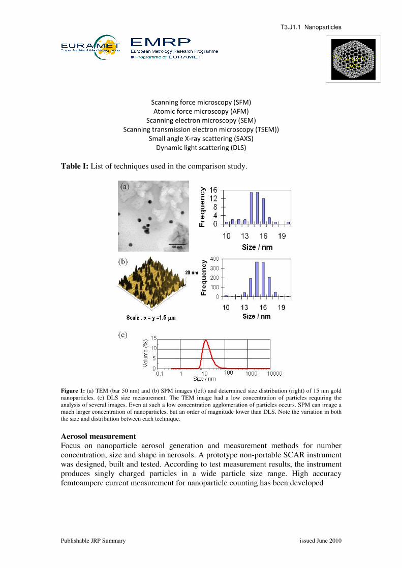
Figure 1: (a) TEM (bar 50 nm) and (b) SPM images (left) and determined size distribution (right) of 15 nm gold
nanoparticles. (c) DLS size measurement. The TEM image had a low concentration of particles requiring the
analysis of several images. Even at such a low concentration agglomeration of particles occurs. SPM can image a
much larger concentration of nanoparticles, but an order of magnitude lower than DLS. Note the variation in both
the size and distribution between each technique.
Aerosol measurement
Focus on nanoparticle aerosol generation and measurement methods for number
concentration, size and shape in aerosols. A prototype non-portable SCAR instrument
was designed, built and tested. According to test measurement results, the instrument
produces singly charged particles in a wide particle size range. High accuracy
femtoampere current measurement for nanoparticle counting has been developed
T3.J1.1 Nanoparticles
Publishable JRP Summary issued June 2010
High Aspect Ratio (HAR) Nanoparticles
High-aspect ratio nanoparticles and nanotubes are still a challenging topic for precise
measurement technology, since only spherical reference materials are available on the
market. This work package is devoted to the improvement of this situation and to
investigate options for an appropriate measurement technology and standardisation in
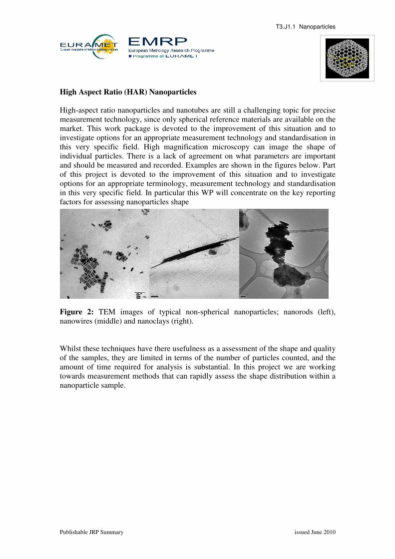
this very specific field. High magnification microscopy can image the shape of
individual particles. There is a lack of agreement on what parameters are important
and should be measured and recorded. Examples are shown in the figures below. Part
of this project is devoted to the improvement of this situation and to investigate
options for an appropriate terminology, measurement technology and standardisation
in this very specific field. In particular this WP will concentrate on the key reporting
factors for assessing nanoparticles shape
Figure 2: TEM images of typical non-spherical nanoparticles; nanorods (left),
nanowires (middle) and nanoclays (right).
Whilst these techniques have there usefulness as a assessment of the shape and quality
of the samples, they are limited in terms of the number of particles counted, and the
amount of time required for analysis is substantial. In this project we are working
towards measurement methods that can rapidly assess the shape distribution within a
nanoparticle sample.
0.2 �m0.2 �m
T3.J1.1 Nanoparticles
Publishable JRP Summary issued June 2010
JRP Contract Number: T3 J1.1
JRP Title - JRP Acronym: Nanoparticles
JRP start date and duration: 1 June 2008 (3 years)
Date of this
Publishable JRP Summary:
June 2010
JRP-Coordinator:
Name, Title, Organisation: Dr Alexandre Cuenat, Senior Research Scientist, National
Organisation, Country: Centro Español de Metrología, CEM, Spain; Czech Metrological Institute, CMI, CZ; Institutul National de Metrologie, INM, RO Istituto Nazionale di Ricerca Metrologica, INRIM, IT; Federal Office of Metrology, METAS, CH; Centre for Metrology and Accreditation, MIKES, FI;
Physikalisch-Technische Bundesanstalt, PTB, DE
The research within this EURAMET joint research project receives funding from the European Union
Seventh Framework Programme, ERA-NET Plus, under the iMERA-Plus Project – Grant Agreement