30
Trachea diseases and esophagus diseases The Otolaryngology Faculty of Shanghai Jiao Tong University School of Medicine Xiang Mingliang
| Date post: | 18-Dec-2015 |
| Category: |
Documents |
| Upload: | harold-lane |
| View: | 219 times |
| Download: | 3 times |
Trachea diseases and esophagus diseases
The Otolaryngology Faculty of Shanghai Jiao Tong University School
of Medicine
Xiang Mingliang
Anatomy of trachea and bronchus
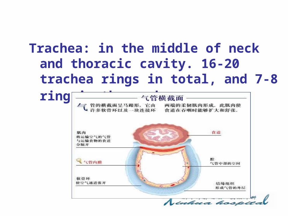
Trachea: in the middle of neck and thoracic cavity. 16-20 trachea rings in total, and 7-8 ring in the neck.
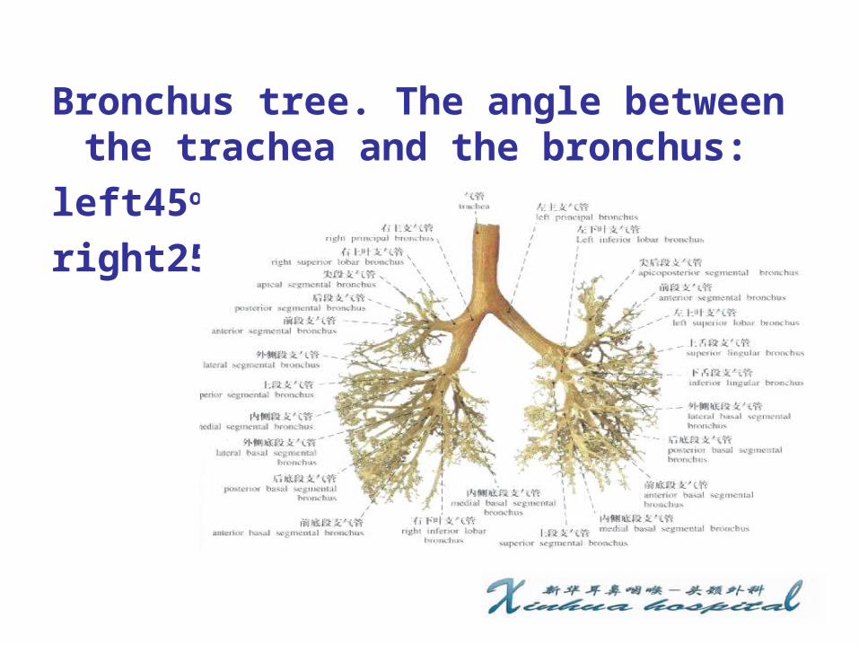
Bronchus tree. The angle between the trachea and the bronchus:
left45o,right25o
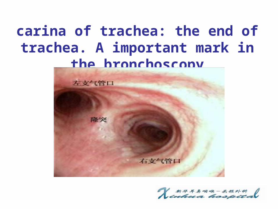
carina of trachea: the end of trachea. A important mark in
the bronchoscopy

Anatomy of esophagus
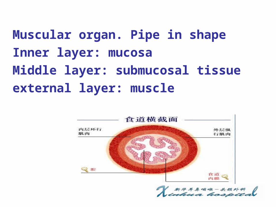
Muscular organ. Pipe in shapeInner layer: mucosaMiddle layer: submucosal tissue external layer: muscle

Physiological stenosis in esophagus
1 ) from upper incisor 16cm2 ) from upper incisor 23cm3 ) from upper incisor 27cm4 ) from upper incisor 36cm
foreign body in trachea and bronchus
Source: external、 internal
Classification of foreign body: plant: peanut、 seeds.
animal: fishbone、 bone pieces
mineral: iron nail
synthetic product: dental
prosthesis

90% cases﹤5 years, 80% cases ﹤ 3 years
causes: 1 ) poorly chew function,
poorly pharyngeal reflex
2 ) unconscious, aspiration
3) unhealthy habit: foreign body
in mouth
4 ) medical:
Factors associated with clinic presentation• foreign body sorts : peanut , chemical
bronchitis• Size:• Shape:• Stay time:• Stay site• Infection or not:
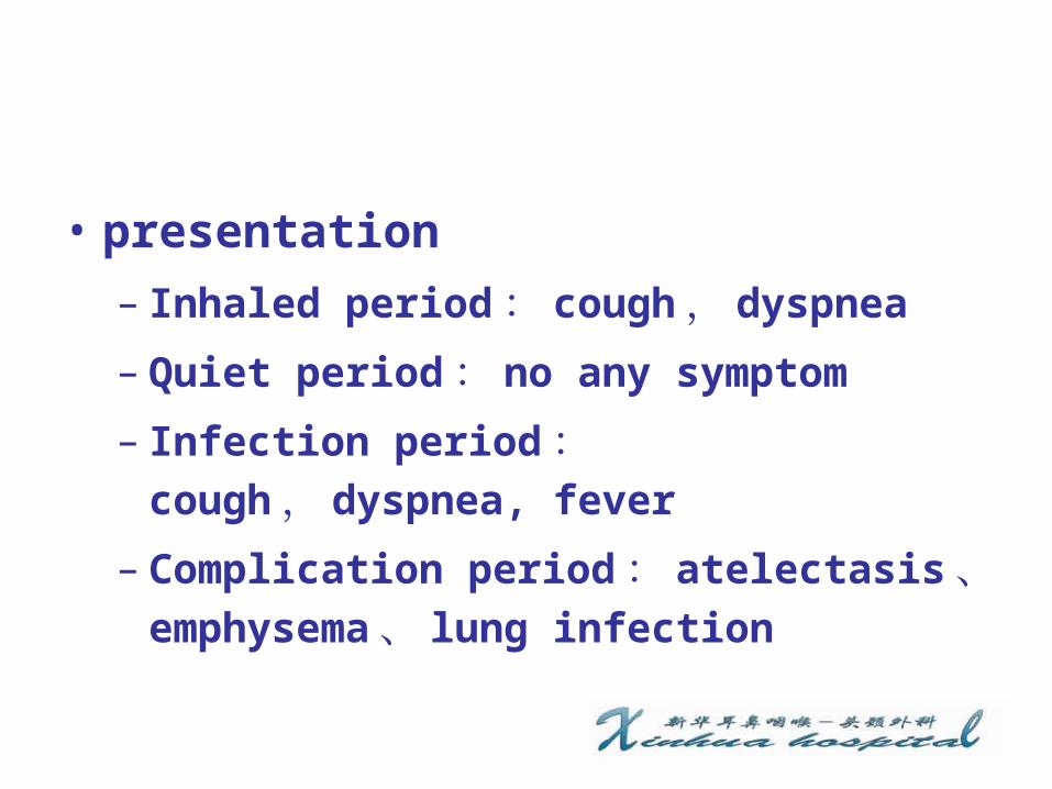
• presentation– Inhaled period: cough, dyspnea
– Quiet period: no any symptom
– Infection period: cough, dyspnea, fever
– Complication period: atelectasis、emphysema、 lung infection

diagnosis: history of foreign
body, dyspnea、 cough, disorder
sign in X-ray
X-ray: 1 ) obstructive emphysema
2 ) obstructive atelectasisCT scan: three-dimensional rebuild
treatment: bronchoscopy


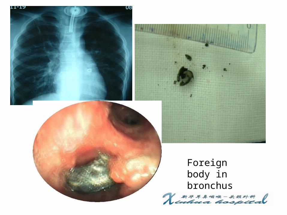
Foreign body in bronchus
Foreign body in bronchus

Foreign body in trachea

Foreign body in trachea

Foreign body in bronchus
Foreign body in esophagus
cause
• More cases in old: poorly chew function,
disesthesia of mouth, dental prosthesis
• More cases in children : like to put toy
in his mouth
• Eating too quick
• Stenosis or lump in esophagus
• suicide

Sorts of foreign body • animal : fishbone,bone pieces, meat lump
• metals : coin• synthetic product : dental prosthesis
• plant : kernel

Location of foreign body
• The first stenosis: most seen
• The second stenosis
• The third stenosis

Clinic symptoms •dysphagia•odynophagia•dyspnea : large foreign body

X-ray examination
nonopaque: positive in barium x-ray
study
opaque: x-ray flat. entopic and
lateral examination of esophagus

complication
• Esophagus perforation
• Infection of periesophagus organ :neck,mediastinum
• Large artery disruption
• Tracheal-esophageal fistula

diagnosis: history of foreign
body, dysphagia,odynophagia , diso
rder sign in X-ray
treatment:• esophagoscopy• Electronic gastroscopy• open: foreign body in out of esophagus,
injury of large artery

Foreign body in esophagus

Foreign body in esophagus

Thanks


![Diseases of the Lungs...August 1898.] DISEASES OF THE LUNGS.307 ing off more abruptly from the trachea than the left, is a mistake, arising from the fact that the eparterial bronchus](https://static.documents.pub/doc/80x56/5ee1f44ead6a402d666ca367/diseases-of-the-lungs-august-1898-diseases-of-the-lungs307-ing-off-more-abruptly.jpg)