Page 1
Tramtrack Protein - DNA Interactions: A Cross-Linking Study*
Dimitrii E. Kamashev§, AnnaV. Balandina, Vadim L. Karpov
From the Laboratory of Chromatin Structure and Function, W. Engelhardt Institute of
Molecular Biology, Russian Academy of Sciences, 32, Vavilov Str., 117984
MOSCOW, Russia.
RUNNING TITLE
Tramtrack Protein - DNA Interactions: A Cross-Linking Study
1
Copyright 2000 by The American Society for Biochemistry and Molecular Biology, Inc.
JBC Papers in Press. Published on August 29, 2000 as Manuscript M001691200 by guest on A
pril 8, 2018http://w
ww
.jbc.org/D
ownloaded from
Page 2
SUMMARY
Interaction of the Tramtrack protein from D. melanogaster with DNA was
analyzed by a cross-linking method. Tramtrack residues cross-linkable to the partly
depurinated DNA were identified by direct sequencing. The N-terminal α-amino
group of the protein DNA binding domain was found to be the major product of
cross-linking. The location of the N-terminus on the DNA was determined by
identification of the DNA bases which were cross-linked to the protein α-amino
group. We conclude that accessory N-terminal peptide preceding the first zinc finger
of Tramtrack directly interacts with DNA, both in specific and nonspecific DNA –
protein complexes. Our finding explains the role in the protein binding of the DNA
bases outside of the direct interaction with the zinc fingers.
2
by guest on April 8, 2018
http://ww
w.jbc.org/
Dow
nloaded from
Page 3
Cys2-His2 zinc fingers consist of a 30 amino acid residues sequence, which is
folded around a zinc ion to form a stable structure (1). Zinc-fingers are DNA binding
motifs and occur repeated in tandem. At least two adjacent zinc fingers appear to be
necessary for binding (2). The zinc finger is an independent folding motif (3)
consisting of a small β-sheet and an α-helix stabilized in a compact globular
structure by the bound zinc (4 - 6). Base-specific contacts are made in the major
groove of DNA and are mediated by amino acids in positions 1, 2, 3, and 6, of the α-
helical "reading head" (4 - 6). The role of the linkers between finger domains is not
as clear: linkers may make no contacts with DNA and do not affect on the relative
orientation of adjacent fingers (5), but in some cases they appear to participate in
DNA – protein interactions (6). In the case of at least three zinc finger proteins,
several amino acids N-terminal to the first finger are necessary for specific DNA
binding. For the yeast transcription factor ADR1, containing two zinc finger motifs, it
was demonstrated that residues outside zinc finger motif are important for DNA
binding (7 - 9). Eleven residues N-terminal to the formal start of the first finger motif
of yeast SWI5 protein are essential for stabilizing the folded form of finger 1 and for
DNA binding (3). Furthermore, seven residues N-terminal the first finger were shown
to be required for sequence-specific DNA-binding of Tramtrack protein (10).
To clarify the role of the N-terminal peptide preceding the zinc finger domain
we have studied the Tramtrack protein (Ttk1) from D. melanogaster. Ttk is involved in
the regulation of Drosophila embryonic development and it binds to at least four
DNA-binding sites (11, 12). Ttk is a sequence-specific DNA-binding protein, which
contains two adjacent zinc fingers. The 66-residue DNA - binding domain (TTK
3
by guest on April 8, 2018
http://ww
w.jbc.org/
Dow
nloaded from
Page 4
DBD) is able to bind DNA specifically (10). Seven residues N-terminal to the first
finger motif of TTK DBD are essential for DNA binding (10). The crystal structure of
the domain complexed with DNA is known (5). The overall three-dimensional
structure of the Ttk zinc fingers is the same as that of other zinc finger proteins.
Although three residues N-terminal to the conventional finger motif fold to form a
third strand to the β-sheet and lie too distant from DNA to interact directly with DNA
bases (5). Goal of our study was to investigate Ttk binding to DNA in solution and to
compare the results with the crystallographic data. This was accomplished by cross-
linking of TTK DBD with DNA, that was activated by partial depurination. Covalent
adduct formation between activated DNA and the protein implied that the amino acid
residue was in close proximity to the individual apurinic DNA site (13 - 16). We have
identified the traced contacts of TTK DBD on the level of amino acids and DNA base
pairs. It emerged that TTK DBD N-terminus cross-linked to the DNA base pairs
corresponding to both specific and non-specific binding. Thus, Ttk accessory peptide
N-terminal to the first zinc finger is involved in the direct interaction with DNA like
ADR1 (8, 9).
4
by guest on April 8, 2018
http://ww
w.jbc.org/
Dow
nloaded from
Page 5
EXPERIMENTAL PROCEDURES
Tramtrack protein--The 66 residue DNA-binding domain of the Tramtrack
protein was kindly supplied by Louise Fairall (10) and stored in 20 mM MES (pH 6.5),
100 mM NaCl, 0.1 mM ZnSO4, 0.5 mM PMSF, 50% glycerol at –20°C.
Methylation and partial depurination of DNA—As Ttk DNA binding site we
used 22 bp fragment of the #16 binding site from the ftz promoter upstream element
(5, 10, 11). Ttk DNA binding site was prepared by annealing of the gel-purified
complementary strands. Mild depurination of DNA includes DNA methylation by
dimethyl sulfate and spontaneous elimination of methylated purines. Double-
stranded DNA in 100 µl of 30 mM HEPES (pH 7.4), 50 mM NaCl, 0.1 mM EDTA was
methylated with 25 µl of dimethyl sulfate freshly dissolved in 500 mM HEPES. A final
concentration of dimethyl sulfate was 35 mM, reaction was carried out at 25°C for 30
min. DNA was precipitated to remove dimethyl sulfate and depurinated by incubation
at 42°C for 16h in 50 µl of 30 mM HEPES (pH 7.4), 50 mM NaCl, 0.1 mM EDTA.
Methylation rate was measured for 5’-labeled DNA by the cleavage at the
methylated base with 10% piperidine at 95°C for 30 min and analysis of the DNA
fragments on 20% denaturing polyacrylamide gel. Quantification of the bands
intensities allowed us to find the necessary methylation conditions. Ttk DNA binding
site of 22 bp was methylated with dimethyl sulfate so that 30% of the DNA molecule
had one, and only one, modified purine and less than 9% had more than one
modified purine.
Ttk cross-linking to partly depurinated DNA--DNA-protein cross-linking was
carried out in 100 µl of the binding buffer containing 20 mM MES (pH 6.5), 10 mM
5
by guest on April 8, 2018
http://ww
w.jbc.org/
Dow
nloaded from
Page 6
HEPES (pH 7.4), 100 mM NaCl, 0.05 mM ZnSO4, 0.1% NP40, 0.1mg/ml BSA, 2 mM
MgCl2, 0.1 mM PMSF, 10 % glycerol. One-end-labeled partly depurinated DNA was
incubated with TTK DBD at room temperature for 15 min and 200 mM pyridine-
borane complex dissolved in methanol was added to the final concentration 10 mM
(14, 16). After incubation for 1.5 h at 37°C generated cross-links were additionally
reduced with NaBH4 (final concentration of 0.5 mg/ml). To maintain pH, HEPES (pH
7.4) was added to 25 mM. Following to incubation at room temperature for 20 min, an
equal amount of NaBH4 and HEPES were added and reducing was carried out for an
extra 20 min.
Purification of cross-links and trypsin digestion--Reduced cross-links were
loaded onto 16% discontinuous polyacrylamide gel containing 0.1% SDS and 7M
urea. Radioactive bands were excised from the gel. DNA-linked protein was eluted
from the gel by incubation in 300 µl of 10 mM Tris-mercaptoacetic acid (pH 8.0),
0.1% Triton X100, 0.1 mM EDTA, 0.1 mM PMSF at 45°C for 4 h, eluant was
collected; then procedure was repeated under the same conditions and eluants were
pooled. Protein cross-linked to DNA was digested with 1 µg of trypsin (Boehlinger
Manheim, sequencing grade) at 37°C for 6 h in 100 mM NH4CO3 , 0.2% Triton X-
100, 1 mM dithiotreitol.
A+G sequencing reaction--The 5’-labeled DNA or DNA-linked peptides were
fragmented at purines by the treatment with 2% diphenylamine in 70% formic acid
aqueous solution at 37°C for 30 min. Mixtures were extracted by diethylether for five
times and dried. DNA fragments as well as DNA-linked peptides were analyzed on
20% polyacrylamide gel containing 7M urea, 90 mM Tris-borate and 0.2 mM EDTA.
6
by guest on April 8, 2018
http://ww
w.jbc.org/
Dow
nloaded from
Page 7
Exonuclease digestion--To digest 3’-end of peptide-linked oligonucleotides,
T4 DNA polymerase was used as 3’ -> 5’ exonuclease. Peptide-linked
oligonucleotides were treated with 0.2 units of polymerase in the buffer containing 50
mM Tris-Cl (pH 8.0), 10 mM MgCl2, 5 mM 2-mercaptoethanol and 0.2% Triton X-
100 at 37°C for 30 min. Products were precipitated and resolved in polyacrylamide
gel. Treatment of the peptide-linked oligonucleotides with DNA Polymerase I Klenow
fragment yields the same products (not shown).
Purification and sequencing of the linked peptides--Uniformly labeled
poly[d(AT)] was partly depurinated and cross-linked with TTK DBD as described
above. The DNA moiety of the linked complexes was digested with 2%
diphenylamine in 70% formic acid at 70°C for 70 min to a short radioactive
oligonucleotide tag covalently bound to the protein. After digestion with trypsin and
subsequent fractionating by gel-electrophoresis, the radioactive bands were eluted
from the gel as described above and treated with 0.5 units of alkaline phosphatase in
50 mM Tris-Cl, pH 8.0 at 37°C for 30 min. Resultant dephosphorylated nucleotide-
peptides were re-electrophoresed under the same conditions, eluted from the gel
and sequenced by Edman degradation.
7
by guest on April 8, 2018
http://ww
w.jbc.org/
Dow
nloaded from
Page 8
RESULTS
Ttk cross-linking to partly depurinated DNA binding site--In this work we
analyzed both specific and nonspecific Ttk - DNA complexes. To determine the
amino acid residues and DNA bases close to each other in Ttk - DNA complexes, we
used zero-length DNA - protein cross-linking. DNA activated by partial depurination
can bind covalently with the N-terminal α amino group, ε-amino group of lysines,
and the imidazole group of histidines (13, 14, 16). Cross-linking of these residues
within the DNA - protein complex is considered to provide direct evidence for their
proximity to the modified sites on DNA. Thus, the main goal of the in vitro cross-
linking studies is the identification of the amino acid residues cross-linked to DNA
and the positions of the cross-links on the DNA (15).
An end-labeled 22 bp DNA fragment containing the Ttk binding site was
methylated with dimethyl sulfate so that less than 9% of the DNA molecules had
more than one modified purine. Methylated DNA was depurinated by heating.
Elimination of purines causes formation of the aldehyde group on DNA that is able to
form cross-links with the protein residues (15). We verified that Tramtrack binding to
the partly depurinated DNA is still specific. The DNA was titrated by TTK DBD in
cross-linking buffer and complex formation was analyzed by gel-electrophoresis.
Band-shift assay (Fig. 1) shows that zinc-fingers of Ttk bind depurinated DNA
binding site as strong as non-modified one.
Partly depurinated DNA was incubated with TTK DBD as it described in
Experimental section and the generated cross-links were purified by electrophoresis
under denaturing conditions in a discontinuous SDS/urea-polyacrylamide gel. Fig.
2A shows that the major product of cross-linking was well separated from both non-
8
by guest on April 8, 2018
http://ww
w.jbc.org/
Dow
nloaded from
Page 9
cross-linked DNA and protein. The yield of the cross-linking was1-4%.
Identification of the purines cross-linked to Ttk--In order to separate different
possible amino acids of Ttk that were cross-linked to DNA, the DNA-linked protein
was digested with trypsin and the resultant DNA-linked peptides were separated by
gel-electrophoresis. Four radioactive bands containing Ttk peptides linked to 5’-
labeled full length strands of 22-bp DNA (Fig. 2B) were excised from the gel after
autoradiography and eluted.
In order to identify the positions of cross-linking on the DNA, 5’-labeled DNA
strands cross-linked to peptides were subjected to the A+G sequencing reaction.
The interpretation rests on the assumption that the sequencing ladder must be
interrupted at and above the position of the cross-linked purine, because of the
retardation imposed by a covalently-attached peptide moiety (Fig. 3). The retarded
bands migrate between the full-length (non-cleaved) DNA fragment linked to peptide
and the DNA fragment corresponding to the site of cross-linking.
Bands DA1 - DA4 and DB1 - DB4 presented in Fig. 2B may arise from the
peptides cross-linked to several purines on the DNA strand. In such a case,
sequencing pattern will be a superposition of the patterns corresponding to cross-
links through several purines. To separate them, the DNA-linked peptides were
digested with 3’ -> 5’ exonuclease (Klenow fragment of E. coli DNA polymerase I or
T4 DNA polymerase); the products were fractionated by gel-electrophoresis (Fig. 4)
and eluted from the gel. Exonuclease stops close to the site of cross-linking and the
number of the removed nucleotides depends on the distance between the 3’-end of
the strand and the site of cross-linking. In this way cross-links to the different DNA
sites can be separated. All peptides linked to strand A migrate after digestion with
9
by guest on April 8, 2018
http://ww
w.jbc.org/
Dow
nloaded from
Page 10
exonuclease as a single band (Fig. 4) and consequently peptides must be cross-
linked to a single DNA site. By contrast, peptides linked to strand B migrate after
treatment with exonuclease as several bands. Thus, they were cross-linked to
several sites of DNA strand B. The separated products of exonuclease digestion
were subjected to the A+G sequencing reaction, and electrophoresis were carried out
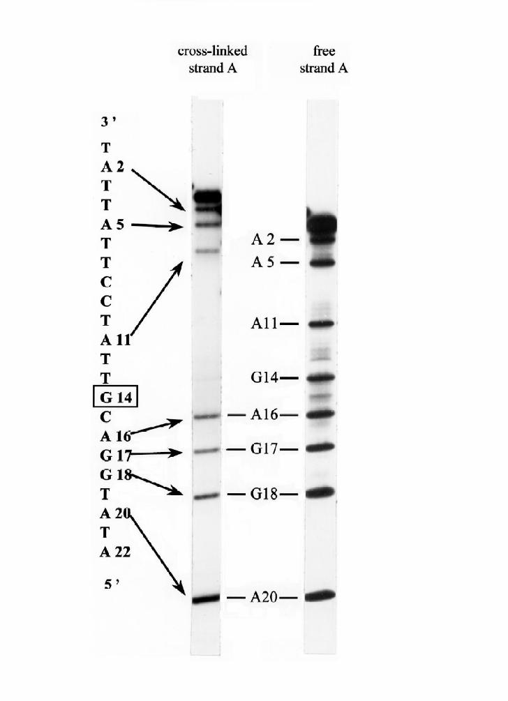
under denaturing conditions (Fig. 5). It can be seen that G14 of strand A is the first
"absent" purine in the sequencing ladders. Thus, G14 on strand A is the site where
all four peptides were cross-linked to strand A. DNA-linked peptide DB1 has four
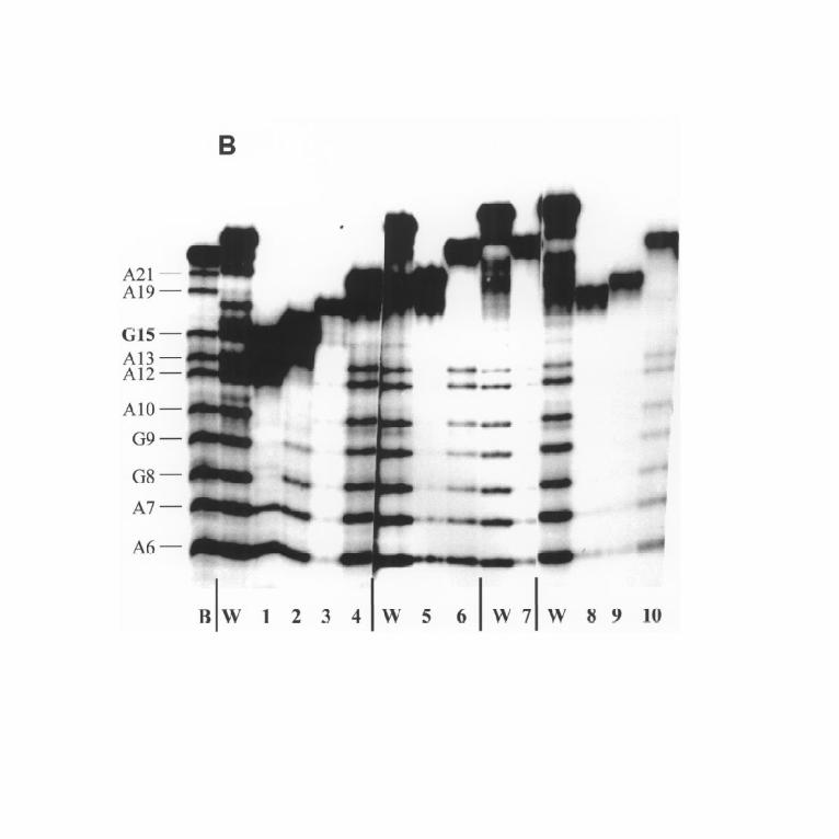
products of exonuclease digestion (marked 1-4 in Fig. 4, lane 8). The A+G
sequencing reaction, and subsequent electrophoresis shows that the products carry
cross-links through the sites G8, A10, A13,and G15 (Fig. 5B, lanes 1 - 4). DNA-
linked peptide DB2 has two products of digestion, both cross-linked through the site
G15 (Fig.5B, lanes 5 and 6). DB3 was transformed into the only peptide, cross-
linked through the site G15. Finally, DNA-linked peptide DB4 was cross-linked
through the sites G8 and G15. Thus, our approach make it possible to determine the
protein cross-linked DNA bases precisely, here we first used this technique.
Identification of Ttk amino acids cross-linkable to DNA--We would like to
compare the amino acids of Ttk cross-linkable to the different DNA sequences such
as specific and non-specific DNA sites. For this purpose we modified the method of
the cross-links analysis. The protein was cross-linked to DNA and DNA moiety of
the cross-linked complexes was digested with micrococcal nuclease, so that only a
short oligonucleotide tag is covalently bound to the protein (13, 16). The non-
digested DNA tag, that corresponds to the site of cross-linking was labeled with
32P-ATP and T4 polynucleotide kinase. Subsequently, the protein was digested with
10
by guest on April 8, 2018
http://ww
w.jbc.org/
Dow
nloaded from
Page 11
trypsin, and the resultant nucleotide-linked peptides were separated by gel-
electrophoresis and visualized by autoradiography (Fig. 6). This permits comparison
of the nucleotide-peptide patterns corresponding to Ttk complexed to DNA of
different lengths and sequences. The maps of cross-linked nucleotide-peptides were
obtained for complexes of TTK DBD with its binding site (lane 1, Fig. 6) and non-
specific sequences (lanes 2 and 3, Fig. 6). Nucleotide-peptide patterns of the non-
specific poly[d(AT)] and 49-bp DNA fragment and of the Ttk binding site are almost
identical.
To identify the cross-linked peptides, we prepared them in quantities sufficient
for direct sequencing. As cross-linking to specific DNA binding site and to poly[d(AT)]
results in the same nucleotied-peptides, the latter one was used for preparative
cross-linking. Uniformly labeled poly[d(AT)] was partly depurinated and cross-linked
with TTK DBD. The DNA moiety of the linked complexes was digested with formic
acid / diphenylamine to a short radioactive oligonucleotide tag covalently bound to
the protein and bearing 5’-phosphate. After digestion with trypsin and subsequent
fractionating by gel-electrophoresis, the radioactive bands were eluted from the gel
(Fig. 6) and treated with alkaline phosphatase. During re-electrophoresis under the
same conditions the nucleotide - peptides migrate slower than before the treatment
with phosphatase due to the loss of the 5’-phosphates in the oligonucleotide moiety
of the nucleotide-peptides (migration rate decrease is about 70% of the original
mobility, Fig. 6). Thus, nucleotide-peptides were purified not only from the other
nucleotide-peptides (during the first electrophoresis) but also from the non-labeled
peptides which were not retarded during the second electrophoresis. Radioactive
nucleotide-peptides were eluted from the gel and sequenced by Edman degradation.
11
by guest on April 8, 2018
http://ww
w.jbc.org/
Dow
nloaded from
Page 12
Peptides eluted from the bands N1, N2,and N3 are found to have the same sequence
?EFTK (residues 1 - 5 of TTK DBD), peptides from bands NN1 and NN2 (the latter is
not shown in Fig. 6) have also the same sequence ?EFTKEGEHTYR (residues 1 -
12). The question mark (?) stands for Met-1, which was not identified. This proves
that it is modified by cross-linking. Both peptide sequences contain possible amino
acids that can be cross-linked to sites of DNA depurination - Met-1, Lys-5 and His-
9. Since the two latter residues were identified on the peptide sequencing
chromatography, they were not cross-linked to DNA. Thus, TTK DBD N terminus is
the major product of cross-linking of the protein to partly depurinated DNA. The
finding that several peptides (marked NN) were the result of non-complete digestion
with trypsin explains why we had several bands corresponding to cross-linking
through a single amino acid.
12
by guest on April 8, 2018
http://ww
w.jbc.org/
Dow
nloaded from
Page 13
DISCUSSION
We have used DNA – protein cross-linking techniques (15) to study the
complex between the 66-residue DNA - binding domain of the Tramtrack protein
(TTK DBD) and both a specific and a non-specific DNA binding sites. In both cases
the major product of TTK DBD – DNA cross-linking was identified to be the N-
terminal α-amino group of the protein. We conclude that the N-terminus of the TTK
DBD is in the contact with DNA in both specific and non-specific complexes with
DNA. DNA was mildly methylated and depurinated to generate aldehyde groups that
are able to form Shiff bases (cross-links) with either N-terminal α-amino group of
proteins, ε-amino group of lysines, and imidazole groups of histidines (13, 14, 16).
Formation of such a zero-length cross-links provides evidence for the close vicinity
of the corresponding DNA and protein groups. In contrast, the absence of cross-links
between protein residue and DNA does not necessarily indicate that protein side
chains are remote from DNA (13, 15). The chemistry used here favors the formation
of cross-links preferentially through N-terminal α↑amino groups, then through
lysines, and to a less extent through histidines (14, 16).
Five bases of the specific Ttk - DNA binding site were found to contact the N-
terminus of the TTK DBD. The contact points can be divided into two sites on the
DNA sequence; one includes base pairs 13-15, and the other base pairs 8-10 (Fig.
7). If the protein had no particular position on DNA, its N-terminus should have
contacted all possible DNA base pairs, i.e., base pairs 1 - 22. Instead we obtained
only two sites of contact, corresponding to two specific positions of the protein on the
DNA, and, as expected, the location of the protein on the DNA is not random. Within
these two regions TTK DBD N-terminus does not contact only one base pair, and
13
by guest on April 8, 2018
http://ww
w.jbc.org/
Dow
nloaded from
Page 14
hence appears to be flexible, similarly to many other protein tails (9).
The N-terminus of the TTK DBD is residue 184 of the intact 68.5 kDa
Tramtrack protein (11). The sequence of the TTK DBD construct we studied starts
from mefTKEGEHT, with first three residues encoded by the vector. The
corresponding sequence in the intact protein is svrnycTKEGEHT. As can be seen,
the positively charged group of the arginine in the intact protein is replaced in the
construct with an N-terminal α-amino group. We speculate that in the full length
protein the arginine residue would make contact with the same bases of the DNA
binding site as does the N-terminus of the TTK DBD in our cross-linking studies.
The yield of cross-links is proportional to the length of time apurinic sites and
protein residues are in close contact. For the specific TTK DBD/DNA complex 90% of
the total cross-links involve base pairs 13-15 of binding site. Thus, the interaction of
the N-terminus of TTK DBD with base pairs 13-15 corresponds to the specific
complex. This observation is in agreement with the footprinting data - binding of the
TTK DBD protects these sites from the attack of hydroxyl radicals, methylation and
DNaseI cleavage (10). Base pairs 13-15 observed to be in contact with the N-
terminus of the TTK DBD fall within the minimal binding site of Ttk which retains full
binding affinity (corresponding to base pairs 5 -15 with an overhanging adenine at
each 5’ end (10), see Fig. 7). We attribute the interaction of the TTK DBD N-terminus
with base pairs 8-10 to non-specific one since: 1) it is minor (10%) product of the N
-termini cross-linking; 2) in the crystal structure these bases are involved in specific
contacts with residues from the zinc-finger “reading heads” (5). Thus, an interaction
of the TTK DBD N-terminus with bases 8-10 is only possible in the absence of
specific binding.
14
by guest on April 8, 2018
http://ww
w.jbc.org/
Dow
nloaded from
Page 15
Residues N-terminal to the formal start of the first zinc-finger have been
shown to play a role in the binding of certain zinc-finger proteins to DNA. Such
proteins contain relatively few, two or three, zinc finger motifs. For the yeast
transcription factor SWI5, eleven residues N-terminal to the first zinc-finger are
necessary for stable folding and the binding of the protein to DNA (3). The NMR
structure of SWI5 shows that a 15 residue region N-terminal to the finger motifs
forms part of the structure of the first finger domain adding a β↑strand and α-helix
(17). Comparison of methylation interference footprinting study of SWI5 containing 10
and 22 residues N-terminal the first zinc finger motif shows that the presence of
these residues enlarge the region of DNA protected by protein (17). Our finding that
the N-terminus of the TTK DBD interacts with the DNA bases remote from the
specific binding site of the zinc-finger domain with DNA is in agreement with these
data. Another example of the participation of the N-terminal tail of the zinc finger
protein in DNA binding comes from the yeast ADR1 protein. The minimal DNA
binding domain of the protein includes two zinc-fingers and an accessory sequence
of 20 residues N-terminal to the first finger motif (7). NMR studies of the protein (8, 9)
show that the N-terminal sequence is unstructured and highly flexible in the absence
of DNA. Upon binding to DNA the tail region becomes more protected from solvent
exchange and exhibits reduced motions. It was proposed that the N-terminal region
of ADR1 lies on the surface of DNA (8, 9).
For Tramtrack protein our conclusion is that the N-terminal region preceding
the first zinc-finger of Tramtrack is essential for DNA binding due to its direct
interaction with DNA at base pairs 13 – 15, 5’ to the sites of zinc fingers interactions
located at base pairs 7 –11 (Fig. 7). In the crystal structure of the TTK DBD - DNA
15
by guest on April 8, 2018
http://ww
w.jbc.org/
Dow
nloaded from
Page 16
complex, residues N-terminal to the first zinc-finger form a third strand to the β-
sheet and do not interact directly with DNA (5). The distance between G15 and the
closest side chain of the TTK DBD, exceeds 12 Å (5). Hence, the protein side chains
are too distant from G15 to make a contact with this base in TTK DBD - DNA
complex. Nevertheless it was clearly shown by the footprinting studies that G15
belongs to the minimal Ttk DNA binding site (10). This result is consistent with our
finding that the residues preceding first zinc finger make contact with this base. Our
data show that in solution the N-terminal region is unfolded and interacts with DNA
rather than with the first zinc- finger of Tramtrack. Tramtrack has several natural
binding sites within the ftz upstream enhanced element and zebra elements (10, 11,
12), where base pairs 13-15 are not conserved (see Fig. 7). Thus, interaction of
Tramtrack residues preceding the first zinc finger with base pairs 13 – 15 of DNA is
likely to be non-specific, contributing to affinity rather than to specificity. To prove the
idea that these base pairs being within Ttk minimal binding site do no contribute to
DNA recognition we checked the protein binding to Ttk binding site carrying
mutations. Wild-type base pairs were replaced with base pairs which were never
found within natural Tramtrack binding sites. Fig. 1 shows that substitution of base
pairs 13, 14, and 15 decrease the protein binding only twice (G15C) or trice (A13G,
and C14G) which indicates that DNA – protein interaction remains specific. We
believe that the interaction of the N-terminus of the TTK DBD with DNA contributes
to DNA binding by orientating the protein along DNA. This interaction also takes
place when the protein is bound to non-specific DNA and hence may play role in the
search for the specific binding site.
16
by guest on April 8, 2018
http://ww
w.jbc.org/
Dow
nloaded from
Page 17
LEGENDS TO FIGURES
Fig. 1. DNA binding assay for TTK DBD. Non-modified and partly depurinated
Ttk DNA binding site as well as Ttk DNA binding sites with indicated base
substitutions were tested for the protein binding. Increasing amount of TTK DBD was
incubated with 5‘-labeled 22 bp DNA fragments (1.5 nM) and the protein
concentrations were the same for each DNA. Free and bound DNA were separated
by electrophoresis in 8% polyacrylamide gel buffered with 45 mM Tris-borate (pH
8.3). DNA sequences are listed in Fig. 7.
Fig. 2. Detection and purification of TTK DBD cross-linked to the DNA. A, TTK
DBD was cross-linked to one-end-labeled 22-bp Ttk DNA binding site and
electrophoresed in SDS / urea discontinuous gel. Lane A, strand A is 5‘-labeled, lane
B, strand B is 5‘-labeled. “DNA” indicates position of non-linked DNA strands. Arrow
indicates position of non-linked TTK DBD revealed by Coomassie blue staining. B,
Separation of the DNA-linked peptides. DNA-linked protein was treated with trypsin,
and resultant DNA-linked peptides were separated by gel-electrophoresis. Lanes A
and B, 5‘-labeled strands A and B, respectively. Lanes TA and TB, tryptic digestion
of the major products of cross-linking through strand A and strand B, respectively.
Bands, which were then cut out from the gel, are marked.
Fig. 3. Experimental approach for identification of the cross-linked DNA
bases. Purified 5’-labeled DNA-linked peptide DA2 (see Fig. 2B) and 5’-labeled
DNA strand A were subjected to A+G reaction of sequencing protocol and gel-
17
by guest on April 8, 2018
http://ww
w.jbc.org/
Dow
nloaded from
Page 18
electrophoresed. Uninterrupted arrows show the non-retarded DNA fragments,
dotted arrows show the bands retarded by the linked peptide. Marked guanine was
identified as the site of cross-linking.
Fig. 4. Purification of the products of exonuclease digestion of the DNA-linked
peptides. DNA-linked peptides DA1 - DA4 ( lanes 1 - 4) and DB1 - DB4 (lanes 8, 7,
6, 5, respectively) were digested with 3’ -> 5’ exonuclease and gel-electrophoresed.
Lanes A and B, 5’-labeled strands A and B. Bands, which were then cut out from the
gel are marked for strand B.
Fig. 5. Identification of the sites of the protein cross-linking. A, Peptides DA1 -
DA4 linked to the strand A either digested (lanes 1 - 4) or non-digested (lanes 5 - 8)
with exonuclease were subjected to "A+G" reaction and gel-electrophoresed. Lanes
A, ladder of the strand A after "A+G" reaction. B, Peptides linked to the strand B either
digested (lanes 1 - 10) or non-digested (lanes W) with exonuclease were subjected
to "A+G" reaction and gel-electrophoresed. Lane B, ladder of the strand B after
"A+G" reaction.
Fig. 6. Mapping of the cross-linked nucleotide-peptides. TTK DBD was
cross-linked to 22-bp Ttk binding site, 49-bp nonspecific DNA and poly[d(AT)]
(lanes 1-3, respectively), digested with micrococcal nuclease, labeled at the site of
cross-linking and digested with trypsin. Lane T, TTK DBD cross-linked to uniformly
labeled poly[d(AT)], treated with 2% diphenylamine/ 70% formic acid at 70°C and
digested with trypsin. Lanes N1, N2, N3, and NN1, nucleotide-peptides after first
gel-electrophoresis of purification and dephosphorylation.
18
by guest on April 8, 2018
http://ww
w.jbc.org/
Dow
nloaded from
Page 19
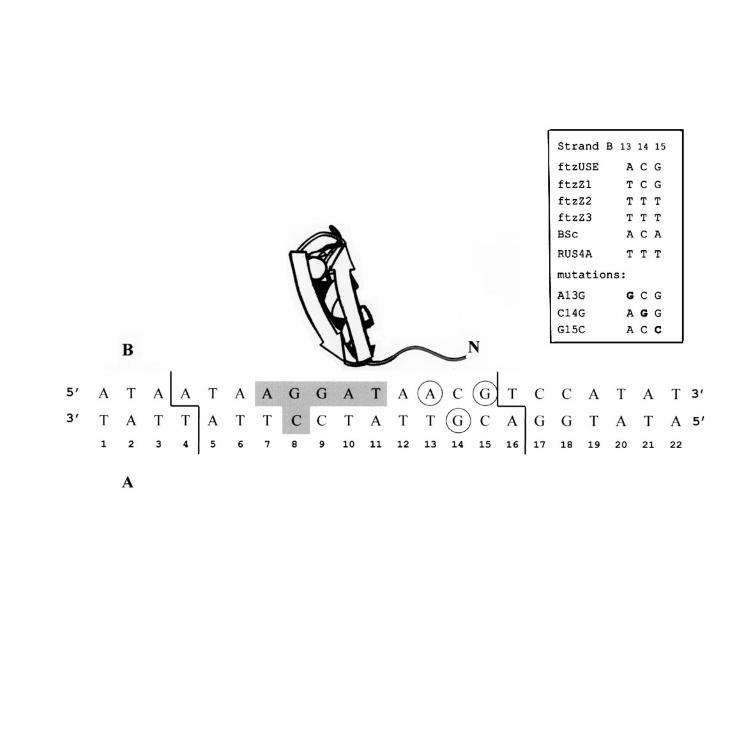
Fig. 7. Ttk zinc-finger 1 - DNA complex. The overall architecture of the
complex established by X-ray studies is taken from ref. (5). Sites of cross-linking of
TTK DBD N terminus corresponding to the specific complex are circled. Accessory
sequence of 10 residues N-terminal to the first finger was found to be turned to DNA.
Bases those makes specific contacts with Ttk zinc fingers (5) are shadowed, borders
of the minimal protein binding site (10) are marked. Right: base pairs found in
positions 13 – 15 of Ttk natural binding sites (11, 12, cited in 10) and their
substitutions studied in this work.
19
by guest on April 8, 2018
http://ww
w.jbc.org/
Dow
nloaded from
Page 20
REFERENCES
1. Miller J., McLachlan , A. D., and Klug A. (1985) EMBO J. 4, 1609-1614
2. Jacobs, G. H. (1992) EMBO J. 11, 4507-4517
3. Nakaseko, Y., Neuhaus, D., Rhodes, D., and Klug, A. (1992) J. Mol. Biol. 228,
619-636
4. Pavletich, N. P. and Pabo, C. O., (1991) Science 252, 809-817
5. Fairall, L., Schwabe, J. W. R., Chapman, L., Finch, J. T., and Rhodes D. (1993)
Nature 366, 483-487
6. Wuttke, D. S., Foster, M. P., Case, D. A., Gottesfeld, J. M., and Wright, P. E.
(1997) J. Mol. Biol. 273, 183-206
7. Thukral, S. K., Eisen, A., and Young, E. T. (1991) Mol. Cell. Biol. 11, 1566-1577
8. Schmiedeskamp, M., and Klevit, R. E. (1997) Biochemistry 36, 14003-14011
9. Hyre, D. E. and Klevit, R. E. (1998) J. Mol. Biol. 279, 929-943
10. Fairall, L., Harrison, S. D., Travers, A. A., Rhodes, D.(1992) J. Mol. Biol. 226,
349-366
11. Harrison, S. D. and Travers, A. A. (1990) EMBO J. 9, 207-216
12. Brown, J. L., Sonoda, S., Ueda, H., Scott, M. P., and Wu, C. (1991) EMBO J.
10, 665-674
13. Ebralidse, K. K., Grachev, S. A., and Mirzabekov, A. D. (1988) Nature 331,
365-367
14. Nacheva, G. A., Guschin, D. Yu., Preobrazhenskaya, O. V., Karpov, V. L.,
Ebralidse, K. K. and Mirzabekov A. D. (1989) Cell 58, 27-36
20
by guest on April 8, 2018
http://ww
w.jbc.org/
Dow
nloaded from
Page 21
15. Mirzabekov, A. D., Bavykin, S. G., Belyavsy, A. V.,Karpov, V. L.,
Preobrazhenskaya, O. V., Shick, V. V. and Ebralidse, K. K. (1989)
Methods Enzymol. 170, 386-407
16. Kamashev, D., Esipova, N. G., Ebralidse, K., and Mirzabekov, A. D. (1995)
FEBS Lett. 375, 27 – 30
17. Dutnall, R. N., Neuhaus, D., and Rhodes, D. (1996) Structure 4, 599-611
21
by guest on April 8, 2018
http://ww
w.jbc.org/
Dow
nloaded from
Page 22
FOOTNOTES
* This work was supported by grants 94-04-12344 and 96-04-50484 from Russian
Fund Fundamental Investigations. D.K. is a recipient of Short Term Fellowship of
EMBO. The costs of publication of this article were defrayed in part by the payment of
page charges. This article must therefore be hereby marked “advertisement” in
accordance with 18 U.S.C. Section 1734 solely to indicate this fact.
§ To whom correspondence should be addressed: Engelhardt Institute of Molecular
Biology, 32, Vavilov Str., 117984 MOSCOW, Russia. Fax: (7095) 135-14-05; E-
mail: [email protected]
1 The abbreviations used are: Ttk, D. melanogaster Tramtrack protein; TTK DBD,
Tramtrack DNA binding domain, PMSF, phenylmethylsulfonyl fluoride.
22
by guest on April 8, 2018
http://ww
w.jbc.org/
Dow
nloaded from
Page 23
ACKNOWLEGEMENTS
We thank Daniela Rhodes for stimulating discussions and carrying out major
part of this work in her laboratory in LMB/ MRC, Cambridge, UK and Louise Fairall for
the generous gift of TTK DBD.
23
by guest on April 8, 2018
http://ww
w.jbc.org/
Dow
nloaded from
Page 24
by guest on April 8, 2018
http://ww
w.jbc.org/
Dow
nloaded from
Page 25
by guest on April 8, 2018
http://ww
w.jbc.org/
Dow
nloaded from
Page 26
by guest on April 8, 2018
http://ww
w.jbc.org/
Dow
nloaded from
Page 27
by guest on April 8, 2018
http://ww
w.jbc.org/
Dow
nloaded from
Page 28
by guest on April 8, 2018
http://ww
w.jbc.org/
Dow
nloaded from
Page 29
by guest on April 8, 2018
http://ww
w.jbc.org/
Dow
nloaded from
Page 30
by guest on April 8, 2018
http://ww
w.jbc.org/
Dow
nloaded from
Page 31
by guest on April 8, 2018
http://ww
w.jbc.org/
Dow
nloaded from
Page 32
Dimitrii E. Kamashev, Anna V. Balandina and Vadim L. KarpovTramtrack Protein - DNA Interactions: A Cross-Linking Study
published online August 29, 2000J. Biol. Chem.
10.1074/jbc.M001691200Access the most updated version of this article at doi:
Alerts:
When a correction for this article is posted•
When this article is cited•
to choose from all of JBC's e-mail alertsClick here
by guest on April 8, 2018
http://ww
w.jbc.org/
Dow
nloaded from