+ All Categories
Home > Documents > Transarterial Sorafenib Chemoembolization: Preliminary Study of Technical Feasibility in a Rabbit...

Transarterial Sorafenib Chemoembolization: Preliminary Study of Technical Feasibility in a Rabbit...

Date post: 08-Dec-2016
Category:
Upload: nishant
View: 213 times
Download: 1 times
Share this document with a friend
7
LABORATORY INVESTIGATION Transarterial Sorafenib Chemoembolization: Preliminary Study of Technical Feasibility in a Rabbit Model Ron C. Gaba, MD, Felix Y. Yap, BSE, Elizabeth M. Martinez, BS, Yongchao Li, MS, Grace Guzman, MD, Ahmad Parvinian, BS, Richard B. van Breemen, PhD, and Nishant Kumar, MD ABSTRACT Purpose: To test the feasibility of targeted intraarterial administration of the tyrosine kinase inhibitor chemotherapeutic agent sorafenib to inhibit embolotherapy-induced tumor angiogenesis and reduce systemic drug side effects. Materials and Methods: The left hepatic lobes of five New Zealand White rabbits (mean weight, 2.7 kg 0.2) were treated with chemoembolization with sorafenib and ethiodized oil emulsion, followed by immediate euthanasia. Postprocedure noncontrast computed tomography (CT) was used to evaluate intrahepatic chemotherapy mixture distribution. Liquid chromatography/tandem mass spectrometry (LC-MS/MS) was then used to directly measure sorafenib concentration in the treated liver tissue. Histopathologic assessment of treated left lobes was performed to identify any immediate toxic effects of the sorafenib solution. Results: Lobar sorafenib chemoembolization was successfully performed in all cases via the left hepatic artery. Sorafenib and ethiodized oil (mean, 6.4 mg 3.8 and 0.95 mL 0.7, respectively) were injected, and CT confirmed targeted left hepatic lobe sorafenib emulsion delivery in all cases. Corresponding LC-MS/MS analysis yielded a mean sorafenib concentration of 94.2 mg/ mL 48.3 in treated left lobe samples (n ¼ 5), significantly greater than typical therapeutic drug levels (2–10 mg/mL) achieved with oral sorafenib systemic therapy. Histopathologic assessment showed only mild or moderate nonspecific ballooning degeneration in zone 3 hepatocytes, without tissue necrosis. Conclusions: Targeted transarterial sorafenib delivery is feasible and results in higher tissue drug levels than reported for systemic sorafenib therapy, without immediate histopathologic tissue toxicity. Future studies should aim to determine the utility of sorafenib chemoembolization in reducing hypoxia-induced vasculogenesis in liver tumors. ABBREVIATIONS C max = peak plasma concentration, HCC = hepatocellular carcinoma, HIF = hypoxia-inducible factor, LC-MS/MS = liquid chromatography/tandem mass spectrometry, SRM = selected reaction monitoring, UHPLC = ultra–high-pressure liquid chromatography, VEGF = vascular endothelial growth factor, VEGFR = vascular endothelial growth factor receptor Transarterial chemoembolization takes advantage of the hepatic arterial derivation of hepatocellular carcinoma (HCC) perfusion for targeted chemotherapeutic agent delivery and tumor devascularization (1). Although embolization of hepatic arteries supplying liver malignancies results in hypoxia and tumor necrosis, this process also induces angiogenesis (2). Induction of ischemia within liver neoplasms has been shown, in rabbit models, to result in increased intratumoral expression of hypoxia-inducible factor (HIF)–1a among residual surviving cells (3). When combined in cellular nuclei with HIF-1b, operational HIF-1 is formed and initiates a cascade of gene expression of proangiogenic factors and altered glucose metabolism to counteract nutritional deprivation incipient with hypoxia (2). The array of upregulated factors is diverse, but prominently features vascular endothelial growth factor (VEGF), which results in aggressive tumor ontogenesis and vascularity (4) & SIR, 2013 J Vasc Interv Radiol 2013; XX:]]]]]] http://dx.doi.org/10.1016/j.jvir.2013.01.488 None of the authors have identified a conflict of interest. From the Department of Radiology and Interventional Radiology Section (R.C.G., F.Y.Y., A.P., N.K.), Department of Medicinal Chemistry and Pharma- cognosy and College of Pharmacy (E.M.M., Y.L., R.B.v.B.), and Department of Pathology (G.G.), University of Illinois Hospital and Health Sciences System, 1740 W. Taylor St., MC 931, Chicago IL 60612 (G.G.). Received October 5, 2012; final revision received January 13, 2013; accepted January 14, 2013. Address correspondence to R.C.G.; E-mail: [email protected]
Transcript

LABORATORY INVESTIGATION

Transarterial Sorafenib Chemoembolization:Preliminary Study of Technical Feasibility in a Rabbit

Model

Ron C. Gaba, MD, Felix Y. Yap, BSE, Elizabeth M. Martinez, BS,Yongchao Li, MS, Grace Guzman, MD, Ahmad Parvinian, BS,

Richard B. van Breemen, PhD, and Nishant Kumar, MD

ABSTRACT

Purpose: To test the feasibility of targeted intraarterial administration of the tyrosine kinase inhibitor chemotherapeutic agent

sorafenib to inhibit embolotherapy-induced tumor angiogenesis and reduce systemic drug side effects.

Materials and Methods: The left hepatic lobes of five New Zealand White rabbits (mean weight, 2.7 kg � 0.2) were treated with

chemoembolization with sorafenib and ethiodized oil emulsion, followed by immediate euthanasia. Postprocedure noncontrast

computed tomography (CT) was used to evaluate intrahepatic chemotherapy mixture distribution. Liquid chromatography/tandem

mass spectrometry (LC-MS/MS) was then used to directly measure sorafenib concentration in the treated liver tissue. Histopathologic

assessment of treated left lobes was performed to identify any immediate toxic effects of the sorafenib solution.

Results: Lobar sorafenib chemoembolization was successfully performed in all cases via the left hepatic artery. Sorafenib and

ethiodized oil (mean, 6.4 mg � 3.8 and 0.95 mL � 0.7, respectively) were injected, and CT confirmed targeted left hepatic lobe

sorafenib emulsion delivery in all cases. Corresponding LC-MS/MS analysis yielded a mean sorafenib concentration of 94.2 mg/

mL � 48.3 in treated left lobe samples (n ¼ 5), significantly greater than typical therapeutic drug levels (2–10 mg/mL) achieved with

oral sorafenib systemic therapy. Histopathologic assessment showed only mild or moderate nonspecific ballooning degeneration in

zone 3 hepatocytes, without tissue necrosis.

Conclusions: Targeted transarterial sorafenib delivery is feasible and results in higher tissue drug levels than reported for systemic

sorafenib therapy, without immediate histopathologic tissue toxicity. Future studies should aim to determine the utility of sorafenib

chemoembolization in reducing hypoxia-induced vasculogenesis in liver tumors.

ABBREVIATIONS

Cmax = peak plasma concentration, HCC = hepatocellular carcinoma, HIF = hypoxia-inducible factor, LC-MS/MS = liquidchromatography/tandem mass spectrometry, SRM = selected reaction monitoring, UHPLC = ultra–high-pressure liquidchromatography, VEGF = vascular endothelial growth factor, VEGFR = vascular endothelial growth factor receptor

Transarterial chemoembolization takes advantage of the

hepatic arterial derivation of hepatocellular carcinoma

(HCC) perfusion for targeted chemotherapeutic agent

& SIR, 2013

J Vasc Interv Radiol 2013; XX:]]]–]]]

http://dx.doi.org/10.1016/j.jvir.2013.01.488

None of the authors have identified a conflict of interest.

From the Department of Radiology and Interventional Radiology Section

(R.C.G., F.Y.Y., A.P., N.K.), Department of Medicinal Chemistry and Pharma-

cognosy and College of Pharmacy (E.M.M., Y.L., R.B.v.B.), and Department

of Pathology (G.G.), University of Illinois Hospital and Health Sciences

System, 1740 W. Taylor St., MC 931, Chicago IL 60612 (G.G.). Received

October 5, 2012; final revision received January 13, 2013; accepted January

14, 2013. Address correspondence to R.C.G.; E-mail: [email protected]

delivery and tumor devascularization (1). Although

embolization of hepatic arteries supplying liver

malignancies results in hypoxia and tumor necrosis, this

process also induces angiogenesis (2). Induction of

ischemia within liver neoplasms has been shown, in

rabbit models, to result in increased intratumoral

expression of hypoxia-inducible factor (HIF)–1a among

residual surviving cells (3). When combined in cellular

nuclei with HIF-1b, operational HIF-1 is formed and

initiates a cascade of gene expression of proangiogenic

factors and altered glucose metabolism to counteract

nutritional deprivation incipient with hypoxia (2). The

array of upregulated factors is diverse, but prominently

features vascular endothelial growth factor (VEGF), which

results in aggressive tumor ontogenesis and vascularity (4)

Gaba et al ’ JVIR2 ’ Transarterial Sorafenib Chemoembolization in a Rabbit Model

and connotes increased tumoral propensity for metastasis

and invasive behavior (5). VEGF exerts its downstream

effects by interaction with a tyrosine kinase receptor,

designated VEGF receptor (VEGFR).

Receptor tyrosine kinase inhibitors are a class of drugs that

interrupt signaling pathways involved in tumor progression

and angiogenesis (6). In biochemical assays and murine

models, sorafenib (BAY 43-9006; Nexavar; Bayer,

Leverkusen, Germany) has been shown to potently inhibit

multiple receptor tyrosine kinases, including VEGFR

subtypes, involved in tumor angiogenesis. Immunohistologic

correlation with murine tumors in mice treated with sorafenib

demonstrates a decrease in tumoral microvessel density (6),

and in vitro assays and murine models have demonstrated

suppression of HCC cell proliferation and induction of

apoptosis in a dose-dependent manner (7). Clinically,

sorafenib has been used in the treatment of patients with

advanced HCC. Double-blind randomized controlled phase 3

clinical trials of sorafenib in such patients have shown delay

in time to progression and increase in overall survival (8,9).

However, the adverse effect profile of this agent, which is at

present commercially available only as an oral formulation

for systemic delivery, limits medication compliance, with

notable side effects including diarrhea and hand–foot syn-

drome that occur at a statistically significantly higher rate than

in placebo comparisons (8). To date, other routes of sorafenib

instillation have not been widely explored; the ability to

infuse sorafenib by using a transcatheter intraarterial route has

the potential to deliver high localized drug concentrations

directly to tumor to reduce the proangiogenic cascade

stimulated by hypoxic conditions precipitated during

chemoembolization while decreasing systemic drug side

effects. As such, the goal of the present project was to

assess the feasibility of transarterial hepatic delivery of a

lipid-emulsified preparation of sorafenib in a rabbit model.

MATERIALS AND METHODS

Animal care and use committee approval was obtained for

this prospective study. The experimental protocol consisted

of several steps: (i) production of a lipid-emulsified

sorafenib preparation, (ii) in vivo intravascular delivery

of the agent into New Zealand White rabbit livers,

(iii) noninvasive assessment of ethiodized oil and drug

emulsion delivery by computed tomography (CT) imaging,

(iv) liver explantation and liquid chromatography (LC)/

tandem mass spectrometry (LC-MS/MS) direct tissue

chemotherapeutic analysis to determine local drug concen-

trations, and (v) histopathologic analysis to assess for

possible toxic effects of sorafenib solution on hepatic

parenchyma.

Intraarterial Dosing and Preparation ofSorafenib SolutionIn devising a dosing regimen for intraarterial sorafenib,

peak plasma concentrations (Cmax) and therapeutic drug

levels for humans and rabbits were considered. In human

trials, the Cmax for sorafenib, as demonstrated in multiple

discontinuous (10) and continuous (11) dosing trials that

used the maximum tolerated dosage consisting of oral

sorafenib 400 mg twice daily (a regimen widely used

clinically), ranged from approximately 2 to 10 mg/mL;

these plasma concentrations represent therapeutic levels of

sorafenib, which inhibits components of the Raf–mitogen-

activated protein kinase kinase–extracellular signal-related

kinase signaling pathway and receptor tyrosine kinases at

concentrations ranging between 3 and 270 ng/mL (10). In

rabbits, plasma sorafenib levels approximating 5 mg/mL (a

concentration comparable to Cmax achieved in clinical

studies) may be reasonably attained by using an oral

dosing regimen of 30 mg/kg/d (Bayer, unpublished data,

February 2011). Considering that chemoembolization

generally results in local drug concentrations 10–100 times

greater than systemic administration 1), intraarterial dosing

was therefore empirically targeted at approximately 3 mg/

kg. The selected chemoembolization protocol consisted of a

6–12-mg/mL–concentration sorafenib solution emulsified

with an equal volume of ethiodized oil (Lipiodol; Guerbet,

Villepinte, France), and required injection of 1.5–3 mL total

chemotherapeutic emulsion volume for complete dose

administration in a 3.0-kg rabbit.

Sorafenib liquid solution formulation was prepared by

using a solvent of 12.5% Cremophor EL (Sigma-Aldrich,

St. Louis, Missouri), 12.5% ethyl alcohol (Sigma-Aldrich),

and 75% distilled water (Sigma-Aldrich) (6). For solution

preparation, sorafenib powder (provided by Bayer) was

dissolved in a 50% Cremophor EL and 50% ethyl alcohol

mixture at 12–24 mg/mL. Heating of the mixture to 601C

was necessary to get the sorafenib into solution. When the

compound was in solution, distilled water was added

gradually with mixing to generate the 6–12-mg/mL dosing

solution. The sorafenib solution was then allowed to cool

to room temperature before use in chemoembolization

procedures.

Transarterial Sorafenib ChemoembolizationFive male New Zealand White rabbits (mean weight, 2.7

kg � 0.2) underwent sorafenib chemoembolization proce-

dures, which were performed by a single operator (R.C.G.)

after rabbits were intubated and maintained under general

anesthesia (induction with intramuscular ketamine 50–65

mg/kg and intramuscular xylazine 5 mg/kg, maintenance

with inhaled isoflurane 1%–3%). Angiography was per-

formed with a C-arm unit (OEC Medical Systems series

9600; GE Healthcare, Milwaukee, Wisconsin). The femo-

ral artery was accessed through a surgical cutdown and

catheterized with a 3-F vascular sheath (Cook, Blooming-

ton, Indiana). A 2-F JB1 catheter (Cook) was advanced

over a guide wire, and the celiac artery was selectively

catheterized. The catheter was then advanced into the left

hepatic artery. Celiac and hepatic arteriography was

performed via injections of iohexol (Omnipaque-300;

Volume XX ’ Number X ’ Month ’ 2013 3

Amersham, Princeton, New Jersey). After catheter position

was confirmed in the left hepatic artery, selective left

hepatic lobe chemoembolization was performed by injec-

tion of a 1:1 volumetric emulsion of 6–12 mg/mL sorafenib

solution and ethiodized oil (Lipiodol; Guerbet). Under

fluoroscopic visualization, the chemotherapy emulsion

was injected by hand into the catheter. Chemoembolization

was performed to a static angiographic endpoint in all

cases, at which point administration of the chemotherapy

emulsion was ceased, regardless of administered volume.

Immediately after completion of chemoembolization, rab-

bits were euthanized by using a lethal intravenous dose of

390 mg/mL pentobarbital sodium solution (Schering-

Plough, Kenilworth, New Jersey).

Of note, a sixth rabbit (a 3.0-kg female) underwent

bland liver embolization with 12.5% Cremophor EL

(Sigma-Aldrich), 12.5% ethyl alcohol, and 75% distilled

water solution mixed with ethiodized oil in a 1:1 volu-

metric ratio for the purposes of serving as an experimental

control for histologic analysis. As in the chemoemboliza-

tion cohort, the emulsion was injected by hand into a

hepatic artery catheter under fluoroscopic visualization,

and the embolization was performed to a static angio-

graphic endpoint.

CT ImagingCT scans were obtained within 30 minutes of euthanasia

using a BrightSpeed 16 slice scanner (GE Healthcare) to

delineate the anatomic distribution of injected sorafenib

mixture, which was radiopaque as a result of incorporated

ethiodized oil. The CT protocol included helical acquis-

ition with a current of 300–400 mA, voltage of 120 kV,

pitch of 1.375:1, and 0.625-mm acquisition slice thickness.

Animal Necropsy and Tissue HarvestWithin 30 minutes of CT scan completion, rabbit necropsy

was performed and livers were harvested and processed for

LC-MS/MS sorafenib analysis as well as histopathologic

assessment. The explanted livers were separated into left

and right hepatic lobes. Hepatic tissue was harvested for

LC-MS/MS analysis; specimens consisted of two repre-

sentative 2-cm3 samples of treated left hepatic lobe

parenchyma, one from the left medial segment and one

from the left lateral segment, assuming homogeneous

distribution of sorafenib and ethiodized oil emulsion within

the left hepatic lobe as seen on CT imaging. Collected

specimens were stored in 1 mL of sterile saline solution

and were frozen in liquid nitrogen at �801C until the time

of LC-MS/MS analysis.

Measurement of Tissue SorafenibConcentrationA calibration curve was first created for the drug. Standard

sorafenib solutions ranging from 4 mg/mL to 200 mg/mL

(4, 8, 12.5, 25, 50, 100, and 200 mg/mL) were used to

create the standard curve, which was linear with an R2 of

0.999. Sample preparation was based on the method of

Romisch-Margl et al (12) and modified for the analysis

of sorafenib by using ultra–high-pressure LC/MS-MS

(UHPLC/MS-MS). Briefly, approximately 100 mg of liver

tissue was homogenized in phosphate buffer (10 mM, pH

7.4) containing 8% ethanol volumetric ratio (ie, vol/vol) to

give a homogenate containing 100 mg tissue per milliliter.

N-(4-phenoxyphenyl)-N0-phenylurea (Sigma-Aldrich) was

added at a concentration of 10 mg/mL as an internal

standard. Protein precipitation was carried out by using

100 mL of homogenized tissue by adding 400 mL of

acetonitrile/ethanol (4:1 vol/vol). After centrifugation at

12,000g for 20 minutes (41C), the supernatant was

removed and evaporated to dryness under a stream of nitro-

gen. The residue was reconstituted in 50 mL of methanol/

water (1:1 vol/vol).

UHPLC/MS-MS was carried out by using a Nexera

UHPLC system (Shimadzu, Kyoto, Japan) interfaced with

a model LCMS 8040 triple-quadrupole mass spectrometer

(Shimadzu). Separations were carried out by using a BEH

Shielded C18 column (2.0 mm � 50 mm; internal diame-

ter, 1.7 mm; Waters, Milford, Massachusetts) at 45 1C and a

1-minute gradient from 25% to 75% acetonitrile in 0.1%

aqueous formic acid. The injection volume was 1 mL, and

the flow rate was 0.6 mL/min. The column was reequili-

brated for 1 minute between injections. Sorafenib and

N-(4-phenoxyphenyl)-N0-phenylurea were measured by

using positive ion electrospray with selected reaction

monitoring (SRM) of the transitions from mass-to-charge

ratios of 465 to 252 and from 305 to 186, respectively. The

SRM dwell time was 10 ms per transition. Samples were

analyzed in triplicate.

Tissue Histopathologic AssessmentAfter removal of representative specimens for LC-MS/MS

analysis, the remainder of treated left lobes was fixed in

10% neutral buffered formalin solution, embedded in

paraffin, sectioned, and stained with hematoxylin and eosin

for histopathologic analysis. The reviewing pathologist

(G.G.) was blinded to the procedure performed, and

specimens were evaluated with the intent of ensuring that

the sorafenib solution, which contained a low concentration

of ethyl alcohol, did not exhibit immediate hepatotoxic

properties. Stained sections were examined at low power to

identify any regions of gross lobular ballooning degener-

ation or coagulative necrosis. Ballooning degeneration is a

form of hepatocyte injury characterized by cellular enlarge-

ment and paleness as a result of the presence of irregular

wispy or clumpy cytoplasm (13). This was followed by

high-power examination of hepatic lobules with particular

attention to zone 3 hepatocytes, central veins, and large

bile ducts. Hepatocyte ballooning degeneration, when

present, was graded as absent, mild, moderate, or severe,

and tissue necrosis, when present, was estimated by visual

inspection and expressed as a percentage of liver area for

each slice.

Figure 1. Sorafenib chemoembolization and CT imaging. Left hepatic arteriogram (a) from rabbit liver sorafenib chemoembolizationshows catheter tip (arrowhead) at left, where 8 mg sorafenib was administered. Subsequent CT image in the same rabbit (b) revealsleft hepatic lobe (white arrowheads) distribution of radiopaque chemotherapy emulsion (black arrowheads), without nontarget righthepatic lobe (arrows) delivery.

Gaba et al ’ JVIR4 ’ Transarterial Sorafenib Chemoembolization in a Rabbit Model

Statistical AnalysisStatistical analysis was implemented by using a commer-

cially available statistics program (SPSS Statistics version

17.0; SPSS, Chicago, Illinois). Results are reported as

means � standard deviation.

RESULTS

Sorafenib solution was easily prepared in 6–12-mg/mL

concentrations. Sorafenib chemoembolization was success-

fully performed in all five rabbits (Fig 1), and selective

administration was performed from the left hepatic artery

in all five cases. A mean of 6.4 mg � 3.8 of sorafenib was

administered, and a mean of 0.85 mL � 0.7 of ethiodized

oil was injected (Table). Postprocedure CT showed

targeted left hepatic lobe chemotherapy emulsion delivery

in all cases (Fig 1).

Tissue harvesting and processing was successfully per-

formed in all five chemoembolized rabbits. LC-MS/MS

analysis of the tissue specimens for sorafenib measurement

was technically successful in all cases (Table, Fig 2). Mean

overall tissue sorafenib concentration was 94.2 mg/mL � 48.3

among all five rabbits. Mean left medial and left lateral lobe

tissue sorafenib concentrations were 92.2 � 46.8 mcg/mL and

96.3 � 55.1 mcg/mL, respectively. Variability between

sorafenib levels in the left medial and left lateral liver

Table . Tissue Sorafenib Concentration and Histologic Analysis

Rabbit* Sorafenib Dose (mg) Ethiodized Oil Volume (mL)

1 3 0.5

2 8 2

3 12 1

4 6 0.5

5 3 0.25

Values presented as means � standard deviation where applicableLS ¼ lateral segment, MS ¼ medial segment.nAll animals received treatment to the left liver lobe.

segments was 9% � 7 (range, 1%–17%; Table). Tissue

histopathologic examination showed only mild (n ¼ 3) or

moderate (n ¼ 2) nonspecific ballooning degeneration in zone

3 hepatocytes (Table, Fig 3). No tissue necrosis was observed.

Of note, histologic findings in the vehicle control rabbit treated

with bland oily embolization comprised hepatocyte ballooning

degeneration. These findings were in line with histopathologic

changes reported in the literature, namely hepatocellular

degeneration (14), as well as the findings observed in the

sorafenib chemoembolization cohort.

DISCUSSION

The efficacy of transarterial chemoembolization is based at

least in part on the induction of tumoral hypoxia, which

prompts ischemic tumor necrosis and facilitates intracel-

lular transit of chemotherapeutic agents (15). Although

tumor response rates after chemoembolization are

generally favorable, treatment may be incomplete in as

many as 40% of cases, with such tumors showing partial

necrosis (16). In these instances, residual cancer cells are

able to contribute to the angiogenic diathesis that may play

a role in the inception of HCC recurrence. Hypoxia

stimulates HCC angiogenesis by promoting transcription

of VEGF (17), a potent mitogen that facilitates cellular

migration during angiogenesis. Clinically, increased serum

Histologic Alterations

Sorafenib Level (lg/mL)

MS LS

Zone 3 mild degeneration 51.1 � 9.1 44.1 � 3.7

Zone 3 mild degeneration 132.9 � 10.9 160.6 � 2.3

Zone 3 moderate degeneration 152.4 � 3.3 151.3 � 6.7

Zone 3 moderate degeneration 60.8 � 2.6 68.6 � 3.1

Zone 3 mild degeneration 63.6 � 3.4 60.6 � 6.7

.

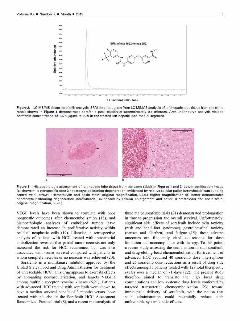
Figure 2. LC-MS/MS tissue sorafenib analysis. SRM chromatogram from LC-MS/MS analysis of left hepatic lobe tissue from the samerabbit shown in Figure 1 demonstrates sorafenib peak elution at approximately 0.4 minutes. Area-under-curve analysis yieldedsorafenib concentration of 132.9 mg/mL � 10.9 in the treated left hepatic lobe medial segment.

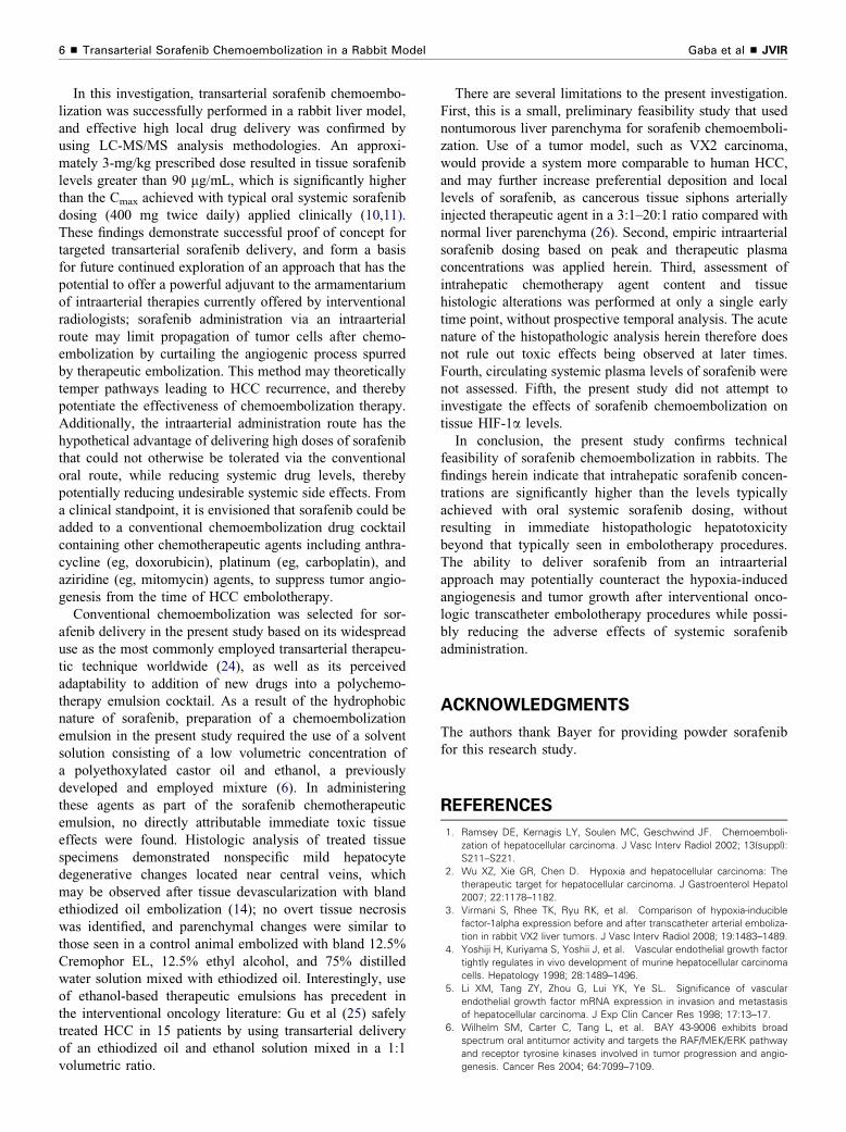
Figure 3. Histopathologic assessment of left hepatic lobe tissue from the same rabbit in Figures 1 and 2. Low-magnification image(a) shows mild nonspecific zone 3 hepatocyte ballooning degeneration, evidenced by relative cellular pallor (arrowheads) surroundingcentral vein (arrow). (Hematoxylin and eosin stain; original magnification, �2.5.) Higher magnification (b) better demonstrateshepatocyte ballooning degeneration (arrowheads), evidenced by cellular enlargement and pallor. (Hematoxylin and eosin stain;original magnification, � 20.)

Volume XX ’ Number X ’ Month ’ 2013 5

VEGF levels have been shown to correlate with poor

prognostic outcomes after chemoembolization (18), and

histopathologic analyses of embolized tumors have

demonstrated an increase in proliferative activity within

residual neoplastic cells (19). Likewise, a retrospective

analysis of patients with HCC treated with transarterial

embolization revealed that partial tumor necrosis not only

increased the risk for HCC recurrence, but was also

associated with worse survival compared with patients in

whom complete necrosis or no necrosis was achieved (20).

Sorafenib is a multikinase inhibitor approved by the

United States Food and Drug Administration for treatment

of unresectable HCC. This drug appears to exert its effects

by abrogating neovascularization, and targets VEGFR

among multiple receptor tyrosine kinases (6,21). Patients

with advanced HCC treated with sorafenib were shown to

have a median survival benefit of 3 months versus those

treated with placebo in the Sorafenib HCC Assessment

Randomized Protocol trial (8), and a recent metaanalysis of

three major sorafenib trials (21) demonstrated prolongation

in time to progression and overall survival. Unfortunately,

significant side effects of sorafenib include skin toxicity

(rash and hand–foot syndrome), gastrointestinal toxicity

(nausea and diarrhea), and fatigue (15); these adverse

outcomes are frequently cited as reasons for dose

limitation and noncompliance with therapy. To this point,

a recent study assessing the combination of oral sorafenib

and drug-eluting bead chemoembolization for treatment of

advanced HCC required 40 sorafenib dose interruptions

and 25 sorafenib dose reductions as a result of drug side

effects among 35 patients treated with 128 total therapeutic

cycles over a median of 71 days (22). The present study

therefore aimed to translate the high local drug

concentrations and low systemic drug levels conferred by

targeted transarterial chemoembolization (23) toward

intrahepatic delivery of sorafenib, with the notion that

such administration could potentially reduce such

unfavorable systemic side effects.

Gaba et al ’ JVIR6 ’ Transarterial Sorafenib Chemoembolization in a Rabbit Model

In this investigation, transarterial sorafenib chemoembo-

lization was successfully performed in a rabbit liver model,

and effective high local drug delivery was confirmed by

using LC-MS/MS analysis methodologies. An approxi-

mately 3-mg/kg prescribed dose resulted in tissue sorafenib

levels greater than 90 mg/mL, which is significantly higher

than the Cmax achieved with typical oral systemic sorafenib

dosing (400 mg twice daily) applied clinically (10,11).

These findings demonstrate successful proof of concept for

targeted transarterial sorafenib delivery, and form a basis

for future continued exploration of an approach that has the

potential to offer a powerful adjuvant to the armamentarium

of intraarterial therapies currently offered by interventional

radiologists; sorafenib administration via an intraarterial

route may limit propagation of tumor cells after chemo-

embolization by curtailing the angiogenic process spurred

by therapeutic embolization. This method may theoretically

temper pathways leading to HCC recurrence, and thereby

potentiate the effectiveness of chemoembolization therapy.

Additionally, the intraarterial administration route has the

hypothetical advantage of delivering high doses of sorafenib

that could not otherwise be tolerated via the conventional

oral route, while reducing systemic drug levels, thereby

potentially reducing undesirable systemic side effects. From

a clinical standpoint, it is envisioned that sorafenib could be

added to a conventional chemoembolization drug cocktail

containing other chemotherapeutic agents including anthra-

cycline (eg, doxorubicin), platinum (eg, carboplatin), and

aziridine (eg, mitomycin) agents, to suppress tumor angio-

genesis from the time of HCC embolotherapy.

Conventional chemoembolization was selected for sor-

afenib delivery in the present study based on its widespread

use as the most commonly employed transarterial therapeu-

tic technique worldwide (24), as well as its perceived

adaptability to addition of new drugs into a polychemo-

therapy emulsion cocktail. As a result of the hydrophobic

nature of sorafenib, preparation of a chemoembolization

emulsion in the present study required the use of a solvent

solution consisting of a low volumetric concentration of

a polyethoxylated castor oil and ethanol, a previously

developed and employed mixture (6). In administering

these agents as part of the sorafenib chemotherapeutic

emulsion, no directly attributable immediate toxic tissue

effects were found. Histologic analysis of treated tissue

specimens demonstrated nonspecific mild hepatocyte

degenerative changes located near central veins, which

may be observed after tissue devascularization with bland

ethiodized oil embolization (14); no overt tissue necrosis

was identified, and parenchymal changes were similar to

those seen in a control animal embolized with bland 12.5%

Cremophor EL, 12.5% ethyl alcohol, and 75% distilled

water solution mixed with ethiodized oil. Interestingly, use

of ethanol-based therapeutic emulsions has precedent in

the interventional oncology literature: Gu et al (25) safely

treated HCC in 15 patients by using transarterial delivery

of an ethiodized oil and ethanol solution mixed in a 1:1

volumetric ratio.

There are several limitations to the present investigation.

First, this is a small, preliminary feasibility study that used

nontumorous liver parenchyma for sorafenib chemoemboli-

zation. Use of a tumor model, such as VX2 carcinoma,

would provide a system more comparable to human HCC,

and may further increase preferential deposition and local

levels of sorafenib, as cancerous tissue siphons arterially

injected therapeutic agent in a 3:1–20:1 ratio compared with

normal liver parenchyma (26). Second, empiric intraarterial

sorafenib dosing based on peak and therapeutic plasma

concentrations was applied herein. Third, assessment of

intrahepatic chemotherapy agent content and tissue

histologic alterations was performed at only a single early

time point, without prospective temporal analysis. The acute

nature of the histopathologic analysis herein therefore does

not rule out toxic effects being observed at later times.

Fourth, circulating systemic plasma levels of sorafenib were

not assessed. Fifth, the present study did not attempt to

investigate the effects of sorafenib chemoembolization on

tissue HIF-1a levels.

In conclusion, the present study confirms technical

feasibility of sorafenib chemoembolization in rabbits. The

findings herein indicate that intrahepatic sorafenib concen-

trations are significantly higher than the levels typically

achieved with oral systemic sorafenib dosing, without

resulting in immediate histopathologic hepatotoxicity

beyond that typically seen in embolotherapy procedures.

The ability to deliver sorafenib from an intraarterial

approach may potentially counteract the hypoxia-induced

angiogenesis and tumor growth after interventional onco-

logic transcatheter embolotherapy procedures while possi-

bly reducing the adverse effects of systemic sorafenib

administration.

ACKNOWLEDGMENTS

The authors thank Bayer for providing powder sorafenib

for this research study.

REFERENCES

1. Ramsey DE, Kernagis LY, Soulen MC, Geschwind JF. Chemoemboli-

zation of hepatocellular carcinoma. J Vasc Interv Radiol 2002; 13(suppl):

S211–S221.

2. Wu XZ, Xie GR, Chen D. Hypoxia and hepatocellular carcinoma: The

therapeutic target for hepatocellular carcinoma. J Gastroenterol Hepatol

2007; 22:1178–1182.

3. Virmani S, Rhee TK, Ryu RK, et al. Comparison of hypoxia-inducible

factor-1alpha expression before and after transcatheter arterial emboliza-

tion in rabbit VX2 liver tumors. J Vasc Interv Radiol 2008; 19:1483–1489.

4. Yoshiji H, Kuriyama S, Yoshii J, et al. Vascular endothelial growth factor

tightly regulates in vivo development of murine hepatocellular carcinoma

cells. Hepatology 1998; 28:1489–1496.

5. Li XM, Tang ZY, Zhou G, Lui YK, Ye SL. Significance of vascular

endothelial growth factor mRNA expression in invasion and metastasis

of hepatocellular carcinoma. J Exp Clin Cancer Res 1998; 17:13–17.

6. Wilhelm SM, Carter C, Tang L, et al. BAY 43-9006 exhibits broad

spectrum oral antitumor activity and targets the RAF/MEK/ERK pathway

and receptor tyrosine kinases involved in tumor progression and angio-

genesis. Cancer Res 2004; 64:7099–7109.

Volume XX ’ Number X ’ Month ’ 2013 7

7. Liu L, Cao Y, Chen C, et al. Sorafenib blocks the RAF/MEK/ERK

pathway, inhibits tumor angiogenesis, and induces tumor cell apoptosis

in hepatocellular carcinoma model PLC/PRF/5. Cancer Res 2006; 66:

11851–11858.

8. Llovet JM, Ricci S, Mazzaferro V, et al. Sorafenib in advanced

hepatocellular carcinoma. N Engl J Med 2008; 359:378–390.

9. Keating GM, Santoro A. Sorafenib: a review of its use in advanced

hepatocellular carcinoma. Drugs 2009; 69:223–240.

10. Strumberg D, Clark JW, Awada A, et al. Safety, pharmacokinetics, and

preliminary antitumor activity of sorafenib: a review of four phase I trials

in patients with advanced refractory solid tumors. Oncologist 2007; 12:

426–437.

11. Strumberg D, Richly H, Hilger RA, et al. Phase I clinical and pharma-

cokinetic study of the novel Raf kinase and vascular endothelial growth

factor receptor inhibitor BAY 43-9006 in patients with advanced refrac-

tory solid tumors. J Clin Oncol 2005; 23:965–972.

12. Romisch-Margl W, Prehn C, Bogumil R, Rohring C, Suhre K, Adamski

J. Procedure for tissue sample preparation and metabolite extraction

for high-throughput targeted metabolomics. Metabolomics 2012; 8:

133–142.

13. Yerian L. Histopathological evaluation of fatty and alcoholic liver

diseases. J Dig Dis 2011; 12:17–24.

14. Akashi Y, Koreeda C, Mizuno T, Inoue K, Kawa SK, Tanaka Y. Hepatic

parenchymal changes after the intraarterial injection of lipiodol in tumor-

bearing rabbits. Invest Radiol 1993; 28:128–132.

15. Lewandowski RJ, Geschwind JF, Liapi E, Salem R. Transcatheter

intraarterial therapies: rationale and overview. Radiology 2011; 259:

641–657.

16. Lewandowski RJ, Mulcahy MF, Kulik LM, et al. Chemoembolization for

hepatocellular carcinoma: comprehensive imaging and survival analysis

in a 172-patient cohort. Radiology 2010; 255:955–965.

17. Kim KR, Moon HE, Kim KW. Hypoxia-induced angiogenesis in human

hepatocellular carcinoma. J Mol Med (Berl) 2002; 80:703–714.

18. Poon RT, Lau C, Yu WC, Fan ST, Wong J. High serum levels of vascular

endothelial growth factor predict poor response to transarterial chemo-

embolization in hepatocellular carcinoma: a prospective study. Oncol Rep

2004; 11:1077–1084.

19. Kim YB, Park YN, Park C. Increased proliferation activities of vascular

endothelial cells and tumour cells in residual hepatocellular carcinoma

following transcatheter arterial embolization. Histopathology 2001; 38:

160–166.

20. Adachi E, Matsumata T, Nishizaki T, Hashimoto H, Tsuneyoshi M,

Sugimachi K. Effects of preoperative transcatheter hepatic arterial che-

moembolization for hepatocellular carcinoma. The relationship between

postoperative course and tumor necrosis. Cancer 1993; 72:3593–3598.

21. Zhang T, Ding X, Wei D, et al. Sorafenib improves the survival of

patients with advanced hepatocellular carcinoma: a meta-analysis of

randomized trials. Anticancer Drugs 2010; 21:326–332.

22. Pawlik TM, Reyes DK, Cosgrove D, Kamel IR, Bhagat N, Geschwind

JF. Phase II trial of sorafenib combined with concurrent transarterial

chemoembolization with drug-eluting beads for hepatocellular carci-

noma. J Clin Oncol 2011; 29:3960–3967.

23. Raoul JL, Heresbach D, Bretagne JF, et al. Chemoembolization of

hepatocellular carcinomas. A study of the biodistribution and pharmaco-

kinetics of doxorubicin. Cancer 1992; 70:585–590.

24. Satake M, Uchida H, Arai Y, et al. Transcatheter arterial chemoembo-

lization (TACE) with lipiodol to treat hepatocellular carcinoma: survey

results from the TACE study group of Japan. Cardiovasc Intervent Radiol

2008; 31:756–761.

25. Gu YK, Luo RG, Huang JH, Si QJ, Li XX, Gao F. Transarterial

embolization ablation of hepatocellular carcinoma with a lipiodol-ethanol

mixture. World J Gastroenterol 2010; 16:5766–5772.

26. Kennedy AS, Nutting C, Coldwell D, Gaiser J, Drachenberg C. Patho-

logic response and microdosimetry of (90)Y microspheres in man: review

of four explanted whole livers. Int J Radiat Oncol Biol Phys 2004; 60:

1552–1563.


Recommended