Open Access Journal of Pharmaceutical Research ISSN: 2574-7797 Transferosomes for Trans-Nasal Brain Delivery of Clonazepam: Preparation, Optimization, Ex-Vivo Cytotoxicity and Pharmacodynamic Study J Pharm Res Transferosomes for Trans-Nasal Brain Delivery of Clonazepam: Preparation, Optimization, Ex-Vivo Cytotoxicity and Pharmacodynamic Study Nour SA*, Abdelmalak NS and Naguib MJ Department of Pharmaceutics, Faculty of Pharmacy, Cairo University, Cairo, Egypt *Corresponding author: Samia A Nour, Department of Pharmaceutics, Faculty of Pharmacy, Cairo University, Cairo, Egypt, Email: [email protected]Abstract Clonazepam (CZ) is a benzodiazepine derivative that is used mainly in the treatment of status epilepticus (SE). Intranasal (IN) administration of nano-carrier systems generally have the ability to deliver the drug to the brain through olfactory and trigeminal nerves pathways circumventing the blood brain barrier (BBB). The aim of the present work was to enhance intranasal (IN) brain delivery of clonazepam in an attempt to treat status epileticus (SE). In order to achieve this goal, clonazepam transferosomes (TF) were prepared via thin film hydration technique (TFH) adopting 2 2 X 3 1 full factorial design (FFD). The twelve formulae were evaluated in terms of entrapment efficiency (EE), particle size (PS), polydispersity index (PDI), zeta potential (ZP) and in-vitro release. TF1 prepared using sodium deoxycholate (SDC) as edge activator (EA), lipid to edge activator molar ratio of 10:1 and 10 mg as initial drug amount showed the highest desirability value of 0.833. The optimized formula showed minor changes to sheep nasal mucosa upon ex-vivo cytotoxicity study and significant delay in the onset of pentylenetetrazole (PTZ) induced seizures. The declared results reveal the ability of the developed TF to be a strong potential candidate for the emergency treatment of SE. Keywords: Intranasal; Transferosomes; Clonazepam; Cytotoxicity; Pentylenetetrazole. Abbreviations: TEM-Transmission Electron Microscopy; BBB-Blood Brain Barrier; CMC-Critical Micelle Concentration; DSC-Differential Scanning Calorimetry; EE-Entrapment Efficiency Introduction Epilepsy represents a group of brain disorders which have in common the occurrence of spontaneous and recurring epileptic seizures i.e., hyper synchronous electrical discharge in brain network [1]. Status epilepticus (SE) is a medical emergency that is associated with high morbidity and mortality [2]. It is defined as a prolonged seizure or multiple seizures with incomplete return to baseline [3]. Neuronal damage in SE results from sustained NMDA-mediated neuronal stimulation which leads to apoptosis [4]. When these neuronal cells are depolarized, the Mg 2+ ions diffuses outward, allowing sodium ions and Ca 2+ to flood the cell, resulting in a cascade of Ca +2 -mediated cytotoxic events, leading to neuronal injury, cell lysis, and cell death [5]. Treatment of SE should aim to immediately terminate the seizures with prevention of their recurrence [6]. Research Article Volume 1 Issue 2 Received Date: February 27, 2017 Published Date: March 09, 2017
Transcript
Open Access Journal of Pharmaceutical Research
ISSN: 2574-7797
Transferosomes for Trans-Nasal Brain Delivery of Clonazepam: Preparation, Optimization, Ex-Vivo Cytotoxicity and Pharmacodynamic Study J Pharm Res
Epilepsy represents a group of brain disorders which have in common the occurrence of spontaneous and recurring epileptic seizures i.e., hyper synchronous electrical discharge in brain network [1]. Status epilepticus (SE) is a medical emergency that is associated
with high morbidity and mortality [2]. It is defined as a prolonged seizure or multiple seizures with incomplete return to baseline [3]. Neuronal damage in SE results from sustained NMDA-mediated neuronal stimulation which leads to apoptosis [4]. When these neuronal cells are depolarized, the Mg 2+ ions diffuses outward, allowing sodium ions and Ca 2+ to flood the cell, resulting in a cascade of Ca +2-mediated cytotoxic events, leading to neuronal injury, cell lysis, and cell death [5]. Treatment of SE should aim to immediately terminate the seizures with prevention of their recurrence [6].
Research Article
Volume 1 Issue 2
Received Date: February 27, 2017
Published Date: March 09, 2017
Open Access Journal of Pharmaceutical Research
Nour SA et al. Transferosomes for Trans-Nasal Brain Delivery of Clonazepam: Preparation, Optimization, Ex-Vivo Cytotoxicity and Pharmacodynamic Study. J Pharm Res 2017, 1(2): 000107.
Clonazepam (CZ) is a nitro benzodiazepine derivative. Its structure is 5-(2-Chlorophenyl)-7-nitro-2,3-dihydro-1,4-benzodiazepin-2-one. It acts via modulating gamma-amino-butyric acid (GABA) function in the brain, via the benzodiazepine receptor which in turn leads to enhanced GABA ergic inhibition of neuronal firing. In addition clonazepam decreases the utilization of 5-HT (serotonin) by neurons and has been shown to bind tightly to central type benzodiazepine receptors [7]. It is said to be among the class of “highly potent” benzodiazepines. Clonazepam is available as tablet dosage forms, with peak plasma concentration between 1 and 4 hours after ingestion [8]. It is extensively metabolised in the liver, its principal metabolite being 7-aminoclonazepam, which has no antiepileptic activity. It is about 85% bound to plasma proteins and estimations of its elimination half-life range from about 20 to 40 hours [9]. As a highly lipophilic molecule and highly protein bound, CZ is characterized by high systemic distribution in fat leading to adverse effects such as palpitation, hair loss and anorexia among others [10]. Drug delivery to the brain is a challenge because of the presence of the blood brain barrier (BBB). Intranasal administration of the neurotherapeutics results into direct delivery of the drug to the brain circumventing the BBB. This route is generally non-invasive, convenient, self-administered, along with rapid drug absorption leading to quick onset of action [11]. On the other hand, oral or intravenous drug delivery is inconvenient and impractical for the control of convulsions due to patient’s inability to swallow and the need for qualified personnel for intravenous administration. Transferosomes (TF) are highly adaptable and stress-responsive, complex aggregates [12]. Each transfersome consists of at least one inner aqueous compartment, which is surrounded by a lipid bilayer with specially tailored properties [13] and a bilayer softening component (such as a biocompatible surfactant) that increases lipid bilayer flexibility and permeability [14]. They have been used to target olanzapine directly to the brain after intranasal application according to the work of
Salama et al. [15]. The results demonstrated the ability of the developed nanocarriers for brain targeting of the drug. Hence, the aim of the present study was to develop CZ loaded TF intended for brain delivery of the drug via the intranasal route in an attempt for immediate control of SE.
Materials and Methods
Materials
Clonazepam (CZ) was a gift from Amoun Pharmaceuticals ( Elobour city- Egypt), Sodium deoxycholate (SDC) was purchased from Sigma Chemical Co. (USA), Labrafil (LB) was a gift from Gattefosse (France), Phospholipon 90G was obtained as a gift from Lipiod GmbH (Ludwigshafen, Germany). Spectra/Pore® dialysis membrane (12,000–14,000 molecular weight cut off) was purchased from Spectrum Laboratories Inc. (CA, USA). Ethanol and methylene chloride were purchased from El-Nasr Chemical Co. (Cairo, Egypt).
Experimental Design
In order to investigate the influence of formulation variables on the transferosomes characteristics, CZ loaded transferosomes were prepared according to full factorial design (FFD) (22 X 31) using Design Expert® software (Version 7, Stat-Ease Inc., Minneapolis, MN). The independent variables investigated were X1: edge activator type (EA), X2: lipid: edge activator molar ratio and X3: initial drug amount. For X1 and X3, the levels of each factor were designated as (-1, +1). Concerning X2, the designated levels were (-1, 0, +1). The corresponding actual values of the independent variables are shown in Table 1. The compositions of the 12 formulae of the full factorial design are shown in Table 2. Analysis of variance (ANOVA) was carried out to estimate the significance of model and term. Probability p-values (p< 0.05) denoted significance.
Factors Levels
-1 0 1
X1:EA type SDC
LB
X2:lipid : EA ratio 10:1 30:1 50:1
X3:initial drug amount 10
20
EA: edge activator, SDC: sodium deoxycholate, LB: labrafil Table 1: The Independent Variables and their Levels for 21 X 31Factorial Design for CZ Loaded TF.
Open Access Journal of Pharmaceutical Research
Nour SA et al. Transferosomes for Trans-Nasal Brain Delivery of Clonazepam: Preparation, Optimization, Ex-Vivo Cytotoxicity and Pharmacodynamic Study. J Pharm Res 2017, 1(2): 000107.
Table 2: Composition of 22 X 31FFD for CZ transferosomes
Preparation of Clonazepam Loaded TF
TF were prepared adopting the thin film hydration technique [16]. Predetermined weights of CZ (10, 20 mg) and 300 mg mixture of phospholipon 90G and the EA (SDC or LB) according to the specified molar ratio were accurately weighed and dissolved in 10 ml methylene chloride in a one liter round- bottomed flask. Methylene chloride was slowly evaporated under vacuum at 50 ͦ C for 30 min. using rotary evaporator at 90 rpm such that a thin dry film of the components was formed on the inner wall of the flask . The dried thin film was hydrated with 10 ml distilled water by rotating the flask in water bath at 30 ͦ C by rotary evaporator with the aid of small glass beads (25 small glass beads, each of a diameter of2mm)at 210 rpm for one hour under normal pressure. The prepared TF where then refrigerated (5°C±3) in amber colored vials for further investigation.
In-Vitro Evaluation of the Prepared TF
Determination of entrapment efficiency: Methanol was selected as an appropriate solvent for the lysis of the prepared transferosomes [17]. Total amount of the drug (drug content) of the prepared formulae was determined by dissolving 0.5ml of the prepared TF in methanol and then measuring the UV absorbance using spectrophotometer at the predetermined λmax of CZ in methanol after performing the necessary dilution. In order to determine EE, CZ containing transferosomes were separated from the unentrapped drug by filtrating the formed suspension using Whatman filter paper
(Grade No. 1, 11 µm) [18]. Owing to its extremely low solubility in water, the unentrapped CZ was sedimented and retained over the filter paper. 0.5 ml of the separated vesicles (the filtrate) was disrupted by sonication with methanol and the concentration of the entrapped drug was measured spectrophotometrically at the same λmax. The EE was calculated using the following formula:
𝐄𝐄 % =𝐚𝐦𝐨𝐮𝐧𝐭 𝐂𝐙 𝐞𝐧𝐭𝐫𝐚𝐩𝐩𝐞𝐝 (𝐦𝐠)
𝐭𝐨𝐭𝐚𝐥 𝐚𝐦𝐨𝐮𝐧𝐭 𝐨𝐟𝐂𝐙 𝐦𝐠 𝐗𝟏𝟎𝟎
The measurements were done in triplicates and the results were recorded. Determination of Particle Size (PS), Polydispersity Index (PDI) and Zeta Potential (ZP): The mean PS, PDI and ZP were determined by Zetasizer (Malvern Instruments Ltd., Worcester-shire, UK) at 25 °C adopting the dynamic light-scattering method. The results were recorded in triplicates. Determination of Deformability Index: Comparative measurement of elasticity wasper formed by extruding vesicles suspension through nylon millipore filters with a pore size of 200 nm [19] at a constant pressure of 2.5 bar [20]. Elasticity of the vesicle membrane was expressed in terms of deformability index (DI) according to the following equation [21]:
𝐷𝐼 = 𝐽 𝑟𝑣
𝑟𝑝
Where DI is the deformability index (g), J is the weight of dispersion extruded in 2 minutes, rv is the size of vesicles
2
Open Access Journal of Pharmaceutical Research
Nour SA et al. Transferosomes for Trans-Nasal Brain Delivery of Clonazepam: Preparation, Optimization, Ex-Vivo Cytotoxicity and Pharmacodynamic Study. J Pharm Res 2017, 1(2): 000107.
after extrusion (nm), and rp is the pore size of the barrier (nm). In-vitro Release: The CZ release from the developed TF was assessed in triplicates using the membrane diffusion technique [22]. Volumes of the filtered transferosomes containing 1mg of the drug were placed in a dialysis bag (soaked overnight in distilled water) immersed in 50 mls of the release medium (ethanol: water 1:1) [23]. The bottles were then shaken in a thermostatically controlled shaking water bath operating at 100 shake per minute and a temperature of 37 °C±0.5 [24]. At predetermined time intervals, three mls of the release media were withdrawn and immediately replaced by an equal volume of fresh medium. Drug amount was then assayed spectrophotometrically at λmax 309 nm. Percentage of drug released was calculated and plotted versus time. The same procedure was repeated using the drug solution (in ethanol/water mixture in the ratio of 1:1 and conc. of 1mg/2ml) to test the ability of the drug to cross the dialysis bag. The time required for the release of 50% of the loaded drug (t50%) and dissolution efficiency (DE) were also calculated and statistically analyzed. Kinetic analysis was subsequently done according to zero and first orders and Higushi diffusion model [25] .The model with the highest coefficient of determination was considered the best fitting. Selection of the Optimized TF Formula: Desirability was calculated using Design-Expert® software and considered to optimize the studied responses depending on the provided results. The significant responses were taken into considerations while the non-significant factors were not [26]. The TF formula with the highest desirability value (close to 1) was subjected for further investigation. Differential Scanning Calorimetry (DSC): Samples (2mg) of pure drug , components (Phospholipon 90G and SDC) and drug loaded optimized formula (TF1) were heated in an aluminuim pan at a rate of 5oC /min in an atmosphere of nitrogen to 400oC and the thermo grams were recorded (15). Transmission Electron Microscopy (TEM): One drop of the optimized formula (TF1) was placed on a copper grid and the excess was removed using a filter paper and left to dry at room temperature. Then, one drop of 2% phospho-tungstic acid aqueous solution was added (negative staining, to improve contrast between the inorganic materials and the underlying supporting film)
and the excess was similarly removed and similarly dried. Finally, the grid was examined under a transmission electron microscope (Jeol JEM 2100, Tokyo, Japan). Effect of storage: The investigated formula (TF1) was assessed following storage at refrigerator (5°C±3) over four weeks. At the end of the storage period, transferosomes were evaluated with respect to their appearance, EE, PS, and Q 8h. Statistical analysis of the obtained results was performed by Student’s- t-test using SPSS 17.0® software (Chicago, USA). Differences at p ≤ 0.05 were considered significant. The release profile of the stored transferosomes was compared to that of the freshly prepared ones according to the model independent mathematical approach of Moore and Flanner [27]. The similarity factor (f2) was calculated according to the following equation:
𝒇𝟐
= 𝟓𝟎𝒍𝒐𝒈{[𝟏 + 𝟏
𝒏 (𝑹𝒕
𝒏
𝒕=𝟏−𝑻𝒕)
𝟐]−𝟎.𝟓𝑿𝟏𝟎𝟎}
where n is the number of sampling points, Rt and Tt are the mean percent released from reference (fresh) and from test (stored) at time t, respectively. An f2 value ≥ 50 indicates that the release profiles are similar, whereas smaller values may imply dissimilar release profiles. Ex-Vivo Assessment of Nasal Cytotoxicity of the Optimized Formula The protocol of the study (PI 1114) was reviewed and approved by Research Ethics Committee-Faculty of Pharmacy, Cairo University (REC-FOPCU) in Egypt. Histopathological analysis was done on isolated sheep nasal mucosa to assess the possible local cytotoxic effects of the developed CZ loaded transferosomes. Segments from the anterior and posterior nasal mucosa were dissected from the head of a 1.3 old sheep weighing 55 kg obtained from the local slaughter house (Cairo-Egypt), within 10 minutes of the sacrifice. The nasal cavity was exposed longitudinally. The obtained tissues were immediately washed with normal saline. The excised tissues were then randomly allocated to three groups so that each group contains equal number of anterior and posterior segments. Group one was treated with pH 6.4phosphate buffer saline (PBS) as negative control [28], group two received isopropyl alcohol as a positive control [29] and the third group was exposed to CZ-loaded TF, for two hours. All the three groups received equal volumes of the treatment (2ml). The pieces were washed with distilled water and preserved in 10% formalin in saline solution [30]. The mucosal tissues were stained with hematoxylin and eosin and examined using light microscope (National model 138, China).
Open Access Journal of Pharmaceutical Research
Nour SA et al. Transferosomes for Trans-Nasal Brain Delivery of Clonazepam: Preparation, Optimization, Ex-Vivo Cytotoxicity and Pharmacodynamic Study. J Pharm Res 2017, 1(2): 000107.
The pharmacodynamic study was done according to the protocol proposed by Florence et al. [31]. Briefly, male Swiss albino mice weighing from 25 to 35 g were randomly distributed to four different groups (each of 15 mice). Mice were treated with different preparations where group I received intransal saline as a negative control, group II was administered CZ intranasal solution (CZS i.n.), Group III received CZ intravenous solution (CZS i.v.) and Group IV was treated by CZ intranasal optimized transferosomes (TF1i.n.) in a dose of 4mg/kg of body weight [32]. At 15, 30 and 45 minutes after drug administration, five mice were withdrawn from each group and injected intraperitoneal with pentylenetetrazole (PTZ) (100mg/kg of body weight) [33]. The time required for the onset of seizures from the time of injection of PTZ was recorded and taken as an evaluation parameter. Statistical analysis of the obtained results was performed by Post-Hoc test using SPSS 17.0® software (Chicago, USA).
Results and Discussion
TF are biocompatible and biodegradable as they are made from natural phospholipids similar to liposomes. They have high entrapment efficiency, in case of lipophilic drug near to 90% [34]. Also, TF have the ability to protect the encapsulated drug (such as: protein and peptides) from metabolic degradation. They act as depots, releasing
their contents slowly and gradually. They can be used for both systemic as well as topical delivery of drugs [12]. In the present study, twelve formulae were developed. The ability of TF for direct nose-to-brain delivery was investigated depending on the elasticity properties of the developed nano-carriers.
In-vitro Evaluation
Entrapment efficiency: All of the TF formulations had total drug content ≈ 100%. The entrapment efficiency ranged from 25.37%±3.25 (TF10) to 92.31%±0.41 (TF3) (Table 3). EE values were subjected to polynomial analysis using two factor interaction models. The statistical analysis revealed that the three investigated factors, namely, EA type (X1), lipid: EA molar ratio (X2) and initial drug amount (X3) can significantly (P<0.0001) affect the ability of the drug to be incorporated in the TF. Adequate precision was calculated by the Design-Expert® software to demonstrate the signal to noise ratio, whereas a ratio greater than 4 is desirable indicating the validity of the utilized model to navigate the design space [35]. On the other hand, predicted R2 was calculated as a measure of how good the model could predict a response value by comparing the calculated value with the adjusted R2 [36]. R2, adjusted R2, predicted R2 and adequate precision values are presented in Table 4. Adequate precision was 47.145 with reasonable difference between the predicted R2 (0.9858) and the adjusted R2 (0.9921).
Nour SA et al. Transferosomes for Trans-Nasal Brain Delivery of Clonazepam: Preparation, Optimization, Ex-Vivo Cytotoxicity and Pharmacodynamic Study. J Pharm Res 2017, 1(2): 000107.
*Total drug content in all of these formulae was ≈100%. EE: entrapment efficiency, PS: particle size, PDI: Polydispersity index, ZP: zeta potential Q8h: amount released after 8 hrs and t50%: time for the release of 50% of the drug, DI: deformability index, DE: dissolution efficiency Table 3: Measured Responses of FFD of Clonazepam Loaded Transferosomes
Table 4: R2 Squared, Adjusted R2, Predicted R2and Adequate Precision Values for the Measured Variables According to 22X 31 FFD.
Concerning X1(EA type), TF prepared using SDC as a surfactant showed higher entrapment levels in comparison to those prepared using LB. This may be due to the fact that SDC has lower molecular weight if compared to Labrafil, thus smaller weight of surfactant is used in each corresponding ratio. In addition, smaller critical micelle concentration (CMC) of Labrafil in comparison to SDC may indicate the formation of Labrafil micelles hence solubliziing the drug and decreasing the ability of the formed TF to entrap the drug [37]. With respect to X2 (lipid: edge activator molar ratio), it was obvious that EE increased with decreasing EA molar ratio. High concentration of EA produced a destabilization of the lipid bilayer [38], which could lead to pore formation and thus, decreased EE [39]. The high EA level may also cause solubilization of the drug which in turn may lead to diffusion of the drug into the aqueous medium during preparation of the transferosomes [15]. In addition, upon increasing the EA beyond certain
threshold, micelles are formed, leading to a decrease in EE [40]. These micelles were reported to have a lower drug carrying capacity and poor skin permeation due to their structural features [41]. On the other hand, increasing initial amount of the drug (X3) significantly lowered entrapment efficiency. This may be due to the ability of 10mg of CZ to saturate the amount of the lipid used reaching high EE as 92.31%±0.41 (TF3) and 86.22%±0.53(TF9) indicating that the maximum loading capacity of the transferosomes was reached. Further increase in the nominal drug amount could even decrease the efficiency to entrap the drug [42]. Particle size, Polydispersity Index and Zeta Potential: The mean PS of the prepared TF formulae ranged from 104.5 nm±0.42 (TF4) to 959.7 nm±61.23 (TF9).The statistical analysis using two factor interaction model revealed that the three investigated factors can significantly (P<0.0001) affect PS. Adequate precision was 17.235 with reasonable difference between the predicted R2 (0.8635) and the adjusted R2 (0.9237).
Open Access Journal of Pharmaceutical Research
Nour SA et al. Transferosomes for Trans-Nasal Brain Delivery of Clonazepam: Preparation, Optimization, Ex-Vivo Cytotoxicity and Pharmacodynamic Study. J Pharm Res 2017, 1(2): 000107.
In general, TF formulated using SDC as EA showed smaller particle size in comparison to those prepared using LB (X1). This may be due to the higher molecular weight of labrafil, thus the formed vesicle adopted a larger orientation in order to accommodate Labrafil which finally resulted in higher PS [43]. On the other hand, lipid: edge activator molar ratio (X2) significantly affected PS of the prepared transferosomal vesicles where increasing EA molar ratio lead to lower PS. At high level of EA, the interfacial tension will be lowered leading to formation of smaller nanovesicles [44]. Besides, the small vesicle diameters at high EA concentrations might be due to the formation of micelles instead of vesicles which are smaller in size [40]. Surprisingly, statistical analysis revealed that increasing initial drug amount from 10 to 20 mg (X3) lead to higher particle size. This could be mainly explained on the basis that increasing initial drug amount together with lower entrapment efficiency, resulting from this increase, increased the viscosity of the medium which ultimately lead to an increase in the viscous forces resisting droplet breakdown and thus bigger particles are formed, resulting in increased particle size [45]. Concerning PDI, the values obtained ranged between 0.24±0.01 to 0.65±0.03 (Table 3) which could be within the acceptable range according to Cho et al. [46] Who stated that the acceptable range should be between 0.05 to 0.7.Statistical analysis revealed that both X1 and X3could significantly affect the broadness of the size distribution. Generally, LB showed higher PDI values in comparison to SDC. This may be due to the combination of two aforementioned factors, namely, the lower CMC value of Labrafil®- leading to the formation of micelles with small PS- and the larger orientation of the formed transferosomes to accommodate LB molecule. These two factors may result in broader distribution of particle size and thus higher PDI values. On the other hand, increasing initial drug amount from 10 mg to 20 mg (X3) resulted in significantly higher values of PDI. This may be due to more drug molecules precipitation per nucleation site and a less uniform distribution for the PS with higher PDI values [47]. Zeta potential (ZP) can be considered as an important indicator of physical stability of nano-dispersions [48]. A higher electric charge on the surface of the nanoparticles will prevent aggregation because of the strong repellent forces among particles giving more stable dispersions [49]. Generally, ZP values above 20 mV indicate that nano-suspensions are well dispersed with considerable
stability [50]. Results are showed in Table 3. Phospholipon 90G is a zwitterionic compound with an isoelectric point [pI] between 6 and 7. Under experimental conditions of pH around 7, where the pH was higher than its pI, Phospholipon 90Gcarried a net negative charge. Statistical analysis of ZP revealed that the three investigated factors could significantly influence the measured variable (P<0.0001). Adequate precision was 18.014 with reasonable difference between the predicted R2 (0.8578) and the adjusted R2 (0.9205). Labrafil transferosomes (X1) generally exhibited higher zeta potential. This may be due to the higher amount of labrafil used together with lower CMC value resulting in profound decrease in surface tension and surface free energy at the interface of the formed transferosomal vesicle [51]. This could be also the basis to explain that higher EA molar ratio led to significantly higher ZP values. On increasing EA molar ratio (X2), more surfactant was presented at the surface of the formed TF, thus decreasing the surface tension of the surrounding water layer and decreasing the surface free energy. Knowing that ZP was determined for the filtered transferosomes after removal of the unentrapped drug , and that initial drug amount of 10mg showed higher EE if compared to 20mg (X3) , could explain the reason why those transferosomes showed higher (p<0.0214) ZP . The anion form of CZ is predominant at neutral pH [52], therefore increasing the EE (X3=10mg) led to higher ZP levels. In general, negatively charged nano-carriers formulations strongly improve permeation of drugs through biological barriers [53]. Deformability Index: The crucial feature which enables transfersomal vesicles to cross biological membranes, when compared with liposomes, is their elasticity [54]. The presence of edge activators (EA) is the reason why transferosomes have the advantage of being ultradeformabile vesicles [55]. The ANOVA of deformability index using two factor model interaction revealed the model to be statistically significant (p<0.0001) with an adequate precision of 14.866 and a reasonable difference between the predicted R2 (0.8012) and the adjusted R2 (0.8889). Both X2 and X3were found to have significant effect on the measured variable. For X2 increasing EA molar ratio was found to increase the ability of the transferosomsal vesicle to deform (P<0.0026). These results are in line with those declared by Salama et al. [15] and Gupta et al. [39] stating that increasing EA molar ratio would endly result in
Open Access Journal of Pharmaceutical Research
Nour SA et al. Transferosomes for Trans-Nasal Brain Delivery of Clonazepam: Preparation, Optimization, Ex-Vivo Cytotoxicity and Pharmacodynamic Study. J Pharm Res 2017, 1(2): 000107.
higher deformable properties of the transferosomes. This might be attributed to fluidization of the lipid bilayer. Interestingly, initial lower drug amount (X3) showed significantly higher deformability index (p<0.0001). This may be due to higher EE recorded with lower initial drug amount, thus increasing the amount of the drug in the formed vesicles. The simultaneous presence of the hydrophobic clonazepam, which is supposed to be intercalated in the bilayer, and EA led to a decrease in lamellar phase stability with a consequent increase in vesicle elasticity [56].
In-vitro Release: In-vitro cumulative release profiles of the drug from different transferosomes are shown in Figure 1. The passage of CZ from drug solution through the dialysis bag was investigated as control. It reached 82.3% within 2 hrs; this suggested that the drug could freely diffuse through dialysis membrane [57]. By the second hour, the percentage drug released from SDC containing TF (TF1-6) were 61.57%±0.74, 66.22 %±6.46, 68.49 %±4.57, 64.40 %±4.45, 63.19 %±1.51 and 67.92 %±0.83, respectively. While those prepared using LB (TF7-12) were 59.92 %±0.78, 56.68%±0.84, 62.49 %±2.12, 49.81 %±1.04, 69.34 %±0.16 and 68.01 %±0.48, respectively.
Figure 1: In-vitro Release Profiles of Clonazepam from Transferosomes containing A) SDC and Ten mg CZ, B) SDC and Twenty mg CZ, C) LB and Ten mg CZ and D) LB and Twenty mg CZ.
ANOVA of Q8h revealed that increasing initial amount of the drug could significantly (P< 0.0001) lead to lower release rate and lower Q8h. This may be due to the fact that lower initial amount of the drug (X3=10 mg) generally showed smaller particle size. Decreased average particle size, increased the effective surface area exposed
to the drug release media, subsequently resulted in an increased initial release [58]. The adequate precision calculated was 7.954 with reasonable difference between the predicted R2 (0.5498) and the adjusted R2 (0.7304). The time required for the release of 50% of the loaded drug (t50%) from transferosomes ranged from 2.49 h±0.1
Open Access Journal of Pharmaceutical Research
Nour SA et al. Transferosomes for Trans-Nasal Brain Delivery of Clonazepam: Preparation, Optimization, Ex-Vivo Cytotoxicity and Pharmacodynamic Study. J Pharm Res 2017, 1(2): 000107.
(TF11) to 12.92 h±0.29 (TF6) (Table 3). ANOVA of the obtained results declared that the three investigated factors could significantly affect t50% with R2 value of 0.9724. Adequate precision was 25.34 with reasonable difference between the predicted R2 (0.9189) and the adjusted R2 (0.9547). For EA type used (X1), it was deduced that Labrafil® decreased the time required to reach 50%release of the drug (t50%). This may be due to the ability of Labrafil® to destabilize the lipid bilayer [59] leading to faster release of the drug and thus lowering t50% . The effect of the other two variables (X2 and X3) on t50% could be easily explained looking at the particle size analysis. Increasing particle size associated with decreasing EA amount (X2) and increasing initial drug amount (X3) led to lowering total surface area exposed to release medium which in turn decreased the release rate of the drug and increase t50%. Dissolution efficiency for the prepared TF ranged from 0.55±0.00 (TF10) to 0.72±0.00 (TF3) (Table 3). Its ANOVA
points out that all of the three investigated factors had significant effect on the measured variable. SDC (X1) and 10 mg of CZ (X3) generally lead to higher dissolution efficiency in comparison to Labrafil® and 20 mg drug, respectively. This may be due to smaller particle size associated with TF prepared using the formers leading to increased exposed surface area and higher dissolution efficiency. Also TF prepared using 30:1 lipid: EA molar ratio (X2) showed lower DE than those prepared using 10:1. This can be explained in the light that the former had larger particle size in comparison to the latter leading to decreased surface area and increased DE. The in-vitro drug release profiles of the investigated transferosomes could be best fitted to Higuchi-diffusion model (highest R2) (Table 5). These results are in accordance with the release results declared by Patel et al. [60] and Ghanbarzadeh and Arami [61] who formulated curcumin and diclofenac loaded transferosomes, respectively. The release media allows the transferosomal vesicle to swell releasing the drug [62].
Formula code Coefficient of determination
Zero order First order Higuchi diffusion
TF1 0.832 0.732 0.941
TF2 0.819 0.678 0.926
TF3 0.758 0.639 0.873
TF4 0.737 0.629 0.871
TF5 0.778 0.645 0.884
TF6 0.784 0.694 0.873
TF7 0.865 0.717 0.956
TF8 0.762 0.623 0.863
TF9 0.868 0.773 0.957
TF10 0.9 0.745 0.978
TF11 0.832 0.732 0.941
TF12 0.669 0.575 0.8
Table 3: Fitting of Clonazepam Release Data from Transferosomes to Certain Models.
Selection of the optimized TF formula: In general, the goal of the optimization of pharmaceutical formulations is to determine the levels of variables required to produce a high quality product. Desirability was calculated by Design-Expert® software and considered to optimize the studied responses depending on the provided results. Optimization of all investigated factors simultaneously
was almost impossible as the optimum condition obtained with one response might be associated with the worst result in another one for the same formula. Clonazepam transferosomes were optimized for the responses Y1 (EE, %), Y2 (PS, nm), Y3 (PDI), Y4 (ZP, mV), Y5 (DI, gm), Y6 (Q8h, %), Y7 (t50%, h) and Y8 (DE). The aim was to maximize EE, ZP, DI, Q8h and DE while PS, PDI and
Open Access Journal of Pharmaceutical Research
Nour SA et al. Transferosomes for Trans-Nasal Brain Delivery of Clonazepam: Preparation, Optimization, Ex-Vivo Cytotoxicity and Pharmacodynamic Study. J Pharm Res 2017, 1(2): 000107.
t50% were minimized. Highest desirability value obtained was 0.833 and it was devoted to the following composition X1=SDC X2=10:1 and X3=10mg (TF1). Differential Scanning Calorimetry (DSC): The DSC study was done for CZ , phospholipon 90G, SDC and for CZ loaded transferosomes optimized formula (TF1). Figure 2 shows the DSC thermo gram of CZ with a sharp characteristic endothermic peak at 238 ͦC indicating its crystalline state. Concerning thermo grams of phospholipon 90G and SDC , small endothermic peaks were detected at 162 ͦC and 339.7 ͦC, respectively, indicating their melting points. The DSC thermo gram of CZ optimized formula (TF1) reveals shifting of the SDC endothermic peak to 320 °C and disappearance of the characteristic endothermic peaks of CZ and phospholipon 90G. Disappearance of the endothermic peak of phospholipid might indicate perturbance of the packing characteristics of the lipid bilayer and its fluidization by SDC [63]. On the other hand, shifting of the SDC endothermic peak might be related to a sort of physical interaction between SDC, phospholipid, and the drug. Disappearance of the characteristic endothermic peak of the drug might indicate the entrapment of the drug in the transferosomes [64].
Figure 2: DSC Thermograms of Clonazepam, Phospholipon 90G, SDC, and the Optimized Transferosomes (TF1).
Transmission Electron Microscopy (TEM): Photomicrographs of CZ loaded transferosomes (TF1) are illustrated in figure 3 (A and B). It is obvious that the vesicles appeared as irregular spherical shape, which may be due to the presence of higher content of surfactant (3B) [65]. Also, the particle size was in a good agreement with that measured by laser scattering technique.
Figure 3: TEM Photomicrographs of Clonazepam Optimized Transferosomes (TF1)
Effect of storage: There was no observed aggregation or change in the appearance of the optimized transferosomes (TF1) on storage at 5°C±3 for four weeks.
The recorded EE, PS and Q8h were 83.65 %±4.08, 128.43 nm±2.83 and 92.33 %±0.23, respectively. Statistical analysis revealed that there was no significant difference
Open Access Journal of Pharmaceutical Research
Nour SA et al. Transferosomes for Trans-Nasal Brain Delivery of Clonazepam: Preparation, Optimization, Ex-Vivo Cytotoxicity and Pharmacodynamic Study. J Pharm Res 2017, 1(2): 000107.
(p > 0.05) in the measured variables of the stored vesicles when compared to the freshly prepared ones. Figure 4 show that both of the fresh and stored transferosomes exhibited similar release profiles. Calculating similarity factor (f2) produced a value of 72.10 indicating that storage at the specified conditions had no marked effect on the release of the drug. These results are in accordance with Al-mehallawi et al. [37] who formulated transferosomes containing the drug ciprofloxacin. Their results demonstrated physical stability of the developed nanocarriers to six month at the same storage conditions.
Figure 4: In-vitro Release Profiles of Clonazepam from Optimized Transferosomes TF1 (fresh and stored).
Ex-vivo Assessment of Nasal Cytotoxicity As depicted in (Figure: 5A), anterior part of the nasal mucosa treated with PBS as a negative control, revealed no change in the histological structures with normal stratified squamousepithelium and intact underlying connective tissue containing sebaceous glands and hair follicles. Upon exposure to isopropyl alcohol, (figure: 5B) sloughing of the epidermal lining with disfiguration of the underlying tissue was observed. On the other hand, applying TF1 to the anterior segment of the sheep nasal mucosa led to only some focal thinning of the epithelium without affecting the underlying tissue. Examining the posterior part (Figure: 5C), treated with pH 6.4 PBS, revealed normal pseudo stratified columnar epithelium with submucosa, submucosal glands and cartilaginous layer (Figure: 6A). On exposure to isopropyl alcohol, sloughing of the epithelium was noticed with complete distortion of the submucosal layer (Figure: 6B).
Figure 5: Photomicrographs of the Anterior Segments of Sheep Nasal Mucosa Treated with pH 6.4 PBS (negative control, A), Isopropyl alcohol (positive control, B), and CZ-Loaded Transferosomes (C) (100X).
For TF1 (Figure: 6C), minor inflammation was noticed in the posterior part of the sheep nasal mucosa associated with number of inflammatory cells infiltration. These results are in line with Salama et al. [15] who developed olanzapine transferosomes prepared using SDC. Upon application of the formed TF, mild-to-moderate reversible inflammation of the nasal epithelium was reported.
Figure 6: Photomicrographs of the Posterior Segments of Sheep Nasal Mucosa Treated with pH 6.4 PBS (negative control, A), Isopropyl alcohol (positive control, B) and CZ-Loaded Transferosomes (C) (100X).
Open Access Journal of Pharmaceutical Research
Nour SA et al. Transferosomes for Trans-Nasal Brain Delivery of Clonazepam: Preparation, Optimization, Ex-Vivo Cytotoxicity and Pharmacodynamic Study. J Pharm Res 2017, 1(2): 000107.
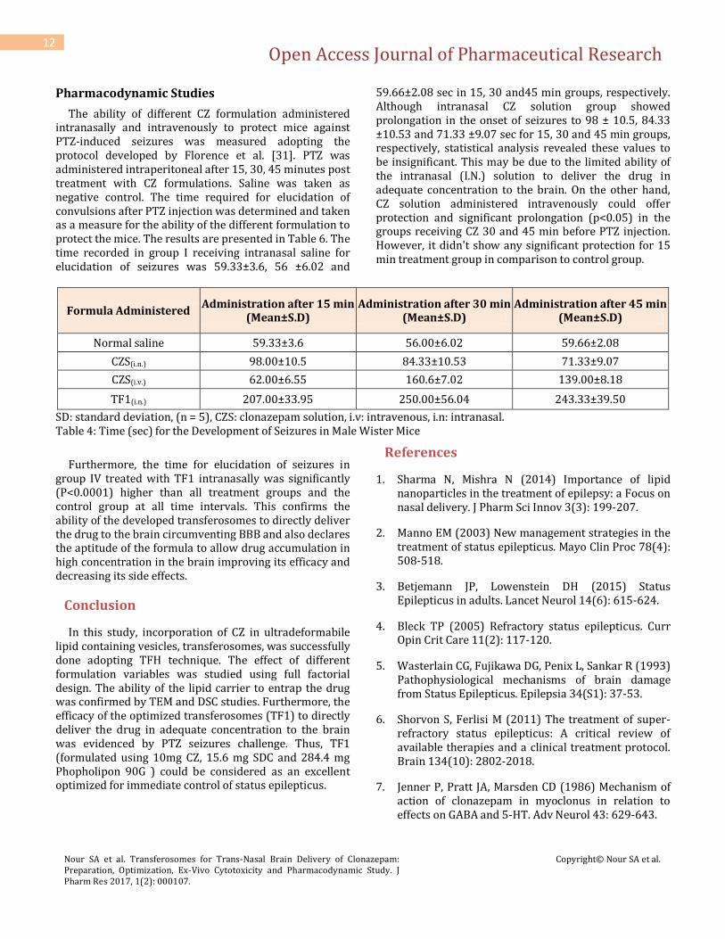
The ability of different CZ formulation administered intranasally and intravenously to protect mice against PTZ-induced seizures was measured adopting the protocol developed by Florence et al. [31]. PTZ was administered intraperitoneal after 15, 30, 45 minutes post treatment with CZ formulations. Saline was taken as negative control. The time required for elucidation of convulsions after PTZ injection was determined and taken as a measure for the ability of the different formulation to protect the mice. The results are presented in Table 6. The time recorded in group I receiving intranasal saline for elucidation of seizures was 59.33±3.6, 56 ±6.02 and
59.66±2.08 sec in 15, 30 and45 min groups, respectively. Although intranasal CZ solution group showed prolongation in the onset of seizures to 98 ± 10.5, 84.33 ±10.53 and 71.33 ±9.07 sec for 15, 30 and 45 min groups, respectively, statistical analysis revealed these values to be insignificant. This may be due to the limited ability of the intranasal (I.N.) solution to deliver the drug in adequate concentration to the brain. On the other hand, CZ solution administered intravenously could offer protection and significant prolongation (p<0.05) in the groups receiving CZ 30 and 45 min before PTZ injection. However, it didn't show any significant protection for 15 min treatment group in comparison to control group.
Formula Administered Administration after 15 min
(Mean±S.D) Administration after 30 min
(Mean±S.D) Administration after 45 min
(Mean±S.D)
Normal saline 59.33±3.6 56.00±6.02 59.66±2.08
CZS(i.n.) 98.00±10.5 84.33±10.53 71.33±9.07
CZS(i.v.) 62.00±6.55 160.6±7.02 139.00±8.18
TF1(i.n.) 207.00±33.95 250.00±56.04 243.33±39.50
SD: standard deviation, (n = 5), CZS: clonazepam solution, i.v: intravenous, i.n: intranasal. Table 4: Time (sec) for the Development of Seizures in Male Wister Mice
Furthermore, the time for elucidation of seizures in group IV treated with TF1 intranasally was significantly (P<0.0001) higher than all treatment groups and the control group at all time intervals. This confirms the ability of the developed transferosomes to directly deliver the drug to the brain circumventing BBB and also declares the aptitude of the formula to allow drug accumulation in high concentration in the brain improving its efficacy and decreasing its side effects.
Conclusion
In this study, incorporation of CZ in ultradeformabile lipid containing vesicles, transferosomes, was successfully done adopting TFH technique. The effect of different formulation variables was studied using full factorial design. The ability of the lipid carrier to entrap the drug was confirmed by TEM and DSC studies. Furthermore, the efficacy of the optimized transferosomes (TF1) to directly deliver the drug in adequate concentration to the brain was evidenced by PTZ seizures challenge. Thus, TF1 (formulated using 10mg CZ, 15.6 mg SDC and 284.4 mg Phopholipon 90G ) could be considered as an excellent optimized for immediate control of status epilepticus.
References
1. Sharma N, Mishra N (2014) Importance of lipid nanoparticles in the treatment of epilepsy: a Focus on nasal delivery. J Pharm Sci Innov 3(3): 199-207.
2. Manno EM (2003) New management strategies in the treatment of status epilepticus. Mayo Clin Proc 78(4): 508-518.
3. Betjemann JP, Lowenstein DH (2015) Status Epilepticus in adults. Lancet Neurol 14(6): 615-624.
4. Bleck TP (2005) Refractory status epilepticus. Curr Opin Crit Care 11(2): 117-120.
5. Wasterlain CG, Fujikawa DG, Penix L, Sankar R (1993) Pathophysiological mechanisms of brain damage from Status Epilepticus. Epilepsia 34(S1): 37-53.
6. Shorvon S, Ferlisi M (2011) The treatment of super-refractory status epilepticus: A critical review of available therapies and a clinical treatment protocol. Brain 134(10): 2802-2018.
7. Jenner P, Pratt JA, Marsden CD (1986) Mechanism of action of clonazepam in myoclonus in relation to effects on GABA and 5-HT. Adv Neurol 43: 629-643.
Nour SA et al. Transferosomes for Trans-Nasal Brain Delivery of Clonazepam: Preparation, Optimization, Ex-Vivo Cytotoxicity and Pharmacodynamic Study. J Pharm Res 2017, 1(2): 000107.
8. Cheze M, Deveaux M, Lenoan A, Pepin G, Author C, et al. (2005) Clonazepam, bromazepam and Zolpidem in hair of victims of drug facilitated crimes : quantitative analysis by LC-MS / MS and correlation with self-report. Ann Toxicol Anal 17(4): 269-273.
9. Andre M, Boutroy MJ, Dubruc C, Thenot JP, Bianchetti G, et al. (1986) Clonazepam pharmacokinetics and therapeutic efficacy in neonatal seizures. Eur J Clin Pharmacol 30(5): 585-589.
10. Roche (2009) Klonopin tablets (clonazepam).
11. Parvathi M (2012) Intranasal drug delivery to brain: an Overview. Int J Res Pharm 2(3): 889-895.
12. Walve JR, Bakliwal SR, Rane BR, Pawar SP (2011) Transfersomes: a Surrogated carrier for transdermal drug delivery system. Int J Appl Biol Pharm Technol 2(1): 204-213.
13. Abdallah MH (2013) Transfersomes as a transdermal drug delivery system for enhancement the antifungal activity of nystatin. Int J Pharm Pharm Sci 5(4): 560-567.
14. Venkatesh DN, Kalyani K, Tulasi K, Priyanka VS, Ali SKA, et al. (2014) Transferosomes: A novel technique for transdermal drug delivery. Int J Res Pharm Nano Sci 3(4): 266-276.
15. Salama HA, Mahmoud AA, Kamel AO, Abdel Hady M, Awad GA (2012) Brain delivery of olanzapine by intranasal administration of transfersomal vesicles. J Liposome Res 22(4): 336-345.
16. Sultana SS, Sailaja KA (2015) Formulation and evaluation of diclofenac sodium transferosomes using different surfactants by thin film hydration method. Der Pharm Lett 7(11): 43-53.
17. Shaji J, Lal M (2014) Novel double loaded transferosomes : evidence of superior anti-inflammatory efficacy- a comparative study. Int J Curr Pharm Res 6(2): 16-25.
18. Aburahma MH, Abdelbary GA (2012) Novel diphenyl dimethyl bicarboxylate provesicular powders with enhanced hepatocurative activity: Preparation, optimization, in vitro/in vivo evaluation. Int J Pharm 422(1-2): 139-150.
19. Lei W, Yu C, Lin H, Zhou X (2013) Development of tacrolimus-loaded transfersomes for deeper skin
penetration enhancement and therapeutic effect improvement in vivo. Asian J Pharm Sci 8(6): 336-345.
20. Irfan M, Verma S, Ram A (2012) Preparation and characterization of Ibuprofen loaded transferosome as a novel carrier for transdermal drug delivery system. Asian J Pharm Clin Res 5(3): 162-165.
21. Gupta PN, Mishra V, Rawat A, Dubey P, Mahor S, et al. (2005) Non-invasive vaccine delivery in transfersomes, niosomes and liposomes: A comparative study. Int J Pharm 293(1-2): 73-82.
22. Nour SA, Abdelmalak NS, Naguib MJ, Rashed HM, Ibrahim AB (2016) Intranasal brain-targeted clonazepam polymeric micelles for immediate control of status epilepticus: in vitro optimization, ex vivo determination of cytotoxicity, in vivo biodistribution and pharmacodynamics studies. Drug Deliv 23(9): 3681-3695.
23. Sharma D, Maheshwari D, Philip G, Rana R, Bhatia S, et al. (2014) Formulation and optimization of polymeric nanoparticles for intranasal delivery of Lorazepam using Box-Behnken design : In vitro and in vivo evaluation. Biomed Res Int 2014: 1-14.
24. Yang ZZ, Zhang YQ, Wanga ZZ, Wu K, Lou JN, et al. (2013) Enhanced brain distribution and pharmacodynamics of rivastigmine by liposomes following intranasal administration. Int J Pharm 452(1-2): 344-354.
25. Higuchi T (1963) Mechanism of sustained action medication. J Pharm Sci 52(12): 1145-1149.
26. Nour SA, Abdelmalak NS, Naguib MJ (2015) Bumadizone calcium dihydrate microspheres compressed tablets for colon targeting: formulation, optimization and in vivo evaluation in rabbits. Drug Deliv 22(3): 286-297.
27. Moore J and Flanner H (1996) Mathematical comparison of curves with an emphasis on in-vitro dissolution profiles. Pharm Technol 20(6): 64-74.
28. Jagtap P, Jadhav K, Dand N (2015) Formulation and ex vivo evaluation of solid lipid nanoparticles ( SLNS ) based hydrogel for intranasal drug delivery. Int J Medical, Heal Biomed Bioeng Pharm Eng 9(1): 43-53.
Nour SA et al. Transferosomes for Trans-Nasal Brain Delivery of Clonazepam: Preparation, Optimization, Ex-Vivo Cytotoxicity and Pharmacodynamic Study. J Pharm Res 2017, 1(2): 000107.
29. Kumar A, Sharma P, Chaturvedi A, Jaiswal D, Bajpai M, et al. (2009) Formulation development of sertraline hydrochloride microemulsion for intranasal delivery. Int J ChemTech Res 1(4): 941-947.
30. Al-Saraj A (2010) Use of saturated sodium chloride solution as a tissue fixative. Iraqi J Vet Sci 24(1): 53-58.
31. Florence K, Manisha L, Kumar BA, Ankur K, Kumar MA, et al. (2011) Intranasal clobazam delivery in the treatment of status epilepticus. Pharm Nanotechnol 100(2): 692-703.
32. Cote CJ, Lerman J, Anderson B (2013) A Practice of Anesthesia for Infants and Children: Expert Consult: Online and Print 5th(Edn.) Elsevier Health Sciences pp.1168.
33. Jelenkovic AV, Jovanovic MD, Stanimirovic DD, Bokonjic DD, Ocic GG, et al. (2008) Beneficial effects of Ceftriaxone against pentylenetetrazole-evoked convulsions. Exp Biol Med 233(11): 1389-1394.
34. Sachan R, Parashar T, Singh V, Singh G, Tyagi S, et al. (2013) Drug carrier transferosomes: a novel tool for transdermal drug delivery. Int J Res Dev Pharm Life Sci 2(2): 309-316.
35. Ahmad A, Alkharfy KM, Wani TA, Raish M (2015) Application of Box-Behnken design for ultrasonic-assisted extraction of polysaccharides from Paeonia emodi. Int J Biol Macromol 72: 990-997.
36. Annadurai G, Ling LY, Lee JF (2008) Statistical optimization of medium components and growth conditions by response surface methodology to enhance phenol degradation by Pseudomonas putida. J Hazard Mater 151(1): 171-178.
37. Al-Mahallawi AM, Khowessah OM, Shoukri RA (2014) Nano-transfersomal ciprofloxacin loaded vesicles for non-invasive trans-tympanic ototopical delivery: In-vitro optimization, ex-vivo permeation studies, and in-vivo assessment. Int J Pharm 472(1-2): 304-314.
38. Duangjit S, Opanasopit P, Rojanarata T, Ngawhirunpat T (2011) Characterization and in vitro skin permeation of meloxicam-loaded liposomes versus Ttransfersomes. J Drug Deliv 1-9.
39. Gupta PN, Mishra V, Singh P, Rawat A, Dubey P, et al. (2005) Tetanus toxoid-loaded transfersomes for
40. Basha M, Abd El-Alim SH, Shamma RN, Awad GE (2013) Design and optimization of surfactant-based nanovesicles for ocular delivery of Clotrimazole. J Liposome Res 23(3): 203-210.
41. Gupta A, Aggarwal G, Singla S, Arora R (2012) Transfersomes: A novel vesicular carrier for enhanced transdermal delivery of sertraline: Development, characterization, and performance evaluation. Sci Pharm 80(4): 1061-1080.
43. Gref R, Luck M, Quellec P, Marchand M, Dellacherie E, et al. 2000 Stealth’ corona-core nanoparticles surface modified by polyethylene glycol (PEG): influences of the corona (PEG chain length and surface density) and of the core composition on phagocytic uptake and plasma protein adsorption. Colloids Surfaces B Biointerfaces 18(3-4): 301-313.
44. Dora CP, Singh SK, Kumar S, Datusalia AK,Deep A (2010) Development and characterization of nanoparticles of glibenclamide by solvent displacement method. Acta Pol Pharm 67(3): 283-290.
45. Sharma N, Madan P, Lin S (2016) Effect of process and formulation variables on the preparation of parenteral paclitaxel-loaded biodegradable polymeric nanoparticles: A co-surfactant study. Asian J Pharm Sci 11(3): 404-416.
46. Cho HJ, Park JW, Yoon IS, Kim DD (2014) Surface-modified solid lipid nanoparticles for oral delivery of docetaxel: Enhanced intestinal absorption and lymphatic uptake. Int J Nanomedicine 9(1): 495-504.
47. Aghajani M, Shahverdi AR, Amani A (2012) The use of artificial neural networks for optimizing polydispersity index (PDI) in nanoprecipitation process of acetaminophen in microfluidic devices. AAPS PharmSciTech 13(4): 1293-1301.
Nour SA et al. Transferosomes for Trans-Nasal Brain Delivery of Clonazepam: Preparation, Optimization, Ex-Vivo Cytotoxicity and Pharmacodynamic Study. J Pharm Res 2017, 1(2): 000107.
49. Honary S, Zahir F (2013) Effect of zeta potential on the properties of nano-drug delivery systems - A Review ( Part 2 ). Trop J Pharm Res 12(2): 2650-273.
50. Hornig S, Bunjes H, Heinze T (2009) Preparation and characterization of nanoparticles based on dextran-drug conjugates. J Colloid Interface Sci 338(1): 56-62.
51. Taylor MJ, Tanna S, Sahota T (2010) In vivo study of a polymeric glucose-sensitive insulin delivery system using a rat model. J Pharm Sci 99(10): 4215-4227.
52. Garcia DA, Perillo MA (1999) Benzodiazepine localization at the lipid-water interface: effect of membrane composition and drug chemical structure. Biochim Biophys Acta 1418(1): 221-231.
53. Sinico C, Manconi M, Peppi M, Lai F, Valenti D, et al. (2005) Liposomes as carriers for dermal delivery of tretinoin: In vitro evaluation of drug permeation and vesicle-skin interaction. J Control Release 103(1): 123-136.
54. Goindi S, Kumar G, Kumar N, Kaur A (2013) Development of novel elastic vesicle-based topical formulation of cetirizine dihydrochloride for treatment of atopic dermatitis. AAPS Pharm Sci Tech 14(4): 1284-1293.
55. Jadupati M, Amites G, Kumar NA (2012) Transferosomes: an opportunistic carrier for transdermal drug delivery system. Int Res J Pharm 3(3): 35-38.
56. Manconi M, Caddeo C, Sinico C, Valenti D, Mostallino MC, et al. (2011) Ex vivo skin delivery of diclofenac by transcutol containing liposomes and suggested mechanism of vesicle-skin interaction. Eur J Pharm Biopharm 78(1): 27-35.
57. Wei Z, Hao J, Yuan S, Li Y, Juan W, et al. (2009) Paclitaxel-loaded pluronic P123/F127 mixed polymeric micelles: Formulation, optimization and in vitro characterization. Int J Pharm 376(1-2): 176-185.
58. Bhagav P, Upadhyay H, Chandran S (2011) Brimonidine tartrate–eudragit long-acting nanoparticles: Formulation, optimization, in vitro and in vivo evaluation. AAPS PharmSciTech 12(4): 1087-1101.
59. Baboota S, Shakeel F, Ahuja A, Ali J, Shafiq S (2007) Design, development and evaluation of novel nanoemulsion formulations for transdermal potential of celecoxib. Acta Pharm 57(3): 315-332.
60. Patel R, Singh SK, Singh S, Sheth NR, Gendle R (2009) Development and characterization of curcumin loaded transfersome for transdermal delivery. J Pharm Sci Res 1(4): 71-80.
61. Ghanbarzadeh S, Arami S (2013) Enhanced transdermal delivery of diclofenac sodium via conventional liposomes, ethosomes, and transfersomes. BioMed Res Int 1-7.
62. Narasaiah VL, Padmabhushanam P, Kishore VS (2014) Design, development and characterization of lovastatin transferosomal loaded gels for transdermal drug delivery. World J Pharm Res 3(9): 1498-1501.
63. Dragicevic-Curic N, Friedrich M, Petersen S, Scheglmann D, Douroumis D, Plass W, et al. (2011) Assessment of fluidity of different invasomes by electron spin resonance and differential scanning calorimetry. Int J Pharm 412(1-2): 85-94.
64. Leyva-Gomez G, Gonzalez-Trujano ME, Lopez-Ruiz E, Couraud PO, Wekslerg B, et al. (2014) Nanoparticle formulation improves the anticonvulsant effect of Clonazepam on the Pentylenetetrazole-induced seizures : Behavior and electroencephalogram. Pharm Nanotechnol 103(8): 2509-2519.
65. Choi J, Cho S, Yun J, Yu Y, Cho C (2015) Ethosomes and transfersomes for topical delivery of ginsenoside Rh1 from red ginseng : Characterization and in vitro evaluation. J Nanosci Nanotechnol 15(8): 5660-5662.