Transgenic Mice Expressing Green Fluorescent Proteinunder the Control of the Melanocortin-4 Receptor Promoter
Hongyan Liu,1* Toshiro Kishi,2* Aaron G. Roseberry,1 Xiaoli Cai,1 Charlotte E. Lee,3 Jason M. Montez,1
Jeffrey M. Friedman,1 and Joel K. Elmquist2,3
1Laboratory of Molecular Genetics, Howard Hughes Medical Institute, The Rockefeller University, New York, New York 10021, 2Department of Neurology,Beth Israel Deaconess Medical Center, and Program in Neuroscience, Harvard Medical School, Boston, Massachusetts 02215, and 3Department of Medicineand Division of Endocrinology, Beth Israel Deaconess Medical Center, Harvard Medical School, Boston, Massachusetts 02215
The melanocortin-4 receptor (MC4-R) is an important regulator of energy homeostasis, and evidence suggests that MC4-R-expressingneurons are downstream targets of leptin action. MC4-Rs are broadly expressed in the CNS, and the distribution of MC4-R mRNA hasbeen analyzed most extensively in the rat. However, relatively little is known concerning chemical profiles of MC4-R-expressing neurons.The extent to which central melanocortins act presynaptically or postsynaptically on MC4-Rs is also unknown. To address these issues, wehave generated a transgenic mouse line expressing green fluorescent protein (GFP) under the control of the MC4-R promoter, using amodified bacterial artificial chromosome. We have confirmed that the CNS distribution of GFP-producing cells is identical to that ofMC4-R mRNA in wild-type mice and that nearly all GFP-producing cells coexpress MC4-R mRNA. For example, cells coexpressing GFPand MC4-R mRNA were distributed in the paraventricular hypothalamic nucleus (PVH) and the dorsal motor nucleus of the vagus(DMV). MC4-R promotor-driven GFP expression was found in PVH cells producing thyrotropin-releasing hormone and in cholinergicDMV cells. Finally, we have observed that a synthetic MC3/4-R agonist, MT-II, depolarizes some GFP-expressing cells, suggesting thatMC4-Rs function postsynaptically in some instances and may function presynaptically in others. These studies extend our knowledge ofthe distribution and function of the MC4-R. The transgenic mouse line should be useful for future studies on the role of melanocortinsignaling in regulating feeding behavior and autonomic homeostasis.
IntroductionThe melanocortin-4 receptor (MC4-R) regulates food intake andbody weight in rodents and humans (Butler and Cone, 2002).This G-protein-coupled receptor is expressed widely in the CNS(Mountjoy et al., 1994; Kishi et al., 2003). Genetic evidence dem-onstrates that MC4-R blockade produces obesity syndromes. Forexample, obesity in Ay mice results from ectopic expression ofagouti protein in the brain, which is an endogenous antagonist formelanocortin receptors including the MC4-R (Spiegelman andFlier, 1996; Fan et al., 1997; Graham et al., 1997; Ollmann et al.,1997). MC4-R �/� mice (Huszar et al., 1997) and humans(Vaisse et al., 1998; Farooqi et al., 2000) display a similar obesitysyndrome. Transgenic overexpression of agouti-related protein(AgRP), another endogenous MC3/4-R antagonist, also results inobesity (Graham et al., 1997; Ollmann et al., 1997). Pro-opiomelanocortin (POMC) is the precursor of �-MSH, an en-
dogenous MC3/4-R agonist, and POMC-null mice (Yaswen et al.,1999) and humans (Krude et al., 1998) also exhibit an obesephenotype.
A large body of evidence has shown that leptin (Zhang et al.,1994; Friedman and Halaas, 1998; Saper et al., 2002) stimulatesmelanocortin signaling. The arcuate nucleus of the hypothala-mus (Arc) has been established as a site of leptin action in theCNS. The Arc contains two counterpoised neuronal populationsproducing distinct melanocortins, i.e., �-MSH and AgRP (Bro-berger et al., 1998a; Elias et al., 1998). �-MSH and AgRP neuronscoexpress the signaling or long-form leptin receptor (Mercer etal., 1996; Cheung et al., 1997). Leptin has been shown to increasePOMC mRNA (Schwartz et al., 1997; Thornton et al., 1997; Mi-zuno et al., 1998). In contrast, AgRP mRNA is increased duringfasting when leptin levels rapidly fall and in leptin-deficientob/ob mice, and its expression is reduced by exogenous leptin(Hahn et al., 1998). Leptin-induced anorexia can be suppressedby melanocortin receptor antagonism (Seeley et al., 1997), andMC4-R antagonists also block leptin-induced increases of un-coupling protein-1 in brown adipose tissue of fasted rats (Scar-pace et al., 1997; Kotz et al., 1998; Satoh et al., 1998). Finally, thethyrotropin-releasing hormone (TRH) gene is transcriptionallyregulated by leptin and melanocortins (Harris et al., 2001; Feketeet al., 2000). Taken together, these observations suggest that sub-sets of MC4-R-expressing neurons mediate some of the leptin
Received March 18, 2003; revised June 9, 2003; accepted June 13, 2003.This work was supported by National Institutes of Health Grants DK56116, DK53301, and MH61583 to T.K. and
J.K.E. We thank Dr. Andrew Cubitt from Aurora Bioscience for the Sapphire DNA, Dr. Nathaniel Heintz and XiangdongYang for the shuttle vector PSV1, Dr. Stanley J. Watson for the AVP and CRH probes, and Dr. Rexford S. Ahima for theTRH probe.
*H.L. and T.K. contributed equally to this work.Correspondence should be addressed to Dr. Jeffrey M. Friedman, Box 305, Howard Hughes Medical Institute, The
The Journal of Neuroscience, August 6, 2003 • 23(18):7143–7154 • 7143
actions on autonomic and endocrine responses, and on feedingbehavior. Importantly, leptin-independent melanocortin signal-ing pathways also exist (Boston et al., 1997; Butler et al., 2001).
However, despite the established importance of the MC4-R,relatively little is known regarding chemical and electrophysio-logical profiles of MC4-R-expressing cells. In addition, neuro-anatomic studies of the MC4-R have been limited by the lack ofhigh-affinity antibodies. To address these issues, we generated atransgenic mouse line in which green fluorescent protein (GFP)is expressed under the control of the MC4-R promoter, by usinga modified bacterial artificial chromosome (Yang et al., 1997).The characteristics of this mouse line and the MC4-R-expressingcells are described in detail below.
Materials and MethodsProduction of the MC4R-Tau-Sapphire transgenic (MC4-R/GFP) mouse.Sapphire is a blue-shifted GFP variant from Aurora Biosciences (La Jolla,CA). Bacterial artificial chromosome (BAC) filters (BAC mouse II) andclones were obtained from Genome Systems (St. Louis, MO). Tau-Sapphire fusion protein and polyA signal were inserted into the ATG siteof a MC4-R-containing BAC using a method developed by Yang et al.(1997) to modify BAC in Escherichia coli host bacterial. Kozak sequencewas added before Tau, a microtubule-binding protein (Callahan andThomas, 1994). PolyA signal was PCR amplified from nucleotide 697–1088 of SV40 polyA of pREP7 (Invitrogen, Carlsbad, CA). The modifi-cation of the BAC requires a shuttle vector (PSV1) that has three features:a temperature-sensitive origin of replication, a RecA gene to introducerecombination in a recombination-deficient E. coli host, and a tetracy-cline resistance gene (Tet�). The shuttle vector has 2.5 kb and 1.7 kbarms of MC4-R sequence flanking Tau-Sapphire polyA sequence. Theshuttle vector was transformed into the DH10B E. coli host harboring theMC4-R BAC. After two homologous recombination events, the modifiedMC4-R BAC was selected by temperature and antibiotic sensitivity. Thehomologous recombination of the modified BAC was confirmedthrough Southern analysis. The BAC DNA was then purified withCesium chloride gradient, linearized with NotI, further purified withSepharose CL-4b (Sigma, St. Louis, MO) column, and injected intopronuclei of CBA/C57Bl6 F1 mice by the Rockefeller transgenic facil-ity. The incorporation of the transgene in mouse genome was identi-fied through Southern blots and PCR. The genotyping primers for thetransgene is GFPF1 5�-CCGAGGATCCTACCATGGTGAG-CAAGGGCGA-3� and GFP407R 5�-CAGCTTGTGCCCCAGGATGT.
Animals and histology. The MC4-R/GFP mice and C57BL/6 mice(25–35 gm; Jackson Laboratory, Bar Harbor, ME) were housed with foodand water available ad libitum in a light-controlled (12 hr light/darkcycle; lights on 7 A.M.) and temperature-controlled (21.5–22.5°C) envi-ronment. The animals and procedures used in this study were in accor-dance with the guidelines of The Rockefeller Animal Research Center aswell as with the guidelines and approval of the Harvard Medical Schooland Beth Israel Deaconess Medical Center Institutional Animal Care andUse Committees. Mice were deeply anesthetized with intraperitonealinjection of chloral hydrate (350 mg/kg) and perfused transcardially withDEPC-treated 0.9% saline, followed by 50 ml of 10% neutral bufferedformalin. Brains were removed, stored in the same fixative for 4 hr at 4°C,immersed in 20% sucrose in DEPC-treated PBS, pH 7.0, at 4°C over-night, and were cut coronally at 25 �m into 1:5 equal series on a freezingmicrotome. Sections were stored at �20°C in an antifreeze solution(Simmons et al., 1989) until sections were treated for immunohisto-chemistry (IHC) and/or in situ hybridization histochemistry (ISHH).
IHC for GFP. To determine GFP-producing brain sites, IHC was per-formed using seven mice (four mice of line 21; three mice of line 30), asreported previously (Elmquist and Saper, 1996; Elias et al., 1998). Sec-tions were pretreated with 0.3% hydrogen peroxide in PBS, pH 7.4, for 30min at room temperature and then incubated in 3% normal donkeyserum (Jackson ImmunoResearch Laboratories Inc., West Grove, PA)with 0.25% Triton X-100 in PBS (PBT) for 1 hr, followed by overnightincubation in a GFP rabbit primary antiserum (Molecular Probes, Eu-
gene, OR; 1:20,000 in PBT) at room temperature. After washing in PBS,sections were incubated in biotinylated donkey anti-rabbit IgG (JacksonImmunoResearch Laboratories Inc.; 1:1,000) for 1 hr at room tempera-ture and then incubated in a solution of ABC (Vectastain Elite ABC Kit;Vector Laboratories, Burlingame, CA; 1:500) dissolved in PBS for 1 hr.After washing in PBS, the sections were incubated in a solution of 0.04%DAB (Sigma) and 0.01% hydrogen peroxide dissolved in PBS. Brainsections were mounted onto gelatinized slides, air-dried, dehydrated inascending concentrations of ethanol, cleared in xylenes, and were thencoverslipped with Permaslip (Alban Scientific, St. Louis, MO). GFP-immunoreactive (GFP-IR) cells were plotted using a camera lucida at-tached to a microscope (Zeiss Axioscope). Coverslips were then re-moved, and the sections were conterstained with thionin to determinenuclear boundaries (Kishi et al., 2000).
Single-label ISHH for MC4-R. To examine the distribution of MC4-RmRNA in the wild-type (WT) mouse brain, single-label ISHH was per-formed as reported recently from our laboratory (Marcus et al., 2001;Kishi et al., 2003), based on a modification of the protocol previouslyreported by our and other laboratories (Simmons et al., 1989; Chan et al.,1993; Brady et al., 1994; Ericsson et al., 1995; Elmquist et al., 1998; Elias etal., 1999). Tissue sections were mounted onto SuperFrost slides (FisherScientific, Pittsburgh, PA), air-dried, and stored in desiccated boxes at�20°C. Before hybridization, sections were fixed in 4% formaldehyde inDEPC-treated PBS, pH 7.0, for 20 min at 4°C, dehydrated in ascendingconcentrations of ethanol, cleared in xylenes for 15 min, rehydrated indescending concentrations of ethanol, and placed in prewarmed sodiumcitrate buffer (95–100°C; pH 6.0). Slides were then placed in a Sharp(Nahwah, NJ) R-510C commercial microwave oven (1100 W) for 10 minat 70% power (temperature, 95–100°C), dehydrated in ascending con-centrations of ethanol, and air-dried.
The MC4-R probe was made using a DNA fragment corresponding tonucleotides 806 –1400 of the rat MC4R transcript (Kishi et al., 2003).BLAST sequence alignment indicates 89% identity between the rat se-quence and nucleotides 665–999 of the mouse MC4-R transcript (Gen-Bank accession number NM_016977). To generate antisense 35S-labeledcRNA, the plasmids were linearized by digestion with NcoI and subjectedto in vitro transcription with SP6 RNA polymerase according to the man-ufacturer’s protocol (Promega, Madison, WI). For generation of sense35S-labeled cRNA, the plasmids were linearized by digestion with SalIand subjected to in vitro transcription with T7 RNA polymerase accord-ing to the manufacturer’s protocol (Ambion, Austin, TX). The 35S-labeled cRNA probe for the MC4-R mRNA was then diluted to 10 6
cpm/ml in a hybridization solution composed of 50% formamide, 10 mM
Tris-HCl (Gibco-BRL, Bethesda, MD), pH 8.0, 5.0 mg of tRNA (Invitro-gen), 10 mM DTT, 10% dextran sulfate, 0.3 M NaCl, 1 mM EDTA, pH 8.0,and 1� Denhardt’s solution (Sigma).
Hybridization solution and a coverslip were applied to each slide, andsections were incubated for 12–16 hr at 57°C. Coverslips were then re-moved, and slides were washed with 2� SSC, pH 7.0. Sections were thenincubated in 0.002% RNAase A (Roche Molecular Biochemicals, India-napolis, IN) with 0.5 M NaCl, 10 mM Tris-HCl, pH 8.0, and 1 mM EDTAfor 30 min. Subsequently, sections were washed in decreasing concentra-tions of SSC containing 0.25% DTT: 2� SSC at 50°C for 1 hr, 0.2� SSCat 55°C for 1 hr, and 0.2� SSC at 60°C for 1 hr. Sections were nextdehydrated in graded ethanol (50, 70, 80, and 90%) containing 0.3 M
NH4OAc, followed by 100% ethanol. Slides were air-dried and placed inx-ray film cassettes with BMR-2 film (Kodak, Rochester, NY) for 2–3 d.Slides were then dipped in NTB2 photographic emulsion (Kodak), dried,and stored in desiccated and foil-wrapped boxes at 4°C for 3– 4 weeks.Finally, slides were developed with D-19 developer (Kodak), counter-stained with thionin, dehydrated in graded ethanols, cleared in xylenes,and coverslipped with Permaslip.
In a WT mouse, an adjacent series of sections was stained with thioninto identify nuclear boundaries (Marcus et al., 2001; Kishi et al., 2003).Control experiments to confirm the specificity of this protocol includedhybridization with sense probes and with antisense probes after treat-ment with RNAase A (200 �g/ml).
Dual-label ISHH/IHC. IHC was coupled with free-floating ISHH todemonstrate cells coexpressing GFP and MC4-R mRNA in three mice of
7144 • J. Neurosci., August 6, 2003 • 23(18):7143–7154 Liu et al. • MC4-R/GFP Transgenic Mice
line 21 as well as in two mice of line 30. The procedure was a modificationof that described previously (Priestley et al., 1993; Elias et al., 1998;Yamamoto et al., 2003). Brain sections were first rinsed in DEPC-treatedPBS, pH 7.0, and were pretreated with 1% sodium borohydride (Sigma)in DEPC-PBS for 15 min at room temperature. After washing in DEPC-PBS, sections were rinsed in 0.1 M TEA, pH 8.0, and incubated in 0.25%acetic anhydride in 0.1 M TEA for 10 min. After washing in 2� SSC,sections were incubated in the above-mentioned hybridization solutioncontaining the MC4-R probes diluted to 10 6 cpm/ml for 12–16 hr at57°C. Subsequently, sections were rinsed in 4� SSC and incubated in0.002% RNAase A (Roche Molecular Biochemicals) with 0.5 M NaCl, 10mM Tris-HCl, pH 8, and 1 mM EDTA for 30 min at 37°C. Sections wererinsed with 2� SSC and then with 50% formamide in 0.2� SSC at 50°C.Subsequently, sections were washed in decreasing concentrations of SSC:2� SSC at 50°C for 1 hr, 0.2� SSC at 55°C for 1 hr, and 0.2� SSC at 60°Cfor 1 hr. After washing in PBS, pH 7.4, IHC for GFP was performed asdescribed above. Sections were mounted onto SuperFrost slides (FisherScientific) and were placed in x-ray film cassettes with BMR-2 film(Kodak) for 2–3 d. Slides were then treated as described above withoutcounterstaining.
GFP-expressing cells were chemically defined using the same dual-label free-floating ISHH/IHC method with antisense probes forcorticotropin-releasing hormone (CRH) (Day et al., 2002), arginine va-sopressin (AVP) (Helmreich et al., 1999), orexin (ORX) (Sakurai et al.,1998), melanin-concentrating hormone (MCH) (Qu et al., 1996),GAD67 (Erlander et al., 1991), or ChAT (Kishi et al., 2003). An antisensefor TRH was also used, which was made by using a DNA fragment cor-responding to nucleotides 308 –573 of the mouse preproTRH mRNA(GenBank accession number X59387; a gift from Dr. Rexford S. Ahima,Department of Medicine, Division of Endocrinology, University ofPennsylvania School of Medicine, Philadelphia, PA). In addition, an an-tisense probe for oxytocin was applied, which was generated by using aDNA fragment corresponding to nucleotides 3060 –3256 of the rat oxy-tocin DNA (GenBank accession number X12792). Three transgenic micewere used in each dual-label experiment. The 35S-labeled cRNA probeswere generated from the cDNA templates by in vitro transcription withappropriate polymerases (SP6 for oxytocin, AVP, MCH, GAD67, ChAT,and TRH; T7 for CRH and ORX) according to the manufacturers’ pro-tocol (Promega; Ambion). The period during which slides were exposedto emulsion ranged from 3 to 10 d.
Two methods of scoring double-labeled cells were used (Elias et al.,1999). The less stringent of the two considered cells to contain positivehybridization if the silver grains overlying GFP-IR cell bodies were five-folds above background hybridization levels. Estimates of backgroundhybridization levels were made by calculating the mean number of silvergrains overlying a counting grid (100 �m 2) in the internal capsule. Amore conservative estimate was used that defined cells as double-labeledonly if the silver grains above background conformed to the shape of theGFP-IR cell bodies.
Anatomic analysis and production of photomicrographs. Sections wereanalyzed with a Zeiss Axioskop or a Zeiss Stemi 2000-C dissecting mi-croscope. Cytoarchitectonic details were added by using a camera lucida.Photomicrographs were produced with a Spot digital camera (DiagnosticInstruments, Sterling Heights, MI) attached to the microscopes and anApple Macintosh G3 computer. An image editing software (Adobe Pho-toshop 5.5) was used to combine microphotographs onto plates, andfigures were printed on a dye sublimination printer (Kodak 8670 PS).Only the contrast and brightness were adjusted.
Electrophysiological recording. Young MC4-R/GFP mice (4 – 6 weeks ofage; line 21) were deeply anesthetized with halothane (Halocarbon, RiverEdge, NJ) before decapitation and removal of the entire brain. The brainwas immediately submerged in ice-cold, carbogen-saturated (95%O2/5%CO2) artificial CSF (aCSF), and a brain block containing the hypo-thalamus was made. The aCSF contained (in mM): 126 NaCl, 2.5 KCl, 2.4CaCl2, 1.2 NaH2PO4, 1.2 MgCl2, 21.4 NaHCO3, and 11.1 glucose. Coro-nal sections (180 �m) were cut with a Leica VT1000S vibratome, and theslices were incubated at 37°C for �30 min, followed by incubation atroom temperature until used.
Slices were transferred to the recording chamber and allowed to equil-
ibrate for 10 –20 min before use. The slices were perfused with oxygen-ated aCSF (29 –30°C) at a flow rate of �1.7 ml/min. GFP-positive MC4-Rcells in the paraventricular hypothalamic nucleus (PVH) and the dorso-medial nucleus (DMH) were visualized using epifluorescence andinfrared-differential interference contrast (IR-DIC) imaging on an up-right Zeiss Axioskop 2FS Plus microscope equipped with filter sets spe-cific for Sapphire GFP (excitation, 395; emission, 510; Chroma Technol-ogy Corp., Brattleboro, VT) and a Sony XC-75 CCD camera. MC4-Rneurons were identified via epifluorescence and then patched under IR-DIC optics. Recordings were performed using a potassium gluconate-based internal solution, which contained (in mM): 128 KGluconate, 10HEPES, 1 EGTA, 10 KCl, 1 MgCl2, 0.3 CaCl2, 5 MgATP, and 0.3 NaGTP,pH 7.35, with KOH. Electrodes were borosilicate glass and had resis-tances of �2.5– 4 M� when filled with the KGluconate internal solution,and series resistance values were �10 M� and were not compensated.Trials were excluded if the series resistance increased significantly duringthe experiment. Recordings were made using a HEKA EPC9/2 amplifierunder the control of Pulse software. In addition, a Powerlab 4/20 usingChart software was used for data acquisition during current-clamp re-cordings. Data analysis was performed using PulseFit, Chart, IgorPro,and Microsoft Excel, and figures were created using PulseFit and IgorPro.Mean values for membrane potential were similar after analysis with
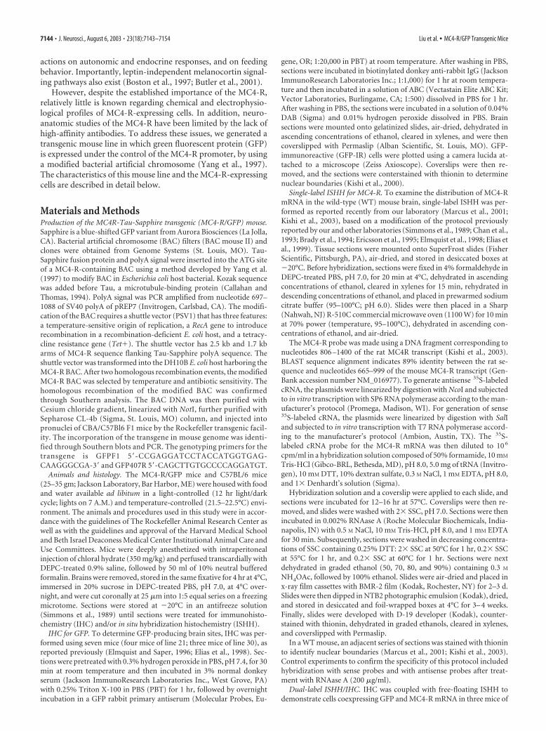
Figure 1. Production of MC4-R/GFP transgenic mice. A, Schematic drawings show insertionof Tau-Sapphire-polyA into the MC4-R BAC by homologous recombination in E. coli cells. B,Southern blot of pulsed field gel electrophoresis of restriction digestion of WT and modifiedMC4-R BAC. C, Southern blot analysis of ApaI digest of genomic DNA from WT, line 21, and line30 transgenic mice.
Liu et al. • MC4-R/GFP Transgenic Mice J. Neurosci., August 6, 2003 • 23(18):7143–7154 • 7145
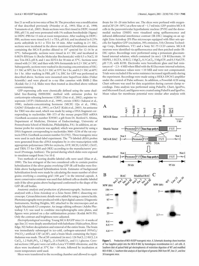
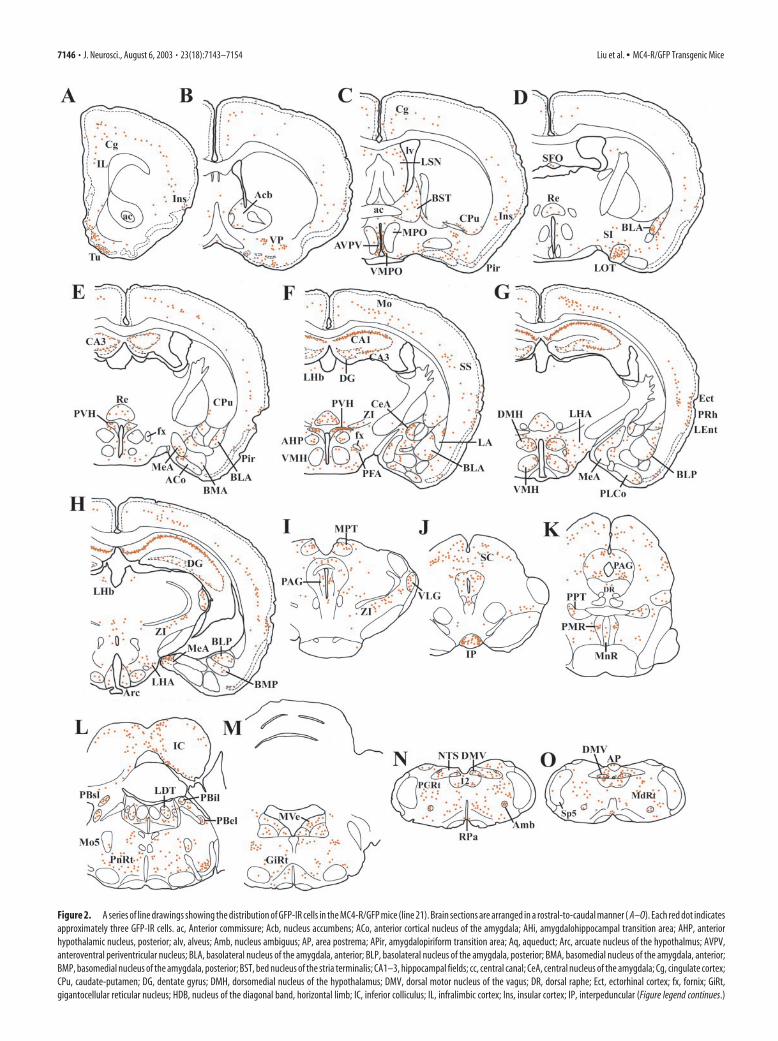
Figure 2. A series of line drawings showing the distribution of GFP-IR cells in the MC4-R/GFP mice (line 21). Brain sections are arranged in a rostral-to-caudal manner ( A–O). Each red dot indicatesapproximately three GFP-IR cells. ac, Anterior commissure; Acb, nucleus accumbens; ACo, anterior cortical nucleus of the amygdala; AHi, amygdalohippocampal transition area; AHP, anteriorhypothalamic nucleus, posterior; alv, alveus; Amb, nucleus ambiguus; AP, area postrema; APir, amygdalopiriform transition area; Aq, aqueduct; Arc, arcuate nucleus of the hypothalmus; AVPV,anteroventral periventricular nucleus; BLA, basolateral nucleus of the amygdala, anterior; BLP, basolateral nucleus of the amygdala, posterior; BMA, basomedial nucleus of the amygdala, anterior;BMP, basomedial nucleus of the amygdala, posterior; BST, bed nucleus of the stria terminalis; CA1–3, hippocampal fields; cc, central canal; CeA, central nucleus of the amygdala; Cg, cingulate cortex;CPu, caudate-putamen; DG, dentate gyrus; DMH, dorsomedial nucleus of the hypothalamus; DMV, dorsal motor nucleus of the vagus; DR, dorsal raphe; Ect, ectorhinal cortex; fx, fornix; GiRt,gigantocellular reticular nucleus; HDB, nucleus of the diagonal band, horizontal limb; IC, inferior colliculus; IL, infralimbic cortex; Ins, insular cortex; IP, interpeduncular (Figure legend continues.)
7146 • J. Neurosci., August 6, 2003 • 23(18):7143–7154 Liu et al. • MC4-R/GFP Transgenic Mice
both Chart and Pulse, and the data reported here were obtained usingChart. All p values reported were from paired t tests.
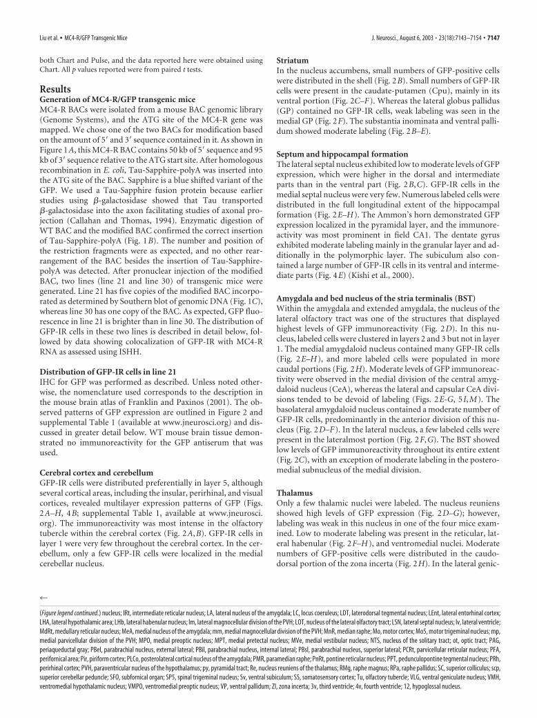
ResultsGeneration of MC4-R/GFP transgenic miceMC4-R BACs were isolated from a mouse BAC genomic library(Genome Systems), and the ATG site of the MC4-R gene wasmapped. We chose one of the two BACs for modification basedon the amount of 5� and 3� sequence contained in it. As shown inFigure 1A, this MC4-R BAC contains 50 kb of 5� sequence and 95kb of 3� sequence relative to the ATG start site. After homologousrecombination in E. coli, Tau-Sapphire-polyA was inserted intothe ATG site of the BAC. Sapphire is a blue shifted variant of theGFP. We used a Tau-Sapphire fusion protein because earlierstudies using �-galactosidase showed that Tau transported�-galactosidase into the axon facilitating studies of axonal pro-jection (Callahan and Thomas, 1994). Enzymatic digestion ofWT BAC and the modified BAC confirmed the correct insertionof Tau-Sapphire-polyA (Fig. 1B). The number and position ofthe restriction fragments were as expected, and no other rear-rangement of the BAC besides the insertion of Tau-Sapphire-polyA was detected. After pronuclear injection of the modifiedBAC, two lines (line 21 and line 30) of transgenic mice weregenerated. Line 21 has five copies of the modified BAC incorpo-rated as determined by Southern blot of genomic DNA (Fig. 1C),whereas line 30 has one copy of the BAC. As expected, GFP fluo-rescence in line 21 is brighter than in line 30. The distribution ofGFP-IR cells in these two lines is described in detail below, fol-lowed by data showing colocalization of GFP-IR with MC4-RRNA as assessed using ISHH.
Distribution of GFP-IR cells in line 21IHC for GFP was performed as described. Unless noted other-wise, the nomenclature used corresponds to the description inthe mouse brain atlas of Franklin and Paxinos (2001). The ob-served patterns of GFP expression are outlined in Figure 2 andsupplemental Table 1 (available at www.jneurosci.org) and dis-cussed in greater detail below. WT mouse brain tissue demon-strated no immunoreactivity for the GFP antiserum that wasused.
Cerebral cortex and cerebellumGFP-IR cells were distributed preferentially in layer 5, althoughseveral cortical areas, including the insular, perirhinal, and visualcortices, revealed multilayer expression patterns of GFP (Figs.2A–H, 4B; supplemental Table 1, available at www.jneurosci.org). The immunoreactivity was most intense in the olfactorytubercle within the cerebral cortex (Fig. 2A,B). GFP-IR cells inlayer 1 were very few throughout the cerebral cortex. In the cer-ebellum, only a few GFP-IR cells were localized in the medialcerebellar nucleus.
StriatumIn the nucleus accumbens, small numbers of GFP-positive cellswere distributed in the shell (Fig. 2B). Small numbers of GFP-IRcells were present in the caudate-putamen (Cpu), mainly in itsventral portion (Fig. 2C–F). Whereas the lateral globus pallidus(GP) contained no GFP-IR cells, weak labeling was seen in themedial GP (Fig. 2F). The substantia inominata and ventral palli-dum showed moderate labeling (Fig. 2B–E).
Septum and hippocampal formationThe lateral septal nucleus exhibited low to moderate levels of GFPexpression, which were higher in the dorsal and intermediateparts than in the ventral part (Fig. 2B,C). GFP-IR cells in themedial septal nucleus were very few. Numerous labeled cells weredistributed in the full longitudinal extent of the hippocampalformation (Fig. 2E–H). The Ammon’s horn demonstrated GFPexpression localized in the pyramidal layer, and the immunore-activity was most prominent in field CA1. The dentate gyrusexhibited moderate labeling mainly in the granular layer and ad-ditionally in the polymorphic layer. The subiculum also con-tained a large number of GFP-IR cells in its ventral and interme-diate parts (Fig. 4E) (Kishi et al., 2000).
Amygdala and bed nucleus of the stria terminalis (BST)Within the amygdala and extended amygdala, the nucleus of thelateral olfactory tract was one of the structures that displayedhighest levels of GFP immunoreactivity (Fig. 2D). In this nu-cleus, labeled cells were clustered in layers 2 and 3 but not in layer1. The medial amygdaloid nucleus contained many GFP-IR cells(Fig. 2E–H), and more labeled cells were populated in morecaudal portions (Fig. 2H). Moderate levels of GFP immunoreac-tivity were observed in the medial division of the central amyg-daloid nucleus (CeA), whereas the lateral and capsular CeA divi-sions tended to be devoid of labeling (Figs. 2E-G, 5 I,M). Thebasolateral amygdaloid nucleus contained a moderate number ofGFP-IR cells, predominantly in the anterior division of this nu-cleus (Fig. 2D–F). In the lateral nucleus, a few labeled cells werepresent in the lateralmost portion (Fig. 2F,G). The BST showedlow levels of GFP immunoreactivity throughout its entire extent(Fig. 2C), with an exception of moderate labeling in the postero-medial subnucleus of the medial division.
ThalamusOnly a few thalamic nuclei were labeled. The nucleus reuniensshowed high levels of GFP expression (Fig. 2D–G); however,labeling was weak in this nucleus in one of the four mice exam-ined. Low to moderate labeling was present in the reticular, lat-eral habenular (Fig. 2F–H), and ventromedial nuclei. Moderatenumbers of GFP-positive cells were distributed in the caudo-dorsal portion of the zona incerta (Fig. 2H). In the lateral genic-
4
(Figure legend continued.) nucleus; IRt, intermediate reticular nucleus; LA, lateral nucleus of the amygdala; LC, locus coeruleus; LDT, laterodorsal tegmental nucleus; LEnt, lateral entorhinal cortex;LHA, lateral hypothalamic area; LHb, lateral habenular nucleus; lm, lateral magnocellular division of the PVH; LOT, nucleus of the lateral olfactory tract; LSN, lateral septal nucleus; lv, lateral ventricle;MdRt, medullary reticular nucleus; MeA, medial nucleus of the amygdala; mm, medial magnocellular division of the PVH; MnR, median raphe; Mo, motor cortex; Mo5, motor trigeminal nucleus; mp,medial parvicellular division of the PVH; MPO, medial preoptic nucleus; MPT, medial pretectal nucleus; MVe, medial vestibular nucleus; NTS, nucleus of the solitary tract; ot, optic tract; PAG,periaqueductal gray; PBel, parabrachial nucleus, external lateral; PBil, parabrachial nucleus, internal lateral; PBsl, parabrachial nucleus, superior lateral; PCRt, parvicellular reticular nucleus; PFA,perifornical area; Pir, piriform cortex; PLCo, posterolateral cortical nucleus of the amygdala; PMR, paramedian raphe; PnRt, pontine reticular nucleus; PPT, pedunculopontine tegmental nucleus; PRh,perirhinal cortex; PVH, paraventricular nucleus of the hypothalamus; py, pyramidal tract; Re, nucleus reuniens of the thalamus; RMg, raphe magnus; RPa, raphe pallidus; SC, superior colliculus; scp,superior cerebellar peduncle; SFO, subfornical organ; SP5, spinal trigeminal nucleus; Sv, ventral subiculum; SS, somatosensory cortex; Tu, olfactory tubercle; VLG, ventral geniculate nucleus; VMH,ventromedial hypothalamic nucleus; VMPO, ventromedial preoptic nucleus; VP, ventral pallidum; ZI, zona incerta; 3v, third ventricle; 4v, fourth ventricle; 12, hypoglossal nucleus.
Liu et al. • MC4-R/GFP Transgenic Mice J. Neurosci., August 6, 2003 • 23(18):7143–7154 • 7147
ulate nucleus, labeled cells were observed exclusively in the ven-tral part (Fig. 2H, I).
HypothalamusThere was an aggregation of many GFP-IR cells in the anteroventralperiventricular nucleus (Fig. 2C). Labeling in the ventromedial pre-optic nucleus was less intense (Fig. 2C,D). The medial preoptic andmedian preoptic nuclei contained only a few labeled cells. GFP im-munoreactivity was undetectable in the suprachiasmatic and su-praoptic nuclei. The PVH displayed a unique pattern of labeling (Fig.2E,F). Within the PVH, the density of GFP-IR cells was highest inthe lateral half of the posterior division (Figs. 2F, 5J,N). The anteriorparvicellular division showed light labeling, whereas the medialmagnocellular division also contained a moderate number of labeledcells. Only a few GFP-positive cells were also present in the lateralmagnocellular division. On the other hand, the medial parvicellulardivision of the PVH was unlabelled.
In the tuberal hypothalamus, GFP immunoreactivity was ev-ident in the DMH (Fig. 2G). The labeled cells were populatedpreferentially in the caudal portion of the ventral division of theDMH, whereas labeling in the compact and dorsal DMH divi-sions was more intense in more rostral portions. GFP-positivecells were also present in an area closely dorsal to the DMH re-ferred to as the dorsal hypothalamic area in the rat (Saper, 1995).The ventromedial nucleus (VMH) also displayed low to moder-ate labeling, which was denser in more dorsomedial portions ofthis nucleus (Fig. 2G,H). In addition, GFP-IR cells were observedto surround the ventrolateral surface of the VMH. Relativelysmall numbers of labeled cells were scattered in the lateral hypo-thalamic area (LHA), including the perifornical area (PFA) (Figs.2E–H, 5K,O). The arcuate nucleus displayed a relatively low levelof GFP immunoreactivity in the caudal one-third of this nucleus(Figs. 2H, 4H). GFP-IR cells were also present in the most super-ficial layer of the median eminence.
Midbrain, pons, and medulla oblongataWithin the pretectal area, GFP expression was most prominent inthe medial pretectal nucleus (Fig. 2 I). The superior colliculuscontained a large number of labeled cells, many of which wereconcentrated in the intermediate gray layer (Fig. 2 J,K). The peri-aqueductal gray matter displayed moderate levels of labelingthroughout its full rostral-to-caudal extent (Fig. 2 I–K). In thiscolumnar structure, labeling was most conspicuous in the ven-trolateral division (Figs. 2K, 4L). Intense labeling was evident inthe dorsolateral portion of the interpeduncular nucleus (Fig. 2 J).In the inferior colliculus, GFP-IR cells were distributed mainlyin the medial portion of the external cortex (Fig. 2 L). Thepedunculopontine tegmental (Fig. 2 K) and laterodorsal teg-mental (Fig. 2 L) nuclei exhibited moderate to high levels ofGFP immunoreactivity.
The lateral parabrachial nucleus also displayed GFP expres-sion, which was intense in the superior division, moderate in thedorsal and internal divisions, and weak in the central, external,and ventral divisions (Fig. 2L). Very few labeled cells were ob-served in the medial parabrachial nucleus. Whereas the parame-dian raphe exhibited moderate levels of GFP immunoreactivity,fewer labeled cells were observed in the dorsal raphe, medianraphe, raphe magnus, and raphe pallidus (Fig. 2K,N,O). Moder-ate numbers of GFP-positive cells were scattered throughout theentire rostrocaudal and mediolateral extent of the nucleus of thesolitary tract (Fig. 2N,O). The dorsal motor nucleus of the vagus(Fig. 5L,P) and nucleus ambiguus contained moderate numbersof labeled cells (Fig. 2N,O). The hypoglossal nucleus showed
moderate labeling (Fig. 2N,O). Moderate to high levels of GFPimmunoreactivity were observed in reticular nuclei including thepontine, parvicellular, gigantocellular, and the medullary nuclei(Fig. 2K–O).
Distribution of GFP-IR cells in line 30The distribution of GFP-IR cells in line 30 was nearly identical tothat of line 21 with the following exceptions. In line 30, layer 1 ofthe cerebral cortex did not show GFP expression. GFP-IR cellswere localized in layer 5 of the ectorhinal, perirhinal, and visualcortices in line 30. The dentate gyrus also revealed a discrepancy:GFP-IR cells were distributed in the polymorphic layer of line 30,whereas labeling was seen preferentially in the granular layer ofline 21. In line 30, the nucleus accumbens and medial septumtended to contain more abundant GFP-positive cells. The ven-tromedial hypothalamic nucleus of line 30 contained very fewGFP-IR cells. In line 30, GFP immunoreactivity was not detectedin the median eminence and raphe pallidus.
Distribution of MC4-R mRNA in WT miceAs described, lines 21 and 30 of the MC4-R/GFP mice displayednearly identical distribution patterns of GFP-IR cells. The pat-terns of GFP expression were almost identical to that of MC4-RmRNA in WT mice (Figs. 2– 4), with only a few exceptions (sup-plemental Table 1, available at www.jneurosci.org). For example,the rostral portion of the medial parvicellular division of the PVHdisplayed weak MC4-R hybridization in WT mice (Figs. 3D, 4F),even though GFP-IR cells were not distributed in this PVH por-tion of both lines. MC4-R mRNA expression was observed in theraphe pallidus of the WT mice (Fig. 4M). As mentioned, how-ever, GFP-positive cells were not distributed in this nucleus ofline 30. In the WT mice, MC4-R mRNA was detected in multiplelayers of the visual, ectorhinal, and perirhinal cortices, whereasGFP-IR cells were localized in layer 5 of these cortical fields in line30. Conversely, the nucleus reuniens of both lines produced GFP,but MC4-R hybridization was undetectable in this thalamic nu-cleus of the WT mice. In line 30, the same discrepancy was ob-served in the polymorphic layer of the dentate gyrus.
In addition, the relative expression levels of MC4-R mRNA inthe WT mice were highly consistent with those of GFP in eachbrain site of both lines. Control ISHH experiments demonstratedno MC4-R-specific hybridization.
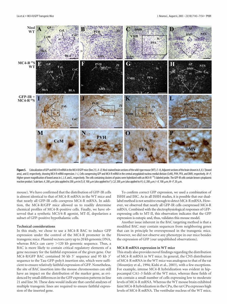
Colocalization of GFP and MC4-R mRNAA combination of ISHH and IHC revealed that nearly all GFP-IRcells coexpressed MC4-R mRNA in both lines of MC4-R/GFPmice. Representative examples of the colocalization in the medialdivision of the CeA, PVH, PFA, and in the dorsal motor nucleusof the vagus (DMV) are shown in Figure 5. However, in a smallnumber of cases, GFP was expressed in sites where MC4-RmRNA was below the limit of detection. In line 21, for example,single-labeled GFP-positive cells were distributed in layer 1 of thecerebral cortex, medial cerebellar nucleus, and in the medianeminence, although the WT mice did not display detectableMC4-R hybridization in these sites. Similarly, in line 30, the poly-morphic layer of the dentate gyrus contained single-labeledGFP-IR cells, despite undetectable MC4-R hybridization in thislayer of the WT mice. We cannot rule out that these few examplesof discordance may represent ectopic expression of the transgene.However, it is important to note that in the vast majority of sites,the expression pattern was topographically identical to MC4-RmRNA in WT animals. Moreover, GFP-IR cells were found tocoexpress MC4-R mRNA in nearly all of these sites. Taken to-
7148 • J. Neurosci., August 6, 2003 • 23(18):7143–7154 Liu et al. • MC4-R/GFP Transgenic Mice
gether, these findings demonstrate the usefulness of this mousemodel in anatomic, molecular, and physiological studies.
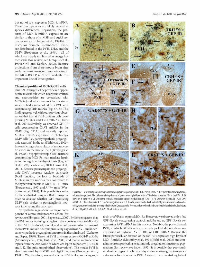
Chemical profiles of cells that produce GFP markerfor MC4-RTo identify chemical phenotypes of MC4-R-positive cells in thebrain sites involved in energy homeostasis, we performed a seriesof dual-label experiments, using line 21 of the MC4-R/GFP mice.The PVH contained cells coexpressing GFP and TRH mRNA(Fig. 6A,B). We also observed that a few GFP-IR cells producingGAD67 mRNA were scattered in the PFA (Fig. 6G–J), BST, CPu,and in the cerebral cortex.
In the DMV, GFP expression was observed in cells coexpress-ing ChAT mRNA (Fig. 6K,L), a marker for autonomic pregan-glionic neurons. The LHA including the PFA was observed to
contain moderate numbers of GFP-IR cells as well as cells ex-pressing ORX or MCH mRNA. However, we did not detect thecolocalization of GFP and ORX mRNA or MCH mRNA.
Electrophysiological responses of GFP-expressing cells to amelanocortin receptor ligandTo determine whether MC4-R/GFP cells were able to respond di-rectly to MC4-R agonists, electrophysiological patch-clamp record-ings were performed on GFP-labeled neurons from line 21 mice. Asmentioned, this line exhibited brighter GFP fluorescence. Whole-cell current-clamp recordings were made from cells within the DMHand PVH of the hypothalamus (Fig. 7). As observed with other hy-pothalamic cells (our unpublished observations), MC4-R cells in thehypothalamus showed very high input resistance (�1 G�) and firedaction potentials spontaneously. To test the ability of MC4-R/GFP
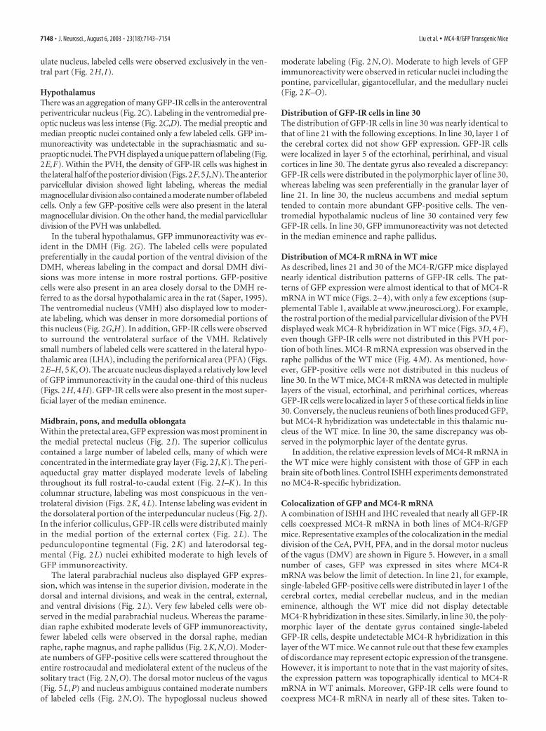
Figure 3. A series of low-power photomicrographs summarizing MC4-R mRNA expression sites across the WT mouse brain. Brain sections are arranged in a rostral-to-caudal manner ( A–L). Scalebar (in L), 1 mm ( A–L). (For list of abbreviations, see Fig. 2 legend.)
Liu et al. • MC4-R/GFP Transgenic Mice J. Neurosci., August 6, 2003 • 23(18):7143–7154 • 7149
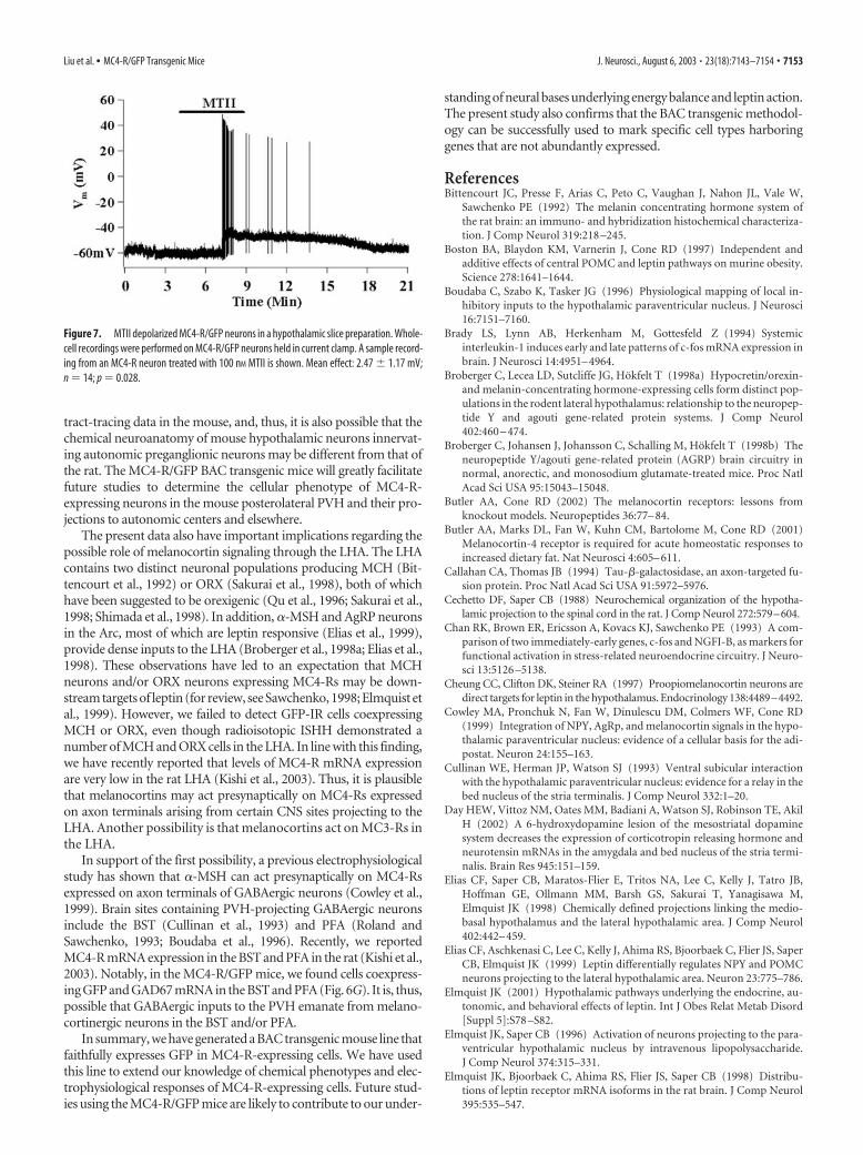
cells to respond to MC4-R agonists, slices were perfused with theMC3/4-R agonist, MTII, in the extracellular medium. MTII induceda significant depolarization of MC4-R/GFP cells (2.47 � 1.17 mV;n 14; p 0.028). Despite this highly significant average effect, onlysix of the 14 cells that were tested showed a direct response to MTII.All of the responding neurons did respond similarly with a signifi-cant depolarization. The lack of effect in some cells would be seen incases in which MC4-R is trafficked to presynaptic terminals becausestimulation of presynaptic receptor would not be expected to have
an effect on total cellular electrical activity. Whereas MTII is a non-specific MC3/MC4-R agonist, the effect of MTII is likely to be me-diated by MC4-Rs because MC3-Rs were not reported to be ex-pressed in the DMH and PVH (Roselli-Rehfuss et al., 1993).
Discussion
In this study, we have generated a transgenic mouse-expressingGFP under the control of the MC4-R promoter (MC4-R/GFP
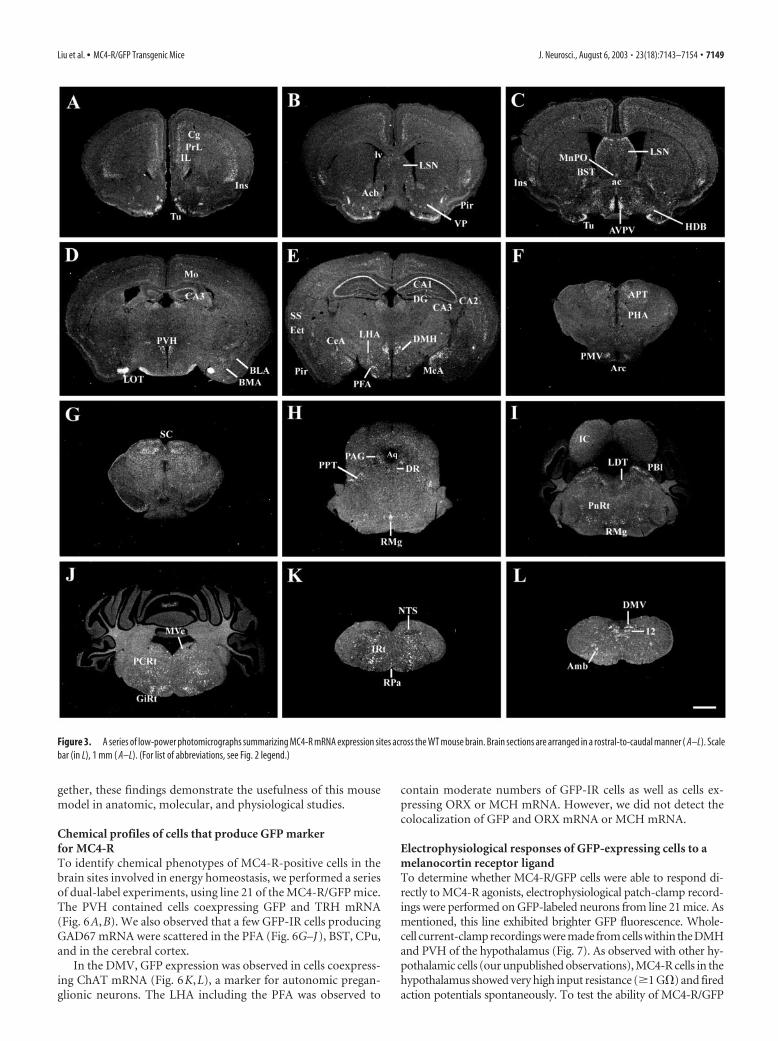
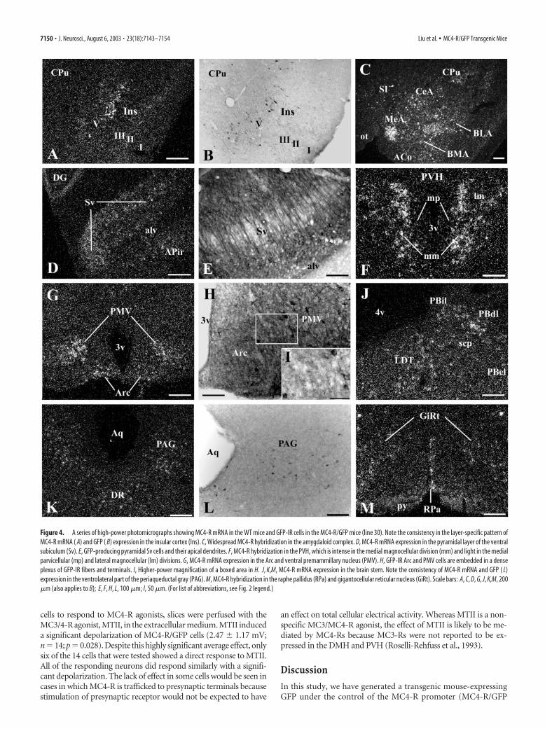
Figure 4. A series of high-power photomicrographs showing MC4-R mRNA in the WT mice and GFP-IR cells in the MC4-R/GFP mice (line 30). Note the consistency in the layer-specific pattern ofMC4-R mRNA ( A) and GFP ( B) expression in the insular cortex (Ins). C, Widespread MC4-R hybridization in the amygdaloid complex. D, MC4-R mRNA expression in the pyramidal layer of the ventralsubiculum (Sv). E, GFP-producing pyramidal Sv cells and their apical dendrites. F, MC4-R hybridization in the PVH, which is intense in the medial magnocellular division (mm) and light in the medialparvicellular (mp) and lateral magnocellular (lm) divisions. G, MC4-R mRNA expression in the Arc and ventral premammillary nucleus (PMV). H, GFP-IR Arc and PMV cells are embedded in a denseplexus of GFP-IR fibers and terminals. I, Higher-power magnification of a boxed area in H. J, K,M, MC4-R mRNA expression in the brain stem. Note the consistency of MC4-R mRNA and GFP ( L)expression in the ventrolateral part of the periaqueductal gray (PAG). M, MC4-R hybridization in the raphe pallidus (RPa) and gigantocellular reticular nucleus (GiRt). Scale bars: A, C, D, G, J, K,M, 200�m (also applies to B); E, F, H, L, 100 �m; I, 50 �m. (For list of abbreviations, see Fig. 2 legend.)
7150 • J. Neurosci., August 6, 2003 • 23(18):7143–7154 Liu et al. • MC4-R/GFP Transgenic Mice
mouse). We have confirmed that the distribution of GFP-IR cellsis almost identical to that of MC4-R mRNA in the WT mice andthat nearly all GFP-IR cells coexpress MC4-R mRNA. In addi-tion, the MC4-R/GFP mice allowed us to readily determinechemical profiles of MC4-R-positive cells. Finally, we have ob-served that a synthetic MC3/4-R agonist, MT-II, depolarizes asubset of GFP-positive hypothalamic cells.
Technical considerationsIn this study, we chose to use a MC4-R BAC to induce GFPexpression under the control of the MC4-R promoter in thetransgenic mice. Plasmid vectors carry up to 20 kb genomic DNA,whereas BACs can carry 120 kb genomic sequence. Thus, aBAC is more likely to contain critical regulatory elements of agene necessary for the faithful expression of the given gene. OurMC4-R/GFP BAC contained 50 kb 5� sequence and 95 kb 3�sequence to the Tau-GFP-polyA insertion site, which were suffi-cient to ensure relatively faithful expression of GFP. Nonetheless,the site of BAC insertion into the mouse chromosomes can stillhave an impact on the distribution of the marker gene, as evi-denced by small differences in the GFP expression patterns in line21 and line 30. These data would indicate that careful analyses ofmultiple transgenic lines are required to ensure faithful expres-sion of the inserted gene.
To confirm correct GFP expression, we used a combination ofISHH and IHC. As in all ISHH studies, it is possible that our dual-label method is not sensitive enough to detect MC4-R mRNA. How-ever, we observed that nearly all GFP-IR cells coexpressed MC4-RmRNA. Combined with the electrophysiological responses of GFP-expressing cells to MT-II, this observation indicates that the GFPexpression is eutopic and, thus, validates this mouse model.
Another issue inherent in the BAC targeting method is that amodified BAC may contain sequences from neighboring genesthat can in principle be overexpressed in the transgenic mice.However, we did not observe any phenotype in our mice besidesthe expression of GFP (our unpublished observations).
MC4-R mRNA expression in WT miceThis study also provides novel findings regarding the distributionof MC4-R mRNA in WT mice. In general, the CNS distributionof MC4-R mRNA in the WT mice was analogous to that of the rat(Mountjoy et al., 1994; Kishi et al., 2003), with a few exceptions.For example, intense MC4-R hybridization was evident in hip-pocampal CA1–3 fields of the WT mice, whereas these fields ofrats contain a small number of cells expressing low to moderatelevels of MC4-R mRNA. Whereas the WT mouse brain exhibitedfaint MC4-R hybridization in the CPu, the rat CPu expresses highlevels of MC4-R mRNA. The vestibular nucleus of the WT mice,
Figure 5. Colocalization of GFP and MC4-R mRNA in the MC4-R/GFP mice (line 21). A–D, Nissl-stained brain sections of the wild-type mouse (WT). E–H, Adjacent sections of the brain shown in A, B, C (boxedarea), and D, respectively, showing MC4-R mRNA expression. I–L, Cells coexpressing GFP and MC4-R mRNA in the central amygdaloid nucleus medial division (CeM), PVH, PFA, and DMV, respectively. M–P,Higher-power magnification of boxed areas in I, J, K, and L, respectively. The cells containing clusters of grains were hybridized with an MC4-R 35S-labeled probe. The GFP-IR cells contain brown cytoplasmicreaction product. Scale bars: A, 200 �m (also applied to 200 �m in E); B, 100 �m (also applied to F ); C,D, 200 �m (also applied to H ); G, 200 �m; I–K, 100 �m; M–P, 20 �m.
Liu et al. • MC4-R/GFP Transgenic Mice J. Neurosci., August 6, 2003 • 23(18):7143–7154 • 7151
but not of rats, expresses MC4-R mRNA.These discrepancies are likely viewed asspecies differences. Regardless, the pat-terns of MC4-R mRNA expression aresimilar to those of �-MSH and AgRP ax-ons in mice (Broberger et al., 1998b). Inmice, for example, melanocortin axonsare distributed in the PVH, LHA, and theDMV (Broberger et al., 1998b), all ofwhich are deeply implicated in energy ho-meostasis (for review, see Elmquist et al.,1999; Grill and Kaplan, 2002). Becauseprojections from these mouse brain sitesare largely unknown, retrograde tracing inthe MC4-R/GFP mice will facilitate thisimportant line of investigation.
Chemical profiles of MC4-R/GFP cellsOur BAC transgenic line provides an oppor-tunity to establish which neurotransmittersand neuropetides are colocalized withMC4-Rs (and which are not). In this study,we identified a subset of GFP-IR PVH cellscoexpressing TRH mRNA (Fig. 6A,B). Thisfinding agrees well with our previous obser-vation that the rat PVH contains cells coex-pressing MC4-R and TRH mRNAs (Harriset al., 2001). Similarly, we observed GFP-IRcells coexpressing ChAT mRNA in theDMV (Fig. 6K,L) and recently reportedMC4-R mRNA expression in cholinergicDMV cells (i.e., parasympathetic pregangli-onic neurons) in the rat (Kishi et al., 2003).In considering a dense plexus of melanocor-tin axons in the mouse PVH (Broberger etal., 1998b), hypophysiotropic TRH neuronscoexpressing MC4-Rs may mediate leptinaction to regulate the thyroid axis (Legradiet al., 1998; Fekete et al., 2000; Harris et al.,2001). Because parasympathetic pregangli-onic DMV neurons regulate pancreatic�-cell function, the lack or blockade ofMC4-Rs in this nucleus may contribute tothe hyperinsulinemia in MC4-R �/� mice(Huszar et al., 1997) and Avy/� mice (War-britton et al., 1994). This possibility can befurther evaluated using our BAC transgenicmice to analyze whether GFP-producingDMV cells project to postganglionic neu-rons innervating the pancreas.
Sympathetic regulation is a major com-ponent of central melanocortin action (forreview, see Elmquist, 2001; Saper et al., 2002). Evidence suggests thatthe PVH relays leptin signaling from the arcuate nucleus to MC4-Rsin the PVH. The dorsal, ventral, and lateral parvicellular divisions ofthe rat PVH contain neurons producing oxytocin or AVP and inner-vate sympathetic preganglionic neurons in the spinal cord (Cechettoand Saper, 1988). These rat PVH divisions express MC4-R mRNA(Mountjoy et al., 1994; Kishi et al., 2003) and receive melanocortininputs from the Arc, some of which are leptin responsive (T. Kishiand J. K. Elmquist, unpublished observations). The mouse PVH isalso innervated by �-MSH and AgRP neurons (Broberger et al.,1998b). We, therefore, assessed whether PVH cells producing oxy-
tocin or AVP also express MC4-Rs. However, we observed only a fewGFP-IR cells coexpressing oxytocin mRNA and no GFP-IR cells co-expressing AVP mRNA in this nucleus. Notably, the posterolateralPVH, in which GFP-IR cells are densely packed, did not show anyexpression of oxytocin, AVP, TRH, or CRH mRNA. Because thelateral parvicellular division of the rat PVH expresses high levels ofMC4-R mRNA (Mountjoy et al., 1994; Kishi et al., 2003) and con-tains neurons projecting to autonomic preganglionic neuronal pop-ulations (for review, see Saper, 1995), it is possible that previouslyunidentified types of cells may relay melanocortin signals to regulateautonomic function via the PVH. As noted, there is a striking lack of
Figure 6. A series of photomicrographs showing chemical profiles of MC4-R/GFP cells. The GFP-IR cells contain brown cytoplas-mic reaction product. The cells containing clusters of grains were hybridized with a 35S-labeled probe for TRH in the PVH ( A, B),oxytocin in the PVH (C D), CRH in the central amygdaloid nucleus medial division (CeM) ( E, F ), GAD67 in the PFA ( G–J), or ChATmRNA ( K, L). Boxed areas in A, C, E, G,K are magnified in B, D, F, J, and L, respectively. A cell indicated by an arrowhead and anothercell by two arrowheads in G are magnified in H and I, respectively. Arrows and arrowheads indicate double-labeled cells. Scale bars:A, C,K, 100 �m; E, 200 �m; B, D, F, H–J,L, 20 �m; G, 50 �m.
7152 • J. Neurosci., August 6, 2003 • 23(18):7143–7154 Liu et al. • MC4-R/GFP Transgenic Mice
tract-tracing data in the mouse, and, thus, it is also possible that thechemical neuroanatomy of mouse hypothalamic neurons innervat-ing autonomic preganglionic neurons may be different from that ofthe rat. The MC4-R/GFP BAC transgenic mice will greatly facilitatefuture studies to determine the cellular phenotype of MC4-R-expressing neurons in the mouse posterolateral PVH and their pro-jections to autonomic centers and elsewhere.
The present data also have important implications regarding thepossible role of melanocortin signaling through the LHA. The LHAcontains two distinct neuronal populations producing MCH (Bit-tencourt et al., 1992) or ORX (Sakurai et al., 1998), both of whichhave been suggested to be orexigenic (Qu et al., 1996; Sakurai et al.,1998; Shimada et al., 1998). In addition, �-MSH and AgRP neuronsin the Arc, most of which are leptin responsive (Elias et al., 1999),provide dense inputs to the LHA (Broberger et al., 1998a; Elias et al.,1998). These observations have led to an expectation that MCHneurons and/or ORX neurons expressing MC4-Rs may be down-stream targets of leptin (for review, see Sawchenko, 1998; Elmquist etal., 1999). However, we failed to detect GFP-IR cells coexpressingMCH or ORX, even though radioisotopic ISHH demonstrated anumber of MCH and ORX cells in the LHA. In line with this finding,we have recently reported that levels of MC4-R mRNA expressionare very low in the rat LHA (Kishi et al., 2003). Thus, it is plausiblethat melanocortins may act presynaptically on MC4-Rs expressedon axon terminals arising from certain CNS sites projecting to theLHA. Another possibility is that melanocortins act on MC3-Rs inthe LHA.
In support of the first possibility, a previous electrophysiologicalstudy has shown that �-MSH can act presynaptically on MC4-Rsexpressed on axon terminals of GABAergic neurons (Cowley et al.,1999). Brain sites containing PVH-projecting GABAergic neuronsinclude the BST (Cullinan et al., 1993) and PFA (Roland andSawchenko, 1993; Boudaba et al., 1996). Recently, we reportedMC4-R mRNA expression in the BST and PFA in the rat (Kishi et al.,2003). Notably, in the MC4-R/GFP mice, we found cells coexpress-ing GFP and GAD67 mRNA in the BST and PFA (Fig. 6G). It is, thus,possible that GABAergic inputs to the PVH emanate from melano-cortinergic neurons in the BST and/or PFA.
In summary, we have generated a BAC transgenic mouse line thatfaithfully expresses GFP in MC4-R-expressing cells. We have usedthis line to extend our knowledge of chemical phenotypes and elec-trophysiological responses of MC4-R-expressing cells. Future stud-ies using the MC4-R/GFP mice are likely to contribute to our under-
standing of neural bases underlying energy balance and leptin action.The present study also confirms that the BAC transgenic methodol-ogy can be successfully used to mark specific cell types harboringgenes that are not abundantly expressed.
Sawchenko PE (1992) The melanin concentrating hormone system ofthe rat brain: an immuno- and hybridization histochemical characteriza-tion. J Comp Neurol 319:218 –245.
Boston BA, Blaydon KM, Varnerin J, Cone RD (1997) Independent andadditive effects of central POMC and leptin pathways on murine obesity.Science 278:1641–1644.
Boudaba C, Szabo K, Tasker JG (1996) Physiological mapping of local in-hibitory inputs to the hypothalamic paraventricular nucleus. J Neurosci16:7151–7160.
Brady LS, Lynn AB, Herkenham M, Gottesfeld Z (1994) Systemicinterleukin-1 induces early and late patterns of c-fos mRNA expression inbrain. J Neurosci 14:4951– 4964.
Broberger C, Lecea LD, Sutcliffe JG, Hokfelt T (1998a) Hypocretin/orexin-and melanin-concentrating hormone-expressing cells form distinct pop-ulations in the rodent lateral hypothalamus: relationship to the neuropep-tide Y and agouti gene-related protein systems. J Comp Neurol402:460 – 474.
Broberger C, Johansen J, Johansson C, Schalling M, Hokfelt T (1998b) Theneuropeptide Y/agouti gene-related protein (AGRP) brain circuitry innormal, anorectic, and monosodium glutamate-treated mice. Proc NatlAcad Sci USA 95:15043–15048.
Butler AA, Marks DL, Fan W, Kuhn CM, Bartolome M, Cone RD (2001)Melanocortin-4 receptor is required for acute homeostatic responses toincreased dietary fat. Nat Neurosci 4:605– 611.
Callahan CA, Thomas JB (1994) Tau-�-galactosidase, an axon-targeted fu-sion protein. Proc Natl Acad Sci USA 91:5972–5976.
Cechetto DF, Saper CB (1988) Neurochemical organization of the hypotha-lamic projection to the spinal cord in the rat. J Comp Neurol 272:579–604.
Chan RK, Brown ER, Ericsson A, Kovacs KJ, Sawchenko PE (1993) A com-parison of two immediately-early genes, c-fos and NGFI-B, as markers forfunctional activation in stress-related neuroendocrine circuitry. J Neuro-sci 13:5126 –5138.
Cheung CC, Clifton DK, Steiner RA (1997) Proopiomelanocortin neurons aredirect targets for leptin in the hypothalamus. Endocrinology 138:4489–4492.
Cowley MA, Pronchuk N, Fan W, Dinulescu DM, Colmers WF, Cone RD(1999) Integration of NPY, AgRp, and melanocortin signals in the hypo-thalamic paraventricular nucleus: evidence of a cellular basis for the adi-postat. Neuron 24:155–163.
Cullinan WE, Herman JP, Watson SJ (1993) Ventral subicular interactionwith the hypothalamic paraventricular nucleus: evidence for a relay in thebed nucleus of the stria terminalis. J Comp Neurol 332:1–20.
Day HEW, Vittoz NM, Oates MM, Badiani A, Watson SJ, Robinson TE, AkilH (2002) A 6-hydroxydopamine lesion of the mesostriatal dopaminesystem decreases the expression of corticotropin releasing hormone andneurotensin mRNAs in the amygdala and bed nucleus of the stria termi-nalis. Brain Res 945:151–159.
Elias CF, Saper CB, Maratos-Flier E, Tritos NA, Lee C, Kelly J, Tatro JB,Hoffman GE, Ollmann MM, Barsh GS, Sakurai T, Yanagisawa M,Elmquist JK (1998) Chemically defined projections linking the medio-basal hypothalamus and the lateral hypothalamic area. J Comp Neurol402:442– 459.
Elias CF, Aschkenasi C, Lee C, Kelly J, Ahima RS, Bjoorbaek C, Flier JS, SaperCB, Elmquist JK (1999) Leptin differentially regulates NPY and POMCneurons projecting to the lateral hypothalamic area. Neuron 23:775–786.
Elmquist JK (2001) Hypothalamic pathways underlying the endocrine, au-tonomic, and behavioral effects of leptin. Int J Obes Relat Metab Disord[Suppl 5]:S78 –S82.
Elmquist JK, Saper CB (1996) Activation of neurons projecting to the para-ventricular hypothalamic nucleus by intravenous lipopolysaccharide.J Comp Neurol 374:315–331.
Elmquist JK, Bjoorbaek C, Ahima RS, Flier JS, Saper CB (1998) Distribu-tions of leptin receptor mRNA isoforms in the rat brain. J Comp Neurol395:535–547.
Figure 7. MTII depolarized MC4-R/GFP neurons in a hypothalamic slice preparation. Whole-cell recordings were performed on MC4-R/GFP neurons held in current clamp. A sample record-ing from an MC4-R neuron treated with 100 nM MTII is shown. Mean effect: 2.47 � 1.17 mV;n 14; p 0.028.
Liu et al. • MC4-R/GFP Transgenic Mice J. Neurosci., August 6, 2003 • 23(18):7143–7154 • 7153
Elmquist JK, Elias CF, Saper CB (1999) From lesions to leptin: hypotha-lamic control of food intake and body weight. Neuron 22:221–232.
Ericsson A, Liu C, Hart RP, Sawchenko PE (1995) Type-1 interleukin-1receptor in the rat brain: distribution, regulation, and relationship to sitesof IL-1-induced cellular activation. J Comp Neurol 361:681– 698.
Erlander MG, Tillakarante NJ, Feldblum S, Patel N, Tobin AJ (1991) Twogenes encode distinct glutamate decarboxylase. Neuron 7:91–100.
Fan W, Boston BA, Kesterson RA, Hruby VJ, Cone RD (1997) Role of mela-nocortinergic neurons in feeding and the agouti obesity syndrome. Na-ture 385:165–168.
Farooqi IS, Yeo GS, Keogh JM, Aminian S, Jebb SA, Butler G, Cheetham T,O’Rahilly S (2000) Dominant and recessive inheritance of morbid obe-sity associated with melanocortin 4 receptor deficiency. J Clin Invest106:271–279.
Fekete C, Legradi G, Mihaly E, Huang Q-H, Tatro JB, Rand WM, EmersonCH, Lechan RM (2000) �-Melanocyte-stimulating hormone is con-tained in nerve terminals innervating thyrotropin-releasing hormone-synthesizing neurons in the hypothalamic paraventricular nucleus andprevents fasting-induced suppression of prothyrotropin-releasing hor-mone gene expression. J Neurosci 20:1550 –1558.
Franklin KBJ, Paxinos G (2001) The mouse brain in stereotaxic coordinates,Ed 2. San Diego: Academic.
Friedman JM, Halaas JL (1998) Leptin and the regulation of body weight inmammals. Nature 395:763–770.
Graham M, Shutter JR, Sarmiento U, Sarosi I, Stark KL (1997) Overexpres-sion of Agrt leads to obesity in transgenic mice. Nat Genet 17:273–274.
Grill HJ, Kaplan JM (2002) The neuroanatomical axis for control of energybalance. Front Neuroendocrinol 23:2– 40.
Hahn TM, Breininger JF, Baskin DG, Schwartz MW (1998) Coexpression ofAgrp and NPY in fasting-activated hypothalamic neurons. Nat Neurosci1:271–272.
Harris M, Aschkenasi C, Elias CF, Chandrankunnel A, Nillni EA, BjoorbaekC, Elmquist JK, Flier JS, Hollenberg AN (2001) Transcriptional regula-tion of the thyrotropin-releasing hormone gene by leptin and melanocor-tin signaling. J Clin Invest 107:111–120.
Helmreich DL, Watkins LR, Deak T, Maier SF, Akil H, Watson SJ (1999)The effect of stressor controllability on stress-induced neuropeptidemRNA expression within the paraventricular nucleus of the hypothala-mus. J Neuroendocrinol 11:121–128.
Huszar D, Lynch CA, Fairchild-Huntress V, Dunmore JH, Fang Q, BerkemeierLR, Gu W, Kesterson RA, Boston BA, Cone RD, Smith FJ, Campfield LA,Burn P, Lee F (1997) Targeted disruption of the melanocortin-4 receptorresults in obesity in mice. Cell 88:131–141.
Kishi T, Tsumori T, Ono K, Yokota S, Ishino H, Yasui Y (2000) Topograph-ical organization of projections from the subiculum to the hypothalamusin the rat. J Comp Neurol 419:205–222.
Kishi T, Aschkenasi CJ, Lee CE, Mountjoy KG, Saper CB, Elmquist JK (2003)Expression of melanocortin 4 receptor mRNA in the central nervoussystem of the rat. J Comp Neurol 457:213–235.
Kotz CM, Briggs JE, Pomonis JD, Grace MK, Levine AS, Billington CJ (1998)Neural site of leptin influence on neuropeptide Y signaling pathwaysaltering feeding and uncoupling protein. Am J Physiol 275:R478 –R484.
Krude H, Biebermann H, Luck W, Horn R, Brabant G, Gruters A (1998)Severe early-onset obesity, adrenal insufficiency and red hair pigmenta-tion caused by POMC mutations in humans. Nat Genet 19:155–157.
Legradi G, Emerson CH, Ahima RS, Rand WM, Flier JS, Lechan RM (1998)Arcuate nucleus ablation prevents fasting-induced suppression of Pro-TRH mRNA in the hypothalamic paraventricular nucleus. Neuroendo-crinology 68:89 –97.
Marcus JN, Aschkenasi CJ, Lee CE, Chemelli RM, Saper CB, Yanagisawa M,Elmquist JK (2001) Differential expression of orexin receptors 1 and 2 inthe rat brain. J Comp Neurol 435:6 –25.
Mercer JG, Hoggard N, Williams LM, Lawrence CB, Hannah LT, Morgan PJ,Trayhurn P (1996) Coexpression of leptin receptor preproneuropeptideY mRNA in arcuate nucleus of mouse hypothalamus. J Neuroendocrinol8:733–735.
Mizuno TM, Kleopoulos SP, Bergen HT, Roberts JL, Priest CA, Mobbs CV(1998) Hypothalamic pro-opiomelanocortin mRNA is reduced by fasting inob/ob and db/db mice, but is stimulated by leptin. Diabetes 47:294–297.
Mountjoy KG, Mortrud MT, Low MJ, Simerly RB, Cone RD (1994) Local-ization of the melanocortin-4 receptor (MC4-R) in neuroendocrine andautonomic control circuits in the brain. Mol Endocrinol 8:1298 –1308.
Ollmann MM, Wilson BD, Yang Y-K, Kerns JA, Chen Y, Gantz I, Barsh GS(1997) Antagonism of central melanocortin receptors in vitro and in vivoby agouti-related protein. Science 278:135–138.
Priestley JV, Wotherspoon G, Savery D, Averill S, Rattray M (1993) A com-bined in situ hybridization and immunofluorescence procedure allowingvisualisation of peptide mRNA and serotonin in single sections. J Neuro-sci Methods 48:99 –110.
Qu D, Ludwig DS, Gammeltoft S, Piper M, Pelleymounter MA, Cullen MJ,Mathes WF, Przypek J, Kanarek R, Maratos-Flier E (1996) A role formelanin-concentrating hormone in the central regulation of feeding be-haviour. Nature 380:243–247.
Roland BL, Sawchenko PE (1993) Local origins of some GABAergic projec-tions to the paraventricular and supraoptic nuclei of the hypothalamus inthe rat. J Comp Neurol 332:123–143.
Roselli-Rehfuss L, Mountjoy KG, Robbins LS, Mortrud MT, Low MJ, TatroJB, Entwistle ML, Simerly RB, Cone RD (1993) Identification of a recep-tor for � melanotropin and other proopiomelanocortin peptides in thehypothalamus and limbic system. Proc Natl Acad Sci USA 90:8856 – 8860.
Sakurai T, Amemiya A, Ishii M, Matsuzaki I, Chemelli RM, Tanaka H, Wil-liams SC, Richardson JA, Kozlowski GP, Wilson S, Arch JRS, BuckinghamRE, Haynes AC, Carr SA, Annan RS, McNulty DE, Liu W-S, Terrett JA,Elshourbagy NA, Bergsma DJ, Yanagisawa M (1998) Orexins and orexinreceptors: a family of hypothalamic neuropeptides and G protein-coupled receptors that regulate feeding behavior. Cell 92:573–585.
Saper CB (1995) Central autonomic system. In: The rat nervous system(Paxinos G, ed), pp 107–135. San Diego: Academic.
Saper CB, Chou TC, Elmquist JK (2002) The need to feed: homeostatic andhedonic control of eating. Neuron 36:199 –211.
Satoh N, Ogawa Y, Katsuura G, Numata Y, Masuzaki H, Yoshimasa Y, NakaoK (1998) Satiety effect and sympathetic activation of leptin are mediatedby hypothalamic melanocortin system. Neurosci Lett 249:107–110.
Sawchenko PE (1998) Toward a new neurobiology of energy balance, appe-tite, and obesity: the anatomists weigh in. J Comp Neurol 402:435– 441.
Scarpace PJ, Matheny M, Pollock BH, Tumer N (1997) Leptin increasesuncoupling protein expression and energy expenditure. Am J Physiol273:E226 –E230.
Seeley RJ, Yagaloff KA, Fisher SL, Burn P, Thiele TE, van Dijk G, Baskin DG,Schwartz MW (1997) Melanocortin receptors in leptin effects. Nature390:349.
Shimada M, Tritos N, Lowell BB, Flier JS, Maratos-Flier E (1998) Mice lack-ing melanin concentrating hormone are hypophagic and lean. Nature396:670 – 674.
Simmons DM, Arriza JL, Swanson LW (1989) A complete protocol for insitu hybridization of messenger RNAs in brain and other tissues withradiolabelled single stranded RNA probes. J Histotechnol 12:169 –181.
Spiegelman BM, Flier JS (1996) Adipogenesis and obesity: rounding out thebig picture. Cell 87:377–389.
Thornton JE, Cheung CC, Clifton DK, Steiner RA (1997) Regulation of hy-pothalamic proopiomelanocortin mRNA by leptin in ob/ob mice. Endo-crinology 138:5063–5066.
Vaisse C, Clement K, Guy-Grand B, Froguel P (1998) A frameshift mutationin human MC4R is associated with a dominant form of obesity. Nat Genet20:113–114.
Warbritton A, Gill AM, Yen TT, Bucci T, Wolff GL (1994) Pancreatic isletcells in preobese yellow A vy/� mice: relation to adult hyperinsulinemiaand obesity. Proc Soc Exp Biol Med 206:145–151.
Yamamoto H, Kishi T, Lee CE, Choi BJ, Fang H, Hollenberg AN, Drucker DJ,Elmquist JK (2003) Glucagon-like peptide-1 responsive catecholamineneurons in the area postrema link peripheral glucagon-like peptide-1action with central autonomic control sites. J Neurosci 23:2939 –2946.
Yang XW, Model P, Heintz N (1997) Homologous recombination basedmodification in Escherichia coli and germline transmission in transgenicmice of a bacterial artificial chromosome. Nat Biotechnol 15:859 – 866.
Yaswen L, Diehl N, Brennan MB, Hochgeschwender U (1999) Obesity inthe mouse model of pro-opiomelanocortin deficiency responds to pe-ripheral melanocortin. Nat Med 5:1066 –1070.
Zhang Y, Proenca R, Maffei M, Barone M, Loepold L, Friedman JM (1994)Positional cloning of the mouse obese gene and its human homologue.Nature 372:425– 432.
7154 • J. Neurosci., August 6, 2003 • 23(18):7143–7154 Liu et al. • MC4-R/GFP Transgenic Mice