Proc. NatL Acad. Sci. USA Vol. 79, pp. 1912-1916, March 1982 Developmental Biology Full-term development after transplantation of parthenogenetic embryonic nuclei into fertilized mouse eggs (nuclear transplantation/parthenogenesis/LT/Sv mise) PETER C. HOPPE* AND KARL ILLMENSEEt *The Jackson Laboratory, Bar Harbor, Maine 04609; and tDepartment of Animal Biology, University of Geneva, CH-1211 Geneva, Switzerland Communicated by Robert W. Briggs, December 14, 1981 ABSTRACT Diploid parthenogenetically activated oocytes were obtained after gonadotropin-induced ovulation of virgin fe- males of the LT/Sv (LT) inbred mouse strain. These oocytes cleave spontaneously and develop into blastocysts which implant in the uterus but die within a few days. We examined the devel- opmental potential of nuclei from parthenogenetic embryos after transplantation into fertilized eggs. The inner cell mass (ICM) and trophectoderm (TE) of LT parthenogenetic blastocysts were me- chanically isolated and dissociated into single cells. Their nuclei were then injected into fertilized C57BL/6J eggs from which the male and female pronuclei were removed. Of 94 eggs injected with TE cell nuclei, 4 embryos developed to the morula stage; all 4 showed abnormalities and subsequently became arrested in de- velopment. Enzyme analysis of these embryos revealed that TE cell nuclei could neither independently initiate nor support preim- plantation development. However, of 54 eggs injected with nuclei from ICM cells, 3 morulae and 3 blastocysts developed and en- zyme analyses of them confirmed that the preimplantation de- velopment of 2 embryos was supported by transplanted parthen- ogenetic nuclei. In another experimental series, 3 morulae and 4 blastocysts developed from 107 eggs injected with ICM nuclei and were transferred to uteri of foster mothers to ascertain their post- implantation development. Four female offspring were born and all of them showed a diploid karyotype and expressed enzyme ac- tivity of only the LT genotype. One femaleproved to be fertile and transmitted the parthenogenetic genome to the next generation. These results demonstrate that the nucleus from LT partheno- genetic blastocysts contains a complete genome necessary to sup- port development of an adult mouse. Therefore, the early postim- plantation death of parthenogenetic embryos does not seem to be related to an aberrant genotype but rather to undefined mecha- nisms associated with fertilization and normal morphogenetic processes. Parthenogenesis-i.e., development without fertilization- occurs most frequently in invertebrate species but is also observed, although to a limited extent, in reptiles (1), fishes (2), and birds (3). Amphibian oocytes can be activated experi- mentally to commence cleaving, and a small proportion of the developing embryos reach the adult stage (4). Artificial -acti- vation of mammalian oocytes results in only limited develop- ment with death of the embryo occurring within a few days after implantation (5, 6). In females of the inbred mouse strain LT/ Sv (LT), approximately 10% of the oocytes from spontaneous and gonadotropin-induced ovulation cleave in the absence of fertilization and develop into diploid blastocysts (7-9). The mechanism for activation and subsequent diploidy is unknown. These blastocysts can implant when transferred to uteri of pseu- dopregnant foster mothers but die soon after implantation. Ex- perimentally activated mouse oocytes also die shortly after im- plantation (5, 6, 10, 11). On the other hand, when the activated LT oocytes do not ovulate but remain in the ovary, they can develop into ovarian teratomas (7). These tumors are usually benign and differentiate into various tissues derived from the three germ layers. Only occasionally do malignant tumors occur within the ovary. After being injected into genetically different blastocysts, single cells from these teratocarcinomas are able to contribute to the de- velopment of several organs of chimeric mice (12). Parthenogenetic LT embryonic cells have been "rescued" by aggregating them with fertilized embryos (8, 13) or by injection of parthenogenetic embryonic cells into normal blastocysts (9). The resulting chimeric mice exhibited the parthenogenetic ge- notype (LT) in their coat color and glucosephosphate isomerase activity in several adult tissues. One chimera gave birth to off- spring whose phenotypes were derived from the parthenoge- netic genome (8). This suggests that the early mortality of LT parthenogenetic embryos may involve organismic rather than genetic factors. In these experiments, however, cells from par- thenogenetic embryos have always been associated with normal cells of the chimeric tissues which, in principle, could com- pensate for cell defects due to parthenogenesis. In the present study, we examined the developmental po- tential of nuclei from cells of spontaneously activated LT par- thenogenetic blastocysts by transplanting them into fertilized but enucleated eggs. This experimental approach unequivocally demonstrated that the parthenogenetic genome in inner cell mass (ICM) cells can fully support normal development. MATERIALS AND METHODS Recipient Zygote Collection. Recipient C57BL/6J (B6) fer- tilized eggs, approximately 10 hr after ovulation, were collected in culture medium (14) containing bovine testis hyaluronidase (Sigma) at 1 mg/ml for removing cumulus cells. The pronuclear- stage eggs were washed in fresh medium to remove residual hyaluronidase and cumulus cells and were incubated at 370C in culture medium with 5 jig of cytochalasin B (Aldrich) per ml under paraffin oil in an atmosphere of 5% C02/5% 02/90% N2 for at least 1 hr before microsurgical manipulation. Donor Cell Isolation. Donor parthenogenetic embryos were obtained from LT mice after ovulation was induced with preg- nant mare's serum and human chorionic gonadotropin. Ap- proximately 10% of ovulated LT oocytes cleave spontaneously (7). Parthenogenetic morulae or early blastocysts were flushed from the uteri on day 4 (approximately 80 hr after administration of the gonadotropin) and cultured overnight. This culture pe- riod was necessary in order to allow the parthenogenetic blas- Abbreviations: B6, C57BL/6J; LT, LT/Sv; ICR, ICR/Swiss; TE, tro- phectoderm; ICM, inner cell mass; GPI, glucosephosphate isomerase (EC 5.3.1.9). The publication costs of this article were defrayed in part by page charge payment. This article must therefore be hereby marked "advertise- ment" in accordance with 18 U. S. C. §1734 solely to indicate this fact. 1912
Transcript
Proc. NatL Acad. Sci. USAVol. 79, pp. 1912-1916, March 1982Developmental Biology
Full-term development after transplantation of parthenogeneticembryonic nuclei into fertilized mouse eggs
PETER C. HOPPE* AND KARL ILLMENSEEt*The Jackson Laboratory, Bar Harbor, Maine 04609; and tDepartment of Animal Biology, University of Geneva, CH-1211 Geneva, Switzerland
Communicated by Robert W. Briggs, December 14, 1981
ABSTRACT Diploid parthenogenetically activated oocyteswere obtained after gonadotropin-induced ovulation of virgin fe-males of the LT/Sv (LT) inbred mouse strain. These oocytescleave spontaneously and develop into blastocysts which implantin the uterus but die within a few days. We examined the devel-opmental potential of nuclei from parthenogenetic embryos aftertransplantation into fertilized eggs. The inner cell mass (ICM) andtrophectoderm (TE) of LT parthenogenetic blastocysts were me-chanically isolated and dissociated into single cells. Their nucleiwere then injected into fertilized C57BL/6J eggs from which themale and female pronuclei were removed. Of94 eggs injected withTE cell nuclei, 4 embryos developed to the morula stage; all 4showed abnormalities and subsequently became arrested in de-velopment. Enzyme analysis of these embryos revealed that TEcell nuclei could neither independently initiate nor support preim-plantation development. However, of 54 eggs injected with nucleifrom ICM cells, 3 morulae and 3 blastocysts developed and en-zyme analyses of them confirmed that the preimplantation de-velopment of 2 embryos was supported by transplanted parthen-ogenetic nuclei. In another experimental series, 3 morulae and 4blastocysts developed from 107 eggs injected with ICM nuclei andwere transferred to uteri offoster mothers to ascertain their post-implantation development. Four female offspring were born andall of them showed a diploid karyotype and expressed enzyme ac-tivity ofonly the LT genotype. One femaleproved to be fertile andtransmitted the parthenogenetic genome to the next generation.These results demonstrate that the nucleus from LT partheno-genetic blastocysts contains a complete genome necessary to sup-port development of an adult mouse. Therefore, the early postim-plantation death of parthenogenetic embryos does not seem to berelated to an aberrant genotype but rather to undefined mecha-nisms associated with fertilization and normal morphogeneticprocesses.
Parthenogenesis-i.e., development without fertilization-occurs most frequently in invertebrate species but is alsoobserved, although to a limited extent, in reptiles (1), fishes(2), and birds (3). Amphibian oocytes can be activated experi-mentally to commence cleaving, and a small proportion of thedeveloping embryos reach the adult stage (4). Artificial -acti-vation of mammalian oocytes results in only limited develop-ment with death ofthe embryo occurring within a few days afterimplantation (5, 6). In females of the inbred mouse strain LT/Sv (LT), approximately 10% of the oocytes from spontaneousand gonadotropin-induced ovulation cleave in the absence offertilization and develop into diploid blastocysts (7-9). Themechanism for activation and subsequent diploidy is unknown.These blastocysts can implant when transferred to uteri ofpseu-dopregnant foster mothers but die soon after implantation. Ex-
perimentally activated mouse oocytes also die shortly after im-plantation (5, 6, 10, 11).On the other hand, when the activated LT oocytes do not
ovulate but remain in the ovary, they can develop into ovarianteratomas (7). These tumors are usually benign and differentiateinto various tissues derived from the three germ layers. Onlyoccasionally do malignant tumors occur within the ovary. Afterbeing injected into genetically different blastocysts, single cellsfrom these teratocarcinomas are able to contribute to the de-velopment of several organs of chimeric mice (12).
Parthenogenetic LT embryonic cells have been "rescued" byaggregating them with fertilized embryos (8, 13) or by injectionof parthenogenetic embryonic cells into normal blastocysts (9).The resulting chimeric mice exhibited the parthenogenetic ge-notype (LT) in their coat color and glucosephosphate isomeraseactivity in several adult tissues. One chimera gave birth to off-spring whose phenotypes were derived from the parthenoge-netic genome (8). This suggests that the early mortality of LTparthenogenetic embryos may involve organismic rather thangenetic factors. In these experiments, however, cells from par-thenogenetic embryos have always been associated with normalcells of the chimeric tissues which, in principle, could com-pensate for cell defects due to parthenogenesis.
In the present study, we examined the developmental po-tential of nuclei from cells of spontaneously activated LT par-thenogenetic blastocysts by transplanting them into fertilizedbut enucleated eggs. This experimental approach unequivocallydemonstrated that the parthenogenetic genome in inner cellmass (ICM) cells can fully support normal development.
MATERIALS AND METHODSRecipient Zygote Collection. Recipient C57BL/6J (B6) fer-
tilized eggs, approximately 10 hr after ovulation, were collectedin culture medium (14) containing bovine testis hyaluronidase(Sigma) at 1 mg/ml for removing cumulus cells. The pronuclear-stage eggs were washed in fresh medium to remove residualhyaluronidase and cumulus cells and were incubated at 370Cin culture medium with 5 jig ofcytochalasin B (Aldrich) per mlunder paraffin oil in an atmosphere of5% C02/5% 02/90% N2for at least 1 hr before microsurgical manipulation.
Donor Cell Isolation. Donor parthenogenetic embryos wereobtained from LT mice after ovulation was induced with preg-nant mare's serum and human chorionic gonadotropin. Ap-proximately 10% of ovulated LT oocytes cleave spontaneously(7). Parthenogenetic morulae or early blastocysts were flushedfrom the uteri on day 4 (approximately 80 hr after administrationof the gonadotropin) and cultured overnight. This culture pe-riod was necessary in order to allow the parthenogenetic blas-
The publication costs ofthis article were defrayed in part by page chargepayment. This article must therefore be hereby marked "advertise-ment" in accordance with 18 U. S. C. §1734 solely to indicate this fact.
1912
Developmental Biology: Hoppe and Illmensee
tocysts to expand fully and to facilitate the mechanical separa-tion of the ICM from the trophectoderm (TE). The ICM or TEwas dissociated into individual cells by a short incubation inCa2+- and Mg2"-free Hanks' solution (GIBCO) containing 1%pancreatin (GIBCO) and 0.25% trypsin (GIBCO) followed byrapid pipetting of the cells in Ca2"- and Mg2+-free Hanks' so-lution containing 5 kug of DNase per ml. The isolated cells wereincubated at 370C in Dulbecco's modified Eagle's medium plus10% fetal calf serum under paraffin oil in an atmosphere of5%C02/5% 02/90% N2 until transplantation of their nuclei intorecipient eggs.
Micromanipulation. Usually 5 zygotes and about 15-20 do-nor cells were transferred to a welled microscope slide con-taining a drop of culture medium with cytochalasin B underhalofluorocarbon oil (Voltalef 10S). A fertilized egg was firmlyattached to a holding pipette and an ICM or TE cell was suckedinto the transfer pipette whose diameter was smaller than thatof the cell, causing its rupture but leaving the intact nucleuswithin the pipette. The nucleus was immediately injected intothe egg cytoplasm and both pronuclei were sucked into the samepipette before its withdrawal from the egg (15). Eggs survivingnuclear transplantation were subsequently cultured (14) duringtheir preimplantation development.
Preimplantation and Postimplantation Development. Par-ticipation of the transplanted ICM or TE nucleus in preim-plantation development was determined by analyzing thestrain-specific allelic variants of glucosephosphate isomerase(GPI) by using cellulose acetate electrophoresis (16). Individualembryos at 5 days after nuclear transplantation were treated forenzyme analysis as described (15). Postimplantation develop-ment of day 5 nuclear transplant embryos occurred after theirtransfer into uteri of day 3 ICR/Swiss (ICR) pseudopregnantfoster mothers who were allowed to carry the embryos to term.Offspring were analyzed for coat color and GPI expression todetermine whether development was exclusively derived fromthe transplanted LT parthenogenetic nucleus. For chromosomeanalysis, short-term skin and organ cultures were establishedfrom the nuclear transplant mice. The cultured cells were fur-ther processed for karyotyping as described (15).
RESULTSPreimplantation Development. Data in Table 1 (series A)
illustrate the proportion of recipient B6 eggs surviving the in-jection ofeither ICM orTE cell nuclei from LT parthenogeneticblastocysts and the subsequent embryonic development ofthese eggs after 5 days in culture. Fifty-two percent of the eggssurvived transplantation of an 1CM cell nucleus and removalofpronuclei whereas only 30% ofthe eggs remained intact afterTE cell nuclear injection. A larger proportion of the ICM than
Table 1. Preimplantation development supported by nuclei fromICM or TE cells from parthenogenetic mouse LT blastocysts aftertransplantation into fertilized but enucleated B6 eggs
the TE surviving nuclear transplant eggs cleaved (57% versus
41%), although no difference was observed in the proportionof cleaving eggs developing to morulae or blastocysts betweenthe two groups (38% versus 36%). These results are similar tothose of earlier studies using nuclei from ICM or TE cells offertilized blastocysts (15). In order to investigate whether thetransplanted nuclei from TE or ICM cells had promoted preim-plantation development, some of the resulting embryos (Fig.1) were prepared for GPI assay.
Enzyme analysis was carried out on seven preimplantationstage embryos (three 4- to 8-cell embryos and four morulae, allof which appeared to be abnormal) that developed after trans-plantation ofTE cell nuclei. Three embryos (Fig. 2A, lanes 4,5, and 7) expressed only the B6 parental GPI variant as a resultof incomplete enucleation. Four embryos (Fig. 2A, lanes 1, 2,3, and 6) exhibited both parental and the heteropolymeric hy-brid bands, probably due to a possible coexistence between a
residual egg genome and the transplanted nucleus. Alterna-tively, the egg-specific GPI contribution may have been derivedfrom a maternal cytoplasmic mRNA or enzyme pool. No em-
bryos developed exclusively from TE cell nuclei of partheno-genetic blastocysts, as judged from our GPI analysis. Similarrestrictions in developmental potency of transplanted TE cellnuclei have also been observed in normal blastocysts (15).
Eight embryos (two 4- to 8-cell embryos, four morulae, andtwo blastocysts) developing after ICM nuclear transplantationwere analyzed for their GPI activity (Fig. 2B). Two early em-
bryos expressed only the GPI variant of the B6 strain (lanesland 2), presumably due to incomplete enucleation. These em-
bryos arrested at the 4- and 5-cell stages, respectively. Twoembryos revealed both the LT and B6 variants with the het-eropolymeric hybrid band (lanes 4 and 6) as a result of incom-plete enucleation or maternally derived mRNA, giving rise toGPI heterodimers during translation. Two embryos exhibitedboth parental variants without the hybrid band (lanes 3 and 5).Such a GPI pattern may have originated from incomplete enu-
cleation and subsequent segregation of the remaining egg nu-
clear genome into one cleavage blastomere and functional in-tegration ofthe transplanted nucleus into the other blastomere,thus giving rise to two different cell lineages. The remainingtwo embryos showed only the LT specific band (lanes 7 and 8).The presence of a faint recipient egg-specific enzyme band inthese two embryos most likely resulted from residual GPI ofmaternal origin. This enzyme analysis ofearly embryonic stagesdemonstrates that ICM cell nuclei of LT parthenogenetic blas-tocysts support normal preimplantation development after theirtransplantation into fertilized eggs.
Postimplantation Development. In this experiment (Table1, series B), the proportion of eggs surviving after transplan-tation of an ICM cell nucleus from LT parthenogenetic blas-tocysts was less (36% versus 52%) than observed in the preim-plantation studies (Table 1, series A). However, a largerproportion of the surviving eggs cleaved (66% versus 57%) al-though fewer cleaving eggs developed to morulae or blastocysts(28% versus 38%). Seven experimental embryos and 23 controlalbino ICR embryos were transferred to the uteri ofthree pseu-
dopregnant ICR foster mothers (Table 2). Two of the three fe-males became pregnant; one foster mother gave birth to one
dark-eyed female and seven albino control mice. The otherpregnant female gave birth prematurely to three dark-eyed fe-male babies and five albino control fetuses that died shortly afterbirth.
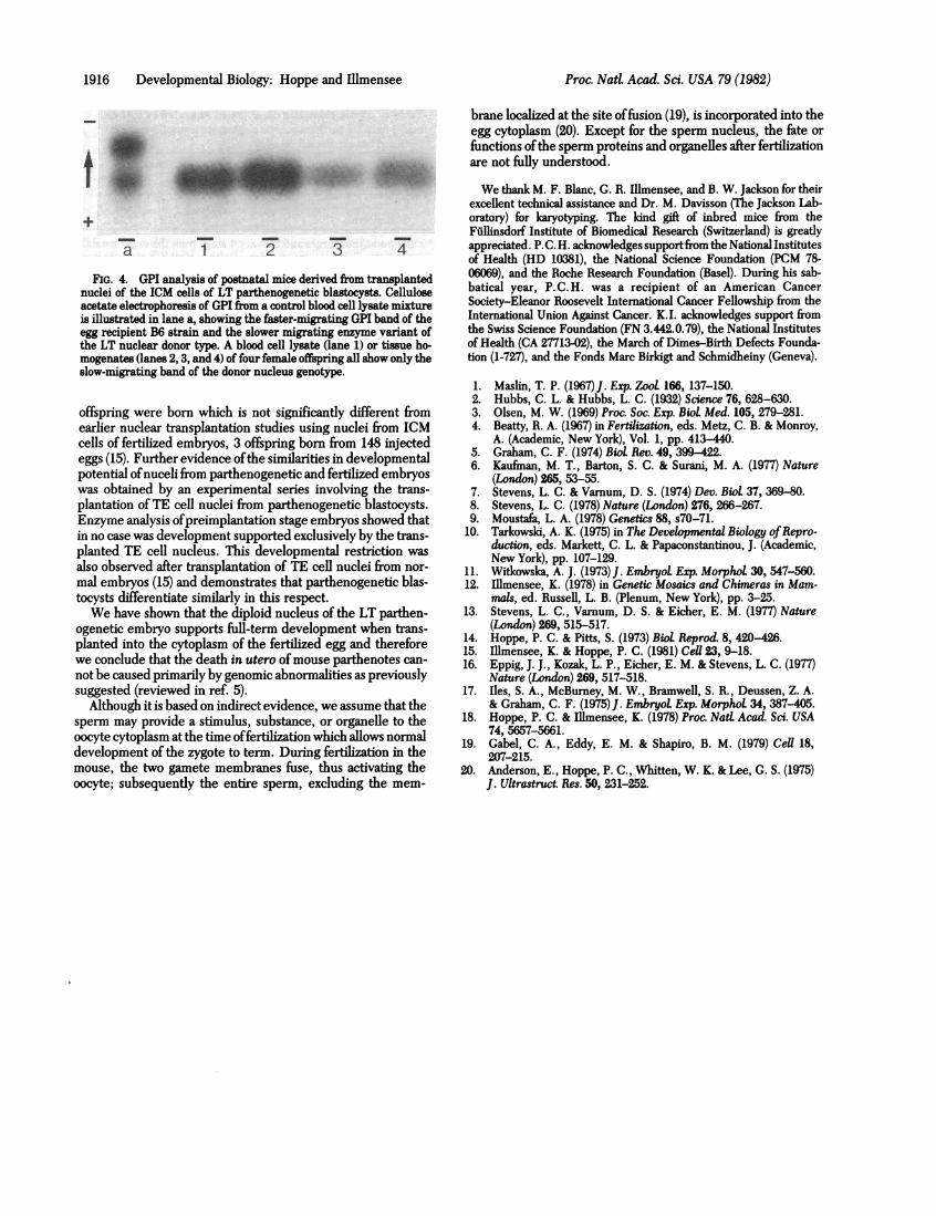
The dark-eyed baby from the first foster mother subse-quently developed the light coat color characteristic of the LTstrain (Fig. 3), and analysis of a blood cell lysate showed onlythe LT strain-specific variant of GPI (Fig. 4, lane 1). Matings
As comparison and control, see our previously published data withICM and TE nuclei from normal blastocysts (15).
Proc. Natl. Acad. Sci. USA 79 (1982) 1913'
1914 Developmental Biology: Hoppe and Illmensee
A
4
I~~~~~~~~Sl*
5
2
6
.t
'A
"'t. %
3
... .--
SI
I,
..
_ aon't n
B 1 2?
5
3 4
t acaren>/4ke*.Or 4 8,
6
FIG. 1. Nuclear transplant embryos at various preimplantation stages of development. Nuclei fromlTE and ICM cells of LT parthenogeneticblastocysts were-transplanted into fertilized but enucleated B6 eggs. (A) TE series: (1 and2) 4-c6l stage; (3) 8-cell stage; (4-7) early morulae. Allembryos appear to be morphologically abnormal. (B) ICM series: (1 and2) 4- to 5-cell stage; (3-6) morulae; (7 and 8) blastocysts apparently normalmorphologically. For each embryo, the corresponding GPI pattern is shown in Fig. 2.
of this female to males of the LT strain have produced 33 (15female and 18 male) normal LT offspring. Enzyme analysis oftissue homogenates from the other three dark-eyed femalesexpressed only the LT variant of GPI (Fig. 4, lanes 2, 3, and4). Chromosomal analysis revealed- that all four nuclear-trans-plant mice had a diploid karyotype indicating two X chromo-somes. Our data demonstrate that the nucleus of the ICM cellof the LT parthenogenetic blastocyst will support full-term de-velopment when transplanted into the fertilized egg.
DISCUSSIONMouse oocytes can be parthenogenetically activated either ex-
perimentally (5) or as occurs spontaneously in the LT inbred
strain (7). Parthenogenetic oocytes cleave and can develop intonormal-appearing blastocysts which implant but die within a
few days (10, 11). There is no evidence confirming the birth ofa mouse parthenote developing from an unfertilized egg. Onthe other hand, chimeric mice derived from aggregation ofdip-loid LT parthenogenetic and normal fertilized embryos exhibitsignificant cellular contributions, including the germ line, fromthe parthenogenetic embryonic cells (7, 8, 13). Apparentlythese cells do survive to the adult stage when associated withnormal cells. Similarly, cells of parthenogenetic embryos pro-liferate and differentiate into adult-appearing cells when trans-planted to extrauterine sites such as the testis (7) or kidney (17).
In LTfemales, parthenogenetic oocytes remaining within the
Table 2. Postimplantation development supported by ICM cellnuclei from LT parthenogenetic blastocysts transplanted intofertilized but enucleated B6 eggs
Embryos Mice bornGenotype of embryo transferred No. %
LT nuclear transplant 7* 4t 57ICR control 23 12 52
* Embryos at the morula and blastocyst stage from the experimentalseries B (see Table 1) were transferred with control ICR embryos intouteri of three ICR foster mothers.
tThree female mice died after premature birth.
oftatto((lapa'he'p11
GI
ofdeiGPegifro:B6butexjGP
ovE
an(
ger
injection of these LT teratocarcinoma cells into blastocysts re-J& ^ isults the birth ofchimeric which the injected cell
j t w participated in normal development and differentiation (12).The contribution or mechanism by which parthenogenetic cellscan be developmentally "rescued" when in contact with normal
---------- embryonic cells or adult somatic cells has not yet been deter-a 1 2 3 4 5 6 7 8 mined. The birth ofisodiploid mice developed from the genome
FIG. 2. GPI analysis by microelectrophoresis of total cell lysates of only one parent, either maternal or paternal in origin (18),
single preimplantation stage embryos developing after transplan- demonstrates that the cause of early parthenote death is not ation of nuclei from TE (A) or ICM (B) from LT parthenogenetic blas- consequence of containing only the maternal genome. Devel-cystsintofertilizedbutenucleatedB6 eggs. Abloodcelllysate control opment of these completely homozygous mice also arguesme a) of an F1 hybrid mouse (LT x B6) shows the fast-migrating GPI against the possible expression of recessive lethal genes as thettern of the B6, the slower migrating GPI variant of the LT, and the primary cause of parthenogenetic embryonic mortality interopolymericband. (A) From sevenembryos developing aftertrans- inbred strains of mice. Although it cannot be ruled out that in-
mntation of nuclei from TE cells, three express only the B6 parental?I (lanes 4, 5, and 7) and the four other embryos show the GPI pattern teraction of normal cells with parthenogenetic cells allows fur-
the F1 hybrid (lanes 1, 2, 3, and 6). (B) In contrast, two embryos ther development beyond the usual lethal period by compen-
veloped from the transplanted ICM cell nuclei as evidenced by their sating for possible genetic defects, the evidence suggests that'I expression of only the LT parental type (lanes 7 and 8). The faint the arrested development ofLT parthenogenetic embryos mayg-specificband observed inthese two blastocysts most likely resulted involve aberrant organismic rather than genetic factors. It ism a persisting maternal GPI pool. Two embryos exhibited only the therefore desirable to ascertain the developmental capacity ofipattern (lanes 1 and 2), two embryos showed both parental bands the enome of arthenogenetic embros directl b transplan-t not the heteropolymeric band (lanes 3 and 5), and two embryos t ge gofeofparthenogeneticfembryosirtly by bt enu-pressed the characteristic F1 hybrid pattern (lanes 4 and 6). For each tation of their 1CM and TE cell nuclei into fertilized but enu-
'I test, the corresponding embryo is shown in Fig. 1. cleated mouse eggs.The results from nuclear transplantation experiments dem-
ary develop into ovarian teratomas which are usually benign onstrate that nuclei from the ICM ofparthenogenetic embryosd are composed of differentiated cells derived from all three supported development of full-term offspring. Of 107 eggs in-rm layers (7). Infrequently these tumors are malignant, and jected with nuclei and subsequently enucleated, four female
A BFIG. 3. (A) Mice from the recipient B6 and donor LT inbred strains, showing their different phenotypes in coat color. (B) ICR foster mother with
an albino ICR control offspring and a female whose coat color demonstrates that her development was supported by the transplanted nucleus ofan ICM cell from an LT parthenogenetic blastocyst.
+
Proc. Nad Acad. Sci. USA 79 (1982) 1915
J..1
1916 Developmental Biology: Hoppe and Illmensee
a
FIG. 4. GPI analynuclei of the ICM cellacetate electrophoresiis illustrated in lane aegg recipient B6 straithe LT nuclear donormogenates (lanes 2,3,slow-migrating band o
offspring were bornearlier nuclear transcells of fertilized emeggs (15). Further evpotential ofnuceli frwas obtained by an
plantation ofTE cellEnzyme analysis ofpin no case was develcplanted TE cell nuc
also observed after t
mal embryos (15) an(
tocysts differentiateWe have shown tU
ogenetic embryo supplanted into the cytcwe conclude that thenot be caused primarsuggested (reviewed
Although it is base(sperm may provide a
oocyte cytoplasm at tIdevelopment of the zmouse, the two gamioocyte; subsequently
brane localized at the site offusion (19), is incorporated into theegg cytoplasm (20). Except for the sperm nucleus, the fate orfunctions ofthe sperm proteins and organelles after fertilizationare not fully understood.
MR" - ~We thank M. F. Blanc, G. R. Illmensee, and B. W. Jackson for theirexcellent technical assistance and Dr. M. Davisson (The Jackson Lab-oratory) for karyotyping. The kind gift of inbred mice from theFfillinsdorf Institute of Biomedical Research (Switzerland) is greatly
2 T T appreciated. P.C.H. acknowledges supportfrom the National Institutesof Health (HD 10381), the National Science Foundation (PCM 78-
sis of postnatal mice derived from transplanted 06069), and the Roche Research Foundation (Basel). During his sab-Is of LT parthenogenetic blastocysts. Cellulose batical year, P.C. H. was a recipient of an American Cancers of GPI from a control blood cell lysate mixture Society-Eleanor Roosevelt International Cancer Fellowship from theL, showing the faster-migrating GPI band of the International Union Against Cancer. K.I. acknowledges support fromin and the slower migrating enzyme variant of the Swiss Science Foundation (FN 3.442.0.79), the National Institutestype. A blood cell lysate (lane 1) or tissue ho- of Health (CA 27713-02), the March of Dimes-Birth Defects Founda-and 4) of four female offspring all show only the tion (1-727), and the Fonds Marc Birkigt and Schmidheiny (Geneva).)f the donor nucleus genotype.
1. Maslin, T. P. (1967)J. Exp. Zool 166, 137-150.2. Hubbs, C. L. & Hubbs, L. C. (1932) Science 76, 628-630.
which is not significantly different from 3. Olsen, M. W. (1969) Proc. Soc. Exp. Biol Med. 105, 279-281.plantation studies using nuclei from ICM 4. Beatty, R. A. (1967) in Fertilization, eds. Metz, C. B. & Monroy,ibryos, 3 offspring born from 148 injected A. (Academic, New York), Vol. 1, pp. 413-440.ridence ofthe similarities in developmental 5. Graham, C. F. (1974) Biol Rev. 49, 399-422.c
1parthenogenetic and fertilized embryos 6. Kaufinan, M. T., Barton, S. C. & Surani, M. A. (1977) Natuream parthenogenetic and fertilized embryos (London) 265, 53-55.experimental series involving the trans- 7. Stevens, L. C. & Varnum, D. S. (1974) Dev. Bol. 37, 369-0.
I nuclei from parthenogenetic blastocysts. 8. Stevens, L. C. (1978) Nature (London) 276, 266-267.reimplantation stage embryos showed that 9. Moustafa, L. A. (1978) Genetics 88, s70-71.)pment supported exclusively by the trans- 10. Tarkowski, A. K. (1975) in The Developmental Biology ofRepro-leus. This developmental restriction was duction, eds. Markett, C. L. & Papaconstantinou, J. (Academic,
ofTEellnulei frm nor-New York), pp. 107-129.ransplantation of TE cell nuclei from nor- 11. Witkowska, A. J. (1973)J. Embryol Exp. Morphol 30, 547-560.d demonstrates that parthenogenetic blas- 12. Illmensee, K. (1978) in Genetic Mosaics and Chimeras in Mam-similarly in this respect. mals, ed. Russell, L. B. (Plenum, New York), pp. 3-25.iat the diploid nucleus of the LT parthen- 13. Stevens, L. C., Varnum, D. S. & Eicher, E. M. (1977) Natureports full-term development when trans- (London) 269, 515-517.)plasm of the fertilized egg and therefore 14. Hoppe, P. C. & Pitts, S. (1973) Biol. Reprod. 8, 420-426.15. Illmensee, K. & Hoppe, P. C. (1981) Cell 23, 9-18.death in utero of mouse parthenotes can- 16. Eppig, J. J., Kozak, L. P., Eicher, E. M. & Stevens, L. C. (1977)ily by genomic abnormalities as previously Nature (London) 269, 517-518.in ref. 5). 17. Iles, S. A., McBurney, M. W., Bramwell, S. R., Deussen, Z. A.d on indirect evidence, we assume that the & Graham, C. F. (1975)J. Embryol Exp. Morphol 34, 387-405.a stimulus, substance, or organelle to the 18. Hoppe, P. C. & Illmensee, K. (1978) Proc. Natl Acad. Sci. USAhe time offertilization which allows normal 74, 5657-5661.
_tr.Dfertilization in the 19. Gabel, C. A., Eddy, E. M. & Shapiro, B. M. (1979) Cell 18,zygote to term. During Iertszatlon m tne 207-215.fete membranes fuse, thus activating the 20. Anderson, E., Hoppe, P. C., Whitten, W. K. & Lee, G. S. (1975)Z the entire sperm, excluding the mem- J. Ultrastruct. Res. 50, 231-252.