Name: ........................................................................... ( ) Class: 3 ............. Date: .................... Topic: Transport in Humans Learning Outcomes: Students should be able to: Describe the circulatory system as a system of tubes with a pump and valves to ensure one-way flow of blood Describe the double circulation in terms of a low pressure circulation to the lungs and a high pressure circulation to the body tissues and relate these differences to the different functions of the two circuits Describe the structure and function of the heart in terms of muscular contraction and the working of valves *State that heart action is initiated at the pacemaker (sino-atrial node) Outline the cardiac cycle in terms of what happens during systole and diastole with involvement of the heart valves (Histology of the heart muscles, names of nerves and transmitter substances are not required) Describe the effect of exercise on heart rate and its significance Identify the main blood vessels to and from the heart, lungs, liver and kidney Relate the structure of arteries, veins and capillaries to their functions and be able to recognize these vessels from photomicrographs *State the origin of blood pressure *Describe how blood pressure is measured *Describe how a pulse is generated State the functions of various blood components: red blood cells - haemoglobin and oxygen transport white blood cells - phagocytosis, antibody formation and tissue rejection platelets - fibrinogen to fibrin, causing clotting plasma - transport of blood cells, ions, soluble food substances, hormones, carbon dioxide, urea, vitamins, plasma proteins Identify red and white blood cells as seen under the microscope on prepared slides, and in diagrams and photomicrographs Explain the role of haemoglobin in the transport of oxygen *Explain the significance of the Bohr shift in the oxygen dissociation curve List the different ABO blood groups and all possible combinations for the donor and recipient in blood transfusion *Differentiate between the innate and adaptive immune systems in the generation of an immune response Describe the transfer of materials between capillaries and tissue fluid Describe coronary heart disease in terms of the occlusion of coronary arteries and list the possible causes, such as diet, stress and smoking, stating the possible preventive measures *Explain the cause of deep vein thrombosis and its prevention HWA CHONG INSTITUTION (HIGH SCHOOL) SECONDARY 3 BIOLOGY 2012 NOTES Secondary 3 Biology, Transport in Humans, Notes, 2012, CHS 1
Describe the circulatory system as a system of tubes with a pump and valves to ensure one-way flow of blood Describe the double circulation in terms of a low pressure circulation to the lungs and a high pressure circulation to the body tissues and relate these differences to the different functions of the two circuits Describe the structure and function of the heart in terms of muscular contraction and the working of valves *State that heart action is initiated at the pacemaker (sino-atrial node)Outline the cardiac cycle in terms of what happens during systole and diastole with involvement of the heart valves (Histology of the heart muscles, names of nerves and transmitter substances are not required) Describe the effect of exercise on heart rate and its significance Identify the main blood vessels to and from the heart, lungs, liver and kidney Relate the structure of arteries, veins and capillaries to their functions and be able to recognize these vessels from photomicrographs*State the origin of blood pressure *Describe how blood pressure is measured *Describe how a pulse is generated State the functions of various blood components:
red blood cells - haemoglobin and oxygen transport white blood cells - phagocytosis, antibody formation and tissue rejection platelets - fibrinogen to fibrin, causing clotting plasma - transport of blood cells, ions, soluble food substances, hormones, carbon dioxide, urea, vitamins, plasma proteins
Identify red and white blood cells as seen under the microscope on prepared slides, and in diagrams and photomicrographs Explain the role of haemoglobin in the transport of oxygen *Explain the significance of the Bohr shift in the oxygen dissociation curve List the different ABO blood groups and all possible combinations for the donor and recipient in blood transfusion *Differentiate between the innate and adaptive immune systems in the generation of an immune response Describe the transfer of materials between capillaries and tissue fluid Describe coronary heart disease in terms of the occlusion of coronary arteries and list the possible causes, such as diet, stress and smoking, stating the possible preventive measures *Explain the cause of deep vein thrombosis and its prevention
Secondary 3 Biology, Transport in Humans, Notes, 2012, CHS1
ABO blood typing system
Blood typing involves the two types of molecules called:
AntigenProtein or polysaccharide, that stimulates the immune system to react, such as to produce antibodies
AntibodyProtein produced in response to the presence of antigen
Each antibody combines with a specific antigen.
The most common system for typing blood is the ABO systemIn the ABO system, the presence or absence of type A and type B antigens on red blood cells determines a person’s blood typeFor example:
A person has type A blood, the A antigen is on his/her red blood cellsThis molecule is not a foreign antigen to this individual, although it can be an antigen to a recipient who does not have type A blood
There are four types of blood: A, B, AB and O
Within the plasma, there are antibodies to the antigens that are not present on the person’s red blood cellsThese antibodies are called anti-A and anti-BIt is reasonable that type A blood would have anti-B and not anti-A antibodies in the plasmaIf anti-A antibodies were present in the plasma, agglutination, or clumping of red blood cells would occur
Antigens on an erythrocyte (Red blood cell)
A red blood cell (RBC) has three different antigens on the surface of its membraneThe antigens are glycoproteins with unique molecular shapesThey have molecular weights of 200,000 to 300,000
Secondary 3 Biology, Transport in Humans, Notes, 2012, CHS2
Three different types of blood antibodies that circulate in the plasmaEach antibody has two combining sites where it attaches to the complimentary antigen on the surface of a red blood cell (RBC) membraneAnti-A and Anti-B antibodies are glycoproteins with a molecular weight of about 900,000Anti-Rh antibodies are smaller glycoproteins with a molecular weight of about 150,000
!Type A erythrocyte
Red blood cells (RBCs): Type A Positive (left) and type A Negative (right). Both types have the A antigen, but only the A Positive (left) has the Rh antigen.
Type B erythrocyte
Red blood cells (RBCs): Type B Positive (left) and type B Negative (right). Both types have the B antigen, but only the B Positive (left) has the Rh antigen.
Secondary 3 Biology, Transport in Humans, Notes, 2012, CHS3
Type AB erythrocyte
Red blood cells (RBCs): Type AB Positive (left) and type AB Negative (right). Both types have A and B antigens, but only the AB Positive (left) has the Rh antigen.
Type O erythrocytes
Red blood cells (RBCs): Type O Positive (left) and type O Negative (right). Both types are without A and B antigens, but the O Positive (left) have the Rh antigen. Type O Negative (right) has none of the antigens (A, B or Rh) on its membrane.
Agglutination (clumping)
Agglutination (clumping) of type A red blood cells (RBCs) by anti-A antibodiesThe antibodies have two combining sites and are able to attach to the A antigens on adjacent RBCs, thus causing the RBCs to bond together
Agglutination of red blood cells can cause blood to stop circulating in small blood vessels, and this can lead to organ damageIt is also followed by hemolysis, which may cause the death of the individual
Blood transfusion
For a recipient to receive blood from a donor, the recipient's plasma must not have an antibody that causes the donor’s cells to agglutinateImportant to determine each person’s blood typeBlood that is of good quality and free of infectious agents
Secondary 3 Biology, Transport in Humans, Notes, 2012, CHS4
Blood typing kit
Using the antibodies derived from plasma (antibodies are immobilized onto the blood typing kit) to determine the blood type If clumping occurs after a sample of blood is exposed to a particular antibody; the person has that type of blood
Rhesus system (Rh system) - For Enrichment
Another important antigen in matching blood type is the Rh factor.Rh+ (Rh positive)
Antigen is present on the red blood cells.Rh- (Rh negative)
Antigen is not present on the red blood cells. Individuals normally do not have antibodies to the Rh factor, but they may make them when exposed to the Rh factor. Anti – Rh antibodies are used for blood testingWhen Rh+ blood is mixed with anti – Rh antibodies, agglutination occurs
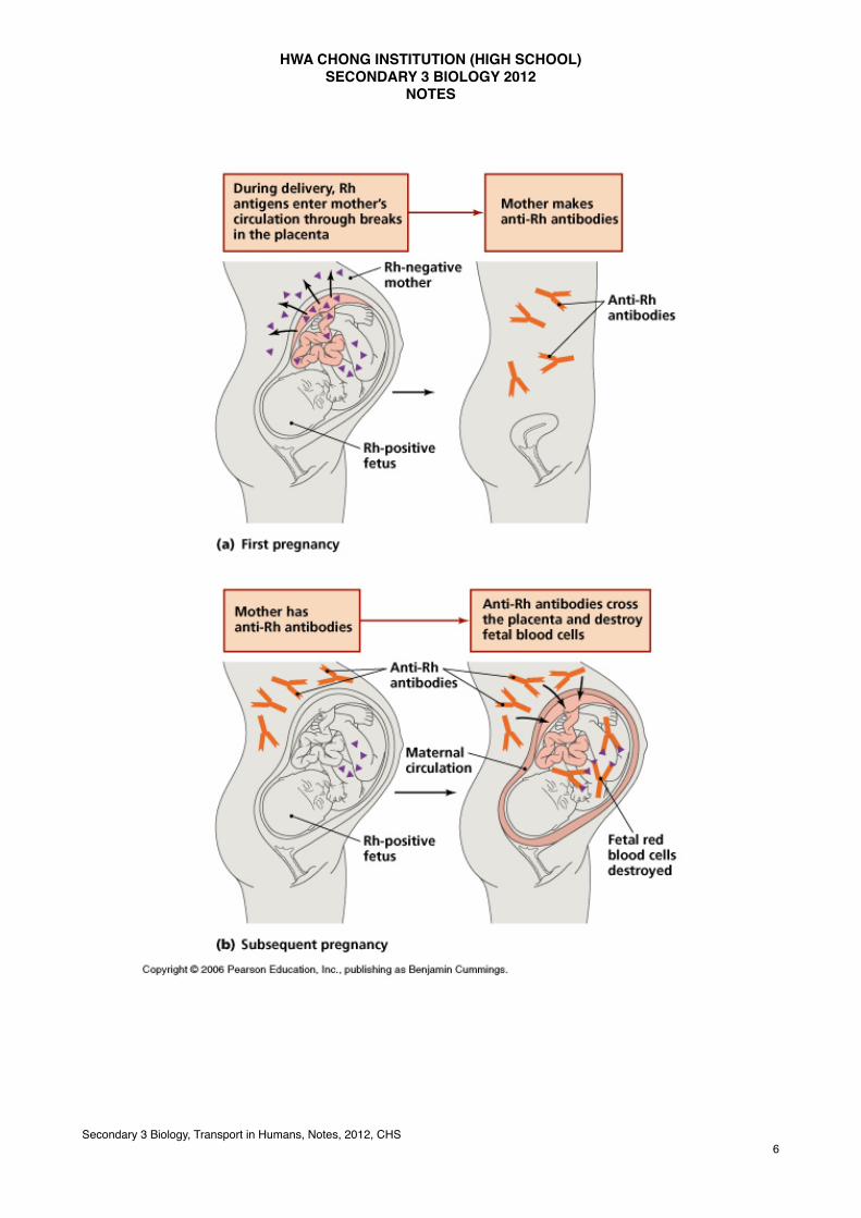
Hemolytic disease of the newborn
During pregnancy, if the mother is Rh- and the father is Rh+, there is a possibility that the child is Rh+.The Rh+ red blood cells may begin leaking across the placenta into the mother’s cardiovascular system, as placental tissues normally break down before and at birth.The presence of these Rh antigens causes the mother to produce anti – Rh antibodies. In this or a subsequent pregnancy with another Rh+ baby, the anti – Rh antibodies produced by the mother may cross the placenta and destroy the child’s red blood cells. The Rh problem is prevented by giving the Rh- women an Rh immunoglobulin injection either midway through the pregnancy or no later than 72 hours after giving birth to any Rh+ child.Injection contains anti – Rh antibodies that attack any of the baby’s red blood cells in the mother’s blood before these cells can stimulate her immune system to produce her own antibodies.
Secondary 3 Biology, Transport in Humans, Notes, 2012, CHS6
Component of Blood
The red blood cells (erythrocytes)
Small, biconcave disks that lacks a nucleus when matureOccurs in great quantities4 – 6 million red blood cells per mm3 of whole bloodAbsence of a nucleus provides more space for the pigment haemoglobinRespiratory pigment because it transports oxygenA red blood cell contains about 200 million haemoglobin moleculesIf this much haemoglobin is suspended within the plasma rather than enclosed within the cells, blood would be so viscous; the heart would have difficulty pumping it
Secondary 3 Biology, Transport in Humans, Notes, 2012, CHS7
The iron portion of haemoglobin carries oxygen, a molecule that cells require for cellular respirationHb + O2 ↔ HbO2
Haemoglobin, which is combined with oxygen, is called oxyhaemoglobinFormed in the lungs and has a bright red colourHaemoglobin, which has given up oxygen to tissue fluid, is called de-oxyhaemoglobinDark purplish colour
The life cycle of an erythrocyte
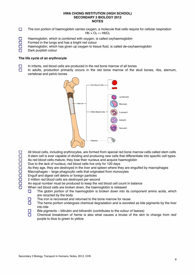
In infants, red blood cells are produced in the red bone marrow of all bonesIn adults, production primarily occurs in the red bone marrow of the skull bones, ribs, sternum, vertebrae and pelvic bones
All blood cells, including erythrocytes, are formed from special red bone marrow cells called stem cellsA stem cell is ever capable of dividing and producing new cells that differentiate into specific cell types.As red blood cells mature, they lose their nucleus and acquire haemoglobinDue to the lack of nucleus, red blood cells live only for 120 daysAs they age, they are destroyed in the liver and spleen where they are engulfed by macrophagesMacrophages – large phagocytic cells that originated from monocytesEngulf and digest cell debris or foreign particles2 million red blood cells are destroyed per secondAn equal number must be produced to keep the red blood cell count in balanceWhen red blood cells are broken down, the haemoglobin is released
The globin portion of the haemoglobin is broken down into its component amino acids, which are recycled by the bodyThe iron is recovered and returned to the bone marrow for reuseThe heme portion undergoes chemical degradation and is excreted as bile pigments by the liver into bileBile pigments – bilirubin and biliverdin (contributes to the colour of faeces)Chemical breakdown of heme is also what causes a bruise of the skin to change from red/purple to blue to green to yellow
Secondary 3 Biology, Transport in Humans, Notes, 2012, CHS8
The number of red blood cells produced increases whenever arterial blood carries a reduced amount of oxygen
E.g. when an individual first takes up residence at a high altitude (acclimatisation) or loses red blood cells or full use of their lungs.
Kidneys accelerate their release of erythropoietin, a hormone that is carried in blood to red bone marrowSpeeds up the maturation of cells that are in the process of becoming red blood cellsLiver and other tissues also produce erythropoietinMass – produced through biotechnologySometimes abused by athletes in order to raise their red blood cell counts and thereby increase the oxygen – carrying capacity of their blood
Anemia
When there is an insufficient number of red blood cells or the cells do not have enough haemoglobin, the individual suffers from anemia
Tired, run – down feelingDiets do not contain enough iron or folic acidWhole – grain cereals (rich in iron and folic acid)
Pernicious anemiaDigestive tract is unable to absorb enough vitamin B12, found in diary products, fish, eggs and poultryEssential for proper formation of red blood cellsImmature red blood cells tend to accumulate in the bone marrow in large quantitiesSpecial diet and administration of vitamin B12 by injection is an effective treatment
Hemolytic anemia (Hemolysis – rupturing of red blood cells) Increased rate of red blood cells destruction
Secondary 3 Biology, Transport in Humans, Notes, 2012, CHS9
Sickle – cell disease Hereditary condition in which the individual has sickle-shaped red blood cells that tends to rupture as they pass through the narrow capillaries.
Leukocytes (White blood cells)
Leukocytes differ from erythrocytes in that they are:Usually largerHave a nucleusLack haemoglobinThere are only 5000 to 11000 per mm3 of blood
Fight infectionLeukocytes are derived from stem cells in the red bone marrowUndergo several maturation stagesProduction of leukocytes increases whenever the body is invaded by pathogensHormones called colony – stimulating factors are released by white blood cells, and circulate back to the bone marrow, stimulating an increased productionRed blood cells are confined to the blood, but leukocytes are able to squeeze through pores in the capillary wallsTherefore, they are found in tissue fluid and lymphWhen there is an infection, leukocytes greatly increase in number
Secondary 3 Biology, Transport in Humans, Notes, 2012, CHS10
Types of leukocytes - For Enrichment
Leukocytes are classified into the granular leukocytes and the agranular leukocytesBoth types of cells have granules in the cytoplasm surrounding the nucleus, but the granules are more visible upon staining in granular leukocytesGranules contain various enzymes and proteins, which help leukocytes, defend the bodyThree types of granular leukocytes and two types of agranular leukocytesDiffer by the size of the cell and shape of the nucleusDiffer in their functions
Secondary 3 Biology, Transport in Humans, Notes, 2012, CHS11
Granular leukocytes
Neutrophils, eosinophils and basophils are granular leukocytes
Neutrophils Most abundant of the white blood cellsMultilobed nucleus joined by nuclear threadsAlso called polymorphonuclearFirst type of leukocytes to respond to an infection and they engulf pathogens during phagocytosis
What is phagocytosis?
The process of engulfing and ingesting foreign particles, such as bacteria, by the leukocytes is known as phagocytosisA phagocyte first engulfs the bacteria by flowing over them and enclosing themThe phagocyte then ingests the bacteriaThe ingested bacteria will be digested by the phagocyteIn the process of ‘fighting’ with the bacteria at the site of the wound, some of the phagocytes are killedThese dead phagocytes, together with the dead bacteria, form pus
Secondary 3 Biology, Transport in Humans, Notes, 2012, CHS12
Eosinophils
Bilobed nucleusIncrease in number when there is a parasitic worm infection or in the case of allergic reactions
Basophils
U – shaped or lobed nucleusBasophils enter the tissues and are believed to become mast cells, which release the histamine associated with allergic reaction Histamine dilates blood vessels and causes contraction of smooth muscle
Agranular leukocytes
Monocytes and lymphocytes are agranular leukocytesResponsible for specific defense to particular pathogens and their toxins (poisonous substances)Pathogens have molecules called antigens that allow the immune system to recognize them as foreign
Monocytes
Kidney – shaped nucleusLargest of the leukocytesAfter taking residence in the tissues, they differentiate into even larger macrophagesPhagocytose pathogens, old cells and cellular debrisStimulate other leukocytes to defend the body
Secondary 3 Biology, Transport in Humans, Notes, 2012, CHS13
Lymphocytes
Spherical – shaped nucleus2 types – B lymphocytes and T lymphocytesB lymphocytes protect us by producing antibodiesT lymphocytes protect us by destroying any cell that has foreign antigens.
Secondary 3 Biology, Transport in Humans, Notes, 2012, CHS16
Model of Immune Response:Speed and Specificity
Response
time after infection
Innate immune responseAdaptive immune response
Defence mechanisms of the body
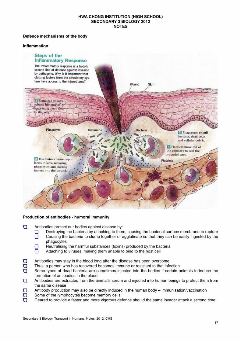
Inflammation
Production of antibodies - humoral immunity
Antibodies protect our bodies against disease by:Destroying the bacteria by attaching to them, causing the bacterial surface membrane to ruptureCausing the bacteria to clump together or agglutinate so that they can be easily ingested by the phagocytesNeutralising the harmful substances (toxins) produced by the bacteriaAttaching to viruses, making them unable to bind to the host cell
Antibodies may stay in the blood long after the disease has been overcomeThus, a person who has recovered becomes immune or resistant to that infectionSome types of dead bacteria are sometimes injected into the bodies if certain animals to induce the formation of antibodies in the bloodAntibodies are extracted from the animal’s serum and injected into human beings to protect them from the same diseaseAntibody production may also be directly induced in the human body – immunisation/vaccinationSome of the lymphocytes become memory cellsGeared to provide a faster and more vigorous defence should the same invader attack a second time
Secondary 3 Biology, Transport in Humans, Notes, 2012, CHS17
Cell mediated immunity
When antibody – mediated immunity is ineffectiveWhen foreign organisms have successfully invaded and are multiplying inside the cells of the bodyInfected cells display the foreign antigens on their surfaceLymphocytes – cytotoxic cells (N K cells) produce proteins which create holes in the infected cellProtein from the cytotoxic cells will enter the infected cell and trigger programmed cell death
Secondary 3 Biology, Transport in Humans, Notes, 2012, CHS18
Test yourself – Name the leukocytes!
Leukemia
Abnormally large number of immature leukocytes that fill the red bone marrowPrevents erythrocytes developmentAnemia results and the immature leukocytes offer little protection from disease Type of cancer
Blood platelets (thrombocytes)
Not true cellsMembrane – bound fragments of cytoplasm from certain bone marrow cellsPlay a part in clotting of blood
Blood clotting
When blood vessels are damaged, damaged tissues and blood platelets release an enzyme known as thrombokinaseThrombokinase converts the protein prothrombin, normally present in the plasma, into thrombinCalcium ions must be present before this can take placeThrombin is also an enzyme. It catalyses the conversion of soluble protein fibrinogen to insoluble threads of fibrinFibrin threads entangle blood cells and the whole mass form a clotIn undamaged blood vessels, the blood does not clotPresence of an anti – clotting substance called heparinProduced in the liverThrombokinase neutralizes the action of heparin so that clotting can take placeWhen blood clots, a yellowish liquid called serum is left behindSerum has the same composition as plasma except that it lacks the clotting factor
Secondary 3 Biology, Transport in Humans, Notes, 2012, CHS20
thrombokinase
Real world application - warfarin
Warfarin (Coumadin®) is the most commonly used prescription medication for preventing harmful blood clots from forming or from growing larger. Warfarin belongs to a class of drugs called anticoagulants, which simply means medications that prevent the blood from clotting. People often call these drugs "blood thinners."
Secondary 3 Biology, Transport in Humans, Notes, 2012, CHS21
It is vital that our bodies can form blood clots to control bleeding. However, many medical conditions and inherited factors can make a person more likely to form abnormal blood clots. Abnormal blood clots are dangerous because they can block the flow of blood to parts of the body like the heart, lungs, or brain.
Some conditions that are treated with warfarin on a short-term or long-term basis include:
Irregular heartbeatHeart valve replacementPrevious heart attack or strokeBlood clot in a vein (venous thrombosis or deep vein thrombosis)Blood clot in the lung (pulmonary embolism)Certain orthopedic surgeries, such as knee or hip replacementInherited blood clotting disorders, such as Factor V Leiden
Haemophilia
Inherited clotting disorder due to deficiency in a clotting factorSlightest bump can cause the affected person to bleed into the jointsNormal clotting mechanism is greatly impairedSlight injuries may cause a person to bleed to death or die of internal bleeding