Page 1
Michael Sapateiro Luís 2013 1
MICHAEL SAPATEIRO LUÍS
TRASTUZUMAB IN ADVANCED GASTRIC CANCER:
PROPORTION OF ELIGIBLE CASES IN A SINGLE CENTRE
Dissertação de Candidatura ao grau
de Mestre em Oncologia submetida ao
Instituto de Ciências Biomédicas de
Abel Salazar da Universidade do Porto.
Orientador – Doutor Lúcio Lara Santos
Afiliação – Instituto de Ciências
Biomédicas Abel Salazar da
Universidade do Porto.
Page 2
Michael Sapateiro Luís 2013 2
AGRADECIMENTOS
Quero prestar os meus agradecimentos ao Prof. Doutor Lúcio Lara Santos, por todo o
tempo disponibilizado e pela valiosa orientação na elaboração desta dissertação.
Um grande obrigado à Ana Tavares, companheira de percurso neste projecto, pelo
apoio e persistência com que contribuiu para que em conjunto o trabalho tomasse
forma.
Ao Prof. Doutor Fernando Schmitt e à Dr.ª Dina Leitão pela contribuição fundamental
na análise patológica.
Ao Prof. Doutor Jorge Valente, Prof. Rui Henrique e Dr. Luís Pedro Afonso, pela
disponibilidade, apoio e amabilidade.
À Dr.ª Ana Raimundo e Dr.ª Deolinda Pereira, pelas valiosas sugestões feitas ao longo
trabalho.
Ao Dr. António Miguel Abreu e Dr. Ramon Mello, cujo apoio foi fundamental na
conclusão deste trabalho.
Aos colegas de trabalho que contribuíram com sugestões e disponibilizaram o seu
tempo.
À minha família e amigos, pela paciência manifestada nos dias de maior
indisponibilidade e por serem o meu sustentáculo.
À Ana Isabel, que por causa desta tarefa teve de prescindir de algumas coisas e a
quem dedico este trabalho.
Page 3
Michael Sapateiro Luís 2013 3
No contexto da realização deste trabalho, foram publicados os seguintes textos:
Artigo de revisão: Michael Luis, Ana Tavares, Luis-Silva Carvalho, Lucio Lara-
Santos, António Araújo, Ramon A de Mello. Personalizing therapies in
advanced gastric cancer – molecular mechanisms, current biomarkers and
novel anti-HER2 therapies World J Gastroenterol. 2013 October 14; 19(38):
6383–6397
Capítulo de livro: Michael Luis, Ramon A de Mello. HER2 Over-Expression and
Gastric Cancer: Molecular Mechanisms and Target Therapies. In: Atta-ur-
Rahman and M. Iqbal Choudhary (eds.), Frontiers in Anti-Cancer Drug
Discovery, Volume (2), Bentham Science Publishers, 2013. ISBN: 978-1-
60805-809-9
Page 4
Michael Sapateiro Luís 2013 4
ABSTRACT
Gastric cancer is the fourth most commonly diagnosed cancer and the second leading
cause of cancer death worldwide. HER2 (human epidermal growth factor receptor 2),
an oncogene of the HER family of growth factors has been shown to be overexpressed
in gastric cancer. Trastuzumab is a monoclonal antibody targeting HER2, which
demonstrated to significantly prolong overall survival in association with standard
chemotherapy in this patients in the ToGA (Trastuzumab for GAstric Cancer) trial.
These data contributed for the approval of trastuzumab in combination therapy in the
treatment of metastatic adenocarcinoma of the stomach or gastro-esophageal junction
by various international authorities. Aiming to address the particular issues of the
patients diagnosed and treated at Instituto Português de Oncologia do Porto (IPOP), a
retrospective cohort study was designed in order to estimate the proportion of patients
eligible for trastuzumab therapy at this particular centre.
Ninety-eight patients admitted during 2005 and 2006 at IPOP with metastatic or locally
advanced gastric or GEJ intestinal-type adenocarcinoma were included and
characterized according to clinicopathological variables. HER2-status was determined
by immunohistochemistry. Ambiguous cases were analyzed by silver in-situ
hybridization.
Overall survival in the study sample was 36 months [24-48], with a disease-free
survival of 12 months [8-16]. Overall positivity for HER2 was 16.3%. HER2 correlated
significantly with pathological stage, N-status and N-ratio (p < 0.05). Overall survival in
HER2- patients was 43 vs. 19 months in HER2 positive patients (p=0.032).
Our data suggest an important role for HER2 as prognostic factor. The methods used
for HER2 assessment proved to be efficient. Anti-HER2 therapeutics in gastric cancer
seem to have a role the adjuvant setting and in patients with locally advanced disease.
Key-words (MESH): stomach neoplasms, esophageal neoplasms, trastuzumab,
ERBB2, amplification, immunohistochemistry, in situ hybridisation
Page 5
Michael Sapateiro Luís 2013 5
RESUMO
O cancro gástrico é a 4ª neoplasia mais frequentemente diagnosticada e a 2ª causa de
morte por cancro no mundo. O oncogene HER2 (human epidermal growth factor
receptor 2), pertencente à família HER dos factores de crescimento, encontra-se
sobre-expresso no cancro gástrico. O trastuzumab é um anticorpo monoclonal dirigido
ao HER2, que demonstrou prolongar a sobrevivência em associação com
quimioterapia nos doentes que sobre-expressam o HER2, no ensaio clínico ToGA
(Trastuzumab for GAstric Cancer). Estes dados contribuíram para a aprovação do
trastuzumab em terapêutica combinada no tratamento do adenocarcinoma metastático
do estômago e da junção esófago-gástrica (JEG). No intuito de perceber a realidade
existente no Instituto Português de Oncologia do Porto (IPOP), foi desenhado um
estudo de coorte retrospectivo, com o objectivo primário de estimar a proporção de
doentes potencialmente elegíveis para terapêutica com trastuzumab.
Noventa e oito doentes admitidos durante 2005 e 2006 com o diagnostico de
adenocarcinoma do tipo intestinal do estômago ou JEG foram incluídos e analizados
quanto a variáveis clinicopatológicas. O status HER2 foi determinado por
imunohistoquímica, tendo os casos ambíguos sido analisados por hibridização in situ
com prata.
A sobrevivência global foi de 36 meses [24-48], com uma sobrevivência livre de
doença de 12 meses [8-16]. A positividade global para o HER2 foi de 16,3%. O status
HER2 correlaciona-se significativamente com o estádio patológico, positividade
ganglionar e razão de gânglios metastizados / gânglios excisados (p < 0.05). A
sobrevivência global em doentes HER2- foi de 43 vs. 19 meses em doentes HER2+
(p=0.032).
Os dados sugerem um papel importante para o HER2 como factor de prognóstico. Os
métodos utilizados para a determinação do status HER2 demonstraram ser eficazes. A
terapêutica HER2 no cancro gástrico parece ter um papel no contexto adjuvante e em
doentes com doença localmente avançada.
Page 6
Michael Sapateiro Luís 2013 6
"É preciso fazer um esforço contínuo para amar o presente. Viver pelo
passado, pelo que se fez, pelo que se conseguiu, é o mesmo que alimentar
uma fome premente com banquetes de outrora."
Miguel Torga
Page 7
Michael Sapateiro Luís 2013 7
CONTENTS
Introduction 8
Material and Methods 34
Results 37
Discussion 54
Conclusion 56
References 57
Page 8
Michael Sapateiro Luís 2013 8
INTRODUCTION
Epidemiology of Gastric Cancer
Gastric cancer is the fourth most commonly diagnosed cancer and the second leading
cause of cancer death worldwide, with an estimated 990.000 new cases and 738.000
cancer deaths in 2008 (1). In the northern region of Portugal, gastric cancer has an
incidence rate of about 47.4/100.000 person-years in men and 28/100.000 person-
years in women (2). Incidence rates show considerable geographic variation, varying
from 3.3 per 100.000 in men and 2.0 in women in Egypt to 65.9 in men and 25.9 in
women in Korea. Highest rates are found in Asian and South American countries.
These large regional variations possibly reflect different prevalences of Helicobacter
pylori infection, which is responsible for more than 60% of gastric cancer globally (3).
About 85% of gastric cancers are adenocarcinomas, with 15% comprised of
lymphomas, gastrointestinal stromal tumors (GIST) and leiomyosarcomas. Gastric
adenocarcinomas can be subdivided into diffuse and intestinal types based on the
Lauren classification, which constitute pathological entities with distinct epidemiological
and prognostic features (4). In diffuse type gastric adenocarcinoma cell cohesion is
absent; they occur throughout the entire stomach and more often in younger patients
than in intestinal type adenocarcinoma and carry a worse prognosis. Intestinal type
gastric adenocarcinoma presents cohesive cells arranged in glandlike structures; they
occur more commonly in the antrum and lesser curvature of the stomach (4-6).
Different etiologic factors seem to be involved, as incidence of diffuse type is similar in
most populations while intestinal type predominates in high risk regions. A decrease in
gastric cancer incidence in the United States in the last 70 years has been mainly
observed for the intestinal type, associated with Helicobacter pylori infection and
therefore reflecting improved sanitary conditions and food conservation methods,
including refrigeration; on the other hand, the incidence of diffuse type gastric cancer
has remained constant over time and seems to be increasing recently, both in Western
and Eastern parts of the world (7-9).
Tumors at the junction of the esophagus with the stomach are oftentimes of either
gastric or esophageal origin and are therefore referred as gastroesophageal junction
(GEJ) cancers. Although the incidence and mortality of gastric cancer are globally
decreasing, the incidence of GEJ cancer is increasing in Europe and the United States
(10, 11). This increase is thought to reflect an increase in gastroesophageal reflux
disease associated with the increasing prevalence of obesity in these countries. In the
last three decades, a tendency towards more proximal located gastric cancer (cardia
Page 9
Michael Sapateiro Luís 2013 9
vs. corpus and pylorus) is emerging, probably explained by a parallel increase of
Barrett’s esophagus and the decline in Helicobacter pylori infection (10, 12).
In western countries, 50 - 80% of patients present with inoperable advanced or
metastatic disease, which in combination with patient co-morbidities, age (the majority
of patients is > 65 years old) and the relative chemoresistance of the disease
contributes to poor overall survival (OS). A prognostic index for locally advanced and
metastatic esophago-gastric cancer (the Royal Marsden Hospital Prognostic Index)
was initially published in 2004, with data supporting its validity presented in 2009 (13,
14). Four independent poor prognostic factors are taken in consideration: performance
status ≥ 2, liver metastasis, peritoneal metastasis, and serum alkaline phosphatase ≥
100 U/L. Patients are subdivided into good (no risk factor), moderate (1 or 2 risk
factors), and poor (3 or 4 risk factors) risk groups. Applying this index to the data of the
REAL-2 study (described below) resulted in statistically significant differences between
the 3 risk groups in terms of median survival and 1-year survival rates (median survival
times for good, moderate, and poor risk groups were 12.7, 8.6, 4.3 months
respectively; 1-year survival rates were 52.4%, 33.1% and 13.7% respectively (14).
Two-thirds of patients amenable to curative surgical resection will have recurrence
within 2 years (15-17). The late presentation contributes to the grim prognosis
associated with gastric cancer, with 5-year survival rate for metastatic gastric cancer
being estimated at 5-20% and median survival times ranging from 7 to 10 months (18,
19). The survival rates of patients with resectable disease ranges from 10-30% (20).
Combination chemotherapy has been shown to improve overall survival in comparison
with single-agent chemotherapy or best supportive care (21). Current management
typically consists of fluoropyrimidine and platinum-based combinations with or without a
third drug (usually docetaxel or epirubicin) (22). However, data available have been
conflicting when it comes to evaluate the clinical outcome with different treatment
strategies, which can explain the lack of global consensus for adjuvant chemotherapy
(23, 24). Considering the poor prognosis of advanced gastric or GEJ cancer, new
therapeutic regimens with acceptable toxicity have been actively pursued.
Pathology of gastric cancer
Sporadic intestinal type gastric adenocarcinoma is thought to arise in a multi-step
process of intestinal metaplasia – dysplasia – carcinoma, involving mutations or
deregulation of genes regulating key events in the cancerization process such as
apoptosis, cell cycle regulation, proliferation and metabolism, in a similar fashion of the
progression of colon adenomas to carcinoma (25). Therefore, accumulation of
molecular genetic alterations involving activation of oncogenes and inactivation of
Page 10
Michael Sapateiro Luís 2013 10
tumor suppressor genes are likely involved in the process of carcinogenesis. Other
implicated genes include p53, CHD1, SMAD4, FHIT and CDKN2A. On the other hand,
no precursor cells have been clearly identified in diffuse type gastric adenocarcinoma,
but aberrations in the FGFR2/ErbB3/PI3 kinase pathway are frequent in this kind of
neoplasia (26-29).
Information on the specific pathways involved is scarce; however, HER2 has been
shown to be overexpressed/amplified in gastric cancer and correlation between HER2
expression and intestinal type gastric cancer has been reported (30). HER2 is a
member of the epidermal growth factor receptor (EGFR) family of growth factors, with
intrinsic protein tyrosine kinase activity and its increased activity is an assumed
mechanism underlying cell transformation. The EGFR family includes four structurally
related members, ErbB1 (HER1), also known as EGFR, ErbB2 (HER2), ErbB3 (HER3)
and ErbB4 (HER4) (31). These receptors are known to be activated by homo- or
heterodimerization induced by ligands. However, no ligand has as far been identified
for HER2, which is therefore an orphan receptor. It is believed that the receptor
homodimerizes independently of a ligand or heterodimerizes with another ligand-bound
member of the EGFR family for activation (32). Ligand-independent homodimerization
will occur in the setting of HER2 overexpression. The HER2 protein, a 185 kDa protein
(p185) encoded by a gene located on chromosome 17q21 is a transmembrane tyrosine
kinase receptor with an extracellular ligand-binding domain; a short transmembrane
domain and an intracellular domain with kinase activity (see Fig. 1).
Figure 1 The HER2 receptor
Page 11
Michael Sapateiro Luís 2013 11
As aforementioned, the receptor is activated through homodimerization or
heterodimerization, leading to a cascade of events that involves autophosphorilation
and activation of the tyrosine kinase domain, Ras/Raf/mitogen-activated protein kinase
(MAPK) pathway, phospholipase C-γ (PLC γ) and phosphatidylinositol-3-kinase
(PI3K)/Akt/mammalian target of rapamycin (mTOR) (see Fig.2).
Figure 2 HER2-activated pathways
Furthermore, HER2 receptors have been found in nuclear localization, where they can
act as transcription factors for cycline D1 and p53 (33, 34). Therefore, HER2 (also
known as c-erbB-2/neu) is an oncogene that is involved in the regulation of cell
proliferation, differentiation, motility and apoptosis (35-39). Despite being an orphan
receptor it is known that, if overexpressed, the receptor becomes a preferential binding
partner for other family members, leading to ligand-independent hetero- and
homodimerization. This ligand-independent dimerization is better understood in the
light of crystal structure studies, which show fixed conformations resembling a ligand
activated state and interaction with other family members in the absence of any ligand
and therefore activating the mentioned oncogenic pathways (27, 40-43). Heterodimers
of HER2 with other members of the HER family, particularly with HER3, are the most
Page 12
Michael Sapateiro Luís 2013 12
mitogenic dimers and HER2 increases the affinity of EGFR, HER3 and HER4 to their
own ligands (32, 44-46).
Understanding the molecular structure of HER2 has allowed integrating the
mechanisms through which the receptor can be approached as a therapeutic target
(see Figs. 1 and 3).
Figure 3 HER2 ultrastructure
The extracellular domain can be subdivided into four subdomains. Subdomains II and
IV are involved in the process of dimerization and are the binding sites for pertuzumab
and trastuzumab respectively, two of the most well studied HER2 inhibitors, whereas
subdomains I and III are binding sites of potential ligands. Crystallographic data
confirm that most receptors exist in an open configuration, where subdomains I and III
associate to leave the dimerization arm protruded outside of subdomains II and IV. This
might explain the role of HER2 as a preferred dimerization partner among the HER
family. However, interesting data suggests that there is no lack of autoinhibitory
interdomain interactions in HER2, as these are maintained and even extended when
compared to EGFR. Some authors therefore suggest that HER2 signalling may be
regulated by ligands that we do not know yet, just as in EGFR (26, 47). The
transmembrane domain of HER2 plays an important role in the process of dimerization
and oncogenic mutations in this region are known. At last, the intracellular domain
contains the active enzyme site and activates different downstream pathways by
phosphorylation (42, 48-50).
Page 13
Michael Sapateiro Luís 2013 13
Treatment of gastric cancer
Surgery constitutes a fundamental modality in treatment of resectable gastric cancer,
with endoscopic submucosal dissection constituting a valid alternative to surgery in
early gastric cancer (51). However, the optimal extent of lymph node dissection is a
matter of debate, with different strategies applied in the western and eastern world,
particularly Japan (27). Japanese practice advocates a D2 resection (en-bloc resection
of perigastric and celiac axis nodes). Even with radical node dissection, survival
remains poor, with 5-year survival rates typically < 50% for stage II disease with
surgery alone (52). In a Dutch trial comparing D1 vs. D2 node dissection, 15-year
survival was reported to be 33% for stage II and 19% for stage IIIA disease (53). While
D2 dissection was associated with lower recurrence of the disease, higher surgical
mortality and complication rate was observed and the authors found no difference in
OS. Likewise, D2 resection + para-aortic nodal dissection seems not to improve OS or
relapse-free survival; however a recent systematic review found no increase in post-
operative mortality with this technique, favouring D2 resection (53-56). Although
surgery constitutes the only potentially curative procedure, most patients eventually
recur in regional or distant sites, even after radical resection as mentioned above.
Outcomes of operable cancer have improved since the introduction of multimodality
therapies in the neoadjuvant/adjuvant setting (57).
In 1999, Nakajima et al. presented the results of a phase III clinical trial of adjuvant
chemotherapy after curative gastrectomy in macroscopically serosa-negative gastric
cancer. A total of 579 patients were enrolled in the study, randomly allocated for
adjuvant chemotherapy with mitomycin +5-fluorouracil (5-FU) for 3 weeks past surgery
and uracil + tegafur for 18 months. No significant difference in survival between the
groups was found (OS 82.9% in control group vs. 85.8% in treated patients) (58).
Adjuvant chemoradiation has been shown to improve OS compared to surgery alone
(59), but in the ARTIST (Adjuvant Chemoradiation Therapy in Stomach Cancer) trial,
which compared adjuvant chemotherapy with adjuvant chemoradiation after D2
resection, the addition of radiotherapy to chemotherapy with capecitabine + cisplatin
did not significantly reduce recurrence after curative resection and D2 lymph node
dissection (60).
Perioperative chemotherapy was investigated by the MAGIC (Medical Research
Council Adjuvant Gastric Infusional Chemotherapy) trial, where patients were treated
with 3 cycles of 5-FU + cisplatin + epirubicin before and after radical surgery
experienced a significant improvement in 5-year OS (23% vs. 36.3%), higher likelihood
of progression-free survival (PFS), higher rate of curative surgery, reduced tumor size
and less advanced nodal disease in comparison with the surgery-only group (27, 60,
Page 14
Michael Sapateiro Luís 2013 14
61). In the French FNLCC Accord07-FFCD 9703 study, R0 resection rate after
neoadjuvant therapy (cisplatin + 5-FU) was improved compared to surgical resection
alone (84% vs. 73%, p = 0.04). Gain in the 5 year-DFS rate was 13% (34% vs. 21%, p
= 0.0033) and in the 5- year OS rate was 38% vs. 24% (p = 0.021), favouring
neoadjuvant chemotherapy (62).
In another trial, adjuvant oral fluoropyrimidine monotherapy (with S-1) showed
promising results, with 3-year OS rates of 80.1% in the chemotherapy group and
70.1% in the surgery-only group (63). The potential of adjuvant chemotherapy was
addressed by a metanalysis with data from 3838 patients, demonstrating a median OS
of 7.8 years following adjuvant chemotherapy vs. 4.9 years following surgery alone,
with the authors concluding that adjuvant 5-FU-based chemotherapy is associated with
improvement in OS and is therefore recommended for patients who have not received
perioperative treatments after gastric cancer resection (24).
In metastatic gastric cancer, combination chemotherapy improves OS in comparison
with single-agent chemotherapy or best supportive care (21). Objective response rates
are estimated as 10 – 30% for single-agent therapy and 30 – 60% for combination
therapy (64, 65). Current management typically consists of fluoropyrimidine-based and
platinum-based combinations with or without a third drug (usually docetaxel or
epirubicin) (22). In a meta-analysis comprising 13 randomized trials, drug regimens
containing 5-FU, anthracyclines and cisplatin achieved superior survival results
compared to cisplatin + 5-FU or antracycline + 5-FU combinations; furthermore,
regimens including irinotecan demonstrated a non-significant trend towards better
survival (66).
ECF (epirubicin + cisplatin + 5-FU) demonstrated response rates of 42.4%, with
median survival of 9.4 months. MCF (mitomycin + cisplatin + 5-FU) had equivalent
efficacy, but quality of life was superior with ECF (67). In the REAL-2 (Randomized
ECF for Advanced and Locally Advanced Esophagogastric Cancer 2) trial conducted
by Cunningham et al., 1002 patients were randomly assigned to receive triplet therapy
with epirubicin and cisplatin + either 5-FU (ECF) or capecitabine (ECX) or triplet
therapy with epirubicin and oxaliplatin + either 5-FU (EOF) or capecitabine (EOX). It
demonstrated OSs between 9.3 and 11.2 months (with epirubicin + oxaliplatin +
capecitabine). Toxic side-effects of capecitabine and 5-FU were comparable.
Compared with cisplatin, oxaliplatin was associated with lower incidences of grade 3 or
4 neutropenia, alopecia, renal toxicity, and thromboembolism but with higher
incidences of grade 3 or 4 diarrhea and neuropathy (22, 68). Similar values were found
in a recent single-center trial with an overall response rate of 51.1% and median OS of
10.4 months (69). A median OS of 9.2 months was reported by Van Cutsem et al. in a
Page 15
Michael Sapateiro Luís 2013 15
phase III clinical trial with patients receiving a combination therapy of docetaxel +
cisplatin + 5-FU (vs. 8.6 months with cisplatin + 5-FU, p = 0.02) (70). OSs of 10.5 and
10.7 months were reported in phase III trials with capecitabine + cisplatine and 5-FU +
leucovirin + oxiplatin respectively (70-72). Irinotecan showed promising results as 2nd
line therapy in metastatic gastric or GEJ cancer. In a phase III trial involving 40
patients, irinotecan 250mg/m2 every 3 weeks was compared to best supportive care.
Stable disease was found in 58% of patients; median survival in the irinotecan arm was
of 123 days and 72.5 days in the best supportive care arm (73). The SPIRITS trial, a
phase III trial which enrolled 305 patients, compared S-1 + cisplatin vs. S-1 alone in
chemotherapy-naïve patients with advanced gastric cancer. Median OS was longer in
patients assigned to S-1 + cisplatin (13 months) than in those assigned to S-1 alone
(11 months). PFS was also significantly longer in patients assigned to S-1 + cisplatin
than in those assigned to S-1 alone (median PFS of 6 months vs 4 months) (74).
However, taken together, data available have been conflicting when it comes to
evaluate the clinical outcome with different treatment strategies, which can explain the
lack of global consensus for the choice of chemotherapy regimens (21, 23, 24). Even if
adenocarcinomas of the GEJ have different clinicopathologic features as compared
with distal gastric cancers, the chemotherapy approaches remain similar for both
entities (10, 11, 75-77). Despite the benefits of chemotherapy in the palliative setting,
the prognosis of advanced gastric cancer remains poor (78, 79).
New therapeutic regimens with acceptable toxicity have been actively pursued, namely
molecular targeting agents. Several drugs are objects of current interest, such as
bevacizumab, cetuximab, erlotinib and sorafenib. In this context, bevacizumab (a
monoclonal antibody against VEGF-A) added to docetaxel + cisplatin + 5-FU showed a
median OS of 16.2 months and 6-month PFS of 79% in metastatic gastroesophageal
adenocarcinoma (80). With bevacizumab added to a regimen of docetaxel + cisplatin +
irinotecan, a partial response in 63% and stable disease in 30% of patients was
achieved (81). Cetuximab, a recombinant monoclonal antibody, binds to the
extracellular domain of the human EGFR, competitively inhibiting the binding of
epidermal growth factor and other ligands. In pre-treated patients, cetuximab shows
poor response rates, in the order of 6% (82). Better results have been observed in the
first line metastatic setting combining cetuximab with various chemotherapy regimens,
yielding response rates between 41% and 69% and median OS between 9.0 and 16.6
months (79). Erlotinib is a small molecule TKI that inhibits EGFR autophosphorilation
and therefore downstream signal transduction (79). In a phase II trial of erlotinib in GEJ
junction and gastric adenocarcinomas, an overall response probability of 9% was
estimated, all of them occurring in GEJ cancer patients. Median survival was 6.7
Page 16
Michael Sapateiro Luís 2013 16
months in GEJ and 3.5 months in gastric cancer. Interestingly, no somatic mutations of
the EGFR exons 18, 19, or 21 or amplification of EGFR were detected. The authors
concluded that erlotinib showed activity in GEJ adenocarcinoma, but appeared inactive
in gastric cancer (83). Sorafenib is a multi-target TKI, against RAFK, PDGFR-β, VEGF-
2 and VEGFR-3. In a phase II trial by the SWOG, 53 patients with metastatic or locally
advanced unresectable disease were treated with sorafenib + docetaxel + cisplatin.
Median OS was 14.9 months with a median PFS of 5.8 months (84).
HER2 as a therapeutic target
The importance of addressing HER2 as a therapeutic target is underscored by a
number of molecular and pathological findings. Upregulated HER2 level causes
tumorigenesis and the level of HER2 gene expression is much higher in cancer cells
than that in non-malignant adult cells. HER2 overexpression is found in both primary
tumours and metastasized organs. (85) Furthermore, HER2 is the preferred
dimerization partner for other HER receptors in the activation of HER signaling
pathways, and the HER2 containing heterodimers have the highest mitogenic potential
among all HER complexes. (35) HER2 overexpression has been reported in breast,
lung, salivary gland, ovary, colon, prostate and pancreatic cancers. (86)
About 10–34% of invasive breast cancers present HER2 overexpression, which stands
as a poor prognosis marker for chemo- and endocrine therapy but at the same time as
a positive predictive marker for treatment with trastuzumab, which has shown survival
advantage in early and metastatic disease and is a part of standard care. (87).
Trastuzumab proved to be effective as adjuvant treatment in breast cancer with HER2
overexpression, with various chemotherapy regimens (88-94). In the adjuvant setting,
the HERA trial showed that trastuzumab treatment for one year after chemotherapy
correlates with improved status at four-year follow up and improved outcome (95).
Neoadjuvant chemotherapy is increasingly becoming standard for patients with
inflammatory or locally advanced breast cancer, being currently increasingly used in
patients with operable disease (96). In the 2010 NOAH phase III trial, the addition of
trastuzumab to anthracycline and taxane-based chemotherapy was assessed in two
arms of HER2-positive patients with locally advanced and inflammatory breast cancer.
The complete pathological response was higher in the trastuzumab-treated arm than in
the standard arm (87% vs. 74%) (97). The GeparQuattro phase III trial showed a
better complete pathological response in the trastuzumab arm when compared with the
standard chemotherapy arm 31.7% vs. 15.7% respectively (98).
In metastatic breast cancer, a therapeutic role for trastuzumab is also established,
particularly in combination regimens. In a phase I study by Nakayama et al., the
Page 17
Michael Sapateiro Luís 2013 17
association of S-1 and trastuzumab attained overall response and disease control rates
of 33.3 and 83.3% respectively (99). Morrow et al. evaluated the combination of
trastuzu-mab and everolimus (a mTOR inhibitor), in HER2-positive metastatic breast
cancer. This association showed good performance in patients who had previously
been treated with a trastuzumab-based regimen, with a clinical benefit rate of 34%, a
partial response in 15% of patients and a stable disease rate of 19% (100).
In the particular case of breast cancer, recognition of the molecular signature of HER2
overexpression / HER2 amplification using immunohistochemistry (IHC) or in situ
hybridization (ISH) is widely used to tailor therapeutic regimens involving trastuzumab.
Clinical application of HER2 targeted therapy
Prognostic significance
The most important prognostic factor for gastric cancer is the TNM stage which
evaluates depth of invasion, involvement of lymph nodes and distant metastasis (64,
88). HER2 overexpression/amplification is currently demonstrated to occur in about 7-
34% of gastric and GEJ cancers, with considerable variation regarding the assay used.
However, the correlation between the expression of HER2 protein and the prognosis of
gastric cancer is still controversial. (101-103)
Initial works addressing the prognostic significance of HER2 amplification reported a
negative effect on OS and a role as marker of poor short term prognosis began to
emerge (57, 104). However, conflicting results regarding the prognostic value of HER2
have been published in the last twenty years. While some studies found a negative
effect of HER2 on prognosis with reduction in OS (30, 88, 104-111), others found no
relationship (112-115) and a trend towards improved survival was found in one cohort
(116). A recent systematic analysis by Jørgensen et al. found that the majority of
publications (71%) that fulfilled the selection criteria for the analysis, associated HER2-
positive status with poor survival and clinicopathological characteristics such as serosal
invasion, lymph node metastases, disease stage or distant metastases (117). Chua
and Merrett recently reviewed 49 studies with data regarding the relation of HER2 with
clinicopathological variables and survival and concluded that HER2 overexpression is
associated with poorer survival; results pertaining other variables were not conclusive
(118). Furthermore, HER2 overexpression has been suggested as a molecular
abnormality in the development of intestinal type gastric cancer and HER2 expression
increases with disease progression in some studies, leading to the suggestion that the
initial timing of this event probably occurs in early stages. Barros-Silva et al. found
overexpression and amplification in both components of mixed tumours (with intestinal
Page 18
Michael Sapateiro Luís 2013 18
and diffuse types) and HER2 amplification in early stages, supporting the idea of
amplification in an early stage of carcinogenesis (88). This idea is further supported by
the high levels of concordance between primary tumours and paired metastatic sites
found by some authors, suggesting HER2 amplification as an early event and not
acquired at a later moment by cells with metastatic potential (119). Kataoka et al. on
the other hand found no HER2 positivity in the diffuse component of mixed type cases,
but also found HER2 overexpression in early TNM T1a cases, pointing towards an
early event (105, 120, 121). Although these data tend to establish HER2 as a potential
negative prognostic factor in gastric cancer, the relation seems not to be as consistent
as in breast cancer (117). In fact, recent studies demonstrate no significant prognostic
effect. In a study involving 381 metastatic gastric/GEJ cancer patients, Yanjigian et al.
found that patients with HER2-positive gastric cancer had longer median OS compared
with HER2-negative gastric cancer patients, but on multivariate analysis HER2 status
was not an independent prognostic factor (122). Okines et al. analysed the prognostic
role of HER2 in the MAGIC trial (see above), concluding that HER2 status is not an
independent prognostic factor in early esophagogastric adenocarcinoma (123).
Terashima et al. found no correlation with OS in 829 stage II/III resected gastric cancer
patients (124). Hsu et al. investigated 1036 gastric cancer patients undergoing
curative-intent resection. Although HER2 positivity emerged as a favourable prognostic
factor for stage III-IV gastric cancer on univariate analysis, it failed to do so on
multivariate adjustment (125).
Despite these conflicting results, it seems likely that HER2 is not associated with an
adverse prognosis in gastric and GEJ cancer in an extent similar to breast cancer;
nevertheless, inhibition of the HER2 pathway in patients with HER2 amplification
demonstrated clinical benefits, as will be discussed in the following section.
Preclinical data
Overexpression of HER2 in gastric cancer cells was first related in 1986 by Sakai et al.
and Fukushige et al. (126, 127). Preclinical models of gastric cancer were successful in
proving the inhibitory effect of trastuzumab on human gastric cancer cell lines in vitro
and in mice xenografts in vivo, with additive and synergistic antineoplasic effects in
combination with chemotherapy (128-132). A study by Tanner et al. points out a gastric
cancer cell line that was as sensitive to trastuzumab as a breast cancer cell line, both
of them with amplified HER2 (30), while Matsui et al. reported suppression of tumor
growth in a xenograft model (130). Enhanced antineoplasic effects were observed with
capecitabine, cisplatin, docetaxel, paclitaxel and irinotecan (128), and a further
synergistic effect with cisplatin has been found by Kim et al. (131).
Page 19
Michael Sapateiro Luís 2013 19
Clinical data
As noted earlier, although information on the specific pathways involved is scarce,
HER2 has been shown to be amplified in gastric cancer and HER2 is progressively
regarded as an important biomarker and driver of carcinogenesis in gastric cancer, with
studies pointing out amplification or overexpression in 7-34% of tumours, mainly in the
intestinal type and in GEJ and proximal tumours (30, 64, 133). The correlation of
intestinal type histology with HER2 overexpression (75% vs. 9% in diffuse type) may be
in part explained by E-cadherin mutations (134). In gastric cancer, HER2 gene
amplification associates inversely with E-cadherin mutations, which are far more
common in diffuse type gastric cancer than in the intestinal type (134).
The broad variation in numbers concerning amplification/overexpression is due in part
to a lack of a standardized definition of HER2 positivity in gastric cancer, histological
variation within gastric cancer tissues, different scoring systems and laboratorial
methods. Taking into account the investigational data and clinical experience from
breast cancer and considering the possible benefits of introducing HER2 targeted
therapy in gastric cancer, a standardized scoring system was developed and validated
for the ToGA (Trastuzumab for Gastric Cancer) trial.
The ToGA trial constitutes a milestone, establishing trastuzumab as the first biological
therapy that demonstrated survival benefits in gastric cancer exhibiting HER2
expression (128, 130). ToGA was a multicenter, international trial, undertaken in 24
countries (101). It evaluated the combination of trastuzumab with standard
chemotherapy (cisplatin + either capecitabine or 5-FU) in advanced (inoperable locally
advanced, recurrent or metastatic) HER2-positive gastric and GEJ cancer as a first-line
therapy vs. chemotherapy alone. Patients were treated with six cycles of chemotherapy
in both treatment arms, with patients in the experimental arm continuing to be treated
with trastuzumab until disease progression. Cisplatin 80 mg/m² was given on day 1 by
intravenous infusion. Capecitabine 1000 mg/m² was given orally twice a day for 2
weeks followed by a 1-week rest or 5-FU 800 mg/m² per day was given by continuous
infusion on days 1–5 of each cycle. Trastuzumab was given intravenously at a loading
dose of 8 mg/kg on day 1 of the first cycle, followed by 6 mg/kg afterwards.
The primary objective of the study was to compare OS in both arms, and the secondary
objectives were to compare PFS, time to progression, overall response rate, disease
control, duration of response and quality of life between the two treatment arms.
Among 3665 tumor tissue specimens screened for HER2 positivity, 22% were HER2
positive (34% of the intestinal type vs. 6% of diffuse and 20% of mixed types).
Assessment was done with IHC and fluorescence ISH (FISH), according to Fig. 4.
Page 20
Michael Sapateiro Luís 2013 20
Figure 4 HER2 assesment in the TOGA trial
The highest rate was observed in 34% of GEJ cancer and 20% of gastric cancer
samples, (135) which is in conformity with other studies where positivity rates for the
GEJ are between 24-35% and in gastric cancer samples comprise 9.5-21%. (12, 30,
64, 136, 137)
The combination of trastuzumab with chemotherapy in advanced HER2-positive cancer
patients led to significantly better OS compared to the same chemo-therapeutic
regimen alone (median OS in the combination therapy group was 13.8 months against
11.1 months in the chemotherapy-alone group). This effect was observed in patients
with intestinal type gastric cancer but not in those with diffuse type gastric cancer. (26,
101) Median PFS (6.7 vs. 5.5 months) and radiological response rate (47% vs. 35%,)
also improved with trastuzumab therapy. Exploring these data further, a sub-group
analysis of the ToGA study which excluded patients with IHC 0-1+ FISH+ disease,
found a main gain in medial survival of 4.2 months, comparable to the figures in breast
cancer (57). In fact, patients with strongest HER2 expression (IHC 3 + FISH +) gained
the greatest benefit, with a median survival of 17.9 months in patients treated with
trastuzumab vs. 12.3 months with chemotherapy alone. A summary of selected clinical
trials of trastuzumab in gastro-esophagic cancer can be found in table 1.
Page 21
Michael Sapateiro Luís 2013 21
Reference Phase Treatment n OS
(mo)
PFS
(mo)
%RR %CR %PR
Bang et al. (101)
III 5FU + cisplatin or
capecitabine +
cisplatin
290 11.1 5.5 34.5 N/A N/A
Trastuzumab +
5FU + cisplatin or
trastuzumab +
capecitabine +
cisplatin
294 13.8 6.7 47.3 N/A N/A
Cortés-Funes et
al. (138)
II Trastuzumab +
cisplatin
21 N/A N/A 41.1 5.8 35
Egamberdiev et
al. (139)
II Trastuzumab +
leucovirin +
cisplatin + 5FU
16 N/A 8.3 54.5 N/A N/A
Leucovirin + +
cisplatin + 5FU
18 N/A 5.2 33.3 N/A N/A
Gravalos et al.
(140)
II Trastuzumab +
cisplatin
22 N/A 5.1 32 N/A N/A
Table 1 Trials of trastuzumab in gastro-esophagic cancer
Abbreviations: RR, response rate; CR, complete response; PR, partial response; N/A,
not available.
In 2007, Cortés-Funes et al. presented preliminary results of a phase II study involving
21 chemothreapy-naïve patients with HER2 overexpressing locally advanced or
metastatic gastric cancer. Trastuzumab at a loading dose of 8 mg/kg and maintenance
dose of 6 mg/kg and cisplatin 75 mg/m2 were administered every 21 days until
progression, unacceptable toxicity or withdrawal of consent. Response rate was of
35%, with 17% of patients achieving stabilization. The tolerability profile was
favourable; no grade 4 toxicity was observed and most the frequent grade 3 events
were asthenia, nausea or vomiting, diarrhea, hiporexia and neutropenia (138). Data
from another preliminary phase II study involving 16 gastric cancer patients were
presented by Egamberdiev et al. in 2011. Trastuzumab 6mg/kg was administered once
in addition to cisplatin 100 mg/m2 during 3 days + 5-FU 1000 mg/m2 3 days + leucovirin
100 mg/m2 3 days, every 3 weeks. Authors reported an objective response rate of
54.5% in the combined therapy group vs. 33.3% in the chemotherapy-only group and a
median remission duration of 8.3 vs. 5.2 months (139). In a recent phase II study
Page 22
Michael Sapateiro Luís 2013 22
carried out by Gravalos et al., chemo-naïve patients with non-resectable advanced or
metastatic gastric or GEJ adenocarcinoma overexpressing HER2 were treated with
trastuzumab 8 mg/kg as loading dose and 6 mg/kg in subsequent cycles + cisplatin 75
mg/m2 every 3 weeks. Twenty-two out of 228 patients (9,6%) enrolled had HER2
overexpression. An overall response rate of 32% was found, with disease control
achieved in 64% of patients; median time to progression was 5.1 months. No grade 4
toxicities occurred, whereas most frequent grade 3 adverse events were asthenia,
neutropenia, anorexia, diarrhoea and abdominal pain. Interestingly, higher baseline
HER2 extracellular domain levels associated with better response to therapy (140).
In more recent studies, HER2 overexpression was found to be lower than previously
reported, especially in distant gastric cancers (141). Resectable gastric cancer has
reported HER2-positive ratios of 8.1% and 11.7%, suggesting that in resectable gastric
cancer HER2 positive status might be less frequent than in advanced gastric cancer
(142, 143). In this behalf, it is important to consider the possible benefits of
trastuzumab in the adjuvant setting for earlier stages of the disease; however activity of
targeted therapeutics in advanced disease should not automatically be extrapolated
into the adjuvant setting, as results may be misleading (27). Trials have been initiated
which intend to investigate anti-HER2 therapeutics in this setting (144, 145). Early
onset gastric cancer (presenting at or under the age of 45) seems to have lower HER2
overexpression than in late onset cases, with possible different molecular genetic
pathways (146-148).
Anti-HER2 agents
Trastuzumab
Trastuzumab is a fully humanized monoclonal antibody that binds to the extracellular
domain of the receptor and acts by blockage of HER2 receptor cleavage and inhibiting
dimerization, as well as by the induction of antibody-dependent cellular cytotoxicity
(ADCC), increasing endocytosis of the receptor and possibly through anti-angiogenic
effects (149-151). It was developed in the 1990s, after murine monoclonal antibodies
directed to the extracellular domain of HER2 were produced and evaluated in cell lines
and xenografts (129, 152, 153).
In January 2010, based on ToGA trial results as previously discussed, the European
Medicines Agency (EMA) granted approval to trastuzumab plus chemotherapy in the
treatment of with IHC 3+ or 2+/metastatic adenocarcinoma of the stomach or GEJ
(154). The U.S. Food and Drug Administration (FDA) approved trastuzumab for HER2
overexpressing patients, without further specification (155).
Page 23
Michael Sapateiro Luís 2013 23
Pharmacokinetics and pharmacodynamics
Most data regarding the pharmacokinetic and pharmacodynamic profiles of
trastuzumab stem from studies in breast cancer. A low systemic clearance (5.15 ± 2.45
mL/kg/day) and volume of distribution (44 mL/kg) have been described. Serum
minimum concentrations of 10 µg/mL are needed to attain anti-proliferative effects and
ADCC. With the usual loading dose of 4 mg/kg followed by 2 mg/kg/week, trastuzumab
achieves and maintains serum minimum concentrations greater than 20 µg/mL. Recent
results demonstrate that trastuzumab 6 mg/kg every 3 weeks lead to the same plasma
trough levels as trastuzumab 2 mg/kg weekly. Trastuzumab has been found not to
exhibit dose-related nonlinear pharmacokinetics and the value of half-life of
trastuzumab has an estimated value of 28.5 days (156, 157). No relevant drug
interactions have been reported to date and elimination pathways remain largely
unknown (158). Targeted delivery systems involving anti-HER2 antibody mediated
nano-scaled systems, drug conjugates, and fusion proteins are under active
investigation (35, 159, 160).
Safety
The most commonly described adverse events with trastuzumab are infusion-
related, described as fever, rigors, chills, nausea, dyspnea, and hypotension, and are
present in about 40% of patients after the first administration and in 5% with
subsequent treatment (78). Trastuzumab has been extensively evaluated in breast
cancer with a wide range of chemotherapeutic agents showing no significant
overlapping toxicity, with one important exception, regarding an increased risk of
cardiotoxicity. Trastuzumab-related cardiac dysfunction is largely reversible on
withdrawal of the antibody. However, significant cardiopathy such as valvular heart
disease, angina pectoris, previous transmiocardial infarction and heart failure with left
ventricular ejection fraction (LVEF) ≤ 50% or a drop > 10% from baseline LVEF are
generally regarded as counter-indications for trastuzumab use (57). With the
chemotherapy doublet regimen evaluated in the ToGA trial, trastuzumab contributed
with little added toxicity; no increase in chemotherapy related grade 3–4 toxicities (68%
both arms) or cardiac events (6% both arms) were found. Nonetheless the number of
patients with cardiac dysfunction (considered a ≥10% drop in LVEF to an absolute
value <50%) was low in both arms (5% trastuzumab + chemotherapy vs. 1%
chemotherapy alone). The European Society for Medical Oncology (ESMO) (161),
issued a statement regarding the cardiac monitoring of patients receiving trastuzumab.
Clinical evaluation and assessment of cardiovascular risk factors and comorbidities
should be performed in every patient proposed for treatment with trastuzumab (162).
Page 24
Michael Sapateiro Luís 2013 24
While screening algorithms for trastuzumab-induced cardiomyopathy provide guidance,
patient–based strategies of surveillance remain important. Many clinical trials involving
patients with metastatic breast cancer include a screening study to document the
baseline LVEF, followed by serial monitoring at 8-to-16-week intervals (163).
In the ToGA trial, serious adverse events were reported in 32% of patients treated with
trastuzumab + chemotherapy and 28% in the chemotherapy group; with treatment-
related mortality of 3% and 1% respectively. The adverse events were similar between
both groups, with no difference in the overall rate of adverse events. Nausea,
neutropenia, vomiting, and anorexia were the most frequently reported adverse events.
Patients treated with trastuzumab + chemotherapy had slightly higher rates of
diarrhoea, stomatitis, anemia, thrombocytopenia, fatigue, chills, weight loss, pyrexia,
mucosal inflammation, and nasopharyngitis (101). In a phase II study with trastuzumab
and cisplatin as first-line therapy in GEJ and gastric cancer, trastuzumab showed a
favourable toxicity profile (140).
Resistance to trastuzumab
Whilst data regarding mechanisms of resistance to trastuzumab in gastroesophageal
cancer is scarce, important information can be retrieved from previous knowledge in
the treatment of breast cancer. Primary resistance to single-agent trastuzumab in
HER2-overexpressing metastatic breast carcinomas is described in 66 - 88% of cases,
with resistance eventually ensuing after a relatively short treatment period; in fact, the
majority of patients who achieve an initial response to trastuzumab-based regimens
develop resistance within 1 year (PFS between 6.7 and 7.4 months) (134, 164-166).
Proposed resistance mechanisms include aberrations in the PI3K/AKT/mTOR pathway
with or without loss of the PTEN (phosphatase and tensin homologue protein) tumor
suppressor gene, accumulation of truncated forms of the HER2 receptor that lack the
extracellular trastuzumab-binding domain (collectively known as p95HER2), loss of
phosphatase, activation of other tyrosine kinase receptors including the insulin-like
growth factor receptor (IGF-1R), increased expression of membrane-associated
glycoprotein (MUC4) and cyclin E overexpression (96, 134, 166).
PTEN inhibits PI3K, thereby inhibiting the PI3K/AKT/mTOR pathway. Loss of this tumor
suppressor gene leads to at least partial resistance to trastuzumab. Indeed, both PIK3
mutations and PTEN loss were associated with inferior PFS and OS in a retrospective
study of 256 HER2-positive metastatic breast cancer patients treated with trastuzumab
(167). A potential role for PI3K, AKT or mTOR inhibitors seems to exist, since these
agents preclude the constitutive activation of this pathway, reversing PTEN loss-
induced trastuzumab resistance (100, 168-170).
Page 25
Michael Sapateiro Luís 2013 25
Truncated forms of HER2 which arise through the proteolytic shedding of the
extracellular domain of full-length HER2 or by alternative translation initiation from two
methionine residues are the predominant HER2 forms in some tumours. The biological
function of p95HER2 has not been fully characterized, though overexpression of
p95HER2 has been shown to lead to growth of tumor xenografts in nude mice. The
p95HER2 protein has kinase activity, and this activity is required for tumor growth;
however, the mechanisms involved and its possible relationship with those used by full-
length HER2 are still unknown. Importantly, since p95HER2 lacks the binding site for
trastuzumab, it conveys resistance to this antiboby. p95HER2 is ex-pressed in
approximately 30% of HER2-positive breast tumours and is correlated with poor
disease-free survival (DFS) and increased nodal metastasis when compared with
patients that express full-length HER2 (96, 171). p95HER2 can therefore be seen as a
prognostic and predictive biomarker in breast cancer. In one study analysing 93
metastatic breast tumors, patients overexpressing p95HER2 were found to have a
higher incidence of lung metastases and had significantly shorter PFS and OS with
trastuzumab treatment in comparison with patients expressing only the full-length
receptor (172).
Tumors that express p95HER2 may be resistant to trastuzumab but sensitive to the
inhibitory effects of lapatinib, a low-molecular-weight dual tyrosine kinase inhibitor (TKI)
of HER1/2 that has activity in patients with HER2-expressing tumors that are resistant
to trastuzumab. Combination of trastuzumab with lapatinib has been evaluated in
women with HER2-positive, trastuzumab-refractory metastatic breast cancer. Lapatinib
with trastuzumab was superior to lapatinib alone in clinical benefit: complete response,
partial response, and stable disease for ≥ 24 weeks was observed in 24.7% of patients
in the combination arm vs. 12.4% in the monotherapy arm (173, 174). According to
some authors this combination could provide a chemotherapy-free option after first line
chemotherapy + trastuzumab (166).
Increased signalling through other receptor TKIs including EGFR, HER3, MET and
IGF-1R has been found in cells resistant to HER2-targeting treatments (166).
PI3K/AKT/mTOR pathway activation through upregulation of HER3 signalling was
demonstrated after exposure of breast cancer cells to HER TKIs (175). On the other
hand, pertuzumab, a HER2-HER3 dimerization inhibitor has demonstrated activity
against trastuzumab resistant breast cancer cells (176). Taking this findings into
account, HER3 seems to play an important role in the mechanism of trastuzumab
resistance.
In preclinical studies, co-expression of HER2 and IGF-1R in breast cancer cells
resulted in loss of sensitivity to trastuzumab, conversely, inhibiting ligand-mediated
Page 26
Michael Sapateiro Luís 2013 26
activation of IGF-1R restored sensitivity to trastuzumab, therefore pointing towards a
possible strategy to reduce or delay trastuzumab resistance (177, 178).
Overcoming resistance to trastuzumab
Strategies to overcome trastuzumab resistance imply the important fact that many
HER2-positive gastric tumours retain dependency on downstream signalling via the
HER2 pathway. Therefore, besides other anti-HER2 agents (described in the following
section), a focus on targeting these downstream signalling molecules has emerged
(179, 180). Implied targets include mTOR inhibitors, HSP90 inhibitors and MET
inhibitors; particularly interesting data exists concerning the possibility to combine
some of these agents with anti-HER2 agents on which a patient has progressed, as the
potential to reverse resistance to trastuzumab has been demonstrated (181-183).
Adjuvant treatment
In this behalf, it is important to consider the possible benefits of trastuzumab in the
adjuvant setting for earlier stages of the disease; however activity of targeted
therapeutics in advanced disease should not automatically be extrapolated into the
adjuvant setting, as results may be misleading (27). Trials have been initiated which
intend to investigate anti-HER2 therapeutics in this setting (144, 145). Early onset
gastric cancer (presenting at or under the age of 45) seems to have lower HER2
overexpression than in late onset cases, with possible different molecular genetic
pathways (146-148).
Maintenance therapy
From a clinical perspective, data known from breast cancer suggest that trastuzumab
administration after disease progression might have benefits in OS (91, 134). In an
observational of study of 623 patients, median time to progression was longer in
patients who continued trastuzumab beyond progression than in those who stopped
(10.2 vs. 7.1 months) (184). Data from an interventional study involving 156 patients
revealed OS rates of 20.4 vs. 25.5 months and response rates of 27 vs. 48.1% in
patients who stopped and continued trastuzumab beyond progression, respectively.
Continuation of trastuzumab beyond progression was not associated with increased
toxicity (185). However, the issue is still a matter of debate, as increasing therapeutic
options pose a challenge on the best possible sequencing and combinations of these
interventions (186-188).
Page 27
Michael Sapateiro Luís 2013 27
Perioperative treatment
Perioperative chemotherapy regimens have shown promising results in gastric cancer.
The MAGIC trial randomized over 500 patients to either surgery alone or perioperative
chemotherapy consisting of epirubicin, cisplatin and fluorouracil (3 cycles before and 3
cycles after surgery). This triplet therapy demonstrated a decrease in tumor size and
improved PFS and OS in comparison with surgery alone (61, 123). In addition, some
data indicate that response to neoadjuvant treatment is a major predictive factor of
survival after curative surgical resection (189).
Although there is no trial so far reporting results on the role of trastuzumab in the
neoadjuvant setting, a number of case reports with trastuzumab-containing
neoadjuvant chemotherapy regimens have been published, with interesting outcomes;
complete pathological responses were attained in 2 cases and a partial response with
tumor mass reduction allowing for an extensive surgery in another case (190-192).
Other anti-HER2 agents
Lapatinib
Lapatinib is a dual tyrosine kinase inhibitor (TKI) active on both EGFR and HER2, with
known activity in trastuzumab resistant advanced breast cancer; data suggests that
there is no cross-resistance with trastuzumab and lapatinib restored trastuzumab
sensitivity in preclinical models. (57, 193, 194) Wainberg et al. evaluated the effect of
lapatinib in HER2-amplified cell lines and xenograft models, concluding that lapatinib
inhibits the growth of HER2-amplified cancer cell lines, induces cell cycle arrest and
apoptosis and acts synergistically with trastuzumab (195).
It is approved as combination therapy with capecitabine for patients with HER2-
overexpressing breast cancer with prior progression on trastuzumab, an anthracycline
and a taxane (196). In a phase II trial conducted by Galsky et al., patients with HER2
amplified gastro-esophageal, bladder, ovarian, or uterine tumours were enrolled into a
double-blinded randomized discontinuation study of lapatinib 1500 mg per os a day. Of
a total of 141 patients screened, 32 patients with HER2 amplified tumours were
enrolled in the study. At 3 months, 1 (3%) patient had a complete response (CR), 9
(28%) had stable disease, 20 (63%) had progressive disease, and 2 (6%) were
unknown.
Unfortunately, due to low response rate and slow enrolment, the study had to be closed
early. Concerning gastro-esophageal cancer, a modest CR rate of 6.25% was reported
(197). A phase II study with lapatinib as first-line therapy in 47 patients with advanced
Page 28
Michael Sapateiro Luís 2013 28
gastric cancer showed modest single-agent activity, with 12% response rate, 20%
disease stabilization, 7% of patients experiencing partial response and a median OS of
5 months, less than that seen with conventional cytotoxic chemotherapy (198). Another
phase II study of lapatinib monotherapy in patients with HER2-overexpressing GEJ or
esophageal cancer reported limited single-agent activity, with no objective responses
and stable disease in 8% of pacients (199). Lapatinib in conjunction with capecitabine
in the first line treatment of HER2 positive metastatic gastric cancer setting was
addressed in a multicenter phase II trial, reporting a response rate of 22% and stable
disease rate of 45% (200).
In another phase II trial, partial response of 24% and stable disease in 34% of patients
was reported with lapatinib + capecitabine. Most frequent grade 3 and 4 side effects
were anorexia, hand-foot syndrome, anemia and nausea; no significant cardiotoxicity
was reported (201). Two phase III studies evaluating the role of lapatinib in
combination with chemotherapy in advanced esophagogastric cancer are currently
being conducted, the LOGIC trial (202, 203) (combination of lapatinib with oxaliplatin
and capecitabine as first-line treatment) and the TYTAN trial (204, 205) (lapatinib in
combination with weekly paclitaxel in second-line setting).
Data from the TYTAN trial were presented at ASCO GI 2013. 430 patients were
randomized, with an OS of 11 months for the experimental arm vs. 8.9 months for the
paclitaxel-alone arm; the subgroup of patients with HER2 3+ expression score attained
an OS of 14 months.
As previously stated, dual blockade with lapatinib and trastuzumab in metastatic breast
cancer patients that progressed on trastuzumab-containing regimens improved PFS
and clinical response rate (174); a clinical case reported durable stable disease in a
patient treated with this strategy despite progression during prior chemotherapy with
trastuzumab (206).
Pertuzumab
Pertuzumab is a monoclonal antibody targeting HER2 in domain II (see Fig. 1),
preventing formation of the highly mitogenic HER2/HER3 dimer. Available data stem
mostly from breast cancer. As with trastuzumab, the antibody is not effective in patients
without amplification of HER2 (207). In the phase III CLEOPATRA study, 808 patients
with HER2-positive metastatic breast cancer received placebo + trastuzumab +
docetaxel (control group) or pertuzumab + trastuzumab + docetaxel (pertuzumab
group). Median PFS was 12.4 months in the control group vs. 18.5 months in the
pertuzumab group. The hazard ratio for the addition of pertuzumab to docetaxel +
trastuzumab for PFS was 0.62, with moderate toxicity added by the second antibody
Page 29
Michael Sapateiro Luís 2013 29
(208). Pre-clinical results show potentiation of trastuzumab antitumour activity when
combined with pertuzumab (209). Pertuzumab is currently under investigation in a
phase II study, in the first line gastric setting in combination with trastuzumab and
platinum-fluoropyrimidine based chemotherapy (210).
T-DM1
Trastuzumab emtansine (T-DM1) is an antibody-drug conjugate, which combines
trastuzumab with the targeted delivery of the cytotoxic agent DM1, a derivative of
maytansine, and a potent antimicrotubule agent. As systemic therapy, gastrointestinal
toxicity limits the therapeutic usefulness of the agent (166). In xenograft models, T-
DM1 was found more effective than trastuzumab alone, with positive results
independent of the tumor burden at therapy initiation or preceding treatment with
trastuzumab (159). In a phase II study by Burris et al., T-DM1 had robust single-agent
activity in patients with heavily pretreated, HER2-positive metastatic breast cancer, with
a favourable toxicity profile (211). In breast cancer, the EMILIA trial assigned patients
with HER2-positive advanced breast cancer, previously treated with trastuzumab and a
taxane, to T-DM1 or lapatinib + capecitabine. Median PFS was 9.6 months with T-DM1
versus 6.4 months with lapatinib plus capecitabine; with an objective response rate of
43.6% for T-DM1 (212). Taken together, results from preclinical studies and in breast
cancer clinical trials point out T-DM1 as a promising agent to be evaluated in gastric
cancer. Currently, a phase II-III study is ongoing to evaluate T-DM1 versus taxane in
patients with previously treated locally advanced or metastatic HER2+ gastric and GEJ
cancer. (213)
Pan-HER TKIs
Irreversible small molecule pan-HER TKIs causes tumor regression in HER2-
overexpressing human gastric cancer xenograft models. They act by inhibition of
HER family receptor phosphorylation and blocking of hetero-dimerization among them.
Pre-clinical data reveal a synergistic effect with other molecular targeted agents
(including trastuzumab) and chemotherapeutic agents (214). Currently investigated
pan-HER TKIs include dacomitinib and afatinib. Dacomitinib (PF00299804) is a pan-
HER small molecule inhibitor, with antitumor activity reported in HER2-positive gastric
cancer cell lines and xenografts, and synergy observed with several commonly used
cytotoxics (5-FU, cisplatin, docetaxel and paclitaxel), targeted agents such as
trastuzumab (214).
Page 30
Michael Sapateiro Luís 2013 30
Other HER2-directed strategies
HER2 vaccines, both DNA and peptide-based, are actively researched in the field of
breast cancer and results indicate a possible future role for this modality in combination
with other HER2 targeted therapies. A phase I study carried out by Hamilton et al.
combined HER2 immunization with lapatinib found this combination to be safe and
immunogenic, however, the anticancer activity of immunization-induced antibodies is
still not well characterized (215). Successful repression of the HER2 gene by the
means of adenovirus constructs rises expectations for possible applications in cancer
treatment (216). Radioimmunotherapy is another possible application of HER2 directed
homing, namely 212Pb conjugated with trastuzumab in intraperitoneal cancer showed
interesting results (217, 218).
HER2 testing in gastric cancer
As mentioned before, HER2 overexpression is currently estimated to occur in about 7-
34% of gastric and GEJ cancers as a whole, with considerable variation regarding the
assay used (101-103). There is some controversy about the concordance between
HER2 overexpression and HER2 amplification in gastric cancer. In breast cancer,
standardized methods of FISH and IHC assessments have been developed, with
concordance rates between the two methods around 73-98%, and overexpression is
regarded as achieved primarily through gene amplification (219, 220). In gastric cancer
however, earlier studies did not observe high concordance between the two methods
and overexpression without amplification was described in some studies, with some
authors postulating possible alternative mechanisms of overexpression by
transcriptional activation by other genes or post-transcriptional events (64, 221).
Recent studies report high concordance between overexpression assessed by IHC and
amplification by FISH or chromogenic ISH (CISH), with both surgical resected material
and biopsy specimens suitable for evaluating gastric cancer for HER2 status (222).
Tsapralis et al. recently concluded that in gastric cancer HER2 amplification is the main
mechanism of HER2 overexpression, as happens in breast cancer (223).
It is important to stress the recent development of validated methods in identifying
suitable patients for trastuzumab therapy, which differ from the methods used in breast
cancer (16, 36, 224, 225). Testing of HER2 status by IHC differs from breast cancer in
fundamental aspects: the IHC 2+/3+ score is attributed even though membranous
staining is incomplete if membrane staining is clearly detectable even at low
Page 31
Michael Sapateiro Luís 2013 31
magnification or medium magnification; membrane staining at the appropriate intensity
found in at least 10% of tumor cells is restricted to resection specimens (see table 2).
Score Surgical specimen – staining
pattern
Biopsy specimen –
staining pattern
HER2
overexpression
assessment
0 No reactivity or membranous
reactivity in <10% of tumour cells
No reactivity or no
membranous reactivity
in any tumour cell
Negative
1+ Faint or barely perceptible
membranous reactivity in ≥10% of
tumour cells; cells are reactive only in
part of their membrane
Tumour cell cluster with a
faint or barely
perceptible membranous
reactivity
irrespective of percentage of
tumour cells
stained
Negative
2+ Weak to moderate complete,
basolateral or lateral membranous
reactivity in ≥10% of tumour cells
Tumour cell cluster with a
weak to
moderate complete,
basolateral or lateral
membranous reactivity
irrespective of
percentage of tumour cells
stained
Equivocal
3+ Strong complete, basolateral or lateral
membranous reactivity in ≥10% of
tumour cells
Tumour cell cluster with a
strong complete,
basolateral or lateral
membranous
reactivity irrespective of
percentage of
tumour cells stained
Positive
Table 2 IHC scoring for HER2 in gastric and GEJ cancer, adapted from ref. (101)
Page 32
Michael Sapateiro Luís 2013 32
Evaluation of HER2 amplification by in situ hybridization is similar to breast cancer with
HER2 / CEP17 ratio (HER2 gene copy number per tumor cell to the chromosome 17
copy number) values ≥2.0 indicating HER2 gene amplification. Using methods
developed for breast cancer in gastric cancer can yield markedly different results
according to some authors. Therefore, new scoring criteria have been validated and
refined during the ToGA (101) trial and are internationally regarded as standard in the
determination of HER2 positivity (226).
Some particularities in the laboratorial assessments are still under debate. For
example, heterogeneity in gastric cancer tissue is higher than in breast cancer (5% vs.
1.5%), and therefore CISH, by allowing easier morphological examination, may have
an advantage over FISH (224, 227). This also raises questions about the adequacy of
biopsy specimens and the number that need to be analysed (228). Concordance
between IHC HER2 status between biopsies and gastrectomy material was found to be
74.1% in one study (229). Using CISH, amplification can only be assessed
semiquantitatively and quantitative polymerase chain reaction (PCR) techniques have
been proposed as alternative. Multiplex ligation-dependent probe amplification (MLPA)
has been tested in breast cancer and could be a possible technique in gastric cancer
also (12). Another important question refers to the differences between HER2 results
on primary tumours and corresponding metastases. In a study by Bozzeti et al. a high
concordance between HER2 status on primary tumours and metastases is described,
suggesting that in gastric cancer HER2 status is maintained in most cases unchanged
during the metastatic process (119). On the other hand, Kim et al. found discordance in
HER2 amplification between primary tumours and metastatic lesions and attributed this
to heterogeneity within primary tumors (230). The debate on hetero- or homogeneity of
HER2 amplification by its turn raises the question on the feasibility of tissue
microarrays vs. whole-tissue sections in practice. In a study of 2009 cases, HER2
positivity was detected in 12.3% of whole-tissue sections and in 17% of tissue
microarrays (231). Marx et al. described HER2 amplification as being highly
homogenous (232), while Yang et al. describe intratumoral heterogeneity (233).
Discussion about the ideal methodology for HER2 testing in gastric cancer is ongoing,
(234, 235) with many studies evaluating new methods, including dual colour silver ISH
(SISH) (236). Kim et al. found high concordance between methods in a study where
HER2 amplification by FISH and real-time PCR and HER2 expression by IHC were
performed (237). Other studies revealed similar results and CISH and SISH are
emerging as a reasonable alternative to FISH, with less expensive costs and
necessary equipment (222, 238). A high concordance was comproved using the latter
method compared to FISH in breast cancer (96%) (239). Advantages of SISH include
Page 33
Michael Sapateiro Luís 2013 33
the possibility of being realized in automatic processing equipment, the use of a
conventional clear-field light microscope (as opposed to fluorescence microscopy in
FISH) and the easy archiving of glass slides with less signal loss compared to other
ISH methods, contributing to reduction of costs (240).
The primary objective was to estimate the proportion of patients with gastric cancer or
GEJ cancer admitted at IPOP eligible for trastuzumab therapy. Secondary objectives
were the characterization of the study population according to clinicopathological
variables and to obtain values of OS and PFS.
Page 34
Michael Sapateiro Luís 2013 34
MATERIAL AND METHODS
Type of study
Quantitative, non-interventional, retrospective cohort study
Inclusion criteria
The study population consists of patients with diagnosed gastric or GEJ cancer
admitted at IPOP from 01.01.2005 to 31.12.2006, as retrieved from the centre
database. The following selection criteria are applied:
Patients with stage IV (according to AJCC 7th Edition) and locally advanced
disease are selectable, with locally advanced disease being defined as
T4NxM0, T3NxM0, T2N+M0 or T1N+M0
Patients without clearly defined date of first observation, without available
tumour samples in the institutional archive or lacking variables essential for the
clinicopathological characterization described below are excluded
In a first step, this population is characterized according to clinicopathological variables
such as age, gender, treatment, TNM stage, survival and N-ratio. A second step
involves the selection of cases potentially eligible for trastuzumab treatment according
to EMA approval (metastatic, i.e. stage IV adenocarcinoma of the stomach or GEJ) and
locally advanced disease. In a third step, cases of this latter group are analyzed for
HER2 overexpression/amplification according to the methods used in the ToGA trial.
Tumour sample inclusion criteria
Tumour samples fulfil the following criteria:
Histologically confirmed gastric or GEJ adenocarcinoma of the intestinal
type;
Surgical specimens or biopsy specimens with sufficient invasive tumour
tissue for HER2 overexpression assessment and
Specimens must be available at the Department of Pathology of IPOP.
HER2 status testing
HER2 testing is applied according to the methods described in the ToGA trial (Fig 5).
(16, 36, 101, 226)
Immunohistochemistry
IHC targeting the HER2 protein was carried out in 3µm thick tissue sections, on
adhesive glass slides (Super Frost Plus Menzel®), using a Ventana® Benchmark
Page 35
Michael Sapateiro Luís 2013 35
ULTRA (Ventana Medical Systems®, S.A. Illkirch, France) automatized equipment.
The Her2/neu TesT (4B5) antibody and the DAB Ultraview Universal detection kit, both
by Ventana Medical Systems® were used.
Silver in-situ hybridization
SISH was processed using the mentioned Ventana® Benchmark ULTRA system, with
the following kits: Ultraview SISH DNP detection kit, Ultraview RED ISH DIG detection
kit and Inform Her2 Dual ISH DNA probe cocktail.
Pathological evaluation
The glass slides used for HER2-status analysis were prepared by Ana Sousa Tavares,
Pathology Technician and afterwards selected by Pathologist Dr. Luís Pedro Afonso,
both working at IPOP. The slides were processed by Dr. Dina Leitão at Instituto de
Patologia e Imunologia Molecular da Universidade do Porto (IPATIMUP). Pathological
evaluation was performed by Professor Fernando Schmitt.
Figure 5
HER2 testing algorithm, adapted from ref. (135)
IHC scoring refers to the criteria outlined in table 1. FISH is considered positive if the
ratio of the HER2 gene copy number per tumour cell to the chromosome 17 copy
number (HER2 / CEP17 ratio) is ≥ 2.
Statistical analysis
Categorical data was analyzed with the χ2 - test. Survival curves are obtained
according to the Kaplan-Meier method and the significance of differences between
survival curves is determined using the log-rank test. Patients without known date of
death will are censored at the date last known to be alive. Values o p < 0.05 are
Page 36
Michael Sapateiro Luís 2013 36
considered statistically significant. Analysis was performed using the computer
program IBM® SPSS Statistics for Windows®, Version 20.0 (Armonk, USA).
Page 37
Michael Sapateiro Luís 2013 37
RESULTS
Histology
Image 1 Hematoxilin-eosin staining of gastrectomy sample, with intestinal-type adenocarcinoma
(a – 10x, b – 40x amplification)
Image 2 IHC for HER2 a – negative (10x), b – 1+ (40x), c – 3+ (10x), d – 3+ (40x)
Page 38
Michael Sapateiro Luís 2013 38
Image 3 SISH for HER2. Chromosome 17 marked red and the HER2 gene in black, 20 and 40x
Description of the sample
General sample
In the years of 2005 and 2006, 720 patients were admitted with a diagnosis of gastric
cancer. Male patients comprise 60.6% of the sample. Table 3 depicts the proportion of
the different histophatological types (missing cases in the following tables due to lack of
clinical information).
Histophatological classification Proportion
Adenocarcinoma 88.8%
Intestinal 42.2%
Diffuse 22.2%
Mixed 8.3%
Mucinous 5.3%
NOS 10.6%
Lymphoma 5.6%
GIST 1%
Other 4.6%
Table 3
Study sample According to the selection criteria previously enumerated (MATERIALS AND METHODS), 98
patients were included.
Page 39
Michael Sapateiro Luís 2013 39
Figure 6
Selection criteria flow-chart
Male patients constitute 57.1% of the sample. Median age at diagnosis was 66 years
[35 – 89]. No statistically significant difference was found between genders regarding
age at diagnosis (p = 0.9). Properties of the study sample can be found in table 4.
Patients admitted with the diagnosis of gastric cancer n = 720
Stage IV and locally advanced disease
Intestinal adenocarcinoma n = 146
n = Histologic material present at IPOP
n = 108
Enough available clinical data n = 98
Page 40
Michael Sapateiro Luís 2013 40
Gender n / %
Male 56 (55.1%)
Female 42 (44.9%)
Age Median 66 years
Tumour
Grade
1 6 (6.1%)
2 51 (52%)
3 34 (34.7%)
Stage
I 3 (3.1%)
II 44 (44.9%)
III 37 (37.8%)
IV 14 (14.3%)
Topography
Body (including curvatures) 38 (38.8%)
Antrum and pylorus 38 (38.8%)
Overlapping 12 (12.2%)
GEJ 6 (6.1%)
Fundus 2 (2%)
Undefined 2 (2%)
Table 4
Treatment
Surgery
Primary surgery was realized in 93.4% of patients; intent was curative in 68 (80%)
cases and palliative in 17 (20%). Lymphadenectomy was reported as D2 in 2/3 of
surgeries. Extent of resection was classified as R0 in 71.8%, R1 in 10.3% and R2 in
12.8% of cases, with undetermined extent in 5.1%. Mean N-ratio was 20%. A
correlation between resection and surgical intent is depicted in table 5.
Surgical intent Resection p
R0 R1 R2
Palliative 0 (0%) 5 (35.7%) 9 (64.3%) p < 0.001
Curative 55 (91.7%) 4 (6.7%) 1 (1.7%)
Table 5
Page 41
Michael Sapateiro Luís 2013 41
Chemotherapy
Adjuvant chemotherapy was administered in 6 patients submitted to surgery, while
palliative chemotherapy following after surgery was administered in 10 patients, with a
cisplatin/5-FU regimen (cisplatin 100 mg/m2 on day 1 + 5-FU 1000 mg/m2/day during 5
days, cycles interval 28 days). Primary palliative chemotherapy (without prior surgery)
was used in 2 patients; with the same regimen described above. Neoadjuvant
chemotherapy was used in 2 patients, also with a cisplatin/5-FU regimen.
Table 6 describes the use of adjuvant chemotherapy according to stage, N+, resection,
node sampling and N+ status.
Factor Adjuvant chemotherapy p
No Yes
Stage
I 3 (100%) 0 (0%) NS
II 44 (100%) 0 (0%)
III 31 (83.8%) 6 (16.2%)
Resection
0 54 (96.4%) 2 (3.6%) NS
1 6 (75%) 2 (25%)
2 8 (80%) 2 (20%)
N sampling
≥ 15 65 (92.9%) 5 (7.1%) NS
< 15 13 (92.9%) 1 (7.1%)
N+ status
N- 27 (100%) 0 (0%) NS
N+ 51 (89.5%) 6 (10.5%)
Table 6
Page 42
Michael Sapateiro Luís 2013 42
Survival analysis - overall survival
Median OS was 36 months [24-48], 87% at 1 year, 59% at 2 years and 37% at 5 years.
Graph 1
OS by stage
Stage Median OS (months)
p < 0.001
I 88
II 72 [61-83] (mean)
III 22 [19-25]
IV 18 [16-20]
Table 7
Page 43
Michael Sapateiro Luís 2013 43
Graph 2
OS by age (early-onset gastric cancer)
Age group n Median OS (months)
p = 0.081 ≥ 45 86 37 [24-50]
< 45 (early-onset) 4 13 [6-40]
Table 8
Graph 3
Page 44
Michael Sapateiro Luís 2013 44
OS by resection
R n Median OS (months)
p = 0.011 0 56 88 [20-156]
1 8 43 [21-66]
2 10 20 [18-23]
Table 9
Graph 4
OS by N sampling
N sampling n Median OS (months)
p = 0.145 ≥ 15 16 43 [31-55]
< 15 74 22 [16-28]
Table 10
Page 45
Michael Sapateiro Luís 2013 45
Graph 5
OS by N status
N status Mean OS (months)
p < 0.001 N - 80 [67-92]
N + 40 [32-48]
Table 11
Graph 6
Page 46
Michael Sapateiro Luís 2013 46
OS by histological grade
Only 7 tumours were classified as G1 and 6 tumours had undetermined histological
grade. A comparison between grades 2 and 3 shows no statistically significant
difference in OS.
Histological grade n Median OS (months)
p = 0.231 2 45 43 [11-75]
3 32 22 [13-31]
Table 12
Graph 7
OS by N-ratio (ratio between metastatic and examined lymph nodes)
N-ratio n Median OS (months)
p < 0.001
0 25 81 [70-93] (mean)
1-9% 11 43 [11-75]
10-25% 22 28 [19-37]
>25% 29 19 [15-23]
Table 13
Page 47
Michael Sapateiro Luís 2013 47
Graph 8
Survival analysis - Disease-free Survival (stages I-III)
Median DFS was 12 months [8-16], 53% at 1 year, 12% at 2 years.
Graph 9
Page 48
Michael Sapateiro Luís 2013 48
DFS by stage
Only 3 patients with stage I disease were included into the sample.
Stage n Median DFS (months)
p = 0.090 II 11 16 [7-25]
III 21 12 [9-15]
Table 14
Graph 10
DFS by age (early-onset gastric cancer)
Age group n Median DFS (months)
p = 0.074 ≥ 45 29 12 [9-16]
< 45 (early-onset) 3 21 [7-35]
Table 15
Page 49
Michael Sapateiro Luís 2013 49
Graph 11
DFS by N sampling
N sampling n Median DFS
(months) p = 0.914
≥ 15 25 14 [10-18]
< 15 7 9 [3-15]
Table 16
Page 50
Michael Sapateiro Luís 2013 50
Graph 12
DFS by N status
N status n Median DFS (months)
p = 0.673 N - 4 12 [1-23]
N + 28 12 [8-16]
Table 17
Graph 13
Page 51
Michael Sapateiro Luís 2013 51
DFS by histological grade
In the subgroup of stage I-III, only 5 tumours were classified as G1, and 4 tumours had
undetermined histological grade. A comparison between grades 2 and 3 shows no
statistically significant difference in OS.
Histological grade n Median DFS
(months)
p = 0.331 1 3 18 [6-34]
2 17 12 [5-19]
3 12 12 [8-16]
Table 18
Graph 14
Page 52
Michael Sapateiro Luís 2013 52
HER2
The following tables describe the sample in terms of HER2-status.
HER2 by IHQ
0 1+ 2+ 3+
79 (80.6%) 1 (1.0%) 4 (4.1%) 14 (14.3%)
HER2 by SISH
- +
2 2
HER2 final
- +
82 (83.7%) 16 (16.3%)
Table 19
HER2 Stage
p = 0.05
I II III IV
- 3 (3.7%) 42 (51.2%) 29 (35.4%) 8 (9.8%)
+ 0 (0%) 2 (12.5%) 8 (50%) 6 (37.5%)
Table 20
HER2 N status
p = 0.006
- +
- 28 (34.1%) 54 (65.9%)
+ 0 (0%) 16 (100%)
Table 21
HER2 M status
p = 0.016
- +
- 71 (86.6%) 99 (11%)
+ 9 (56.2%) 6 (37.5%)
Table 22
HER2 n Mean OS (months)
p = 0.032 - 76 43 [21-65]
+ 14 19 [17-21]
Table 23
Page 53
Michael Sapateiro Luís 2013 53
HER2 survival analysis
Graph 15
HER2 Mean DFS (months)
p = 0.572 - 12 [7-17]
+ 13 [9-18]
Table 24
Graph 16
Page 54
Michael Sapateiro Luís 2013 54
DISCUSSION
Assessing HER2-status has become standard practice in managing breast cancer, as
a reliable predictor of response to biological therapeutics with agents targeting the
HER2 receptor. Apart from breast cancer, other malignancies were studied for possible
therapeutic approaches and gastric cancer followed as the second neoplasia with
approved anti-HER2 treatment. Our main objective was to assess the proportion of
patients with gastric cancer or GEJ cancer admitted at IPOP eligible for trastuzumab
therapy, taking the new histopathological classification standards developed in the
context of the ToGA trial into account. On the other hand, this work incorporates a
wider project, where the costs of this treatment are determined. Therefore, SISH was
selected as the technique employed to assess amplification, due to its inherent lower
cost.
The composition of the general sample generally follows a distribution similar to that
discribed in the literature. In our study sample, we wanted to include not only
metastatic cases but also patients with locally advanced disease, in order to better
understand in which way these patients differ from the metastatic cases and if there is
a potential role for the use of adjuvant anti-HER2 therapy in this subgroup. In fact,
adjuvant treatment with anti-HER2 agents in gastric cancer is currently under
discussion and investigation (188).
Primary treatment for gastric cancer remains surgery. In our series, patients that
underwent surgery with a curative intent attained a R0 ressection in 91.7%.
Adjuvant and neoadjuvant chemotherapy were not frequently used in our sample,
which can be explained taking into account the years involved (2005 and 2006), where
the role of this therapeutic modalities was yet to be clarified further. Nevertheless, data
reveal a tendency for the use of adjuvant chemotherapy in stage III patients, patients
with incomplete ressections and positive N status. Survival analysis regarding adjuvant
chemotherapy is limited by the low number of patients that underwent this treatment.
Median OS was 36 months. As expected, TNM staging was highly correlated with
different OS between stages, which confirms TNM staging as the most important
prognostic factor for gastric cancer. Median survival of 18 months for stage IV is
superior than expected, possibly reflecting the low number of cases (n=14). There was
a low number of early-onset gastric cancer (n=4); generally in literature gastric cancer
diagnosed under 45 is described as making up < 10% of all cases. Data reveal a
tendency for a worse prognosis in this subgroup of patients, in line with previously
published findings (148). N status significantly influenced OS, therefore further
Page 55
Michael Sapateiro Luís 2013 55
reinforcing the theoretical advantage of adjuvant therapy in these patients. N-ratio, in
accordance with previous publications, significantly influenced OS (241).
Median DFS was 12 months [8-16], 53% at 1 year, 12% at 2 years, in other words, in
our sample about half the patients reccur at 1 year and most of the remaining reccured
during the following year. Although not statistically significant, there seems to be a
tendency confirming the higher odds for early recurrence in stage III vs. stage II
patients. As opposed to the role in OS, N-status did not influence recurrence in a
statiscally significant way.
Global HER2-positivity was 16.3%, which is in accordance with expected values using
the new scoring methods. In a large series published before the new ToGA criteria and
with patients from the same centre, positivity rate in intestinal type gastric cancer was
about half of the reported in this series (88). Our positivity rate fits into the 9.5-21%
positivity rate found in the literature and described in the introduction. The even higher
ToGA trial positvity rates are probably due to the higher number of GEJ cancers
analysed, which were only in number of 6 in this series.
Analysing HER2-status by stage, it becomes apparent that HER2-positive cases are
diagnosed at later stages, with none of the stage I patients in our sample showing
HER2 amplification. Further decomposing these analysis, N-status and M-status are
independently affected by HER2 status, while T is not (p= 0.51). In our sample, OS
was negatively affected by HER2-positivity, whereas it had no influence in DFS. The
high difference in median OS (43 vs. 19 months) certainly suffers from a small sample
size, however, it seem undeniable that patients with locally advanced disease can
benefit from anti-HER2 therapy.
As a retrospective work, this study suffers from its inherent limitations. Time and cost
restraints imposed selection criteria that deserve a further explanation. While the total
number of patients admitted at IPO during the 2 years considered in the study totalizes
720 patients, many had diagnostic biopsy and surgery in other hospitals and were
referred for a second clinical opinion or in order to realize chemotherapy after primary
surgical treatment. The number therefore decreased to 108 patients, of which 98 had
sufficient clinical information accessible in a feasible schedule to be included in the
study sample.
Various efforts in detecting potential molecular targets in different cancer cell receptors
are being undertaken, which may lead to the development of novel agents directed
against kinases (242, 243). As exciting as the evolution of anti-HER2 therapy in gastric
cancer may be, strategies to overcome resistance need to be pursued while developing
new treatment strategies. A better understanding of the HER signalling pathways and a
deeper knowledge about downstream molecules and other signalling pathways
Page 56
Michael Sapateiro Luís 2013 56
including Wnt/β-catenin and TGF- β/SMAD may contribute to one day achieve
multitargeted and network-based therapy possible (179, 244). In such a perspective, a
combination of agents will target different crosstalk pathways and contribute to more
effective therapies (12).
CONCLUSION
Anti-HER2 therapies have established themselves as valuable partners in the
therapeutic strategies against breast cancer and are now part of the standard of care in
gastric and GEJ cancer. Our data suggest an important role for theses agents in the
adjuvant setting and for locally advanced stages.
However, some open questions remain, regarding the role of these agents in adjuvant
therapy, the safety in combination with other chemotherapeutic regimens, the optimal
duration of treatment and its usage after disease progression. Addressing resistance
and combination therapy with other targeted agents will certainly pose challenges in
the future. Further studies in the perioperative and adjuvant settings and in earlier
disease stages are warranted, hopefully further extending the survival benefits already
found.
Page 57
Michael Sapateiro Luís 2013 57
REFERENCES
1. Jemal A, Center MM, DeSantis C, Ward EM. Global patterns of cancer
incidence and mortality rates and trends. Cancer epidemiology, biomarkers &
prevention : a publication of the American Association for Cancer Research,
cosponsored by the American Society of Preventive Oncology. 2010;19(8):1893-907.
Epub 2010/07/22.
2. Registo Oncológico da Região Norte - RORENO. 2006; Available from:
www.roreno.com.pt/.
3. Parkin DM. The global health burden of infection-associated cancers in the year
2002. International journal of cancer Journal international du cancer.
2006;118(12):3030-44. Epub 2006/01/13.
4. Mayer R. Gastrointestinal Tract Cancer. In: Fauci A, Braunwald E, Kasper D,
Hauser S, Longo D, Jameson J, et al., editors. Harrison's Principals of Internal
Medicine 17th ed. New York: McGrawHill; 2008. p. 571-2.
5. Lauren P. The Two Histological Main Types of Gastric Carcinoma: Diffuse and
So-Called Intestinal-Type Carcinoma. An Attempt at a Histo-Clinical Classification. Acta
pathologica et microbiologica Scandinavica. 1965;64:31-49. Epub 1965/01/01.
6. Yamashita K, Sakuramoto S, Katada N, Futawatari N, Moriya H, Hirai K, et al.
Diffuse type advanced gastric cancer showing dismal prognosis is characterized by
deeper invasion and emerging peritoneal cancer cell: the latest comparative study to
intestinal advanced gastric cancer. Hepato-gastroenterology. 2009;56(89):276-81.
Epub 2009/05/21.
7. Kountouras J, Zavos C, Chatzopoulos D, Katsinelos P. New aspects of
Helicobacter pylori infection involvement in gastric oncogenesis. The Journal of
surgical research. 2008;146(1):149-58. Epub 2007/08/28.
8. Maruyama K, Kaminishi M, Hayashi K, Isobe Y, Honda I, Katai H, et al. Gastric
cancer treated in 1991 in Japan: data analysis of nationwide registry. Gastric cancer :
official journal of the International Gastric Cancer Association and the Japanese
Gastric Cancer Association. 2006;9(2):51-66. Epub 2006/06/13.
9. Henson DE, Dittus C, Younes M, Nguyen H, Albores-Saavedra J. Differential
trends in the intestinal and diffuse types of gastric carcinoma in the United States,
1973-2000: increase in the signet ring cell type. Archives of pathology & laboratory
medicine. 2004;128(7):765-70. Epub 2004/06/25.
10. Blot WJ, McLaughlin JK. The changing epidemiology of esophageal cancer.
Seminars in oncology. 1999;26(5 Suppl 15):2-8. Epub 1999/11/24.
Page 58
Michael Sapateiro Luís 2013 58
11. Botterweck AA, Schouten LJ, Volovics A, Dorant E, van Den Brandt PA. Trends
in incidence of adenocarcinoma of the oesophagus and gastric cardia in ten European
countries. International journal of epidemiology. 2000;29(4):645-54. Epub 2000/08/03.
12. Moelans CB, van Diest PJ, Milne AN, Offerhaus GJ. Her-2/neu testing and
therapy in gastroesophageal adenocarcinoma. Pathology research international.
2011;2011:674182. Epub 2010/12/29.
13. Chau I, Norman AR, Cunningham D, Waters JS, Oates J, Ross PJ. Multivariate
prognostic factor analysis in locally advanced and metastatic esophago-gastric cancer-
-pooled analysis from three multicenter, randomized, controlled trials using individual
patient data. Journal of clinical oncology : official journal of the American Society of
Clinical Oncology. 2004;22(12):2395-403. Epub 2004/06/16.
14. Chau I, Ashley S, Cunningham D. Validation of the Royal Marsden hospital
prognostic index in advanced esophagogastric cancer using individual patient data
from the REAL 2 study. Journal of clinical oncology : official journal of the American
Society of Clinical Oncology. 2009;27(19):e3-4. Epub 2009/05/28.
15. Field K, Michael M, Leong T. Locally advanced and metastatic gastric cancer:
current management and new treatment developments. Drugs. 2008;68(3):299-317.
Epub 2008/02/09.
16. Albarello L, Pecciarini L, Doglioni C. HER2 testing in gastric cancer. Advances
in anatomic pathology. 2011;18(1):53-9. Epub 2010/12/21.
17. Catalano V, Labianca R, Beretta GD, Gatta G, de Braud F, Van Cutsem E.
Gastric cancer. Critical reviews in oncology/hematology. 2009;71(2):127-64. Epub
2009/02/24.
18. Kamangar F, Dores GM, Anderson WF. Patterns of cancer incidence, mortality,
and prevalence across five continents: defining priorities to reduce cancer disparities in
different geographic regions of the world. Journal of clinical oncology : official journal of
the American Society of Clinical Oncology. 2006;24(14):2137-50. Epub 2006/05/10.
19. Roth AD, Ajani J. Docetaxel-based chemotherapy in the treatment of gastric
cancer. Annals of oncology : official journal of the European Society for Medical
Oncology / ESMO. 2003;14 Suppl 2:ii41-4. Epub 2003/06/18.
20. Green D, Ponce de Leon S, Leon-Rodriguez E, Sosa-Sanchez R.
Adenocarcinoma of the stomach: univariate and multivariate analysis of factors
associated with survival. American journal of clinical oncology. 2002;25(1):84-9. Epub
2002/02/02.
21. Wagner AD, Grothe W, Haerting J, Kleber G, Grothey A, Fleig WE.
Chemotherapy in advanced gastric cancer: a systematic review and meta-analysis
Page 59
Michael Sapateiro Luís 2013 59
based on aggregate data. Journal of clinical oncology : official journal of the American
Society of Clinical Oncology. 2006;24(18):2903-9. Epub 2006/06/20.
22. Cunningham D, Starling N, Rao S, Iveson T, Nicolson M, Coxon F, et al.
Capecitabine and oxaliplatin for advanced esophagogastric cancer. The New England
journal of medicine. 2008;358(1):36-46. Epub 2008/01/04.
23. Mari E, Floriani I, Tinazzi A, Buda A, Belfiglio M, Valentini M, et al. Efficacy of
adjuvant chemotherapy after curative resection for gastric cancer: a meta-analysis of
published randomised trials. A study of the GISCAD (Gruppo Italiano per lo Studio dei
Carcinomi dell'Apparato Digerente). Annals of oncology : official journal of the
European Society for Medical Oncology / ESMO. 2000;11(7):837-43. Epub 2000/09/21.
24. Paoletti X, Oba K, Burzykowski T, Michiels S, Ohashi Y, Pignon JP, et al.
Benefit of adjuvant chemotherapy for resectable gastric cancer: a meta-analysis. JAMA
: the journal of the American Medical Association. 2010;303(17):1729-37. Epub
2010/05/06.
25. Lauwers GY. Defining the pathologic diagnosis of metaplasia, atrophy,
dysplasia, and gastric adenocarcinoma. Journal of clinical gastroenterology. 2003;36(5
Suppl):S37-43; discussion S61-2. Epub 2003/04/19.
26. Yamashita K, Sakuramoto S, Watanabe M. Genomic and epigenetic profiles of
gastric cancer: potential diagnostic and therapeutic applications. Surgery today.
2011;41(1):24-38. Epub 2010/12/31.
27. Bystricky B, Okines A, Cunningham D. Targeting Her-2 in gastric cancer -
incorporation of trastuzumab into the treatment of operable disease. Gastrointestinal
Cancer: Targets and Therapy. 2011;1:41-52.
28. Becker KF, Keller G, Hoefler H. The use of molecular biology in diagnosis and
prognosis of gastric cancer. Surgical oncology. 2000;9(1):5-11. Epub 2001/08/30.
29. Demash DV, Bazas VM, Lukianova NY, Rozumiy DO, Chekhun VF. Molecular
profile of gastric cancer as a basis of individualized treatment and prognosis of disease
outcome. Experimental oncology. 2011;33(3):182-5. Epub 2011/10/01.
30. Tanner M, Hollmen M, Junttila TT, Kapanen AI, Tommola S, Soini Y, et al.
Amplification of HER-2 in gastric carcinoma: association with Topoisomerase IIalpha
gene amplification, intestinal type, poor prognosis and sensitivity to trastuzumab.
Annals of oncology : official journal of the European Society for Medical Oncology /
ESMO. 2005;16(2):273-8. Epub 2005/01/26.
31. Yarden Y, Sliwkowski MX. Untangling the ErbB signalling network. Nature
reviews Molecular cell biology. 2001;2(2):127-37. Epub 2001/03/17.
32. Worthylake R, Opresko LK, Wiley HS. ErbB-2 amplification inhibits down-
regulation and induces constitutive activation of both ErbB-2 and epidermal growth
Page 60
Michael Sapateiro Luís 2013 60
factor receptors. The Journal of biological chemistry. 1999;274(13):8865-74. Epub
1999/03/20.
33. Lin SY, Makino K, Xia W, Matin A, Wen Y, Kwong KY, et al. Nuclear localization
of EGF receptor and its potential new role as a transcription factor. Nature cell biology.
2001;3(9):802-8. Epub 2001/09/05.
34. Williams CC, Allison JG, Vidal GA, Burow ME, Beckman BS, Marrero L, et al.
The ERBB4/HER4 receptor tyrosine kinase regulates gene expression by functioning
as a STAT5A nuclear chaperone. The Journal of cell biology. 2004;167(3):469-78.
Epub 2004/11/10.
35. Tai W, Mahato R, Cheng K. The role of HER2 in cancer therapy and targeted
drug delivery. Journal of controlled release : official journal of the Controlled Release
Society. 2010;146(3):264-75. Epub 2010/04/14.
36. Ruschoff J, Dietel M, Baretton G, Arbogast S, Walch A, Monges G, et al. HER2
diagnostics in gastric cancer-guideline validation and development of standardized
immunohistochemical testing. Virchows Archiv : an international journal of pathology.
2010;457(3):299-307. Epub 2010/07/29.
37. Coussens L, Yang-Feng TL, Liao YC, Chen E, Gray A, McGrath J, et al.
Tyrosine kinase receptor with extensive homology to EGF receptor shares
chromosomal location with neu oncogene. Science. 1985;230(4730):1132-9. Epub
1985/12/06.
38. Akiyama T, Sudo C, Ogawara H, Toyoshima K, Yamamoto T. The product of
the human c-erbB-2 gene: a 185-kilodalton glycoprotein with tyrosine kinase activity.
Science. 1986;232(4758):1644-6. Epub 1986/06/27.
39. Popescu NC, King CR, Kraus MH. Localization of the human erbB-2 gene on
normal and rearranged chromosomes 17 to bands q12-21.32. Genomics.
1989;4(3):362-6. Epub 1989/04/01.
40. Graus-Porta D, Beerli RR, Daly JM, Hynes NE. ErbB-2, the preferred
heterodimerization partner of all ErbB receptors, is a mediator of lateral signaling. The
EMBO journal. 1997;16(7):1647-55. Epub 1997/04/01.
41. Cho HS, Mason K, Ramyar KX, Stanley AM, Gabelli SB, Denney DW, Jr., et al.
Structure of the extracellular region of HER2 alone and in complex with the Herceptin
Fab. Nature. 2003;421(6924):756-60. Epub 2003/03/01.
42. Garrett TP, McKern NM, Lou M, Elleman TC, Adams TE, Lovrecz GO, et al.
The crystal structure of a truncated ErbB2 ectodomain reveals an active conformation,
poised to interact with other ErbB receptors. Molecular cell. 2003;11(2):495-505. Epub
2003/03/07.
Page 61
Michael Sapateiro Luís 2013 61
43. Olayioye MA, Graus-Porta D, Beerli RR, Rohrer J, Gay B, Hynes NE. ErbB-1
and ErbB-2 acquire distinct signaling properties dependent upon their dimerization
partner. Molecular and cellular biology. 1998;18(9):5042-51. Epub 1998/08/26.
44. Pinkas-Kramarski R, Shelly M, Glathe S, Ratzkin BJ, Yarden Y. Neu
differentiation factor/neuregulin isoforms activate distinct receptor combinations. The
Journal of biological chemistry. 1996;271(32):19029-32. Epub 1996/08/09.
45. Hendriks BS, Orr G, Wells A, Wiley HS, Lauffenburger DA. Parsing ERK
activation reveals quantitatively equivalent contributions from epidermal growth factor
receptor and HER2 in human mammary epithelial cells. The Journal of biological
chemistry. 2005;280(7):6157-69. Epub 2004/12/02.
46. Rubin I, Yarden Y. The basic biology of HER2. Annals of oncology : official
journal of the European Society for Medical Oncology / ESMO. 2001;12 Suppl 1:S3-8.
Epub 2001/08/28.
47. Alvarado D, Klein DE, Lemmon MA. ErbB2 resembles an autoinhibited
invertebrate epidermal growth factor receptor. Nature. 2009;461(7261):287-91. Epub
2009/09/01.
48. Park JW, Neve RM, Szollosi J, Benz CC. Unraveling the biologic and clinical
complexities of HER2. Clinical breast cancer. 2008;8(5):392-401. Epub 2008/10/28.
49. Wieduwilt MJ, Moasser MM. The epidermal growth factor receptor family:
biology driving targeted therapeutics. Cellular and molecular life sciences : CMLS.
2008;65(10):1566-84. Epub 2008/02/09.
50. Telesco SE, Radhakrishnan R. Atomistic insights into regulatory mechanisms of
the HER2 tyrosine kinase domain: a molecular dynamics study. Biophysical journal.
2009;96(6):2321-34. Epub 2009/03/18.
51. Tanaka M, Ono H, Hasuike N, Takizawa K. Endoscopic submucosal dissection
of early gastric cancer. Digestion. 2008;77 Suppl 1:23-8. Epub 2008/02/08.
52. Wagner AD, Wedding U. Advances in the pharmacological treatment of gastro-
oesophageal cancer. Drugs & aging. 2009;26(8):627-46. Epub 2009/08/19.
53. Bonenkamp JJ, Songun I, Hermans J, Sasako M, Welvaart K, Plukker JT, et al.
Randomised comparison of morbidity after D1 and D2 dissection for gastric cancer in
996 Dutch patients. Lancet. 1995;345(8952):745-8. Epub 1995/03/25.
54. Songun I, Putter H, Kranenbarg EM, Sasako M, van de Velde CJ. Surgical
treatment of gastric cancer: 15-year follow-up results of the randomised nationwide
Dutch D1D2 trial. The lancet oncology. 2010;11(5):439-49. Epub 2010/04/23.
55. Sasako M, Sano T, Yamamoto S, Kurokawa Y, Nashimoto A, Kurita A, et al. D2
lymphadenectomy alone or with para-aortic nodal dissection for gastric cancer. The
New England journal of medicine. 2008;359(5):453-62. Epub 2008/08/02.
Page 62
Michael Sapateiro Luís 2013 62
56. Wang Z, Chen JQ, Cao YF. Systematic review of D2 lymphadenectomy versus
D2 with para-aortic nodal dissection for advanced gastric cancer. World journal of
gastroenterology : WJG. 2010;16(9):1138-49. Epub 2010/03/06.
57. Okines AF, Cunningham D. Trastuzumab in gastric cancer. Eur J Cancer.
2010;46(11):1949-59. Epub 2010/06/15.
58. Nakajima T, Nashimoto A, Kitamura M, Kito T, Iwanaga T, Okabayashi K, et al.
Adjuvant mitomycin and fluorouracil followed by oral uracil plus tegafur in serosa-
negative gastric cancer: a randomised trial. Gastric Cancer Surgical Study Group.
Lancet. 1999;354(9175):273-7. Epub 1999/08/10.
59. Macdonald JS, Smalley SR, Benedetti J, Hundahl SA, Estes NC,
Stemmermann GN, et al. Chemoradiotherapy after surgery compared with surgery
alone for adenocarcinoma of the stomach or gastroesophageal junction. The New
England journal of medicine. 2001;345(10):725-30. Epub 2001/09/08.
60. Lee J, Lim do H, Kim S, Park SH, Park JO, Park YS, et al. Phase III trial
comparing capecitabine plus cisplatin versus capecitabine plus cisplatin with
concurrent capecitabine radiotherapy in completely resected gastric cancer with D2
lymph node dissection: the ARTIST trial. Journal of clinical oncology : official journal of
the American Society of Clinical Oncology. 2012;30(3):268-73. Epub 2011/12/21.
61. Cunningham D, Allum WH, Stenning SP, Thompson JN, Van de Velde CJ,
Nicolson M, et al. Perioperative chemotherapy versus surgery alone for resectable
gastroesophageal cancer. The New England journal of medicine. 2006;355(1):11-20.
Epub 2006/07/11.
62. Boige V, Pignon J, Saint-Aubert B. Final results of a randomized trial comparing
preoperative 5-fluorouracil (F) cisplatin (P) to surgery alone in adeno-carcinoma of
stomach and lower esophagus (ASLE): FNLCC accord 07-FFCD 9703 trial. Journal of
Clinical Oncology. 2007;25:4510.
63. Sakuramoto S, Sasako M, Yamaguchi T, Kinoshita T, Fujii M, Nashimoto A, et
al. Adjuvant chemotherapy for gastric cancer with S-1, an oral fluoropyrimidine. The
New England journal of medicine. 2007;357(18):1810-20. Epub 2007/11/06.
64. Gravalos C, Jimeno A. HER2 in gastric cancer: a new prognostic factor and a
novel therapeutic target. Annals of oncology : official journal of the European Society
for Medical Oncology / ESMO. 2008;19(9):1523-9. Epub 2008/04/29.
65. Sastre J, Garcia-Saenz JA, Diaz-Rubio E. Chemotherapy for gastric cancer.
World journal of gastroenterology : WJG. 2006;12(2):204-13. Epub 2006/02/17.
66. Wagner AD, Grothe W, Haerting J, Kleber G, Grothey A, Fleig WE.
Combination chemotherapies in advanced gastric cancer: An updated meta-analysis.
Journal of Clinical Oncology. 2007;25(18S (June 20 Supplement)):4555.
Page 63
Michael Sapateiro Luís 2013 63
67. Ross P, Nicolson M, Cunningham D, Valle J, Seymour M, Harper P, et al.
Prospective randomized trial comparing mitomycin, cisplatin, and protracted venous-
infusion fluorouracil (PVI 5-FU) With epirubicin, cisplatin, and PVI 5-FU in advanced
esophagogastric cancer. Journal of clinical oncology : official journal of the American
Society of Clinical Oncology. 2002;20(8):1996-2004. Epub 2002/04/17.
68. Okines AF, Norman AR, McCloud P, Kang YK, Cunningham D. Meta-analysis
of the REAL-2 and ML17032 trials: evaluating capecitabine-based combination
chemotherapy and infused 5-fluorouracil-based combination chemotherapy for the
treatment of advanced oesophago-gastric cancer. Annals of oncology : official journal
of the European Society for Medical Oncology / ESMO. 2009;20(9):1529-34. Epub
2009/05/29.
69. Xiang XJ, Qiu F, Xiong JP, Zhang L, Yu F, Feng M, et al. A phase II trial of
epirubicin, oxaliplatin, and capecitabine (EOX) as first-line chemotherapy in advanced
gastric cancer: a Chinese single-center experience. Chemotherapy. 2010;56(3):171-7.
Epub 2010/04/29.
70. Van Cutsem E, Moiseyenko VM, Tjulandin S, Majlis A, Constenla M, Boni C, et
al. Phase III study of docetaxel and cisplatin plus fluorouracil compared with cisplatin
and fluorouracil as first-line therapy for advanced gastric cancer: a report of the V325
Study Group. Journal of clinical oncology : official journal of the American Society of
Clinical Oncology. 2006;24(31):4991-7. Epub 2006/11/01.
71. Kang YK, Kang WK, Shin DB, Chen J, Xiong J, Wang J, et al.
Capecitabine/cisplatin versus 5-fluorouracil/cisplatin as first-line therapy in patients with
advanced gastric cancer: a randomised phase III noninferiority trial. Annals of oncology
: official journal of the European Society for Medical Oncology / ESMO.
2009;20(4):666-73. Epub 2009/01/21.
72. Al-Batran SE, Hartmann JT, Probst S, Schmalenberg H, Hollerbach S, Hofheinz
R, et al. Phase III trial in metastatic gastroesophageal adenocarcinoma with
fluorouracil, leucovorin plus either oxaliplatin or cisplatin: a study of the
Arbeitsgemeinschaft Internistische Onkologie. Journal of clinical oncology : official
journal of the American Society of Clinical Oncology. 2008;26(9):1435-42. Epub
2008/03/20.
73. Thuss-Patience PC, Kretzschmar A, Deist T, Hinke A, Bichev D, Lebedinzew B,
et al. Irinotecan versus best supportive care (BSC) as second-line therapy in gastric
cancer: A randomized phase III study of the Arbeitsgemeinschaft Internistische
Onkologie (AIO). Journal of Clinical Oncology. 2009;27(15S):Abstract 4540.
Page 64
Michael Sapateiro Luís 2013 64
74. Koizumi W, Narahara H, Hara T, Takagane A, Akiya T, Takagi M, et al. S-1 plus
cisplatin versus S-1 alone for first-line treatment of advanced gastric cancer (SPIRITS
trial): a phase III trial. The lancet oncology. 2008;9(3):215-21. Epub 2008/02/20.
75. Bertuccio P, Chatenoud L, Levi F, Praud D, Ferlay J, Negri E, et al. Recent
patterns in gastric cancer: a global overview. International journal of cancer Journal
international du cancer. 2009;125(3):666-73. Epub 2009/04/22.
76. Devesa SS, Blot WJ, Fraumeni JF, Jr. Changing patterns in the incidence of
esophageal and gastric carcinoma in the United States. Cancer. 1998;83(10):2049-53.
Epub 1998/11/25.
77. El-Serag HB. Time trends of gastroesophageal reflux disease: a systematic
review. Clinical gastroenterology and hepatology : the official clinical practice journal of
the American Gastroenterological Association. 2007;5(1):17-26. Epub 2006/12/05.
78. Meza-Junco J, Au HJ, Sawyer MB. Critical appraisal of trastuzumab in
treatment of advanced stomach cancer. Cancer management and research. 2011;3:57-
64. Epub 2011/05/11.
79. Cappetta A, Lonardi S, Pastorelli D, Bergamo F, Lombardi G, Zagonel V.
Advanced gastric cancer (GC) and cancer of the gastro-oesophageal junction (GEJ):
focus on targeted therapies. Critical reviews in oncology/hematology. 2012;81(1):38-
48. Epub 2011/01/25.
80. Kelsen D, Jhawer M, Ilson D, Tse A, Randazzo J, Robinson E, et al. Analysis of
survival with modified docetaxel, cisplatin, fluorouracil (mDCF), and bevacizumab
(BEV) in patients with metastatic gastroesophageal (GE) adenocarcinoma: Results of a
phase II clinical trial. Journal of Clinical Oncology. 2009;27(15S (May 20
Supplement)):4512.
81. Enzinger PC, Ryan DP, Regan EM, Lehman N, Abrams TA, Hezel AF, et al.
Phase II trial of docetaxel, cisplatin, irinotecan, and bevacizumab in metastatic
esophagogastric cancer Journal of Clinical Oncology. 2008;26(15S):4552.
82. Tebbutt NC, Sourjina T, Strickland AH, Van Hazel GA, Pavlakis N, Ganju V, et
al. ATTAX2: Docetaxel plus cetuximab as second-line treatment for docetaxel-
refractory oesophago-gastric cancer - Final results of a multicentre phase II trial by the
AGITG. Journal of clinical oncology : official journal of the American Society of Clinical
Oncology. 2008;26(May 20 suppl):abstr 15554.
83. Dragovich T, McCoy S, Fenoglio-Preiser CM, Wang J, Benedetti JK, Baker AF,
et al. Phase II trial of erlotinib in gastroesophageal junction and gastric
adenocarcinomas: SWOG 0127. Journal of clinical oncology : official journal of the
American Society of Clinical Oncology. 2006;24(30):4922-7. Epub 2006/10/20.
Page 65
Michael Sapateiro Luís 2013 65
84. Sun W, Powell ME, O’Dwyer P, Ansari RH, Benson AB. A phase II study:
Combination of sorafenib with docetaxel and cisplatin in the treatment of metastatic or
advanced unresectable gastric and gastroesophageal junction (GEJ) adenocarcinoma
(ECOG 5203). Journal of Clinical Oncology. 2008;26(15S):4535.
85. Niehans GA, Singleton TP, Dykoski D, Kiang DT. Stability of HER-2/neu
expression over time and at multiple metastatic sites. Journal of the National Cancer
Institute. 1993;85(15):1230-5. Epub 1993/08/04.
86. Yan SY, Hu Y, Fan JG, Tao GQ, Lu YM, Cai X, et al. Clinicopathologic
significance of HER-2/neu protein expression and gene amplification in gastric
carcinoma. World journal of gastroenterology : WJG. 2011;17(11):1501-6. Epub
2011/04/08.
87. Ross JS, Slodkowska EA, Symmans WF, Pusztai L, Ravdin PM, Hortobagyi
GN. The HER-2 receptor and breast cancer: ten years of targeted anti-HER-2 therapy
and personalized medicine. The oncologist. 2009;14(4):320-68. Epub 2009/04/07.
88. Barros-Silva JD, Leitao D, Afonso L, Vieira J, Dinis-Ribeiro M, Fragoso M, et al.
Association of ERBB2 gene status with histopathological parameters and disease-
specific survival in gastric carcinoma patients. British journal of cancer.
2009;100(3):487-93. Epub 2009/01/22.
89. Dowsett M, Bartlett J, Ellis IO, Salter J, Hills M, Mallon E, et al. Correlation
between immunohistochemistry (HercepTest) and fluorescence in situ hybridization
(FISH) for HER-2 in 426 breast carcinomas from 37 centres. The Journal of pathology.
2003;199(4):418-23. Epub 2003/03/14.
90. Slamon DJ, Leyland-Jones B, Shak S, Fuchs H, Paton V, Bajamonde A, et al.
Use of chemotherapy plus a monoclonal antibody against HER2 for metastatic breast
cancer that overexpresses HER2. The New England journal of medicine.
2001;344(11):783-92. Epub 2001/03/15.
91. Piccart-Gebhart MJ, Procter M, Leyland-Jones B, Goldhirsch A, Untch M, Smith
I, et al. Trastuzumab after adjuvant chemotherapy in HER2-positive breast cancer. The
New England journal of medicine. 2005;353(16):1659-72. Epub 2005/10/21.
92. Romond EH, Perez EA, Bryant J, Suman VJ, Geyer CE, Jr., Davidson NE, et al.
Trastuzumab plus adjuvant chemotherapy for operable HER2-positive breast cancer.
The New England journal of medicine. 2005;353(16):1673-84. Epub 2005/10/21.
93. Joensuu H, Bono P, Kataja V, Alanko T, Kokko R, Asola R, et al. Fluorouracil,
epirubicin, and cyclophosphamide with either docetaxel or vinorelbine, with or without
trastuzumab, as adjuvant treatments of breast cancer: final results of the FinHer Trial.
Journal of clinical oncology : official journal of the American Society of Clinical
Oncology. 2009;27(34):5685-92. Epub 2009/11/04.
Page 66
Michael Sapateiro Luís 2013 66
94. Smith I, Procter M, Gelber RD, Guillaume S, Feyereislova A, Dowsett M, et al.
2-year follow-up of trastuzumab after adjuvant chemotherapy in HER2-positive breast
cancer: a randomised controlled trial. Lancet. 2007;369(9555):29-36. Epub 2007/01/09.
95. Gianni L, Dafni U, Gelber RD, Azambuja E, Muehlbauer S, Goldhirsch A, et al.
Treatment with trastuzumab for 1 year after adjuvant chemotherapy in patients with
HER2-positive early breast cancer: a 4-year follow-up of a randomised controlled trial.
The lancet oncology. 2011;12(3):236-44. Epub 2011/03/01.
96. de Mello RA, de Vasconcelos A, Ribeiro RA, Pousa I, Afonso N, Pereira D, et
al. Insight into p95HER2 in breast cancer: molecular mechanisms and targeted
therapies. Recent patents on DNA & gene sequences. 2012;6(1):56-63. Epub
2012/01/14.
97. Gianni L, Eiermann W, Semiglazov V, Manikhas A, Lluch A, Tjulandin S, et al.
Neoadjuvant chemotherapy with trastuzumab followed by adjuvant trastuzumab versus
neoadjuvant chemotherapy alone, in patients with HER2-positive locally advanced
breast cancer (the NOAH trial): a randomised controlled superiority trial with a parallel
HER2-negative cohort. Lancet. 2010;375(9712):377-84. Epub 2010/02/02.
98. Untch M, Rezai M, Loibl S, Fasching PA, Huober J, Tesch H, et al. Neoadjuvant
treatment with trastuzumab in HER2-positive breast cancer: results from the
GeparQuattro study. Journal of clinical oncology : official journal of the American
Society of Clinical Oncology. 2010;28(12):2024-31. Epub 2010/03/24.
99. Nakayama T, Morita S, Takashima T, Kamigaki S, Yoshidome K, Ito T, et al.
Phase I study of S-1 in combination with trastuzumab for HER2-positive metastatic
breast cancer. Anticancer research. 2011;31(9):3035-9. Epub 2011/08/27.
100. Morrow PK, Wulf GM, Ensor J, Booser DJ, Moore JA, Flores PR, et al. Phase
I/II study of trastuzumab in combination with everolimus (RAD001) in patients with
HER2-overexpressing metastatic breast cancer who progressed on trastuzumab-based
therapy. Journal of clinical oncology : official journal of the American Society of Clinical
Oncology. 2011;29(23):3126-32. Epub 2011/07/07.
101. Bang YJ, Van Cutsem E, Feyereislova A, Chung HC, Shen L, Sawaki A, et al.
Trastuzumab in combination with chemotherapy versus chemotherapy alone for
treatment of HER2-positive advanced gastric or gastro-oesophageal junction cancer
(ToGA): a phase 3, open-label, randomised controlled trial. Lancet.
2010;376(9742):687-97. Epub 2010/08/24.
102. Takehana T, Kunitomo K, Kono K, Kitahara F, Iizuka H, Matsumoto Y, et al.
Status of c-erbB-2 in gastric adenocarcinoma: a comparative study of
immunohistochemistry, fluorescence in situ hybridization and enzyme-linked immuno-
Page 67
Michael Sapateiro Luís 2013 67
sorbent assay. International journal of cancer Journal international du cancer.
2002;98(6):833-7. Epub 2002/04/12.
103. Ougolkov A, Yamashita K, Bilim V, Takahashi Y, Mai M, Minamoto T. Abnormal
expression of E-cadherin, beta-catenin, and c-erbB-2 in advanced gastric cancer: its
association with liver metastasis. International journal of colorectal disease.
2003;18(2):160-6. Epub 2003/01/28.
104. Yonemura Y, Ninomiya I, Yamaguchi A, Fushida S, Kimura H, Ohoyama S, et
al. Evaluation of immunoreactivity for erbB-2 protein as a marker of poor short term
prognosis in gastric cancer. Cancer research. 1991;51(3):1034-8. Epub 1991/02/01.
105. Mizutani T, Onda M, Tokunaga A, Yamanaka N, Sugisaki Y. Relationship of C-
erbB-2 protein expression and gene amplification to invasion and metastasis in human
gastric cancer. Cancer. 1993;72(7):2083-8. Epub 1993/10/01.
106. Motojima K, Furui J, Kohara N, Izawa K, Kanematsu T, Shiku H. erbB-2
expression in well-differentiated adenocarcinoma of the stomach predicts shorter
survival after curative resection. Surgery. 1994;115(3):349-54. Epub 1994/03/01.
107. Nakajima M, Sawada H, Yamada Y, Watanabe A, Tatsumi M, Yamashita J, et
al. The prognostic significance of amplification and overexpression of c-met and c-erb
B-2 in human gastric carcinomas. Cancer. 1999;85(9):1894-902. Epub 1999/05/01.
108. Allgayer H, Babic R, Gruetzner KU, Tarabichi A, Schildberg FW, Heiss MM. c-
erbB-2 is of independent prognostic relevance in gastric cancer and is associated with
the expression of tumor-associated protease systems. Journal of clinical oncology :
official journal of the American Society of Clinical Oncology. 2000;18(11):2201-9. Epub
2000/06/01.
109. Pinto-de-Sousa J, David L, Almeida R, Leitao D, Preto JR, Seixas M, et al. c-
erb B-2 expression is associated with tumor location and venous invasion and
influences survival of patients with gastric carcinoma. International journal of surgical
pathology. 2002;10(4):247-56. Epub 2002/12/20.
110. Uchino S, Tsuda H, Maruyama K, Kinoshita T, Sasako M, Saito T, et al.
Overexpression of c-erbB-2 protein in gastric cancer. Its correlation with long-term
survival of patients. Cancer. 1993;72(11):3179-84. Epub 1993/12/01.
111. Ananiev J, Gulubova M, Manolova I, Tchernev G. Prognostic significance of
HER2/neu expression in gastric cancer. Wiener klinische Wochenschrift. 2011;123(13-
14):450-4. Epub 2011/07/09.
112. Tateishi M, Toda T, Minamisono Y, Nagasaki S. Clinicopathological significance
of c-erbB-2 protein expression in human gastric carcinoma. Journal of surgical
oncology. 1992;49(4):209-12. Epub 1992/04/01.
Page 68
Michael Sapateiro Luís 2013 68
113. Ohguri T, Sato Y, Koizumi W, Saigenji K, Kameya T. An immunohistochemical
study of c-erbB-2 protein in gastric carcinomas and lymph-node metastases: is the c-
erbB-2 protein really a prognostic indicator? International journal of cancer Journal
international du cancer. 1993;53(1):75-9. Epub 1993/01/02.
114. Lee HR, Kim JH, Uhm HD, Ahn JB, Rha SY, Cho JY, et al. Overexpression of c-
ErbB-2 protein in gastric cancer by immunohistochemical stain. Oncology.
1996;53(3):192-7. Epub 1996/05/01.
115. Sasano H, Date F, Imatani A, Asaki S, Nagura H. Double immunostaining for c-
erbB-2 and p53 in human stomach cancer cells. Human pathology. 1993;24(6):584-9.
Epub 1993/06/01.
116. Grabsch H, Sivakumar S, Gray S, Gabbert HE, Muller W. HER2 expression in
gastric cancer: Rare, heterogeneous and of no prognostic value - conclusions from 924
cases of two independent series. Cellular oncology : the official journal of the
International Society for Cellular Oncology. 2010;32(1-2):57-65. Epub 2010/03/09.
117. Jorgensen JT, Hersom M. HER2 as a Prognostic Marker in Gastric Cancer - A
Systematic Analysis of Data from the Literature. Journal of Cancer. 2012;3:137-44.
Epub 2012/04/07.
118. Chua TC, Merrett ND. Clinicopathologic factors associated with HER2-positive
gastric cancer and its impact on survival outcomes--a systematic review. International
journal of cancer Journal international du cancer. 2012;130(12):2845-56. Epub
2011/07/23.
119. Bozzetti C, Negri FV, Lagrasta CA, Crafa P, Bassano C, Tamagnini I, et al.
Comparison of HER2 status in primary and paired metastatic sites of gastric
carcinoma. British journal of cancer. 2011;104(9):1372-6. Epub 2011/04/14.
120. Kataoka Y, Okabe H, Yoshizawa A, Minamiguchi S, Yoshimura K, Haga H, et
al. HER2 expression and its clinicopathological features in resectable gastric cancer.
Gastric cancer : official journal of the International Gastric Cancer Association and the
Japanese Gastric Cancer Association. 2012(Mar 14. [Epub ahead of print]). Epub
2012/03/14.
121. Fornaro L, Lucchesi M, Caparello C, Vasile E, Caponi S, Ginocchi L, et al. Anti-
HER agents in gastric cancer: from bench to bedside. Nature reviews Gastroenterology
& hepatology. 2011;8(7):369-83. Epub 2011/06/08.
122. Janjigian YY, Werner D, Pauligk C, Steinmetz K, Kelsen DP, Jager E, et al.
Prognosis of metastatic gastric and gastroesophageal junction cancer by HER2 status:
a European and USA International collaborative analysis. Annals of oncology : official
journal of the European Society for Medical Oncology / ESMO. 2012;23(10):2656-62.
Epub 2012/06/13.
Page 69
Michael Sapateiro Luís 2013 69
123. Okines AF, Thompson LC, Cunningham D, Wotherspoon A, Reis-Filho JS,
Langley RE, et al. Effect of HER2 on prognosis and benefit from peri-operative
chemotherapy in early oesophago-gastric adenocarcinoma in the MAGIC trial. Annals
of oncology : official journal of the European Society for Medical Oncology / ESMO.
2012. Epub 2012/12/13.
124. Terashima M, Kitada K, Ochiai A, Ichikawa W, Kurahashi I, Sakuramoto S, et al.
Impact of Expression of Human Epidermal Growth Factor Receptors EGFR and
ERBB2 on Survival in Stage II/III Gastric Cancer. Clinical cancer research : an official
journal of the American Association for Cancer Research. 2012;18(21):5992-6000.
Epub 2012/09/15.
125. Hsu JT, Chen TC, Tseng JH, Chiu CT, Liu KH, Yeh CN, et al. Impact of HER-2
overexpression/amplification on the prognosis of gastric cancer patients undergoing
resection: a single-center study of 1,036 patients. The oncologist. 2011;16(12):1706-
13. Epub 2011/12/07.
126. Sakai K, Mori S, Kawamoto T, Taniguchi S, Kobori O, Morioka Y, et al.
Expression of epidermal growth factor receptors on normal human gastric epithelia and
gastric carcinomas. Journal of the National Cancer Institute. 1986;77(5):1047-52. Epub
1986/11/01.
127. Fukushige S, Matsubara K, Yoshida M, Sasaki M, Suzuki T, Semba K, et al.
Localization of a novel v-erbB-related gene, c-erbB-2, on human chromosome 17 and
its amplification in a gastric cancer cell line. Molecular and cellular biology.
1986;6(3):955-8. Epub 1986/03/01.
128. Fujimoto-Ouchi K, Sekiguchi F, Yasuno H, Moriya Y, Mori K, Tanaka Y.
Antitumor activity of trastuzumab in combination with chemotherapy in human gastric
cancer xenograft models. Cancer chemotherapy and pharmacology. 2007;59(6):795-
805. Epub 2006/10/13.
129. Kasprzyk PG, Song SU, Di Fiore PP, King CR. Therapy of an animal model of
human gastric cancer using a combination of anti-erbB-2 monoclonal antibodies.
Cancer research. 1992;52(10):2771-6. Epub 1992/05/15.
130. Matsui Y, Inomata M, Tojigamori M, Sonoda K, Shiraishi N, Kitano S.
Suppression of tumor growth in human gastric cancer with HER2 overexpression by an
anti-HER2 antibody in a murine model. International journal of oncology.
2005;27(3):681-5. Epub 2005/08/04.
131. Kim SY, Kim HP, Kim YJ, Oh do Y, Im SA, Lee D, et al. Trastuzumab inhibits
the growth of human gastric cancer cell lines with HER2 amplification synergistically
with cisplatin. International journal of oncology. 2008;32(1):89-95. Epub 2007/12/22.
Page 70
Michael Sapateiro Luís 2013 70
132. Gong SJ, Jin CJ, Rha SY, Chung HC. Growth inhibitory effects of trastuzumab
and chemotherapeutic drugs in gastric cancer cell lines. Cancer letters.
2004;214(2):215-24. Epub 2004/09/15.
133. Bang Y, Chung H, Xu J, Lordick F, Sawaki A, Lipatov O, et al. Pathological
features of advanced gastric cancer (GC): Relationship to human epidermal growth
factor receptor 2 (HER2) positivity in the global screening programme of the ToGA trial.
J Clinical Oncology. 2009;27(15s):4556.
134. Rose JS, Bekaii-Saab TS. New developments in the treatment of metastatic
gastric cancer: focus on trastuzumab. OncoTargets and therapy. 2011;4:21-6. Epub
2011/05/10.
135. De Vita F, Giuliani F, Silvestris N, Catalano G, Ciardiello F, Orditura M. Human
epidermal growth factor receptor 2 (HER2) in gastric cancer: a new therapeutic target.
Cancer treatment reviews. 2010;36 Suppl 3:S11-5. Epub 2010/12/07.
136. Hede K. Gastric cancer: trastuzumab trial results spur search for other targets.
Journal of the National Cancer Institute. 2009;101(19):1306-7. Epub 2009/09/17.
137. Polkowski W, van Sandick JW, Offerhaus GJ, ten Kate FJ, Mulder J, Obertop H,
et al. Prognostic value of Lauren classification and c-erbB-2 oncogene overexpression
in adenocarcinoma of the esophagus and gastroesophageal junction. Annals of
surgical oncology. 1999;6(3):290-7. Epub 1999/05/26.
138. Cortés-Funes H, Rivera F, Alés I. Phase II of trastuzumab and cisplatin in
patients with advanced gastric cancer with HER2/neu overexpression/amplification.
Journal of clinical oncology : official journal of the American Society of Clinical
Oncology. 2007;25:1-2 [abstract 4613].
139. Egamberdiev D, Djuraev M, Tuydjanova K, Nematov O. Our experience in the
use of trastuzumab in patients with advanced stomach cancer. Annals of oncology :
official journal of the European Society for Medical Oncology / ESMO. 2010;21(Suppl
8):S839.
140. Gravalos C, Gomez-Martin C, Rivera F, Ales I, Queralt B, Marquez A, et al.
Phase II study of trastuzumab and cisplatin as first-line therapy in patients with HER2-
positive advanced gastric or gastroesophageal junction cancer. Clinical & translational
oncology : official publication of the Federation of Spanish Oncology Societies and of
the National Cancer Institute of Mexico. 2011;13(3):179-84. Epub 2011/03/23.
141. Boers JE, Meeuwissen H, Methorst N. HER2 status in gastro-oesophageal
adenocarcinomas assessed by two rabbit monoclonal antibodies (SP3 and 4B5) and
two in situ hybridization methods (FISH and SISH). Histopathology. 2011;58(3):383-94.
Epub 2011/02/18.
Page 71
Michael Sapateiro Luís 2013 71
142. Kataoka Y, Okabe H, Yoshizawa A, Minamiguchi S, Yoshimura K, Haga H, et
al. HER2 expression and its clinicopathological features in resectable gastric cancer.
Gastric cancer : official journal of the International Gastric Cancer Association and the
Japanese Gastric Cancer Association. 2012. Epub 2012/03/14.
143. Kunz PL, Mojtahed A, Fisher GA, Ford JM, Chang DT, Balise RR, et al. HER2
expression in gastric and gastroesophageal junction adenocarcinoma in a US
population: clinicopathologic analysis with proposed approach to HER2 assessment.
Applied immunohistochemistry & molecular morphology : AIMM / official publication of
the Society for Applied Immunohistochemistry. 2012;20(1):13-24. Epub 2011/05/28.
144. Radiation Therapy Oncology Group. Radiation Therapy, Paclitaxel, and
Carboplatin With or Without Trastuzumab in Treating Patients With Esophageal
Cancer. 2010; Available from: http://clinicaltrials.gov/ct2/show/NCT01196390.
145. Hoffmann-La Roche. A Study of Capecitabine (Xeloda) in Combination With
Trastuzumab (Herceptin) and Oxaliplatine in Patients With Resectable Gastric Cancer.
2010; Available from: http://clinicaltrials.gov/ct2/show/NCT01130337.
146. Carvalho R, Milne AN, van Rees BP, Caspers E, Cirnes L, Figueiredo C, et al.
Early-onset gastric carcinomas display molecular characteristics distinct from gastric
carcinomas occurring at a later age. The Journal of pathology. 2004;204(1):75-83.
Epub 2004/08/13.
147. Milne AN, Carvalho R, Morsink FM, Musler AR, de Leng WW, Ristimaki A, et al.
Early-onset gastric cancers have a different molecular expression profile than
conventional gastric cancers. Modern pathology : an official journal of the United States
and Canadian Academy of Pathology, Inc. 2006;19(4):564-72. Epub 2006/02/14.
148. Milne AN, Sitarz R, Carvalho R, Carneiro F, Offerhaus GJ. Early onset gastric
cancer: on the road to unraveling gastric carcinogenesis. Current molecular medicine.
2007;7(1):15-28. Epub 2007/02/22.
149. Stoss O, Nagelmeier I, Zielinski D, Rüschoff J. Stoss, O. Nagelmeier, I.
Zielinski, D. Rüschoff, J. The ToGA (Trastuzumab for GAstric Cancer) Trial:
Importance from a Biomarker Perspective. 2010; 52 - 3]. Available from:
www.dako.com.
150. Petit AM, Rak J, Hung MC, Rockwell P, Goldstein N, Fendly B, et al.
Neutralizing antibodies against epidermal growth factor and ErbB-2/neu receptor
tyrosine kinases down-regulate vascular endothelial growth factor production by tumor
cells in vitro and in vivo: angiogenic implications for signal transduction therapy of solid
tumors. The American journal of pathology. 1997;151(6):1523-30. Epub 1997/12/24.
151. Baselga J, Swain SM. Novel anticancer targets: revisiting ERBB2 and
discovering ERBB3. Nature reviews Cancer. 2009;9(7):463-75. Epub 2009/06/19.
Page 72
Michael Sapateiro Luís 2013 72
152. Fendly BM, Winget M, Hudziak RM, Lipari MT, Napier MA, Ullrich A.
Characterization of murine monoclonal antibodies reactive to either the human
epidermal growth factor receptor or HER2/neu gene product. Cancer research.
1990;50(5):1550-8. Epub 1990/03/01.
153. Hancock MC, Langton BC, Chan T, Toy P, Monahan JJ, Mischak RP, et al. A
monoclonal antibody against the c-erbB-2 protein enhances the cytotoxicity of cis-
diamminedichloroplatinum against human breast and ovarian tumor cell lines. Cancer
research. 1991;51(17):4575-80. Epub 1991/09/01.
154. European Medicines Agency. Assessment Report for Herceptin2010. Available
from: http://www.ema.europa.eu/docs/en_GB/document_library/EPAR_-
_Assessment_Report_-_Variation/human/000278/WC500074921.pdf.
155. Food and Drug Administration. U.S. BL 103792 Supplement: Trastuzumab
Genentech, Inc. . 2010; Available from:
www.accessdata.fda.gov/drugsatfda_docs/label/2010/103792s5250lbl.pdf.
156. Harris KA, Washington CB, Lieberman G, Lu JF, Mass R, Bruno R. A
population pharmacokinetic model for trastuzumab (Herceptin) and implications for
clinical dosing. Proc Am Soc Clin Oncol. 2002; 21(123a):[abstract 488].
157. Leyland-Jones B, Gelmon K, Ayoub JP, Arnold A, Verma S, Dias R, et al.
Pharmacokinetics, safety, and efficacy of trastuzumab administered every three weeks
in combination with paclitaxel. Journal of clinical oncology : official journal of the
American Society of Clinical Oncology. 2003;21(21):3965-71. Epub 2003/09/26.
158. Leveque D, Gigou L, Bergerat JP. Clinical pharmacology of trastuzumab.
Current clinical pharmacology. 2008;3(1):51-5. Epub 2008/08/12.
159. Barok M, Tanner M, Koninki K, Isola J. Trastuzumab-DM1 is highly effective in
preclinical models of HER2-positive gastric cancer. Cancer letters. 2011;306(2):171-9.
Epub 2011/04/05.
160. Zhou XX, Ji F, Zhao JL, Cheng LF, Xu CF. Anti-cancer activity of anti-
p185HER-2 ricin A chain immunotoxin on gastric cancer cells. Journal of
gastroenterology and hepatology. 2010;25(7):1266-75. Epub 2010/07/03.
161. Bovelli D, Plataniotis G, Roila F. Cardiotoxicity of chemotherapeutic agents and
radiotherapy-related heart disease: ESMO Clinical Practice Guidelines. Annals of
oncology : official journal of the European Society for Medical Oncology / ESMO.
2010;21 Suppl 5:v277-82. Epub 2010/06/29.
162. Fiuza M, Magalhães A. Trastuzumab and Cardiotoxicity. In: Fiuza M, editor.
Cardiotoxicity of Oncologic Treatments: InTech; 2012.
163. Hudis CA. Trastuzumab--mechanism of action and use in clinical practice. The
New England journal of medicine. 2007;357(1):39-51. Epub 2007/07/06.
Page 73
Michael Sapateiro Luís 2013 73
164. Gajria D, Chandarlapaty S. HER2-amplified breast cancer: mechanisms of
trastuzumab resistance and novel targeted therapies. Expert review of anticancer
therapy. 2011;11(2):263-75. Epub 2011/02/24.
165. Nahta R, Yu D, Hung MC, Hortobagyi GN, Esteva FJ. Mechanisms of disease:
understanding resistance to HER2-targeted therapy in human breast cancer. Nature
clinical practice Oncology. 2006;3(5):269-80. Epub 2006/05/10.
166. Okines AF, Cunningham D. Trastuzumab: a novel standard option for patients
with HER-2-positive advanced gastric or gastro-oesophageal junction cancer.
Therapeutic advances in gastroenterology. 2012;5(5):301-18. Epub 2012/09/14.
167. Razis E, Bobos M, Kotoula V, Eleftheraki AG, Kalofonos HP, Pavlakis K, et al.
Evaluation of the association of PIK3CA mutations and PTEN loss with efficacy of
trastuzumab therapy in metastatic breast cancer. Breast cancer research and
treatment. 2011;128(2):447-56. Epub 2011/05/20.
168. Nagata Y, Lan KH, Zhou X, Tan M, Esteva FJ, Sahin AA, et al. PTEN activation
contributes to tumor inhibition by trastuzumab, and loss of PTEN predicts trastuzumab
resistance in patients. Cancer cell. 2004;6(2):117-27. Epub 2004/08/25.
169. Lu CH, Wyszomierski SL, Tseng LM, Sun MH, Lan KH, Neal CL, et al.
Preclinical testing of clinically applicable strategies for overcoming trastuzumab
resistance caused by PTEN deficiency. Clinical cancer research : an official journal of
the American Association for Cancer Research. 2007;13(19):5883-8. Epub 2007/10/03.
170. Andre F, Campone M, O'Regan R, Manlius C, Massacesi C, Sahmoud T, et al.
Phase I study of everolimus plus weekly paclitaxel and trastuzumab in patients with
metastatic breast cancer pretreated with trastuzumab. Journal of clinical oncology :
official journal of the American Society of Clinical Oncology. 2010;28(34):5110-5. Epub
2010/10/27.
171. Saez R, Molina MA, Ramsey EE, Rojo F, Keenan EJ, Albanell J, et al. p95HER-
2 predicts worse outcome in patients with HER-2-positive breast cancer. Clinical
cancer research : an official journal of the American Association for Cancer Research.
2006;12(2):424-31. Epub 2006/01/24.
172. Sperinde J, Jin X, Banerjee J, Penuel E, Saha A, Diedrich G, et al. Quantitation
of p95HER2 in paraffin sections by using a p95-specific antibody and correlation with
outcome in a cohort of trastuzumab-treated breast cancer patients. Clinical cancer
research : an official journal of the American Association for Cancer Research.
2010;16(16):4226-35. Epub 2010/07/29.
173. Gajria D, Gonzalez J, Feigin K, Patil S, Chen C, Theodoulou M, et al. Phase II
trial of a novel capecitabine dosing schedule in combination with lapatinib for the
Page 74
Michael Sapateiro Luís 2013 74
treatment of patients with HER2-positive metastatic breast cancer. Breast cancer
research and treatment. 2012;131(1):111-6. Epub 2011/09/08.
174. Blackwell KL, Burstein HJ, Storniolo AM, Rugo H, Sledge G, Koehler M, et al.
Randomized study of Lapatinib alone or in combination with trastuzumab in women
with ErbB2-positive, trastuzumab-refractory metastatic breast cancer. Journal of clinical
oncology : official journal of the American Society of Clinical Oncology.
2010;28(7):1124-30. Epub 2010/02/04.
175. Sergina NV, Rausch M, Wang D, Blair J, Hann B, Shokat KM, et al. Escape
from HER-family tyrosine kinase inhibitor therapy by the kinase-inactive HER3. Nature.
2007;445(7126):437-41. Epub 2007/01/09.
176. Lee-Hoeflich ST, Crocker L, Yao E, Pham T, Munroe X, Hoeflich KP, et al. A
central role for HER3 in HER2-amplified breast cancer: implications for targeted
therapy. Cancer research. 2008;68(14):5878-87. Epub 2008/07/18.
177. Lu Y, Zi X, Zhao Y, Mascarenhas D, Pollak M. Insulin-like growth factor-I
receptor signaling and resistance to trastuzumab (Herceptin). Journal of the National
Cancer Institute. 2001;93(24):1852-7. Epub 2001/12/26.
178. Browne BC, Crown J, Venkatesan N, Duffy MJ, Clynes M, Slamon D, et al.
Inhibition of IGF1R activity enhances response to trastuzumab in HER-2-positive
breast cancer cells. Annals of oncology : official journal of the European Society for
Medical Oncology / ESMO. 2011;22(1):68-73. Epub 2010/07/22.
179. Pazo Cid RA, Anton A. Advanced HER2-positive gastric cancer: Current and
future targeted therapies. Critical reviews in oncology/hematology. 2012. Epub
2012/10/02.
180. Smyth EC, Cunningham D. Targeted Therapy for Gastric Cancer. Current
treatment options in oncology. 2012;13:377–89. Epub 2012/05/04.
181. Van Cutsem E, Yeh K, Bang Y. Phase III trial of everolimus (EVE) in previously
treated patients with advanced gastric cancer (AGC): GRANITE-1. Journal of clinical
oncology : official journal of the American Society of Clinical Oncology. 2012;30(suppl
4; asbtr LBA3).
182. Lu C, Liu D, Jin J, Deokar H, Zhang Y, Buolamwini JK, et al. Inhibition of gastric
tumor growth by a novel Hsp90 inhibitor. Biochemical pharmacology. 2013;85(9):1246-
56. Epub 2013/02/19.
183. Nahta R, O'Regan RM. Evolving strategies for overcoming resistance to HER2-
directed therapy: targeting the PI3K/Akt/mTOR pathway. Clinical breast cancer.
2010;10 Suppl 3:S72-8. Epub 2010/12/01.
184. Extra JM, Antoine EC, Vincent-Salomon A, Delozier T, Kerbrat P, Bethune-
Volters A, et al. Efficacy of trastuzumab in routine clinical practice and after progression
Page 75
Michael Sapateiro Luís 2013 75
for metastatic breast cancer patients: the observational Hermine study. The oncologist.
2010;15(8):799-809. Epub 2010/07/31.
185. von Minckwitz G, du Bois A, Schmidt M, Maass N, Cufer T, de Jongh FE, et al.
Trastuzumab beyond progression in human epidermal growth factor receptor 2-positive
advanced breast cancer: a german breast group 26/breast international group 03-05
study. Journal of clinical oncology : official journal of the American Society of Clinical
Oncology. 2009;27(12):1999-2006. Epub 2009/03/18.
186. Jahanzeb M. Continuing trastuzumab beyond progression. Journal of clinical
oncology : official journal of the American Society of Clinical Oncology.
2009;27(12):1935-7. Epub 2009/03/18.
187. Valabrega G, Aglietta M, Montemurro F. Trastuzumab beyond disease
progression: case closed? Journal of clinical oncology : official journal of the American
Society of Clinical Oncology. 2009;27(27):e121-2; author reply e4-5. Epub 2009/08/19.
188. Boku N. HER2-positive gastric cancer. Gastric cancer : official journal of the
International Gastric Cancer Association and the Japanese Gastric Cancer
Association. 2013. Epub 2013/04/09.
189. Lowy AM, Mansfield PF, Leach SD, Pazdur R, Dumas P, Ajani JA. Response to
neoadjuvant chemotherapy best predicts survival after curative resection of gastric
cancer. Annals of surgery. 1999;229(3):303-8. Epub 1999/03/17.
190. Sbitti Y, Essaidi I, Debbagh A, Kadiri H, Oukabli M, Moussaid Y, et al. Is there
any advantage to combined trastuzumab and chemotherapy in perioperative setting
her 2neu positive localized gastric adenocarcinoma? World journal of surgical
oncology. 2011;9:112. Epub 2011/10/01.
191. Wang J, Saukel GW, Garberoglio CA, Srikureja W, Hsueh CT. Pathological
complete response after neoadjuvant chemotherapy with trastuzumab-containing
regimen in gastric cancer: a case report. Journal of hematology & oncology. 2010;3:31.
Epub 2010/09/11.
192. Khaledy C, Ashouri S, Hiyama D, Sadeghi S. Trastuzumab based Neoadjuvant
chemotherapy for Locally Advanced HER2 Over Expressing Gastric Adenocarcinoma.
Proceedings of UCLA Healthcare. 2013;17.
193. Geyer CE, Forster J, Lindquist D, Chan S, Romieu CG, Pienkowski T, et al.
Lapatinib plus capecitabine for HER2-positive advanced breast cancer. The New
England journal of medicine. 2006;355(26):2733-43. Epub 2006/12/29.
194. Ritter CA, Perez-Torres M, Rinehart C, Guix M, Dugger T, Engelman JA, et al.
Human breast cancer cells selected for resistance to trastuzumab in vivo overexpress
epidermal growth factor receptor and ErbB ligands and remain dependent on the ErbB
Page 76
Michael Sapateiro Luís 2013 76
receptor network. Clinical cancer research : an official journal of the American
Association for Cancer Research. 2007;13(16):4909-19. Epub 2007/08/19.
195. Wainberg ZA, Anghel A, Desai AJ, Ayala R, Luo T, Safran B, et al. Lapatinib, a
dual EGFR and HER2 kinase inhibitor, selectively inhibits HER2-amplified human
gastric cancer cells and is synergistic with trastuzumab in vitro and in vivo. Clinical
cancer research : an official journal of the American Association for Cancer Research.
2010;16(5):1509-19. Epub 2010/02/25.
196. Zagouri F, Papadimitriou CA, Dimopoulos MA, Pectasides D. Molecularly
targeted therapies in unresectable-metastatic gastric cancer: a systematic review.
Cancer treatment reviews. 2011;37(8):599-610. Epub 2011/06/17.
197. Galsky MD, Von Hoff DD, Neubauer M, Anderson T, Fleming M, Nagarwala Y,
et al. Target-specific, histology-independent, randomized discontinuation study of
lapatinib in patients with HER2-amplified solid tumors. Investigational new drugs.
2012;30(2):695-701. Epub 2010/09/22.
198. Iqbal S, Goldman B, Fenoglio-Preiser CM, Lenz HJ, Zhang W, Danenberg KD,
et al. Southwest Oncology Group study S0413: a phase II trial of lapatinib (GW572016)
as first-line therapy in patients with advanced or metastatic gastric cancer. Annals of
oncology : official journal of the European Society for Medical Oncology / ESMO.
2011;22(12):2610-5. Epub 2011/03/19.
199. Hecht J, Urba S, Koehler M. Lapatinib monotherapy in recurrent upper
gastrointestinal malignancy: phase II efficacy and biomarker analyses. Proc GI
ASCO2008.
200. Pishvaian M, Sakaeva D, Hsieh R. A global, multi-center phase II trial of
lapatinib plus capecitabine in gastric cancer. Journal of clinical oncology : official
journal of the American Society of Clinical Oncology. 2011;29(suppl 4; abstr 88).
201. Lenz H, Zhang J, Kemner AM, Kaneko T, Yang D, Franklin N, et al. Lapatinib +
capecitabine in advanced gastric cancer: An open-label phase II study of non ERBB2-
targeted disease. Annals of oncology : official journal of the European Society for
Medical Oncology / ESMO. 2010;21(Suppl 8):S817.
202. GlaxoSmithKline LOGiC - Lapatinib Optimization Study in ErbB2 (HER2)
Positive Gastric Cancer: A Phase III Global, Blinded Study Designed to Evaluate
Clinical Endpoints and Safety of Chemotherapy Plus Lapatinib. 2008; Available from:
http://clinicaltrials.gov/ct2/show/NCT00680901.
203. Hecht JR, Urba SG, Koehler M, Ellis C, Gagnon R, Kemner A, et al. Lapatinib
monotherapy in recurrent upper gastrointestinal malignancy: Phase II efficacy and
biomarker analyses. Proceedings of the Gastrointestinal Cancers Symposium,
ASCO2008.
Page 77
Michael Sapateiro Luís 2013 77
204. GlaxoSmithKline Lapatinib in Combination With Weekly Paclitaxel in Patients
With ErB2 Amplified Advanced Gastric Cancer. 2007; Available from:
http://clinicaltrials.gov/ct2/show/NCT00486954.
205. Satoh T, Bang Y, Wang J, Xu J, Chung HC, Yeh K, et al. Interim safety analysis
from TYTAN: A phase III Asian study of lapatinib in combination with paclitaxel as
second-line therapy in gastric cancer. Journal of clinical oncology : official journal of the
American Society of Clinical Oncology. 2010;28(15s):[abstr 4057].
206. Shitara K, Mizota A, Yatabe Y, Kondo C, Nomura M, Yokota T, et al. Lapatinib
plus trastuzumab for a patient with heavily pre-treated gastric cancer that progressed
after trastuzumab. Japanese journal of clinical oncology. 2011;41(5):663-5. Epub
2011/02/22.
207. Gianni L, Llado A, Bianchi G, Cortes J, Kellokumpu-Lehtinen PL, Cameron DA,
et al. Open-label, phase II, multicenter, randomized study of the efficacy and safety of
two dose levels of Pertuzumab, a human epidermal growth factor receptor 2
dimerization inhibitor, in patients with human epidermal growth factor receptor 2-
negative metastatic breast cancer. Journal of clinical oncology : official journal of the
American Society of Clinical Oncology. 2010;28(7):1131-7. Epub 2010/02/04.
208. Baselga J, Cortes J, Kim SB, Im SA, Hegg R, Im YH, et al. Pertuzumab plus
trastuzumab plus docetaxel for metastatic breast cancer. The New England journal of
medicine. 2012;366(2):109-19. Epub 2011/12/14.
209. Yamashita-Kashima Y, Iijima S, Yorozu K, Furugaki K, Kurasawa M, Ohta M, et
al. Pertuzumab in combination with trastuzumab shows significantly enhanced
antitumor activity in HER2-positive human gastric cancer xenograft models. Clinical
cancer research : an official journal of the American Association for Cancer Research.
2011;17(15):5060-70. Epub 2011/06/28.
210. Hoffmann-La Roche A Study of Pertuzumab in Combination With Trastuzumab
and Chemotherapy in Patients With HER2-Positive Advanced Gastric Cancer. 2011;
Available from: http://clinicaltrials.gov/ct2/show/NCT01461057.
211. Burris HA, 3rd, Rugo HS, Vukelja SJ, Vogel CL, Borson RA, Limentani S, et al.
Phase II study of the antibody drug conjugate trastuzumab-DM1 for the treatment of
human epidermal growth factor receptor 2 (HER2)-positive breast cancer after prior
HER2-directed therapy. Journal of clinical oncology : official journal of the American
Society of Clinical Oncology. 2011;29(4):398-405. Epub 2010/12/22.
212. Verma S, Miles D, Gianni L, Krop IE, Welslau M, Baselga J, et al. Trastuzumab
emtansine for HER2-positive advanced breast cancer. The New England journal of
medicine. 2012;367(19):1783-91. Epub 2012/10/02.
Page 78
Michael Sapateiro Luís 2013 78
213. A Study of Trastuzumab Emtansine Versus Taxane in Patients With Advanced
Gastric Cancer. 2012; Available from: http://clinicaltrials.gov/show/NCT01641939.
214. Nam HJ, Ching KA, Kan J, Kim HP, Han SW, Im SA, et al. Evaluation of the
antitumor effects and mechanisms of PF00299804, a pan-HER inhibitor, alone or in
combination with chemotherapy or targeted agents in gastric cancer. Molecular cancer
therapeutics. 2012;11(2):439-51. Epub 2011/12/03.
215. Hamilton E, Blackwell K, Hobeika AC, Clay TM, Broadwater G, Ren XR, et al.
Phase I clinical trial of HER2-specific immunotherapy with concomitant HER2 kinase
inhibtion. Journal of translational medicine. 2012;10:28. Epub 2012/02/14.
216. Loewenstein PM, Green M. Expression of the Adenovirus Early Gene 1A
Transcription-Repression Domain Alone Downregulates HER2 and Results in the
Death of Human Breast Cancer Cells Upregulated for the HER2 Proto-Oncogene.
Genes & cancer. 2011;2(7):737-44. Epub 2011/12/31.
217. Yong KJ, Milenic DE, Baidoo KE, Brechbiel MW. (212)Pb-radioimmunotherapy
induces G(2) cell-cycle arrest and delays DNA damage repair in tumor xenografts in a
model for disseminated intraperitoneal disease. Molecular cancer therapeutics.
2012;11(3):639-48. Epub 2012/01/13.
218. Milenic DE, Wong KJ, Baidoo KE, Nayak TK, Regino CA, Garmestani K, et al.
Targeting HER2: a report on the in vitro and in vivo pre-clinical data supporting
trastuzumab as a radioimmunoconjugate for clinical trials. mAbs. 2010;2(5):550-64.
Epub 2010/08/19.
219. Schnitt SJ. Breast cancer in the 21st century: neu opportunities and neu
challenges. Modern pathology : an official journal of the United States and Canadian
Academy of Pathology, Inc. 2001;14(3):213-8. Epub 2001/03/27.
220. Pauletti G, Godolphin W, Press MF, Slamon DJ. Detection and quantitation of
HER-2/neu gene amplification in human breast cancer archival material using
fluorescence in situ hybridization. Oncogene. 1996;13(1):63-72. Epub 1996/07/04.
221. Kameda T, Yasui W, Yoshida K, Tsujino T, Nakayama H, Ito M, et al.
Expression of ERBB2 in human gastric carcinomas: relationship between p185ERBB2
expression and the gene amplification. Cancer research. 1990;50(24):8002-9. Epub
1990/12/15.
222. Yano T, Doi T, Ohtsu A, Boku N, Hashizume K, Nakanishi M, et al. Comparison
of HER2 gene amplification assessed by fluorescence in situ hybridization and HER2
protein expression assessed by immunohistochemistry in gastric cancer. Oncology
reports. 2006;15(1):65-71. Epub 2005/12/06.
223. Tsapralis D, Panayiotides I, Peros G, Liakakos T, Karamitopoulou E. Human
epidermal growth factor receptor-2 gene amplification in gastric cancer using tissue
Page 79
Michael Sapateiro Luís 2013 79
microarray technology. World journal of gastroenterology : WJG. 2012;18(2):150-5.
Epub 2012/01/19.
224. Hofmann M, Stoss O, Shi D, Buttner R, van de Vijver M, Kim W, et al.
Assessment of a HER2 scoring system for gastric cancer: results from a validation
study. Histopathology. 2008;52(7):797-805. Epub 2008/04/22.
225. Ross JS, Mulcahy M. HER2 Testing in Gastric/Gastroesophageal Junction
Adenocarcinomas: Unique Features of a Familiar Test. Gastrointestinal cancer
research : GCR. 2011;4(2):62-6. Epub 2011/06/16.
226. Ruschoff J, Nagelmeier I, Baretton G, Dietel M, Hofler H, Schildhaus HU, et al.
[Her2 testing in gastric cancer. What is different in comparison to breast cancer?]. Der
Pathologe. 2010;31(3):208-17. Epub 2010/05/06. Her2-Diagnostik beim
Magenkarzinom. Was ist anders im Vergleich zum Mammakarzinom?
227. Moelans CB, Milne AN, Morsink FH, Offerhaus GJ, van Diest PJ. Low
frequency of HER2 amplification and overexpression in early onset gastric cancer. Cell
Oncol (Dordr). 2011;34(2):89-95. Epub 2011/03/12.
228. Ruschoff J, Hanna W, Bilous M, Hofmann M, Osamura RY, Penault-Llorca F, et
al. HER2 testing in gastric cancer: a practical approach. Modern pathology : an official
journal of the United States and Canadian Academy of Pathology, Inc. 2012;25(5):637-
50. Epub 2012/01/10.
229. Lee S, de Boer WB, Fermoyle S, Platten M, Kumarasinghe MP. Human
epidermal growth factor receptor 2 testing in gastric carcinoma: issues related to
heterogeneity in biopsies and resections. Histopathology. 2011;59(5):832-40. Epub
2011/11/19.
230. Kim MA, Lee HJ, Yang HK, Bang YJ, Kim WH. Heterogeneous amplification of
ERBB2 in primary lesions is responsible for the discordant ERBB2 status of primary
and metastatic lesions in gastric carcinoma. Histopathology. 2011;59(5):822-31. Epub
2011/11/19.
231. Kim KC, Koh YW, Chang HM, Kim TH, Yook JH, Kim BS, et al. Evaluation of
HER2 protein expression in gastric carcinomas: comparative analysis of 1,414 cases of
whole-tissue sections and 595 cases of tissue microarrays. Annals of surgical
oncology. 2011;18(10):2833-40. Epub 2011/04/07.
232. Marx AH, Tharun L, Muth J, Dancau AM, Simon R, Yekebas E, et al. HER-2
amplification is highly homogenous in gastric cancer. Human pathology.
2009;40(6):769-77. Epub 2009/03/10.
233. Yang J, Luo H, Li Y, Li J, Cai Z, Su X, et al. Intratumoral heterogeneity
determines discordant results of diagnostic tests for human epidermal growth factor
Page 80
Michael Sapateiro Luís 2013 80
receptor (HER) 2 in gastric cancer specimens. Cell biochemistry and biophysics.
2012;62(1):221-8. Epub 2011/09/20.
234. Ross JS. Point: Fluorescence in situ hybridization is the preferred approach
over immunohistochemistry for determining HER2 status. Clinical chemistry.
2011;57(7):980-2. Epub 2011/05/12.
235. Bloom KJ, Cote RJ. Counterpoint: Both immunohistochemistry and
fluorescence in situ hybridization play important roles for HER2 evaluation. Clinical
chemistry. 2011;57(7):983-5. Epub 2011/05/12.
236. Garcia-Garcia E, Gomez-Martin C, Angulo B, Conde E, Suarez-Gauthier A,
Adrados M, et al. Hybridization for human epidermal growth factor receptor 2 testing in
gastric carcinoma: a comparison of fluorescence in-situ hybridization with a novel fully
automated dual-colour silver in-situ hybridization method. Histopathology.
2011;59(1):8-17. Epub 2011/07/21.
237. Kim MA, Jung EJ, Lee HS, Lee HE, Jeon YK, Yang HK, et al. Evaluation of
HER-2 gene status in gastric carcinoma using immunohistochemistry, fluorescence in
situ hybridization, and real-time quantitative polymerase chain reaction. Human
pathology. 2007;38(9):1386-93. Epub 2007/06/09.
238. Yan B, Yau EX, Bte Omar SS, Ong CW, Pang B, Yeoh KG, et al. A study of
HER2 gene amplification and protein expression in gastric cancer. Journal of clinical
pathology. 2010;63(9):839-42. Epub 2010/08/11.
239. Bartlett JM, Campbell FM, Ibrahim M, Wencyk P, Ellis I, Kay E, et al.
Chromogenic in situ hybridization: a multicenter study comparing silver in situ
hybridization with FISH. American journal of clinical pathology. 2009;132(4):514-20.
Epub 2009/09/19.
240. Nitta H, Hauss-Wegrzyniak B, Lehrkamp M, Murillo AE, Gaire F, Farrell M, et al.
Development of automated brightfield double in situ hybridization (BDISH) application
for HER2 gene and chromosome 17 centromere (CEN 17) for breast carcinomas and
an assay performance comparison to manual dual color HER2 fluorescence in situ
hybridization (FISH). Diagnostic pathology. 2008;3:41. Epub 2008/10/24.
241. Alatengbaolide, Lin D, Li Y, Xu H, Chen J, Wang B, et al. Lymph node ratio is
an independent prognostic factor in gastric cancer after curative resection (R0)
regardless of the examined number of lymph nodes. American journal of clinical
oncology. 2013;36(4):325-30. Epub 2012/05/02.
242. Kiyose SI, Nagura K, Tao H, Igarashi H, Yamada H, Goto M, et al. Detection of
kinase amplifications in gastric cancer archives using fluorescence in situ hybridization.
Pathology international. 2012. Epub 2012/06/14.
Page 81
Michael Sapateiro Luís 2013 81
243. Ishikawa T, Seto M, Banno H, Kawakita Y, Oorui M, Taniguchi T, et al. Design
and synthesis of novel human epidermal growth factor receptor 2 (HER2)/epidermal
growth factor receptor (EGFR) dual inhibitors bearing a pyrrolo[3,2-d]pyrimidine
scaffold. Journal of medicinal chemistry. 2011;54(23):8030-50. Epub 2011/10/19.
244. Roukos DH. Targeting gastric cancer with trastuzumab: new clinical practice
and innovative developments to overcome resistance. Annals of surgical oncology.
2010;17(1):14-7. Epub 2009/10/21.


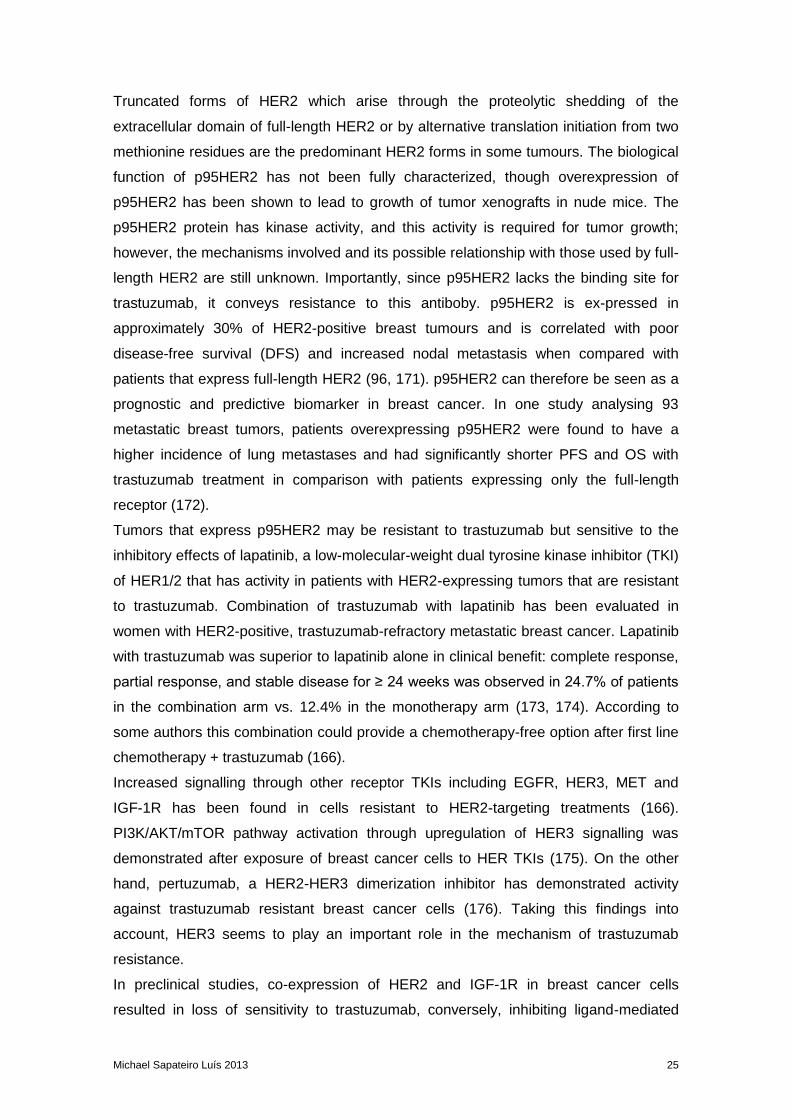


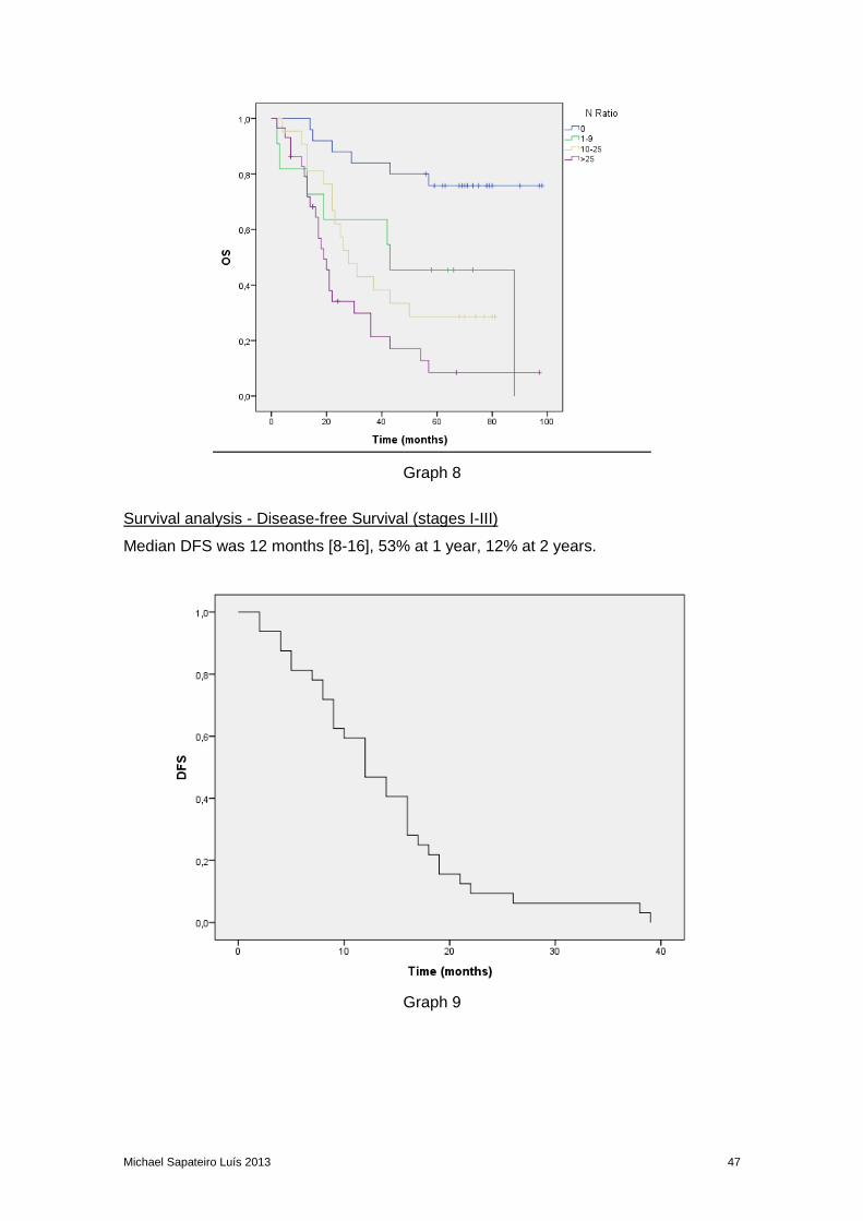


![[Ghiduri][Cancer]Gastric Cancer](https://static.documents.pub/doc/80x56/55cf9399550346f57b9de771/ghiduricancergastric-cancer.jpg)