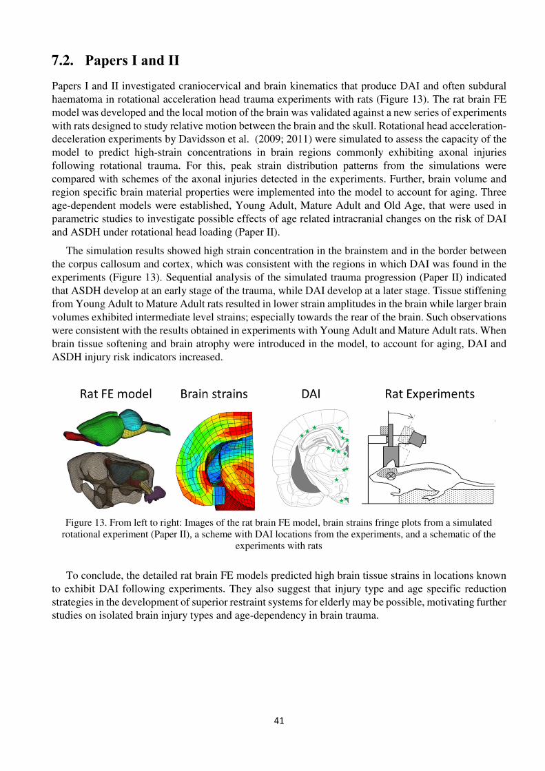
THESIS FOR THE DEGREE OF DOCTOR OF PHILOSOPHY in MACHINE AND VEHICLE SYSTEMS Traumatic Brain Injuries: Animal Experiments and Numerical Simulations to Support the Development of a Brain Injury Criterion JACOBO ANTONA-MAKOSHI Division of Vehicle Safety Department of Applied Mechanics CHALMERS UNIVERSITY OF TECHNOLOGY Gothenburg, Sweden, 2016
Transcript
THESIS FOR THE DEGREE OF DOCTOR OF PHILOSOPHY
in
MACHINE AND VEHICLE SYSTEMS
Traumatic Brain Injuries:
Animal Experiments and Numerical Simulations to
Support the Development of a Brain Injury
Criterion
JACOBO ANTONA-MAKOSHI
Division of Vehicle Safety
Department of Applied Mechanics
CHALMERS UNIVERSITY OF TECHNOLOGY
Gothenburg, Sweden, 2016
II
Traumatic Brain Injuries:
Animal Experiments and Numerical Simulations to Support the
Antona-Makoshi, J., Davidsson, J., Risling, M., Ejima, S., Ono, K., 2014. Validation of Local Brain
Kinematics of a Novel Rat Brain Finite Element Model under Rotational Acceleration, International
Journal of Automotive Engineering, Vol.5, No.1, pp.31-37.
Division of work between authors: Antona-Makoshi designed the study and conducted the modelling.
Davidsson supervised the study and conducted the experiments. Antona-Makoshi supported Davidsson
with the preparation and execution of the experiments. Risling provided environments for
experimental training and medical images. Antona-Makoshi wrote the paper. Davidsson provided
active support in writing the paper. Ejima, Risling and Ono supervised, provided scientific guidance
and reviewed the paper.
Paper II
Antona-Makoshi, J., Eliasson, E., Davidsson, J., Ejima, S., Ono, K. 2015. Effect of Aging on Brain
Injury Prediction in Rotational Head Trauma—A Parameter Study with a Rat Finite Element Model.
Traffic Injury Prevention, Vol.16, No sup1, pp. S91-S99.
Division of work between authors: Antona-Makoshi and Eliasson planned the study together under the
supervision of Davidsson and Ejima. Simulation work was conducted by Eliasson under the guidance
of Antona-Makoshi. Antona-Makoshi led the writing of the paper with the support of Eliasson.
Davidsson provided experimental data, scientific guidance and active support in writing the paper.
Ejima and Ono supervised, provided scientific guidance and reviewed the paper.
Paper III
Antona-Makoshi, J., Davidsson, J., Ejima, S., & Ono, K. 2012. Reanalysis of Monkey Head
Concussion Experiment Data using a Novel Monkey Finite Element Model to Develop Brain Tissue
Injury Reference Values. IRCOBI Conference, Dublin, Republic of Ireland.
Division of work between authors: Antona-Makoshi conducted the literature review, experimental data
re-organisation and analysis, modelling and wrote the paper. Davidsson supervised on a daily basis and
provided active support in writing the paper. Ejima and Ono supervised, provided scientific guidance
and reviewed the paper.
V
Paper IV
Antona-Makoshi, J., Davidsson, J., Ejima, S., Ono, K., Brolin, K., Anata, K., 2013. Correlation of
Global Head Kinematics and Brain Tissue Injury Predictors to Experimental Concussion Derived from
Monkey Head Trauma Experiments. IRCOBI Conference, Gothenburg, Sweden.
Division of work between authors: Antona-Makoshi conducted the literature review, experimental data
re-organisation and analysis, and modelling. Anata provided support with the experimental data
analysis. Antona-Makoshi wrote the paper. Davidsson and Brolin provided scientific guidance and
active support in writing the paper. Ejima and Ono supervised, provided scientific guidance and
reviewed the paper.
Paper V
Antona-Makoshi, J., Davidsson, J., Ejima, S., Ono, K., 2016. Development of a Comprehensive Injury
Criterion for Moderate and Mild Traumatic Brain Injuries. International Journal of Automotive
Engineering, Vol.7, No.2, pp. 69-75.
Division of work between authors: Antona-Makoshi conducted the literature review, experimental data
re-organisation and analysis, and modelling. Antona-Makoshi wrote the paper. Davidsson provided
scientific guidance and active support in writing the paper. Ejima and Ono supervised, provided
scientific guidance and reviewed the paper.
Paper VI
Antona-Makoshi, J., Holcombe, S., Ono, K., Davidsson, J., 2016. Development of a Brain Injury
Criterion and Associated Thresholds. In preparation for Journal submission.
Division of work between authors: Antona-Makoshi and Davidsson planned the study together.
Antona-Makoshi conducted the literature review, the modelling, and wrote the paper. Holcombe
performed the programming to process the data analysis, provided scientific guidance and reviewed
the paper. Ono supervised and provided scientific guidance. Davidsson provided scientific guidance
and active support in writing the paper.
VI
Preface
The work presented in this thesis was carried out at the Japan Automobile Research Institute, Department
of Safety Research and at the Injury Prevention Group, at the Vehicle Safety Division, Department of
Applied Mechanics at Chalmers University of Technology under the supervision of Associate Professor
Johan Davidsson and Professor Karin Brolin from 2011 to 2016. The research was funded by the Japan
Automobile Research Institute Advanced Research Program and by Chalmers Area of Advance, Transport.
VII
Acknowlegements
To Eri, Nina, Koto ... and those to come (?): This thesis has been conducted in parallel to the construction
of this family. It has taken away a lot of precious time of the, so far, happiest period of my life. Eri, I
admire your strength and generosity during the challenging pregnancies, deliveries and care of our kids.
Not a day goes by without me thinking about how fortunate I am for having you all. I love you.
To Johan Davidsson: Being a good PhD supervisor is an extremely difficult task, but you have
succeeded and in a way that always kept my motivation high. You have also been the key person that
boosted my interest in both science and education. You have my deepest respect and lifetime
appreciation. Thank you.
To Susumu Ejima: This thesis started the day I let my frustrations go... and threw them at you. My
personal and professional integration in Japan has been, by far, my most challenging undertaking. You
played a major role to make a success of it. You have been my educator, my colleague, my boss and my
friend during the most critical years of this thesis. Thank you.
To Koshiro Ono: I remember the day you came to my desk with a bunch of old and dusty documents.
Those documents contained what is, in my humble opinion, the most valuable experimental data set in
the history of impact biomechanics research. Your legacy is in good hands. Especial acknowledgement
for that email you sent me when I was at high risk of derailing (2012/03/17). Thank you.
To Karin Brolin, Mats Svensson and Mårten Risling: For your valuable supervision and guidance at
different stages of this thesis and your always encouraging words. Thank you.
To Kozo Watanabe: None of the wonderful things that have happened to me since 2008, including
this thesis, would have been possible without your unconditional support. For any reason, I felt you
treated me as you would have your own son. I hope you feel proud of me as a father would. Thank you.
To my colleagues at JARI: Former president Toshio Kobayashi, Minoru Sakurai, Kunio Yamazaki,
Takeshi Harigae, and Atsuhiro Konosu for giving me the freedom I needed to develop my scientific
curiosity. Yoshihiro Yamamoto for generosity taking the majority of the 'dirty work', allowing me to
concentrate on this thesis. Hisashi Imanaga, Koji Mikami and Ryohei Honma for those countless
refreshing coffee breaks and nomikai. Thank you.
To Sven Holcombe: I am not sure if I ever told you, but I am convinced that your frustrating
experiences in Japan were crucial for my success. I enjoyed our technical and philosophical talks related
to this thesis, but I have enjoy the rest of our talks even more. Also, congratulations for the news you
gave me a few days before printing this thesis. I can’t wait until the date we can all meet up. Cheers
mate.
To Orlando Sanchez and Sébastien Vauclair: The first year of this thesis we spent together in
Gothenburg was as wonderful as our times as students at Chalmers. I am happy to know that life keeps
on giving me chances to meet you from time to time. Thank you friends.
A mis padres y hermanos: Gracias por apoyarme en todo lo que he hecho en mi vida. Las
circunstancias me ha llevado muy lejos de vostros, pero nunca he dejado de percibir vuestro apoyo.
Espero que estéis orgullosos de mi. Os quiero.
VIII
Nomenclature
α Rotational/Angular Acceleration
ω Rotational/Angular Velocity
2D Two-dimensional
3D Three-dimensional
AIS Abbreviated Injury Scale
ATD Anthropomorphic Test Device
BrIC Brain Rotational Injury Criterion
BITS Brain Injury Threshold Surface
CSDM Cumulative Strain Damage Measure
CSF Cerebro Spinal Fluid
CG Centre of Gravity
CNS Central Nervous System
CR Centre of Rotation
CT Computed Tomography
DAI Diffuse Axonal Injury
FE Finite Element
FMVSS Federal Motor Vehicle Safety Standards
GAMBIT Generalised Acceleration Model for Brain Injury Threshold
Global NCAP Global New Car Assessment Program
HIC Head Injury Criterion
HIP Head Injury Power
JARI Japan Automobile Research Institute
KTH Kungliga Tekniska Högskolan
MPS Maximum Principal Strain
MRI Magnetic Resonance Imaging
MS Milliseconds
M/S Meter per second
NHP Non-Human Primate
NHTSA National Highway Traffic Safety Administration, USA
PMHS Post Mortem Human Subject
RIC Rotational Injury Criterion
RMDM Relative Motion Damage Measurement
RVCI Rotational Velocity Criterion Index
SDH Sub-Dural Haematoma
SIMon Simulated Injury Monitor
SUFEHM Strasbourg University Finite Element Head Model
TBI Traumatic Brain Injury
THUMS Total Human Model for Safety, developed by Toyota Motor Corporation
in cooperation with Toyota Central R&D Labs Inc.
VMS Von Mises Stress
WHO World Health Organization
WSU Wayne State University, USA
WSTC Wayne State Tolerance Curve
IX
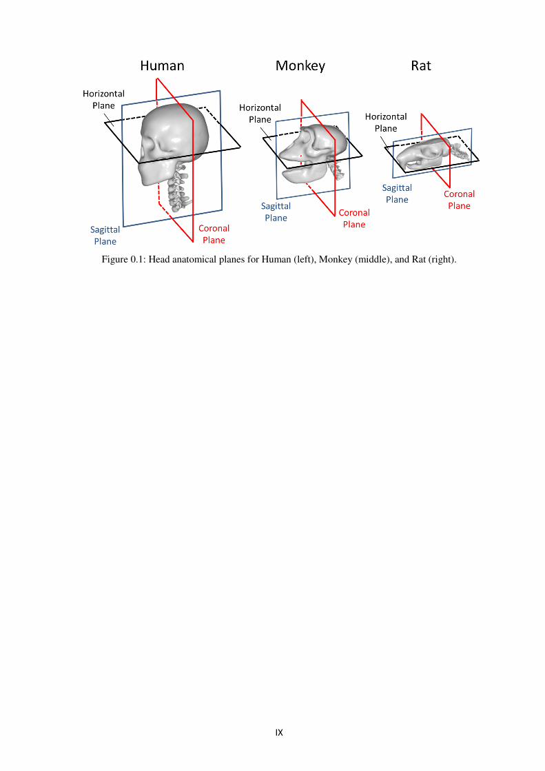
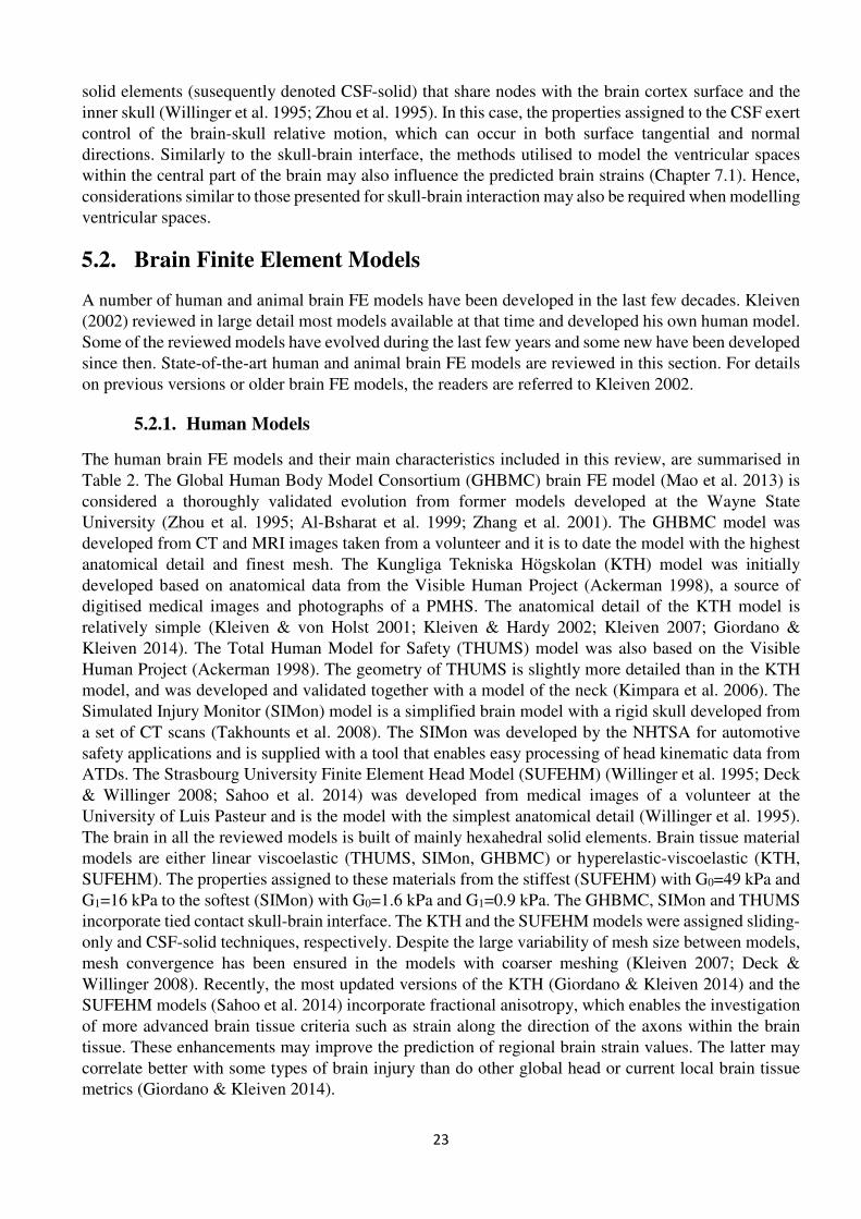
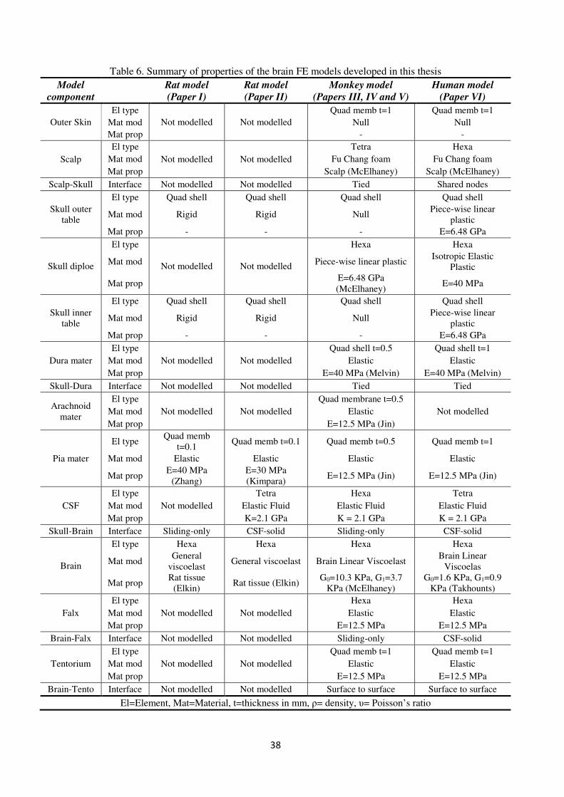
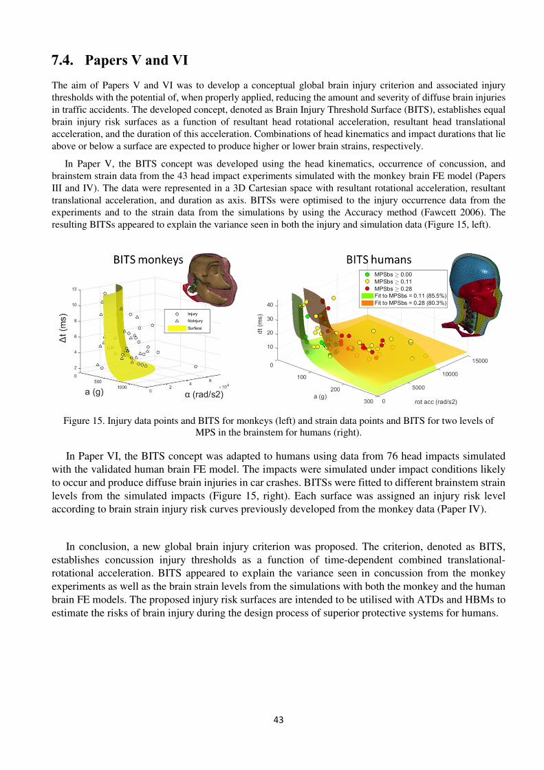
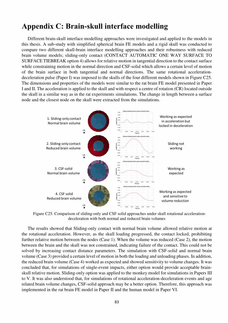
Figure 0.1: Head anatomical planes for Human (left), Monkey (middle), and Rat (right).
X
XI
Table of Contents
Abstract ................................................................................................................................................. III
List of Appended Papers ..................................................................................................................... IV
Preface ................................................................................................................................................... VI
Acknowlegements ................................................................................................................................ VII
Nomenclature .................................................................................................................................... VIII
Table of Contents ................................................................................................................................. XI
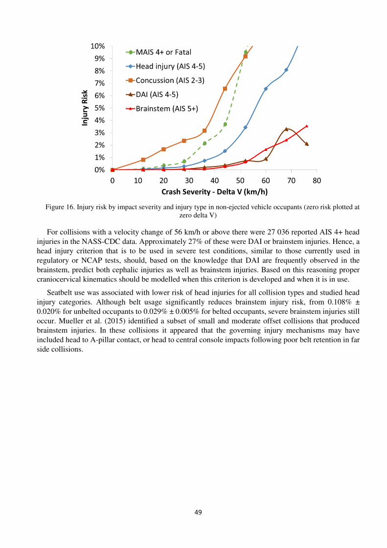
SAH: Sub-arachnoid haemorrhage, LOC: Loss of Consciousness, DAI: Diffuse Axonal Injury
8
with prolonged loss of consciousness of varying duration (Gennarelli et al. 1987; Gennarelli et al. 2003).
Diffuse brain Injuries are associated with mechanical disruption of axons that is produced when brain
tissue is subjected to strains above a recoverable limit (Galbraith et al. 1993; Bain & Meaney 2000;
Anderson et al. 2003). Diffuse brain injuries are usually produced by head impacts that produce abrupt
head motions, typically of combined translational and angular nature (Ono et al. 1980; Newman et al.
2000; Anderson et al. 2003; King et al. 2003).
The term concussion is controversial; its definitions have evolved over time, and past and present
diagnosis of concussions in clinical setting differ (Gennarelli & Wodzin 2006; Carroll et al. 2010).
Concussions have historically been defined based on symptoms. Past definitions of concussion (classical
concussion or cerebral concussion) required symptoms such as loss of corneal reflex, apnoea,
bradycardia or loss of consciousness prolonged for varying periods of time. Currently, a concussion
(often called mild Traumatic Brain Injury) is diagnosed when the patient appears to be confused but loss
of consciousness or amnesia is not required. In addition pathological findings in standard imaging are
not expected. While the symptoms following concussions are relatively well established, its pathology
is not fully understood; and diffuse and gross tissue lesions are absent. The detailed injury mechanism
responsible for concussion is still to be determined whereas some information on the pathology is
available; injuries to afferents of the cortex and normal cellular activity disruption in the reticular
activating system in the brainstem (Jefferson 1944; Adelstein 1978; Ropper & Gorson 2007; Pearce
2008). The immediate effects include neuronal swelling, inflammation, axonal disruption, as well as
metabolic and autonomic changes. Pathophysiological changes that occur following a concussion have
been described as a multilayered neuro-metabolic cascade whereby affected cells typically recover,
although under certain circumstances they might degenerate and die (Giza & Hovda 2001).
Concussions commonly occur without skull fractures (Got et al. 1983; Gennarelli et al. 1987).
Concussions are the most common moderate-to-serious TBIs in motor vehicle crashes (Viano &
Parenteau 2015) accounting for 60% of all AIS2+ injuries to the head (Table 8 in Addendum). The injury
risk approximately increases linearly with crash severity for collision velocity changes below 35 km/h
(Figure 16 in Addendum). DAI occur in traffic less frequently than concussions and at higher collision
speeds (with a velocity change of 56 km/h or above), but their consequences are often devastating. Fifty
percent of the DAI cases suffer moderate to severe deficits, 21% of the cases have vegetative survival,
and 7% are fatal (Gennarelli & Thibault 1982; Melvin & Yoganandan 2015).
3.3. Focal Brain Injuries
Contusions and lacerations are the most frequently found focal brain lesions and consist of
heterogeneous areas of necrosis, pulping, infarction, or micro haemorrhages that can occur in different
locations of the brain. Contusions may be produced due to direct contact between the cortex and the
deforming skull at the site of impact (coup contusions). Contusions can also occur at remote sites from
the impact (contrecoup contusions) or by direct contact between the cerebrum and the rough internal
surfaces of the skull basal bone. Cerebral contusions are in general more consistently associated with
skull fractures than other intracerebral injuries as well as frequently associated with concussion
symptoms (Styrke et al. 2007). Cerebral Contusions have been reported as an incidence of 20-30% of
severe injuries (Khoshyomn & Tranmer 2004) and up to 89% of the brains CT examined post-mortem.
Despite their large incidence, cerebral contusions can heal without medical intervention and are
frequently associated with other brain injuries of more clinical relevance. This is not the case for
brainstem contusions, which are usually classified as critical and are of high clinical relevance.
Intracranial haemorrhages often comprise life-threatening vasculature related injuries produced by
violent motions of the brain inside the skull. Blood may accumulate in different intracranial regions
which may increase intracranial pressure. Such increase of pressure may require surgical intervention.
9
Acute Subdural Haemorrhages (ASDHs) are commonly produced when the brain move relative the skull,
the vasculature that bridges the brain’s surface to the various dural sinuses is torn (Gennarelli & Thibault
1982) and blood accumulates between the dura mater and the brain. ASDHs are the most common type
of focal injuries with an incidence of 5 to 30% among severely head-injured patients. ASDHs can occur
in moderate head impacts producing combined translation and rotation head kinematics (Ono et al. 1980).
Mortality rates for ASDH range from 30 to 60% (Cooper 1982; Gennarelli & Thibault 1982). Intra
Cerebral Haemorrhages and Subarachnoid Haemorrhages are examples of other forms of focal injuries
less frequent than ASDH in traffic accidents. Intracerebral haemorrhages are collections of blood within
the cerebral parenchyma that develop superficially and extend deeply into the lateral ventricles, into the
corpus callosum and the brainstem and can be distinguished from contusions and ASDH by CT scans
(Cooper 1982; Melvin & Yoganandan 2015). Intracerebral haemorrhages are commonly associated with
temporal bone fractures (Asha’ari et al. 2012).
10
11
4. Review of Brain Injury Experiments and Accident
Studies
Methods that allow the establishment of injury thresholds can roughly be divided into three main
categories: experiments with volunteers, reconstruction of accidents, and experiments with animals. The
latter are the main focus of this thesis and are hence reviewed thoroughly in this section.
4.1. Volunteer Experiments and Accident Reconstructions
Snyder (1971) summarised a number of free-fall and sled tests with military volunteers conducted at the
Aeronautical Research Laboratory base in New Mexico, USA. These data provided evidence of brain
injury tolerance decreasing with increasing pulse durations, supporting the need to account for the
duration of the injurious events in the development of preventive strategies. Ewing et al. (1975) subjected
volunteers to rotational head accelerations in the sagittal plane up to 2.7 krad/s2 and durations over 20
ms without finding any adverse effects in the volunteers. Currently, strict restrictions apply for the usage
of volunteers in impact biomechanics research, hence the need for alternative research methodologies.
An alternative data source for the establishment of injury thresholds and risk functions is real-life events,
such as motor sports car crashes and contact sports, in which the participants are at risk of suffering head
impacts. Instrumentation has largely been applied to helmets of American football players to capture
head kinematics during no-concussive and concussive hits. This type of data have been used to propose
concussion thresholds (Rowson et al. 2009, 2012). However, this use of such a data source has a number
of limitations; lack of accuracy of the accelerometer measurements due to uncoupling between the
instrumented helmets and the head of the players, difficulties in isolating directional effects of impact,
limiting the opportunity to understand the craniocervical kinematics that produce the injuries, and study
groups limited to young and fit athletes. In addition, while practicing sports, the players are under high
cardiorespiratory activity. The influence accelerated breathing and high pulse may have on the brain
conditions and physiology needs to be clarified. Most of these limitations can be complemented with
animal testing.
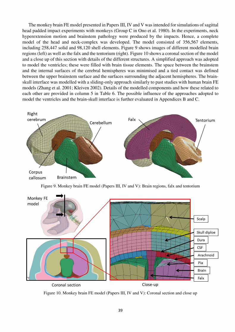
4.2. Animal Experiments
Successful animal brain injury models should produce the injury type that is the object of the study in
an isolated, controlled and repeatable manner. The injury type and severity should be representative of
those seen in the clinical setting. A large number of models have been developed and used for the
clarification of the injury mechanisms and to develop improved injury diagnosis methods and treatment
methods. However, the biomechanical response has been characterised in very few models. Such
characterisation is a requirement for the development of brain injury criteria and associated risk functions
using data from reconstruction of the experiments using computational models of the animal. In this
chapter animal TBI models developed for biomechanics research are reviewed. Emphasis is given to
those models that have been successfully utilised to generate consistent and large data sets of graded
injury severity and considered suitable for reconstructions using FE models of the experimental set-up.
Special focus is on concussion and DAI models as these are of high clinical relevance in motor vehicle
crashes (Viano & Parenteau 2015, Addendum).
Early experimental animal research studies focused on understanding the mechanisms and
establishing thresholds for whiplash-type brain and neck injuries. Ommaya et al. (1966; 1971) reported
a series of experiments in which injuries were produced in primates by either direct impact to the
occipital zone of the head or by hyper-extension head loading caused by impact to the base of a mobile
chair carrying the seated animal. In total, 42 squirrel monkeys (brain mass 20-27 gr.), 83 Rhesus monkeys
12
(brain mass 70-100 gr.) and 11 Chimpanzees (350-500 gr.) were utilised for these experiments. Cerebral
concussions were scored according to loss of coordinated response to external stimuli, duration of
apnoea and bradycardia. Displacement of the head and body was recorded by cinematography at 3,000
to 5,000 frames per second and values for rotational acceleration were calculated from displacement
data obtained from film analysis. Rotational accelerations ranging from 20 to 1,000 krad/s2 with
durations from 1 to 12 ms were estimated (Ommaya et al. 1967). By comparing the results from the
direct head impact experiments with the head acceleration experiments, it was shown that higher levels
of angular motion were required to produce cerebral concussion in the head acceleration experiments
(whiplash). Hence, suggesting that approximately half of the potential for brain injury during impact to
the unprotected movable head is directly proportional to the amount of head rotation. The remaining risk
of brain injury was attributed to the contact phenomena of the impact. The experiments by Ommaya et
al. laid the foundation for contemporary experimental works and highlighted the importance of the
methods used to load the head in the experiments.
Based on the method used to load the head, experimental brain injury models can be classified as
impact models or acceleration models. The objective with impact models is to reproduce head impacts
to a structure while the objective with acceleration models is to produce the head response without skull
bone deformations. Figure 1 illustrates examples of acceleration models for primates (Gennarelli et al.
1972) and for rats (Davidsson et al. 2009) and impact models for primates (Ono et al. 1980) and for rats
(Marmarou et al. 1994).
Figure 1. Schematic illustrations of acceleration models (a and c) and impact models (b and d) designed for
primates and rats.
4.2.1. Head Acceleration Models
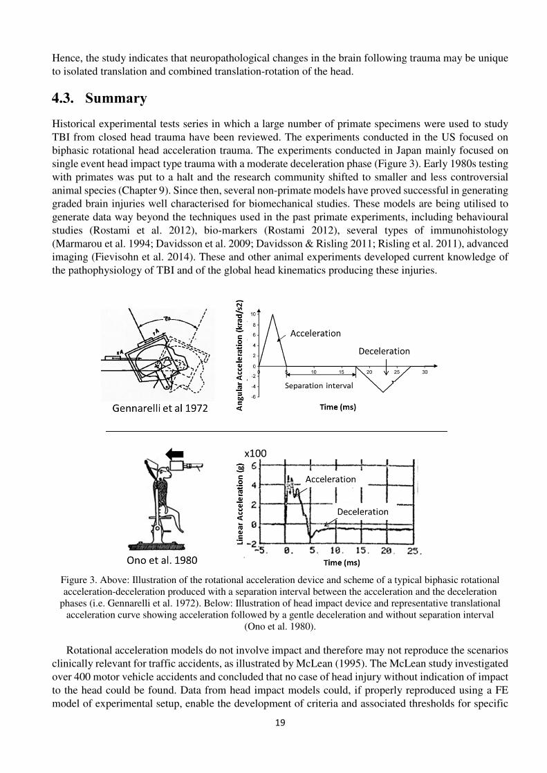
Gennarelli et al. (1971; 1972) conducted a series of experiments in which accelerations in the sagittal
plane were induced to the head of squirrel monkeys. A test apparatus consisting of a cylindrical cam
13
attached to the periphery of a flywheel (HAD-II) was utilised to impose controlled accelerations to a
helmet tightly fastened to the head of the animals. The translational acceleration and both components
of the rotational acceleration (tangential and radial) were measured at the head centre of gravity. The
imposed loading followed a biphasic pulse consisting of varying acceleration-duration followed by
varying deceleration-duration with a separation time interval of 3 ms. Both phases of the pulses were
approximately triangular in shape. In one series of experiments, 12 specimens were subjected to
predominantly translational acceleration-deceleration loading with peak accelerations ranging from 665
g to 1,230 g and peak deceleration values of approximately one-half of these values. In another series of
experiments, 13 specimens were subjected to predominantly rotational acceleration-deceleration loading
with peak positive tangential accelerations at the head centre of gravity ranging from 348 g to 1,025 g
and the centre of rotation located at the base of the neck. In these experiments the deceleration
magnitudes were nearly the same as the acceleration magnitudes. Typical duration of the acceleration
and the deceleration pulses were 1.8 and 3.3 ms, respectively. Brain pathology occurred in both series,
but with a greater frequency and severity in the group of animals predominantly exposed to rotational
head loading. Subdural haematoma was produced in the frontal region in half of the animals
predominantly subjected to translational acceleration loading. More extensive haematomas, including in
the temporal regions were found in all animals exposed to rotational loading. Predominant translational
loading did not produce concussion while predominant rotational loading produced concussion lasting
from 2 to 12 minutes.
Abel et al. (1978) extended the Gennarelli et al. (1972) work with a series of experiments with larger
sized primates focused on producing subdural haematomas and cerebral concussions. The original test
HAD-II apparatus was substituted by a HYGE pneumatic actuator coupled to a cam-linkage mechanism
that allowed delivering controlled accelerations-decelerations to the head of larger sized species. Forty
rhesus monkeys were exposed to single biphasic rotational acceleration-deceleration trauma in the
sagittal plane. Peak rotational accelerations ranged from 18 to 120 krad/s2. Peak values of the tangential
component of the rotational accelerations at the centre of gravity of the head ranged up to 1,300 g.
Although typical acceleration-deceleration pulses from this new device had similar separation time
intervals (4ms) as the HAD-II device, peak acceleration magnitudes were lower than peak deceleration
magnitudes. Acceleration and deceleration pulses had approximate durations of 6 and 3.5 ms,
respectively. Physiological and neurological data recorded included electrocardiogram,
electroencephalogram, systemic arterial pressure, intracranial pressure, respiration and corneal reflex.
The post trauma symptoms were evaluated using the Experimental Trauma Score (ETS). This score was
defined by the researchers involved in the experiments and was based on simple but objective
physiological, behavioural and neurological variables as follows. An animal with ETS 0 was one in
whom no physiological, behavioural or neurological changes were noted following the test. In ETS 1,
blood pressure or heart rate were the only variables that changed. ETS 2 included a brief period of apnoea
in addition to blood pressure or heart rate changes but the animal was conscious. ETS 3 involved a very
brief duration of unconsciousness followed by complete neurological recovery. ETS 4 involved
unconsciousness from the time of impact which remained for 5-15 minutes. Most animals in this
category exhibited prolonged behavioural abnormality including timidity, and disinterest in their
environment, but ate and drank normally. ETS 5 implied neurological death (never recovering from
unconsciousness) within 6 hours from the trauma. ETS 6 was applied to cases in which extremely high
head accelerations produced brainstem laceration and instantaneous death. According to this scale, the
term cerebral concussion typically referred to at the time of the experiments would correspond with ETS
grades ranging from 3 to 6. The pathological injury outcome was analysed in relation to the ETS grade
scored at the experiments. Cases of ETS grade 2 or lower showed little abnormality in the form of
occasional cortical contusions in the frontal lobes. SAH and ASDH could be observed for ETS 3 and
higher. Brainstem haemorrhage was only reported in fatal levels of ETS. ASDH occurrence was shown
to correlate well with the onset of tangential head accelerations occurring at values of 700 g. This study
14
pioneered in establishing graded correlation between physiological symptoms immediately after impact
and macroscopic brain pathology.
Gennarelli et al. 1982 conducted experiments with 23 rhesus monkey and 22 baboon specimens
aiming at the generation of graded DAI and their accompanying neurological deficits. The head of the
rhesus specimens was subjected to a single biphasic rotational acceleration-deceleration, by rotating it
through a 60 degree arc, in the sagittal plane, the coronal plane, or a 45 degree oblique plane between
these two planes. The peak deceleration was approximately three times the peak acceleration and ranged
from 70 krad/s2 to 180 krad/s2. The duration of these decelerations ranged from 6 and 9 ms, with a
tendency for shorter durations to correspond with higher peaks and longer durations with lower peaks
(Gennarelli et al. 1987). Neurological assessment was conducted in the 45 animals. Cerebral concussion,
characterised by unconsciousness that lasted less than 15 minutes followed by good recovery, occurred
in 15 cases, the majority of them from the sagittal group. Mild and moderate levels of prolonged coma
(unconsciousness lasting between 15 minutes and 6 hours) occurred in 10 of the animals. Severe
prolonged traumatic coma (more than 6 hours) was produced in 13 cases, all of them from the coronal
loading group. Four of these animals never recovered from coma. The remaining ten awakened from
coma but never recovered enough function to eat or drink. Neuropathology analysis was conducted in
26 of the 45 tested specimens. DAI was evaluated microscopically and the output was graded 1 to 3
according to its extent. DAI grade 1 was scored when axonal damage was confined to the white matter
of the cerebral hemispheres. DAI grade 2 when tissue tears (characterised by axonal and small vascular
damage) in the corpus callosum or the central brain area in addition to the cerebral hemispheres were
found. DAI grade 3 included axonal damage in the brainstem in addition to the axonal damage found in
grade 2. Overall, it was concluded that as duration of coma increased from concussion to prolonged
traumatic coma, the incidence and the severity of DAI increased. Intensity of axonal damage was found
to be proportional to lesions in the corpus callosum, and axonal damage produced by coronal head
acceleration was a major cause of prolonged traumatic coma and its sequelae.
Smith et al. (1997; 2000) developed a head acceleration model to explore potential anatomical origins
of posttraumatic coma. The HAD-II device was utilised to study DAI in miniature pigs (brain mass 80
to 90 gr.) by applying pulses of varying duration. Anaesthetised specimens were subjected to head
rotational acceleration in the coronal plane (transverse to the brainstem) and compared to specimens
subjected to similar rotational acceleration levels applied in the horizontal plane (circumferential to the
brainstem). Peak decelerations applied were approximately three times the peak accelerations and ranged
from 60 krad/s2 to 180 krad/s2 with durations between 4 and 6 ms. Immediate prolonged coma, assessed
by changes in EEG (slowing down of alpha rhythm in the frontal and parietal regions and intermittent
rhythmic high amplitude delta and theta activity in all regions), was consistently produced by head
horizontal plane rotation, but not by head coronal plane rotation. Immuno-histochemical examination of
the injured brains revealed that DAI was produced by head rotation in both planes in all animals.
However, extensive axonal damage in the brainstem was found in the pigs injured via head horizontal
plane rotation. No relationship was found between coma and the extent of axonal damage in other brain
regions. In these animals, the severity of coma was found to correlate with both the extent of axonal
damage in the brainstem and the applied loading conditions. The study suggested head loading
directional dependence in producing DAIs in the brainstem. These observations may however be
affected by the characteristic neck anatomy of the pigs that does not rotate in the coronal plane. In
addition, interpreting the implications for humans is difficult due to differences in head and neck
orientation between bipeds and quadrupeds (Margulies & Coats 2012).
Davidsson et al. (2009; 2011) developed a model in which the head of rats (brain mass 1.7 to 2 gr.)
is exposed to biphasic rotational acceleration-deceleration in the sagittal plane. The model has been
extensively utilised for behavioural and pathological studies and has been successful in producing graded
DAI with no obvious signs of contusions, intra-cerebral haemorrhages and skull fractures. Prior to
15
trauma, a curved aluminium plate is glued to the skull after removing skin and periosteum from the
cranial vaults and bones. This plate is secured to a bar that rotates around a horizontal axis perpendicular
to the sagittal plane of the animal. The trauma is produced when a solid brass weight hits a polyurethane
bumper on the bar which subjects the animal’s head to an extension motion. Applied acceleration can
be varied up to 2 Mrad/s2 while the duration remains at about 0.4 ms. The acceleration is followed by
deceleration with a magnitude of roughly one-fourth of the acceleration magnitude and 0.5 ms duration.
Both pulses are approximately triangular shaped with a separation between them of approximately 1 ms.
After the trauma, the animals have been utilised for behavioural studies (Rostami et al. 2012) or
sacrificed at different times post trauma for pathological studies (Davidsson et al. 2009; Davidsson &
Risling 2011). Presence of haematoma is assessed macroscopically. Detection of injured axons and
decaying axons is carried out by through tissue staining followed by inspection with confocal
microscopes. The overall findings using this model can be summarised as follows: producing graded
DAIs in the corpus callosum, the border between the corpus callosum and cortex and in tracts in the
brainstem with no obvious signs of contusions, haemorrhages and skull fractures (Davidsson & Risling
2011), initiation of inflammatory responses (Risling et al. 2011), limited behavioural changes (Rostami
et al. 2012) and changes to a large number of genes (Davidsson & Risling 2011). Predominantly head
rotational acceleration-deceleration experiments in the sagittal plane conducted with this model have
shown a threshold of 1 Mrad/s2 above which DAI is likely to occur in young adult rats (Davidsson et al.
2009; Davidsson & Risling 2011). Similarly, for COX2 presence and S100 concentrations at >0.9
Mrad/s2, the number of stained cells in the cortex and hippocampus as well as the concentration increased
(Davidsson et al. 2009). Subdural bleedings typically occurred in animals exposed to severe trauma.
Overall, this rotational trauma model produces graded axonal injury, in a repeatable and controlled
manner with a limited amount other types of TBIs and as such is useful in the study of injury
biomechanics, diagnostics, and treatment strategies following DAIs (Davidsson & Risling 2011).
4.2.2. Head Impact Models
Ono et al. 1980; Kanda et al. 1981; Sakai et al. 1982 and Kikuchi et al. 1982 conducted a large series of
head impact experiments with primates at the Japan Automobile Research Institute (JARI). The
experiments were conducted under the auspices of the Ministry of Transportation of Japan through
grants-in-aid of the Automobile Bureau Safety section and with the involvement of major academic
institutions in Japan. The large majority of the specimens utilised throughout the series were Rhesus
monkeys or Japanese monkeys (phylogenetically similar) although a limited amount of squirrel monkeys,
baboons and chimpanzees were included in these studies. The motion of the head and torso of the
specimens was captured by high speed cameras at 2,000 or 4,000 frames per second. Physiological
measures such as respiration, blood pressure, electrocardiogram and electroencephalogram were
recorded prior, during and after the impacts. Neurological measures including corneal and light reflex,
response to painful stimuli, and eye movement were measured from 10 seconds after the impact.
Pathological analysis included macroscopic examination and microscopic observation of formalin-fixed
brain tissue samples prepared shortly after death or sacrifice of the animals following the tests. The
severity of any concussion was evaluated according to symptoms (persistent loss of corneal reflex,
cessation of respiration and blood pressure disturbances) immediately after impact, according to
definitions in use at the time of the experiments (Committee on Terminology in Neurotraumatology of
the World Federation of Neurological Society 1979 ). The experiments comprised a total of 89 specimens
and 193 impacts delivered with different apparatus and variety of loading conditions. Figure 2
summarises the experiments; the name of each group, the number of impacts and number of specimens
in each group, the target injury severity level, schematic illustrations of the specimens and the techniques
used to deliver the impacts, and the main characteristic head kinematics producing the impacts.
16
Ono et al. 1980 presented the first three groups of impacts (A, B and C) consisting of sagittal head
impacts with different loading conditions. Group A consisted of single fatal impacts to 27 specimens (21
frontal and 6 occipital). The impacts were delivered over a broad area of contact through a plaster mask
fitted to the head which produced high levels of virtually pure translational motion of the head. The head
was measured in plane accelerations using four linear accelerometers mounted on the skull. Average
resultant translational accelerations of the head ranged from 240 to 1,100 g with the duration of these
accelerations ranging from 3 to 18 ms. All the 27 cases scored concussion, 13 survived the impact, 7
received SAH and only 1 skull fracture was reported. Noticeably, no contusion was produced in this
group. Group B comprised of 18 animals subjected to a single occipital impact to the device that
constrained the head. This device rotated with respect of a horizontal axis approximately passing through
the base of the cervical spine. Head translational and rotational accelerations in group B (and groups C
and D) were measured with a nine accelerometer system mounted on the skull. Average resultant head
translational accelerations ranged from 500 to 800 g with the duration of these accelerations of 1 to 5
ms. Average resultant rotational accelerations were in the range of 50 to 290 krad/s2 with durations
equivalent to those of the translational accelerations. All the 18 cases in this group scored concussion,
12 survived the impact, 11 received SAH, 12 contusion, and 6 skull fractures. Group C comprised a
series of comparatively milder padded impacts in which subjects were impacted repeatedly with
increasing impact severity conditions until concussion was produced. A total of 73 impacts were
delivered to the frontal or occipital part of the head of the 21 specimens. The impacts produced average
resultant head translational accelerations ranging between 140 to 500 g with durations from 1 to 14 ms.
Resultant rotational accelerations ranged from 6 to 120 krad/s2. Skull fractures were found in 4 of the 21
specimens. SAH was found in 10 of the specimens, SDH in 6 and contusion in 6. Brainstem injuries
accompanied by deep-seated haemorrhage was found in 5 specimens. Additional detailed analysis of the
outcome of this group after excluding the cases with skull fracture (Kanda et al. 1981) revealed frequent
haemorrhages and circulatory disturbances in the midbrain, pons and medulla oblongata, suggesting that
Figure 2. Summary of JARI primate head impact experiments carried out 1976 to 1978 (Ono et al. 1980; Kanda et al. 1981; Sakai et al. 1982 and Kikuchi et al. 1982)
17
non-fatal brainstem lesions in the absence of skull fractures were associated with the development of
concussion symptoms.
Sakai et al. (1982) and Kikuchi et al. (1982) separately presented a group of coronal impacts (group
D). The animals in this group were subjected to similar loading and experimental methods as Group C,
but with the impacts being delivered to the temporal region of the head. A total of 75 impacts of
increasing severity until concussion, were delivered to the head of 23 specimens. Velocities of the
impactor ranged from 10 to 27 m/s. Average resultant head translational accelerations ranged from 70 to
1,310 g with durations of these accelerations ranging from 2 to 43 ms. Skull fractures were found in 6
of the 23 specimens. These fractures were accompanied by massive brain injuries that terminated the
specimen within 15 minutes. Brain pathology was found in 18 of the specimens, including 9 specimens
with SAH, 5 with SDH, and 4 with brainstem haemorrhages. Sakai et al. (1982) compared the outcome
of these lateral impacts and the sagittal impacts by Ono et al. (1980). This comparison revealed, contrary
to the experiments with the acceleration-deceleration device reported in Gennarelli et al. (1982), that the
tolerance for concussion and severe pathological brain injuries was higher for lateral impacts than that
of sagittal impacts.
The overall key findings of the JARI primate head trauma impact series could be summarised as
follows; impacts delivered to the front, rear or lateral region of the head of primates with three different
experimental models produced single phase head acceleration events. The resultant head accelerations
were either translational (Group A), predominantly rotational (Group B) or a combination of these
(Groups C and D) with variable durations and severities. Concussion symptoms were produced as a
result of all used impact directions, regardless of the experimental method utilised to deliver the impact.
High levels of primarily head translational acceleration produced fatal concussion and SAH brain
injuries without skull fractures. ASH was produced in all groups, with higher rates when the heads were
exposed to predominantly rotation accelerations (Group B). SDHs were produced only when the impact
was delivered using a padded impactor surface (Groups C and D). This device resulted in combined
translational-rotational head acceleration. Tolerance to lateral impacts was higher than those that
produced head accelerations in the sagittal plane. A conservative curve that defined the threshold for
concussion as a function of head translational acceleration in the sagittal plane and duration of the
acceleration was drawn (Ono et al. 1980).
Marmarou et al. (1994) and Abd-Elfattah Foda and Marmarou (1994) developed a weight drop rat
head injury model. The model allows a weight to fall onto a metallic disc fixed to the top part of the
intact skull of the animal which is supported by a foam bed. The disc and the foam bed were utilised to
mitigate the risk of skull fracture. A total of 161 anaesthetised adult rats were subjected to injurious
impacts. The severity of the impact was controlled by varying the mass of the weight and the drop height.
A first group, consisting of 54 rats, was designed to establish the impact conditions that would produce
severe injuries with a mortality rate of approximately 50% and reduced risk of skull fractures. These
conditions were established in a test with a 450 gr weight falling from a 2 m height resulted in a mortality
rate of 44% with a low incidence (12.5%) of skull fractures. Mathematical analysis with lumped mass
models estimated that this mass-height combination resulted in a head acceleration of 900 g and brain
compression of 0.28 mm. In a second group of impacts, comprising 107 animals, the mass of the weight
was set to 450 gr and the drop height was adjusted to either 1 m or 2 m. This group aimed at producing
brain injuries of different severities and at determining the primary cause of death. Some specimens were
mechanically ventilated and others were allowed to breathe spontaneously. Pathological studies using
light and electron microscopy were also performed at 1, 6, 24, or 72 hours, or 10 days after the
experiment. Severe impacts were typically followed by apnoea, convulsions, and moderate hypertension.
The surviving rats developed decortication flexion deformity of the forelimbs, with behavioural
depression and loss of muscle tone. No fatalities occurred with the 1 m level injury, while 59% mortality
was seen with the 2 m level injury. However, the pathological changes observed in both groups were
18
similar. The mortality rate decreased markedly in animals mechanically ventilated during the impact.
Hence, the main cause of death was attributed to central respiratory depression. Gross pathological
examination did not reveal supratentorial focal brain lesions. Petechial haemorrhages were noticed in
the brainstem at the 2 m level injury. Microscopically, the model produced a graded widespread injury
of the neurons, axons, and microvasculature. Neuronal injury was mainly observed bilaterally in the
cerebral cortex. The trauma resulted in massive DAIs that involved the corpus callosum, internal capsule,
optic tracts, cerebral and cerebellar peduncles, and the long tracts in the brainstem. This model was
capable of producing DAIs in rats with low incidence of skull fracture and controlled mortality. These
injuries were produced by a head impact that produced nearly pure translational head motion.
Anderson et al. (2003) conducted a series of head impact experiments with sheep. The study aimed
at the production of axonal injury while measuring head impact dynamics and the subsequent kinematics
of the head. Eleven anaesthetised specimens were instrumented to measure physiological and
biomechanical data. Each animal was subjected to a single controlled impact delivered to the left
temporal region of the skull. Respiration rate, blood pressure, heart rate, electro-cardiogram, and
intracranial pressure were monitored throughout the duration of the experiment for physiological
assessment. A nine accelerometer array was secured to the skull with screws. A modified captive bolt
gun, mounted on a rigid frame, was used to deliver the impact to the sheep head. The gun was modified
to measure striker velocity and impact force. The striker had a mass of 395 g, and presented a spherical
contact surface on impact. The impact force was measured using a force transducer placed between the
body of the striker and the tip of the striker. Following a 4 hour survival period after the trauma, the
animal was terminated. The brains were collected and formaldehyde fixed for a period of two weeks.
Thereafter, immunochemical analysis was conducted for the detection of injured axons through tissue
staining followed by inspection with light microscopes. Three coronal sections from each brain were
examined in detail. Resulting impact forces varied between 5 and 7.4 kN over a duration of
approximately 2 to 3 ms. Head translational accelerations ranged from 700 to 1,800 g. Impact velocities
varied between 23 and 45 m/s. Rotational head accelerations ranged from 81 to 227 krad/s2. Skull
fractures occurred in 8 of the 11 experiments. Five of these fractures were of depressed type. Gross
pathology revealed cerebral contusions adjacent to the site of the impact on the skull, SAH at the site of
impact and sometimes in the contrecoup site. In the most severe cases, fractures caused extensive SAH
and lacerations in the cortex in addition to contusions adjacent to the impact site. Microscopic pathology
of the brains revealed that all animals sustained widespread axonal injury in several regions of the brain.
These regions commonly included the thalamus, hippocampus, margins of the lateral ventricles, digitate
cortical white matter and the corpus callosum. The anterior section usually exhibited less axonal injury
than the medial and posterior sections and the left (impact) hemisphere in all cases. Overall it was
concluded that the distribution of axonal injury in the sheep brain was related to the severity of the
impact to the head and that this distribution was not independent from skull fractures.
Fievisohn et al. (2014; 2015) recently developed two novel injury devices to characterise impact-
induced traumatic brain injury with mini pigs. Eleven animals were exposed in each device. The first
model imparts pure translational acceleration to the head by means of a drop tower. Translational head
accelerations with this model ranged from 27 to 70 g. The second model produced combined
translational-rotational acceleration of the head using a compound drop pendulum. Translational
accelerations ranging from 40 to 96 g and rotational accelerations from 1 to 3.8 krad/s2 were produced
with this model. The objective of the study was to evaluate the neuropathology associated with two
injury devices. Proton magnetic resonance spectroscopy was utilised to quantify metabolic changes and
immunohistology was utilised to evaluate axonal damage. The results revealed that both input modes
led to similar neurofilament damage, which indicates axonal disruption. The results revealed that the
two models produced similar metabolic changes, mainly in the inflammatory response and myelin
disruption, but also several differences in brain metabolites, suggesting distinct underlying mechanisms.
19
Hence, the study indicates that neuropathological changes in the brain following trauma may be unique
to isolated translation and combined translation-rotation of the head.
4.3. Summary
Historical experimental tests series in which a large number of primate specimens were used to study
TBI from closed head trauma have been reviewed. The experiments conducted in the US focused on
biphasic rotational head acceleration trauma. The experiments conducted in Japan mainly focused on
single event head impact type trauma with a moderate deceleration phase (Figure 3). Early 1980s testing
with primates was put to a halt and the research community shifted to smaller and less controversial
animal species (Chapter 9). Since then, several non-primate models have proved successful in generating
graded brain injuries well characterised for biomechanical studies. These models are being utilised to
generate data way beyond the techniques used in the past primate experiments, including behavioural
studies (Rostami et al. 2012), bio-markers (Rostami 2012), several types of immunohistology
(Marmarou et al. 1994; Davidsson et al. 2009; Davidsson & Risling 2011; Risling et al. 2011), advanced
imaging (Fievisohn et al. 2014). These and other animal experiments developed current knowledge of
the pathophysiology of TBI and of the global head kinematics producing these injuries.
Rotational acceleration models do not involve impact and therefore may not reproduce the scenarios
clinically relevant for traffic accidents, as illustrated by McLean (1995). The McLean study investigated
over 400 motor vehicle accidents and concluded that no case of head injury without indication of impact
to the head could be found. Data from head impact models could, if properly reproduced using a FE
model of experimental setup, enable the development of criteria and associated thresholds for specific
Figure 3. Above: Illustration of the rotational acceleration device and scheme of a typical biphasic rotational acceleration-deceleration produced with a separation interval between the acceleration and the deceleration
phases (i.e. Gennarelli et al. 1972). Below: Illustration of head impact device and representative translational acceleration curve showing acceleration followed by a gentle deceleration and without separation interval
(Ono et al. 1980).
20
injuries relevant in traffic accidents. In addition, improving the applicability of the animal data to humans,
which is one of the aims of this thesis, will most likely be more difficult for non-primate animals than
for primate animals. The reason for this is the fact that differences in brain size, brain geometry, relative
dimensions and location of important structures within the brain, tissue properties, the amount of folding
of the cerebrum, etc. are larger between smaller animal species and humans than between non-human
primates and humans. Hence, the head impact experiments carried out at JARI (Ono et al. 1980; Kanda
et al. 1981; Sakai et al. 1982 and Kikuchi et al. 1982) were considered the most appropriate for the
development of tissue and global level brain injury criterion and associated thresholds for concussions
relevant for traffic accidents. Nevertheless, an attempt was carried out in this thesis to use data from
sagittal plane rotational trauma experiments carried out with rats (Davidsson et al. 2009) to develop a
risk function for DAIs.
The contribution of the magnitude of head angular motion to the development of diffuse brain injuries
has been demonstrated with head acceleration models in primates (Gennarelli et al. 1971; 1972; Abel et
al. 1978; Gennarelli et al. 1982), in swine (Smith et al. 1997; 2000) and in rats (Davidsson et al. 2009;
2011). Combined translational-rotational motion of the head produced by impact experiments to
unconstrained heads has been shown to produce diffuse brain injuries often involving the brainstem in
primates (Groups C and D in the JARI experiments by Ono et al. 1980 and Sakai et al. 1982) sheep
(Anderson et al. 2003) and swine (combined input injury device by Fievisohn et al. 2014). Head impact
induced pure translational head motion has been shown to produce concussion symptoms and
subarachnoid haematoma in the absence of contusions and skull fractures with primates (Group A in
Ono et al. 1980). Comparable pure translational models have also produced DAIs of different severity
and spread as detected by immunohistochemistry with swine (translational-input injury device by
Fievisohn et al. 2014) and rats (Abd-Elfattah Foda & Marmarou 1994; Marmarou et al. 1994). Evidence
of increasing injury risks with increasing pulse durations has been established for both head acceleration
and head impact primate models (Ommaya & Gennarelli 1974; Ono et al. 1980). This decrease of injury
tolerance with increasing pulse durations is consistent with that observed in free-falls of military human
volunteers (Snyder 1971). Finally, the review of animal experiments suggests that the direction of head
acceleration may influence injury outcome (Gennarelli et al. 1982; Smith et al. 2000). However,
evidence of this is scarce and the results in these studies appear to be contradictory to another study with
primates (Sakai et al. 1982). In addition, these results were obtained using primates and swine, and may
not be valid for humans as the neck and brain anatomy differ between the species (Margulies & Coats
2012).
21
5. Review of Brain Finite Element Modelling
The FE method is a computer-based numerical technique for approximate calculation of the behaviour
of solid deformable and rigid structures (Hallquist 2006). The structures are broken down into many
small continuous compatible elements called meshing. Six equations are solved to calculate the motion
and deformation of each element for several time steps; from time zero to a predefined termination time.
Three of them are the equilibrium equations according to the fundamentals of physics defined by
Newton’s Law. The other three equations require strain/stress relations which are a function of the
defined material properties. The latter are preferably defined based on tissue experiments (McElhaney
2005). The time step utilised to solve the equations is calculated based on size, mass and material
properties of each element. The smallest time step needs to be small enough to ensure a stable numerical
solution and, at the same time, be large enough so it can be handled by the available computational
resources.
TBIs result from mechanical energy transferred to the brain from physical forces that act directly on
the head or are transmitted through the head-neck complex (Melvin & Yoganandan 2015). The direction,
type, rapidity, and magnitude of head motions determine the strains in the brain. A particular set of input
variables will produce a particular and unique strain field/time profile, which will result in clinically
apparent symptoms and pathological alterations (Gennarelli 2015). Most symptoms are commonly
observable right after trauma while pathologies and some symptoms develop over time as these are part
of a biological process. While brain FE models currently cannot simulate these processes they are applied
under the assumption that injurious biological processes are triggered when the brain tissue reaches a
certain level of deformation.
To estimate the deformation level/strain fields that produce the injuries in the brain, it is common to
apply external loads to the FE model equalling measures from real world events or experiments with
animals that led to injuries. The correlation between the estimated stresses/strains sustained by the brain
tissue and the injury outcome in the real world events or experiments can be determined. Axonal damage,
characteristic of diffuse brain injuries, is produced when brain tissue is subjected to strain (Galbraith et
al. 1993; Bain & Meaney 2000; Anderson et al. 2003). Subdural haematomas are produced when violent
motions of the brain inside the skull tear the vasculature between the brain cortex and the skull
Kleiven 2014). The Total Human Model for Safety (THUMS) model was also based on the Visible
Human Project (Ackerman 1998). The geometry of THUMS is slightly more detailed than in the KTH
model, and was developed and validated together with a model of the neck (Kimpara et al. 2006). The
Simulated Injury Monitor (SIMon) model is a simplified brain model with a rigid skull developed from
a set of CT scans (Takhounts et al. 2008). The SIMon was developed by the NHTSA for automotive
safety applications and is supplied with a tool that enables easy processing of head kinematic data from
ATDs. The Strasbourg University Finite Element Head Model (SUFEHM) (Willinger et al. 1995; Deck
& Willinger 2008; Sahoo et al. 2014) was developed from medical images of a volunteer at the
University of Luis Pasteur and is the model with the simplest anatomical detail (Willinger et al. 1995).
The brain in all the reviewed models is built of mainly hexahedral solid elements. Brain tissue material
models are either linear viscoelastic (THUMS, SIMon, GHBMC) or hyperelastic-viscoelastic (KTH,
SUFEHM). The properties assigned to these materials from the stiffest (SUFEHM) with G0=49 kPa and
G1=16 kPa to the softest (SIMon) with G0=1.6 kPa and G1=0.9 kPa. The GHBMC, SIMon and THUMS
incorporate tied contact skull-brain interface. The KTH and the SUFEHM models were assigned sliding-
only and CSF-solid techniques, respectively. Despite the large variability of mesh size between models,
mesh convergence has been ensured in the models with coarser meshing (Kleiven 2007; Deck &
Willinger 2008). Recently, the most updated versions of the KTH (Giordano & Kleiven 2014) and the
SUFEHM models (Sahoo et al. 2014) incorporate fractional anisotropy, which enables the investigation
of more advanced brain tissue criteria such as strain along the direction of the axons within the brain
tissue. These enhancements may improve the prediction of regional brain strain values. The latter may
correlate better with some types of brain injury than do other global head or current local brain tissue
metrics (Giordano & Kleiven 2014).
24
Despite the large differences between human brain FE models, the level of validation that can be
achieved with these models is limited by the availability of experimental data. In the past, brain models
used to be validated with intracranial pressure data measured from PMHS head impact tests (Nahum et
al. 1977; Trosseille et al. 1992). However, this method proved insufficient for models that are to be
applied for research of brain injury types in which brain tissue strains may be the predominant injury
mechanism (Bradshaw et al. 2001), such as diffuse brain injuries. Currently, models are validated with
data obtained from series of experiments with PMHSs in which the motion of opaque markers in the
brain at head impacts were recorded by means of high-speed x-ray (Al-Bsharat et al. 1999; Hardy et al.
2001; Hardy et al. 2007). Although not a direct measurement of brain tissue strains, this approach is
believed to provide a better insight of the reliability of predicting tissue strains with the brain FE models.
The human brain models included in this review have been, to different extents and level of accuracy,
validated with local brain motion data (Al-Bsharat et al. 1999; Hardy et al. 2001; Hardy et al. 2007). The
most recent version of the KTH model (Giordano & Kleiven 2014) has also been validated against strain
data estimated from the motion data by Hardy et al. (2007).
Applications of human brain FE models are multiple. One example is the investigation of injury
causation mechanisms by detailed reconstruction and simulation of traffic accidents that have resulted
in TBIs. The accidents are simulated with the brain FE models and the results from the simulations are
compared with the type, extent and location of the brain injuries based on the medical records of the
patients (Kleiven 2007; Deck & Willinger 2008; Fahlstedt et al. 2012). Such an approach, although
limited by the uncertainties inherent to accident reconstructions, can support the clarification of the head
kinematics that produced the injuries. Furthermore, if applied to a sufficient number of cases, it can also
provide means of evaluation of the reliability of brain injury criteria (Deck & Willinger 2008). Another
application of human brain FE models is the development of brain injury criteria and associated
thresholds by matching reconstructions of real world head impact events through simulations of these
events with human brain FE models. This method has been applied to series of concussive and non-
concussive head sport impacts. The head kinematics were measured from the head impacts and applied
to the rigid skull of the FE models (Kleiven 2007; Kimpara & Iwamoto 2012). The underlying
assumption of this simulation technique is that the effect of the impact event such as skull deformation
on injury risk may be neglected. This assumption may apply for concussions, since they usually occur
without skull fractures, suggesting small skull deformation. A limitation of this method is that
concussions are usually diagnosed based on symptoms at different times following the impact and
standard imaging is usually normal. Hence, the correlation between the results from the simulations and
the concussions are made based on a limited understanding of the head kinematics causing the injuries.
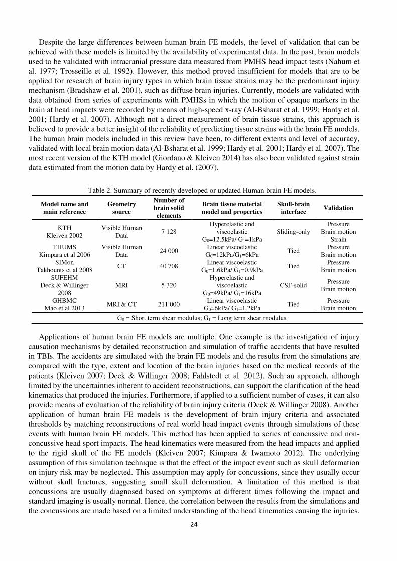
Table 2. Summary of recently developed or updated Human brain FE models.
Model name and
main reference
Geometry
source
Number of
brain solid
elements
Brain tissue material
model and properties
Skull-brain
interface Validation
KTH Kleiven 2002
Visible Human Data
7 128 Hyperelastic and
viscoelastic G0=12.5kPa/ G1=1kPa
Sliding-only Pressure
Brain motion Strain
THUMS Kimpara et al 2006
Visible Human Data
24 000 Linear viscoelastic G0=12kPa/G1=6kPa
Tied Pressure
Brain motion SIMon
Takhounts et al 2008 CT 40 708
Linear viscoelastic G0=1.6kPa/ G1=0.9kPa
Tied Pressure
Brain motion SUFEHM
Deck & Willinger 2008
MRI 5 320 Hyperelastic and
viscoelastic G0=49kPa/ G1=16kPa
CSF-solid Pressure
Brain motion
GHBMC Mao et al 2013
MRI & CT 211 000 Linear viscoelastic
G0=6kPa/ G1=1.2kPa Tied
Pressure Brain motion
G0 = Short term shear modulus; G1 = Long term shear modulus
25
A further example of the application of human brain FE models is the evaluation of the ability of global
brain injury criteria to correlate with brain tissue strains in loading scenarios from crash tests with ATDs
and PMHSs in which more severe brain injuries such as DAI may occur (Takhounts et al. 2013; Gabler
et al. 2015; Yanaoka et al. 2015). Since the loads were applied to rigid models of the skull in these
studies, this method tends to oversee that the injuries, the object of this study, commonly occur in
combination with skull fractures or skull bone deformations. In addition, since TBIs cannot be diagnosed
with ATDs or PMHS, this method will require a complementary methodology to establish injury
thresholds (Takhounts et al. 2013).
5.2.2. Animal Models
In order to improve the understanding of the relation between animal head kinematics and the injuries
produced, animal experiments can be simulated with FE models of the specimens. Contrary to human
models, animal brain FE models are not thoroughly validated and their application is limited to
investigations of specific injury mechanisms or to develop local tissue injury risk functions. Examples
of these models and applications are presented in Table 3. Anderson (2004) developed a sheep brain FE
model to simulate the dynamics of skull and brain during sheep closed head impacts experiments
(Anderson et al. 2003). Validation was attempted by comparing pressure measurements in the
simulations with those measured from the experiments. A number of rat brain FE models have also been
developed. Mao et al. (2006) developed an anatomically detailed rat brain FE model. The cortical motion
of the model was partially validated with in-vivo indentation test data. The validated model was utilised
to simulate controlled open head cortical impact experiments and to investigate mechanisms for
contusions. Baumgarter et al. (2009) developed a rat brain FE model from MRI and CT images for
investigations of DAI from rotational acceleration-deceleration experiments (Davidsson et al. 2009).
The model has been enhanced by incorporating brain region dependent tissue properties (Finan et al.
2012) and validated against brain-skull motion experimental data obtained in Paper I in the current thesis
(Ren et al. 2015). The validated FE model has been utilised to simulate the rotational acceleration-
deceleration experiments (Davidsson et al. 2009) and to develop brain tissue injury risk curves that relate
DAI risk from the experiments with tissue strain criteria from the simulations.
5.3. Summary
Medical imaging technology, advances in meshing techniques as well as software and hardware
development have boosted the development and application of detailed brain FE models. Human brain
FE models are technically mature tools with multiple research and industrial applications. The
achievable level of full scale brain motion validation in all models is limited by a reduced set of local
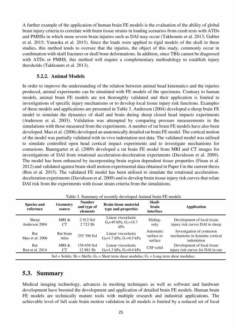
Table 3. Summary of recently developed Animal brain FE models
Species and
reference
Geometry
source
Number
and type of
elements
Brain tissue material
type and properties
Skull-
brain
interface
Application
Sheep Anderson 2004
MRI & CT
2 912 Sol 2 723 Sh
Linear viscoelastic G0=49 kPa, G1=16.7
kPa
Sliding-only
Development of local tissue injury risk curves DAI in sheep
Rat Mao et al. 2006
Rat brain Atlas
255 700 Sol Linear viscoelastic
G0=1.7 kPa, G1=0.5 kPa
Automatic surface to
surface
Investigation of contusion mechanisms in dynamic cortical
indentation
Rat Ren et al. 2014
MRI & CT
156 656 Sol 12 881 Sh
Linear viscoelastic G0=1.3 kPa, G1=0.4 kPa
CSF-solid Development of local tissue
injury risk curves for DAI in rats
Sol = Solids; Sh = Shells; G0 = Short term shear modulus; G1 = Long term shear modulus;
26
brain motion experiments conducted with cadavers (Hardy et al. 2001; Hardy et al. 2007). As human
brain FE models become more accurate, the models are adapted to predict regional brain deformations
in response to complex head motions such as those seen in traffic accidents. Animal brain FE models
are usually less validated than human models and their applications are limited to reduced sets of
experiments. However, in combination with controlled experimental models and improved invasive and
non-invasive injury diagnosis techniques, the animal brain FE model may enable the development of
injury risk functions that can be used to predict brain injuries at the tissue level.
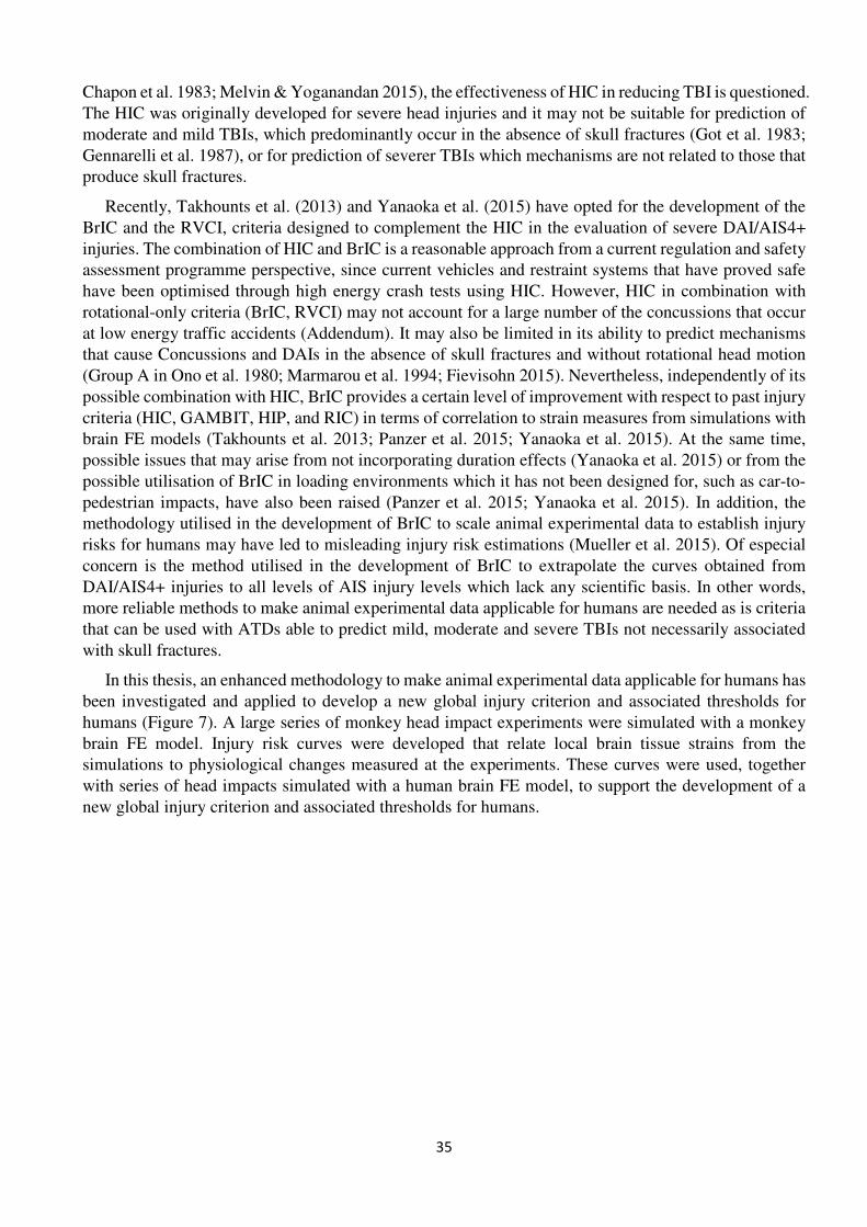
In this thesis one rat, one monkey and one human brain FE model have been developed from CT and
MRI images of the corresponding specimens (Chapter 7.1). Brain tissue linear viscoelastic properties in
line with some current state-of-the-art brain FE models were utilised. Emerging advanced brain tissue
modelling techniques that consider anisotropy were not studied in this thesis (Giordano & Kleiven 2014;
Sahoo et al. 2014). Sliding-only and CSF-solid skull-brain interaction modelling techniques were also
investigated and implemented in the models. These investigations and implementations were supported
with a limited set of original experiments conducted with rats to validate local brain motion in the rat
model. The skull of the rat FE model was modelled as rigid and the model was utilised to simulate head
sagittal rotational acceleration-deceleration experiments with rats. The monkey and the human models,
which were intended for simulations of head impact events, were developed together with a complete
model of the skull, neck and surrounding soft tissues. The monkey and the human brain FE models were
combined to investigate improved methods to make animal experimental data applicable for humans,
and to support the development of brain injury criteria and associated risk functions.
27
6. Review of Brain Injury Criteria, Thresholds and Risk
Functions
Current vehicle safety regulations such as the FMVSS use ATDs for the evaluation of safety through
crash tests. In addition to the physical ATDs, the vehicle industry uses mathematical models of the ATDs
and of humans to develop and evaluate the performance of new products in both crash tests and simulated
crashes. Regardless of the tools used, safety evaluations require injury criteria and injury risk functions
that relate measurements from the physical tests or simulated crashes with risk of injuries in humans.
Depending on the tools utilised and the injuries studied, such measurements can vary from tissue level
(i.e. brain tissue strain estimated using FE models) to global level (i.e. head accelerations measured with
sensors installed in ATDs). Tissue criteria rely on the capacity of brain FE models to predict proper
tissue response and that the parameter chosen to describe the response correlates to risk of injury. Global
brain injury criteria stand on the premise that there is a relationship between head kinematics and brain
tissue responses and that this can predict the risk of a certain brain injury (Newman 2015).
A number of studies have suggested brain injury thresholds for humans based on a single parameter
describing brain tissue strains or head kinematics. Within these, angular head motion has received
especial attention due to its well established contribution in producing brain injuries in experiments with
animals (Ommaya et al. 1967; Margulies & Thibault 1992; Davidsson et al. 2009). However, the same
animal studies have also suggested that a single metric such as peak rotational acceleration may not fully
characterise the diffuse brain injury spectrum, from concussion to severe DAI (Newman 1986;
Yoganandan et al. 2008). Hence, global criteria that include variables such as time histories of
translational and rotational head motion parameters have also been proposed and are currently under
evaluation. A review of local and global brain injury criteria with simple or multiple variables, as well
as their associated thresholds and risk functions is provided in this section.
6.1. Local Tissue Injury Criteria and Associated Injury Risk
Human and animal brain FE models have been utilised to develop local tissue injury criteria and
associated risks. Zhang et al. (2004) utilised the Wayne State University human brain model to
reconstruct football impacts and proposed shear stress in the brainstem to be the best injury predictor for
concussions. Kleiven (2007) utilised the KTH human brain model to simulate series of concussive and
non-concussive head impacts in American football and developed injury risk curves. Among others,
strain in grey matter and corpus callosum, as well as stress at the brainstem were identified as some of
the significant predictors for concussion. By applying logistic regression to each of the identified
parameters, injury risk functions were calculated and thresholds suggested. For example, 50%
probability of concussion was associated with a 26% strain in the grey matter and a 21% strain in the
corpus callosum. Takhounts et al. (2003; 2008; 2013) developed the Cumulative Strain Damage
Measurement (CSDM) which is a local tissue criterion that calculates the cumulative volume of brain
tissue experiencing tensile strains over a predefined critical level. Based on reconstructions of scaled
animal experimental data processed with the SIMon human brain FE model, a CSDM of 0.425 was
associated with a 30% probability of DAI/AIS4+ injury (Takhounts et al. 2013).
Anderson (2004) simulated sheep head impact experiments (Anderson et al. 2003) with a sheep brain
FE model. DAI risk curves were developed by comparing the results from the simulations with the
amount and distribution of DAIs observed following the experiments. To quantify the amount and
distribution of damaged axons, a grading system defined by the researchers involved was used in
conjunction with a 4-mm grid placed over brain sections. The indicated level of stress that corresponded
to approximately 10-20% risk of an intermediate level of axonal injury severity and distribution was
28
approximately 20 kPa. Lamy et al. (2013), reconstructed sagittal head rotational acceleration-
deceleration experiments with rats (Davidsson et al. 2009) using an FE model of the animals, estimated
brain tissue first principal strain at 3.9 and 4.7% thresholds for DAIs as detected with immunochemistry.
The Anderson (2004) and the Lamy et al. (2013) studies did however not explain how the proposed
thresholds relate to humans.
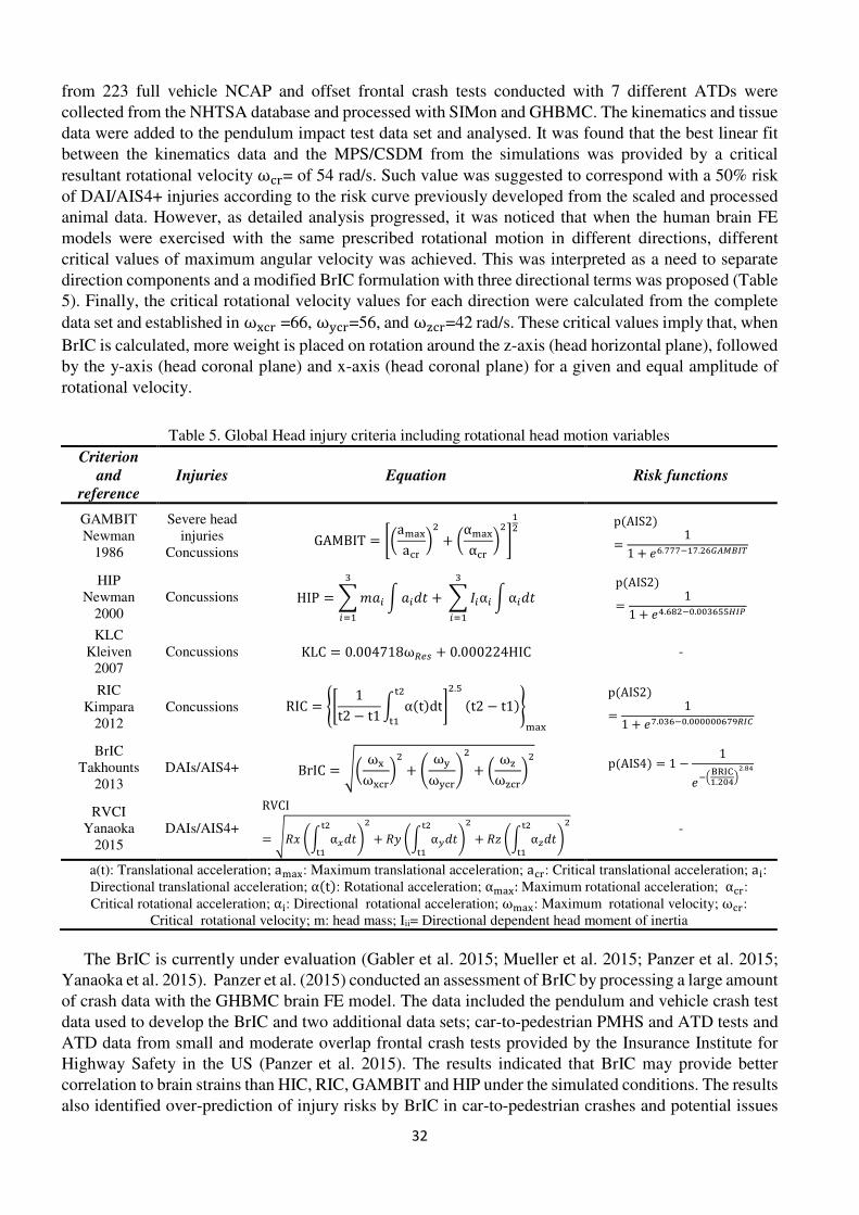
6.2. The Head Injury Criterion
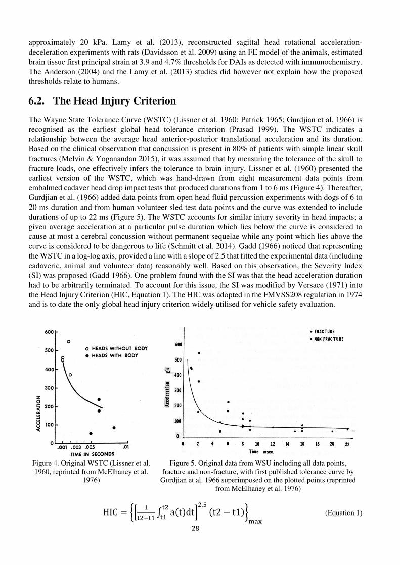
The Wayne State Tolerance Curve (WSTC) (Lissner et al. 1960; Patrick 1965; Gurdjian et al. 1966) is
recognised as the earliest global head tolerance criterion (Prasad 1999). The WSTC indicates a
relationship between the average head anterior-posterior translational acceleration and its duration.
Based on the clinical observation that concussion is present in 80% of patients with simple linear skull
fractures (Melvin & Yoganandan 2015), it was assumed that by measuring the tolerance of the skull to
fracture loads, one effectively infers the tolerance to brain injury. Lissner et al. (1960) presented the
earliest version of the WSTC, which was hand-drawn from eight measurement data points from
embalmed cadaver head drop impact tests that produced durations from 1 to 6 ms (Figure 4). Thereafter,
Gurdjian et al. (1966) added data points from open head fluid percussion experiments with dogs of 6 to
20 ms duration and from human volunteer sled test data points and the curve was extended to include
durations of up to 22 ms (Figure 5). The WSTC accounts for similar injury severity in head impacts; a
given average acceleration at a particular pulse duration which lies below the curve is considered to
cause at most a cerebral concussion without permanent sequelae while any point which lies above the
curve is considered to be dangerous to life (Schmitt et al. 2014). Gadd (1966) noticed that representing
the WSTC in a log-log axis, provided a line with a slope of 2.5 that fitted the experimental data (including
cadaveric, animal and volunteer data) reasonably well. Based on this observation, the Severity Index
(SI) was proposed (Gadd 1966). One problem found with the SI was that the head acceleration duration
had to be arbitrarily terminated. To account for this issue, the SI was modified by Versace (1971) into
the Head Injury Criterion (HIC, Equation 1). The HIC was adopted in the FMVSS208 regulation in 1974
and is to date the only global head injury criterion widely utilised for vehicle safety evaluation.
HIC = �� ����� a t�dt��� �.� t2 − t1����� (Equation 1)
Figure 4. Original WSTC (Lissner et al. 1960, reprinted from McElhaney et al.
1976)
Figure 5. Original data from WSU including all data points, fracture and non-fracture, with first published tolerance curve by
Gurdjian et al. 1966 superimposed on the plotted points (reprinted from McElhaney et al. 1976)
29
Currently, HIC15 and HIC36 (with integration time intervals of 15 and 36 ms, respectively) are used with skull fracture probabilities of 31% at HIC15 = 700 and 48% at HIC36 = 1000 (Eppinger et al. 1999). An AIS 4+ head injury risk curve based on HIC15 was developed by Prasad and Mertz (1985). Curves for other levels of AIS were developed by NHTSA based on the AIS 4+ curve developed by Prasad and Mertz (NHTSA, 2000)
The WSTC and the HIC are criticised for not considering factors that are important to brain injury. Such factors include the impact direction and area of contact, stiffness of the impacting surface, and rotational accelerations of the head induced by oblique impacts or when the torso is restrained (Newman 1986; McElhaney 2005). The effectiveness of HIC in reducing TBI is questioned due to the lack of a consistent relationship between linear skull fractures and neural injuries (Gennarelli 1980; Cooper 1982; Chapon et al. 1983; Nahum & Melvin 2012). In addition, the HIC was developed from test data that generated severe head injuries and it may not be suitable for prediction of moderate and milder forms of TBI, which predominantly occur in the absence of skull fractures (Got et al. 1983; Gennarelli et al. 1987, Addendum). Within all the limitations attributed to HIC, not accounting for rotational head motion has received the most attention, leading to a number of proposals for injury criteria that explicitly account for head rotational kinematics variables.
6.3. Rotational Acceleration Thresholds
Ewing et al. (1975) subjected military volunteers restrained with a 4-point belt to frontal sled
decelerations. These produced head rotational accelerations in the sagittal plane of up to 2.7 krad/s2 and
durations longer than 20 ms without any adverse effects on the volunteers. Löwenhielm (1975) proposed
a tolerance value in the sagittal plane of 4.5 krad/s2 for gliding contusions based on bridging vein rupture
produced by head impact tests with PMHSs. Pellman et al. (2004) suggested a 50% risk of concussion
for head rotational acceleration peaks of 5.5 krad/s2 obtained from head impact measurements taken
through instrumented helmets in American football games. A similar methodology was followed by
Rowson et al. (2012) to suggest a slightly higher threshold of 6.4 krad/s2.
Table 4. Suggested rotational acceleration thresholds (in krad/s2)
AIS Ewing
1975
Löwenhei
m 1975
Ommay
a
1984
Davidsso
n
2009
Margulie
s
1992
Pellman
2004
Rowson
2012
Gennarel
li
2003
Injury type
(example, AIS 2005)
Plane Sagittal Sagittal Sagittal Sagittal Coronal All All All 0 <2.7 No injury
1 <1.7 3 mild concussion; No
LOC
2 1.7 5.5 6.4 4.5 Brief LOC; Skull
fracture
3 4.5 3 8 LOC 1-6 hours,
contusion 4 3.9 10 9 12 DAI
5 4.5 14.5 DAI in corpus callosum and
brainstem
LOC = Loss of consciousness; DAI = Diffuse Axonal Injury
Animal experiments are commonly utilised to estimate brain injury thresholds. Ommaya (1967)
proposed cerebral concussion thresholds as a function of head rotational acceleration using primates and,
in a successive study, as a function of both head rotational acceleration and rotational velocity change
(Ommaya et al 1971). The data were scaled to be applicable for humans by applying scaling as suggested
by Holbourn (1943). In Ommaya (1984) relationships between brain injuries at different AIS levels and
30
peak head sagittal rotational acceleration were established for angular velocities above 30 rad/s (Table
4). Margulies and Thibault (1992) suggested a DAI specific threshold of 9 krad/s2 for rotation in the
coronal plane. This threshold was obtained from impacts to the head of PMHSs and data from
experiments in which baboons were subjected to rotational head acceleration-deceleration with
acceleration peaks ranging from 7 krad/s2 to 20 krad/s2 (Gennarelli et al. 1982). Davidsson et al. (2009)
estimated the sagittal head rotational acceleration threshold for DAI in humans to be 10 krad/s2 for a
pulse duration of 4 ms. This estimate was based on extrapolated results from rotational acceleration-
deceleration experiments with rats. Gennarelli et al. (2003) proposed several rotational acceleration
thresholds for diffuse brain injuries ranging from mild concussion to severe DAI. The proposed values
were based on a review of the existing literature, including the scaled DAI (AIS4) data from primate
experiments (Margulies & Thibault 1992) and American football concussion (AIS2) data (Newman et
al. 2000). The thresholds for these two types of injury were plotted versus their corresponding AIS level
and a linear fit was assumed to extrapolate the two data points to all AIS levels of diffuse brain injuries.
Several studies have suggested that contusion, concussion and DAI thresholds are a function of some
measures of acceleration level and duration (Gurdjian et al. 1966; Ommaya et al. 1967; Ono et al. 1980).
Hence, it has been suggested that diffuse brain injuries cannot be captured by a single metric of head
kinematics (Yoganandan et al. 2008). An example to illustrate this is presented in Figure 6 which shows
experimental data points from concussed and non-concussed primate head impact experiments
represented in an acceleration amplitude vs duration plot (Ommaya et al. 1967). The figure shows that,
as the duration of the event increased, the threshold for concussion decreased. Hence, suggesting the
need of accounting for the duration of the events in the development of brain injury criteria.
Figure 6. Tolerance curve for rotational acceleration amplitude versus duration
for concussed and non-concussed primates (Ommaya 1967)
6.4. Global Injury Criteria and Associated Injury Risks based on
Rotational Head Motion
Global brain injury criteria that explicitly account for angular head motion variables are summarised in
Table 5. When available, their associated thresholds or risk functions are referenced in the table. Some
of these criteria account for angular head motion only and are meant to be combined with the HIC for
head injury evaluation. Others combine both translational and rotational accelerations as an attempt to
capture the kinematics typical in head impacts.
31
The Generalized Acceleration Model for Brain Injury Threshold (GAMBIT) combines resultant head
translational and rotational acceleration peaks to establish thresholds for severe head injuries (Newman
1986). PMHS head impact data that produced skull fractures and primate head impact tests data that
produced prolonged (more than 6 hours) unconsciousness and DAI (Gennarelli et al. 1972) were used
in the development of GAMBIT. The method used to scale the animal data to be applicable for humans
was not clarified. GAMBIT = 1 corresponds with a 50% risk of AIS 3+ injuries with critical translational
and rotational acceleration critical values of 250 g and 10 krad/s2, respectively. Subsequent work
including American football head impacts reconstructed with helmeted ATDs was conducted and
concussion/AIS2 injury risk curves for GAMBIT were proposed (Newman et al. 2000). At the time of
developing the GAMBIT, the need of accounting for directional and time dependency was suggested,
but not taken into account since the data and tools available at the time were insufficient to develop
thresholds that accounted for these characteristics. The Head Injury Power (HIP) is meant to account for
directional dependent translational and rotational effects as well as durations (Newman et al. 2000). HIP
stands on the hypothesis that the maximum rates of translational and rotational energy are the controlling
mechanism in brain injuries induced by head impacts. Direction dependency is assigned by weighting
the corresponding direction component with the corresponding moment of inertia of the head. Due to
lack of human data, mass and moments of inertia from an ATD head were applied to the equation. HIP
was developed from the same reconstructions of helmeted head impacts as the GAMBIT and a
concussion AIS 2 injury risk curve was also developed (Newman et al. 2000). According to this risk
curve, a HIP of 12.5 kW corresponds to a risk of concussion of 50%. Kleiven (2007) proposed, based
on reconstructions of head impacts from American football with the KTH brain model, a linear
combination of HIC36 and the peak change in rotational velocity. The proposed criteria was denoted
KLC. The choice of rotational velocity instead of rotational acceleration was supported by previous
experimental work with PMHSs (Hardy et al. 2001) which suggested that peak change in rotational
velocity could better capture brain motion than peak rotational acceleration. Kimpara and Iwamoto
(2012) applied a similar methodology to reconstruct football impacts with the THUMS model and
developed two injury criteria; the Power Rotational Head Injury Criterion (PRHIC) and the Rotational
Injury Criterion (RIC). PRHIC was an adaptation of the HIP in which only the rotational terms are
considered. RIC is an adaptation of the HIC in which rotational accelerations are considered instead of
the translational.
Recently, the Brain rotational Injury Criterion (BrIC) has been proposed (Takhounts et al. 2013).
BrIC is based on peak rotational velocity in three directions (head x-, y- and z-) and does not account
for time dependency. It is intended to be used together with HIC. Large amount of test and simulation
data were used in the development of BrIC, which consisted of several steps. First, a total of 67
experimental animal injury data including rhesus monkeys, baboons, and miniature pigs were selected
(Abel et al.1978, Stalnaker et al. 1977, Meaney et al. 1993). These data were predominantly comprised
of rotational acceleration-deceleration experiments that produced DAIs. The injuries from the animal
tests were assumed to correspond with AIS 4+ injury in humans. The head motion data from the animal
experiments were scaled in amplitude and time, to account for size differences between the animal and
humans (Takhounts et al. 2003), and applied to the rigid skulls of two different human brain FE models;
the SIMon and the GHBMC. Injury risk curves that relate the tissue strain measurements from the
simulations (MPS and CSDM) with a probability of AIS 4+ were constructed. Thereafter, AIS 1+, 2+,
3+ and 5+ were scaled from the AIS 4+ curve using the ratios between corresponding risk curves for
HIC at 50% risk. Second, 190 tests in which pendulum impacts were delivered to the head of different
ATDs at various impact angles and energies were reconstructed using the two different human brain FE
models. Linear correlation analysis was conducted between the peak values of head kinematic metrics
from the tests and peak MPS and CSDM values from the simulations. From this analysis, it was
concluded that the resultant rotational velocity was the best correlation to the strain measures. Hence,
the BrIC criterion based on peak resultant rotational velocity was proposed. Third, head motion data
32
from 223 full vehicle NCAP and offset frontal crash tests conducted with 7 different ATDs were
collected from the NHTSA database and processed with SIMon and GHBMC. The kinematics and tissue
data were added to the pendulum impact test data set and analysed. It was found that the best linear fit
between the kinematics data and the MPS/CSDM from the simulations was provided by a critical
resultant rotational velocity ω��= of 54 rad/s. Such value was suggested to correspond with a 50% risk
of DAI/AIS4+ injuries according to the risk curve previously developed from the scaled and processed
animal data. However, as detailed analysis progressed, it was noticed that when the human brain FE
models were exercised with the same prescribed rotational motion in different directions, different
critical values of maximum angular velocity was achieved. This was interpreted as a need to separate
direction components and a modified BrIC formulation with three directional terms was proposed (Table
5). Finally, the critical rotational velocity values for each direction were calculated from the complete
data set and established in ω��� =66, ω���=56, and ω���=42 rad/s. These critical values imply that, when
BrIC is calculated, more weight is placed on rotation around the z-axis (head horizontal plane), followed
by the y-axis (head coronal plane) and x-axis (head coronal plane) for a given and equal amplitude of
rotational velocity.
The BrIC is currently under evaluation (Gabler et al. 2015; Mueller et al. 2015; Panzer et al. 2015;
Yanaoka et al. 2015). Panzer et al. (2015) conducted an assessment of BrIC by processing a large amount
of crash data with the GHBMC brain FE model. The data included the pendulum and vehicle crash test
data used to develop the BrIC and two additional data sets; car-to-pedestrian PMHS and ATD tests and
ATD data from small and moderate overlap frontal crash tests provided by the Insurance Institute for
Highway Safety in the US (Panzer et al. 2015). The results indicated that BrIC may provide better
correlation to brain strains than HIC, RIC, GAMBIT and HIP under the simulated conditions. The results
also identified over-prediction of injury risks by BrIC in car-to-pedestrian crashes and potential issues
Table 5. Global Head injury criteria including rotational head motion variables
viscoelast General viscoelast Brain Linear Viscoelast
Brain Linear Viscoelas
Mat prop Rat tissue
(Elkin) Rat tissue (Elkin)
G0=10.3 KPa, G1=3.7 KPa (McElhaney)
G0=1.6 KPa, G1=0.9 KPa (Takhounts)
Falx
El type
Not modelled Not modelled
Hexa Hexa
Mat mod Elastic Elastic
Mat prop E=12.5 MPa E=12.5 MPa
Brain-Falx Interface Not modelled Not modelled Sliding-only CSF-solid
Tentorium
El type
Not modelled Not modelled
Quad memb t=1 Quad memb t=1
Mat mod Elastic Elastic
Mat prop E=12.5 MPa E=12.5 MPa
Brain-Tento Interface Not modelled Not modelled Surface to surface Surface to surface
El=Element, Mat=Material, t=thickness in mm, ρ= density, υ= Poisson’s ratio
39
The monkey brain FE model presented in Papers III, IV and V was intended for simulations of sagittal
head padded impact experiments with monkeys (Group C in Ono et al. 1980). In the experiments, neck
hyperextension motion and brainstem pathology were produced by the impacts. Hence, a complete
model of the head and neck-complex was developed. The model consisted of 356,567 elements,
including 258,447 solid and 98,120 shell elements. Figure 9 shows images of different modelled brain
regions (left) as well as the falx and the tentorium (right). Figure 10 shows a coronal section of the model
and a close up of this section with details of the different structures. A simplified approach was adopted
to model the ventricles; these were filled with brain tissue elements. The space between the brainstem
and the internal surfaces of the cerebral hemispheres was minimised and a tied contact was defined
between the upper brainstem surface and the surfaces surrounding the adjacent hemispheres. The brain-
skull interface was modelled with a sliding-only approach similarly to past studies with human brain FE
models (Zhang et al. 2001; Kleiven 2002). Details of the modelled components and how these related to
each other are provided in column 5 in Table 6. The possible influence of the approaches adopted to
model the ventricles and the brain-skull interface is further evaluated in Appendices B and C.
Figure 9. Monkey brain FE model (Papers III, IV and V): Brain regions, falx and tentorium
Figure 10. Monkey brain FE model (Papers III, IV and V): Coronal section and close up
40
The human brain FE model presented in Paper VI was intended for simulations of head impacts under
a wide range of impact conditions representative of those occurring in real-world car crashes. Hence, a
complete model of the head neck complex was built. The final model consists of a total of 345,557
elements, including 291,948 solids and 53,609 shells. The brain regions, falx and tentorium were
modelled similarly to the monkey model. Figure 11 shows images of these brain regions (left), as well
as the falx and the tentorium (right). Figure 12 shows a coronal section of the human model with details
of the different structures included. Both the ventricular and the brain-skull spaces were filled with CSF-
solid elements that shared nodes with the surrounding regions. Details of the modelled components and
how these related to each other are provided in column 6 in Table 6.
Figure 11. Human brain FE model (Paper VI): Brain regions, falx and tentorium
Figure 12. Human brain FE model (Paper VI): Coronal section
41
7.2. Papers I and II
Papers I and II investigated craniocervical and brain kinematics that produce DAI and often subdural
haematoma in rotational acceleration head trauma experiments with rats (Figure 13). The rat brain FE
model was developed and the local motion of the brain was validated against a new series of experiments
with rats designed to study relative motion between the brain and the skull. Rotational head acceleration-
deceleration experiments by Davidsson et al. (2009; 2011) were simulated to assess the capacity of the
model to predict high-strain concentrations in brain regions commonly exhibiting axonal injuries
following rotational trauma. For this, peak strain distribution patterns from the simulations were
compared with schemes of the axonal injuries detected in the experiments. Further, brain volume and
region specific brain material properties were implemented into the model to account for aging. Three
age-dependent models were established, Young Adult, Mature Adult and Old Age, that were used in
parametric studies to investigate possible effects of age related intracranial changes on the risk of DAI
and ASDH under rotational head loading (Paper II).
The simulation results showed high strain concentration in the brainstem and in the border between
the corpus callosum and cortex, which was consistent with the regions in which DAI was found in the
experiments (Figure 13). Sequential analysis of the simulated trauma progression (Paper II) indicated
that ASDH develop at an early stage of the trauma, while DAI develop at a later stage. Tissue stiffening
from Young Adult to Mature Adult rats resulted in lower strain amplitudes in the brain while larger brain
volumes exhibited intermediate level strains; especially towards the rear of the brain. Such observations
were consistent with the results obtained in experiments with Young Adult and Mature Adult rats. When
brain tissue softening and brain atrophy were introduced in the model, to account for aging, DAI and
ASDH injury risk indicators increased.
To conclude, the detailed rat brain FE models predicted high brain tissue strains in locations known
to exhibit DAI following experiments. They also suggest that injury type and age specific reduction
strategies in the development of superior restraint systems for elderly may be possible, motivating further
studies on isolated brain injury types and age-dependency in brain trauma.
Figure 13. From left to right: Images of the rat brain FE model, brain strains fringe plots from a simulated rotational experiment (Paper II), a scheme with DAI locations from the experiments, and a schematic of the
experiments with rats
42
7.3. Papers III and IV
The aim of Papers III and IV was two-fold: to investigate the injury mechanism that produced concussion
symptoms and some degree of pathology in the experimental setting with primates; to provide brain
tissue injury risk curves to support the development of injury criteria and thresholds indented to be used
in test with ATDs. The monkey brain FE model was developed and partially validated according to
experimental data from the literature. The validated model was utilised to simulate a series of 19 frontal
and 24 occipital non-fatal head impact experiments selected from the past monkey head trauma
experiments (Ono et al. 1980). The impacts were delivered to the head through a padded piston, with
impact velocity, maximum impact stroke and padding rubber properties set for each test, which resulted
in combined rotation and translation of the head. The 19 occipital impacts selected for simulation were
delivered to 7 specimens who suffered 9 concussions (Paper III) and the 24 frontal impacts also selected
for simulations were delivered to 8 specimens who suffered 10 concussions (Paper IV). Records of
concussion, evaluated according to changes in physiological functions such as loss of corneal reflex, and
changes in respiration and cardiovascular patterns during and immediately after the impacts were
matched with the simulation results. Finally, risk functions for concussion as a function of simulated
brain tissue responses were developed.
The simulation results showed high strain concentration in the dorsal part of the brainstem, close to
the skull foramen magnum, which coincided with the locations in which brainstem pathology was
reported in few of the experiments (Figure 14). Risk functions for concussion as a function of strains in
the brainstem were developed. These functions, assuming similar brain tissue mechanical properties and
tissues injury thresholds between primates and humans, can be used to relate brain tissue strains and risk
of injury in studies with human brain FE models.
Figure 14. From left to right, monkey brain FE model images, brain strain fringe plots from a simulated occipital impact experiment, concussion like physiological changes and brainstem pathology from the
experiments and illustration of the experimental model
The findings presented in these papers support the hypothesis suggested at the time of the experiments
whereof brainstem trauma was responsible for the concussion symptoms (Kanda et al. 1981). The
findings also highlight the importance of considering the craniocervical motion in the investigation of
concussion mechanisms and in the development of injury criteria for concussions. In addition, the risk
functions proposed can be used to support the development of a global brain injury criterion for usage
with ATDs.
43
7.4. Papers V and VI
The aim of Papers V and VI was to develop a conceptual global brain injury criterion and associated injury
thresholds with the potential of, when properly applied, reducing the amount and severity of diffuse brain injuries
in traffic accidents. The developed concept, denoted as Brain Injury Threshold Surface (BITS), establishes equal
brain injury risk surfaces as a function of resultant head rotational acceleration, resultant head translational
acceleration, and the duration of this acceleration. Combinations of head kinematics and impact durations that lie
above or below a surface are expected to produce higher or lower brain strains, respectively.
In Paper V, the BITS concept was developed using the head kinematics, occurrence of concussion, and
brainstem strain data from the 43 head impact experiments simulated with the monkey brain FE model (Papers
III and IV). The data were represented in a 3D Cartesian space with resultant rotational acceleration, resultant
translational acceleration, and duration as axis. BITSs were optimised to the injury occurrence data from the
experiments and to the strain data from the simulations by using the Accuracy method (Fawcett 2006). The
resulting BITSs appeared to explain the variance seen in both the injury and simulation data (Figure 15, left).
Figure 15. Injury data points and BITS for monkeys (left) and strain data points and BITS for two levels of
MPS in the brainstem for humans (right).
In Paper VI, the BITS concept was adapted to humans using data from 76 head impacts simulated
with the validated human brain FE model. The impacts were simulated under impact conditions likely
to occur and produce diffuse brain injuries in car crashes. BITSs were fitted to different brainstem strain
levels from the simulated impacts (Figure 15, right). Each surface was assigned an injury risk level
according to brain strain injury risk curves previously developed from the monkey data (Paper IV).
In conclusion, a new global brain injury criterion was proposed. The criterion, denoted as BITS,
establishes concussion injury thresholds as a function of time-dependent combined translational-
rotational acceleration. BITS appeared to explain the variance seen in concussion from the monkey
experiments as well as the brain strain levels from the simulations with both the monkey and the human
brain FE models. The proposed injury risk surfaces are intended to be utilised with ATDs and HBMs to
estimate the risks of brain injury during the design process of superior protective systems for humans.
44
45
8. Addendum: Brainstem Injuries in Motor Vehicle
Crashes
This addendum was authored by Jacobo Antona-Makoshi and Johan Davidsson. Accident data for the
analysis were provided by Chantal Parenteau and David Viano.
8.1. Introduction
The brainstem plays a vital role in maintaining life by controlling important motor nuclei such as the
cardiovascular and the respiratory centres. Anatomically, it is the deepest and lowest part of the brain; it
connects to the diencephalon above and the spinal cord below. Primary lesions include direct superficial
contusion, DAI, primary capillary haemorrhage, and Ponto-medullary rent. Severe brainstem injuries, at
AIS 4 and AIS 5, typically involve tearing in the brainstem area and may occur sometimes without any
other type of brain injury noted (Adelstein 1978). The injury mechanism responsible for these severe
brainstem injuries appears to be craniocervical hyperextension, usually because the face impacts with a
part of the car. Some researchers feel that the severe brainstem injuries are caused by rotational
acceleration of the head. Several studies have suggested that primary brainstem injuries occur following
a direct impact of the brainstem against the tentorial free edge (Oppenheimer 1968; Clifton et al. 1981).
DAI is a severe form of acute diffuse brain injury with poor long term outcome. There is evidence that
DAIs are found in the white matter in several brain regions including the brainstem. In severe forms of
DAI it is also common to observe haemorrhagic lesions in the brainstem (Gean & Marsh 1994). Traffic
accident data have shown that when critical and fatal brain injuries occur, the brainstem is commonly
involved in side impacts (Sunnevång et al. 2015), in frontal and side impacts (Yoganandan et al. 2009),
in small and moderate offset frontal impacts (Mueller et al. 2015) and in rollover crashes (Mattos et al.
2013).
It has been suggested that less violent brainstem traumas produce the loss of consciousness often seen
in concussion, typically graded an AIS 2 or AIS 3 injury, because of the involvement of the reticular
activation formation of the brainstem and diencephalon (Jefferson 1944; Powiertowski & Choróbski
1967; Adelstein 1978; Ropper & Gorson 2007; Pearce 2008). It is thought that the normal cellular
activities in the reticular activating system are disrupted by the trauma but concussion is also thought to
be a milder type of diffuse axonal injury, because axons may be injured to a minor extent due to
stretching. Several other theories of concussion with loss of consciousness have been suggested in the
past, e.g. the vascular, reticular, centripetal, pontine cholinergic and convulsive hypotheses. The latter
implies that loss of consciousness is not due to malfunction of the brainstem reticular activating system
but rather due to functional deafferentation of the cortex as a consequence of diffuse mechanically-
induced depolarisation and synchronised discharge of cortical neurons (Shaw 2002). Researchers have
found that in several of the comatose patients the primary injuries to the brainstem were always part of
more diffuse hemispheric brain damage (Adams et al. 1977). Interestingly, concussions with loss of
consciousness are rare if the head is exposed to translational acceleration, severe enough to produce
cortical contusions and subdural haematomas, without rotational acceleration (Ommaya & Gennarelli
1974).
Despite awareness that the brainstem has very vital functions and is an integrated part of the brain,
and in addition is commonly injured, several brain injury criteria intended for traffic safety
improvements and in helmet evaluation have been developed without properly taking the craniocervical
kinematics into account. One reason for being that the biomechanics of concussion is still unknown and
that craniocervical kinematic motion is less monitored in sports and is commonly rather unknown in
traffic accidents. Another reason is that it has not received as much attention in accidentology studies.
46
There is no descriptive study available that allows placing brainstem injuries in context with other
types of brain injuries in traffic accidents as a function of crash severity. Hence, in this addendum
National Automotive Sampling System - Crashworthiness Data System (NASS-CDS) are used to
estimate the incidence and risk of brainstem injuries in motor vehicle crashes and compare them to other
types of brain injuries considering crash type, crash severity and belt usage.
NASS-CDS is a stratified sample of crashes that are prospectively selected for in-depth investigation.
Most of the vehicles are towed from the scene because of damage. The data includes information based
on crash investigation teams, vehicle registration, medical records, police reports and interviews. The
data was extrapolated to US national estimates using weighting factors.
Viano and Parenteau (2015) recently estimated the annual incidence and risk of brain injuries and
severe head injuries by crash type, severity and seatbelt use in motor vehicle crashes from the NASS-
CDS database. Their study did not specifically look into brainstem injuries.
8.2. Method
The method used in this addendum was identical to that of Viano and Parenteau (2015) with the exception that NASS-CDS data from two additional years was included; data from 1994-2013 was analysed to study head injuries in non-ejected occupants by crash type and belt use. Another exception was that the analysis included brainstem injuries. This study included all crashes and occupants aged 15 years and above. Only light vehicles with model year 1994+ were included in the study.
The crash types were defined as follows:
− Front: Vehicles involved in frontal impacts where the greatest damage was to the front (general area of damage in the front, GAD1 = F). Collisions in which a rollover occurred were excluded from the sample (Rollover ≤0).
− Side: Vehicles involved in side impacts where the greatest damage was to the left or right side (GAD1= L or R). Collisions in which a rollover occurred were excluded from the sample (Rollover ≤0).
− Rear: Vehicles involved in rear impacts where the greatest damage was to the rear (GAD1=B). Collisions in which a rollover occurred were excluded from the sample (Rollover ≤0).
− Rollover: Vehicles involved in rollover were those with a rollover >0.
− Other: Vehicles involved in other impacts where the greatest damage was to the top or undercarriage (GAD1=U or T). Collisions in which a rollover occurred were excluded from the sample (Rollover ≤0).
− Unknown: Vehicles involved in impacts with unknown damage information
The belt use was defined as follows:
It was defined by the variables manual belt use (MANUSE) and automatic belt use (ABELTUSE):
− Belted: if MANUSE = 4:
− Unbelted: if year < 110 and ((MANUSE <= 1) and (1 > ABELTUSE or ABELTUSE = 2)) and if year>109 and (0 <= MANUSE <= 1);
Injury severity were defined as follows:
Injury severity of the occupant was assessed using the Maximum Abbreviated Injury Scale (MAIS) and the “TREATMNT” variable. MAIS represents the assessment of life-threatening injuries at the time of first medical evaluation and not long-term consequences. It ranges from MAIS 0 to 9, where MAIS 9 is an injury with unknown severity. MAIS 4-6 represents a severe-to-unsurvivable injury. Fatality was also used to determine if the occupant died of injuries in the accident. The variable “TREATMNT” was
47
used to define fatality, which is TREATMNT = 1. Because fatalities can occur at any MAIS level, severely injured occupants were defined as those with MAIS 4-6 or fatality. The shorthand notation for this is MAIS 4+F.
Head injuries were classified as:
− Concussion was classified as moderate-to-serious injury (AIS 2-3) to the brain (region90 = 1, struspec = (0, 2, 4, 6, 8, 10) when strutype = 6)
− DAI was classified as severe-to-critical injury (AIS 4-5) to the brain and the brainstem (region90 = 1, struspec = (0, 2, 4, 6, 8, 10) when strutype = 6 and struspec = (2, 4, 6) when strutype = 4)
− AIS 4+ head injury was classified as severe-to-maximum injury (AIS 4-6) to the head (region90 = 1).
− Brainstem injury was classified as critical-to-maximum (AIS 5-6) to the head (region90 = 1 and struspec = 2 when strutype =4)
8.3. Results
Table 7 present all injuries sustained in each of the analysed crash types. Out of the 2 079 821 occupants
involved in a motor vehicle crash 5 443 ± 955 occupants sustained an AIS 4+ head injury and 872 ± 133
a brainstem injury annually.
The injury risk was determined by dividing the number of occupants with a particular injury by the
number of occupants with known injury status (MAIS 0+F). These risks are provided in Table 8. The
risk for severe head injury (AIS 4+) was highest in rollovers (0.73% ± 0.16%) and slightest in rear
impacts (0.16% ± 0.05%). Similarly, the risk of concussion, DAI and severe brainstem injuries were
highest in rollovers while these risks were lowest in frontal impacts.
Out of the occupants with known seatbelt usage 1 489 700 were belted and 360 824 were unbelted in
the impact. Risk for head injury for the belted and unbelted occupants is given in Table 9 and Table 10,
respectively. Seatbelt use was effective in preventing all types of brain injuries, irrespective of the impact
type. Seatbelt use was most effective in preventing DAI at 87% with a 0.223% ± 0.094% risk when
unbelted and 0.0289% ± 0.005% when restrained. The seatbelt was the most effective, for any impact
type, in preventing brainstem injuries in rear-end impacts (94%), DAI in frontal impacts, side impacts
and rollovers (92%) and AIS 4+ head injury in rear-end impacts (92%).
Table 7. Annual Head injury counts and risks by belt use and crash type
Front Side Rear Rollover All
Occupants involved in a tow away motor vehicle crash 893 104 399 607 132 209 151 199 2 079 821
Occupants assigned an AIS value (MAIS 0+F) 862 166 384 414 125 762 142 706 1 975 617
For collisions with a velocity change of 56 km/h or above there were 27 036 reported AIS 4+ head
injuries in the NASS-CDC data. Approximately 27% of these were DAI or brainstem injuries. Hence, a
head injury criterion that is to be used in severe test conditions, similar to those currently used in
regulatory or NCAP tests, should, based on the knowledge that DAI are frequently observed in the
brainstem, predict both cephalic injuries as well as brainstem injuries. Based on this reasoning proper
craniocervical kinematics should be modelled when this criterion is developed and when it is in use.
Seatbelt use was associated with lower risk of head injuries for all collision types and studied head
injury categories. Although belt usage significantly reduces brainstem injury risk, from 0.108% ±
0.020% for unbelted occupants to 0.029% ± 0.005% for belted occupants, severe brainstem injuries still
occur. Mueller et al. (2015) identified a subset of small and moderate offset collisions that produced
brainstem injuries. In these collisions it appeared that the governing injury mechanisms may have
included head to A-pillar contact, or head to central console impacts following poor belt retention in far
side collisions.
Figure 16. Injury risk by impact severity and injury type in non-ejected vehicle occupants (zero risk plotted at
zero delta V)
50
51
9. Ethical Considerations
In this thesis, rat specimens have been utilised to obtain CT and MRI images for FE model development.
In addition, new rotational head trauma experiments were conducted to generate model validation data
(Papers I and II). These imaging studies and experiments were performed at the Neuroscience
Department of the Karolinska Institute, Sweden, in accordance with the Swedish National Guidelines
for Animal Experiments and under the approval of the Animal Care and Use Ethics Committee in
Stockholm, Sweden [doc. N143/09]. In addition, CT and MRI images were obtained from anaesthetised
primate specimens at the Primate Research Institute of the Kyoto University (KUPRI), Japan. These
images were obtained for the development of the monkey FE model (Papers III to V). The experiments
were reviewed and approved by the Animal Welfare and Animal Care Committee ethics panel of the
KUPRI [Permission 2011-134, 7 June, 2011].
All other head trauma data that were re-analysed and reconstructed using FE animal head models in
this thesis were conducted in the past. The experiments with primates were carried out at the JARI from
1976 to 1978 as part of a research programme under the auspice of the Ministry of Transportation of
Japan. Support was granted by the Automobile Bureau Safety section. The experiments were carried out
with the involvement of several major academic institutions in Japan, such as Jikei University School of
Medicine, University of Tokyo, Tokyo Women’s Medical College and Nihon University. The
experiments followed animal care regulations in effect at that time. However, the ethical guidelines were
reviewed and additional primate experiments have not been carried out at JARI since then. At the start
of this thesis, the researchers involved discussed if the utilisation of these data in new studies is ethical.
The researchers found no evidence for the need to obtain additional ethical permission for use of the
experimental data. The arguments concerning the presence of any ethical controversy that could
potentially arise from the utilisation of the primate experimental data are presented below. These
arguments were discussed with a member of the Chalmers University Research Ethics Committee, who
supported the rationale and decisions adopted by the author of this thesis.
In most liberal democratic societies, two main theories, the Consequentialist (Utilitarianism) and the
Deontologist (from the Greek Duty), are currently used to approach any social ethical questions in
general and research ethical questions in particular (Peach 1995). The Consequentialist theory analyses
a problem by focusing on the consequence of an action. For example, in the case of the trauma
experiments comprising living primates, the action would be to carry out the experiments. While the
consequences would be two: 1) the death of a limited amount of specimens and 2) obtaining valuable
data that can, when properly utilised, save human lives and improve the lives of human beings. The
Deontological theory analyses ethical issues from the perspective of what humans must do independently
of the output of results. For example, the duty of a physician is to save lives. Hence, killing humans for
research purposes is not ethically acceptable no matter what the utility of the results is. From a
Consequentialistic perspective, there is no doubt about the important role that the historical primate
experiments have played in establishing important knowledge. Examples of this knowledge are the
improved understanding of the brain injury mechanisms (Chapter 0) and data that was used to establish
head injury criteria (Chapter 6). Besides the utility of the results, researchers have the duty to adapt their
research to the needs of society. When comparing the value of human and animal life, the majority of
society admits differences between humans and animals. However, the sensitivity of society with respect
to animal species is changing towards a more humanised perception of animals, which directly affects
what is considered ethically acceptable. This perception is especially strong for non-human primates.
Hence, from a Deontologist perspective, the past experimental methods in which non-human primates
were subjected to injurious head impacts for the goal of reducing brain injuries in accidents cannot be
considered an ethically acceptable research method in modern society. Since the only negative
consequence identified in the experiments was the suffering and death of specimens, by following the
52
same rationale, the research method proposed in this thesis will not impose any additional suffering or
deaths. Hence, using a computer simulation based method to re-analyse the experiments performed in
the past can thereby be considered an ethically acceptable research methodology. Moreover, the re-
analysis of the experimental data conducted in this thesis was carried out to contribute to the reduction
of brain injuries, and hence to a safer society.
53
10. General Discussion
The main goal of this thesis was to develop a closed head impact criterion and associated thresholds that
will, when properly applied, reduce TBI in traffic accidents. Focus has been on the development of a
closed head impact criterion for use with ATDs. In order to achieve this main goal, several intermediate
objectives have been addressed; definition of the study approach, clarification of the governing global
injury mechanisms, definition of injury criteria that account for such mechanisms, and establishment of
associated thresholds.
10.1. Study Approach
Initially, the means by which the brain injury criterion and thresholds for humans were to be developed
within this thesis were defined. A decision was made at an early stage to include animal experimental
injury data in the development. This called for the use of novel techniques to make the animal data
applicable for humans. It was also decided that the main focus would be on diffuse brain injuries;
particularly including concussions as they represent the most common type of moderate-to-serious type
of brain injury, but with the potential to include DAIs as well as they represent a relevant type of severe-
to-critical injury (Addendum). A scheme of the overall study approach is presented in Figure 17 and a
description of the steps undertaken follows:
1. Selection of animal test data potentially applicable for humans; head rotational acceleration
experiments with rats that mainly produced DAI pathology (Davidsson et al. 2009; 2011) and a sub-
set of head impacts experiments with monkeys that produced concussion symptoms (Ono et al.
1980) were selected for this study (Chapter 4.3).
2. Development of animal brain FE models for reproduction of the animal tests; medical images of rat
and primate specimens were obtained and utilised to build and partially validate detailed brain FE
models of the animals (Chapter 7.1).
3. Clarification of the governing global injury mechanisms in the animal experiments; the rat and the
monkey experiments were simulated with brain FE models of the corresponding animals. Injury
data from the experiments were combined with head-neck motion and brain strain data from the
simulations. Additionally, a sub-study focused on studying the effects of ageing on injury prediction
was conducted with the rat FE model for future integration of ageing in brain injury criteria
development (Paper II).
4. Development of local tissue risk curves that predict diffuse brain injuries; the curves were developed
based on 43 of the monkey head impact experiments and simulations of these experiments with the
Figure 17. Scheme of the study approach to develop a global brain injury criterion for humans
54
monkey brain FE model. The curves relate the probability of developing concussion symptoms with
a measure of brain tissue strains (Paper IV).
5. Development of a conceptual global brain injury criterion and thresholds; the concept, denoted Brain
Injury Threshold Surface (BITS), incorporates three head kinematic variable terms: peak rotational
resultant acceleration, peak translational resultant acceleration, and duration of this acceleration.
BITSs were initially developed based on the monkey data to investigate their potential to capture
both the injury data from the monkey experiments and the brain tissue strain data from the
simulations (Paper V).
6. Transfer the new global brain injury criterion and threshold to be applicable for humans; a detailed
human head-neck FE model was developed from medical images and validated with data from the
literature. The human model was used to simulate 76 head impacts likely to occur and produce
diffuse brain injuries in vehicle crashes. Head kinematics and brain tissue strain data from these
simulated impacts were extracted and applied to develop BITSs for humans (Paper VI). Finally,
each of these BITSs was assigned a probability of concussion injury according to the tissue risk
curves previously developed from the monkey data (Paper IV).
One of the strengths of the approach adopted in this study is that it allowed the combination of animal
tests with FE models of the specimens. This combination provided means to understand the head, neck
and brain kinematics that produced injuries in the experiments (Chapters 7.2, 7.3, and 10.4), which were
later considered in the development of injury criteria (Chapter 10.5). Overall, the basis for the study
approach is the assumptions that brain tissue mechanical properties are similar and that equivalent level
of tissue strain is associated to injury severity levels across the two species. The first assumption is
supported by a study in which monkey and human brain tissue samples were subjected to dynamic
compression, concluding that there were no significant differences between the two species (McElhaney
et al. 1976). The second assumption has also been suggested in several studies that attempted making
injury data applicable to humans based on experiments with monkeys (Margulies & Thibault 1992;
Mendis 1992; Takhounts et al. 2013). However, no comparative studies could be found in which rat and
human brain tissue mechanical properties have been obtained under comparable testing conditions.
Hence, it was decided to withhold the utilisation of rat experimental data during the development of the
brain injury criterion for humans and limit the methodology to apply the animal data for humans to the
monkey data.
10.2. Monkey Experimental Data and Applicability for humans
A sub-group of the JARI monkey head impact experiments (Group C in Ono et al. 1980) was considered
appropriate for the development of tissue and global level brain injury criteria and associated thresholds
for diffuse brain injuries relevant for traffic accidents and primarily targeted in this thesis (Chapter 4.3,
Addendum). The choice of data was based on the technique used to induce trauma in the experiments,
impacts to the unrestrained head, which likely resemble head impact conditions that produce most brain
injuries in traffic accidents (McLean 1995; Yoganandan et al. 2009). It was also based on the severity
and type of injuries produced in the experiments, moderate-to-serious concussions, which account for
60% of all AIS2+ injuries to the head in motor vehicle crashes (Viano & Parenteau 2015, Addendum).
The impacts induced combined translational-rotational motion of the head causing concussions. The
severity of a concussion was evaluated by measuring changes in vital functions such as corneal reflex,
cessation of respiration and blood pressure changes in accordance with an existing definition at the time
of the experiments (Committee on Terminology in Neurotraumatology of the World Federation of
Neurological Society 1979 ). Besides the symptoms, a reduced number of cases scored gross pathology
including contusions SAH and ASDH as detected at autopsy. The animal experiments were reproduced
using FE models of the animal and brain injury criteria that predict the brain injuries were developed for
55
monkeys and humans. Therefore the injury criteria and thresholds proposed in Papers III to VI primarily
predict concussion symptoms as scored at the time of the experiments.
Concussions have historically been defined based on symptoms, their definitions have evolved over
time, and past and present diagnosis of concussions differ (Gennarelli & Wodzin 2006; Carroll et al.
2010). Past definitions (classical concussion or cerebral concussion) required symptoms such as loss of
corneal reflex, apnoea, bradycardia or loss of consciousness prolonged for varying periods of time, and
historically diagnosed regardless of or in combination with pathological findings. Currently, a
concussion is diagnosed when the patient appears to be confused, although loss of consciousness or
amnesia is not required and pathological findings in standard imaging are not expected. In addition, the
definitions of concussion are still subject to debate in both sports and traffic safety related fields
(McCrory 2001; Gennarelli 2015), and the concussions often reported in sports may not necessarily
represent the same mechanisms and severities as those occurring in traffic accidents. In other words, the
concussions reported in the JARI experiments, and hence in the development of injury criteria in this
thesis, may not accurately match what would currently be diagnosed as mild concussion. On one hand,
the occurrence of pathology in few of the selected cases suggests that the majority of the monkey
experimental injury data utilised in this thesis would be representative of Internal organs (Brain) AIS
levels ranging from 1 to 3 (Table 1). On the other hand, the experiments did not produce loss of
consciousness of durations long enough to be considered as Concussive AIS3 or higher (Table 1). Hence,
the injury risk curves developed in this thesis represent a form of concussive injury with limited risk of
pathology that, using the most modern coding, approximate an AIS2 level head injury. These risk curves
provide an opportunity to complement current understanding of the governing mechanisms that produce
mostly mild concussions in sports, with a form of concussions associated to a slightly higher severity,
and of more relevance than other brain injury criteria, i.e. the HIC, for traffic accidents (Addendum).
In order to refine this study and extend it to other injury types and severities, further work is required.
The monkey brain FE model and methodology applied in this thesis can be applied to other groups of
monkey experiments carried out in the past at JARI (Figure 2). The methodology can also be applied to
head acceleration-deceleration experiments reviewed in Chapter 4.2.1 (Abel et al. 1978; Gennarelli et al.
1982). These experiments were essential in establishing a graded correlation between physiological
symptoms immediately after impacts and brain pathology. Such correlation will enable the expansion of
the current study to more severe forms of diffuse brain injuries including severe and critical DAIs.
Summing up all the historical experiments on monkeys reported in the literature, several hundreds of
impacts are already available. A joint research effort between institutions may contribute to the
development of truly comprehensive brain injury criteria and associated risk functions with the potential
to include not only diffuse brain injuries, but also contusions, ASDH, SAH and brainstem injuries, all
of them highly relevant for traffic injuries (Addendum).
10.3. Animal Brain Finite Element Models
Existing animal brain FE models were reviewed (Chapter 5.2.2) and new models of a rat and a monkey
have been developed (Chapter 7.1). One important limitation identified in all animal brain FE models is
the paucity of experimental data for validation of the models. Past FE models have, at best, attempted
validation on intracranial pressure measured at head impacts (Anderson 2004). Therefore, in this thesis,
effort was made to generate a limited set of experiments with rats (Paper I). The experiments generated
brain-skull motion data that were utilised to partially validate the motion of the brain cortex with respect
to the skull in the rat FE model (Paper I and Paper II). The rat brain FE model developed in Paper I,
which includes a skull-brain interface modelled with sliding-only contact and ventricular spaces left
empty, provided brain-skull relative motion in agreement with the experiments. However, tissue strains
were found to be affected by how the ventricles were modelled (Appendix B). The model developed in
Paper II, which includes a skull-brain interface and ventricles filled with solid CSF elements, provided
56
reasonable brain cortex motion and, in addition, prevented strain concentrations around the ventricles
(Appendix B). This modelling approach also provided facilitated modelling of cases with reduced brain
volume (Appendix C), and enhanced the model’s ability to predict high strains in the regions where
DAIs were commonly found following the experiments (Paper II). The fact that models with comparable
levels of brain cortex motion led to different tissue strain predictions in the central parts of the brain
highlighted two possible issues. First, the experimental method applied to measure the brain cortex
motion was insufficient for capturing the motion of the entire brain, which calls for additional
experimental work that allows enhanced model validation. Second, models validated against brain
motion data only, if not accompanied by adequate modelling practices, may not provide reliable brain
tissue strain predictions. When additional validation data is made available and the rat brain FE model
in this thesis has been enhanced, the model can support further investigations to make experimental data
with rats applicable for humans. It can also be applied in a number of studies such as, for example,
investigations of improved brain-skull interface simulation techniques (Klug et al. 2013), incorporation
of brain tissue properties that account for brain tissue property changes and accumulated damage due to
repetitive mild trauma (Gabler et al. 2013), or validation of emerging advanced brain tissue modelling
techniques (Giordano & Kleiven 2014; Sahoo et al. 2014).
In contrast to the rat brain FE model, the monkey brain FE model in Papers III to V was developed
using a pragmatic approach. It was known from an early stage that new primate experimental data were
not expected to be generated in this thesis, and that experimental data for model validation was scarce
and the level of detail of location of injuries reported in past experiments was limited. These limitations
with the data were also considered in the development of the monkey FE model. Despite limited
validation of the brain motion, the sub-studies undertaken to evaluate the adequacy of the approaches
adopted to model the brain-skull interface (sliding-only) and the ventricles (tied-contact) (Chapter 7.1,
Appendix B, and Appendix C), showed that the monkey model was numerically robust. Therefore the
model was considered suitable for simulations of single event head impacts (Papers III to V).
Nevertheless, for future studies additional model validation should be considered. Experiments to obtain
model validation data at sub-injurious levels with living monkeys such as those recently conducted with
human volunteers (Sabet et al. 2008) or with naturally deceased monkeys at higher impact levels may
be considered in the future.
10.4. Governing Global Injury Mechanisms
The combination of animal tests and mathematical modelling of the tests provided a better means to
understand the craniocervical and brain kinematics that produced injuries in the experiments. Due to
significant differences between the head rotational acceleration models and the head impact models they
are discussed separately.
10.4.1. Head Rotational Acceleration-Deceleration
In the head rotational acceleration-deceleration simulations conducted with the rat FE model (Paper II),
the largest brain-skull relative displacements occurred at rotational acceleration, while high strains in
the brain cortex and brainstem occurred both during acceleration and deceleration, but with higher values
immediately after the deceleration (Figure 18). At the initial loading stage, when the head was subjected
to extension, the brain lagged behind the skull due to inertia and viscoelasticity. This produced a relative
motion between the upper surface of the brain cortex and the inner skull, triggering a raise in a RMDM
(ASDH risk indicator) and MPS values (DAI risk indicator) in the forebrain and the brainstem until
reaching peaks shortly after the rotational acceleration peak was reached. Thereafter, the skull was
subjected to deceleration, while the brain cortex and the brainstem were still travelling rearwards. When
the internal regions of the brain eventually caught up with the utmost brain regions and the skull, the
generated momentum for the internal regions produced a second raise the largest MPS as the skull was
57
subjected to flexion. The combined analysis indicated that ASDH, that frequently were observed
following experimental trauma may have been caused at the onset acceleration, while DAI, commonly
observed in the corpus callosum and its borders and in the brainstem, may have been produced by either
the onset acceleration or more likely as a consequence of the combination of the onset acceleration and
the deceleration (Figure 18). These findings are consistent with two studies in which the complete pulses
from past monkey head rotational acceleration-deceleration experiments (Abel et al. 1978; Gennarelli et
al. 1982) were applied to the skull of simplified brain FE models for the clarification of ASDH (Lee et
al. 1987) and DAI mechanisms (Yoganandan et al. 2008). Lee et al. (1987) found that at onset
acceleration the maximum strains occurred in the brain cortex surface elements in regions to which
bridging veins are attached, which suggests that ASDH was produced by the onset acceleration.
Yoganandan et al. (2008) showed that rotational acceleration-deceleration pulses with a certain
separation time between them produce two peak strains. The first of these strain peaks is related to the
acceleration pulse and the second strain peak will depended on the difference in amplitude between the
acceleration and the deceleration peaks, and the separation in time between them.
In summary, head rotational acceleration-deceleration experiments with animals can induce injuries
in association with both the head onset acceleration and the rebound deceleration. In traffic accidents
the highest rotational acceleration is expected to occur when the head of an occupant makes contact with
an airbag or other structures (McLean 1995; Yoganandan et al. 2009). Sometimes the peak acceleration
is preceded by a rather high rotational acceleration due to neck loads induced on the head (Viano et al.
1986). Brain injury criteria should at least include the acceleration during the head contact but preferably
all accelerations that contribute to injury risk. In the development of the brain injury criterion proposed
in this thesis (BITS), focus was on the main event and for this single-event impacts were utilised.
Figure 18 Governing Injury mechanisms in rotational head acceleration-deceleration with rats
Head rotational acceleration (top graph), RMDM as indicator of ASDH risk (middle graph), and MPS as indicator of DAI risk from representative elements of the brain cortex and the brainstem (bottom graph)
58
10.4.2. Head impacts
Analysis of past monkey experiments were carried out by physicians (Kanda et al. 1981). That analysis
was focused on concussion, based on measured physiological changes, such as circulatory disturbances,
and their possible correlation to pathological observations. The latter included studies of haemorrhages
and contusions in the brain tissue. Based on the presence of pathology in the brainstem, Kanda
hypothesised that the changes taking place in these regions were responsible for the concussions. This
hypothesis is supported by a large number of animal experimental studies (Jefferson 1944; Powiertowski
& Choróbski 1967; Adelstein 1978; Ropper & Gorson 2007; Pearce 2008) and by the simulation work
presented in this thesis, for both occipital (Paper III) and frontal (Paper IV) impacts; the simulations
conducted with the monkey FE model provided the highest strains at sites where contusions and SAHs
were reported in the experiments (Ono et al. 1980). It is noticed at this point that the brainstem loading
mechanisms suggested may not be captured by criteria that have been developed with human brain FE
models without consideration of craniocervical motion and that only account for rotational motion (BrIC,
RVCI, RIC in Table 5). Furthermore based on the simulations, it was understood that the frontal and
occipital impact experiments were of a different nature in terms of efficacy of the impact, as well as in
terms of craniocervical kinematics produced by the impacts. Frontal impacts produced higher peak
rotational accelerations while occipital impacts produced higher peak translational accelerations.
However, in both frontal and occipital impacts, the craniocervical kinematics led to compression of the
tissues located in the dorsal brainstem region; especially in a region close to the foramen magnum
(Figure 19). Hence, the simulations provided results that supported grouping frontal and occipital
impacts in the development of a conceptual global injury criteria (Paper V) and that a single criterion
can be used to assess injury risks using an ATD in collisions when head kinematics to the sagittal plane
are limited. This new knowledge was applied when transferring the BITS for monkeys to humans via a
human FE model (Paper VI) although the understanding of craniocervical kinematics and their relation
to brainstem injury risk in side collisions are currently unknown.
10.5. Development of Brain Injury Criteria
The need for developing criteria that can be used with ATDs able to predict predominantly mild and
moderate diffuse brain injuries caused by head impacts in traffic accidents and not necessarily associated
.
Figure 19. Governing Injury mechanisms in occipital (left) and frontal (right) head impacts with monkeys. The illustrations show fringe plots of the MPS in the brain from the simulations (red colour indicates MPS
above 0.18). Red arrows indicate location and direction of the impact. Blue arrows represent translational and rotational accelerations around the centre of gravity of the head. Black arrows indicate the motion of the brain inside the brain cavity relative to the skull. Yellow arrows illustrate the mechanism that produces the increase
of strains in the brainstem.
59
with skull fractures was identified (Addendum and Chapter 6.6). Consequently, a new conceptual global
brain injury criterion, referred to as Brain Injury Threshold Surface (BITS), that accounts for time-
dependent combined translational and rotational head acceleration was developed (Paper V and Paper
VI). Equation 1 presents a BITS generic equation where a��� is the peak translational head resultant
acceleration, ΔT the duration of this acceleration, and α��� the peak rotational resultant acceleration.
The a�� , � , and �� referred to as critical values, are positive constants with the same units as their
corresponding variables. In the development of BITS, a number of decisions had to be made. The
rationales and data that support the proposed concept, its current status, and the required future work are
discussed in this section.
BITS = Y�mno�pq ^ + Yrmnorpq ^ −Yspqst^ (Eq. 1)
10.5.1. Translational vs Rotational Acceleration
BITS incorporates peak translational acceleration and peak rotational acceleration in the same way as
the GAMBIT (Newman 1986). Most TBIs in traffic are produced by head impacts (McLean 1995;
Yoganandan et al. 2009) that produce combined translational-rotational head kinematics (Newman et al.
2000; King et al. 2003; Melvin & Yoganandan 2015). Head impacts delivered to unconstrained heads
of animals that produced combined translational-rotational head kinematics have been shown to cause
diffuse brain injuries often involving the brainstem in primates (groups C and D in the JARI experiments
by Ono et al. 1980; Sakai et al. 1982) and in sheep (Anderson et al. 2003) and in swine (combined input
injury device by Fievisohn et al. 2014). In addition, a number of numerical studies with human brain FE
models have suggested that the use of either translation or rotation may underestimate the risks (DiMasi
et al. 1995; Ueno et al. 1995; Kleiven 2002). In the development of the BITS it was shown that, when
the three terms of the equation were considered, as much as 95% of the strains predicted by the FE
animal model and 72% of the concussions observed in the original experiments could be correctly
classified (Table 3 in Paper V). When the rotational acceleration term was removed from the equation,
these percentages decreased to 91% and 67%, respectively. Similarly, when the translational acceleration
term was removed from the original equation, the initial values were reduced to 80% and 70%. The
contribution of both translational and rotational components to explain the variance in the occurrence of
injury is also illustrated in Figure 20. This figure shows the 43 data points from the simulated monkey
head impact experiments (Papers III to V) in a peak translational acceleration vs peak rotational
acceleration plot. Drawing lines according to the first two terms of the BITS (or the GAMBIT), it is
possible to establish thresholds above which the majority of impacts produced injuries and below which
the majority of impacts did not produce injuries. This addresses the potential benefit of incorporating
both translational and rotational terms in injury criteria as has been done in the GAMBIT and the BITS,
for instance.
60
Figure 20. Peak translational acceleration (in g) versus peak rotational acceleration (rad/s2) from the monkey
sagittal head impact simulations in Papers III to V.
10.5.2. Rotational acceleration vs Rotational velocity
A large number of studies have established peak rotational acceleration thresholds for different injury
types and severities. These studies were mainly based on primate experimental injury data or
reconstructed head impacts in football games (summarised in Table 4). Recently, Hardy et al. (2001;
2007) pointed out the importance of angular velocity in describing brain-skull motion in PMHS
experiments. Peak angular velocity has shown better correlation with tissue strains than peak rotational
acceleration when applying purely rotational impulses (Kleiven 2006) and when processing data from
pendulum impacts to ATD heads (Takhounts et al. 2013) with human brain FE models. Recent
simulation work by Gabler et al. (2015) and Panzer et al. (2015) supports the use of peak rotational
velocity only for short duration impacts, the use of peak rotational acceleration for long duration impacts,
and the need for at least two of three parameters (acceleration, velocity and time) for events of moderate
duration. The rationale stands on a review of theoretical considerations with a 1 degree of freedom
vibration system (von Gierke 1964) and is consistent with results obtained in past monkey head impact
experiments (Ommaya & Hirsch 1971). These experiments showed that, to better capture the variance
in the concussions produced in the primates, rotational acceleration and rotational velocity could be
combined. The BITS incorporates peak rotational acceleration and time, and hence has the potential to
capture brain response at moderate and long duration impacts of interest in traffic accidents, as suggested
by Gabler et al. (2015) and Panzer et al. (2015). For lower duration events, which are typically associated
to hard contacts that may deform the skull causing increased risks of other types of injuries such as
contusions, the BITS incorporates the translational acceleration term in its equation.
10.5.3. Duration vs No Duration
Tolerance levels for several types of TBIs decrease as pulse duration increases. This has been observed
in animal experiments that produced concussions (Gurdjian et al. 1966; Ommaya et al. 1967; Ono et al.
1980), ASDHs (Gennarelli & Thibault 1982 later revised by Lee et al. 1987 and Melvin & Yoganandan
2015), contusions (Ono et al. 1980) and DAI (Gennarelli et al. 1982; Margulies & Thibault 1992). The
decrease is also consistent with that observed in free-falls of military human volunteers (Snyder 1971).
Moreover, such decreases have been shown to be asymptotic (Figure 5 and Figure 6). The BITS
61
incorporates a second order term duration in order to address the duration dependency and the asymptotic
behaviour of this dependency. The results in Table 3 in Paper V showed only a marginal decrease (2%)
of correct classification of both predicted brain tissue strains and predicted risk of concussion when the
duration term was suppressed from the BITS equation. This marginal gain can be explained by the small
range of duration of the head impact in the experimental data used in the study (from 2.4 to 11.7 ms).
When the same comparison was applied to the human head impact simulation data in Paper VI, which
included head impact durations ranging from 4 to 44 ms, the benefit of including the duration term
became more evident (Table 2 in Paper VI). Hence, the inclusion of duration in the BITS will enhance
its usefulness over criteria that do not include duration, e.g. the GAMBIT.
10.5.4. Direction
At this stage in the development, the BITS does not incorporate directional components, i.e. the criterion
does not scale accelerations in different directions differently. Several injury criteria have opted for the
incorporation of direction dependency (BrIC, HIP, RVCI). The review of animal experiments (Chapter
4.3) suggests that the direction of head acceleration may influence injury outcome (Gennarelli et al.
1987). The Gennarelli et al (1987) study suggested that primate tolerance to rotational acceleration-
deceleration was lower in the coronal plane than in the sagittal. These results were contradictory to
another study in which tolerance to head impacts was lower in the sagittal plane than in the coronal
(Sakai et al. 1982). Directional dependency has also been investigated based on simulations with human
brain FE models (Kleiven 2006) and in analysis of concussion impacts in football (Rowson et al. 2012).
Kleiven (2006) exercised a human brain FE model by imposing rotational accelerations in different head
directions and concluded that higher brain strains were produced when rotation was applied on the
coronal plane, which would be consistent with the Gennarelli et al. (1987) experiments with monkeys.
In a later study, Kleiven (2007) simulated 58 concussive and non-concussive football head impacts and
conducted logistic regression analysis for different combinations of rotational and head kinematics.
Significance tests of the regression analysis revealed that some of the significance test scores improved
when directional kinematics was included, while some others did not (Kleiven 2007). Rowson et al.
(2012) selected groups of head impacts that had produced concussions and in which head accelerations
were either primarily sagittal plane rotation or primarily coronal plane rotation. Both groups had similar
average translational and rotational accelerations but sagittal impacts were over-represented (33 cases)
in the sample when compared to coronal impacts (7 cases). This suggests that, in football impacts of
comparable severities, sagittal impacts may produce concussions more frequently than coronal impacts.
This would then be consistent with the Sakai et al. (1982) primate experiments. In other words, more
work is required to clarify the contribution of directional effects on injuries and the need and possible
incorporation of direction dependency in the BITS. Using the monkey brain FE model to simulate the
sagittal and the coronal monkey head acceleration-deceleration by Gennarelli et al. (1987) and the JARI
coronal head impact experiments by Sakai et al. (1982) may provide further insights into the reasons for
contradictory results between the two studies. If simulations of the JARI coronal impacts reveal
brainstem loading mechanisms that can be grouped with sagittal impacts (as has been shown in frontal
and occipital impacts Figure 19), it may be possible to reconstruct the coronal impacts and include these
in the brain tissue injury risk curves, and therefore bypass the need to incorporate directional components
in the BITS. In case different governing mechanisms are identified, the BITS may need to be modified
to incorporate direction dependence.
10.5.5. Transferring the BITS thresholds to humans
The BITS concept, developed using a monkey FE model and data from past monkey experiments, was
transferred to humans in Paper VI. A human FE-model was subjected to a large number of impacts and
the results were used to establish surfaces in the three dimensional space; the parameters were
62
translational acceleration, rotational acceleration and pulse duration, for which the strain was constant.
Tissue injury risk curves developed in Paper IV were used to convert these surfaces to risk of injury. To
allow for this transfer to be carried out consistently, ideally these two models should be developed under
the same modelling principles and using identical models of the materials reproduced by the model. This
was partially achieved in terms of brain anatomical detail and modelling, but brain tissue mechanical
properties differed between models (Chapter 7.1).
The monkey brain FE model was assigned brain tissue mechanical properties in the order of 10 kPa
as obtained from a series of monkey brain tissue dynamic compression tests (McElhaney et al. 1976).
This model was utilised to simulate the monkey head impact experiments and to develop injury risk
curves that relate the risk of developing concussion symptoms in the experiments with MPS in the
brainstem in the simulations (Paper IV). According to the curves developed, MPS of 0.23 represents a
50% risk of concussion. These values are in accordance with two works in which head impacts in football
games had been reconstructed with human brain FE models with brain tissue stiffness comparable to
that of the monkey brain FE model (Kleiven 2007) or stiffer (Zhang et al. 2004). The human brain FE
model developed in Paper VI was initially planned to incorporate brain tissue mechanical properties in
the same range as those that were used in the monkey brain FE model since the brain tissue compression
tests has shown similar responses between both species (McElhaney et al. 1976). However, as the model
development work progressed it was understood that the brain tissue material properties selected were
stiffer when compared to a number of other sources of brain tissue mechanical properties (Chatelin et al.
2010). The first attempt to validate the new human brain model using brain motion data from
experiments with PMHSs (Hardy et al. 2001) confirmed that human brain tissue is softer than previously
reported. Hence, it was decided to implement softer brain tissue properties into the human model, in the
order of 1.6 kPa, to be consistent with different experimental brain tissue mechanical property studies
(Arbogast & Margulies 1997) and other human FE models (Takounts et al. 2008). The human model
with softer properties was finally validated with the brain motion data (Hardy et al. 2001, 2007) and
utilised to simulate 76 head impacts with loading conditions likely to occur in car crashes (Paper VI).
The fact that the brain tissue properties used in the human model were dissimilar to those used in the
monkey model is a limitation that needs to be considered. Future work will include a reduction of
monkey brain tissue stiffness to ensure consistency with the human brain FE model and development of
new brain tissue injury risk curves to tune the human thresholds. Until that work is conducted, the human
thresholds proposed in Paper VI should be considered as preliminary values.
10.6. Limitations and Future Work
The road map that has been utilised to guide the research presented in this thesis is illustrated in Figure
21. The map includes reconstructions of real-world accident data, using physical and virtual models,
applying scaling methods and animal research. The activities addressed within this thesis are highlighted
with a red rectangle. These activities presented a number of limitations which are presented below.
Required future work to account for these limitations and to provide continuation to the research
conducted in this thesis is also presented.
63
Figure 21. Road map for brain injury research including:
The validation of the brain FE models developed in this thesis is limited, especially the validation of
the monkey model; the motion of the brain relative to the skull, which affects the overall brain motion
inside the skull, is not validated. This relative motion possibly influence the results presented in paper
III and IV to some degree. Refinement of the monkey model to incorporate brain tissue properties that
match those used in state of the art human brain FE models (which brain motion relative the skull has
been validated using data from PMHS tests) and to enhance the brain-skull interface modelling is
ongoing. Experiments to obtain brain motion data at sub-injurious levels with living monkeys or with
naturally deceased monkeys at higher impact levels may be considered in the future.
In the monkey experiments used in this study, some of the animals were subjected to subsequent
impacts. However, in the statistical analysis presented in Papers III and IV each impact was assumed to
be an independent variable. Since repeated impacts may affect the injury thresholds of the specimens,
the assumption taken may be affecting the results. Nevertheless, if the results have been affected, they
would be inclined to be affected towards more conservative thresholds as has been assumed that the
injury thresholds of specimens subjected to multiple impacts would be the same as had they not been
subjected to impacts previously. Further work to compensate for previous impacts in statistical analysis
is needed in order to mitigate this limitation.
Concussions represent the most common type of mild and moderate TBI in traffic crashes. Although
there is an agreement on the symptoms observed in concussed individuals, there is no general agreement
on how to quantify these symptoms. This study is based on a specific technique to evaluate concussion
based on changes in physiological measurements caused by a specific method of delivering the impact
to the head, in this case direct padded impacts, and should not be confused with other techniques. Care
must be taken when applying the conclusions from this work to other loading conditions, or other types
and severities of injuries and scoring systems. Furthermore, the detailed injury mechanisms that trigger
concussion symptoms are yet to be clarified. In this thesis any brainstem strains are assumed to be
responsible for the symptoms used in the correlation study between strain measures and symptoms.
However, the assumption remains to be verified. Further experimental research at trauma and at the
64
tissue level is required to clarify the detailed mechanisms for concussion and the validity of the
assumptions used to match tissue data from simulations with the injuries.
Concussions and DAI are considered to be the less and more severe ends of a continuous spectrum
of diffuse brain injuries. BITS as developed in this thesis is mostly predictive of concussions and less of
DAI. The monkey brain FE model and the methodology proposed to make the experimental injury data
applicable to humans can be applied to incorporate simulations of experiments which focused on DAI
(Gennarelli et al. 1982). Such an expansion will allow adapt BITS for prediction of more severe forms
of diffuse brain injuries.
The effect of ageing on brain injury prediction has been studied with the rat brain FE model and
experiments in Paper II. The study approach adopted in this study to develop a brain injury criterion and
associated thresholds from animal tests stands on a series of monkey head trauma impacts conducted in
the past with adult monkey specimens. Other complementary approaches based on collection and
reconstructions of Sports head impact data are limited to young athletes and cannot be applied to studies
on the effect of ageing. In other words, neither the past monkey experiments nor the currently used
approach to collect and reconstruct sports impacts allow the incorporation of ageing into the
development. Additional experiments with rats and simulations with the rat brain FE model may
contribute to fill this gap and support the incorporation of ageing in the development of injury criteria
and thresholds.
A method to make animal experimental data applicable to humans has been proposed. The method
combines animal experiments with simulations of the experiments with FE models of the animals, and
stands on the assumption that brain tissue material properties and injury thresholds are similar for the
studied species. Evidence that supports the assumption for monkey and human brain tissue was found
(McElhaney 1976), but not for rodent and human brain tissue. In order to allow the transfer of data from
rats to humans, further experimental work is required. Future testing with rats should include:
comparative studies on rat and human brain tissue mechanical properties, incorporation of physiological
measurements during and following trauma tests that allow comparing the duration of symptoms. In
addition it would be beneficial to use injury scoring techniques so that the new injury data would be
comparable to past primate injury data as well as comparable to state of the art injury scoring techniques
used for humans.
In the development process of BITS, a human FE model has been utilised and preliminary thresholds
for concussion have been suggested. BITS targets at being applied in crash tests with ATDs. These ATDs
are designed for specific testing conditions and the craniocervical kinematics response to impacts may
differ between ATDs. Hence, evaluation of the applicability of BITS for different ATDs needs to be
conducted. This evaluation has not yet been conducted.
65
11. Contributions
This thesis contributes to traffic safety and the reduction of brain injuries. The brain injury biomechanics
have been studied using mathematical models with particular focus on diffuse brain injuries and the
development of their injury criteria and associated thresholds for concussion for future use with ATDs.
A rat brain FE model was developed and validated with original experimental data. The model was
utilised to show means to simulate aging in human brain FE models. Both the mathematical model and
the experimental data have attracted the interest of several other academic institutions. The experimental
data has been used in validation of FE models developed by the University of Strasbourg (Ren et al.
2014). By making the developed brain FE model and validation data available for other researchers,
additional knowledge will be generated that may contribute to a reduction of brain injuries.
A monkey brain FE model was developed, partially validated and utilised to simulate monkey head
impact experiments for the development of a conceptual brain injury criterion and associated injury
thresholds. The model can also be used in the reconstructions of other past experiments, e.g. those
reported in Gennarelli et al. (1982) or additional sub-sets from the JARI primate head impact
experiments including coronal impacts (Kikuchi et al. 1982; Sakai et al. 1982) or higher severity impacts
(Ono et al. 1980) than those selected for this thesis.
A human brain FE model was developed, validated and utilised in this thesis for the transfer of the
conceptual brain injury criteria and injury risk curve to be valid for humans. The model is currently in
use for numerical reconstructions of accident data collected by JARI. This model, as many others, may
become an important tool for research and applications and as such, further contribute to the reduction
of brain injuries.
Brain tissue strain injury risk curves were constructed from reconstructions of past animal
experiments with known symptoms and pathology and with the absence of skull fractures using the
developed monkey brain FE model. Under certain assumptions, these risk curves can be used with
human brain FE models to estimate risks for concussion injuries. Since the applicability of these curves
is dependent on the brain tissue material model used in the monkey brain FE model, the use of these
curves is somewhat restricted. On the other hand, the brain tissue material model used with the monkey
model can be adapted to match those used with other human brain FE models and provide human model
specific curves.
A new conceptual brain injury criteria and injury threshold (surface) to be used with ATDs that has
the potential to predict the risk of diffuse brain injuries, have been developed. Although additional
evaluation of these using real-world data is pending, these can be considered for use in restraint
evaluations.
66
67
12. Conclusions
In this thesis, the craniocervical and brain kinematics that produce brain injuries in the experimental
setting with animals was investigated by introducing FE models of the animals. One rat and one monkey
brain FE models were developed from medical images of the animals. The rat brain FE model
incorporated region specific material properties from tissue experimental data from the literature and
was validated against full scale experiments with rats specifically designed to study relative motion
between the brain and the skull. The validated rat model was applied to simulate sagittal plane head
rotational acceleration-deceleration experiments with rats to improve the understanding of the trauma.
In addition, the model was utilised in a study focused on the effect of ageing on brain injury prediction.
The monkey FE model was partially validated using experimental data from the literature, and utilised
to simulate series of sagittal head impact experiments conducted in the past to develop tissue strain injury
risk functions.
The rat FE model predicted high-strain concentrations in brain regions commonly exhibiting axonal
injuries following experimental rotational trauma. Sequential analysis of the trauma progression
indicated that ASDH occurred at an early stage of the trauma, while DAI occurred at a later stage. When
reduced brain volume and tissue stiffness were introduced in the model, to simulate an older population,
the ASDH and DAI risks were elevated, respectively. These findings, if applicable to humans, suggest
that injury type and age-specific reduction strategies in the development of superior restraint systems
may be possible with the support of age-specific brain FE models.
In reconstructions, using the monkey brain FE model, of sagittal plane head impact experiments
carried out in the past with monkeys large brainstem strains were predicted. The obtained results support
the hypothesis that concussions are frequently produced due to mechanical loading of the brainstem.
Hence, craniocervical motion must be incorporated in human FE models and ATDs when used in the
development of countermeasures targeting a reduction in concussions.
A method to improve the applicability of animal brain injury experimental data to humans was
investigated. The method uses simulations of the experiments with FE models of the animals and
develops injury risk curves at tissue level. These curves relate probability of injury, as obtained from the
animal experiments with simulated brain strains. By assuming comparable brain tissue mechanical
properties between species, these curves can be applied in human brain FE models to estimate probability
of injuries in humans. In this thesis, brain tissue injury risk curves were constructed from reconstructions
of 43 primate head impact experiments that typically produced concussion symptoms. These curves were
later used to estimate injury risk from brain strain data from 76 impacts simulated with a human brain
FE model that was developed and validated for the purpose of this investigation. Overall, the method
investigated proved to be technically feasible, to provide biomechanically justifiable means to related
head kinematics and brain strains, and to account for contact phenomena typical from vehicle crash like
head impacts.
A conceptual new global brain injury criterion and associated thresholds to be used with ATDs that
have the potential to predict the risk of diffuse brain injuries in traffic crashes have been developed. The
order surfaces as a function of time-dependent and combined translational-rotational kinematics of the
head typical from head impacts in car crashes. BITS appeared to explain the variance seen in concussion
from the monkey experiments as well as the brain strain levels from the simulations with the monkey
and the human brain FE models. Although additional evaluation of the new criterion and risk curves is
pending, e.g. extending of the studies to other types and severities of injuries, adapting to different ATDs,
and reconstructing real world accidents with known injury outcome, these have the potential, when
68
properly applied, to guide the development of superior restraints that reduce the number and severity of
brain injuries in future traffic accidents.
69
13. References
Abd-Elfattah Foda, M. A., & Marmarou, A. (1994). A new model of diffuse brain injury in rats: Part II: Morphological characterization. Journal of neurosurgery, 80(2), 301-313.
Abel, J. M., Gennarelli, T. A., & Segawa, H. (1978). Incidence and severity of cerebral concussion in the rhesus monkey following sagittal plane angular acceleration. SAE Technical Paper.
Ackerman, M. J. (1998). The visible human project. Proceedings of the IEEE conference, 86(3), 504-511.
Adams, J. H., Mitchell, D. E., Graham, D., & Doyle, D. (1977). Diffuse brain damage of immediate impact type. Brain, 100(3), 489-502.
Adelstein, W. (1978). Brain Stem Injuries. Journal of Neuroscience Nursing, 10(3), 112-116.
Al-Bsharat, A. S., Hardy, W. N., Yang, K. H., Khalil, T. B., Tashman, S., & King, A. I. (1999). Brain/skull relative displacement magnitude due to blunt head impact: new experimental data and model. SAE Technical Paper.
Anderson, R. W. (2004). Finite element modelling of impact-induced axonal injury in sheep. Paper presented at the International Neurotrauma Symposium.
Anderson, R. W., Brown, C. J., Blumbergs, P. C., McLean, A. J., & Jones, N. R. (2003). Impact mechanics and axonal injury in a sheep model. Journal of neurotrauma, 20(10), 961-974.
Arbogast, K. B., & Margulies, S. S. (1998). Material characterization of the brainstem from oscillatory shear tests. Journal of biomechanics, 31(9), 801-807.
Asha’ari, Z. A., Ahmad, R., Rahman, J., Yusof, R. A., & Kamarudin, N. (2012). Patterns of intracranial hemorrhage in petrous temporal bone fracture. Auris Nasus Larynx, 39(2), 151-155.
Bain, A. C., & Meaney, D. F. (2000). Tissue-level thresholds for axonal damage in an experimental model of central nervous system white matter injury. Journal of biomechanical engineering,
122(6), 615-622.
Baumgarter, D., Lamy, M., Willinger, R., Choquet, P., Goetz, C., Constantinesco, A., & Davidsson, J. (2009). Finite element analysis of traumatic brain injuries mechanisms in the rat. Paper presented at the IRCOBI conference.
Bener, A., Omar, A. O. K., Ahmad, A. E., Al-Mulla, F. H., & Abdul Rahman, Y. S. (2010). The pattern of traumatic brain injuries: a country undergoing rapid development. Brain injury, 24(2), 74-80.
Bilston, L. E., Liu, Z., & Phan-Thien, N. (1997). Linear viscoelastic properties of bovine brain tissue in shear. Biorheology, 34(6), 377-385.
Bradshaw, D., Ivarsson, J., Morfey, C., & Viano, D. C. (2001). Simulation of acute subdural hematoma and diffuse axonal injury in coronal head impact. Journal of biomechanics, 34(1), 85-94.
70
Bruns, J., & Hauser, W. A. (2003). The epidemiology of traumatic brain injury: a review. Epilepsia,
44(s10), 2-10.
Carroll, C. P., Cochran, J. A., Price, J. P., Guse, C. E., & Wang, M. C. (2010). The AIS-2005 Revision
in Severe Traumatic Brain Injury: Mission Accomplished or Problems for Future Research? Paper presented at the AAAM Conference.
Chapon, A., Verriest, J., Dedoyan, J., Trauchessec, R., & Artru, R. (1983). Research on brain vulnerability from real accidents. ISO document no. Iso/Tc22sc12/Gt6 N, 139.
Chatelin, S., Constantinesco, A., & Willinger, R. (2010). Fifty years of brain tissue mechanical testing: from in vitro to in vivo investigations. Biorheology, 47(5), 255.
Clifton, G. L., McCormick, W. F., & Grossman, R. G. (1981). Neuropathology of early and late deaths after head injury. Neurosurgery, 8(3), 309-314.
Committee on Terminology in Neurotraumatology of the World Federation of Neurological Society. (1979 ). Glossary of Neuro traumatology. Acta Neurochir [Suppl], 25(14).
Cooper, P. R. (1982). Skull fracture and traumatic cerebrospinal fluid fistulas. Head injury. Baltimore
(MD): Williams and Wilkins, 65-82.
Darvish, K., & Crandall, J. (2001). Nonlinear viscoelastic effects in oscillatory shear deformation of brain tissue. Medical engineering & physics, 23(9), 633-645.
Davidsson, J., Angeria, M., & Risling, M. (2009). Injury threshold for sagittal plane rotational induced
diffuse axonal injuries. Paper presented at the IRCOBI conference.
Davidsson, J., Carroll, J., Hynd, D., Lecuyer, E., Song, E., Trosseille, X., . . . Martinez, L. (2014). Development of injury risk functions for use with the THORAX Demonstrator; an updated THOR. Paper presented at the IRCOBI Conference.
Davidsson, J., & Risling, M. (2011). A new model to produce sagittal plane rotational induced diffuse axonal injuries. Front Neurol, 2, 41.
Deck, C., & Willinger, R. (2008). Improved head injury criteria based on head FE model. International
Journal of Crashworthiness, 13(6), 667-678.
DiMasi, F. P., Eppinger, R. H., & Bandak, F. A. (1995). Computational analysis of head impact response
under car crash loadings. Paper presented at the Stapp Car Crash Conference.
Donnelly, B., & Medige, J. (1997). Shear properties of human brain tissue. Journal of biomechanical
engineering, 119(4), 423-432.
Elkin, B. S., Ilankovan, A., & Morrison, B. (2010). Age-dependent regional mechanical properties of the rat hippocampus and cortex. Journal of biomechanical engineering, 132(1), 011010.
Elkin, B. S., Ilankovan, A. I., & Morrison III, B. (2011). A detailed viscoelastic characterization of the P17 and adult rat brain. Journal of neurotrauma, 28(11), 2235-2244.
71
Elkin, B. S., & Morrison, B. (2013). Viscoelastic properties of the P17 and adult rat brain from indentation in the coronal plane. Journal of biomechanical engineering, 135(11), 114507.
Eppinger, R., Sun, E., Bandak, F., Haffner, M., Khaewpong, N., Maltese, M., . . . Tannous, R. (1999). Development of improved injury criteria for the assessment of advanced automotive restraint systems–II. National Highway Traffic Safety Administration, 1-70.
European Comission. (2012). EU Transport in figures 2012.
Ewing, C., Thomas, D., Lustick, L., Becker, E., & Williams, G. (1975). The effect of the initial position of the head and neck on the dynamic response of the human head and neck to-Gx impact acceleration.
Fahlstedt, M., Baeck, K., Halldin, P., Van Der Sloten, J., Goffin, J., Depreitere, B., & Kleiven, S. (2012). Influence of impact velocity and angle in a detailed reconstruction of a bicycle accident. Paper presented at the IRCOBI conference.
Faul, M., Xu, L., Wald, M. M., & Coronado, V. (2010). Traumatic Brain Injury in the United States. Atlanta, GA: Centers for Disease Control and Prevention, National Center for Injury Prevention
and Control.
Fawcett, T. (2006). An introduction to ROC analysis. Pattern recognition letters, 27(8), 861-874.
Fievisohn, E. M. (2015). Traumatic Brain Injury Mechanisms in the Gottingen Minipig in Response to Two Unique Inpute Modes. PhD Thesis, Virginia Tech.
Fievisohn, E. M., Sajja, V. S. S. S., Vandevord, P. J., & Hardy, W. N. (2014). Evaluation of impact-induced traumatic brain injury in the Göttingen Minipig using two input modes. Traffic injury
prevention, 15(sup1), S81-S87.
Fijalkowski, R. J., Yoganandan, N., Zhang, J., & Pintar, F. A. (2009). A finite element model of region-specific response for mild diffuse brain injury. Stapp car crash journal, 53, 193.
Finan, J. D., Elkin, B. S., Pearson, E. M., Kalbian, I. L., & Morrison III, B. (2012). Viscoelastic properties of the rat brain in the sagittal plane: effects of anatomical structure and age. Annals of
biomedical engineering, 40(1), 70-78.
Gabler, L. F., Crandall, J. R., & Panzer, M. B. (2015). Investigating Brain Injury Tolerance in the Sagittal Plane Using a Finite Element Model of the Human Head. International Journal of Automotive
Engineering, 7(1), 37-43.
Gabler, L. F., Stone, J. R., Mourad, P. D., Crandall, J. R., & Salzar, R. S. (2013). Region Specific
Viscoelastic Properties of the Adult Rat Brain under Indentation following Traumatic Brain
Injury. Paper presented at the IRCOBI conference.
Gadd, C. W. (1966). Use of a weighted-impulse criterion for estimating injury hazard. SAE Technical
Paper.
Galbraith, J., Thibault, L., & Matteson, D. (1993). Mechanical and electrical responses of the squid giant axon to simple elongation. Journal of biomechanical engineering, 115(1), 13-22.
72
Galford, J. E., & McElhaney, J. H. (1970). A viscoelastic study of scalp, brain, and dura. Journal of
biomechanics, 3(2), 211-221.
Gayzik, F., Moreno, D., Geer, C., Wuertzer, S., Martin, R., & Stitzel, J. (2011). Development of a full body CAD dataset for computational modeling: a multi-modality approach. Annals of biomedical
engineering, 39(10), 2568-2583.
Gean, A. D., & Marsh, J. L. (1994). Imaging of head trauma. Plastic and Reconstructive Surgery-
Baltimore, 94(6), 887.
Gefen, A., Gefen, N., Zhu, Q., Raghupathi, R., & Margulies, S. S. (2003). Age-dependent changes in material properties of the brain and braincase of the rat. Journal of neurotrauma, 20(11), 1163-1177.
Gennarelli, T., Ommaya, A., & Thibault, L. (1971). Comparison of translational and rotational head
motions in experimental cerebral concussion. Paper presented at the Stapp Car Crash Conference.
Gennarelli, T. A. (1980). Analysis of head injury severity by AIS-80. Paper presented at the AAAM Conference.
Gennarelli, T. A. (2015). The Centripetal Theory of Concussion. Paper presented at the IRCOBI conference.
Gennarelli, T. A., Pintar, F. A., & Yoganandan, N. (2003). Biomechanical tolerances for diffuse brain
injury and a hypothesis for genotypic variability in response to trauma. Paper presented at the AAAM Conference.
Gennarelli, T. A., Thibault, L., & Ommaya, A. K. (1972). Pathophysiologic responses to rotational and translational accelerations of the head. SAE Technical Paper.
Gennarelli, T. A., & Thibault, L. E. (1982). Biomechanics of acute subdural hematoma. Journal of
Trauma and Acute Care Surgery, 22(8), 680-686.
Gennarelli, T. A., Thibault, L. E., Adams, J. H., Graham, D. I., Thompson, C. J., & Marcincin, R. P. (1982). Diffuse axonal injury and traumatic coma in the primate. Annals of neurology, 12(6), 564-574.
Gennarelli, T. A., Thibault, L. E., Tomei, G., Wiser, R., Graham, D., & Adams, J. (1987). Directional dependence of axonal brain injury due to centroidal and non-centroidal acceleration. SAE
Technical Paper.
Gennarelli, T. A., & Wodzin, E. (2006). AIS 2005: a contemporary injury scale. Injury, 37(12), 1083-1091.
Giordano, C., & Kleiven, S. (2014). Evaluation of Axonal Strain as a Predictor for Mild Traumatic Brain Injuries Using Finite Element Modeling. Stapp car crash journal, 58, 29.
Giza, C. C., & Hovda, D. A. (2001). The neurometabolic cascade of concussion. Journal of athletic
training, 36(3), 228.
73
Got, C., Guillon, F., Patel, A., Mack, P., Brun-Cassan, F., Fayon, A., . . . Hureau, J. (1983). Morphological and biomechanical study of 146 human skulls used in experimental impacts, in relation with the observed injuries. SAE Technical Paper.
Gurdjian, E. S., Roberts, V., & Thomas, L. M. (1966). Tolerance curves of acceleration and intracranial pressure and protective index in experimental head injury. Journal of Trauma and Acute Care
Surgery, 6(5), 600-604.
Hallquist, J. O. (2006). LS-DYNA theory manual. Livermore software Technology corporation.
Hardy, W. N., Foster, C. D., Mason, M. J., Yang, K. H., King, A. I., & Tashman, S. (2001). Investigation of Head Injury Mechanisms Using Neutral Density Technology and High-Speed Biplanar X-ray. Stapp car crash journal, 45, 337-368.
Hardy, W. N., Mason, M. J., Foster, C. D., Shah, C. S., Kopacz, J. M., Yang, K. H., . . . Anderst, W. (2007). A study of the response of the human cadaver head to impact. Stapp car crash journal,
51, 17.
Herculano-Houzel, S. (2009). The human brain in numbers: a linearly scaled-up primate brain. Frontiers
in human neuroscience, 3.
Herculano-Houzel, S. (2012). The remarkable, yet not extraordinary, human brain as a scaled-up primate brain and its associated cost. Proceedings of the National Academy of Sciences, 109(Supplement 1), 10661-10668.
Holbourn, A. (1943). Mechanics of head injuries. The Lancet, 242(6267), 438-441.
Holbourn, A. (1956). Private communication to Dr. Sabina Stritch, 13.
Jefferson, G. (1944). The nature of concussion. British medical journal, 1(4330), 1.
Kanda, R., Nakamura, N., Sekino, H., Sakai, H., Masuzawa, H., Mii, K., . . . Sugimori, T. (1981). Experimental Head Injury in Monkeys. Neurologia medico-chirurgica, 21(7), 645-656.
Kent, R. W. (2002). Dynamic response of the thorax : restraint-specific hard tissue injury prediction. PhD Thesis, University of Virginia.
Khoshyomn, S., & Tranmer, B. I. (2004). Diagnosis and management of pediatric closed head injury. Paper presented at the Seminars in pediatric surgery.
Kikuchi, A., Ono, K., & Nakamura, N. (1982). Human head tolerance to lateral impact deduced from
experimental head injuries using primates. Paper presented at the Stapp Car Crash Conference.
Kimpara, H., & Iwamoto, M. (2012). Mild traumatic brain injury predictors based on angular accelerations during impacts. Annals of biomedical engineering, 40(1), 114-126.
Kimpara, H., Nakahira, Y., Iwamoto, M., & Miki, K. (2006). Investigation of anteroposterior head-neck responses during severe frontal impacts using a brain-spinal cord complex FE model. Stapp car
crash journal, 50, 509.
74
King, A. I., Yang, K. H., Zhang, L., Hardy, W., & Viano, D. C. (2003). Is head injury caused by linear
or angular acceleration. Paper presented at the IRCOBI conference.
Kleiven, S. (2002). Finite element modeling of the human head: PhD Thesis, Kungliga Tekniska Högskolan, Sweden.
Kleiven, S. (2006). Evaluation of head injury criteria using a finite element model validated against experiments on localized brain motion, intracerebral acceleration, and intracranial pressure. International Journal of Crashworthiness, 11(1), 65-79.
Kleiven, S. (2007). Predictors for traumatic brain injuries evaluated through accident reconstructions. Stapp car crash journal, 51, 81-114.
Kleiven, S., & Hardy, W. N. (2002). Correlation of an FE Model of the Human Head with Local Brain Motion--Consequences for Injury Prediction. Stapp car crash journal(46), 123-144.
Kleiven, S., & von Holst, H. (2001). Consequences of brain size following impact in prediction of
subdural hematoma evaluated with numerical techniques. Paper presented at the IRCOBI conference.
Klug, C., Sinz, W., Brenn, G., & Feist, F. (2013). Experimental Sphere-in-Sphere Testing for the
Validation of a Numerical Cerebrospinal Fluid Model. Paper presented at the IRCOBI conference.
Löwenhielm, P. (1975). Mathematical simulation of gliding contusions. Journal of biomechanics, 8(6), 351-356.
Lamy, M., Baumgartner, D., Davidsson, J., & Willinger, R. (2013). Traumatic brain injury investigation
using fe modeling of the rat and experimental high amplitude rotations in the sagittal plane. Paper presented at the IRCOBI Conference.
Lee, M.-C., Melvin, J. W., & Ueno, K. (1987). Finite element analysis of traumatic subdural hematoma.
Lissner, H., Lebow, M., & Evans, F. (1960). Experimental studies on the relation between acceleration and intracranial pressure changes in man. Surgery, gynecology & obstetrics, 111, 329-338.
Mao, H., Zhang, L., Jiang, B., Genthikatti, V. V., Jin, X., Zhu, F., . . . Singh, A. (2013). Development of a finite element human head model partially validated with thirty five experimental cases. Journal of biomechanical engineering, 135(11), 111002.
Mao, H., Zhang, L., Yang, K. H., & King, A. I. (2006). Application of a finite element model of the brain to study traumatic brain injury mechanisms in the rat. Stapp car crash journal, 50, 583.
Margulies, S., & Coats, B. (2012). Experimental Injury Biomechanics of the Pediatric Head and Brain Pediatric Injury Biomechanics: Springer.
Margulies, S. S., & Thibault, L. E. (1992). A proposed tolerance criterion for diffuse axonal injury in man. Journal of biomechanics, 25(8), 917-923.
Margulies, S. S., Thibault, L. E., & Gennarelli, T. A. (1990). Physical model simulations of brain injury in the primate. Journal of biomechanics, 23(8), 823-836.
75
Marmarou, A., Foda, M. A. A.-E., Brink, W. v. d., Campbell, J., Kita, H., & Demetriadou, K. (1994). A new model of diffuse brain injury in rats: Part I: Pathophysiology and biomechanics. Journal of
neurosurgery, 80(2), 291-300.
Mattos, G., Grzebieta, R. H., Bambach, M. R., & McIntosh, A. (2013). Head injuries to restrained occupants in single-vehicle pure rollover crashes. Traffic injury prevention, 14(4), 360-368.
McCrory, P. (2001). The nature of concussion: a speculative hypothesis. British journal of sports
medicine, 35(3), 146-147.
McElhaney, J. H. (2005). In search of head injury criteria. Stapp car crash journal, 49, v-xvi.
McElhaney, J. H., Roberts, V. L., & Hilyard, J. F. (1976) Handbook of human tolerance. Tsukuba, Japan: Japan Automobile Research Institute.
McLean, A. J. (1995). Brain injury without head impact? Journal of neurotrauma, 12(4), 621-625.
Melvin, J., & Yoganandan, N. (2015). Biomechanics of Brain Injury: A Historical Perspective Accidental injury: biomechanics and prevention. ed: New York: Springer.
Melvin, J. W., Baron, K. J., Little, W. C., Gideon, T. W., & Pierce, J. (1998). Biomechanical analysis of Indy race car crashes. SAE Technical Paper.
Mendis, K. (1992). Finite element modeling of the brain to establish diffuse axonal injury criteria: PhD Thesis, Ohio State University.
Mendoza-Vazquez, M. (2014). Thoracic injuries in frontal car crashes: risk assessment using a finite element human body model: PhD Thesis, Chalmers University of Technology.
Miller, K., Wittek, A., & Joldes, G. (2009). Biomechanics of the brain for computer-integrated surgery. Acta of bioengineering and biomechanics/Wroclaw University of Technology, 12(2), 25-37.
Mueller, B., MacAlister, A., Nolan, J., & Zuby, D. (2015). Comparison of HIC and BRIC Head Injury
Risk in IIHS Frontal Crash Tests to Real-World Head Injuries. Paper presented at the 24th ESV Conference.
Nahum, A. M., & Melvin, J. W. (2012). Accidental injury: biomechanics and prevention: Springer Science & Business Media.
Nahum, A. M., Smith, R., & Ward, C. C. (1977). Intracranial pressure dynamics during head impact. SAE Technical Paper.
Newman, J. (1986). A generalized acceleration model for brain injury threshold (GAMBIT). Paper presented at the IRCOBI Conference.
Newman, J. (2015). The evolution (and demise?) of Kinematic Brain Injury Metrics. Paper presented at the IRCOBI-NOCSAE-Snell-PDB TBI Workshop.
Newman, J., Barr, C., Beusenberg, M. C., Fournier, E., Shewchenko, N., Welbourne, E., & Withnall, C. (2000). A new biomechanical assessment of mild traumatic brain injury. Part 2: results and
conclusions. Paper presented at the IRCOBI conference.
Ommaya, A. (1984). The head: kinematics and brain injury mechanisms (pp. 117-125): Elsevier: Amsterdam.
Ommaya, A., & Hirsch, A. (1971). Tolerances for cerebral concussion from head impact and whiplash in primates. Journal of biomechanics, 4(1), 13-21.
Ommaya, A., Yarnell, P., Hirsch, A., & Harris, E. (1967). Scaling of Experimental Data on Cerebral Concussion in Sub-Human Primates to Concussion Threshold for Man. SAE Technical Paper.
Ommaya, A. K. (1966). Experimental head injury in the monkey. Paper presented at the Head injury Conference.
Ommaya, A. K. (1985). Biomechanics of head injury: experimental aspects. The biomechanics of trauma,
13, 245-269.
Ommaya, A. K., & Gennarelli, T. A. (1974). Cerebral concussion and traumatic unconsciousness. Brain,
97(1), 633-654.
Ono, K., Kikuchi, A., Nakamura, M., Kobayashi, H., & Nakamura, N. (1980). Human head tolerance to sagittal impact reliable estimation deduced from experimental head injury using subhuman primates and human cadaver skulls. Stapp car crash journal.
Oppenheimer, D. (1968). Microscopic lesions in the brain following head injury. Journal of Neurology,
Neurosurgery, and Psychiatry, 31(4), 299.
Panzer, M. B., Gabler, L. F., & Crandall, J. R. (2015). Evaluation of Kinematic Predictors for Brain
Injury in Multiple Crash Modes. Paper presented at the IRCOBI-NOCSAE-Snell-PDB TBI Workshop.
Patrick, L. M. (1965). Human tolerance to impact-basis for safety design. SAE Technical Paper.
Peach, L. (1995). An introduction to ethical theory. Research ethics: Cases and materials, University of
Indiana Press, Bloomington, 13-26.
Pearce, J. (2008). Observations on concussion. European neurology, 59(3-4), 113-119.
Pellman, E. J., Powell, J. W., Viano, D. C., Casson, I. R., Tucker, A. M., Feuer, H., . . . Robertson, D. W. (2004). Concussion in professional football: epidemiological features of game injuries and review of the literature—part 3. Neurosurgery, 54(1), 81-96.
Prange, M. T., & Margulies, S. S. (2002). Regional, directional, and age-dependent properties of the brain undergoing large deformation. Journal of biomechanical engineering, 124(2), 244-252.
Prasad, P. (1999). Biomechanical basis for injury criteria used in crashworthiness regulations. Paper presented at the IRCOBI Conference.
77
Prasad, P., & Mertz, H. J. (1985). The position of the United States delegation to the ISO Working Group 6 on the use of HIC in the automotive environment. SAE Technical Paper.
Ren, L., Baumgartner, D., Davidsson, J., Yang, J., & Willinger, R. (2014). Effect of the inhomogeneous
brain mechanical characteristic on dynamic responses of head under trauma. Paper presented at the Proceedings of IRCOBI conference, Berlin.
Ren, L., Baumgartner, D., Yang, J., Davidsson, J., & Willinger, R. (2015). Investigation of diffuse axonal injury induced by rotational acceleration via numerical reconstructions of in vivo rat head impact experiments. International Journal of Crashworthiness, 20(6), 602-612.
Risling, M., Plantman, S., Angeria, M., Rostami, E., Bellander, B. M., Kirkegaard, M., . . . Davidsson, J. (2011). Mechanisms of blast induced brain injuries, experimental studies in rats. Neuroimage,
54 Suppl 1, S89-97.
Ropper, A. H., & Gorson, K. C. (2007). Concussion. New England Journal of Medicine, 356(2), 166-172.
Rostami, E. (2012). Traumatic brain injury in humans and animal models. PhD Thesis, Karolinska Institute.
Rostami, E., Davidsson, J., Ng, K. C., Lu, J., Gyorgy, A., Walker, J., . . . Risling, M. (2012). A Model for Mild Traumatic Brain Injury that Induces Limited Transient Memory Impairment and Increased Levels of Axon Related Serum Biomarkers. Front Neurol, 3, 115.
Rowson, S., Brolinson, G., Goforth, M., Dietter, D., & Duma, S. (2009). Linear and angular head acceleration measurements in collegiate football. Journal of biomechanical engineering, 131(6).
Rowson, S., Duma, S. M., Beckwith, J. G., Chu, J. J., Greenwald, R. M., Crisco, J. J., . . . Maerlender, A. C. (2012). Rotational head kinematics in football impacts: an injury risk function for concussion. Annals of biomedical engineering, 40(1), 1-13.
Sabet, A. A., Christoforou, E., Zatlin, B., Genin, G. M., & Bayly, P. V. (2008). Deformation of the human brain induced by mild angular head acceleration. Journal of biomechanics, 41(2), 307-315.
Sahoo, D., Deck, C., & Willinger, R. (2014). Development and validation of an advanced anisotropic visco-hyperelastic human brain FE model. Journal of the mechanical behavior of biomedical
materials, 33, 24-42.
Sakai, H., Nakamura, N., Sekino, H., Kanda, R., Taguchi, Y., Kaneko, D., . . . Aruga, T. (1982). Experimental head injury with lateral impact using monkeys (preliminary report). Neurologia
medico-chirurgica, 22(7), 491-498.
Sawauchi, S., Murakami, S., Ogawa, T., & Abe, T. (2007). [Mechanism of injury in acute subdural hematoma and diffuse brain injury: analysis of 587 cases in the Japan Neurotrauma Data Bank]. No shinkei geka. Neurological surgery, 35(7), 665-671.
Schmitt, K.-U., Zürich, P. F. N. E., Cronin, D. S., Muser, M. H., & Walz, F. (2014). Trauma
Biomechanics: An Introduction to Injury Biomechanics: Springer.
78
Shaw, N. A. (2002). The neurophysiology of concussion. Progress in neurobiology, 67(4), 281-344.
Shen, F., Tay, T., Li, J., Nigen, S., Lee, P., & Chan, H. (2006). Modified Bilston nonlinear viscoelastic model for finite element head injury studies. Journal of biomechanical engineering, 128(5), 797-801.
Shuck, L., & Advani, S. (1972). Rheological response of human brain tissue in shear. Journal of Fluids
Engineering, 94(4), 905-911.
Smith, D. H., Chen, X.-h., Xu, B.-N., McIntosh, T. K., Gennarelli, T. A., & Meaney, D. F. (1997). Characterization of diffuse axonal pathology and selective hippocampal damage following inertial brain trauma in the pig. Journal of Neuropathology & Experimental Neurology, 56(7), 822-834.
Smith, D. H., Nonaka, M., Miller, R., Leoni, M., Chen, X.-H., Alsop, D., & Meaney, D. F. (2000). Immediate coma following inertial brain injury dependent on axonal damage in the brainstem. Journal of neurosurgery, 93(2), 315-322.
Snyder, R. G. (1971). Occupant Impact Injury Tolerances for Aircraft Crashworthiness Design. SAE
Technical Paper.
Stalnaker, R. L., Roberts, V. L., & Mcelhaney, J. H. (1973). Side impact tolerance to blunt trauma. SAE
Technical Paper.
Stemper, B. D., Shah, A. S., Budde, M. D., Chiariello, R., Wilkins, N., Olsen, C., . . . Pintar, F. A. (2015).
Characterization of Differing Time‐Course of Cognitive Deficits and Emotional Changes
Following Rotational Traumatic Brain Injury in the Rat. Paper presented at the IRCOBI Conference.
Styrke, J., Stålnacke, B.-M., Sojka, P., & Björnstig, U. (2007). Traumatic brain injuries in a well-defined population: epidemiological aspects and severity. Journal of neurotrauma, 24(9), 1425-1436.
Sunnevång, C., Sui, B., Lindkvist, M., & Krafft, M. (2015). Census study of real-life near-side crashes with modern side airbag-equipped vehicles in the United States. Traffic injury prevention,
16(sup1), S117-S124.
Takhounts, E. G., Craig, M. J., Moorhouse, K., McFadden, J., & Hasija, V. (2013). Development of brain injury criteria (BrIC). Stapp car crash journal, 57, 243.
Takhounts, E. G., Crandall, J. R., & Darvish, K. (2003). On the importance of nonlinearity of brain tissue under large deformations. Stapp car crash journal, 47, 79.
Takhounts, E. G., Eppinger, R. H., Campbell, J. Q., & Tannous, R. E. (2003). On the development of the SIMon finite element head model. Stapp car crash journal, 47, 107.
Takhounts, E. G., Ridella, S. A., Hasija, V., Tannous, R. E., Campbell, J. Q., Malone, D., . . . Duma, S. (2008). Investigation of traumatic brain injuries using the next generation of simulated injury monitor (SIMon) finite element head model. Stapp Car Crash J, 52, 1-31.
Trosseille, X., Tarriere, C., Lavaste, F., Guillon, F., & Domont, A. (1992). Development of a FEM of
the human head according to a specific test protocol (0148-7191). Retrieved from
79
Ueno, K., Melvin, J., Li, L., & Lighthall, J. (1995). Development of tissue level brain injury criteria by finite element analysis. Journal of neurotrauma, 12(4), 695-706.
Versace, J. (1971). A review of the severity index (0148-7191). Retrieved from
Viano, D. C., Melvin, J. W., McCleary, J. D., Madeira, R. G., Shee, T. R., & Horsch, J. D. (1986). Measurement of head dynamics and facial contact forces in the Hybrid III dummy. SAE
Technical Paper.
Viano, D. C., & Parenteau, C. S. (2015). Concussion, diffuse axonal injury and AIS4+ head injury in motor vehicle crashes. Traffic injury prevention, 16(8), 747-753.
von Gierke, H. (1964). Transient acceleration, vibration and noise problems in space flight Bioastronautics (pp. 27-75): Macmillan New York.
Willinger, R., Taleb, L., & Pradoura, P. (1995). Head biomechanics: from the finite element model to
the physical model. Paper presented at the IRCOBI conference.
World Health Organization. (2013). WHO global status report on road safety 2013: supporting a decade of action.
Yanaoka, T., Dokko, Y., & Takahashi, Y. (2015). Investigation on an Injury Criterion Related to Traumatic Brain Injury Primarily Induced by Head Rotation. SAE Technical Paper.
Yoganandan, N., Gennarelli, T. A., Zhang, J., Pintar, F. A., Takhounts, E., & Ridella, S. A. (2009). Association of contact loading in diffuse axonal injuries from motor vehicle crashes. Journal of
Trauma and Acute Care Surgery, 66(2), 309-315.
Yoganandan, N., Li, J., Zhang, J., Pintar, F. A., & Gennarelli, T. A. (2008). Influence of angular acceleration–deceleration pulse shapes on regional brain strains. Journal of biomechanics,
41(10), 2253-2262.
Zaloshnja, E., Miller, T., Romano, E., & Spicer, R. (2004). Crash costs by body part injured, fracture involvement, and threat-to-life severity, United States, 2000. Accident Analysis & Prevention,
36(3), 415-427.
Zhang, L., Yang, K. H., Dwarampudi, R., Omori, K., Li, T., Chang, K., . . . King, A. I. (2001). Recent advances in brain injury research: a new human head model development and validation. Stapp
Car Crash J, 45(11), 369-394.
Zhang, L., Yang, K. H., & King, A. I. (2004). A proposed injury threshold for mild traumatic brain injury. Journal of biomechanical engineering, 126(2), 226-236.
Zhou, C., Khalil, T. B., & King, A. I. (1995). A new model comparing impact responses of the homogeneous and inhomogeneous human brain. SAE Technical Paper.
Zuckerman, S. L., Kerr, Z. Y., Yengo-Kahn, A., Wasserman, E., Covassin, T., & Solomon, G. S.
(2015). Epidemiology of Sports-Related Concussion in NCAA Athletes From 2009-2010 to 2013-2014
Incidence, Recurrence, and Mechanisms. The American journal of sports medicine, 43(11), 2654-
2662.
80
81
Appendix A: Human Brain Anatomy and Physiology
The human brain is the main organ of the central nervous system (CNS). It is located in the head and
protected by the skull. The weight of an adult human brain is approximately 1.4 kg. It consists of 78%
water, 11% lipids, 8% protein and small amounts of other substances. The human brain is divided into
four major parts: cerebrum, diencephalon, brainstem and cerebellum. The cerebrum is the largest and
most complex part of the CNS. It consists of two cerebral hemispheres which outermost layer is called
the cerebral cortex which is mainly composed by grey matter. The cerebrum enables perceiving,
communicating, memorising, understanding, and initiating voluntary movements. The two hemispheres
are connected through the corpus callosum, which facilitates communication between hemispheres. The
diencephalon, located in the central part of the brain, connects the cerebrum with the brainstem. It
controls the endocrine system, regulates water balance, body temperature, sleep-wake patterns, food
intake and behavioural responses associated with emotion. The brainstem consists of three segments,
midbrain, pons and medulla oblongata, and connects to the diencephalon above and the spinal cord
below. The brainstem plays a vital role in maintaining life since it controls important motor nuclei such
as the cardiovascular and the respiratory centres. The cerebellum is the second largest part of the brain.
It processes information from the cerebral cortex, the brainstem, and sensory receptors to provide
skeletal muscle contraction needed for the coordinated movements in our day-to-day life. Cerebellar
activity occurs subconsciously.
The adult male human brain contains about 86 billion neurons, 19% of which are located in the brain
cortex (Herculano-Houzel 2009). A neuron (or nerve cell) processes and transmits information through
electro-chemical signals. Neurons consist of a cell body, dendrites and an axon. The axons are
surrounded by a myelin sheath, which is white in colour; consequently those parts that contain many
myelinated axons appear white (white matter). The parts that contain concentrations of nerve cell bodies
embedded in a network of nerve processes are grey in colour (grey matter).
The brain is surrounded and protected by three membranes called meninges: pia mater, arachnoid
mater and dura mater. The meninges protect the brain and contribute to ensuring that the nervous tissue
environment is suitable. The dura mater is the outermost membrane and is attached to the internal part
of the skull. The pia mater is the innermost layer; it is large vascularised, attaches to the cerebral cortex
and follows its contour. The arachnoid mater is located between the dura and the pia mater and it is
crossed by the bridging veins. The space between the pia mater and the arachnoid is filled with the CSF,
a water-like fluid that serves as a cushion for the brain. Besides the sub-arachnoid space, within the brain
there are four cavities filled with the fluid. These cavities form the ventricular system. The two lateral
ventricles are located in the cerebrum while the third and fourth ventricles are located in the diencephalon
and behind the pons, respectively.
Figure A22. Sagittal plane view of a human brain MRI
82
Appendix B: Ventricles Modelling
Different ventricles modelling approaches were investigated and applied to the models in this thesis.
Figure B23 compares strain fringe plots of a coronal section from a simulation of an occipital impact
with the monkey brain FE model. The left image shows the monkey brain FE model with a tied-contact
between the upper part of the brainstem and the surrounding structures as utilised in Papers III to V. On
the right image, the same simulation with the tied-contact released is shown. When the contact is
removed, the internal parts of the cerebrum hemispheres tend to implode (black arrows), producing large
element distortions. This suggests that, if the ventricles were modelled without imposing any constraint
to deformation of the central parts of the brain, the model could have induced artifacts. The tied-contact
avoids such distortions, hence suggesting that the modelling approach adopted for the ventricles in the
monkey brain FE model may be an acceptable simplification solution for ventricle modelling.
Tied-contact between brainstem and cerebrum
(Papers III, IV and V) Released tied-contact
(for comparison in this Appendix)
Figure B23. Simplified ventricles modelling approach utilised in the monkey brain FE model
Figure B24 compares a coronal section from the rat brain FE models presented in Paper I and Paper
II, respectively. Strain fringe plots from a simulation of a sagittal head rotational acceleration event with
both versions of the model are also shown. The left image, the rat brain FE model with empty ventricles
(Paper I) and the right image, a similar simulation with the rat brain FE model with CSF-solid elements
filling the ventricles is shown. When the ventricles are left empty, the internal parts of the cerebrum
hemispheres tend to implode (black arrows), producing large element distortions in a similar way as
shown when releasing the tied-contact in the monkey model (Figure B23). This was also solved by filling
the ventricles with CSF-solid elements.
Figure B24. Comparison of ventricles modelling approaches with rat brain FE model: Empty ventricles Paper I (left) and CSF-solid Paper II (right)
In summary, when the ventricular space was modelled in detail but left empty (Paper I), brain
distortions may be induced in the central parts of the brain. This could be solved by restricting relative
motion through a tied contact between the brainstem surface and the surrounding structures (monkey
brain FE model in Papers III to V) or by filling the ventricles with CSF-solid elements (rat brain FE
model in Paper II and human brain FE model in Paper VI). Either of these approaches prevented
excessive distortion of the surroundings of the ventricles.
83
Appendix C: Brain-skull interface modelling
Different brain-skull interface modelling approaches were investigated and applied to the models in
this thesis. A sub-study with simplified spherical brain FE models and a rigid skull was conducted to
compare two different skull-brain interface modelling approaches and their robustness with reduced
brain volume models; sliding-only contact (CONTACT AUTOMATIC ONE WAY SURFACE TO
SURFACE TIEBREAK option 4) allows for relative motion in tangential direction to the contact surface
while constraining motion in the normal direction and CSF-solid which allows a certain level of motion
of the brain surface in both tangential and normal directions. The same rotational acceleration-
deceleration pulse (Paper I) was imposed to the skulls of the four different models shown in Figure C25.
The dimensions and properties of the models were similar to the rat brain FE model presented in Paper
I and II. The acceleration is applied to the skull and with respect a centre of rotation (CR) located outside
the skull in a similar way as in the rat experiments simulations. The change in length between a surface
node and the closest node on the skull were extracted from the simulations.
Figure C25. Comparison of sliding-only and CSF solid approaches under skull rotational acceleration-
deceleration with both normal and reduced brain volumes
The results showed that Sliding-only contact with normal brain volume allowed relative motion at
the rotational acceleration. However, as the skull loading progressed, the contact locked, prohibiting
further relative motion between the nodes (Case 1). When the volume was reduced (Case 2), the motion
between the brain and the skull was not constrained, indicating failure of the contact. This could not be
solved by increasing contact distance parameters. The simulation with CSF-solid and normal brain
volume (Case 3) provided a certain level of motion in both the loading and unloading phases. In addition,
the reduced brain volume (Case 4) worked as expected and showed sensitivity to volume changes. It was
concluded that, for simulations of single-event impacts, either option would provide acceptable brain-
skull relative motion. Sliding-only option was applied to the monkey model for simulations in Papers III
to V. It was also understood that, for simulations of rotational acceleration-deceleration events and age
related brain volume changes, CSF-solid approach may be a better option. Therefore, this approach was
implemented in the rat brain FE model in Paper II and the human model in Paper VI.