60
Traumatic Causes of Stroke Elaine J. Skalabrin MD Vascular Neurology Neurohospitalist-Neurointensivist PeaceHealth Oregon
| Date post: | 08-May-2019 |
| Category: |
Documents |
| Upload: | hoangthuan |
| View: | 216 times |
| Download: | 0 times |
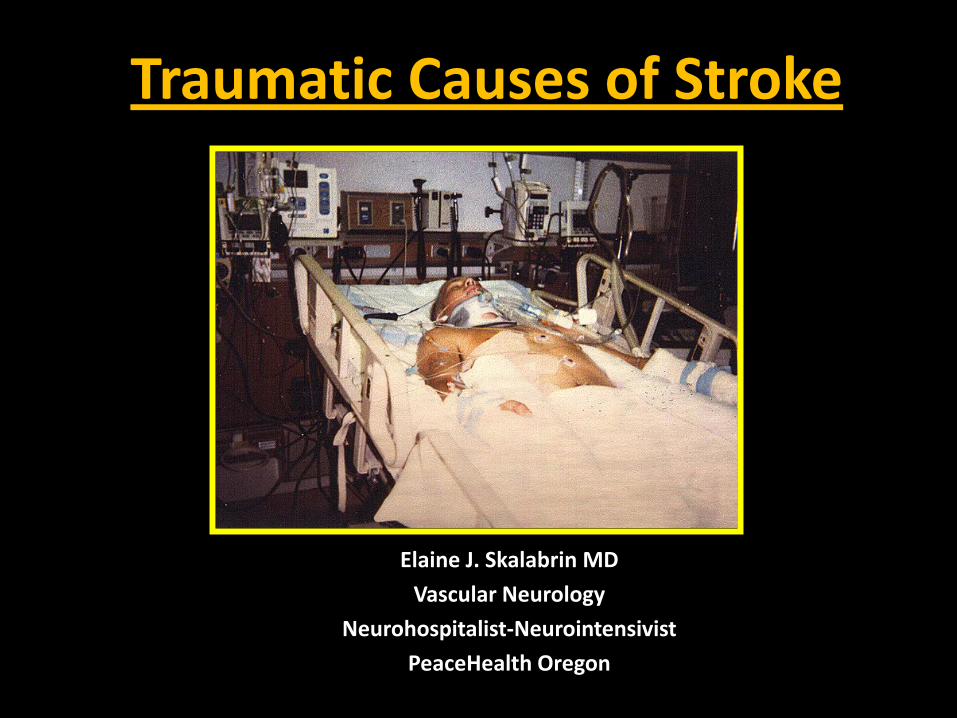
Traumatic Causes of Stroke
Elaine J. Skalabrin MD
Vascular Neurology
Neurohospitalist-Neurointensivist
PeaceHealth Oregon

DISCLOSURES

Objectives
1. Review the clinical features of stroke
2. Understand a variety of mechanisms for stroke in trauma patients
3. Recognize the treatment options and limitation in traumatic causes of stroke

Goal of Therapy in all Neurologic Emergencies
• ACUTE Setting – Identify syndrome
– Take immediate action to reduce disability
– Minimize Risk
• SUBACUTE setting – Understand etiology
– Prevent secondary events
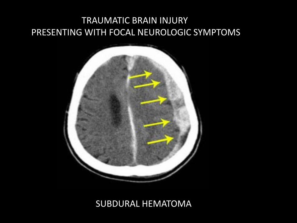
SUBDURAL HEMATOMA
TRAUMATIC BRAIN INJURY PRESENTING WITH FOCAL NEUROLOGIC SYMPTOMS
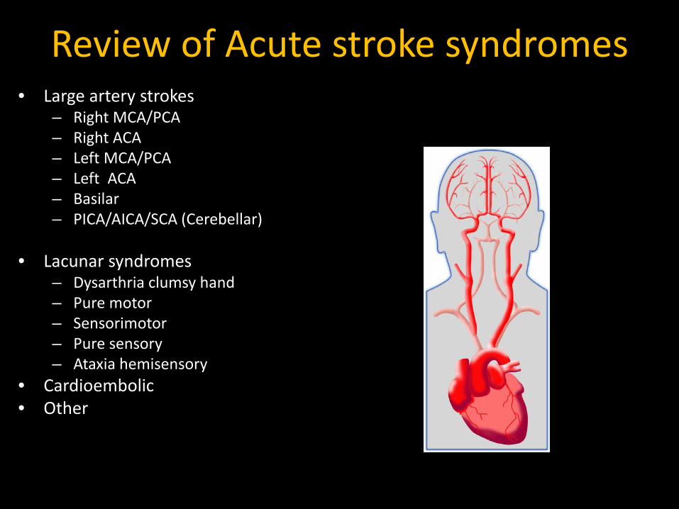
Review of Acute stroke syndromes • Large artery strokes
– Right MCA/PCA – Right ACA – Left MCA/PCA – Left ACA – Basilar – PICA/AICA/SCA (Cerebellar)
• Lacunar syndromes – Dysarthria clumsy hand – Pure motor – Sensorimotor – Pure sensory – Ataxia hemisensory
• Cardioembolic • Other

Traumatic Cause of Acute Stroke • Vascular injuries
– Cervical Dissection
– Cerebral Venous Thrombosis
– Carotid-Cavernous Fistula
– Traumatic Subarachnoid Hemorrhage with Cerebral Vasconstriction
• Hemodynamic – Watershed Infarction
– Cardioembolism
• Special – Paradoxical Embolism
– Coagulopathies

Case #1
• 46 year old male who presents by POV to the ED with neck pain. Earlier that day, he had a fall while skiing. He described he hit a tree branch on the sided of his head which knocked him down. He did not lose consciousness but experience immediate severe neck pain and spasm.
• On exam, he has marked cervical spine tenderness and paraspinal spasm. His neurologic examination is normal
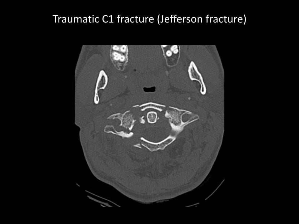
Traumatic C1 fracture (Jefferson fracture)

Case #1
• He is placed in a C-spine collar and given Morphine for pain
• While in the ED, he develops slurred speech and left sided weakness. Initially this is attributed to overmedication. He then progresses to develop severe left hemiplegia and hemineglect

Head CT
IV TPA IS CONTRAINDICATED!
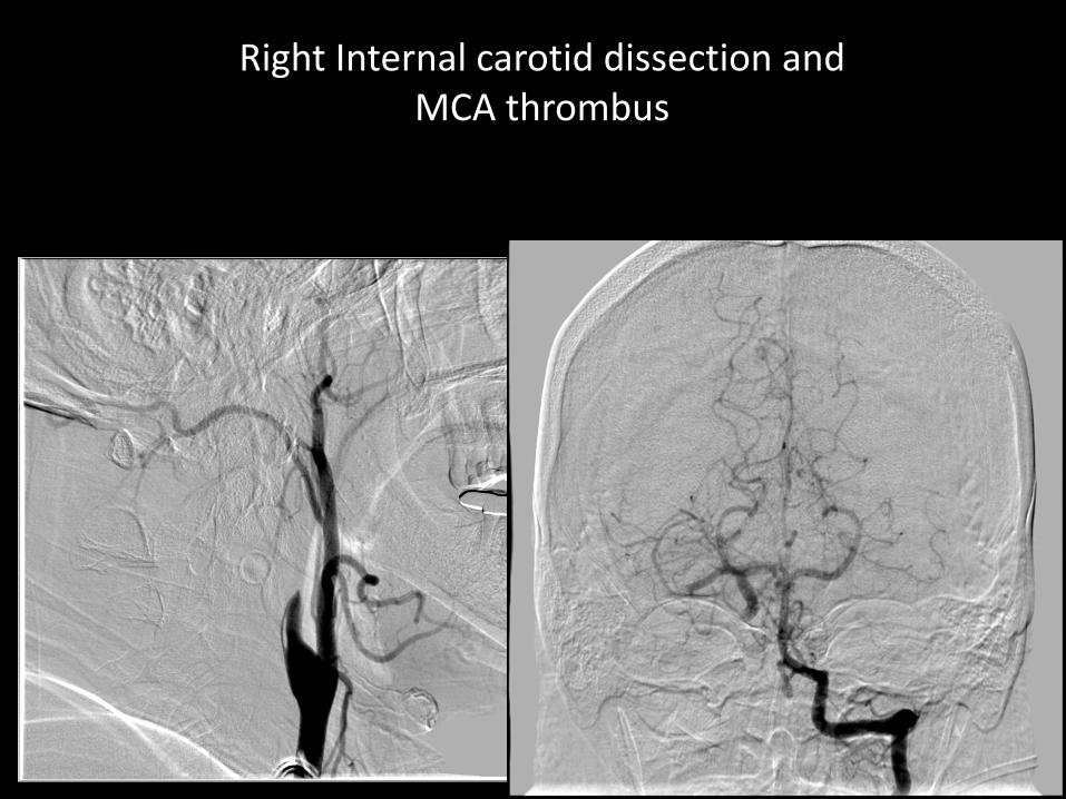
Right Internal carotid dissection and MCA thrombus

Intra- arterial approach: Right carotid stenting with right MCA clot
retrieval and local thrombolysis

Case 2 38 yo white man with left-sided headaches
• Presented to the ED 3 weeks PTA for headache after minor fender-bender.
• Head CT obtained and read as normal.
• Now 3-week h/o intermittent left-sided headaches, localized to behind the left ear. Sometimes blurry vision, nausea, and vomiting with headaches. Pain not relieved by Lortab.
History

History
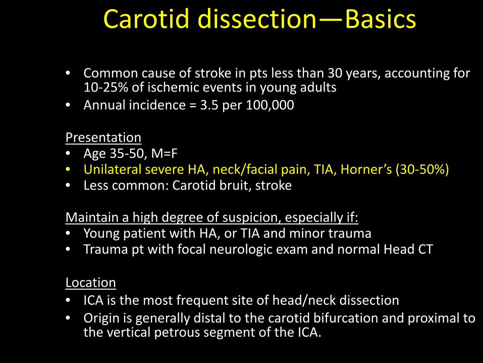
Carotid dissection—Basics
• Common cause of stroke in pts less than 30 years, accounting for 10-25% of ischemic events in young adults
• Annual incidence = 3.5 per 100,000 Presentation • Age 35-50, M=F • Unilateral severe HA, neck/facial pain, TIA, Horner’s (30-50%) • Less common: Carotid bruit, stroke
Maintain a high degree of suspicion, especially if: • Young patient with HA, or TIA and minor trauma • Trauma pt with focal neurologic exam and normal Head CT
Location • ICA is the most frequent site of head/neck dissection • Origin is generally distal to the carotid bifurcation and proximal to
the vertical petrous segment of the ICA.

Carotid dissection
Appearance
• Asymmetric and eccentric narrowed lumen
• Crescentic intramural hematoma
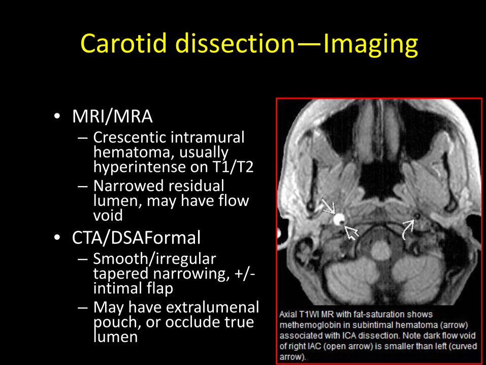
Carotid dissection—Imaging
• MRI/MRA – Crescentic intramural
hematoma, usually hyperintense on T1/T2
– Narrowed residual lumen, may have flow void
• CTA/DSAFormal – Smooth/irregular
tapered narrowing, +/- intimal flap
– May have extralumenal pouch, or occlude true lumen

Carotid dissection Treatment • Goal is to prevent future cerebral ischemia (via arterial
occlusion or thromboembolus) • Antithrombotic therapy is the treatment of choice • Surgical options may be employed if anticoagulation is
contraindicated, including: – Direct repair +/- grafting – Endovascular stenting – STA to MCA bypass (if persistent emboli)
Course • Repeat imaging usually shows restoration (months), or
less commonly, progressive occlusion • Ultimate outcome correlates with severity at time of
diagnosis

Case 4
• 16 yo girl experienced dizziness and nausea after gymnastics practice
• Left school early the next day due to continued nausea and vomiting. Went home to rest.
• Later that evening presented to the ED obtunded.
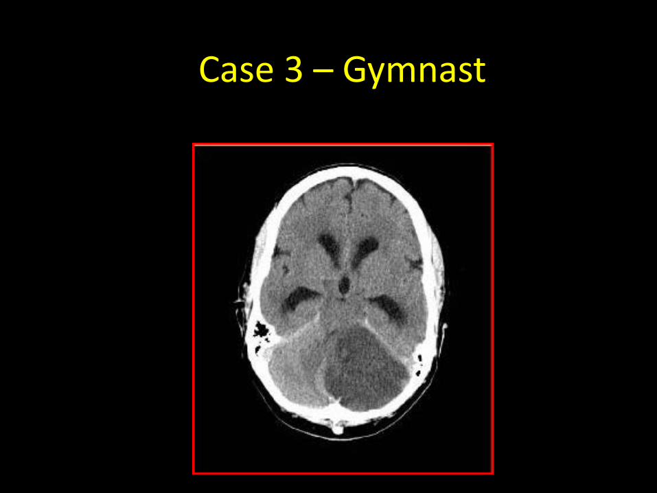
Case 3 – Gymnast

Vertebral artery injury
• Stretching and compression at the atlanto-axial and atlanto-occipital joints during head rotation, makes the 3rd portion of the VA (from C2 to the dura) particularly prone to injury.
• The 2nd portion is injured due to direct trauma from cervical fractures through the transverse foramina.

Symptoms— Vertebral artery injury
• Dizziness
• Nausea and vomiting
• Ataxia
• Facial or body anesthesia
• Swallowing difficulty
• Dysarthria
• Coma, death
Vertebral artery injury—poor outcomes
• Biffl et al reviewed 38 patients with BVAI over a 3.5 year period
• Presentation – Motor vehicle crash most common mechanism – 71% of patients had an associated cervical spine
fractures • No predilection for cervical vertebral level or fracture
pattern
• 24% of all patients with BVAI suffered a posterior circulation stroke.
• The BVAI death rate was 8%

Indications for angiogram in trauma
• Neuro exam not explained by CT
• Expanding cervical hematoma
• Focal neurologic deficit
• Cervical bruit in patient < 50 years old
• Arterial hemorrhage
• Focal neurologic deficit
• Stroke on secondary CT scan

• Treatment was initiated for 282 asymptomatic BCVIs.
• There were bleeding complications in 8 patients total.

• Of the 107 asymptomatic patients who did not receive treatment, 21% had a stroke.
• Of the 50 patients who had a stroke, the mean time to diagnosis was 58 hours after injury.
• Of the 45 patients with stroke (non-catheter related), injury grade was: – 23 Grade I, 19 Grade II – 20 Grade III, 5 Grade IV
• Stroke-related mortality was 30% (15 of 50)

BCVI—Screening
Berne et al, 2010: • 102 pts with BCVI on CTA out of 9935 blunt
trauma patients – 59 CAI – 43 VAI
• Univariant analysis: – Cervicle spine fracture (RR 10.4) – Basilar skull fracture (RR 3.6) – Mandible fracture (RR 2.5)

Case #4
• 18 yo male presents after falling off bike with a left temporal bone fracture. He is treated and released from the hospital and day 3.
• He has progressive intractable headache and on day 5, he presents with progressive confusion


Left temporal edema and hemorrhage
Initial CT 2 days later
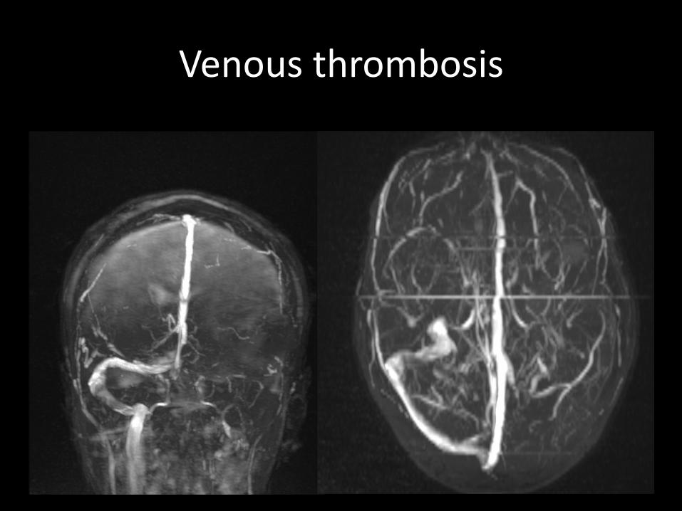
Venous thrombosis
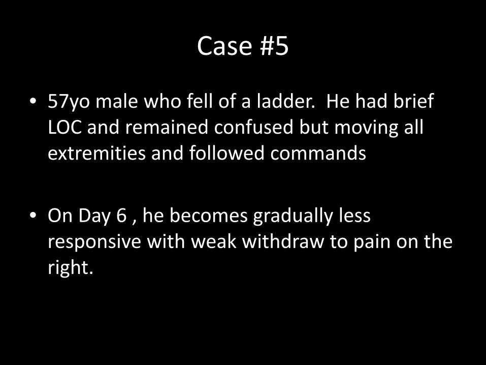
Case #5
• 57yo male who fell of a ladder. He had brief LOC and remained confused but moving all extremities and followed commands
• On Day 6 , he becomes gradually less responsive with weak withdraw to pain on the right.

Traumatic Subarchnoid Hemorrhage
Cerebral angiography is normal

MRI

Angiography
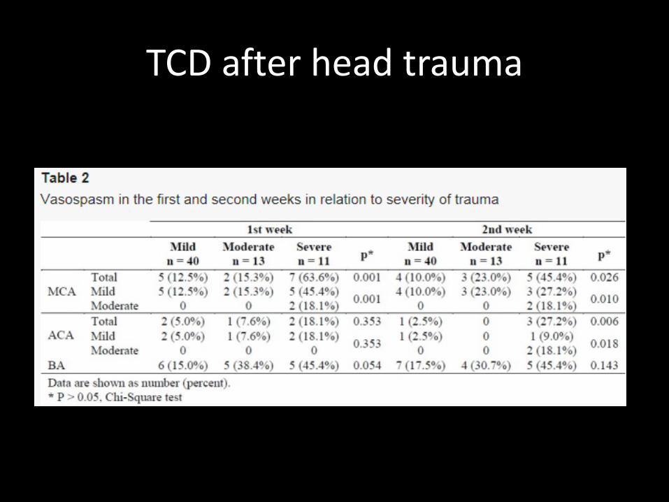
TCD after head trauma

Case #7
• 21 yo man presents with chief complaint of bulging red right eye and pulsitile tinnitus,
• 4 weeks after a MVA

Carotid Cavernous Fistula


Case #8 •
25 Year old man struck by a car moving at a high rate . The patient was combative at the scene and intubated and sedated( GCS 7)
• His head CT was normal • Initial trauma evaluating left tibia, displaced fracture
of the sacrum and symphysis pubis, diaphragmatic herniation of abdominal content and splenic rupture
• On initial ICU assessment he wakens easily , follow command s and move all extremities

Case # 8(cont)
• Patient remained intubated and sedated and required multiple surgeries and diagnostic studies. Routine neurochecks were performed with brief discontinuation of propofol
• On day 3. the propofol was stopped and patient found to be unable to move the left upper and lower extremity above gravity.
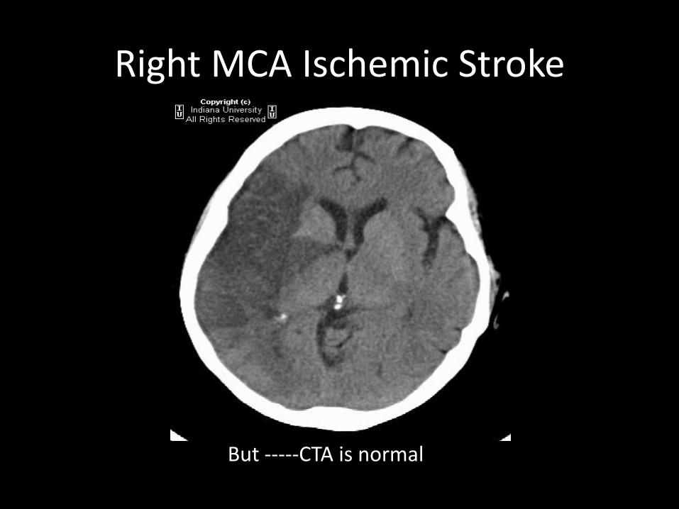
Right MCA Ischemic Stroke
But -----CTA is normal

Deep vein thrombosis
Historical Prospective: Paradoxical Embolism
• Paradoxical embolism first described in 1877 by Connheim
• Defined as embolic entry of a venous thrombus into systemic circulation through a right to left shunt
• Johnson established diagnostic criteria in 1951:
– arterial embolism without evidence of left heart or arterial source
– Abnormal communication between the right and left circulations
– Confirmation of DVT or PE
– A pressure gradient the favors right-left shunt

Patent Foramen Ovale
Prevalence At autopsy 34% in the 1st 3 decades
On TEE 22-26 % of healthy adults
All strokes 10-30%
Cryptogenic stroke 15-50%*
*highest among those with no risk factors
Defined as a defect in the septum primum or secundum with right to left shunt
Most common atrial septal anomaly Many case reports document stroke in the face of known
DVT/PE and PFO
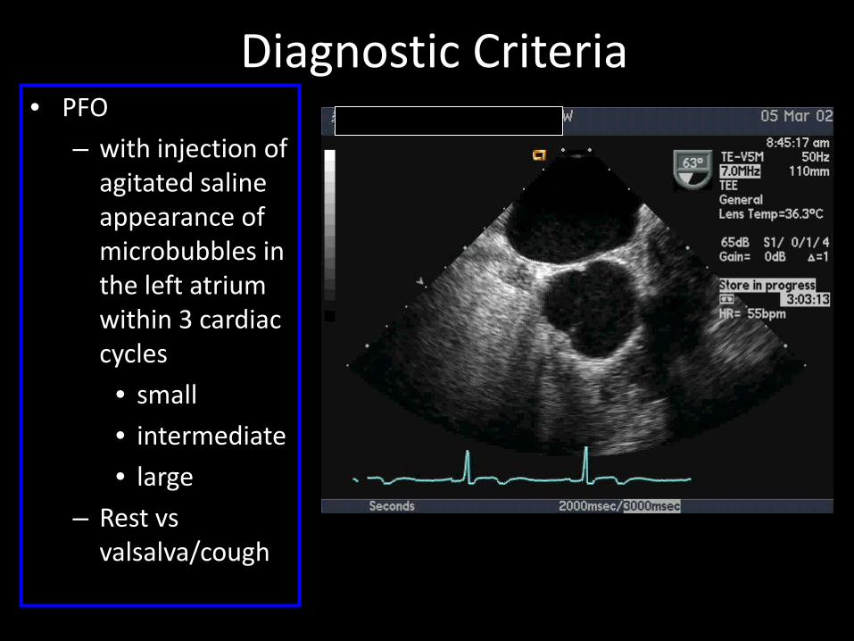
Diagnostic Criteria • PFO
– with injection of agitated saline appearance of microbubbles in the left atrium within 3 cardiac cycles
• small
• intermediate
• large
– Rest vs valsalva/cough

Case # 9
• 19 yo man involved in a motorcycle accident with initial GCS of 13. He sustained a left femur fracture. On day 3, he becomes impulsive , restless and then pulls out his right IJ vein catheter.
• His saturations drop to 70%, he becomes unresponsive. With bag-mask ventilation his saturations improve but he then develops focal motor seizures on the left.

Air embolism ( arterial gas embolism)
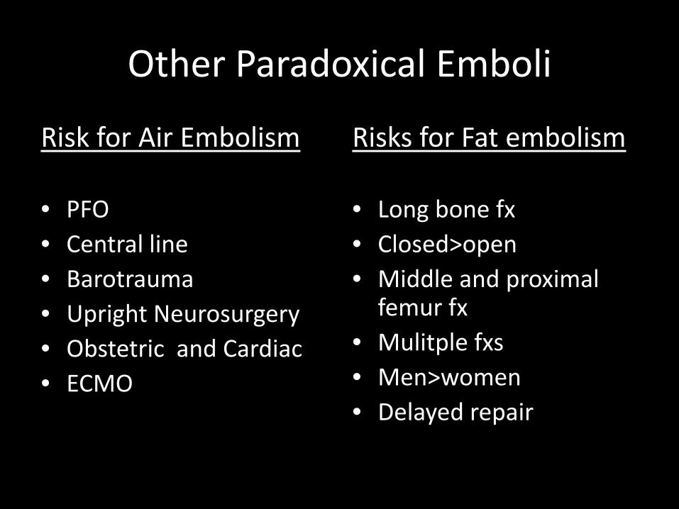
Other Paradoxical Emboli
Risk for Air Embolism
• PFO • Central line • Barotrauma • Upright Neurosurgery • Obstetric and Cardiac • ECMO
Risks for Fat embolism
• Long bone fx • Closed>open • Middle and proximal
femur fx • Mulitple fxs • Men>women • Delayed repair
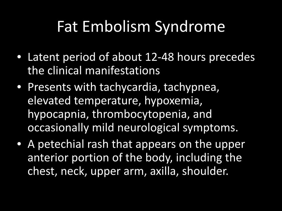
Fat Embolism Syndrome
• Latent period of about 12-48 hours precedes the clinical manifestations
• Presents with tachycardia, tachypnea, elevated temperature, hypoxemia, hypocapnia, thrombocytopenia, and occasionally mild neurological symptoms.
• A petechial rash that appears on the upper anterior portion of the body, including the chest, neck, upper arm, axilla, shoulder.

Fat Embolism Syndrome
• CNS signs: diffuse encephalopathy: acute confusion, stupor, coma, extensor posturing, focal neurological signs or generalized seizure
• Hypoxemia is present in nearly all patients with FES, often to a Pa02 of well below 60 mmHg. Arterial hypoxemia in these patients has been attributed to ventilation-perfusion inequality and intrapulmonary shunting.
• Acute cor pulmonale is manifested by respiratory distress, hypoxemia, hypotension and elevated central venous pressure.

Other mechanisms
Watershed Infarct=
Loss flow across a vessel stenosis with resulting ischemia in areas bordering two vascular territories

Takotsubo Cardiomyopathy Stress-induced cardiomyopathy
Kurisu, S., et al. 2002. American Heart Journal. 143: 448-455.

COAGULOPATHY

SUMMARY
• Cerebrovascular injury with risk for ischemic stroke may occur in setting of even minor trauma
• Intra- arterial approach for acute stroke treatment is necessary in the setting of trauma to minimize bleeding risk.
• More severe TBI is associated with increase risk of vasospasm
• Although uncommon, paradoxical embolism is a risk especially in multitrauma patients
• If the scan does not explain the deficit- LOOK FOR VASCULAR INJURY ACUTELY.


