Dilsher Dhoot, MD Rishi Singh, MD Nathan Steinle, MD TREATING RETINAL DISORDERS: UPDATES FOR THE NEWER RETINA SPECIALIST Supplement to July/August 2018 A continuing medical education (CME) activity provided by Evolve Medical Education LLC and distributed with Retina Today. This CME activity is supported by an educational grant from Regeneron Pharmaceuticals, Inc. Distributed with
Transcript
Dilsher Dhoot, MD
Rishi Singh, MD
Nathan Steinle, MD
TREATING RETINAL DISORDERS: UPDATES FOR THE NEWER RETINA SPECIALIST
Supplement to July/August 2018
A continuing medical education (CME) activity provided by Evolve Medical Education LLC and distributed with Retina Today.
This CME activity is supported by an educational grant from Regeneron Pharmaceuticals, Inc.
Distributed with
CONTENT SOURCEThis continuing medical education (CME) activity captures
content from a roundtable discussion.
ACTIVITY DESCRIPTIONThis activity focuses on providing ongoing and continuous
education with up-to-date information and cases for newer retina specialists involved in the treatment and management of patients with posterior segment diseases.
TARGET AUDIENCEThis certified CME activity is designed for ophthalmologists
and retina specialists involved in the management of patients with retina disorders.
LEARNING OBJECTIVESUpon completion of this activity, the participant should be
able to:• Discuss recent studies that address long-term treatment
outcomes in age-related macular degeneration (AMD).• Develop an individualized treatment plan for patients
with AMD.• Discuss implementing imaging tools as diagnostic therapies.
• Summarize the outcomes of pivotal studies in AMD and diabetic macular edema (DME) and how study results may differ from real-world dosing methods.
• Summarize the comparative Protocol T findings in DME and how to relate that to clinical practice patterns.
• Evaluate practice flow to determine the most efficient patient experience.
• Develop plans to reduce reimbursement denials.
GRANTOR STATEMENTSupported through an educational grant from Regeneron
Pharmaceuticals, Inc.
ACCREDITATION STATEMENTEvolve Medical Education LLC (Evolve) is accredited by
the Accreditation Council for Continuing Medical Education (ACCME) to provide CME for physicians.
CREDIT DESIGNATION STATEMENTEvolve designates this enduring material for a maximum of
1 AMA PRA Category 1 Credit™. Physicians should claim only the credit commensurate with the extent of their participation in the activity.
2 SUPPLEMENT TO RETINA TODAY | JULY/AUGUST 2018
Treating Retinal Disorders: Updates for the Newer Retina Specialist
Release Date: July 2018Expiration Date: July 2019
DILSHER DHOOT, MD California Retina Consultants
Santa Barbara, California
RISHI SINGH, MD Cleveland Clinic Cleveland, Ohio
NATHAN STEINLE, MD California Retina Consultants
Santa Barbara, California
FACULTY
TO OBTAIN AMA PRA CATEGORY 1 CREDIT™To obtain AMA PRA Category 1 Credit™ for this activity, you
must read the activity in its entirety and complete the Pretest/Posttest/Activity Evaluation/Satisfaction Measures Form, which consists of a series of multiple choice questions. To answer these questions online and receive real-time results, please visit evolvemeded.com and click “Online Courses.” Upon completing the activity and self-assessment test, you may print out a CME certificate awarding 1 AMA PRA Category 1 Credit™. Alternatively, please complete the Pretest/Posttest/Activity Evaluation/Satisfaction Measures Form and mail or fax to Evolve Medical Education LLC, 353 West Lancaster Avenue, Second Floor, Wayne, PA 19087; Fax: (215) 933-3950.
DISCLOSURE POLICY It is the policy of Evolve that faculty and other individuals
who are in the position to control the content of this activ-ity disclose any real or apparent conflict of interests relating to the topics of this educational activity. Evolve has full policies in place that will identify and resolve all conflicts of interest prior to this educational activity.
The following faculty/staff members have the following finan-cial relationships with commercial interests:
Dilsher Dhoot, MD, has had a financial agreement or affili-ation during the past year with the following commercial inter-ests in the form of Consultant/Advisory Board/Speaker’s Bureau: Alimera Sciences; Allergan; Genentech; Regeneron Pharmaceuticals, Inc.; Notal Vision; and Santen. Grant/Research Support: Allergan; Genentech; and Regeneron Pharmaceuticals, Inc.
Rishi Singh, MD, has had a financial agreement or affiliation during the past year with the following commercial interests in the form of Consultant/Advisory Board/Speaker’s Bureau: Alcon; Allergan Plc; Genentech; Optos; Regeneron Pharmaceuticals, Inc.; Shire Plc.; and Zeiss. Grant/Research Support: Alcon; Apellis Pharmaceuticals; Genentech; and Regeneron Pharmaceuticals, Inc.
Nathan Steinle, MD, has had a financial agreement or affiliation during the past year with the following commercial interests in the form of Consultant/Advisory Board/Speaker’s Bureau: Alimera Sciences; Genentech; Notal Vision, Regeneron Pharmaceuticals, Inc.; Vortex Surgical; and Zeiss. Grant/Research Support: Genentech and Regeneron Pharmaceuticals, Inc.
EDITORIAL SUPPORT DISCLOSURESErin K. Fletcher, MIT, director of compliance and education,
Susan Gallagher-Pecha, director of client services and project man-agement, Evolve; and Michelle Dalton, writer, have no financial relationships with commercial interests. Jaya Kumar, MD, peer reviewer, has no financial relationships with commercial interests.
OFF-LABEL STATEMENTThis educational activity may contain discussion of published
and/or investigational uses of agents that are not indicated by the FDA. The opinions expressed in the educational activity are those of the faculty. Please refer to the official prescribing infor-mation for each product for discussion of approved indications, contraindications, and warnings.
DISCLAIMER
The views and opinions expressed in this educational activity are those of the faculty and do not necessarily represent the views of Evolve, Retina Today, or Regeneron Pharmaceuticals, Inc.
DIGITAL EDITIONTo view the online version of the material, please visit
evolvemeded.com/online-courses/.
JULY/AUGUST 2018 | SUPPLEMENT TO RETINA TODAY 3
1. PLEASE RATE YOUR CONFIDENCE ON YOUR ABILITY TO INCORPORATE RESEARCH INTO THE CLINIC BASED ON THIS ACTIVITY:
a. Not at all confidentb. Not very confidentc. Neutrald. Confidente. Very confident
2. PLEASE RATE HOW OFTEN YOU INTEND TO APPLY THE RECOMMENDATIONS FROM THE RETINA PANELISTS ON CODING AND BILLING. (BASED ON A SCALE OF 1 TO 5, WITH 1 BEING NEVER AND 5 BEING ALWAYS).
a. 1b. 2c. 3d. 4e. 5
3. ACCORDING TO ASRS SURVEY DATA, HOW LONG DO US PHYSICIANS EXTEND PATIENTS ON A TREAT-AND-EXTEND REGIMEN?
a. 16 or fewer weeksb. 12 or fewer weeksc. 10 or fewer weeksd. 8 or fewer weeks
4. OCT ANGIOGRAPHY (OCT-A) IS USEFUL IN THE FOLLOWING SCENARIOS EXCEPT: a. Patients with an unexplained drop in visual acuity. b. Patients with suspected masquerading syndromes.c. Patients with choroidal neovascularization (CNV).d. Patients with diabetic macular edema (DME).
5. FOR CLINICIANS INTERESTED IN PARTICIPATING IN CLINICAL TRIALS, AT WHAT PHASE ARE EARLY-CAREER INVESTIGATORS LIKELY TO FIND IT EASIEST TO BECOME INVOLVED?
a. Phase 1b. Phase 2c. Phase 3
6. WHICH PART OF RESEARCH INFRASTRUCTURE IS MOST IMPORTANT TO SUC-CESSFUL CLINICAL TRIAL PARTICIPATION?
a. A relationship with the institutional review boardb. A full-time, experienced study coordinatorc. An Early Treatment Diabetic Retinopathy Study (ETDRS) 20-foot study laned. A widefield imaging system
7. WHEN JOINING A PRACTICE FELLOWSHIP, WHAT DO THE PANELISTS CONSIDER THE LEAST IMPORTANT CONSIDERATION?
a. Starting salaryb. Partnership potential c. Geographic location d. The buy-out package
8. MODIFIER 25 IS USED TO __________________a. Schedule injections. b. Repeat imaging in the diseased eye.c. Monitor the fellow eye for disease when no disease is present. d. Monitor the fellow eye for disease progression when disease
is present.
9. MR. SMITH HAS PRESENTED FOR EVALUATION; HE HAS HAD DIABETES FOR MORE THAN 10 YEARS, AND THIS IS HIS FIRST VISIT TO AN EYE CARE PRACTITIO-NER TO EVALUATE FOR DME/DIABETIC RETINOPATHY (DR). YOU SHOULD GET A FLUORESCEIN ANGIOGRAM (FA) __________ AND AN OCT _________
a. at every visit; at every visitb. at baseline and every 6 to 12 months; at every visitc. at every visit; at baseline and every 6 to 12 monthsd. at baseline and then yearly; at baseline and then every 6 to 12 months
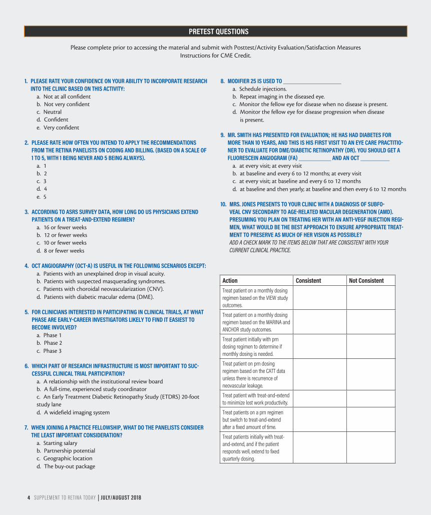
10. MRS. JONES PRESENTS TO YOUR CLINIC WITH A DIAGNOSIS OF SUBFO-VEAL CNV SECONDARY TO AGE-RELATED MACULAR DEGENERATION (AMD). PRESUMING YOU PLAN ON TREATING HER WITH AN ANTI-VEGF INJECTION REGI-MEN, WHAT WOULD BE THE BEST APPROACH TO ENSURE APPROPRIATE TREAT-MENT TO PRESERVE AS MUCH OF HER VISION AS POSSIBLE? ADD A CHECK MARK TO THE ITEMS BELOW THAT ARE CONSISTENT WITH YOUR CURRENT CLINICAL PRACTICE.
PRETEST QUESTIONS
Please complete prior to accessing the material and submit with Posttest/Activity Evaluation/Satisfaction MeasuresInstructions for CME Credit.
4 SUPPLEMENT TO RETINA TODAY | JULY/AUGUST 2018
Action Consistent Not ConsistentTreat patient on a monthly dosing regimen based on the VIEW study outcomes.
Treat patient on a monthly dosing regimen based on the MARINA and ANCHOR study outcomes.
Treat patient initially with prn dosing regimen to determine if monthly dosing is needed.
Treat patient on prn dosing regimen based on the CATT data unless there is recurrence of neovascular leakage.
Treat patient with treat-and-extend to minimize lost work productivity.
Treat patients on a prn regimen but switch to treat-and-extend after a fixed amount of time.
Treat patients initially with treat-and-extend, and if the patient responds well, extend to fixed quarterly dosing.
JULY/AUGUST 2018 | SUPPLEMENT TO RETINA TODAY 5
TREATING RETINAL DISORDERS: UPDATES FOR THE NEWER RETINA SPECIALIST
DEVELOPING INDIVIDUALIZED TREATMENT PLANS Q DILSHER DHOOT, MD: How do you develop an
individualized treatment plan for patients with AMD and DME?
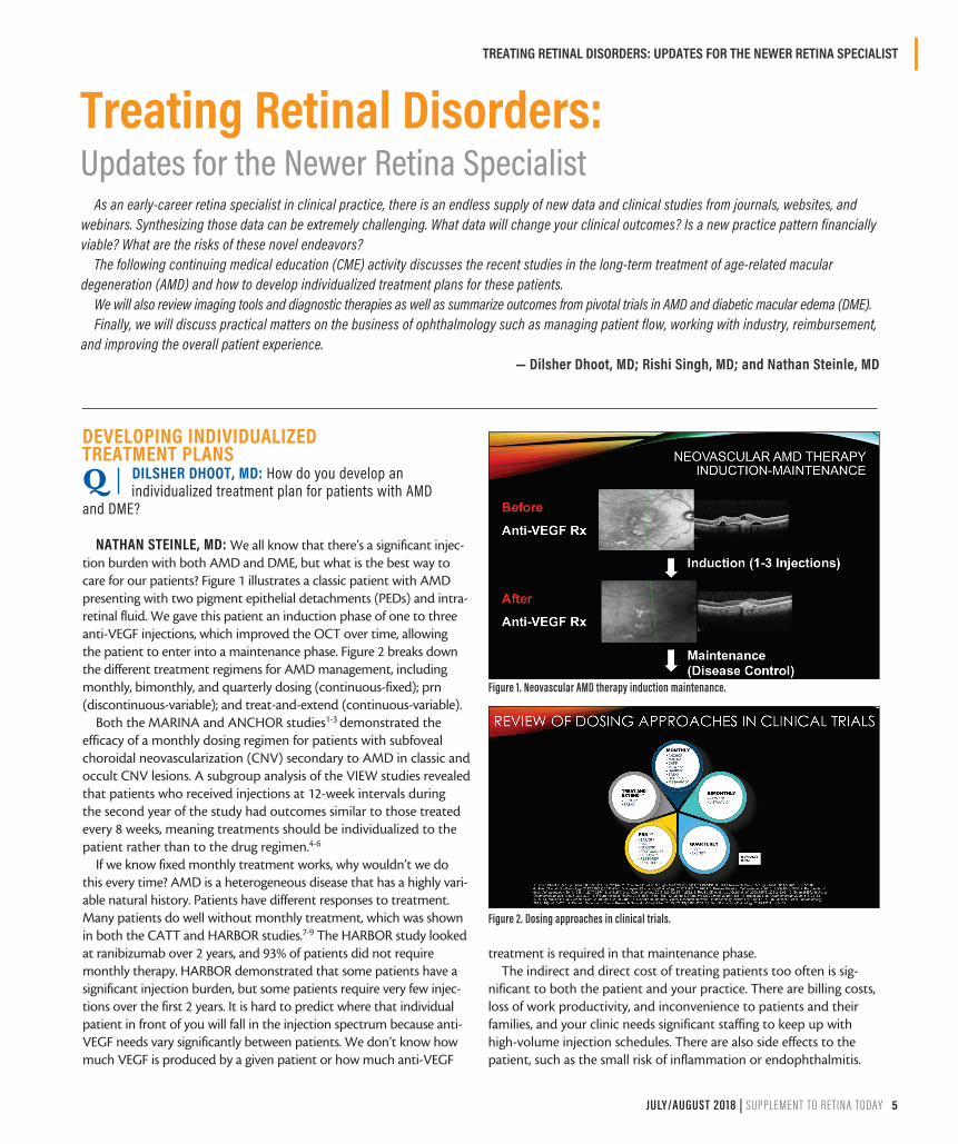
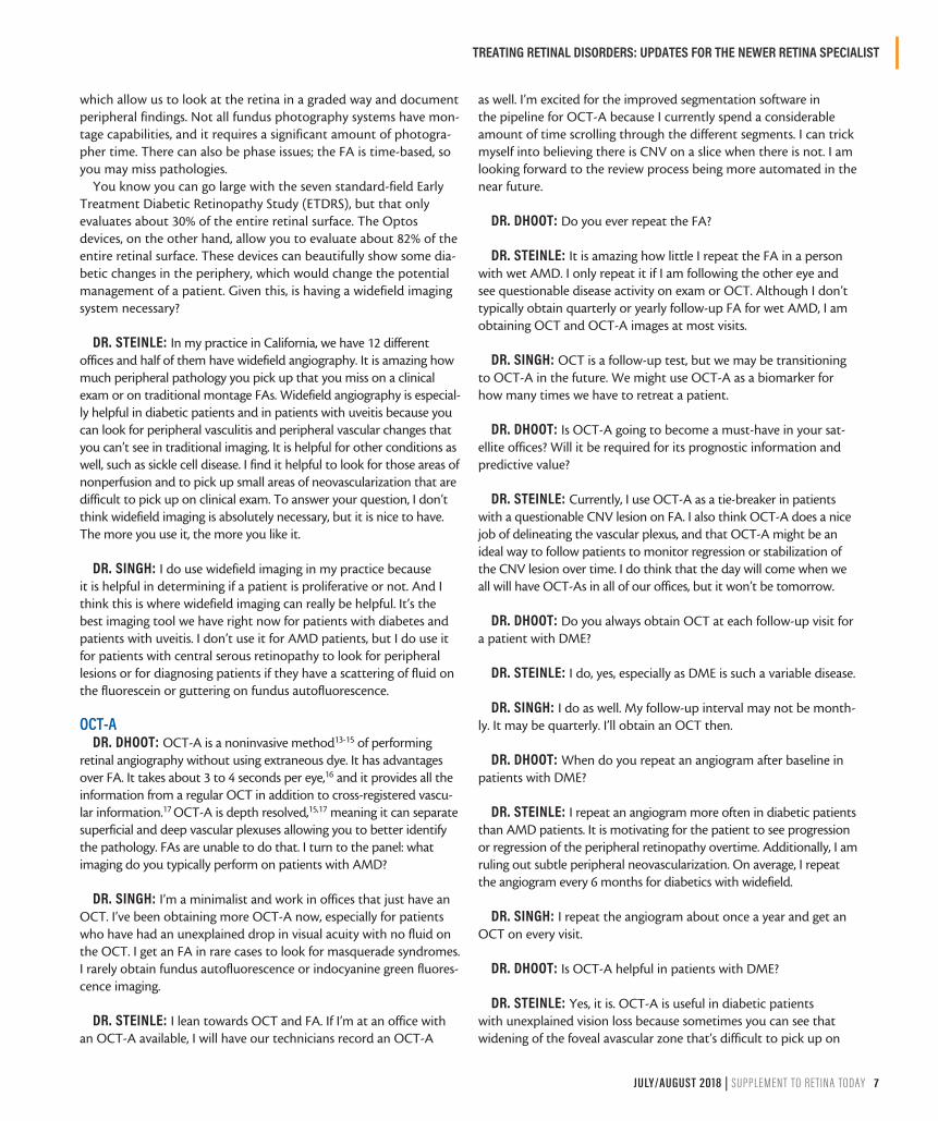
NATHAN STEINLE, MD: We all know that there’s a significant injec-tion burden with both AMD and DME, but what is the best way to care for our patients? Figure 1 illustrates a classic patient with AMD presenting with two pigment epithelial detachments (PEDs) and intra-retinal fluid. We gave this patient an induction phase of one to three anti-VEGF injections, which improved the OCT over time, allowing the patient to enter into a maintenance phase. Figure 2 breaks down the different treatment regimens for AMD management, including monthly, bimonthly, and quarterly dosing (continuous-fixed); prn (discontinuous-variable); and treat-and-extend (continuous-variable).
Both the MARINA and ANCHOR studies1-3 demonstrated the efficacy of a monthly dosing regimen for patients with subfoveal choroidal neovascularization (CNV) secondary to AMD in classic and occult CNV lesions. A subgroup analysis of the VIEW studies revealed that patients who received injections at 12-week intervals during the second year of the study had outcomes similar to those treated every 8 weeks, meaning treatments should be individualized to the patient rather than to the drug regimen.4-6
If we know fixed monthly treatment works, why wouldn’t we do this every time? AMD is a heterogeneous disease that has a highly vari-able natural history. Patients have different responses to treatment. Many patients do well without monthly treatment, which was shown in both the CATT and HARBOR studies.7-9 The HARBOR study looked at ranibizumab over 2 years, and 93% of patients did not require monthly therapy. HARBOR demonstrated that some patients have a significant injection burden, but some patients require very few injec-tions over the first 2 years. It is hard to predict where that individual patient in front of you will fall in the injection spectrum because anti-VEGF needs vary significantly between patients. We don’t know how much VEGF is produced by a given patient or how much anti-VEGF
treatment is required in that maintenance phase.The indirect and direct cost of treating patients too often is sig-
nificant to both the patient and your practice. There are billing costs, loss of work productivity, and inconvenience to patients and their families, and your clinic needs significant staffing to keep up with high-volume injection schedules. There are also side effects to the patient, such as the small risk of inflammation or endophthalmitis.
Treating Retinal Disorders: Updates for the Newer Retina Specialist
As an early-career retina specialist in clinical practice, there is an endless supply of new data and clinical studies from journals, websites, and webinars. Synthesizing those data can be extremely challenging. What data will change your clinical outcomes? Is a new practice pattern financially viable? What are the risks of these novel endeavors?
The following continuing medical education (CME) activity discusses the recent studies in the long-term treatment of age-related macular degeneration (AMD) and how to develop individualized treatment plans for these patients.
We will also review imaging tools and diagnostic therapies as well as summarize outcomes from pivotal trials in AMD and diabetic macular edema (DME). Finally, we will discuss practical matters on the business of ophthalmology such as managing patient flow, working with industry, reimbursement,
and improving the overall patient experience. — Dilsher Dhoot, MD; Rishi Singh, MD; and Nathan Steinle, MD
TREATING RETINAL DISORDERS: UPDATES FOR THE NEWER RETINA SPECIALIST
6 SUPPLEMENT TO RETINA TODAY | JULY/AUGUST 2018
The prn treatment philosophy is to only treat patients if there is dis-ease activity. There are potential limitations of that treatment modal-ity such as recurrence of neovascular leakage, growth of lesion size, and fibrosis. With AMD I’m concerned about having a large subretinal bleed and then having a long-term disciform scar that could have been prevented with more VEGF suppression. We know that multiple recurrences lead to disease progression and poorer long-term visual outcomes in some patients.9 The 5-year CATT data showed that only 50% of the patients had 20/40 or better vision with prn.
Treat-and-extend is very popular in the United States because it is proactive and individualized. It minimizes recurrences and the risk of subretinal fibrosis. It also maximizes long-term visual outcomes and safety through fewer injections and a reduced risk of glaucoma, geo-graphic atrophy, and endophthalmitis. Treat-and-extend is also cost effective because you are minimizing the drug use, time in your clinic, and lost work productivity of the patient and their family.
RISHI SINGH, MD: I tend to dose patients on a prn basis from the beginning just in case they are that one in seven patient who may never need another dose again. I’ll start to employ treat-and-extend in patients if I find after 6 to 9 months that they need continual treatment. One of the problems with continuous-fixed dosing is the high rate of endophthalmitis. In the monthly arm of the CATT trial, the continuous-fixed dosing patients’ rates of endophthalmitis were almost four- and five-fold higher than patients in the prn arm.9
DR. DHOOT: I typically use treat-and-extend from the get-go. If the patient is responding well, I will extend out to 12 weeks. From this point, I will often dose in a fixed quarterly fashion if the patient’s disease activity allows this. Occasionally a patient requests to stop dosing; I reiterate the risk of CNV recurrence and if the patient insists and if the fluorescein angiogram (FA) is quiet, then I’ll turn to prn dosing at that point.
DR. STEINLE: What is your typical injection interval in AMD patients using the treat-and-extend regimen?
DR. SINGH: It is difficult to say and typically drug-dependent. I found a greater durability with branded versus generic drugs. I’ve found that my ability to stretch the interval is much less with beva-cizumab (Avastin, Genentech) then with ranibizumab (Lucentis, Genentech) or aflibercept (Eylea, Regeneron Pharmaceuticals, Inc.). I tend to use those drugs more often with treat-and-extend because my experience hasn’t been the same with bevacizumab. I also don’t think there is a clear answer on the extension threshold. The ATLAS study had patients stretched up to 16 weeks,10 while the ASRS survey data shows the majority of physicians extend to 12 or fewer weeks.11 We don’t know how far patients can be extended.
DR. DHOOT: Most of my patients fall in between 8 and 10 weeks. I tend to cap at 12 weeks because of fear of subretinal hemorrhage and CNV recurrence.
DR. STEINLE: What is your initial anti-VEGF of choice for a new patient with wet AMD?
DR. SINGH: This is a complicated question because of patient finan-cial restrictions. I treat many patients who have Medicare, but many of my patients have no insurance. For those reasons, I tend to start with bevacizumab. However, I’ve been able to achieve more durability with some of the branded drugs versus the non-branded drugs, so I tend to migrate to the other drugs over time. If the patient has good visual acuity with bevacizumab, I’ll keep them on it, but if there is any vari-ability in outcomes, I’ll switch to a branded regimen.
DR. DHOOT: There are definitely external factors that force our hand and make these choices for us. There is a good argument that branded drugs have increased durability. However, I’ll often start with bevacizumab because of insurance factors. I’m always looking for forward progression. If I’m not seeing forward progression with bevacizumab after two or three injections, I will switch to a branded drug, as the insurance allows.
DR. STEINLE: In your geographic area of practice, do you have to utilize step therapy and demonstrate drug failure to insurance carri-ers before switching medications?
DR. SINGH: No, we’re currently able to choose what drugs we want to use, but it is important to understand the diagnosis codes and what is approved. What do you have to document that the patient has exudative AMD? What codes do you have to use to dem-onstrate that they have disease activity? That is where the real prob-lems are because people aren’t aware of the codes needed to ensure they are paid for that visit.
DR. STEINLE: Brolucizumab (RTH258, Novartis) is exiting phase 3 development.12 How will this new agent change the market?
DR. SINGH: One of the big selling points of brolucizumab is the 3-month dosing in 50% of patients.12 We will end up using the drug just from a durability standpoint. It remains to be seen if it is better than what we have now.
DR. DHOOT: It sounds like brolucizumab will be available in 2019, and it will be nice to have another drug in our toolkit. Durability is the key in the treatment of retinal diseases, and if brolucizumab turns out to have a real-world edge in the durability department with equal efficacy, then that will be very attractive.
IMAGING IN AMD/DMEDR. DHOOT: OCT angiography (OCT-A) and widefield imaging are
two imaging modalities that are new in retina practices. But declin-ing reimbursement and increased number of satellite offices make it challenging to afford these imaging modalities at all practice loca-tions. Let us examine these imaging modalities in greater detail.
Widefield ImagingDR. DHOOT: The primary modalities for retinal imaging include
the traditional fundus camera, which is either mydriatic or non-mydriatic. This is being followed by seven-field and montage capabilities. More recently we have had ultrawide-field options,
JULY/AUGUST 2018 | SUPPLEMENT TO RETINA TODAY 7
TREATING RETINAL DISORDERS: UPDATES FOR THE NEWER RETINA SPECIALIST
which allow us to look at the retina in a graded way and document peripheral findings. Not all fundus photography systems have mon-tage capabilities, and it requires a significant amount of photogra-pher time. There can also be phase issues; the FA is time-based, so you may miss pathologies.
You know you can go large with the seven standard-field Early Treatment Diabetic Retinopathy Study (ETDRS), but that only evaluates about 30% of the entire retinal surface. The Optos devices, on the other hand, allow you to evaluate about 82% of the entire retinal surface. These devices can beautifully show some dia-betic changes in the periphery, which would change the potential management of a patient. Given this, is having a widefield imaging system necessary?
DR. STEINLE: In my practice in California, we have 12 different offices and half of them have widefield angiography. It is amazing how much peripheral pathology you pick up that you miss on a clinical exam or on traditional montage FAs. Widefield angiography is especial-ly helpful in diabetic patients and in patients with uveitis because you can look for peripheral vasculitis and peripheral vascular changes that you can’t see in traditional imaging. It is helpful for other conditions as well, such as sickle cell disease. I find it helpful to look for those areas of nonperfusion and to pick up small areas of neovascularization that are difficult to pick up on clinical exam. To answer your question, I don’t think widefield imaging is absolutely necessary, but it is nice to have. The more you use it, the more you like it.
DR. SINGH: I do use widefield imaging in my practice because it is helpful in determining if a patient is proliferative or not. And I think this is where widefield imaging can really be helpful. It’s the best imaging tool we have right now for patients with diabetes and patients with uveitis. I don’t use it for AMD patients, but I do use it for patients with central serous retinopathy to look for peripheral lesions or for diagnosing patients if they have a scattering of fluid on the fluorescein or guttering on fundus autofluorescence.
OCT-ADR. DHOOT: OCT-A is a noninvasive method13-15 of performing
retinal angiography without using extraneous dye. It has advantages over FA. It takes about 3 to 4 seconds per eye,16 and it provides all the information from a regular OCT in addition to cross-registered vascu-lar information.17 OCT-A is depth resolved,15,17 meaning it can separate superficial and deep vascular plexuses allowing you to better identify the pathology. FAs are unable to do that. I turn to the panel: what imaging do you typically perform on patients with AMD?
DR. SINGH: I’m a minimalist and work in offices that just have an OCT. I’ve been obtaining more OCT-A now, especially for patients who have had an unexplained drop in visual acuity with no fluid on the OCT. I get an FA in rare cases to look for masquerade syndromes. I rarely obtain fundus autofluorescence or indocyanine green fluores-cence imaging.
DR. STEINLE: I lean towards OCT and FA. If I’m at an office with an OCT-A available, I will have our technicians record an OCT-A
as well. I’m excited for the improved segmentation software in the pipeline for OCT-A because I currently spend a considerable amount of time scrolling through the different segments. I can trick myself into believing there is CNV on a slice when there is not. I am looking forward to the review process being more automated in the near future.
DR. DHOOT: Do you ever repeat the FA?
DR. STEINLE: It is amazing how little I repeat the FA in a person with wet AMD. I only repeat it if I am following the other eye and see questionable disease activity on exam or OCT. Although I don’t typically obtain quarterly or yearly follow-up FA for wet AMD, I am obtaining OCT and OCT-A images at most visits.
DR. SINGH: OCT is a follow-up test, but we may be transitioning to OCT-A in the future. We might use OCT-A as a biomarker for how many times we have to retreat a patient.
DR. DHOOT: Is OCT-A going to become a must-have in your sat-ellite offices? Will it be required for its prognostic information and predictive value?
DR. STEINLE: Currently, I use OCT-A as a tie-breaker in patients with a questionable CNV lesion on FA. I also think OCT-A does a nice job of delineating the vascular plexus, and that OCT-A might be an ideal way to follow patients to monitor regression or stabilization of the CNV lesion over time. I do think that the day will come when we all will have OCT-As in all of our offices, but it won’t be tomorrow.
DR. DHOOT: Do you always obtain OCT at each follow-up visit for a patient with DME?
DR. STEINLE: I do, yes, especially as DME is such a variable disease.
DR. SINGH: I do as well. My follow-up interval may not be month-ly. It may be quarterly. I’ll obtain an OCT then.
DR. DHOOT: When do you repeat an angiogram after baseline in patients with DME?
DR. STEINLE: I repeat an angiogram more often in diabetic patients than AMD patients. It is motivating for the patient to see progression or regression of the peripheral retinopathy overtime. Additionally, I am ruling out subtle peripheral neovascularization. On average, I repeat the angiogram every 6 months for diabetics with widefield.
DR. SINGH: I repeat the angiogram about once a year and get an OCT on every visit.
DR. DHOOT: Is OCT-A helpful in patients with DME?
DR. STEINLE: Yes, it is. OCT-A is useful in diabetic patients with unexplained vision loss because sometimes you can see that widening of the foveal avascular zone that’s difficult to pick up on
TREATING RETINAL DISORDERS: UPDATES FOR THE NEWER RETINA SPECIALIST
8 SUPPLEMENT TO RETINA TODAY | JULY/AUGUST 2018
traditional angiogram. Also, OCT-A can motivate patients to make lifestyle changes at the earliest stages of microvascular disease.
LONG-TERM TREATMENT OUTCOMES IN AMDQ DR. SINGH: Clinical trials have told us that we can and
do achieve long-term success in managing patients with AMD.1-2,7,9,18-23 How do you maximize and maintain visual gains long-term in these patients? What is the key to successful disease management?
DR. STEINLE: Patient buy-in is the first key to success. If you look at the long-term CATT data, required therapy was 2 years, and then there was a slow decline of vision over time attributable at least in part to undertreatment once strict injection criteria ended as patients exited the study.9 I think patients lose treatment buy-in overtime. Patients need to understand that AMD is a lot like hyper-tension; it's long-term therapy and long-term management. For the practitioner, it’s difficult to set expectations for the patient up front because the disease is unpredictable; you never know how many injections the patient will need. We saw that with the HARBOR data.21,22 At the very least, we need to convey to patients that long-term monitoring is absolutely essential.
DR. DHOOT: You often obtain better buy-in with patients with macular degeneration than the other VEGF-mediated disease states. Physician tolerance for fluid also plays a role. Many patients who are undertreated have chronic fluid. My tolerance for fluid is very low. CATT showed that intraretinal fluid leads to a poorer prognosis than subretinal fluid. You need to keep patients motivated. Another
important point is that patients often think that wet AMD and dry AMD are mutually exclusive. Some patients are not necessarily losing the battle with the wet AMD, but they’re losing the battle with the dry AMD and progression of atrophy.
DR. STEINLE: What is your typical injection technique?
DR. SINGH: We shy away from subconjunctival injections because patients develop fibrosis as they age. I’ve transitioned now to viscous lidocaine, which works well for most patients. It has also been beneficial to the practice and helps with patient flow. Injection frequency really matters in these patients. If you follow patients out to 7 years, like the SEVEN-UP study did, you see the patients actually lost a total of 8.6 letters from the beginning of their treatment.23 There are many different reasons for that such as macular atrophy and geographic atrophy. But to me, the clear key to success is continual follow-up. We need buy-in from the patient and an understanding from them that the treatments are long-term and need to be frequent in nature.
GROWING AND MAINTAINING YOUR REFERRAL BASEQ DR. STEINLE: The goal of any practice is to have
exponential growth, and one way to do so is through patient referrals. I didn’t inherit a practice, and so when I started I didn’t have any patients. By the end of 1 year, I was seeing about 15 to 20 patients a day. That grew to 35 patients per day in 2 years. My clinics have continued to grow each year. How do you ramp up referrals in a private practice?
MAXIMIZING YOUR TIME IN THE CLINICNathan Steinle, MD: Your main goal as a physician is to do what is best for the patient. Over time, as your clinic becomes busy and you have to keep up with the injection volume, you have to become efficient. It is not just about being an excellent physician, it is about being an excellent and efficient physician. There are multiple ways to approach seeing patients efficiently, and not one approach is best. Each depends on the practice environment. Here are a few options.
1. Set an example. One universal key to success is that nothing in your practice will be sustainable without your buy-in and partici-pation. The physician is the person who sets the precedent for the clinic and will set the work ethic for the rest of the staff, who look to you for guidance.
2. Perform a waste walk. Physically walk through the space and ask yourself how the workflow could be improved. What are the steps to get the patient from A to Z, and how many of those steps can be eliminated? Could a different workflow help move those patients more efficiently through the process?
3. Reallocate tasks. Empower your technicians to be problem solvers and to be able to multitask. Ideally, the front desk person should know how to image. The imaging person should know how to manage the front desk. Cross-training is incredibly impor-tant, as this helps address bottlenecks.
4. Take advantage of technology. I highly recommend investing in imaging review software in every patient room. We also have an electronic medical record (EMR) system that allows us to track the flow of patients from the time they check in. We know where
they are in the OCT or fluorescein angiogram (FA) queue. We can track, every day, how quickly patients move through the clinic. We also use time surveys to determine how long it takes a patient to go through the OCT or FA station. These tools give you a metric to follow over time and to improve flow in certain areas. You can use this data to compare satellite offices and improve efficiency across all clinics.
5. Encourage your staff to be proactive. Before clinic begins, staff should test the OCT and send the images to all computers in an office to ensure it is running correctly.
6. Schedule everything. We have three schedules for every clinic (a patient visit schedule, an OCT schedule, and an angiogram schedule). Having multiple schedules eliminates bottlenecks so that you don’t have three FAs back to back, which is impracti-cal. We also review the charts in advance to maximize workflow. Even if you review charts for 10 minutes the night before, that will help you identify time intensive patients to keep the clinic moving on track.
JULY/AUGUST 2018 | SUPPLEMENT TO RETINA TODAY 9
TREATING RETINAL DISORDERS: UPDATES FOR THE NEWER RETINA SPECIALIST
DR. DHOOT: It comes down to the three A’s: availability, affability, and ability. You can be the best physician in the world, but you and your staff have to be accommodating. If your front desk staff is ineffective at getting patients scheduled or calling patients or getting authorizations, that will make you less efficient. Affability means developing rapport and a relationship with your referring doctors. Ability goes with your training and your continued learning of retina.
DR. SINGH: I receive many referrals because I have an open, non-committal, nonjudgmental, conversation with doctors when they call me. That goes a long way when they have a patient to refer. You have to make sure people feel comfortable sending you patients.
DR. STEINLE: When I was starting out in private practice, a general rule I was told was to take optometrists out to lunch and ophthalmologists out to dinner. I invite spouses whenever possible to these events. Once the spouses are friends with your spouse, that can help locking in the referral. Always arrive to business meetings early; it shows that you’re punctual, and you respect their time. Exchange cell phone numbers. I always send a follow-up text with my name and contact information so that they know they can either email or text me anytime.
If a new referral source sends you a surgical case, invite that optometrist or ophthalmologist to come to the OR to watch the case. It is nice for the patient as well because it shows the patient that their optometrist or ophthalmologist is invested in them through the entire surgical experience.
Cater your schedule to the needs of referrals so they feel well taken care of. My front desk staff knows they can always add patients on at the end of any half-session for any referring office that wants a patient to see me quickly. Try to accommodate an open-door policy whenever possible.
You also want to send a letter to the referring provider on all new patients. Consider sending an update letter if the patient requires surgery, has a complication, or is going to see the referring provider soon. A personal phone call is always an excellent touch. Your voice is always more powerful than a letter.
CME Events DR. STEINLE: I have had a great deal of success obtaining referrals
through continuing education (CE) and CME events. Providing CE and CME lectures to your community is a nice way to build a refer-ral network.
Be careful what you present. Because whatever you present, there’s going to be a handful of people in the audience looking at your presentation thinking they have a patient just like the one you are describing and will send them to you.
Keep your slides basic and conversational; don’t use too much text on a slide. Videos are an engaging way to connect with the audience. It is important to be humble and approachable.
Finally, have a hard stop in mind and don’t go over that time limit at the end—respect the audience’s time.
DR. SINGH: CE and CME events are a great idea. It reaches a great deal of people, especially optometrists in your region, who tend to do much of the referring.
Maximizing Patient Satisfaction DR. STEINLE: I recommend taking a few minutes at the end of the
day to call new patients, and thank them for coming in. It also gives you a chance to answer any questions they have and review their treatment plan, which helps with patient satisfaction. I have a box in every chart that I use as space to write a personal note about each patient. This could be the names of their loved one, or their pet’s name, or where they went on a recent trip. It is something you can connect with them about in the future and help build a personal connection with a patient.
Always fill out forms for patients promptly such as forms for the Department of Motor Vehicles or medical leave so you don’t hold up the patient. It also helps to build your network. Patients will judge your clinical performance on how prompt you are with paperwork.
Keep your patients informed. If you are running behind in the clinic and are late seeing patients, make sure you communicate to patients why. If your OR session runs over, inform the waiting room. Once patients hear the word surgery, they are very forgiving.
Schedule time slots based on patient characteristics. I want my less forgiving patients in and out as quickly as possible because they can real-ly upset a waiting room. I put my understanding patients in the middle of a busy clinic in the hopes that they can be a champion for me in the waiting room. Some patients come in with a large entourage and take up a lot of space. I schedule them as the first or last appointment of the day, otherwise their entourage can soak up valuable clinic space.
Schedule complicated cases with the time they deserve. I schedule those patients at the end of the morning right before lunch or the end of the afternoon. That way there’s not that time commitment right afterwards, and I can spend my lunch hour talking with them.
Think of your injection patients as frequent flyers. I want to cater to them because they are going to return frequently. The more you can streamline their visits, the more you can make them buy into the process and have a positive attitude. I like to have injection slots at the beginning of my morning and the beginning of my after-noon. My first five to 10 spots are reserved for injections because I know it is going to be a quick, efficient exam. Overall, you can have 10 to 20 patients out the door very quickly with little wait times.
Finally, the most important thing to maximizing patient satisfac-tion is treating patients like you would like to be treated. That is key.
Newsletters and Social Media DR. STEINLE: We send out practice newsletters every quarter to
patients and referring providers. The newsletter has an ‘eye updates’ segment that discusses upcoming meetings, presentations, and research projects. The newsletter is also a great way to build a referral network because patients will pass them along to friends.
Social media is another great way to reach patients and build a network. For example, we have a Facebook page. Do you utilize social media in your practice?
DR. SINGH: I do, yes, but I think this is an area for growth. In the next decade, social media will really make or break practices. A few weeks ago, I had a patient come in asking me to look at a rash. I told them I don’t typically treat rashes, which confused them. Apparently, I was listed on Google Maps as being an internist. This shows how much your online presence matters. Seven out of 10 patients see you online before meeting
TREATING RETINAL DISORDERS: UPDATES FOR THE NEWER RETINA SPECIALIST
10 SUPPLEMENT TO RETINA TODAY | JULY/AUGUST 2018
you in person.24,25 Whether it is through Facebook, Instagram, Twitter, or Google Maps, you must make sure your credentials are accurate and the information about you online is correct.
DR. DHOOT: I agree. Social media is becoming increasingly impor-tant. Retina has been insulated because we’re not direct-to-consum-er like a dentist is, for example. But your ratings are on the Internet, and patients are looking for them. We have a third party help us with social media and our web presence. Your website is incredibly important as well. Patients are looking for a clean, updated website, and will review it before seeing you in person.
DR. STEINLE: According to academic research conducted on Internet searches, 91% of people do not go further than the first page of search engine results.26 Furthermore, 36% of people do not go beyond the top three search results on the first page.26 That means new patients are making their impressions of you and your practice based on the top of the first page of their Google searches. You would be wise to ensure your patients are reading positive informa-tion about you at the top of their Google search.25,27
INCORPORATING RESEARCH INTO YOUR PRACTICE
DR. DHOOT: Participating in research is extremely rewarding. It keeps you informed on the latest developments in the world of retina. There are also opportunities for your patients to participate in research. They can have access to care even if they’re underinsured. It also gives them access to new and emerging therapies for disease states that may not otherwise be treatable or are currently undertreated.
Research can be remunerative for a practice. It is also beneficial for practice publicity and for networking. Any time you can stand at a podi-um at a national congress and present data is a great thing. You can also publicize that participation in your newsletters and on your website.
Participating in research keeps the job interesting and keeps you engaged. It offers a break from the monotony of performing injec-tions every day.
Types of Research DR. DHOOT: There are many types of research such as retrospec-
tive chart reviews, sub-analyses of existing data, investigated-spon-sored or investigator-initiated trials, and multicenter trials.
In a retrospective chart review, you examine your own patient population and ask a question about that data. It is a great way to do research that has relatively low barriers of entry. In a subanalysis of existing data, you examine existing data sets and ask a question of that data set. You can mine that data and ask a question that’s meaningful and that provides information. Investigator-sponsored trials or investigator-initiated trials are typically performed with an industry sponsor. Finally, a multicenter trial is exactly what it sounds like—research across multiple practices or centers. These can also be done in partnership with industry.
It is exciting to be part of a multicenter trial. When it comes to being invited to be part of a multicenter trial, every study site has a curriculum vitae (CV), so your track record matters. Your prior enrollment is also important. If you don’t enroll patients, industry
may be less likely to involve you in future trials. Your prior perfor-mance and organization in those trials within that company’s pur-view is also important, as is how you managed the data.
Multicenter trials come in three phases: phase 1, phase 2, and phase 3. Phase 3 trials are the easiest to become involved in, followed by phase 2 and phase 3. If you’re starting out in your career, it will be difficult to become involved in a phase 1 trial. However, as your study department expands, and you gain experience with phase 2 and phase 3 trials, you’ll be considered for phase 1 participation.
Building Relationships With IndustryDR. DHOOT: Industry sponsorships are a larger part of research. We
advance in our therapies because of our partnerships with our industry partners. Building relationships with industry starts during training. Even as a fellow, you can engage in the design and logistics of a study. Ask your mentor to allow you to participate. You can be a masked or inject-ing doctor in that study, which is always helpful to have on your CV. Network with medical directors and medical affairs people during fel-lowship. That way you can continue to nurture those relationships when you transition to private practice. Later in your career, as you become an attending out there in practice, become a consultant.
DR. SINGH: Industry collaboration is necessary for us to move the field forward. It is thought leaders who are going to make the contribution to retina to improve therapy, improve outcomes, and decrease treatment burden for our patients.
DR. STEINLE: I completely agree. Participating in research and part-nering with industry also allows you to provide patients with excellent care. Many phase 1 trials, for example, are not available to the standard practice. There are stem cell trials and long-term implant trials going on now that provide patients with another level of care they couldn’t have if their doctors were not involved in research.
DR. SINGH: There is a progression to working with industry. You begin as an investigator in clinical trials. Next, you may become a speaker or trainer as your involvement increases with those trials. You then become a lead investigator on a trial, which helps you become involved in earlier phase studies and advisory boards.
The real question is where to begin? Attend meetings with your local sales representative. This person can be the first step in determining what your next level of interaction is with industry. From there, contact the medical science liaison. This person may have a PhD or MD; their objective is to interact with you and to work with you on your research. Develop industry connections at national meetings. You must take the initiative to network and set up discussions with industry during meet-ings. Follow up your meetings with emails, proposals, and discussion. Ophthalmology, and especially retina, is a very small field. While people might not accept your proposal on the first attempt, your proposal may become relevant to the company in the future.
Try to develop a unique skill set that’s needed in the field and apply that to the project you’re working on. What makes you unique or an expert? Perhaps you’re a good clinical trialist or a good data analyzer.
When you work with industry, your Sunshine Act reporting increas-es. It is important to remember what items fall under the Sunshine
JULY/AUGUST 2018 | SUPPLEMENT TO RETINA TODAY 11
TREATING RETINAL DISORDERS: UPDATES FOR THE NEWER RETINA SPECIALIST
Act because the perception for some of these items is that they are enticement for you to use their drug. Sunshine Act reporting includes a consulting fee, current or prospective ownership or investment inter-est, compensation for services other than consulting, compensation for serving as faculty or as a speaker for an unaccredited and noncertified CME program, an honoraria, gift, entertainment, food and beverage, travel, education, research, and grants, among other factors. Be sure to keep a record of these factors for reporting purposes.
Keys to Participating in ResearchDR. DHOOT: The Diabetic Retinopathy Clinical Research Network
(DRCR) is an important resource for anyone interested in research. The objective of the DRCR is the development of a collaborative network to facilitate multicenter clinical research on diabetic retinopathy, DME, and other retinal diseases. It is funded by the National Eye Institute and the National Institute of Diabetes and Digestive and Kidney Diseases. The DRCR provides a method to ask questions about the various disease states that we treat. The DRCR is one way you could participate in research as a new provider.
The DRCR is looking for collaboration, even with industry. They’ll ask a question, like they did with Protocol T for example, where they asked to compare the three available anti-VEGF agents for DME. The DRCR is often able to secure funding for drugs, like aflibercept or ranibizumab, from the respective manufacturers.
Before you participate in research, you need infrastructure. First, you need a full-time, experienced study coordinator. You need some-one who is organized and able to dedicate his or her time to coor-dination. Having a relationship with the institutional review board (IRB) also helps. You also need just logistical infrastructure, such as an ETDRS 20-foot study lane so you can perform a standardized vision. You also need equipment such as an OCT, widefield imaging system, or fundus camera. If you have the wrong machine, you may not be eligible to be part of the study.
DR. STEINLE: My advice would be don’t consider doing a clinical trial if you don’t have a good clinical coordinator.
DR. SINGH: I agree. My coordinators are like concierges for the patient. These are patients who are giving up their time and their days to participate and help science. The purpose of these coor-dinators is to help move the process along for them and be their resource during the visit. They are the front-facing person the patient sees from beginning to end.
DR. DHOOT: Research begins with an answerable question, one which can be answered in a practical way, and one which is important to answer, meaning others have not exhausted the research around it. This question also requires approval, meaning it must be ethical and be approved by the IRB. We have sworn to do no harm. We want to make sure that whatever we’re doing is safe, and that we’re not put-ting a patient at a disadvantage as far as their treatment.
Research also has to be fundable and publishable. You have to develop a protocol with sound outcome measures and clear inclusion and exclusion criteria. You also need contingency rescue if patients respond poorly. How will you perform the statistics? Is the sample
size ample for the question that you’re asking? How are you going to manage the data that comes in? Will you need computer software to run an algorithm? You’ll also need patient consent and a realistic budget.
DR. SINGH: Investigator-sponsored trials (ISTs) are a great way of
becoming involved in research, and publishing, and getting to the podium with some of the work you have. They can lead to thought-ful discussion around indication of usage beyond the clinical trial. When I formulate an IST, I try to think of things that are practical in nature that you would want to use tomorrow.
DR. STEINLE: I echo those thoughts. Study budgets are difficult to develop, so it is important to seek out advice from someone with experience. Some of the budgets that will be sent to you need nego-tiation before they become practical.
I can’t stress enough the importance of enrolling patients. You want to get off to a good start. If you are going to make a commitment to a study, you have to enroll patients in these trials, especially early on.
DR. SINGH: I agree. You also may want to consider finding a mentor. When I first started doing clinical research, I went to places outside of my own institution and asked them to help me and work with me on bud-gets. You can get really hung up on budgets, but, honestly, as long as you cover your costs in your study, the IST is purely for the benefit of your practice. There’s a potential to reduce your costs and your infrastructure by doing the studies, but I think the bigger thing is the notoriety it brings to your practice. Presenting at the podium of a national meeting is a big deal and is effective from a practice integrity standpoint.
CONTRACT NEGOTIATION AND JOINING A PRACTICE
DR. STEINLE: When you are beginning your career after fellow-ship, it is important to minimize the number of times you are chang-ing practices. There is a tremendous cost involved regarding your future earnings if you are switching practices more than once in your career. Not only do you lose all of your referral providers, you lose your patient base and often restart your associateship again. You are back to square one with a reduced salary for the first several years before you are on a partnership track. If you can select the right prac-tice the first time, you will be much better off as far as your future earning potential, especially toward the end of your career.
I strongly recommend having a contract lawyer review your employment contract. The old saying is that you do not need a con-tract unless there is a problem. That is when contracts are key. You want to make sure the terms are correct and fair. It is also important to connect with providers who have left a practice and find out why they left. Sometimes it is as simple as just a geographic switch, but other times, there is a deeper reason why they left a practice. Industry representatives are also good sources of local knowledge. Talk to industry representatives to get a feel for a certain practice and what the practice’s reputation is.
Starting salary isn’t important, except for maybe in academia. When you are a fellow and reviewing multiple contracts, it is easy to get hung up on the first-year salary because it is such an easy comparison. But
TREATING RETINAL DISORDERS: UPDATES FOR THE NEWER RETINA SPECIALIST
12 SUPPLEMENT TO RETINA TODAY | JULY/AUGUST 2018
what is really important is your long-term potential at that practice. What is the partnership track? The steps to becoming a partner should be very clear. That is much more valuable than the starting salary. Try to ignore the starting salary as much as you can, and never join a practice that doesn’t have clear milestones for becoming a partner.
DR. SINGH: Let the minor details go. Don’t narrow your focus on small items such as paying for equipment or allowances. That isn’t going to matter in the long run. You want to determine the buy-in but also the buy-out. I also agree that it is important to talk to people who have left. They will give you a full view of what is happening at the practice.
DR. DHOOT: Avoid focusing too much on the location. Many fellows will accept a job because it is a fantastic location and they ignore the red flags. Multiple prior physicians leaving is probably the biggest red flag. Another point to consider is the concept of private equity. Private equities are purchasing practices. What’s going to happen if a private equity purchases a practice that you just joined? I don’t know the answer to that, but it’s something to consider.
Reimbursement and Billing Alternatives to Same-Day Multimodal Imaging
DR. STEINLE: Multimodal imaging is helpful but not necessarily cost efficient. You’re only reimbursed for one imaging code per day. Also, there currently is no separate reimbursement coding for OCT-A. If you order an OCT and OCT-A, the OCT-A itself is not reimbursed any more than the OCT alone.
The goal is to eliminate your bottlenecks. If every patient coming through your office is getting an OCT, then you have to make that bottleneck really efficient. A spaghetti diagram can help with mini-mizing those bottlenecks and enhancing patient flow.
Some alternatives to same-day multimodal imaging are separate OCT visits and alternate imaging. Often, patients can come in for a non-dilated, rapid OCT visit ahead of their visit with the doctor. Reward these patients with a premium clinic time, such as the first five or 10 slots in the morning or afternoon. For some patients, the separate OCT visit is completely social. They like to have that extra point of contact, and it is a positive experience for them. Separate OCT appointments are also a good use of technology and office time on non-clinic days when a provider is not in a given office.
Alternate imaging is another option. This allows you to obtain a FA on one visit and an OCT on the next visit, or color photos at the following visit. It is positive from a patient perspective because it allows you to be more efficient with their time.
DR. SINGH: Fundus photography and OCTs are mutually exclusive tests. We don’t tend to do them on the same day unless there’s a real reason to do so. If you’re going to obtain both on the same day, you should bill for the fundus photograph and not the OCT because the fundus photograph pays significantly more than the OCT. For the most part, though, we try to minimize the amount of imaging because of the increased cost of care of these patients. I don’t neces-sarily order OCTs on every patient that walks in the door; instead, I order one based upon a clinical exam and its findings.
Separate Scheduling and Coding ControversiesDR. STEINLE: In my practice, we have three different schedules
ongoing at any time: clinic slots, FA, and OCT. When a patient books an appointment, I know what imaging I’m going to order and make sure I have enough spots available. If you’re going to obtain color photographs, and that same piece of technology does both your color photographs and your FA, you have to make sure those images are appropriately scheduled to avoid overlap. You also want to determine your fastest imaging technician and discover how they can educate others to improve their efficiency.
We discussed earlier about having review software in each room for review of images. I think that’s key. It is worth the expense, and it also helps from both a patient care standpoint and a teaching standpoint.
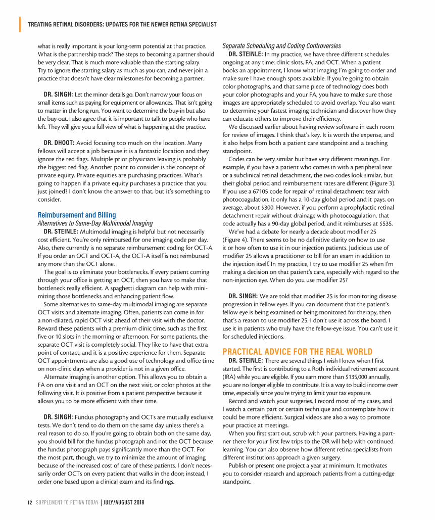
Codes can be very similar but have very different meanings. For example, if you have a patient who comes in with a peripheral tear or a subclinical retinal detachment, the two codes look similar, but their global period and reimbursement rates are different (Figure 3). If you use a 67105 code for repair of retinal detachment tear with photocoagulation, it only has a 10-day global period and it pays, on average, about $300. However, if you perform a prophylactic retinal detachment repair without drainage with photocoagulation, that code actually has a 90-day global period, and it reimburses at $535.
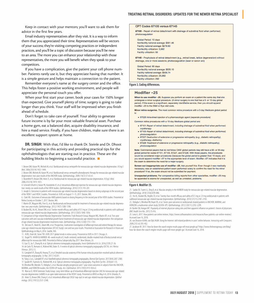
We’ve had a debate for nearly a decade about modifier 25 (Figure 4). There seems to be no definitive clarity on how to use it or how often to use it in our injection patients. Judicious use of modifier 25 allows a practitioner to bill for an exam in addition to the injection itself. In my practice, I try to use modifier 25 when I’m making a decision on that patient’s care, especially with regard to the non-injection eye. When do you use modifier 25?
DR. SINGH: We are told that modifier 25 is for monitoring disease progression in fellow eyes. If you can document that the patient’s fellow eye is being examined or being monitored for therapy, then that’s a reason to use modifier 25. I don’t use it across the board. I use it in patients who truly have the fellow-eye issue. You can’t use it for scheduled injections.
PRACTICAL ADVICE FOR THE REAL WORLDDR. STEINLE: There are several things I wish I knew when I first
started. The first is contributing to a Roth individual retirement account (IRA) while you are eligible. If you earn more than $135,000 annually, you are no longer eligible to contribute. It is a way to build income over time, especially since you’re trying to limit your tax exposure.
Record and watch your surgeries. I record most of my cases, and I watch a certain part or certain technique and contemplate how it could be more efficient. Surgical videos are also a way to promote your practice at meetings.
When you first start out, scrub with your partners. Having a part-ner there for your first few trips to the OR will help with continued learning. You can also observe how different retina specialists from different institutions approach a given surgery.
Publish or present one project a year at minimum. It motivates you to consider research and approach patients from a cutting-edge standpoint.
JULY/AUGUST 2018 | SUPPLEMENT TO RETINA TODAY 13
TREATING RETINAL DISORDERS: UPDATES FOR THE NEWER RETINA SPECIALIST
Keep in contact with your mentors; you’ll want to ask them for advice in the first few years.
Email industry representatives after they visit. It is a way to inform them that you appreciated their time. Representatives will be vectors of your success; they’re visiting competing practices or independent practices, and you’ll be a topic of discussion because you’ll be new to an area. The more you can enhance your relationship with those representatives, the more you will benefit when they speak to your competitors.
If you have a complication, give the patient your cell phone num-ber. Patients rarely use it, but they appreciate having that number. It is a simple gesture and helps maintain a connection to the patient.
Remember everyone’s name at the surgery center and the office. This helps foster a positive working environment, and people will appreciate the personal touch you offer.
When your first start your career, book your cases for 150% longer than expected. Give yourself plenty of time; surgery is going to take longer than you think. Your staff will be impressed when you finish ahead of schedule.
Don’t forget to take care of yourself. Your ability to generate future income is by far your most valuable financial asset. Purchase a home gym, eat a balanced diet, acquire disability insurance, and hire a maid service. Finally, if you have children, make sure there is an excellent support system at home.
DR. SINGH: With that, I’d like to thank Dr. Steinle and Dr. Dhoot for participating in this activity and providing practical tips for the ophthalmologists that are starting out in practice. These are the building blocks to beginning a successful practice. n
1. Brown DM, Kaiser PK, Michels M, et al. Ranibizumab versus verteporfin for neovascular age-related macular degeneration. N Engl J Med. 2006;355(14):1432-1444.2. Brown DM, Michels M, Kaiser PK, et al. Ranibizumab versus verteporfin photodynamic therapy for neovascular age-related macular degeneration: two-year results of the ANCHOR study. Ophthalmology. 2009;116(1):57-65 e5.3. Rosenfeld PJ, Brown DM, Heier JS, et al. Ranibizumab for neovascular age-related macular degeneration. N Engl J Med. 2006;355(14):1419-1431.4. Schmidt-Erfurth U, Kaiser PK, Korobelnik JF, et al. Intravitreal aflibercept injection for neovascular age-related macular degenera-tion: ninety-six-week results of the VIEW studies. Ophthalmology. 2014;121(1):193-201.5. Joseph A. Outcomes in patients with neovascular age-related macular degeneration based on dosing subgroups in the second year of the VIEW 1 and VIEW 2 studies. Presented at: ASRS on August 11-15, 2017. Boston, MA. 6. Clark WL. Outcomes of neovascular AMD patients based on dosing frequency in the second year of the VIEW studies. Presented at Retina Society on October 7, 2017. Boston, MA.7. Martin DF, Maguire MG, Fine SL, et al. Ranibizumab and bevacizumab for treatment of neovascular age-related macular degenera-tion: two-year results. Ophthalmology. 2012;119(7):1388-1398.8. Busbee BG, Ho AC, Brown DM, et al. Twelve-month efficacy and safety of 0.5 mg or 2.0 mg ranibizumab in patients with subfoveal neovascular age-related macular degeneration. Ophthalmology. 2013;120(5):1046-1056.9. Comparison of Age-related Macular Degeneration Treatments Trials Research Group, Maguire MG, Martin DF, et al. Five-year outcomes with anti-vascular endothelial growth factor treatment of neovascular age-related macular degeneration: the comparison of age-related macular degeneration treatments trials. Ophthalmology. 2016;123(8):1751-1761.10. DeCroos FC, Reed DC, Adam MK, et al. Prospective, multicenter investigation of aflibercept treat and extend therapy for neovas-cular age-related macular degeneration (ATLAS Study): one and two year results. Presented at Association for Research in Vision and Ophthalmology on May 4, 2016. Seattle, WA.11. ASRS, Shah GK, Stone TW. ASRS 2017 global trends in retina survey. Presented at ASRS in 2017. Chicago, IL. 12. Dugel PU. HAWK & HARRIER: 48–week results of 2 multi-centered, randomized, double-masked trials of brolucizumab versus aflibercept for neovascular AMD. Presented at Retina Subspecialty Day 2017. New Orleans, LA.13. Gao SS, Jia Y, Zhang M, et al. Optical coherence tomography angiography. Invest Ophthalmol Vis Sci. 2016;57(9):27-36.14. de Carlo TE, Romano A, Waheed NK, Duker JS. A review of optical coherence tomography angiography (OCTA). Int J Retina Vitreous. 2015;1:5.15. Campbell JP, Zhang M, Hwang TS, et al. Detailed vascular anatomy of the human retina by projection-resolved optical coherence tomography angiography. Sci Rep. 2017;7:42201.16. Yang J, Liu L, Campbell JP, et al. Handheld optical coherence tomography angiography. Biomed Opt Expressi. 2017;8(4):2287-2300.17. Spaide RF, Fujimoto JG, Waheed NK, et al. Optical coherence tomography angiography. Prog Retin Eye Res. 2018;64:1-55.18. Bhisitkul RB, Mendes TS, Rofagha S, et al. Macular atrophy progression and 7-year vision outcomes in subjects from the ANCHOR, MARINA, and HORIZON studies: the SEVEN-UP study. Am J Ophthalmol. 2015;159(5):915-924 e2.19. Marcus D, VIEW Extension Study Group. Long-term follow-up of intravitreal aflibercept injection (IAI) for neovascular age-related macular degeneration (nAMD) in an open-label extension of the VIEW1 study. Presented at ARVO on May 4-8, 2014. Orlando, FL. 20. Heier JS, Brown DM, Chong V, et al. Intravitreal aflibercept (VEGF trap-eye) in wet age-related macular degeneration. Ophthal-mology. 2012;119(12):2537-2548.
21. Sadda SR, Tuomi LL, Ding B, et al. Macular atrophy in the HARBOR study for neovascular age-related macular degeneration. Ophthalmology. 2018;125(6):878-886.22. Ho AC, Busbee BG, Regillo CD, et al. Twenty-four-month efficacy and safety of 0.5 mg or 2.0 mg ranibizumab in patients with subfoveal neovascular age-related macular degeneration. Ophthalmology. 2014;121(11):2181-2192.23. Rofagha S, Bhisitkul RB, Boyer DS, et al. Seven-year outcomes in ranibizumab-treated patients in ANCHOR, MARINA, and HORIZON: a multicenter cohort study (SEVEN-UP). Ophthalmology. 2013;120(11):2292-2299.24. Burkle CM, Keegan MT. Popularity of internet physician rating sites and their apparent influence on patients’ choices of physicians. BMC Health Serv Res. 2015;15:416.25. Loria G. 2017. How patients use online reviews. https://www.softwareadvice.com/resources/how-patients-use-online-reviews/. Accessed June 16, 2018. 26. van Deursen AJAM, van Dijk JAGM. Using the Internet: skill related problems in users’ online behavior. Interacting with Computers. 2009;21(5-6):393-402.27. Jacobson M. 2017. How far down the search engine results page will most people go? https://www.theleverageway.com/blog/how-far-down-the-search-engine-results-page-will-most-people-go/. Accessed June 16, 2018.
Figure 3. Coding differences.
Figure 4. Modifier 25.
TREATING RETINAL DISORDERS: UPDATES FOR THE NEWER RETINA SPECIALIST
INSTRUCTIONS FOR CME CREDITTo receive AMA PRA Category 1 Credit™, you must complete the attached Pretest/Posttest/Activity Evaluation/Satisfaction Measures Form and mail
or fax to Evolve Medical Education LLC; 353 West Lancaster Avenue, Second Floor, Wayne, PA 19087; Fax: (215) 933-3950. To answer these questions online and receive real-time results, please visit evolvemeded.com and click “Online Courses.” If you are experiencing problems with the online test, please email us at [email protected]. Certificates are issued electronically; please be certain to provide your email address below.
Please type or print clearly, or we will be unable to issue your certificate.
Name ____________________________________________________________________________________ o MD/DO participant o non-MD participant
Phone (required) ______________________________ o Email (required) _________________________________________________________________
City _____________________________________________________________________ State ____________ Zip ____________________________
License Number _____________________________________________________
DEMOGRAPHIC INFORMATIONProfession
___ MD/DO
___ NP
___ Nurse/APN
___ PA
___ Other
Years in Practice
___ >20
___ 11-20
___ 6-10
___ 1-5
___ <1
Patients Seen Per Week(with the disease targeted in this activity)
___ 0
___ 1-5
___ 6-10
___ 11-15
___ 15-20
___ 20+
Region
___ Northeast
___ Northwest
___ Midwest
___ Southeast
___ Southwest
Setting
___ Solo Practice
___ Community Hospital
___ Government or VA
___ Group Practice
___ Other
___ I do not actively
practice
Models of Care
___ Fee for Service
___ ACO
___ Patient-Centered
Medical Home
___ Capitation
___ Bundled Payments
___ Other
Training of Fellows ___ Yes ___ No
Release Date: July 2018Expiration Date: July 2019
DID THE PROGRAM MEET THE FOLLOWING EDUCATIONAL OBJECTIVES? AGREE NEUTRAL DISAGREE
_____ _____ _____
_____ _____ _____
_____ _____ _____
_____ _____ _____
_____ _____ _____
_____ _____ _____
_____ _____ _____
Discuss recent studies that address long-term treatment outcomes in age-related macular degeneration (AMD).
Develop an individualized treatment plan for patients with AMD.
Discuss implementing imaging tools as diagnostic therapies.
Summarize the outcomes of pivotal studies in AMD and diabetic macular edema (DME) and how study results may differ from real-world dosing methods.
Summarize the comparative Protocol T findings in DME and how to relate that to clinical practice patterns.
Evaluate practice flow to determine the most efficient patient experience.
Develop plans to reduce reimbursement denials.
LEARNING OBJECTIVES
POSTTEST QUESTIONS
1. AFTER REVIEWING THIS ACTIVITY, PLEASE RATE YOUR CONFIDENCE ON YOUR ABILITY TO INCORPORATE RESEARCH INTO THE CLINIC BASED ON THIS ACTIVITY:
a. Not at all confidentb. Not very confidentc. Neutrald. Confidente. Very confident
2. AFTER REVIEWING THIS ACTIVITY, PLEASE RATE HOW OFTEN YOU INTEND TO APPLY THE RECOMMENDATIONS FROM THE RETINA PANELISTS ON COD-ING AND BILLING. (BASED ON A SCALE OF 1 TO 5, WITH 1 BEING NEVER AND 5 BEING ALWAYS).
a. 1b. 2c. 3d. 4e. 5
3. ACCORDING TO ASRS SURVEY DATA, HOW LONG DO US PHYSICIANS EXTEND PATIENTS ON A TREAT-AND-EXTEND REGIMEN?
a. 16 or fewer weeksb. 12 or fewer weeksc. 10 or fewer weeksd. 8 or fewer weeks
4. OCT ANGIOGRAPHY (OCT-A) IS USEFUL IN THE FOLLOWING SCENARIOS EXCEPT: a. Patients with an unexplained drop in visual acuity. b. Patients with suspected masquerading syndromes.c. Patients with choroidal neovascularization (CNV).d. Patients with diabetic macular edema (DME).
5. FOR CLINICIANS INTERESTED IN PARTICIPATING IN CLINICAL TRIALS, AT WHAT PHASE ARE EARLY-CAREER INVESTIGATORS LIKELY TO FIND IT EASIEST TO BECOME INVOLVED?
a. Phase 1b. Phase 2c. Phase 3
6. WHICH PART OF RESEARCH INFRASTRUCTURE IS MOST IMPORTANT TO SUC-CESSFUL CLINICAL TRIAL PARTICIPATION?
a. A relationship with the institutional review boardb. A full-time, experienced study coordinatorc. An Early Treatment Diabetic Retinopathy Study (ETDRS) 20-foot study laned. A widefield imaging system
7. WHEN JOINING A PRACTICE FELLOWSHIP, WHAT DO THE PANELISTS CONSIDER THE LEAST IMPORTANT CONSIDERATION?
a. Starting salaryb. Partnership potential c. Geographic location d. The buy-out package
8. MODIFIER 25 IS USED TO __________________a. Schedule injections. b. Repeat imaging in the diseased eye.c. Monitor the fellow eye for disease when no disease is present. d. Monitor the fellow eye for disease progression when disease is present.
9. MR. SMITH HAS PRESENTED FOR EVALUATION; HE HAS HAD DIABETES FOR MORE THAN 10 YEARS, AND THIS IS HIS FIRST VISIT TO AN EYE CARE PRACTITIO-NER TO EVALUATE FOR DME/DIABETIC RETINOPATHY (DR). YOU SHOULD GET A FLUORESCEIN ANGIOGRAM (FA) ___________AND AN OCT ___________
a. at every visit; at every visitb. at baseline and every 6 to 12 months; at every visitc. at every visit; at baseline and every 6 to 12 monthsd. at baseline and then yearly; at baseline and then every 6 to 12 months
10. MRS. JONES PRESENTS TO YOUR CLINIC WITH A DIAGNOSIS OF SUBFO-VEAL CNV SECONDARY TO AGE-RELATED MACULAR DEGENERATION (AMD). PRESUMING YOU PLAN ON TREATING HER WITH AN ANTI-VEGF INJECTION REGI-MEN, WHAT WOULD BE THE BEST APPROACH TO ENSURE APPROPRIATE TREAT-MENT TO PRESERVE AS MUCH OF HER VISION AS POSSIBLE? ADD A CHECK MARK TO THE ITEMS BELOW THAT ARE CONSISTENT WITH YOUR CURRENT CLINICAL PRACTICE.
Action Consistent Not ConsistentTreat patient on a monthly dosing regimen based on the VIEW study outcomes.
Treat patient on a monthly dosing regimen based on the MARINA and ANCHOR study outcomes.
Treat patient initially with prn dosing regimen to determine if monthly dosing is needed.
Treat patient on prn dosing regimen based on the CATT data unless there is recurrence of neovascular leakage.
Treat patient with treat-and-extend to minimize lost work productivity.
Treat patients on a prn regimen but switch to treat-and-extend after a fixed amount of time.
Treat patients initially with treat-and-extend, and if the patient responds well, extend to fixed quarterly dosing.
Your responses to the questions below will help us evaluate this continuing medical education (CME) activity. They will provide us with evidence that improvements were made in patient care as a result of this activity as required by the Accreditation Council for Continuing Medical Education (ACCME).
Rate your knowledge/skill level prior to participating in this course: 5 = High, 1 = Low __________
Rate your knowledge/skill level after participating in this course: 5 = High, 1 = Low __________
This activity improved my competence in managing patients with this disease/condition/symptom. ____ Yes ____ No
I plan to make changes to my practice based on this activity. _____ Yes _____ No
The design of the program was effective for the content conveyed. ___ Yes ___ No
The content supported the identified learning objectives. ___ Yes ___ No
The content was free of commercial bias. ___ Yes ___ No
The content was relative to your practice. ___ Yes ___ No
The faculty was effective. ___ Yes ___ No
You were satisfied overall with the activity. ___ Yes ___ No
Would you recommend this program to your colleagues? ___ Yes ___ No
Please check the Core Competencies (as defined by the ACCME) that were enhanced through your participation in this activity:
____ Patient Care
____ Practice-Based Learning and Improvement
____ Professionalism
____ Medical Knowledge
____ Interpersonal and Communication Skills
____ System-Based Practice
Additional comments:________________________________________________________________________________________________________________________ I certify that I have participated in this entire activity.
Please identify any barriers to change (check all that apply):
____ Cost ____ Lack of consensus or professional guidelines
____ Lack of administrative support ____ Lack of experience
____ Lack of time to assess/counsel patients ____ Lack of opportunity (patients)
____ Reimbursement/insurance issues ____ Lack of resources (equipment)
This information will help evaluate this CME activity; may we contact you by email in 3 months to see if you have made this change? If so, please provide your email address below._____________________________________________________________________________________________________________________


![Design of lensless retinal scanning display with ...sdl.sjtu.edu.cn/uploadfiles/2019/07/201907112312131213.pdf · coincides with the retina. Virtual retinal display (VRD) [23–25],](https://static.documents.pub/doc/80x56/5f87583e8044cd0abd514c26/design-of-lensless-retinal-scanning-display-with-sdlsjtueducnuploadfiles201907.jpg)


