Page 1
TREATMENT CONSIDERATIONS for MISSING TEETH
Abdolreza Jamilian,
Alireza Darnahal
Ludovica Nucci
Fabrizia D\'Apuzzo
Letizia Perillo
Professor, Fellow of Orthognathic surgery, Department of Orthodontics, Tehran Dental
Branch, Craniomaxillofacial Research Center, Islamic Azad University, Tehran, Iran.
[email protected]
General dentist, Tehran Dental Branch, Craniomaxillofacial Research Center, Islamic Azad
University, Tehran, Iran. [email protected]
Ludovica Nucci, Undergraduate student, Course of Dentistry, Multidisciplinary
Department of Medical-Surgical and Dental Specialties, Second University of Naples,
Naples, Italy. [email protected]
Fabrizia D\'Apuzzo PhD student, Multidisciplinary Department of Medical-Surgical and
Dental Specialties, Second University of Naples, Naples, Italy. [email protected]
Head of Orthodontic Unit and Chair of Postgraduate Orthodontic Program,
Multidisciplinary Department of Medical-Surgical and Dental Specialties, Second University
of Naples, Naples, Italy. [email protected]
Corresponding author:
0098-21-22011892
Abdolreza Jamilian, Professor, Fellow of Orthognathic surgery, Department of orthodontics,
Tehran Dental Branch, Craniomaxillofacial Research Center, Islamic Azad University,
Tehran. Iran.
[email protected]
Page 2
1. Abstract:
Specific terms are used to describe the nature of tooth agenesis. Hypodontia is most
frequently used when describing the phenomenon of congenitally missing teeth. Many
other terms to describe a reduction in the number of teeth appear in the literature:
oligodontia, anodontia, aplasia of teeth, congenitally missing teeth, absence of teeth,
agenesis of teeth and lack of teeth. The term hypodontia is used when one to six teeth,
excluding third molars, are missing, and oligodontia when more than six teeth are absent
(excluding the third molars). The long-term management of hypodontia in the esthetic zone
is a particularly challenging situation. Although there are essentially two distinct
approaches to managing this problem; that is space closure or opening for prosthetic
replacements, implant or autotransplantation. These patients often manifest with many
underlying skeletal and dental problems and a multidisciplinary approach for management
of this condition is recommended. Two treatment approach including space closure and
space reopening are described in details in this chapter.
Keywords: Hypodontia; Missing teeth; Implant; Orthodontic space closure; space reopening
Page 3
2. Introduction
Missing is one of the most dental anomalies in practice of dentistry and they may affect the
self-esteem and social wellbeing of the patients. This condition is often complicated by
dental anomalies associated with hypodontia such as impacted teeth, microdontia, delayed
eruption and taurodontism. Hypodontia reportedly affects between 3% and 8% of the
population. Hypodontia is a common problem seen by the general dentist and is usually
referred to the orthodontist. [1, 2] Agenesis means that a dental bud fails to develop or is not
present at birth. This problem leaves an empty space in the arch which causes plentiful
problems. Specific terms have been used to describe the nature of tooth agenesis.
Anodontia is named complete absence of teeth.
Hypodontia means missing teeth, but usually less than six teeth.
Oligodontia or partial anodontia is defined absence of six or more teeth.
Anodontia and oligodontia are rare, however, hypodontia is relatively a common problem.
Many other terms are also used to describe a reduction in the number of teeth in the
literature such as aplasia of teeth, agenesis of teeth, absence of teeth, lack of teeth, and
congenitally missing teeth. [3, 4]
The aims of this chapter are:
2.1. a. To determine the prevalence of hypodontia
2.2. b. To assess the etiology of hypodontia
2.3. c. Diagnosis of the problem
2.4. d Treatment plan
2.5. d. Decision to open or close space in the dentition
3. Prevalence of hypodontia:
Hypodontia in primary dentition arises in 0.1–0.9 per cent of the
population, with equal frequencies in males and females. This problem
is more common in the upper jaw and it is frequently related with the
upper lateral incisor in the primary dentition. As a general rule, when
Page 4
the primary tooth is missing, its permanent counterpart will be missing.
[1] Hypodontia in the permanent dentition occurs with equal rate in the
upper and lower arches and usually affects the third molar. The type of
agenesis in dentition and prevalence of missing vary with racial and
ethnic groups. However, females are more frequently affected. [2]
Prevalences of hypodontia varies between 1.6 and 9.6 per cent across the
world with exclusion of the third molars. Prevalence of agenesis differs
between continents and races. The occurrence of missing permanent
teeth, excluding the third molar, is 3.4 percent in Swiss, 4.4 percent in the
United States, 6.1 percent in Sweden, 8 per cent in Finland, and 9.6 per
cent in Austria with exclusion of third molar. Japanese people have the
highest rates of agenesis both in primary and permanent dentition.
Australian Aborigines and African Blacks might have a low rate of
missing teeth. The rate of agenesis in Indians has been reported less than
1%. [3, 4] The prevalence of third molar missing has been reported of 9–
37 percent. [2] Muller et al. [5] reported that upper lateral incisors are the
most agenesis teeth (not including third molars). Missing of the upper
lateral incisor is also related to anomalies such as agenesis of other
permanent teeth, undersized maxillary lateral incisors (peg laterals),
palatally position of canines and distal displacement of lower second
premolars. [6-8] Agenesis may arise in isolation, or as part of a syndrome.
Dental anomalies ,especially hypodontia, have frequently been found in
children who also have cleft lip and cleft palate or a syndrome. [9-11]
4. Etiology of hypodontia
Heredity or familial distributionare two of the possible factors associated
with congenitally missing teeth. Graber stated [12], "Congenital partial
anodontia appears to be the result of one or more point mutations in a
closely linked polygenic system, most often transmitted in an autosomal
dominant pattern incomplete penetrance and variable expressivity."
Genetics has a crucial role in hypodontia, as confirmed by studies on
monozygotic twins. The pattern of agenesis can differ between
monozygotic twins, this issue possibly pointing to additional underlying
Page 5
mechanisms, such as epigenetic factors which might be implied
occurrence of two anomalies simultaneously. [13] Genetic, epigenetic and
environmental factors contribute to the development of hypodontia. It
has been shown that genetics has a predominant role in the etiology of
missing teeth. [14] Infection, trauma and drugs, as well as genes
associated with syndromes play a crucial role in hypodontia. Agenesis
may be an isolated condition or a dental appearance of special
syndromes such as cleft lip and palate [9, 15, 16], ectodermal dysplasia.
[17] The isolated one can follow autosomal recessive, dominant, or X-
linked patterns of inheritance. [18] Some studies showed that some
anomalies such as bimaxillary retrusion, mandibular prognathism,
decreased maxillary jaw size and reduced vertical facial dimension in
patients affected with hypodontia. [19, 20] In hereditary cases, missing
has greater incidence when the dental germ is developing after the
adjacent tissues have closed the space needed for the tooth development.
Other scientists reported that delays in tooth development and
reductions in tooth size correlate with agenesis. [21] Both of these might
agree with the terminal reduction theory. Moreover, it has been also
reported that anterior agenesis may depend more on genes while
posterior missing might be sporadic. [22]
5. Diagnosis
Dental agenesis is categorized according to the number of missing
teeth,less than three and six missing teeth are defined as mild and
moderate respectively. Clinical evaluation, radiographic, and dental cast
examinations are required for proper diagnosis. The third molar germ
calcification initiates at the age of about 7.5 and in very few people it
starts at the age 9.5. Thus, by including patients younger than 9
researchers might overestimate the missing of the third molars. This
might explain the high occurrence of agenesis in third molars which has
been reported by some studies.
Page 6
6. Treatment plan
Treatment needs an interdisciplinary approach including operative
dentistry, pediatric dentistry, orthodontics and prosthodontics. Early
extraction of primary canines might guide the eruption of the permanent
canine into the proper position in cases with missing maxillary lateral
and impaction of upper canine. The amount of crowding, type of
malocclusion, facial profile, age of the patient, periodontal conditions,
bone volume in alveolar process, vertical or horizontal growth pattern,
craniofacial morphology and the number of missing teeth should be
considered in treatment plan. There are 2 treatment plans that includes
space reopening or space closing. Space can be reopened for implant
insertion, auto transplantation, prosthetic restoration. Another treatment
plan is space closing which can be done by fixed orthodontics.
7. Space closure versus space opening
Missing of maxillary incisors during the teenage years is a severe
problem and often requires a challenging treatment plan. There are
several solutions for treatment of lacking maxillary incisors including:
crown and bridge, resin-bonded bridge-work, removable partial
dentures, osseointegrated implants, auto transplantation, orthodontic
space closure. [23-27] Each of these methods have their own advantages
and disadvantages; however, opening the space followed by implant
insertion and space closure are the most common treatment options for
tooth replacement. Implant insertion is an optimal treatment plan with
obtaining an ideal occlusion and the indisputable advantage of avoiding
any damage to the adjacent teeth. [23, 28]
Space closing by mesial movement of the posterior teeth is a vital
approach and it provides major satisfactory aesthetic and functional long
term results. Moreover; the result of space closure and all of the changes
in the long term will be natural. It is clear that when implant or any
prostheses are used some changes could happen in the presence of a
Page 7
foreign body. [26, 29, 30] On the other hand, shorter and easier
orthodontic treatment by implant insertion makes the space opening a
favorable treatment approach for replacing missing teeth. Nevertheless,
opening the space and implant insertion have some disadvantages.
Implant insertion is contraindicated in growing patients. Implant must
be postponed until the growth is ceased. If the implant is used at about
18 years of age, the neighbouring teeth and surrounding alveolar bone
may continue to erupt. This eruption results in infraocclusion of the
implant site. There will be a big discrepancy in vertical dimension
between the gingival margin of the implanted tooth and the gingival
margin of the neighbouring teeth. This side effect may appear in few
years after implant insertion in young adult patients and the implant
becomes submerged. [31-34] In patients where maxillary and mandibular
incisors are not in contact with each other, the amount of extrusion might
be 0/2 to 0/3 mm per year. Implant acts like an ankylosed teeth and its
status cannot change in contrast to their adjacent teeth; thus, small
displacement of neighbouring teeth after implant insertion can cause
esthetic complications. [35-37] Infra-positioned implant results in an
unlevelled of gingival margins. This issue is a problematic challenge
especially in patients with a high smile line. Thus it is better not to use
implant in cases with “gummy smile” or vertical growth pattern patients.
[26] Furthermore, it has been reported that above more than 50% of
single-implant crowns at four-year follow-ups have some extent of blue
colouring of the gingiva. [39] Some other side effects such as bleeding on
probing, gingivitis, increased probing depth, periodontitis, Peri-
implantitis, and progressive loss of marginal bone support of the implant
have also been shown in cases with implant insertion. [36, 38, 40, 41]
Besides the most problematic issue of the space opening is that the
teenagers must wait many years after completion of orthodontic
treatment for implant insertion. During this interim phase the patients
must use temporary crowns or restorations that often causes many
difficulties and displacements both on implant site and adjacent teeth.
On the other hand, orthodontic space closure is a practical and safe
procedure that could achieve better long-term results. Moreover, none of
the stated drawbacks have been found in orthodontic space closure [29,
Page 8
42, 43] Nevertheless, orthodontic space closure has its own
disadvantages. Concerns may be related to the complexity of treatment,
the risk for reopening of space, increased functional force on the first
premolar roots. [44] Attempts for closing the space of upper incisors will
tend to retract the anterior teeth, which may be favourable in class II
division I malocclusion with maxillary protrusion. Space closure in the
maxillary arch may well provide reduction of an increased overjet.
However, space closing may be undesirable in class III malocclusion
with maxillary deficiency. Moreover, space closure of a missing upper
lateral incisor results in the canine being displaced mesially into contact
with the central incisor. In this case the canine is more prominent, wider
and darker than the lateral incisor. Canine can be reshaped by selective
grinding of the cusp tip and it needs rebuilding by composite materials
like lateral incisor. In cases with increased overjet or crowding extraction
of the contralateral lateral incisor may help to maintain symmetry and
correct the dental midline. Space reopening is usually the best treatment
option where orthodontic treatment does not need to use the space to
relieve the crowding. In this case any attempt to close the space results in
an unfavourable effect. The major disadvantage of space reopening is
that it requires a foreign body such as permanent prosthesis or implant.
The optimal space required for the prosthesis or implant is usually
determined by two factors. The first one is occlusion and the second is
aesthetic. Ideal overjet and overbite must be provided along with good
Class I malocclusion at the end of the treatment. A maxillary lateral
incisor should be two thirds of the width of the maxillary central incisor.
Providing of these conditions may be difficult due to anchorage
problems associated with reduced numbers of teeth in hypodontia
patients. In cases with extensive space or early loss of teeth which have
resulted in alveolar atrophy, space closure will not be desirable. The
position of the roots of the neighbouring teeth should be estimated
radiographically in space opening cases. Therefore, not only adequate
space must be provided for replacement of the crown but also the roots
of neighbouring teeth should be parallel or slightly divergent to create
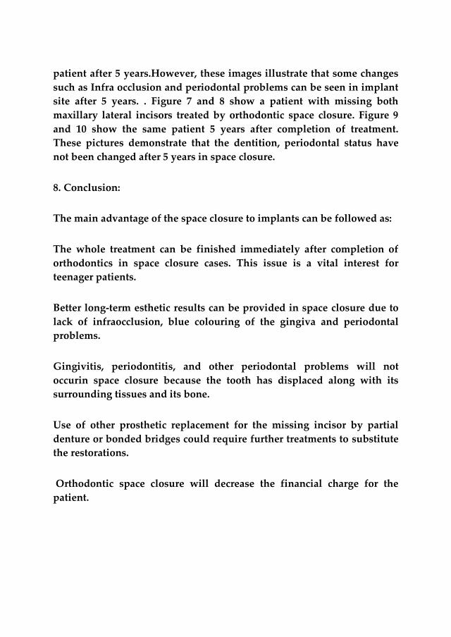
adequate space for implant insertion. Figure 1 to 3 show a patient
immediately after implant insertion and figure 4 to 6 show the same
Page 9
patient after 5 years.However, these images illustrate that some changes
such as Infra occlusion and periodontal problems can be seen in implant
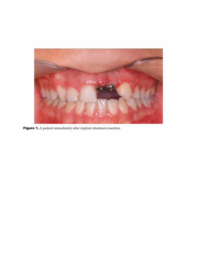
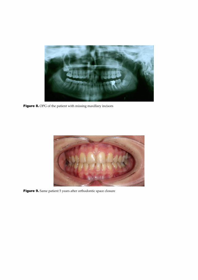
site after 5 years. . Figure 7 and 8 show a patient with missing both
maxillary lateral incisors treated by orthodontic space closure. Figure 9
and 10 show the same patient 5 years after completion of treatment.
These pictures demonstrate that the dentition, periodontal status have
not been changed after 5 years in space closure.
8. Conclusion:
The main advantage of the space closure to implants can be followed as:
The whole treatment can be finished immediately after completion of
orthodontics in space closure cases. This issue is a vital interest for
teenager patients.
Better long-term esthetic results can be provided in space closure due to
lack of infraocclusion, blue colouring of the gingiva and periodontal
problems.
Gingivitis, periodontitis, and other periodontal problems will not
occurin space closure because the tooth has displaced along with its
surrounding tissues and its bone.
Use of other prosthetic replacement for the missing incisor by partial
denture or bonded bridges could require further treatments to substitute
the restorations.
Orthodontic space closure will decrease the financial charge for the
patient.
Page 10
Figure 1. A patient immediately after implant abutment insertion
Page 11
Figure 2. A patient immediately after implant insertion
Figure 3. OPG of the same patient
Page 12
Figure 4. Same patient after 5 years
Figure 5. OPG of the same patient after 5 years
Page 13
Figure 6. Frontal view of the patient after 5 years
Figure 7. A patient with missing maxillary lateral incisors
Page 14
Figure 8. OPG of the patient with missing maxillary incisors
Figure 9. Same patient 5 years after orthodontic space closure
Page 15
Figure 10. OPG of the same patient 5 years after orthodontic space closure
Page 16
9. References
[1] Polder, B.J., et al., A meta-analysis of the prevalence of dental agenesis of permanent
teeth. Community Dent Oral Epidemiol, 2004. 32(3): p. 217-26.
[2] Jamilian, A., et al., Hypodontia and supernumerary and impacted teeth in children with
various types of clefts. Am J Orthod Dentofacial Orthop, 2015. 147(2): p. 221-5.
[3] Vastardis, H., The genetics of human tooth agenesis: new discoveries for understanding
dental anomalies. Am J Orthod Dentofacial Orthop, 2000. 117(6): p. 650-6.
[4] Jamilian, A., L. Perillo, and M. Rosa, Missing upper incisors: a retrospective study of
orthodontic space closure versus implant. Prog Orthod, 2015. 16: p. 2.
[5] Hall, R.K., Congenitally missing teeth--a diagnostic feature in many syndromes of the
head and neck. J Int Assoc Dent Child, 1983. 14(2): p. 69-75.
[6] Aasheim, B. and B. Ogaard, Hypodontia in 9-year-old Norwegians related to need of
orthodontic treatment. Scand J Dent Res, 1993. 101(5): p. 257-60.
[7] Guttal, K.S., et al., Frequency of developmental dental anomalies in the Indian
population. Eur J Dent, 2010. 4(3): p. 263-9.
[8] Carter, N.E., et al., The interdisciplinary management of hypodontia: orthodontics. Br
Dent J, 2003. 194(7): p. 361-6.
[9] Muller, T.P., et al., A survey of congenitally missing permanent teeth. J Am Dent Assoc,
1970. 81(1): p. 101-7.
[10] Garib, D.G., et al., Agenesis of maxillary lateral incisors and associated dental
anomalies. Am J Orthod Dentofacial Orthop, 2010. 137(6): p. 732 e1-6; discussion 732-3.
[11] Peck, S., L. Peck, and M. Kataja, Concomitant occurrence of canine malposition and
tooth agenesis: evidence of orofacial genetic fields. Am J Orthod Dentofacial Orthop, 2002.
122(6): p. 657-60.
[12] Shapira, Y., E. Lubit, and M.M. Kuftinec, Hypodontia in children with various types of
clefts. Angle Orthod, 2000. 70(1): p. 16-21.
[13] Jamilian, A., R. Showkatbakhsh, and M.B. Boushehry, The effect of tongue appliance on
the nasomaxillary complex in growing cleft lip and palate patients. J Indian Soc Pedod Prev
Dent, 2006. 24(3): p. 136-9.
[14] Jamilian, A., F. Nayeri, and A. Babayan, Incidence of cleft lip and palate in Tehran. J
Indian Soc Pedod Prev Dent, 2007. 25(4): p. 174-6.
[15] Graber, L.W., Congenital absence of teeth: a review with emphasis on inheritance
patterns. J Am Dent Assoc, 1978. 96(2): p. 266-75.
Page 17
[16] Backman, B. and Y.B. Wahlin, Variations in number and morphology of permanent
teeth in 7-year-old Swedish children. Int J Paediatr Dent, 2001. 11(1): p. 11-7.
[17] Mirabella, A.D., V.G. Kokich, and M. Rosa, Analysis of crown widths in subjects with
congenitally missing maxillary lateral incisors. Eur J Orthod, 2012. 34(6): p. 783-7.
[18] Srivastava, D., et al., Use of anterior maxillary distraction osteogenesis in two cleft lip
and palate patients. Natl J Maxillofac Surg, 2015. 6(1): p. 80-3.
[19] Jamilian, A., et al., Cleft sidedness and congenitally missing teeth in patients with cleft
lip and palate patients. Prog Orthod, 2016. 17: p. 14.
[20] Lexner, M.O., et al., Anomalies of tooth formation in hypohidrotic ectodermal
dysplasia. Int J Paediatr Dent, 2007. 17(1): p. 10-8.
[21] Ahmad, W., et al., A locus for autosomal recessive hypodontia with associated dental
anomalies maps to chromosome 16q12.1. Am J Hum Genet, 1998. 62(4): p. 987-91.
[22] Tavajohi-Kermani, H., R. Kapur, and J.J. Sciote, Tooth agenesis and craniofacial
morphology in an orthodontic population. Am J Orthod Dentofacial Orthop, 2002. 122(1): p.
39-47.
[23] Endo, T., et al., Hypodontia patterns and variations in craniofacial morphology in
Japanese orthodontic patients. Angle Orthod, 2006. 76(6): p. 996-1003.
[24] Goya, H.A., et al., An orthopantomographic study of hypodontia in permanent teeth of
Japanese pediatric patients. J Oral Sci, 2008. 50(2): p. 143-50.
[25] Galluccio, G. and A. Pilotto, Genetics of dental agenesis: anterior and posterior area of
the arch. Eur Arch Paediatr Dent, 2008. 9(1): p. 41-5.
[26] Rupp, R.P., J.K. Dillehay, and C.F. Squire, Orthodontics, prosthodontics, and
periodontics: a multidisciplinary approach. Gen Dent, 1997. 45(3): p. 286-9.
[27] Ghassemi, M., et al., Orthodontic treatment after autotransplantation. Angle Orthod,
2011. 81(4): p. 721-5.
[28] Zachrisson, B.U., A. Stenvik, and H.R. Haanaes, Management of missing maxillary
anterior teeth with emphasis on autotransplantation. Am J Orthod Dentofacial Orthop, 2004.
126(3): p. 284-8.
[29] Zachrisson, B.U., M. Rosa, and S. Toreskog, Congenitally missing maxillary lateral
incisors: canine substitution. Point. Am J Orthod Dentofacial Orthop, 2011. 139(4): p. 434,
436, 438 passim.
[30] Showkatbakhsh, R. and A. Jamilian, Opening or closing space for replacing upper
incisors. Two case reports. Rev Esp Orthod 2010. 40: p. 181-5.
[31] Zachrisson, B.U., Planning esthetic treatment after avulsion of maxillary incisors. J Am
Dent Assoc, 2008. 139(11): p. 1484-90.
Page 18
[32] Nordquist, G.G. and R.W. McNeill, Orthodontic vs. restorative treatment of the
congenitally absent lateral incisor--long term periodontal and occlusal evaluation. J
Periodontol, 1975. 46(3): p. 139-43.
[33] Robertsson, S. and B. Mohlin, The congenitally missing upper lateral incisor. A
retrospective study of orthodontic space closure versus restorative treatment. Eur J Orthod,
2000. 22(6): p. 697-710.
[34] Bernard, J.P., et al., Long-term vertical changes of the anterior maxillary teeth adjacent
to single implants in young and mature adults. A retrospective study. J Clin Periodontol,
2004. 31(11): p. 1024-8.
[35] Kuijpers, M.A., J. de Lange, and A.V. van Gool, [Maxillofacial growth and dental
implants in the maxillary anterior region]. Ned Tijdschr Tandheelkd, 2006. 113(4): p. 130-3.
[36] Jemt, T., et al., Changes of anterior clinical crown height in patients provided with
single-implant restorations after more than 15 years of follow-up. Int J Prosthodont, 2006.
19(5): p. 455-61.
[37] Spear, F.M., D.M. Mathews, and V.G. Kokich, Interdisciplinary management of single-
tooth implants. Semin Orthod, 1997. 3(1): p. 45-72.
[38] Thilander, B., J. Odman, and U. Lekholm, Orthodontic aspects of the use of oral
implants in adolescents: a 10-year follow-up study. Eur J Orthod, 2001. 23(6): p. 715-31.
[39] Oesterle, L.J. and R.J. Cronin, Jr., Adult growth, aging, and the single-tooth implant. Int
J Oral Maxillofac Implants, 2000. 15(2): p. 252-60.
[40] Chang, M., et al., Implant supported single-tooth replacements compared to
contralateral natural teeth. Crown and soft tissue dimensions. Clin Oral Implants Res, 1999.
10(3): p. 185-94.
[41] Dueled, E., et al., Professional and patient-based evaluation of oral rehabilitation in
patients with tooth agenesis. Clin Oral Implants Res, 2009. 20(7): p. 729-36.
[42] Fransson, C., et al., Extent of peri-implantitis-associated bone loss. J Clin Periodontol,
2009. 36(4): p. 357-63.
[43] Paolantonio, M., et al., Clinical, microbiologic, and biochemical effects of subgingival
administration of a Xanthan-based chlorhexidine gel in the treatment of periodontitis: a
randomized multicenter trial. J Periodontol, 2009. 80(9): p. 1479-92.
[44] Rosa, M. and B.U. Zachrisson, Integrating space closure and esthetic dentistry in
patients with missing maxillary lateral incisors. J Clin Orthod, 2007. 41(9): p. 563-73; quiz
424.
[45] Thordarson, A., B.U. Zachrisson, and I.A. Mjor, Remodeling of canines to the shape of
lateral incisors by grinding: a long-term clinical and radiographic evaluation. Am J Orthod
Dentofacial Orthop, 1991. 100(2): p. 123-32.
Page 19
[46] Czochrowska, E.M., et al., Outcome of orthodontic space closure with a missing
maxillary central incisor. Am J Orthod Dentofacial Orthop, 2003. 123(6): p. 597-603.