CLINICAL ARTICLEJ Kor Neurotraumatol Soc 2010;6:150-153 ISSN 1738-8708
Received: April 26, 2010 / Revised: May 3, 2010Accepted: November 15, 2010Address for correspondence: Dong-Keun Hyun, MDDepartment of Neurosurgery, Inha University Hospital, 7-206 Shinheung-dong 3-ga, Jung-gu, Incheon 400-711, KoreaTel: +82-32-890-2948, Fax: +82-32-890-2947E-mail: [email protected]
Introduction
Arachnoid cyst is a congenital intracranial lesion that caused by abnormal development of meninges. It repre-sent about 1% of all intracranial space-occupying le-sion.1,11,24) In 2.43% of patient who has chronic subdural hematoma or hygroma, arachnoid cyst is observed at mid-dle cranial fossa.21) In young aged patients, because of the possibility of cyst membrane rupture, arachnoid cyst is a risk factor of chronic subdural hematoma.16) In this situa-tion it is considered that craniotomy is required to remove the hematoma inside of the cyst. We report a case that was accompanied with chronic subdural hematoma and arach-noid cyst. The patient was well treated by trephination only. We will report this case with a review of the radio-logical findings.
Case Report
A nineteen year old man was admitted to our hospital because of the 3 day lasting vomiting and headache that developed after mild head trauma during exercise 2 months ago. The patient was alert, and did not show any neurological deficit. The brain computed tomography (CT)
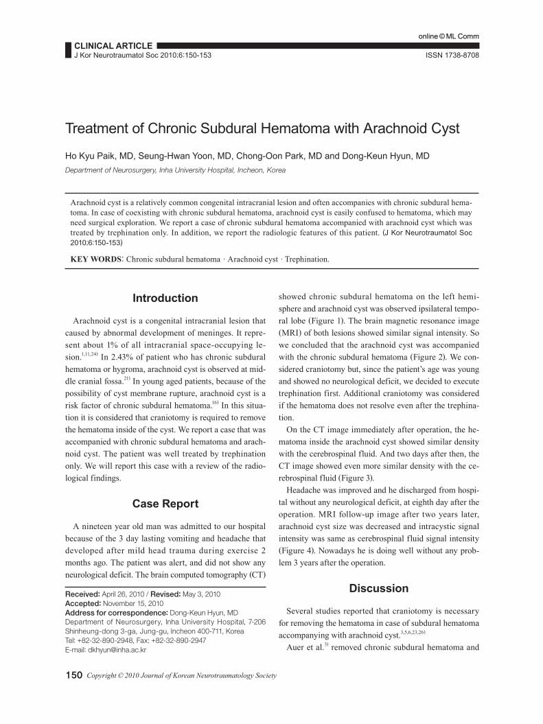
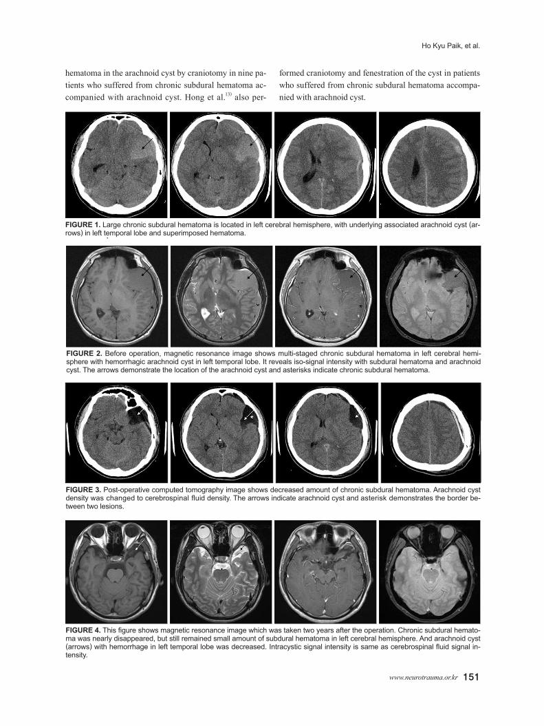
showed chronic subdural hematoma on the left hemi-sphere and arachnoid cyst was observed ipsilateral tempo-ral lobe (Figure 1). The brain magnetic resonance image (MRI) of both lesions showed similar signal intensity. So we concluded that the arachnoid cyst was accompanied with the chronic subdural hematoma (Figure 2). We con-sidered craniotomy but, since the patient’s age was young and showed no neurological deficit, we decided to execute trephination first. Additional craniotomy was considered if the hematoma does not resolve even after the trephina-tion.
On the CT image immediately after operation, the he-matoma inside the arachnoid cyst showed similar density with the cerebrospinal fluid. And two days after then, the CT image showed even more similar density with the ce-rebrospinal fluid (Figure 3).
Headache was improved and he discharged from hospi-tal without any neurological deficit, at eighth day after the operation. MRI follow-up image after two years later, arachnoid cyst size was decreased and intracystic signal intensity was same as cerebrospinal fluid signal intensity (Figure 4). Nowadays he is doing well without any prob-lem 3 years after the operation.
Discussion
Several studies reported that craniotomy is necessary for removing the hematoma in case of subdural hematoma accompanying with arachnoid cyst.3,5,6,23,26)
Auer et al.3) removed chronic subdural hematoma and
Treatment of Chronic Subdural Hematoma with Arachnoid Cyst
Ho Kyu Paik, MD, Seung-Hwan Yoon, MD, Chong-Oon Park, MD and Dong-Keun Hyun, MDDepartment of Neurosurgery, Inha University Hospital, Incheon, Korea
Arachnoid cyst is a relatively common congenital intracranial lesion and often accompanies with chronic subdural hema-toma. In case of coexisting with chronic subdural hematoma, arachnoid cyst is easily confused to hematoma, which may need surgical exploration. We report a case of chronic subdural hematoma accompanied with arachnoid cyst which was treated by trephination only. In addition, we report the radiologic features of this patient. (J Kor Neurotraumatol Soc 2010;6:150-153)
hematoma in the arachnoid cyst by craniotomy in nine pa-tients who suffered from chronic subdural hematoma ac-companied with arachnoid cyst. Hong et al.13) also per-
formed craniotomy and fenestration of the cyst in patients who suffered from chronic subdural hematoma accompa-nied with arachnoid cyst.
FIGURE 1. Large chronic subdural hematoma is located in left cerebral hemisphere, with underlying associated arachnoid cyst (ar-rows) in left temporal lobe and superimposed hematoma.
FIGURE 2. Before operation, magnetic resonance image shows multi-staged chronic subdural hematoma in left cerebral hemi-sphere with hemorrhagic arachnoid cyst in left temporal lobe. It reveals iso-signal intensity with subdural hematoma and arachnoid cyst. The arrows demonstrate the location of the arachnoid cyst and asterisks indicate chronic subdural hematoma.
FIGURE 3. Post-operative computed tomography image shows decreased amount of chronic subdural hematoma. Arachnoid cyst density was changed to cerebrospinal fluid density. The arrows indicate arachnoid cyst and asterisk demonstrates the border be-tween two lesions.
FIGURE 4. This figure shows magnetic resonance image which was taken two years after the operation. Chronic subdural hemato-ma was nearly disappeared, but still remained small amount of subdural hematoma in left cerebral hemisphere. And arachnoid cyst (arrows) with hemorrhage in left temporal lobe was decreased. Intracystic signal intensity is same as cerebrospinal fluid signal in-tensity.
152 J Kor Neurotraumatol Soc 2010;6:150-153
The Meaning of Radiologic Findings of Arachnoid Cyst
But many other studies, including Domenicucci, per-formed trephination only and successfully completed treatment in patients who had chronic subdural hematoma accompanied with arachnoid cyst.4,9,18)
Even though the both lesions are anatomically divided, radiological image finding of both lesions show similar signal intensity. It seems like that the blood product of chronic subdural hematoma could be infiltrated to arach-noid cyst and micro-material could be exchanged between both lesions.8) Also, the reason that the radiological image of the cyst changing to that of the cerebrospinal fluid right after the surgery seems to be the same mechanism.
As explained above, relation of the two lesions makes it possible to remove the hematoma in the cyst by trephina-tion only without any other additive surgery.
Considering that the incidence of complication and the mortality caused by the craniotomy is much higher than that of the trephination, the first treatment of the chronic subdural hematoma accompanied by arachnoid cyst should be trephination.2,10,14,17,19,22,24,25) If this treatment is not good enough to complete the treatment, it means that intracystic hematoma was not originated from subdural hematoma. Therefore, in that situation, craniotomy could be considered as a secondary treatment.3,7,12,14,15,19,20,24,25)
Conclusion
In case of chronic subdural hematoma is accompanied with the arachnoid cyst, both lesions may be anatomically devided. But, considering the microscopic structure of both lesions, infiltration of blood material between the two lesion seems to be possible.8) Because of this reason, the first treatment choice of the chronic subdural hemato-ma accompanied by arachnoid cyst could be trephination only. If this treatment is not good enough to complete the treatment, craniotomy might be considered as a secondary treatment.
REFERENCES1) Albuquerque FC, Giannotta SL. Arachnoid cyst rupture produc-
ing subdural hygroma and intracranial hypertension: case re-ports. Neurosurgery 41:951-955; discussion 955-956, 1997
2) Aoki N, Sakai T. Intraoperative subdural hematoma in a patient with arachnoid cyst in the middle cranial fossa. Childs Nerv Syst 6:44-46, 1990
3) Auer LM, Gallhofer B, Ladurner G, Sager WD, Heppner F, Lechner H. Diagnosis and treatment of middle fossa arachnoid cysts and subdural hematomas. J Neurosurg 54:366-369, 1981
4) Bilginer B, Onal MB, Oguz KK, Akalan N. Arachnoid cyst as-sociated with subdural hematoma: report of three cases and re-view of the literature. Childs Nerv Syst 25:119-124, 2009
5) Chan JY, Huang CT, Liu YK, Lin CP, Huang JS. Chronic subdu-
ral hematoma associated with arachnoid cyst in young adults: a case report. Kaohsiung J Med Sci 24:41-44, 2008
6) Czernicki T, Marchel A, Nowak A, Bojarski P. [Arachnoid cysts of the middle cranial fossa presented as subdural hematomas]. Neurol Neurochir Pol 39:328-334, 2005
7) Domenicucci M, Russo N, Giugni E, Pierallini A. Relationship between supratentorial arachnoid cyst and chronic subdural he-matoma: neuroradiological evidence and surgical treatment. J Neurosurg 110:1250-1255, 2009
8) Domenicucci M, Russo N, Giugni E, Pierallini A. Relationship between supratentorial arachnoid cyst and chronic subdural he-matoma: neuroradiological evidence and surgical treatment J Neurosurg 110:1250-1255, 2009
9) Fuentes S, Palombi O, Pouit B, Bernard C, Desgeorges M. [Arachnoid cysts of the middle fossa and associated subdural he-matoma. Three case reports and review of the literature]. Neuro-chirurgie 46:376-382, 2000
10)Galassi E, Tognetti F, Pozzati E, Frank F. Extradural hematoma complicating middle fossa arachnoid cyst. Childs Nerv Syst 2: 306-308, 1986
11) Harsh GR 4th, Edwards MS, Wilson CB. Intracranial arachnoid cysts in children. J Neurosurg 64:835-842, 1986
12)Helland CA, Wester K. A population based study of intracranial arachnoid cysts: clinical and neuroimaging outcomes following surgical cyst decompression in adults. J Neurol Neurosurg Psy-chiatry 78:1129-1135, 2007
13)Hong JC, Kim MS, Chang CH, Kim SH. Arachnoid cyst with spontaneous intracystic hemorrhage and chronic subdural hema-toma. J Korean Neurosurg Soc 43:54-56, 2008
14)Lund E, Buhl M, Miletic T, Knudsen V. Isodense middle fossa arachnoid cyst and subdural hematoma: a diagnostic problem on CT. J Neuroradiol 14:89-93, 1987
15)Mayr U, Aichner F, Bauer G, Mohsenipour I, Pallua A. Supra-tentorial extracerebral cysts of the middle cranial fossa. A report of 23 consecutive cases of the so-called temporal lobe agenesis syndrome. Neurochirurgia (Stuttg) 25:51-56, 1982
16)Mori K, Yamamoto T, Horinaka N, Maeda M. Arachnoid cyst is a risk factor for chronic subdural hematoma in juveniles: twelve cases of chronic subdural hematoma associated with arachnoid cyst. J Neurotrauma 19:1017-1027, 2002
17)Oberbauer RW, Haase J, Pucher R. Arachnoid cysts in children: a European co-operative study. Childs Nerv Syst 8:281-286, 1992
18)Oka Y, Kumon Y, Ohta S, Sakaki S, Ohue S, Takeda S. Chronic subdural hematoma associated with middle fossa arachnoid cysts--three case reports. Neurol Med Chir (Tokyo) 34:95-99, 1994
19)Page A, Paxton RM, Mohan D. A reappraisal of the relationship between arachnoid cysts of the middle fossa and chronic subdu-ral haematoma. J Neurol Neurosurg Psychiatry 50:1001-1007, 1987
20)Page AC, Mohan D, Paxton RM. Arachnoid cysts of the middle fossa predispose to subdural haematoma formation fact or fic-tion? Acta Neurochir Suppl 42 (Wien):210-215, 1988
21) Parsch CS, Krauss J, Hofmann E, Meixensberger J, Roosen K. Arachnoid cysts associated with subdural hematomas and hygro-mas: analysis of 16 cases, long-term follow-up, and review of the literature. Neurosurgery 40:483-490, 1997
22)Servadei F, Vergoni G, Frattarelli M, Pasini A, Arista A, Fagioli L. Arachnoid cyst of middle cranial fossa and ipsilateral subdu-ral haematoma: diagnostic and therapeutic implications in three cases. Br J Neurosurg 7:249-253, 1993
23)Smith RA, Smith WA. Arachnoid cysts of the middle cranial fossa. Surg Neurol 5:246-252, 1976
www.neurotrauma.or.kr 153
Ho Kyu Paik, et al.
24)Sprung C, Armbruster B, Koeppen D, Cabraja M. Arachnoid cysts of the middle cranial fossa accompanied by subdural effu-sions-experience with 60 consecutive cases. Acta Neurochir (Wien): Epub, 2010
25)van der Meché FG, Braakman R. Arachnoid cysts in the middle cranial fossa: cause and treatment of progressive and non-pro-
26)Wester K, Helland CA. How often do chronic extra-cerebral hae-matomas occur in patients with intracranial arachnoid cysts? J Neurol Neurosurg Psychiatry 79:72-75, 2008