+ All Categories
Home > Documents > Treatment of Class III malocclusion in the primary and ...

Treatment of Class III malocclusion in the primary and ...

Date post: 18-Nov-2021
Category:
Upload: others
View: 3 times
Download: 0 times
Share this document with a friend
75
Graduate Theses, Dissertations, and Problem Reports 2011 Treatment of Class III malocclusion in the primary and early Treatment of Class III malocclusion in the primary and early mixed dentition using the Kiebach Appliance and Protraction mixed dentition using the Kiebach Appliance and Protraction Facemask Facemask Erica W. Reed West Virginia University Follow this and additional works at: https://researchrepository.wvu.edu/etd Recommended Citation Recommended Citation Reed, Erica W., "Treatment of Class III malocclusion in the primary and early mixed dentition using the Kiebach Appliance and Protraction Facemask" (2011). Graduate Theses, Dissertations, and Problem Reports. 3437. https://researchrepository.wvu.edu/etd/3437 This Thesis is protected by copyright and/or related rights. It has been brought to you by the The Research Repository @ WVU with permission from the rights-holder(s). You are free to use this Thesis in any way that is permitted by the copyright and related rights legislation that applies to your use. For other uses you must obtain permission from the rights-holder(s) directly, unless additional rights are indicated by a Creative Commons license in the record and/ or on the work itself. This Thesis has been accepted for inclusion in WVU Graduate Theses, Dissertations, and Problem Reports collection by an authorized administrator of The Research Repository @ WVU. For more information, please contact [email protected].
Transcript

Graduate Theses, Dissertations, and Problem Reports

2011

Treatment of Class III malocclusion in the primary and early Treatment of Class III malocclusion in the primary and early

mixed dentition using the Kiebach Appliance and Protraction mixed dentition using the Kiebach Appliance and Protraction

Facemask Facemask

Erica W. Reed West Virginia University

Follow this and additional works at: https://researchrepository.wvu.edu/etd

Recommended Citation Recommended Citation Reed, Erica W., "Treatment of Class III malocclusion in the primary and early mixed dentition using the Kiebach Appliance and Protraction Facemask" (2011). Graduate Theses, Dissertations, and Problem Reports. 3437. https://researchrepository.wvu.edu/etd/3437

This Thesis is protected by copyright and/or related rights. It has been brought to you by the The Research Repository @ WVU with permission from the rights-holder(s). You are free to use this Thesis in any way that is permitted by the copyright and related rights legislation that applies to your use. For other uses you must obtain permission from the rights-holder(s) directly, unless additional rights are indicated by a Creative Commons license in the record and/ or on the work itself. This Thesis has been accepted for inclusion in WVU Graduate Theses, Dissertations, and Problem Reports collection by an authorized administrator of The Research Repository @ WVU. For more information, please contact [email protected].

i

TREATMENT OF CLASS III MALOCCLUSION IN THE

PRIMARY AND EARLY MIXED DENTITION USING

THE KIEBACH APPLIANCE AND PROTRACTION

FACEMASK

Erica W Reed, DDS

A THESIS

Submitted to: The School of Dentistry

at West Virginia University in partial fulfillment of the requirements

for the degree of

Master of Science In

Orthodontics

Peter Ngan, D.M.D., Chair Chris Martin, D.D.S., M.S.

Thomas Razmus, D.D.S., M.S.

Department of Orthodontics

Morgantown, West Virginia 2011

ii

ABSTRACT

Treatment of Class III Malocclusion in the Primary and Early Mixed

Dentition Using the Kiebach Appliance and Protraction Facemask

Erica W. Reed, DDS

Objectives: To evaluate the short and long-term results of using a Modified Hyrax Expander with

Protraction Facemask at an early age. Methods: Twenty three patients were treated with Dr.

Kiebach’s Modified Hyrax Expander and Protraction Facemask at an early age. Lateral

Cephalograms were taken at three time points: pre-treatment (T1), post-treatment (T2), and 2

years post-treatment (T3) and evaluated using cephalometric analysis. Results: Statistically

significant results were found for all three time points: T2-T1, T3-T2, and T3-T1. The Palatal

Plane, Mandibular Plane, and the Occlusal Plane were the only values to show non-significant

results throughout the three time points. The overjet and correction for T2-T1 was 52% dental

and 48% skeletal. The molar correction for T2-T1 was 115% skeletal and -15% dental. The T3-T2

findings for overjet correction showed a negative 2 mm skeletal correction, but a 1.9 mm dental

correction. The same was true for the molar correction for T3-T2. There was a negative 2.16

mm skeletal relapse, but a positive 1.92 mm dental correction. Evaluating the overall change

using T3-T1, the results showed a mostly dental correction for overjet at 105% and a molar

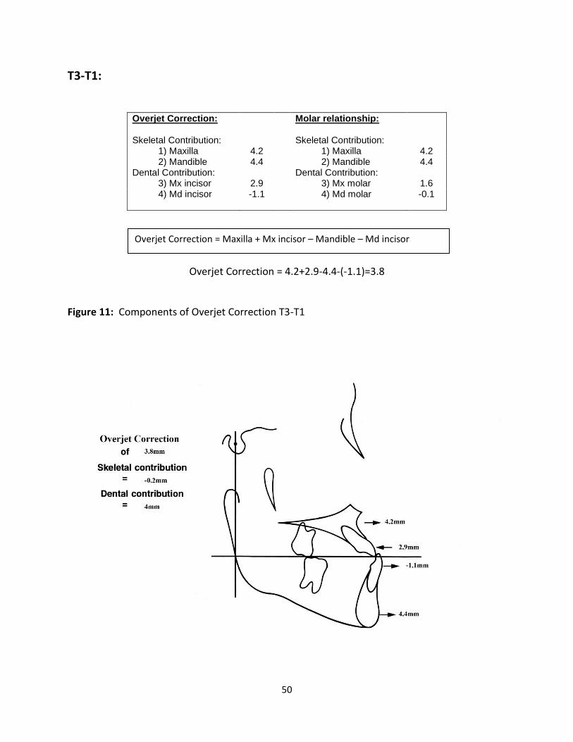
correction that was 113% dental. Overall, the maxilla moved forward 4.2 mm while the

mandible moved forward 4.4 mm. Conclusions: Treatment at a young age using a Modified

Hyrax Expander with Protraction Facemask is successful in treating a Class III malocclusion. The

correction is both skeletal and dental.

iii

DEDICATION

To my husband Jason Lawrence for always being there for me and lending me a supporting hand. You give me strength to push forward when I am weary. You believed in me when I didn’t believe in myself. I’m so glad I get to come home to you every day. You are the light of my life.

To my parents, James and Eleanor Reed, for being such good role models in my life. You are the best parents. You’ve sacrificed so much to help me get to where I am today. You are an inspiration in my life. I could not have done this without you. Thank you for all the support and the phone calls!

To my grandfather Raymond Warren who taught me many things, but above all, that hard work always pays off. Thank you for instilling in me the importance of education and all your help while I was working on mine. I’ll never forget your great stories.

To my sister Vanessa Bennett who always took up for me and taught me fight for what is right. I would not have had the courage to complete this task without your example. Believe it or not, but I have been trying to catch up with you my whole life. Keep me reaching!

To my brother Robert Reed who has taught me to never stop and never give up. You are an inspiration to me. I may never run a marathon, but I know I could because you lead by example.

To my in-laws Patricia Alexander, Aaron Alexander, Patricia Lawrence, Gene Lawrence, John Loving, Marvine Loving, Page Alexander, and Joe Alexander for accepting me into your wonderful family. Thank you.

To my Great Aunt El and Uncle Ban for being so supportive in my educational endeavors. Thank you also for being such great examples of generosity and healing to others. You both are missed.

To my grandparents who are not with us today, Eleanore Warren, Lucy Warren, Mama Tobe and Daddy Tobe; the love and support began with you and you are missed.

To my church family at St. Mark’s Episcopal Church for helping raise me. Your love has stayed with me and been an inspiration during the tough times.

To my teachers who took their time to teach and make a difference in my life. I would not be here without your hard work and dedication.

To my cats Sebastian and Penelope for being the cutest, most loving pets I could ask for. You cheer me up when I am down. I look at my rescue cats and appreciate the roof over my head and the food in my stomach.

iv

ACKNOWLEDGEMENTS

Dr. Peter Ngan – For being a wonderful mentor and advisor. You have built the best residency

program in the world. Your dedication to orthodontics and teaching has given me an invaluable

gift I will pass on to my patients and, hopefully, future residents at WVU.

Dr. Chris Martin – For also being a wonderful mentor and advisor. I will remember your

kindness and support over the past 3 years. I will always be your vice president.

Dr. Thomas Razmus – For your help with my thesis. Thank you for giving your time and being a

friendly face across the hallway.

Dr. Thomas Kiebach – For allowing me to use your clinical records for my thesis. You were

motivational and uplifting throughout this process.

Part time Faculty members: Drs. Tremont, Kirsch, McFarland, Hazey, Jarrett, Foley, Boyles,

Sebbahi, and Gilmore for your pearls of wisdom that I will take with me through the remainder

of my career. Your dedication to the program and profession is remarkable. Thank you for

kindness and everything you have done for me over the years. It was much appreciated.

Drs. Larry and Will Andrews for passing on your knowledge and expertise in orthodontics.

Thank you for being so welcoming to WVU residents!

Staff members Karen, Leona, Carrie, Hillary and Joyce for lending me a helping hand and for

always being there to listen.

Rob VanLaecken – For being such a wonderful classmate who understood the stresses and was a

light-hearted companion through thick and thin. Thanks for being a great friend. You are a

wonderful person and hard worker and I am proud to call you my pier.

Maggie Adams – We have been through thick and thin as well and I’m happy to share the

milestones we have achieved together. You have been a wonderful friend and confidant. I truly

could not have made it through without you! You were always one step (or let’s face it…several

steps) ahead of me and that’s exactly what I needed. Here’s to many more years of friendship!

My fellow residents Thuy, Rajia, Mike B, Mike M, Dean, Colin, Jung Mee, Holly, Ronnie,

Doyong, Chad and Alice for all the great times we shared. Thank you for your help along the

way. Best wishes to all of you!

v

TABLE OF CONTENTS

DEDICATION ................................................................................................................................................. iii

ACKNOWLEDGEMENTS ................................................................................................................................ iv

TABLE OF CONTENTS ..................................................................................................................................... v

LIST OF TABLES ............................................................................................................................................ vii

LIST OF FIGURES ......................................................................................................................................... viii

Chapter 1: Introduction ............................................................................................................................... 1

Background ............................................................................................................................................... 1

Statement of the Problem ........................................................................................................................ 4

Significance of the Problem ...................................................................................................................... 4

Null Hypothesis ......................................................................................................................................... 5

Definition of Terms ................................................................................................................................... 5

Assumptions .............................................................................................................................................. 9

Limitations ................................................................................................................................................ 9

Delimitations ........................................................................................................................................... 10

Chapter II: Review of the Literature ........................................................................................................... 11

Incidence ................................................................................................................................................. 11

Etiology ................................................................................................................................................... 11

Components of Class III malocclusion ..................................................................................................... 12

Diagnosis of Class III malocclusion .......................................................................................................... 15

Treatment of a Class III malocclusion ..................................................................................................... 17

Non-growing patient ........................................................................................................................... 17

Growing patient .................................................................................................................................. 18

Treatment timing for a growing patient ................................................................................................. 20

Chapter III: Materials and Methods ........................................................................................................... 24

Experimental Design and Methods ......................................................................................................... 24

IRB Approval ............................................................................................................................................ 25

Cephalometric Analysis ........................................................................................................................... 25

Sagittal Measurements ........................................................................................................................... 28

Vertical Measurements ........................................................................................................................... 29

Angular Measurements .......................................................................................................................... 30

vi

Method Error .......................................................................................................................................... 32

Evaluation of Overjet and Molar Relationship Correction ...................................................................... 34

Statistical Analysis ................................................................................................................................... 36

Chapter IV: Results ..................................................................................................................................... 37

Cephalometric Measurements ............................................................................................................... 37

Cephalometric Changes of T2-T1, T3-T2, and T3-T1 ............................................................................... 39

Overjet and Molar Relationship Correction ............................................................................................ 43

T2-T1: .................................................................................................................................................. 43

T3-T2: .................................................................................................................................................. 47

T3-T1: .................................................................................................................................................. 50

Chapter V: Discussion................................................................................................................................. 53

Chapter VI: Summary and Conclusions ...................................................................................................... 56

Summary ................................................................................................................................................. 56

Conclusions ............................................................................................................................................. 56

Chapter VII: Recommendations for future research .................................................................................. 59

Appendix A .................................................................................................................................................. 65

vii

LIST OF TABLES

Table 1: Skeletal and Dental Landmarks………………………………………………………………………………….26

Table 2: Definition of Reference Lines………………….………………………………………………………………….27

Table 3: Sagittal Measurements of variables 1-9….………………………………………………………………….29

Table 4: Vertical Measurements of variables 10-16…………………………………………………………………30

Table 5: Angular Measurements of variables 17-25…………………………………………………………………32

Table 6: Reliability Coefficients for all variables at T1, T2, and T3…………………………………………..33

Table 7: Calculation of Overjet and Molar Relationship Changes…………………………………………….34

Table 8: Cephalometric Measurements…………………………………………………………………………………..38

Table 9: Comparison of T2-T1, T3-T1, and T3-T2……………………………………………………………………..42

viii

LIST OF FIGURES

Figure 1: Skeletal and Dental Landmarks………………………………………………………………………………….27

Figure 2: The reference grid used in the sagittal cephalometric analysis…………………………………..28

Figure 3: The reference lines and measuring points used in the vertical analysis…..………………...30

Figure 4: The reference lines and measuring points used for angular analysis………………………….31

Figure 5: Components of Overjet Correction T2-T1…………………………………………………………………..43

Figure 6: Components of Molar Correction T2-T1…………………………………………………………………….44

Figure 7: Pitchfork Analysis for Overjet and Molar Correction T2-T1………………………………………..46

Figure 8: Components of Overjet Correction T3-T2…………………………………………………………………..47

Figure 9: Components of Molar Correction T3-T2…………………………………………………………………….48

Figure 10: Pitchfork Analysis for Overjet and Molar Correction T3-T2……………………….……………..49

Figure 11: Components of Overjet Correction T3-T1………………………………………………………………..50

Figure 12: Components of Molar Correction T3-T1…….…………………………………………………………….51

Figure 13: Pitchfork Analysis for Overjet and Molar Correction T3-T1……….……………………………..52

1

Chapter 1: Introduction

Background

The prevalence of a Class III malocclusion is estimated to be about 5.6% of United States

population.1 Traditional treatment for these individuals include: orthodontic camouflage,

orthopedic correction using functional appliances, and orthognathic surgery. Early orthopedic

therapy including an RPE and facemask can help patients with retrognathic maxillas if it is

provided early. A retrusive maxilla is the underlying cause of a Class III malocclusion in 57% of

patients.2,3 In 42 percent of class III maloclussions mandibular excess is the primary problem.4

Although a Class III malocclusion has a relatively low prevalence, it presents as a

challenging orthodontic problem.5 Historically, the Class III malocclusion was viewed as a

problem of the mandible. A Class III malocclusion and mandibular prognathism were virtually

synonymous.6 Management usually involved chincup appliances to restrain mandibular growth,

camouflage techniques to advance maxillary incisors and retract mandibular incisors, or wait

until growth ceased to pursue orthognathic surgery. 7 Many studies have found that the

primary etiology in a Class III malocclusion is, however, maxillary deficiency. Protraction

facemask in the treatment of Class III malocclusions with maxillary deficiency has become an

acceptable procedure among the orthodontic profession.

Facemask therapy is often supplemented with maxillary expansion. Midface orthopedic

expansion has been recommended for use in conjunction with protraction forces on the maxilla

because it supposedly disrupts the circummaxillary sutural system and facilitates the

2

orthopedic effect of the facemask.8-11 There is evidence that maxillary expansion alone can be

beneficial in treating Class III malocclusions, especially in borderline malocclusions. Haas

reported that palatal expansion produces a forward and downward movement of the maxilla by

affecting the intermaxillary and cirummaxillary sutures and the disruption of these sutures may

help initiate cellular response in the sutures, allowing for a more positive reaction to

protraction force.9,10 RME is also helpful in these patients because maxillary retrognathic

patients also have posterior crossbites due to a deficiency in the transverse dimension as well.

Recommendations on the optimal time to treat a child with a combination of palatal

expansion and facemask therapy have been based primarily on clinical impressions. The

recommended age to begin treatment is between the age of 6 and 8 years after the maxillary

permanent first molars and incisors have erupted.12-14 Studies have employed biologic

indicators such as chronological age, stage of dental development or skeletal age to determine

the impact of age on orthopedic treatment.8,12,15-20 It was found that early treatment,

sometimes beginning as young as age 4, can be effective for orthopedic correction of Class III

malocclusions.12 Several studies found younger patients more responsive to orthopedic

correction. Compliance is also less of an issue at this early age.

Various expansion appliances have been used as anchorage devices for maxillary

protraction. The problem with using bands is breakage around the solder joints. A heavy

0.036” wire is usually soldered to the buccal side of the molar band and extends forward to the

canine area for protraction. A new design that utilizes a stainless steel crown and a removable

arm for maxillary protraction may overcome this problem. This arm resists deformation and

3

fracture while also providing rigidity. The stainless steel crowns keep the anterior teeth away

from occlusion. This design is particularly suitable for patients in the primary dentition.

The objective of this study was to conduct an observational retrospective study to

determine the immediate and long term skeletal and dental effects of this new anchorage

device used in conjunction with a protraction facemask. The experimental group consisted of

23 patients treated with Dr. Kiebach’s Modified Hyrax appliance and protraction facemask

therapy at an early age. Lateral cephalograms were taken before treatment (T1), after

treatment (T2), and approximately 2 years post-treatment (T3). A custom cephalometric

analysis as described by Byork21 and Pancherz22 was used to determine the amount of skeletal

and dental correction. Data will be analyzed using a paired t-test.

4

Statement of the Problem

The purpose of this research project is to determine the skeletal and dentoalveolar

changes in Class III patients treated using Dr. Kiebach’s Modified Hyrax Expander and facemask

therapy in the primary and early mixed dentition.

Significance of the Problem

For young Class III patients with a retrusive maxilla, the treatment of choice is maxillary

expansion combined with a protraction facemask. The timing for this treatment is traditionally

between the ages of six and eight years after the maxillary permanent first molars and incisors

have erupted. Recent studies have shown that the earlier treatment begins, the more the

maxilla will protract.

Dr. Kiebach advocates early treatment and has designed a Modified Hyrax expander

which utilizes Stainless Steel crowns and laser welded arms that prevent breakage and provide

more anchorage for use in the primary and early mixed dentition. The stainless steel crowns

also serve as a bite plate to reduce any interference from the occlusion while protracting. This

study will determine how much skeletal movement patient’s treated in this manner will receive

and document the stability of the movements at this young age.

5

Null Hypothesis

1. There were no significant sagittal changes in patients treated with Dr. Kiebach’s

Modified Hyrax Expander and facemask therapy.

2. There were no significant vertical changes in patients treated with Dr. Kiebach’s

Modified Hyrax Expander and facemask therapy.

3. There were no significant angular changes in patients treated with Dr. Kiebach’s

Modified Hyrax Expander and facemask therapy.

Definition of Terms

1. Maxillary expansion: Separation of the two halves of the maxilla achieved in the

growing individual with the use of and orthopedic expansion device.

2. Maxillary protraction: The orthopedic anterior and downward repositioning of the

maxilla achieved in the growing individual with the use of an orthopedic protraction

facemask appliance.

3. Protraction facemask: An extraoral appliance used to exert a forward and downward

vector of force on the maxilla. Also referred to as protraction headgear.

4. Class III malocclusion: That relationship in which the buccal groove of the mandibular

first permanent molar articulates anteriorly to the mesiobuccal cusp of the maxillary

first permanent molar. A mesial relationship of the lower first molar to the upper

and/or a distal relationship of the upper first molar to the lower.

6

5. Skeletal Class III malocclusion: Skeletal relationship in which either the mandible is

prognathic, the maxilla is retrognathic, or a combination of the two.

6. Pseudo Class III malocclusion: Relationship in which a Class I skeletal pattern, normal

facial profile, and Class I molar relation may occur in centric relation, but a Class III

skeletal and dental pattern are observed in centric occlusion.

7. Centric occlusion: The relationship between upper and lower teeth in normal full

functional closure

8. Centric relation: The relation between upper and lower teeth when both mandibular

condyles are fully seated in their fossa in optimum functional positions.

9. Cephalogram: A term sometimes used as a synonym for cephalometric radiograph.

10. Cephalometric analysis: An evaluation of dental and related skeletal relationships based

on measurements of cephalometric radiographs.

11. Cephalometric radiograph: A radiograph of the head made with precise reproducible

relationships between x-ray source, subject and film. The generally accepted distances

between x-ray source and the center of the subject are 5 feet or 150 centimeters. The

distance between subject and film is usually 15 cm, but may be standardized at different

value or varied with patient size and recorded for each exposure.

12. Cephalometric tracing: A tracing of selected structures from a cephalometric

radiograph, made on translucent drafting paper or digitized on computer software for

purposes of measurement and evaluation.

13. Comprehensive orthodontic therapy: A coordinated approach to improvement of the

overall anatomic and functional relationships of the dentofacial complex, as opposed to

7

partial correction with more limited objectives such as cosmetic improvement.

Comprehensive orthodontic treatement, usually, but not necessarily, utilizes fixed

orthodontic attachments as one treatment modality. May be coordinated with

adjunctive procedures directed at malrelationships within the entire dentofacial

complex.

14. Crossbite: An abnormal relationship of a tooth or teeth to the opposing teeth, in which

normal buccolingual relationships are reversed.

15. Deep bite: Excessive overbite; closed bite

16. Distal: A direction oriented along the dental arch away from the dental midline; right or

left in the anterior segment, posteriorly in the buccal segments.

17. Mesial: Toward or facing the midline, following the dental arch. Used to describe

surfaces of teeth as well as direction.

18. Labial: of or pertaining to the lip. Also used to identify a surface facing the lips or a

direction toward the lips.

19. Lingual: Of or pertaining to the tongue. Used to describe surfaces and directions facing

the tongue.

20. Malocclusion: A deviation in intramaxillary and/or intermaxillary relations of teeth that

presents a hazard to the individual’s well-being. Often associated with other dentofacial

deformities.

21. Mixed dentition: The developmental stage during which both deciduous and

permanent teeth are present in the mouth.

8

22. Occlusion: The relationship of the maxillary and mandibular teeth as they are brought

into functional contact.

23. Open bite: A malocclusion in which some teeth cannot be brought into functional

contact with opposing teeth.

24. Overbite: Vertical overlapping of upper teeth over lower teeth usually measured

perpendicular to the occlusial plane.

25. Overjet: Horizontal projection of upper teeth beyond the lower teeth, usually measured

parallel to the occlusal plane.

26. Prognathic: Forward relationship of the mandible relative to the craniofacial skeleton.

27. Retrognatic: The condition of the maxilla or mandible that is posterior to its normal

relationship with other facial structures.

28. Retrusion: Teeth and/or jaw posterior to their normal positions.

29. Proclination: Anterior angulation of anterior teeth, as opposed to protrusion, which

indicates positional variation.

30. Tipping: Tooth movement, either spontaneous or therapeutic, in which the angulation

of the long axis of the root is changed.

31. Facial concavity: A term applied to the analysis of a profile. The shape is described as

an inwardly rounded curve from the forehead to the lips to the chin. A concave facial

profile is often associated with a Class III malocclusion.

32. Hyrax expander: Commonly used type of banded rapid maxillary expansion appliance.

Bands are placed on the maxillary first molars and first premolars or primary molars.

9

The expansion screw is located in the palate in close proximity to the palatal contour.

Buccal and lingual support wires also may be added for rigidity.

Assumptions

1. It is assumed that the lateral cephalograms were taken with the teeth in centric relation.

2. It is assumed that each child achieved sutural separation with the expander before using

the protraction facemask.

3. It is assumed that each patient wore the facemask for at least 12 hours per day.

Limitations

1. Inconsistency of records due to exfoliation of teeth during treatment. Serial

cephalograms will be taken over time. Treatment begins in the primary dentition and

exfoliation of primary incisors during treatment time will be a source of uncontrolled

error in the treated group.

2. Since different x-ray units were used to collect data all the magnification errors were

accounted for.

3. Cooperation differences between patients such as length of time each child wears the

facemask appliance daily.

4. Samples were not selected at random.

10

5. The total sample was limited to 23 patients due to the availability of records.

6. The T3 follow up cephalograms were taken at different time intervals.

7. The T2 radiographs were taken at different time intervals.

Delimitations

1. One researcher performed all cephalometric tracings and measurements.

2. Patients with craniofacial anomalies were excluded from the study.

3. Patients were limited to those in a primary dentition or early mixed dentition.

4. The experimental group was limited to patients who had acceptable quality radiographs

for the various time points of the study.

11

Chapter II: Review of the Literature

Incidence

The Prevalence of Class III malocclusions vary among different ethnicities. In

Caucasians, the incidence ranges from 1-4% of the population.23-26 Asian societies have a higher

incidence of maxillary deficiency and, therefore, a higher incidence of Class III malocclusions.

The incidence ranges between 4% and 5% among the Japanese and 4% and 14% among the

Chinese.23,27,28 The prevalence is approximately 3-6% in the U.S. black population. Class III

malocclusions are more prevalent in Hispanic populations than in African or Caucasian groups.13

Etiology

The etiology of malocclusions has been studied for many years. Some studies

conducted to learn about the etiology of malocclusions compared identical twins, fraternal

twins, and ordinary siblings. The difficulty in these studies was to confirm that the

environments were the same for both members of a twin pair. Summarizing a number of

research investigations of this type, Lauweryns et al. concluded that about 40% of the dental

and facial variations that lead to malocclusion can be attributed to hereditary factors.29,30 One

of the best known examples of a hereditary Class III malocclusion is that of the Hapsburg’s, a

European royal family. The families mandibular prognathism became known as the Hapsburg

jaw because it recurred over many generations.30 A prognathic mandible was evident in 83% of

the 40 family members whom records were available.31

12

Harris and Johnson also concluded that the heritability of craniofacial or skeletal

characteristics was relatively high, but that the dental characteristics was low.30 Dental

characteristics are caused more from environmental factors; such as tongue posturing and

other habits. Environmental factors, although more rare, can cause Class III malocclusions.

Environmental influences during growth and development consist largely of pressures and

forces related to physiologic activity. How you chew and swallow place pressures against the

jaws and teeth that can affect how jaws grow and teeth erupt in to the jaws.30 An equilibrium

is formed between soft and hard tissues. A large tongue, perhaps in a patient with a thyroid

disorder, can cause the development of mandibular prognathism due to the mandible being

postured forward at all times.30 This constant distraction of the mandibular condyles may

cause excessive mandibular growth in these patients. Mandibular size may also be affected by

functional mandibular shifts due to respiratory needs. A mouth-breather’s tongue tends to be

flat and anteriorly displaced, resulting in the mandibular arch widening laterally and anteriorly.

Components of Class III malocclusion

In a Class III individual, the nasomaxillary complex may be retrusive, the mandible may

be protrusive, or there could be a combination of both. Many years ago, the Class III

malocclusion was viewed as a problem of the mandible. Until the 1960’s and 70’s the terms

Class III and mandibular prognathism were basically synonymous. Although maxillary

protraction using facemask therapy was first described more than a century ago, practitioners

did not begin using it with frequency until the 1960’s.5 In 1997 Lee KG et al. stated that in 42-

13

62% of skeletal Class III malocclusions, a combination of a retrognathic maxilla and a normal to

mildly prognathic mandible exists.32 Other combinations can exist as well including: Maxilla

within normal range and mandibular prognathism, the maxilla and mandible are both

prognathic and the mandible is prognathic and the maxilla is retrognathic.

Patients with a Class III malocclusion usually present with a concave facial profile. A

maxillary deficiency can affect the entire midface causing the areas such as the zygomatic

processes and nasal bridge to appear deficient. The tip of the chin, as well as the lower lip, will

lie somewhere in front of a vertical line drawn from nasion, perpendicular to the Frankfort

horizontal plane. A small maxilla will affect the craniofacial complex in a sagittal dimension as

well causing a skeletally derived posterior dental crossbite. There is also often and increased

lower facial height due to the maxilla not growing downward and forward.

Patients with class III malocclusions may present with varying combinations of skeletal,

dental, and soft tissue combinations. The most common skeletal features include an obtuse

gonial angle, a shortened anterior cranial base, a sagittal discrepancy of the maxilla or

mandible, and an increased lower facial height. Dental findings usually include Angle Class III

molars and canines with retroclined mandibular incisors and proclined maxillary incisors. This

can result in an edge to edge incisor relationship or an anterior crossbite. In profile, the soft

tissue outline appears concave. The nasolabial process is often acute with a retrusive upper lip

and lower lip posturing forward.

With many different presentations and variations of a Class III skeletal and dental

pattern, many question the underlying cause of this malocclusion. Understanding Class III

14

growth trends is needed for effective treatment planning and for knowing the stability of

treatment outcomes. This can help orthodontists when deciding between an orthodontic and

surgical approach to treating this malocclusion. Longitudinal data on Class III subjects indicate

that the rate of maxillary growth in Class III malocclusion during developmental ages is lower

than expected for normal subjects being less than 1 mm per year. Also, mandibular growth is 3

to 4.5 mm per year.3 Miyajima et al studied Japanese female subjects and concluded that the

maxilla exhibited a retrusive position at an early developmental stage and retained a fairly

constant anteroposterior relationship to the cranial base structures with continued

development.33 The mandible was protrusive early in development and became increasingly

prognathic with age.34 A cross-sectional study by Battagel showed that the largest increments

of mandibular length in male subjects occurred at ages of 15 years and older, indicating peak

growth at a late age period.35 The female samples showed that the maximum changes in facial

characteristics occurred between the ages of 11 and 12 years, but continued after 15 years of

age. The control group for the females showed that facial growth had stopped at the age group

of 14 to 17 years, but development remained active in the Class III group.5 Data suggests that

growth trends in Class III malocclusions might be different from normal developmental patterns

because peak growth occurs later and at relatively high rates until young adulthood.5 Adding to

the data of this growth time and rate, a study conducted by Baccetti et al discovered that the

duration of the peak interval of growth is approximately 6 months longer in Class III patients of

both sexes than in those with normal occlusion.5

The average increase in Co-Gn for a Class I patient is 2 to 3 mm. It is much greater in

Class III subjects. Baccetti et al found that mandibular length increased between 6 and 7 mm

15

for males and 4 to 5.5 mm in females in the same skeletal age group measured by patients’

CVM. A similar amount of increase was also found by Miyajima et al in his female Class III

subjects.5,33 He also found that the maxilla showed a retrusive position at an early

developmental stage and retained this anteroposterior position, whereas the mandibular

position worsened with growth.33 There are important clinical implications with these findings

that should be taken into account when treatment planning. With a much longer period of

mandibular growth and the absence of growth by the maxilla, the timing for Orthognathic

surgery should be carefully considered.

Diagnosis of Class III malocclusion

Diagnosing the underlying cause of a Class III malocclusion can come with differing

opinions. In many areas of orthodontics, clinical preference or judgment can play a role.

However, in order to differentiate the underlying cause of a Class III malocclusion, a simplified

method of evaluating patients must be utilized. The following recommendations have been

made in the assessment of Class III patients.36-38

The first step is to take a thorough family history. As mentioned, skeletal relationships

are strongly hereditary and if a close relative required orthognathic surgery to correct a

malocclusion, then this should alert the clinician that the patient may exhibit a potential

skeletal discrepancy. Some Class III individuals can have a differing growth pattern compared

to norms which presents as excessive late mandibular growth. This occurs most frequently in

16

males during their late teens. Clinicians must be aware of this during the exam because a

patient who presents with a less severe Class III skeletal pattern may not stay that way due to

further growth potential.

Second, it is necessary to diagnose the presence of a functional shift or CR/CO

discrepancy. An anterior posturing of the mandible may result when an abnormal contact

encourages the mandible to shift forward. It is important to distinguish the true Class III

malocclusion from a Pseudo-Class III malocclusion. A pseudo-class III patient is usually

characterized by having a Class I skeletal pattern, normal facial profile, and Class I molar

relation in centric relation, but possesses a Class III skeletal and dental pattern in centric

occlusion. The elimination of a CR/CO discrepancy should reveal whether the malocclusion is a

Class I or a compensated Class III malocclusion.

Third, a cephalometric analysis provides a quantitative assessment of the severity of the

Class III malocclusion. This radiograph is always taken in Centric Relation with the mandible

seated in its most superior anterior position. A lateral cephalogram aids in determining the

cause of the malocclusions; whatever the combination of skeletal disharmony it may be.

Finally, the clinical assessment of the patient is very important in diagnosis. The antero-

posterior skeletal base relationship and the vertical facial proportions should be assessed while

the patient is standing upright with a natural head position. Profile disharmonies should be

recorded at this time. The transverse dimension should be assessed along with any facial and

dental asymmetries. The clinical exam includes the TMJ, associated musculature, oral mucosa,

and occlusion. The use of mounted orthodontic study models can be an adjunct to the clinical

17

exam revealing incisor relationship, overjet, overbite, incisor inclination, arch alignment,

midline discrepancies and occlusal disharmonies such as a cant of the maxilla. The

development of a problem list from all acquired data assists in the planning of Class III

treatment.

Treatment of a Class III malocclusion

Non-growing patient

In the past, most of the treatment of Class III malocclusion involved a combination of

orthodontic and orthognathic surgical correction upon completion of active growth of the

patient. If the skeletal discrepancy is large and surgery is not an option, then a fair amount of

negative overjet may still persist after orthodontic treatment.

Orthodontic camouflage can be performed on the growing or non-growing patient. It

usually involves the extraction of mandibular first premolars with or without the extraction of

maxillary second premolars. This extraction pattern is done to camouflage a moderate skeletal

discrepancy when orthopedic correction by growth is not possible or there is dental crowding

which requires extractions to obtain space to align the teeth in the arch. Extracting in Class III

individuals allows the orthodontist to reduce the amount of negative overjet and camouflage

the skeletal discrepancy. When there is doubt about further skeletal growth, orthodontic

camouflage should be deferred until the remaining skeletal growth has been complete.

18

Orthognathic surgery is a treatment alternative that will most likely lead to an ideal

relationship of the maxilla and mandible in severe malocclusions. However, it is very invasive

and financially demanding. Class III malocclusions makes up a small percentage of the

malocclusions in the United States, but they comprise a substantial percentage of patients

seeking orthognathic surgery in adults.39,40 Pre-surgical orthodontic treatment usually involves

the fixed appliances to align the maxillary and mandibular arches, so that they will coordinate

when the skeletal bases are positioned properly in surgery. Since there is equilibrium between

hard and soft tissues, orthodontic decompensation is usually necessary to gain the correct axial

inclination of the incisors.

Growing patient

There is a lot more freedom when treating a growing patient with a Class III

malocclusion. These options include camouflage treatment and, more importantly, functional

orthopedic appliances. The goal of orthopedic correction of skeletal Class III discrepancies is to

control and/or redirect the growth of the mandible and maxilla. Some functional appliances

focus on the mandible, some focus more on the maxilla. The different orthopedic appliances

used in the correction of skeletal Class III malocclusions include the chin cup appliance, the

Frankel III appliance, and the maxillary protraction appliance.

The chin cup appliance which represents one of the oldest orthopedic appliances used

to treat a skeletal Class III malocclusion is rarely used today. This was used heavily in the past

when Class III malocclusions were thought to originate solely due to mandibular prognathism.

These appliances, in order to be successful, were worn throughout growth. This is one of the

19

chin cups draw backs. Another reason for abandoning this treatment is because greater forces

are required to achieve orthopedic effects. It requires 600 to 800 grams of force which can

cause the patient to experience temporomandibular joint problems. The last reason for

discontinuing the chin cup as a treatment of choice is that the positive effects of the chin cup

therapy were often not maintained due to latent mandibular growth.

The Frankel III appliance or FR-3 utilizes the maxillary and mandibular vestibules in the

treatment of Class III malocclusions. The appliance shields the maxilla from the negative

influence of the surrounding soft tissue, which in turn provides a restrictive force on the

mandible.41 Treatment time with the FR-3 can be extensive; up to 24 months for a good result.

The treatment effects include a forward maxillary movement, forward movement of the

maxillary dentition, mandibular growth modification downward and backward, and lingual

tipping of the mandibular incisors. Most practitioners use an FR-3 appliance, if used at all, as a

retainer after facemask therapy is complete.

Facemask therapy in conjunction with maxillary expansion is the orthopedic treatment

of choice today. It is an effective method of treating skeletal Class III malocclusion with

maxillary retognathism and/or mandibular prognathism. The facemask, popularized by Delaire,

uses the chin and forehead for support. The orthopedic force of this appliance is utilized to

protract the maxilla while the chin support serves to redirect mandibular growth. Midfacial

orthopedic expansion has been recommended for use in conjunction with protraction forces on

the maxilla because it supposedly disrupts the circummaxillary sutural system and presumably

facilitates the orthopedic effect of the face mask.8

20

Therapy involves the assisted forward growth of the maxilla which is accomplished by

utilizing elastics to connect a fixed appliance on the posterior teeth to an extraoral anchorage

site. The elastics are secured near the maxillary canines to avoid bite opening. A downward

force of 30 degrees to the occlusal plane provides the greatest translator displacement of the

craniofacial complex along the force application line.42 Anterior displacement requires 600-800

g of force per side. Treatment time varies among individuals, but the average treatment length

is 9 months wearing the facemask for at least 12- 14 hours per day.

Treatment timing for a growing patient

One problem that clinicians have with treating retrusive maxillas early with facemask

therapy is that mandibular growth cannot be predicted.43 One way to predict excessive

mandibular growth is to look at the patients’ family.4 Early treatment in patients with

mandibular excess is not advised because early treatment to correct the prognathism of the

mandible does not result in normal growth thereafter. On the other hand, the window for

treatment of a patient with maxillary deficiency is very narrow. Orthopedic treatment is best

rendered before the onset of puberty.

Over the last 20 years, the use of rapid maxillary expansion with protraction facemask

has gained popularity among clinicians. The treatment effects are a combination of skeletal

and dental modifications in both the maxilla and mandible. Optimal time to treat a child has

been based primarily on clinical impressions with the suggested time between the ages of 6 and

21

8 years. Treating at this early age is reported to remove factors that inhibit growth and

development, such as an anterior crossbite that limits normal alveolar bone growth. Many

investigators have conducted cephalometric studies of children treated with RME/FM to

determine whether biologic indicators such as chronological age, stage of dental development,

or skeletal age impact the orthopedic effects of treatment and future growth.12 Saadia et. al.

found that younger patients show greater, faster results in less time under facemask therapy

with the best results coming from the age group of 3 to 6 years. At this early age, compliance is

improved and psychosocial scars which have been shown to affect patients into adulthood are

reduced due to the patients’ enhanced esthetics after treatment.44 Another study by Kapust

and Turley found that the best age range for facemask therapy was between the ages of 4 and

7 years.45 The 4 to 7 year age group showed statistically greater increases in the SNA angle. It

was almost twice the change in SNA as the older group from 10 to 14 years. Baccetti et al

showed that early treatment groups showed significantly greater advancement of maxillary

structures and significantly more upward and forward direction of condylar growth after

treatment.5

Franchi et al investigated treatment timing for RME/FM based on an early treated group

(ETG) if they were either in the deciduous or early mixed dentition, and late treated group (LTG)

if they were in the late mixed dentition with erupting permanent canines and premolars. The

results showed a significant differential between the groups of 7 mm. The early treated

patients maintained a maxillary/mandibular skeletal relationship within 1 mm because of the

significant favorable skeletal contributions of the maxilla and the mandible. The maxilla

showed a forward movement of 1.8 mm and the mandible expressed a significantly smaller

22

anterior projection of 5 mm compared with the untreated Class III control. In the LTG, the

skeletal movements could not achieve a positive change. The mandible moved forward more

than the maxilla in the LTG and control group. However, treatment in the late mixed dentition

produced significantly smaller increased in total mandibular length with respect to the control.

A significant advancement of the maxilla can be achieved orthopedically only by treating Class

III patients in the deciduous or early mixed dentition phases. About 2 mm of supplementary

forward movement of the maxilla are maintained in treated patients at the completion of

growth when compared with untreated subjects. This movement is not possible in the patients

of late mixed dentition or older. In early developmental phases, mandibular growth control is

associated with a significant decrease of the gonial angle in patients treated with RME/FM

therapy.12 Because Franchi et al compared his treated group to a control group who also had

Class III malocclusions, this allowed them to investigate the craniofacial growth characteristic

for this type of skeletal discrepancy. The observations made in both the early and late control

groups suggest that the skeletal imbalance in Class III malocclusion is established early in life

and is not self-correcting during development.12 These investigators recommend early

intervention for Class III malocclusion although patients treated during the late mixed dentition

can still benefit from RME/FM therapy, but to a lesser degree.

Some of the rationales for early treatment of Class III Malocclusions include:

1. To prevent progressive irreversible soft tissue or bony changes. If the patient

has an uncorrected anterior crossbite, it may lead to abnormal wear of incisors

23

and dental compensation of incisors. Also, expansion in the permanent

dentition can lead to histological changes in the pulp.

2. To improve skeletal discrepancies and provide a more favorable environment for

future growth. This can minimize dental compensations such as overclosure of

the mandible and over retraction of the lower incisors.

3. To improve occlusal function. A class III malocclusion is often accompanied by a

functional shift. Elimination of a functional shift with orthopedic treatment may

help the patient avoid adverse growth potential.

4. To simplify Phase II treatment. Early orthodontic or orthopedic treatment for

mild of moderate Class III patients may eliminate the need for surgery. If the

patient needs surgery, early treatment may minimize the extent of the surgery.

5. To provide more pleasing facial esthetics which can improve the psychosocial

development of the child. Early treatment can improve lip posture and facial

appearance.2

Each case must be considered individually. Factors that determine treatment may

include familial history of a prognathic mandible or patient’s age. Overcorrection is

recommended because these patients tend to grow similarly to untreated Class III patients after

facemask treatment. Currently there is a lack of long-term data to answer the many questions

that continue to plague orthodontists in regard to long-term stability of facemask therapy.45

24

Chapter III: Materials and Methods

Experimental Design and Methods

The study group was composed of 76 consecutively patients treated with protraction

facemask at a very early age using the Modified Hyrax Appliance. Due to exclusion criteria, the

sample size was reduced to 23 patients. The pre-treatment craniofacial morphology had an

average SNA measurement of 80, SNB of 81, ANB of -0.3, and Wits of -4.2 Patients were

excluded if radiographs were not taken at each time point and if the radiographs were not of

sufficient quality. All patients had lateral cephalometric radiographs taken pre-treatment (T1),

post-treatment (T2), and an average of 22 months after removal of the appliance (T3). The

mean age at the start of treatment was 6 years 2 months. The stage of dental development

varied from primary dentition to early mixed dentition. The youngest age was 4 years 4 months

and the oldest age was 10 years 4 months. The treatment time for each time point can be

found in a table located in Appendix A. The average treatment time for T2-T1 was 9 months.

Treatment time varied between 3 months to 16 months. All films were traced by a single

investigator and compared using a customized cephalometric analysis, as described by Bjork21

and Pancherz.22

The Cervical Vertebra Maturation (CVM) for all subjects was an average of CVM 1.0. T1,

T2, and T3 radiographs were all taken before pubertal growth had occurred. Therefore the

treatment group was pooled together for analysis.

25

IRB Approval

IRB exemption was obtained from West Virginia University prior to beginning this study.

Cephalometric Analysis

Lateral cephalograms were obtained from the office of Dr. Keibach. The time points

obtained were Pre-treatment (T1), Post-Treatment (T2), and 22 month after appliance removal

(T3). The radiographs were scanned and placed on a CD and mailed to the school. The files

were downloaded in jpeg format, and digitized in Dolphin Imaging (Dolphin Imaging,

Chatsworth, CA) to adjust for magnification. Each image was then printed 1:1 to ensure there

was no magnification. The files were printed on an Epson Stylus Pro 3880 Printer on quality

photo paper (HP Premium Photo Paper).

All landmarks and tracings were made on the printouts while viewing the original digital

file. Tracings were performed by one operator using a 0.5mm mechanical lead pencil, and

orthodontic protractor, and 0.003 inch matte cephalometric acetate tracing film (3M Unitek,

Monrovia, CA). A custom cephalometric analysis was performed as described by Bjork21 and

Pancherz.22

26

TABLE 1: Skeletal and Dental Landmarks

Name Symbol Definition

Sella S The center of the sella turcica

Nasion N The most anterior point of the nasofrontal suture

Anterior Nasal Spine ANS The apex of the spina nasalis anterior

Posterior Nasal Spine PNS The most posterior point on contour of the palate in the midsagittal plane

Subspinale A pt. The deepest point in the concavity of the anterior maxilla between the ANS and the alveolar crest

Supramentale B pt. The deepest point in the concavity of the anterior mandible between the alveolar crest and pogonion

Pogonion Pg The most prominent point on the chin

Menton Me The deepest point of the mandibular symphysis

Gonion Go The lowest point of the bony contour of the angle of the mandible

Maxillary incisor apex

Isa The root apex of the most prominent maxillary central incisor

Maxillary incisor Is The incisal point of the most prominent maxillary central incisor

Mandibular incisor apex

Iia The root apex of the most prominent mandibular central incisor

Mandibular incisor Ii The incisal point of the most prominent mandibular central incisor

Molar superius mesial cusp

Msc The mesio-buccal cusp tip of the maxillary first permanent molar

Molar Superius Ms The mesial contact point of the maxillary permanent first molar

Molar inferius mesial cusp

Mic The mesial-buccal cusp tip of the mandibular first permanent molar

Molar inferius Mi The mesial contact point of the mandibular first permanent molar

27

Figure 1: Skeletal and Dental Landmarks

Table 2: Definition of Reference Lines

Name Symbol Definition

Sella-Nasion plane SNL Reference line joining Nasion and Sella

Maxillary plane NL Reference line joining anterior nasal spine and posterior nasal spine

Occlusal plane OL Reference line joining maxillary incisal edge and the molar superious mesial cusp tip

Mandibular plane ML Refernce line joining menton and gonion

Occlusal plane perpendicular

OLp Reference line produced by dropping a perpendicular line from sella to the occlusal plane

28

Sagittal Measurements

Figure 2. The reference grid (OLs and OLp) and measuring points used in the sagittal

cephalometric analysis.

Skeletal and dental changes in A-point, Is, Ms, Ii, Mi, and Pogonion compared to OLs and

OLp were measured by forming a reference grid based on the occlusal line (OLs) and occlusal

line perpendicular (OLp) see Figure 2. The reference grid was traced on T1 and used for all

sagittal measurements between OLp and the cephalometric landmarks transferring the grid by

superimposition from T1 to T2 and T3. Sagittal measurments taken can be seen in Table 3. The

measurement for each sagittal measurement was performed with an electronic digital caliper

and measured to the nearest 0.1 mm. The caliper was calibrated to 0.0 mm prior to each

measurement. Lateral cephalograms often present landmarks with right and left images;

therefore, the midpoint bisecting the two images was used.

29

Table 3: Sagittal Measurements of variables 1-9

Variable (mm) Definition

Skeletal Measuring Points:

1. OLp-A Position of maxillary base

2. OLp-Pg Position of mandibular chin

3. Wits Mx and Md position relative to OLs

Dental Measuring Points:

4. Is/OLp Position of maxillary central incisor

5. Ii/OLp Position of mandibular central incisor

6. Overjet Is/OLp minus Ii/OLp

7. Ms/OLp Position of maxillary first permanent molar

8. Mi/OLp Position of Mandibular first permanent molar

9. Molar rel. Molar relationship: Ms/OLp minus Mi/OLp

Vertical Measurements

Vertical measurements used OLs, NL, and ML. A measurement from Nasion to a-point

and ANS to Me was also included. Measurements from T1, T2, and T3 were not superimposed.

The equipment and measurement protocol was exactly the same as used in the Sagittal

Measurement mentioned above. Vertical measurements can be seen in Figure 3 and Table 4.

30

Figure 3: The reference lines and measuring points used in the vertical cephalometric analysis.

Table 4: Vertical Measurments of variables 10-16

Variable (mm) Definition Skeletal measuring points: 10. N-A pt. Maxillary vertical positioning 11. ANS-Me Lower facial height Dental measuring points: 12. Is-NL Position of maxillary central incisor (measured Is ╧ NL) 13. Ii-ML Position of mandibular central incisor (measured Ii ╧ ML) 14. Overbite Distance form Ii ╧ OLs 15. Msc-NL Position of maxillary permanenet forst molar (Msc ╧ NL) 16. Mic-ML Position of mandibular permanent first molar (Mic ╧ ML)

Angular Measurements

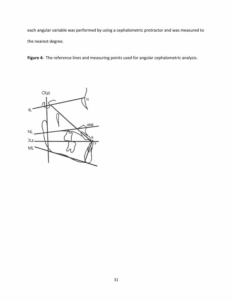
Angular measurements were used in addition to the grid measurements in order to

identify changes in the dentofacial complex. These angular measurements are shown in Figure

4 below. Also, the angular measurements are defined in Table 5 below. The measurement for

31

each angular variable was performed by using a cephalometric protractor and was measured to

the nearest degree.

Figure 4: The reference lines and measuring points used for angular cephalometric analysis.

32

Table 5: Angular Measurements of variables 17-25

Variable (°) Definition

Skeletal measuring points:

17. SNA Maxillary base relative to SNL

18. SNB Mandibular base relative to SNL

19. ANB SNA minus SNB

20. SNL-ML Mandibular plane angle

21. SNL-OL Occlusal plane angle

22. SNL-NL Palatal plane angle

Dental measuring points:

23. Is/SNL Maxillary central incisor angle

24. Ii/ML Mandibular central incisor angle

25. Is/Ii Interincisal angle

All lateral cephalograms were be calibrated to a 1:1 ratio using Dolphin software

(Dolphin Imaging, Chatsworth, CA). Data was analyzed with ANOVA and a multiple comparison

t-test.

Method Error

The reliability of the cephalometric measurements was tested by evaluating the error in

locating, superimposing, and measuring the differences in the landmarks. Pre-treatment (T1),

Post-treatment (T2), and Follow up (T3) radiographs of 6 randomly selected patients were

retraced two weeks after initial tracing and were analyzed to evaluate the error. For all

cephalometric variables, differences between the measurements from the first and second

33

tracings were compared for each individual at T1, T2, and T3. A reliability coefficient was

established for each variable at each time point to determine the degree of reliability (Table 6).

Table 6: Reliability Coefficients for all variables at T1, T2, and T3

Variables Reliability

Sagittal:

Olp-A 0.98

Olp-Pg 0.96

Is-Olp 0.99

Ii-Olp 0.98

Overjet 0.88

Ms-Olp 0.98

Mi-Olp 0.95

Molar Relationship 0.75

Vertical:

N-A 0.95

ANS-Me 0.96

Is-NL 0.97

Ii-ML 0.95

Overbite 0.91

Msc-NL 0.97

Mic-ML 0.96

Angular:

SNA 0.80

SNB 0.92

ANB 0.88

SNL-NL 0.93

SNL-ML 0.94

SNL-OLs 0.95

Is/SNL 0.97

Ii/ML 0.97

Is/Ii 0.97

34

The method of cephalometric analysis used in this study was determined to be reliable.

This included the identification of landmarks, superimposition of radiographs, and the

measurements taken at each time point. Reliability ranged from 0.75 to 0.99, which means

that the method of data collection was reliable.

Evaluation of Overjet and Molar Relationship Correction

To determine the amount of skeletal and dental contribution to the overjet and molar

relationship correction, the amount of dental change in the maxilla and mandible was

calculated. The method of obtaining these measurements is shown below (Table 7).

Table 7: Calculation of Overjet and Molar Relationship Changes

Overjet Molar Relationship

Skeletal contributions: 1. OLp-Apt 2. OLP-Pg

Dental contributions:

3. Is-OLp minus OLp-Apt 4. Ii-OLp minus OLp-Pg

Overjet correction: Sum of 1,2,3,and 4

Skeletal contributions: 1. OLp-Apt 2. OLP-Pg Dental contributions: 3. Ms-OLp minus OLp-Apt 4. Mi-OLp minus OLp-Pg

Molar relationship correction: Sum of 1,2,3,and 4

35

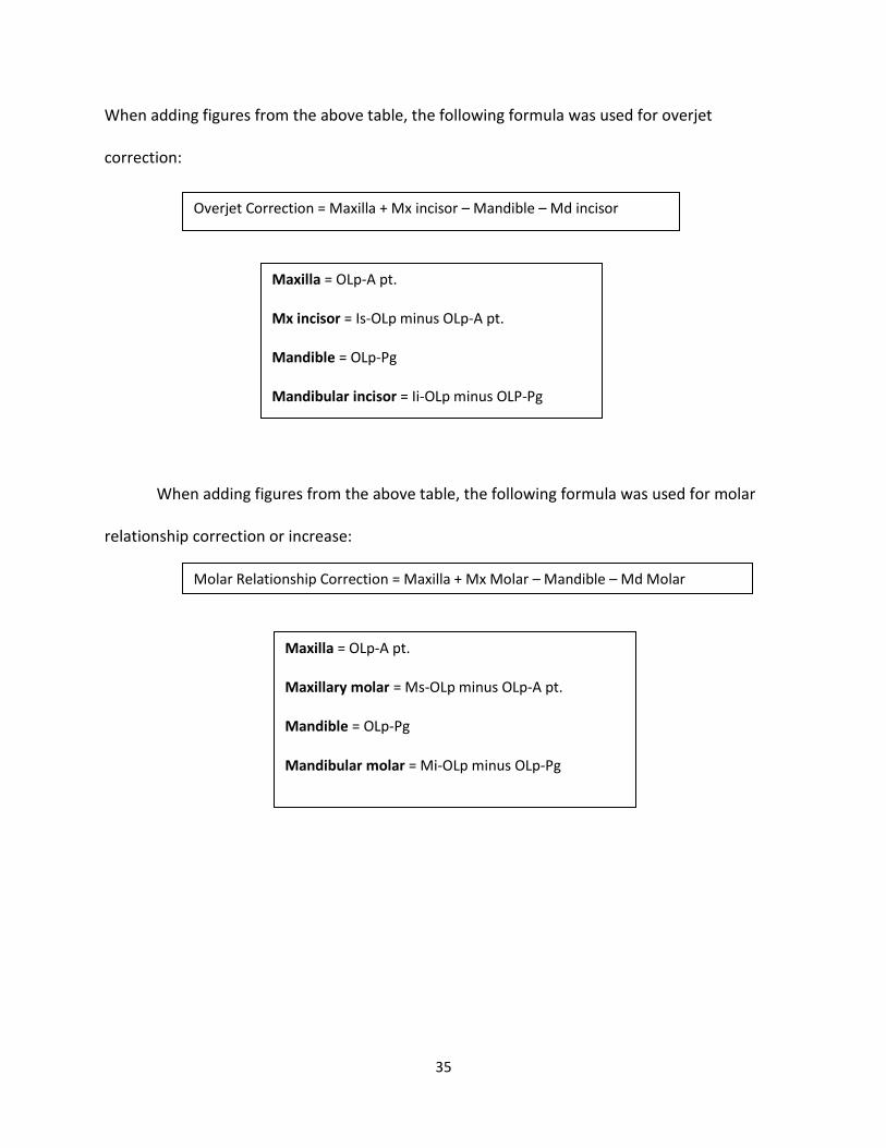
When adding figures from the above table, the following formula was used for overjet

correction:

When adding figures from the above table, the following formula was used for molar

relationship correction or increase:

Overjet Correction = Maxilla + Mx incisor – Mandible – Md incisor

Maxilla = OLp-A pt.

Mx incisor = Is-OLp minus OLp-A pt.

Mandible = OLp-Pg

Mandibular incisor = Ii-OLp minus OLP-Pg

Molar Relationship Correction = Maxilla + Mx Molar – Mandible – Md Molar

Maxilla = OLp-A pt.

Maxillary molar = Ms-OLp minus OLp-A pt.

Mandible = OLp-Pg

Mandibular molar = Mi-OLp minus OLp-Pg

36

Statistical Analysis

A paired t-test was used to compare T1 to T2, T2 to T3, and T1 to T3. This was used on

each variable to identify the overall treatment effects of the Modified Hyrax Expander in

combination with protraction facemask treatment. A level of significance of p<0.05 (95%

confidence interval) was used in this study.

In order to obtain the coefficient of reliability a measurement was made on the initial

tracing and another measurement was made on the same tracing 2 weeks later. When there

are two measurements, the coefficient of reliability is the correlation coefficient of the first and

second measurements. This information is found on Table 6. Correlation coefficients were

reported to determine how strongly the first measurements were associated with the second

measurements of each variable at every time period in six individuals.

37

Chapter IV: Results

Cephalometric Measurements

The measurements for each of the 25 variables were analyzed. The mean, standard

deviation, maximum and minimum for each variable measurement were recorded for each

time period (T1, T2, and T3). Table 8 shows the sagittal, vertical and angular measurements at

time periods T1, T2, and T3.

38

Table 8: Cephalometric Measurements

T1 T2 T3

Variable Mean S.D. Min Max Mean S.D. Min Max Mean S.D. Min Max

Sagittal:

Olp-A pt. 63.81 4.79 56.5 74.45 66.45 4.86 59.17 79.5 68.00 5.13 60 78.9

Olp-Pg 67.79 5.51 59.6 79.33 68.46 6.28 58.75 82.5 72.20 6.64 59.1 89.8

Is-Olp 66.54 6.46 58.5 83.98 70.34 7.12 61 90.68 73.68 7.77 58.78 90.92

Ii-Olp 68.45 6.52 60.22 86.55 68.12 6.89 58.68 86.9 71.79 6.67 61.2 86.47

Overjet -1.90 1.33 -4.37 2.2 2.22 1.40 -.95 5.1 1.93 1.89 -2.42 4.45

Ms-Olp 39.15 5.51 32 51.58 42. 10 4.59 35.62 53.3 44.96 5.70 36 56.26

Mi-Olp 41.59 5.25 34.27 55.05 42.83 5.16 36.84 54.3 45.92 5.72 38 58.26

Molar Relationship -2.39 1.49 -5.05 0.6 -0.72 2.23 -5.75 3.8 -0.96 1.79 -4.56 3.5

Wits -4.2 1.9 -8.4 0.0 -1.0 2.9 -7.1 4.4 -2.5 2.0 -6.8 1.7

Vertical:

Nasion-Apt 44.18 3.14 40.53 50.8 46.55 3.60 41 56.26 49.86 3.69 42.8 60

ANS-Me 52.87 4.56 44.75 63.67 56.08 5.96 46.71 70.23 56.85 5.61 46.85 68.23

Is-NL 22.26 3.27 17.61 31.32 23.59 4.06 18.23 35.4 24.61 3.91 18.73 33.59

Ii-ML 33.07 3.22 28.74 41.64 34.37 3.70 29.13 44.37 35.81 3.56 29.09 44.55

Overbite 1.98 1.61 -1.57 4.31 1.02 1.39 -2.2 2.95 1.42 1.70 -1.2 4.11

Msc-NL 14.11 2.25 10.86 19.4 15.61 2.81 12.3 24.6 16.12 2.79 11.12 23.56

Mic-ML 21.75 2.59 18.94 30.43 22.75 2.86 18.2 30.33 24.02 2.79 19.63 31.55

Angular:

SNA 80.39 4.33 70 90 80.78 3.86 75 86 80.17 3.96 74 87

SNB 80.56 4.28 72 91 78.82 3.98 70 85 79.30 3.37 72 85

ANB -0.26 2.24 -4 6 1.91 2.06 -3 5 0.73 2.61 -4 9

SNL-NL 7.30 3.92 1 17 7.39 3.61 3 16 8.04 3.72 2 18

SNL-ML 32.08 3.67 26 39 33.04 4.18 25 42 31.26 4.35 25 39

SNL-Ols 15.78 4.26 9 24 16.04 3.58 9 25 15.95 3.94 9 23

Is/NL 93.60 9.28 80 113 96.95 8.48 85 113 103.43 9.35 88 120

Ii/ML 87.13 6.67 82 106 85.21 7.61 71 102 90.39 10.12 74 115

Interincisal Angle 148.08 10.22 120 159 145.87 13.50 120 170 136 12.81 117 161

39

Cephalometric Changes of T2-T1, T3-T2, and T3-T1

Changes of cephalometric measurements in patients treated with protraction headgear

before treatment (T1), after treatment (T2) and 22 months after removal of the appliance (T3)

are shown in Table 9. Of the 25 variables investigated significant changes were found in most

of the variables.

Figures 5, 6, and 7 summarize the changes during treatment for T2-T1. Overjet and

sagittal molar relationships improved by an average of 4.1 mm and 1.8 mm, respectively.

Looking at Table 9, 7 out of 9 sagittal measurements were significant. The non-significant

measurements were OLp-Pg and Ii-OLp. This means that Pogonion did not have a significant

change between time points T2 and T1. The lower incisor inclination was also non-significant.

Vertical changes included an overbite decrease of 0.96 mm. This decrease in overbite was due

to primary teeth being exfoliated and permanent central incisors erupting during treatment. It

could also have been due to the Stainless Steel Crown used in the Modified Hyrax Expander. As

the patients wore the facemask, the maxilla grew downward and forward while the mandible

grew vertically as well. Only four out of the nine measurements for the Angular section were

statistically significant. The four that were statistically significant were: SNB, ANB, Is/SNL, and

Ii/ML. This shows that the mandibular prominence changed significantly between T2 and T1

measurements. Also, the maxillary incisor angulation changed significantly. Measurements

that were not statistically significant were SNA, SNL-NL, SNL-ML, SNL-OLs, and Is/Ii. A-point has

variability upon location and the palatal plane, occlusal plane, and mandibular plane did not

40

change significantly between T2 and T1. Also, the interincisal angle did not change significantly

during protraction facemask treatment.

Figures 8, 9, and 10 show the cephalometric changes 22 months after appliance removal

(T3-T2). Significant differences were found in 16 of the 24 variables. Over approximately 2

years, the maxilla continued to move forward by 1.5 mm, while the mandible moved forward

3.7 mm. Most of the overjet and molar correction, therefore, can be attributed to dental

movements. The mean overjet correction decreased by 0.3 mm and the mean molar correction

decreased by 0.2 mm. Wits decreased 1.4 mm showing skeletal relapse as well. There was a

big difference between the Vertical group T2-T1 and T3-T2 (Table 9). In the T2-T1 group, all of

the variables were statistically significant; however, only 4 out of 7 of the variables were

statistically significant for the T3-T2 group. The non-significant findings were ANS-Me,

Overbite, and Msc-NL. The overbite decreased an average of 0.40 mm and this was most likely

due to the error mentioned above. Under the Angular group, 5 of the 9 values were statistically

significant measurements (Table 9). They were ANB, SNL-ML, Is/SNL, Ii/ML, and Is/Ii. This

shows that the incisors changed angulation significantly and the mandibular plane angle also

changed significantly. This also shows that ANB had a significant change, but this change was a

negative number meaning there was skeletal relapse. ANB decreased an average of 1.17

degrees during the 22 months after appliance removal.

Figure 11, 12, and 13 shows the net changes for 9 months of treatment and 22 months

of observation (T3-T1). The maxilla moved forward 4.2 mm and the mandible moved forward

4.4 mm. The maxillary incisor tipped labially 2.9 mm and the mandibular incisor tipped lingually

41

1.1 mm, resulting in a net overjet correction of 3.8 mm. Mesial movement of the maxillary

molars was 1.6 mm, while the mandibular molars moved distally and average of 0.1 mm. A net

improvement in molar relationship was 1.7 mm, contributing to a 113% overall dental

correction for the mandibular molars. Most of the values found in Table 9 for T3-T1 were

statistically significant. This shows there was a positive net change over the 9 months of

treatment and 22 months of observation. Wits maintained a net correction of 1.6 mm. The

measurements that were not statistically significant are as follows: Overbite, SNA, SNL-NL, SNL-

ML, and SNL-OLs. The overbite is most likely non-significant because the primary incisors on

some patients could have exfoliated during treatment. This would have left the permanent

incisors in the process of erupting throughout treatment. SNA was insignificant due to the

difficulty in marking A point. The palatal plane, occlusal plane, and mandibular plane were not

significant for T3-T1. SNL-ML, or mandibular plane, was the only planed measurement to have

a significant finding at any time point measured in this study and it was between T3 and T2.

The values for T2-T1, T3-T2, and T3-T1 are listed below in Table 9.

42

Table 9: Comparison of T2-T1, T3-T1, and T3-T2

Variables T2-T1 T3-T2 T3-T1

Mean S.D P val sig Mean S.D. P val sig Mean S.D. P val sig

Sagittal:

Olp-A 2.6 1.5 .0001 * 1.5 2.0 .0014 * 4.2 2.3 .0001 *

Olp-Pg 0.6 2.7 .2571 NS 3.7 3.6 .0001 * 4.4 4.4 .0001 *

Is-Olp 3.8 2.4 .0001 * 3.3 3.1 .0001 * 7.1 4.1 .0001 *

Ii-Olp -0.3 2.5 .5371 NS 3.6 3.1 .0001 * 3.3 3.7 .0003 *

Overjet 4.1 2.1 .0001 * -0.3 1.7 .4374 NS 3.8 2.4 .0001 *

Ms-Olp 2.9 2.7 .0001 * 2.8 2.7 .0001 * 5.8 3.3 .0001 *

Mi-Olp 1.2 2.1 .0083 * 3.1 3.2 .0002 * 4.3 3.5 .0001 *

Molar Rel 1.6 2.0 .0007 * -0.2 1.6 .4860 NS 1.4 1.7 .0008 *

Wits 3.1 2.6 .0001 * 1.6 2.5 .0049 * -1.4 2.4 .0121 *

Vertical:

N-A 2.3 2.3 .0001 * 3.3 2.2 .0001 * 5.7 2.7 .0001 *

ANS-Me 3.2 2.7 .0001 * 0.7 2.6 .1733 NS 4.0 2.1 .0001 *

Is-NL 1.3 1.5 .0004 * 1.0 2.0 .0251 * 2.3 2.0 .0001 *

Ii-ML 1.3 1.2 .0001 * 1.4 1.2 .0001 * 2.7 1.1 .0001 *

Overbite -0.9 1.7 .0150 * 0.4 1.7 .2548 NS -0.6 1.9 .1659 NS

Msc-NL 1.4 1.6 .0002 * 0.5 1.8 .1886 NS 2.0 1.3 .0001 *

Mic-ML 1.0 1.4 .0035 * 1.3 1.4 .0003 * 2.3 1.6 .0001 *

Angular:

SNA 0.4 2.5 .4671 NS -0.6 2.2 .2002 NS -0.2 2.5 .6833 NS

SNB -1.7 2.4 .0021 * 0.4 1.7 .1848 NS -1.2 2.4 .0211 *

ANB 2.2 2.5 .0004 * -1.2 2.1 .0155 * 1 1.9 .0184 *

SNL-NL 0.1 2.2 .8549 NS 0.6 2.0 .1388 NS 0.7 2.1 .1012 NS

SNL-ML 0.9 2.6 .1020 NS -1.8 2.8 .0068 * -0.8 2.7 .1551 NS

SNL-OLs 0.3 3.2 .7064 NS -0.1 3.6 .9093 NS 0.2 4.3 .8470 NS

Is/SNL 3.3 5.9 .0131 * 6.5 6.3 .0001 * 9.8 8.7 .0001 *

Ii/ML -1.9 4.2 .0426 * 5.2 6.1 .0006 * 3.2 7.4 .0463 *

Is/Ii -2.2 8.8 .2403 NS -9.8 8.2 .0001 * -12.1 10.8 .0001 *

43

Overjet and Molar Relationship Correction

T2-T1:

Overjet Correction: Molar relationship:

Skeletal Contribution: Skeletal Contribution: 1) Maxilla 2.6 1) Maxilla 2.6 2) Mandible 0.7 2) Mandible 0.7 Dental Contribution: Dental Contribution: 3) Mx incisor 1.2 3) Mx molar 0.4 4) Md incisor -1.0 4) Md molar 0.5

Overjet Correction = 2.6+1.2-0.7-(-1.0)=4.1

Figure 5: Components of Overjet Correction T2-T1

Overjet Correction = Maxilla + Mx incisor – Mandible – Md incisor

4.1mm mmmmmmmmmm

1.9 mm

2.2mm

2.6mm

1.2mm

1.0mm

0.7mm

44

Molar Relationship Correction =2.6+.4-0.7-.5=1.8

Figure 6: Components of Molar Correction T2-T1

Molar Relationship Correction = Maxilla + Mx molar – Mandible – Md

molar

1.8mm

2.6mm

0.7mm

0.4mm

0.5mm

1.9mm

-0.1mm

45

The amount of skeletal and dental contribution to the overjet and molar relationship

correction for T2-T1 was calculated using the formulas in Table 7. The amount of overjet

correction was 4.1 mm. The amount of correction attributed to skeletal movement was 1.9 mm

or 46% and the amount of dental correction was 2.2 mm or 54%. The amount of molar

relationship correction was 1.8 mm. The skeletal correction was 105%, leaving -0.1 mm or -5%

attributed to dental movements. Calculations are shown for the overjet and molar relationship

correction above. Diagrams are also provided to illustrate the anterior and posterior

movements of the maxilla, mandible, maxillary incisors, mandibular incisor, maxillary molars,

and mandibular molars (Figures 5 and 6). A pitchfork analysis diagram describing the skeletal

and dental components of overjet and molar correction is shown in Figure 7.

46

Figure 7: Pitchfork Analysis for Overjet and Molar Correction T2-T1

4.1mm 100%

1.8mm 100%

1.9mm 46%

2.2mm 54%

1.9mm 105%

-0.1mm -5%

Maxilla 2.6mm 64%

Mandible 0.7mm -17%

Maxilla 1.2mm 29%

Mandible -1.0mm 24%

Maxilla

2.6mm 145%

Mandible 0.7mm -39%

Maxilla

0.4mm 22%

Mandible 0.5mm -28%

47

T3-T2:

Overjet Correction: Molar relationship:

Skeletal Contribution: Skeletal Contribution: 1) Maxilla 1.5 1) Maxilla 1.5 2) Mandible 3.7 2) Mandible 3.7 Dental Contribution: Dental Contribution: 3) Mx incisor 1.8 3) Mx molar 1.4 4) Md incisor -0.1 4) Md molar -0.6

Overjet Correction = 1.5+1.8-3.7-(-.1)=-0.3

Figure 8: Components of Overjet Correction T3-T2

Overjet Correction = Maxilla + Mx incisor – Mandible – Md incisor

48

Molar Relationship Correction =1.54+1.31-3.7-(-.61)=-0.24

Figure 9: Components of Molar Correction T3-T2

The amount of skeletal and dental contribution to the overjet and molar relationship

correction for T3-T2 was calculated using the formulas in Table 7. The amount of overjet

correction was -0.3 mm. The amount of correction attributed to skeletal movement was -2.2

mm or -733% and the amount of dental correction was 1.9 mm or 633%. The amount of molar

relationship correction was -0.2 mm. The skeletal correction was -2.2mm, or -1100%; leaving

Molar Relationship Correction = Maxilla + Mx molar – Mandible – Md

molar

49

2.0 mm or 1000% attributed to dental movements. Calculations are shown for the overjet and

molar relationship correction above. Diagrams are also provided to illustrate the anterior and

posterior movements of the maxilla, mandible, maxillary incisors, mandibular incisor, maxillary

molars, and mandibular molars (Figures 8 and 9). A pitchfork analysis diagram describing the

skeletal and dental components of overjet and molar correction is shown in Figure 10.

Figure 10: Pitchfork Analysis for Overjet and Molar Correction T3-T2

50

T3-T1:

Overjet Correction: Molar relationship:

Skeletal Contribution: Skeletal Contribution: 1) Maxilla 4.2 1) Maxilla 4.2 2) Mandible 4.4 2) Mandible 4.4 Dental Contribution: Dental Contribution: 3) Mx incisor 2.9 3) Mx molar 1.6 4) Md incisor -1.1 4) Md molar -0.1

Overjet Correction = 4.2+2.9-4.4-(-1.1)=3.8

Figure 11: Components of Overjet Correction T3-T1

Overjet Correction = Maxilla + Mx incisor – Mandible – Md incisor

51

Molar Relationship Correction =4.2+1.6-4.4-(-0.1)=1.5

Figure 12: Components of Molar Correction T3-T1

The amount of skeletal and dental contributions for T3-T1 net overjet and net molar

relationship correction were calculated using the formulas in Table 7. This T3-T1 shows the net

change that occurred over the length of the study which was 31months. The amount of net

overjet correction was 3.8 mm. This measurement is the result of the dental incisor correction

being 4 mm while the skeletal contributions were -0.2 mm. Reviewing the dental movements,

Molar Relationship Correction = Maxilla + Mx molar – Mandible – Md

molar

52

the maxillary incisors moved forward 2.9 mm and the mandibular incisors retruded 1.1 mm

resulting in a total dental correction of 4 mm. The net molar correction was 1.5 mm. The

maxillary molar moved forward 1.6 mm and the mandibular molar moved back 0.1 mm

resulting in a net dental correction for the molars of 1.7 mm. Diagrams and illustrations of

these findings are found above. A pitchfork analysis describing the net skeletal and dental

contributions to overjet and molar relationship correction is shown in the figure below.

Figure 13: Pitchfork Analysis for Overjet and Molar Correction T3-T1

53

Chapter V: Discussion

This research project was completed using patients treated at a young age with a

Modified Hyrax Expander designed by Dr. Kiebach and Facemask Treatment. It shows the

skeletal movements achieved at a young age and also shows the stability of these movements 2

years post-treatment. The maxilla has been shown to be in a retrusive position at an early

developmental age, so early treatment is advocated.48 Previous investigators, such as Saadia et.

al., Turley, Baccetti et. al., and Kapust have found that early treatment produces greater, faster

results with improved compliance. The ages in the early treatment groups were from 3 to 7

years of age. They found that changes were almost twice what older groups between 10 and

14 years gained.5,44,45 The ages compared with this study group were somewhat different. The

youngest treated age for this study was 4 years 4 months with an average of 6 years 2 months.

The oldest age was 10 years 4 months which falls within the classified older group in previous

research completed. Although the oldest age group in this study was over 10 years of age, the

CVM was 1 showing the study group had similar skeletal ages.

Guyer et al found that longitudinal data on Class III subjects showed them to have an

average maxillary growth of less than 1 mm per year and mandibular growth of 3-4.5 mm per

year. This study showed an average maxillary growth of 1.6 mm per year and an average

mandibular growth of 1.7 mm per year. This shows a vastly different skeletal growth pattern

between our treated group and the control study conducted by Guyer et al.5

Franchi et. al. investigated treatment timing for RME/FM. He compared early treatment

including deciduous and early mixed dentition with late treatment that were in the late mixed

54

dentition with erupting canines and premolars. He found that a significant maxillary movement

of about 2mm was maintained in the early treatment group.12 In our study, results following

active treatment showed a forward movement of the maxilla of about 2.6 mm which supports

previous findings. The mandibular forward movement was 0.7 mm during active treatment.

This calculates to an average mandibular growth of less than 1 mm per year. Comparing this to

the average mandibular growth in subjects not undergoing treatment which is approximately 3-

4.5 mm of mandibular growth per year, you will find a significant difference between the two.

The net changes found in this study showed a forward movement of the maxilla of 4.2 mm

between T1 and T3 time points. The mandible, however, did “catch up” with the maxilla having

a total forward movement of 4.4 mm. The mandible outgrew the maxilla, but only slightly,

indicating that a maxillary orthopedic change was achieved and maintained.

Franchi et al also found that early treatment maintained a maxillary/mandibular skeletal

relationship within 1 mm because of the significant favorable skeletal contributions of the

maxilla and mandible from RME/FM treatment.18 This study also supports these previous

findings because the total skeletal differential in growth of the maxilla and mandible over the

31 months of observation was that the mandible outgrew the maxilla by only 0.2 mm.

The average treatment age for this research group was 6 years 2 months at the

beginning of treatment. The 23 samples ranged from 4 years 4 months to 10 years 4 months.

During treatment, the overjet correction attained was 4.1 mm. Most of this correction was

maintained with a final overjet correction of 3.8 mm. This shows that the achieved results were

stable over the approximate 2 year post-treatment time span. The molar correction achieved

55

during treatment was 1.8 mm and the molar correction 22 months later was an average of 1.5

mm. The molar correction was stable after the appliances were removed over approximately 2

years.

The radiographs were traced by the same examiner to reduce error. The method of

cephalometric analysis by Pancherz22 was reliable and the error that did occur was within

acceptable parameters. The angular measurements showed that the Palatal Plane, Occlusal

Plane, and Mandibular Plane did not change significantly during treatment. The only significant

change occurred between T2 and T3 time points with the Mandibular Plane. Results showed a

decrease in the Mandibular Plane Angle which increased again for the time points T1 to T3.

This indicates that the mandibular plane Angle changed with treatment, but reverted back

toward pre-treatment averages by the 2 year post-treatment cephalogram.

Skeletal maturation and age differentiation was not addressed in this project because

the entire study sample size had a CVM I skeletal age for time point T1. There would have been

no difference in the results, so all patients were pooled together.

The research conducted evaluated an active treatment time of 9 months and a follow up

of approximately 2 years after active treatment. This was a long-term observational study

which was able to show skeletal and dental stability over time. The Wits measurement before

treatment was -4.2 and was –2.5 after 31 months of treatment and observation. This shows

that the skeletal correction achieved at a young age was maintained over the 2 years of

observation. Results support previous research advocating early treatment for Class III

malocclusions.

56

Chapter VI: Summary and Conclusions

Summary

The purpose of this study was to investigate skeletal and dental changes associated with

Dr. Kiebach’s Modified Hyrax Expander and Facemask treatment performed at an early age.

This was an observational study which consisted of 23 patients treated by one clinician.

The cephalometric study implored and analysis described by Byork21 and Pancherz.22

Angular cephalometric measurements were completed as well. A matched pairs t-test was

used to evaluate the findings. The following hypothesis were tested:

1. The patients treated with Dr. Kiebach’s Modified Hyrax Expander and facemask therapy

will not show significant dental movements.

2. The patients treated with Dr. Kiebach’s Modified Hyrax Expander and facemask therapy

will not show significant skeletal movements.

Conclusions

The hypothesis was rejected as the following statistically significant changes were

observed:

1. From T1 to T2: Sagittal movements were all statistically significant accept growth at

Pogonion and lower incisor inclination. Vertical movements were all statistically

significant. Statistically significant movements for the angular measurements were SNB,

ANB, Is/SNL and Ii/ML.

57

2. From T2 to T3: All sagittal movements were statistically significant accept the molar

relationship measurement. Vertically significant measurements were Nasion-A point, Is-

NL, Ii-ML, and Mic-ML. For the angular measurements, ANB, SNL-ML, Is/SNL, Ii/ML, and

Is/Ii were statistically significant.

3. From T1 to T3: All movements were statistically significant accept Overbite, SNA, SNL-

NL, SNL-ML, and SNL-OLs.

The net overjet and molar relationship movements showed a forward maxillary skeletal

movement of 2.6 mm in 9 months with RME/FM treatment. There was a continuation of

forward maxillary movement the next 22 months of 1.5 mm. The results show a combination

of skeletal and dental contributions to the Class III malocclusion treated. The treatment at a

young age showed stable results post treatment.

Overall, the net overjet corrections observed by T2-T1 were 46% skeletal and 54% dental.

However, comparing the T3-T1 overjet measurements, the skeletal contribution was -5% and

the dental contributions were 105%. This shows that over time, the class III growth pattern

remained and the skeletal corrections achieved were masked over time by a continued Class III

growth. The same is true for the net molar correction. For T2-T1, the net molar correction was

105% skeletal and -5% dental. By the time the 22 month post-treatment cephalogram was

taken for T3-T1, the skeletal correction was -13% and the dental correction was 113%. Skeletal

changes included forward movement of the maxilla and limited movement of the mandible for

T2-T1. Dental changes for the same time points included proclination of the maxillary incisors

while the mandibular incisors retroclined. Maxillary molars mesialized and average of 0.4 mm

58

while the mandibular molars continued to move forward 0.5 mm. The net change (T3-T1)

shows the maxilla moved forward 4.2 mm and the mandible moved forward 4.2mm. The

maxillary incisors tipped labially 2.9 mm while the mandibular incisors tipped lingually 1.1 mm.

The maxillary molars mesialized 1.6 mm and the mandibular molars distalized 0.1 mm.

59

Chapter VII: Recommendations for future research

1. The treatment group should be compared to a control group matched in skeletal age

and also traced by the same investigator.

2. This study should have a bigger treatment group sample.

3. The sample should have a more narrow age range rather than T1 ages varying from

4 years 4 months to 10 years 4 months.

4. The T3 long term results should be more concise rather than having a wide range for

T2-T3. This time measurement varied from 3 months to over 4 years. A more

consistent post-treatment reference would give the reader a more reliable result.

5. A 3D cone beam study on patients treated with the Modified Hyrax Expander and

Facemask treatment would allow the study of the effects of the devices in 3

dimensions.

60

Reference List:

1. Prevalence of malocclusion and orthodontic treatment need in the United States:

Estimates from the NHANES III Survey. Int J Orthod 1998;13 (2):97-106

2. Ngan P: Early treatment of Class III malocclusion: Is it worth the burden? Am J Orthod

Dentofac Orthop 2005; 129:S82-S85

3. Guyer EC, Ellis EE, McNamara JA et al: Components of Class III malocclusions in juveniles

and adolescents. Angle Orthod 1986 56:7-30

4. Bailey L, Turvey T, Sarver D, Proffit W, Chapter 16: Class III Problems, Proffit W, White R,

Sarver D: Contemporary Treatment of Dentofacial Deformity. 2003; 507-555

5. Baccetti, T: Craniofacial changes in Class III malocclusion as related to skeletal and

dental maturation. Am J Orthod Dentofac Orthop 2007; 132(2):171-178

6. Vaughn G: The effects of maxillary protraction therapy with or without rapid palatal

expansion: A prospective, randomized clinical trial. Am J Orthod Dentofac Orthop

2005; 128 (3): 299-309

7. Turley: Managing the developing Class III malocclusion with palatal expansion and

facemask therapy. Am J Orthod Dentofac Orthop 2002; 122(4):349-352

8. Baccetti, T: Skeletal effects of early treatment of Class III malocclusion with maxillary

expansion and facemask therapy. Am J Orthod Dentofac Orthop 1998; 113(3):333-343

9. Haas AJ: Treatment of maxillary deficiency by opening the midpalatal suture. Angle

Orthod 1965;65:200-217

10. Haas AJ: Palatal expansion: just the beginning of dentofacial orthopedics. Am J Orthod

1970;57:219-255

61

11. McNamara JA Jr, Brudon WL. Orthodontic and orthopedic treatment in the mixed

dentition. Ann Arbor, Mich.: Needham Press, 1993

12. Franchi L; Postpubertal assessment of treatment timing for maxillary expansion and

protraction therapy followed by fixed appliances. Am J Orthod Dentofac Orthop 2004;

126(5):555-568

13. Proffit WR, Fields HW Jr. Contemporary orthodontics 3rd ed. Saint Louis: Mosby; 2000

14. McNamara JA Jr, Brudon WL. Orthodontics and dentofacial orthopedics. Ann Arbor:

Needham Press; 2001

15. Vaik HS. Clinical results of maxillary protraction in Korean children. Am J Orthod

Dentofac Orthop 1995; 108:583-592

16. Merwin D, Ngan P, Hagg U, YiuC, Wei SH. Timing for effective application of anteriorly

directed orthopedic force to the maxilla. Am J Orthod Dentofac Orthop 1997;112:292-9

17. Kapust AJ, Sinclair PM, Turley PK. Cephalometric effects of facemask/expansion therapy

in Class III children: a comparison of three age groups. Am J orthod Dentofac orthop

1998;113:204-12

18. Saadia M, Torres E. Sagittal changes after maxillary protraction with expiation in Class III

patients in the primary, mixed, and late mixed dentitions: a longitudinal retrospective

study. Am J orthod Dentofac Orthoped 2000; 117:669-80

19. Baccetti T, Franchi L, McNamara JA Jr. Treatment and post-treatment craniofacial

changes after rapid maxillary expansion and facemask therapy. Am J Orthod Dentofac

Orthop 2000; 118:404-13

62

20. Cha KS. Skeletal changes of maxillary protraction in patients exhibiting skeletal Class III

malocclusion: a comparison of three skeletal maturation groups. Angle orthod

2003;73:26-35

21. Bjork A: The face in Profile: An anthropological X-ray investigation of Swedish Children

and Conscripts. Lund: Berlingska Boktrycheriet 1947; 40: 58

22. Pancherz H: The mechanism of Class II correction in Herbst appliance treatment, a

cephalometric investigation. Am J Orthod 1982; 82: 107-113

23. Ngan P: Early Timely Treatment of Class III Malocclusion. Seminars in Orthod:11:140-

145, 2005

24. Newman GV: Prevalence of malocclusion in children 6-14 years of age and treatment in

preventable cases. Am J Dent Assoc 52:566-575, 1956

25. Thilander B, Myrberg N: The prevalence of malocclusion in Swedish school children.

Scand J Dent Res 81:12-20, 1973

26. Tschill P, Bacon W, Sonko A: Malocclusion in the deciduous dentition of Caucasian

children. Eur J Orthod 19:361-367, 1997

27. Ishii H, Morita S, Takeuchi Y, et al: Treatment effect of combined maxillary protraction

and chincap appliance in severe skeletal Class III cases. Am J Orthod Dentofacial Orthop

92:304-312, 1987

28. Allwright WC, Burndred WH: A survey of handicapping dentofacial anomalies among

Chinese in Hong Kong. Int Dent J 14:505-519, 1964

29. Lauweryns I, Carels C, Vlietinck R. The use of twins in dentofacial genetic research.

Am J Orthod Dentofac Orthop 103:33-38,1993

63

30. Proffit WR, Fields HW Jr., Sarver DM, Contemporary orthodontics 4th ed. 141-147 Saint

Louis: Mosby Elsevier; 2007

31. McGuigan DG. The Hapsburgs. London, WH Allen, 1966

32. Lee KG, RyuYK, Park YC, Rudolph DJ: A study of holographic interferometry on the

initial reaction of maxillofacial complex during protraction. Am J Orthod Dentofac

Orthoped 1997; 111:623-32

33. Miyajima K, McNamara JA Jr, Kimura T, Murata S, Iizuka T. An estimation of

craniofacial growth in the untreated Class III female with anterior crossbite. Am J

Orthod Dentofacial Orthop 1997; 112:425-34

34. Enrich RE, Brodie AG, Blayney JR: Prevalence of Class I, Class II, and Class III

malocclusions in an urban population: An epidemiological study. J Dent Res

1964;44:947

35. Battagel, JM. The aetiological factors in Class III malocclusion. Eur J Orthod 1993; 15(5):

347-370

36. McIntyre GT: Treatment planning in Class III Malocclusion. Dent Update 2004; Jan-

Feb 31(1): 13-20

37. Bagatel JM: Class III malocclusion; postretention findings. European J of Orthod

1993;15:45-55

38. Murakami C, Hiyama S, Ohayama K: Craniofacial morphology of skeletal Class III

patients. World J Orthod 2005; Spring; 6(1):51-60

39. Macdonald, KE, Kapust AJ, Turley, PK: Cephalometric changes after the correction of

Class III malocclusion with maxillary expansion/facemask therapy. Am J Orthod

Dentofac Orthop 1999, 116(1):13-24

64

40. Graber TM: Orthodontics: principles and practice. Philadelphia:WB Saunders

Company; 1966

41. McNamara JA: Mixed dentition treatment. In: Graber TM, Vanarsdall RL, eds.

Orthodontics. Current principles and techniques, 2nd

ed. St. Louis: Mosby Year Book,

1994; 507-541

42. Tanne K, HiragaJ, Sakuda M: Effects of directions of maxillary protraction forceson

biomechanical changes in craniofacial complex. Europ J Orthod 1989;11:382-395

43. Ngan P: Biomechanics of maxillary expansion and protractioin in Class III patients. Am J

Orthod Dentofacial Orthop 2002, 121:582-583

44. Saadia M, Torres E: Sagittal changes after maxillary protraction with expansion in Class

III patients in the primary, mixed and late mixed dentitions: A longitudinal retrospective

study. Am J Orthod Dentofac Orthop 2000; 117(6):669-680

65

Appendix A

Treatment age for T1, T2 and T3

Treatment Age

T1 T2 T3 4y 11m 5y 7m 6y 1m

7y 8m 9y 5m 11y 6m

4y 6m 5y 6m 6y 10m

4y 10 m 5y 6m 7y 5m

10y 4 m 11y 2m 11y 7m

6y 6m 7y 2m 9y 10m

5y 3m 6y 1m 7y 9m

5y 5y 5m 7y 6m

8y 2m 9y 1m 11y 4m

10y 1m 10y 7m 12y 6m

4y 1m 6y 2m 8y 8m

6y 5m 7y 9m 10y 2m

5y 7m 6y 1m 8y 11m

6y 10m 7y 5m 9y 8m

5y 2m 5y 9m 7y 7m

8y 7m 8y 10m 9y 11m

6y 6m 6y 9m 7y 11m

4y 4m 5y 2m 7y 4m

4y 11m 5y 7m 6y 6m

5y 1m 5y 11m 9y 11m

6y 2m 7y 2m 10y 10m

5y 2m 5y 10m 6y 5m

5y 2m 6y 2m 7y 1m

66

TREATMENT OF CLASS III MALOCCLUSION IN THE

PRIMARY AND EARLY MIXED DENTITION USING

THE KIEBACH APPLIANCE AND PROTRACTION

FACEMASK

Erica W Reed, DDS

Thesis Submitted to the

School of Dentistry at West Virginia University

In partial fulfillment of the requirements

For the degree of Master of Science

APPROVAL OF EXAMINING COMMITTEE

_________________________

Peter Ngan, D.M.D., Chairman

_________________________

Chris Martin, D.D.S., M.S.

_________________________

Thomas Razmus, D.D.S., M.S.

______________________

Date


Recommended