Thlorax (1962), 17, 342. TREATMENT OF SPONTANEOUS PNEUMOTHORAX BY WILLIAM G. SMITH* AND PETER P. G. ROTHWELLt From Sully Hospital (Thoracic Centre), Penarth, Glamorgan The term pneumothorax was first used by Itard in 1803, and in 1826 Laennec first described the clinical features. For many years spontaneous pneumothorax was thought to be tuberculous in origin, and as late as 1931 Palmer and Taft reviewed 70 published cases and added five of their own. They stated that the cause was ulceration of the visceral pleura by a subpleural tuberculous focus in 80 to 90% of adults and in 40 to 50% of children. Kjaergaard (1932), in an important study, described 51 patients followed for three to 18 years. He stressed that few of these patients had cough, sputum, pyrexia, or other evidence of pulmonary tuberculosis, and this disease subse- quently developed in only one of 49 patients. He thought that the pneumothorax resulted from the rupture of a non-tuberculous air vesicle or bleb and suggested the term " pneumothorax simplex " for the occurrence of spontaneous pneumothorax in apparently healthy individuals. This opinion has been confirmed by many workers since then. In the many papers on the subject the main points of discussion have usually centred around treatment, recurrence rates, and possible tubercu- lous aetiology. Interpretation is often difficult because of small numbers of patients and relatively short periods of follow-up. There are few adequate reports in which it is possible to assess any particular form of treatment. The present study is concerned mainly with the efficacy and place of iodized talc pleurodesis. MATERIAL AND METHODS The present series consists of 150 patients admitted to hospital between 1944 and 1960. One hundred and eighteen patients were treated in Sully Hospital, the regional thoracic centre of South Wales; 25 patients were treated in Llandough Hospital and seven in the East Glamorgan Hospital, both general hospitals in the vicinity of Cardiff. All patients were treated by the same consultant thoracic surgeons. One hundred and thirty-three patients have been traced, and these have answered a postal questionnaire and have been examined either by the authors, by the chest clinic physicians, or, in a few instances, by the family * Now at Perth Chest Hospital, Shenton Park, Western Australia. t Now at Hamilton Public Hospital, New Zealand. practitioner. A 1960 chest radiograph was obtained in 133 patients. Two of the original films could not be traced. The pneumothorax was spontaneous in all cases and of the benign " simplex " variety in eighty- four. No pneumothorax was included due to gross extrapulmonary or pulmonary disease, such as pneumonia, infarction, neoplasm, honeycomb lungs, active tuberculosis, trauma, or oesophageal or sub- phrenic lesions. ANALYSIS Table I shows the composition of the series and the age and sex incidence. One man aged 70 died in hospital as a result of the pneumothorax. He TABLE I SPONTANEOUS_PNEUMOTHORAX Admissions 1944 to 1960 No. of patients Male Female Ages: 14 to 79 years (average 40 years) Occupation: Heavy. Medium to light Deaths .. . . . Pneumothorax (in hospital) ? Pneumothorax (at home) Unrelated causes (at home) 173 150 120 (80%) 30 (20%) 37 113 10 I 8 had severe obstructive emphysema with a greatly reduced respiratory reserve and died a few days after admission despite re-expansion of the lung by an intercostal tube and suction. Another patient died at home, probably from tension pneumo- thorax, although no necropsy was made. Of the 43 known Mantoux reactions, nine were negative. Table II shows the age groups concerned. The rather high percentage in the 40 to 60 age group may be explained by the relatively high incidence of chronic bronchitis and emphysema in this study, as in the series of Crowther (1955). TABLE II AGE INCIDENCE Age -2 No. of patients 7 Percentage 5 o 21-30 31-40 41-50 51-60 61-70 71-80 Total 40 27 37 18 16 5 150 26 18 25 12 11 3 100 Table III indicates the increased numbers of patients referred for treatment during the past five years. This reflects the trend towards more active treatment in recent years. on 24 May 2019 by guest. Protected by copyright. http://thorax.bmj.com/ Thorax: first published as 10.1136/thx.17.4.342 on 1 December 1962. Downloaded from
Transcript
Thlorax (1962), 17, 342.
TREATMENT OF SPONTANEOUS PNEUMOTHORAXBY
WILLIAM G. SMITH* AND PETER P. G. ROTHWELLtFrom Sully Hospital (Thoracic Centre), Penarth, Glamorgan
The term pneumothorax was first used by Itardin 1803, and in 1826 Laennec first described theclinical features. For many years spontaneouspneumothorax was thought to be tuberculous inorigin, and as late as 1931 Palmer and Taftreviewed 70 published cases and added five oftheir own. They stated that the cause wasulceration of the visceral pleura by a subpleuraltuberculous focus in 80 to 90% of adults and in40 to 50% of children.
Kjaergaard (1932), in an important study,described 51 patients followed for three to 18years. He stressed that few of these patients hadcough, sputum, pyrexia, or other evidence ofpulmonary tuberculosis, and this disease subse-quently developed in only one of 49 patients. Hethought that the pneumothorax resulted from therupture of a non-tuberculous air vesicle or bleband suggested the term " pneumothorax simplex "for the occurrence of spontaneous pneumothoraxin apparently healthy individuals. This opinionhas been confirmed by many workers since then.
In the many papers on the subject the mainpoints of discussion have usually centred aroundtreatment, recurrence rates, and possible tubercu-lous aetiology. Interpretation is often difficultbecause of small numbers of patients and relativelyshort periods of follow-up. There are fewadequate reports in which it is possible to assessany particular form of treatment. The presentstudy is concerned mainly with the efficacy andplace of iodized talc pleurodesis.
MATERIAL AND METHODSThe present series consists of 150 patients admitted
to hospital between 1944 and 1960. One hundred andeighteen patients were treated in Sully Hospital, theregional thoracic centre of South Wales; 25 patientswere treated in Llandough Hospital and seven in theEast Glamorgan Hospital, both general hospitals inthe vicinity of Cardiff. All patients were treated bythe same consultant thoracic surgeons. One hundredand thirty-three patients have been traced, and thesehave answered a postal questionnaire and have beenexamined either by the authors, by the chest clinicphysicians, or, in a few instances, by the family
* Now at Perth Chest Hospital, Shenton Park, Western Australia.t Now at Hamilton Public Hospital, New Zealand.
practitioner. A 1960 chest radiograph was obtainedin 133 patients. Two of the original films could notbe traced. The pneumothorax was spontaneous in allcases and of the benign " simplex " variety in eighty-four. No pneumothorax was included due to grossextrapulmonary or pulmonary disease, such aspneumonia, infarction, neoplasm, honeycomb lungs,active tuberculosis, trauma, or oesophageal or sub-phrenic lesions.
ANALYSISTable I shows the composition of the series and
the age and sex incidence. One man aged 70 diedin hospital as a result of the pneumothorax. He
TABLE ISPONTANEOUS_PNEUMOTHORAX
Admissions 1944 to 1960No. of patientsMaleFemale
Ages: 14 to 79 years (average 40 years)Occupation: Heavy.
Medium to lightDeaths .. . . .Pneumothorax (in hospital)? Pneumothorax (at home)Unrelated causes (at home)
173150120 (80%)30 (20%)
3711310
I8
had severe obstructive emphysema with a greatlyreduced respiratory reserve and died a few daysafter admission despite re-expansion of the lung byan intercostal tube and suction. Another patientdied at home, probably from tension pneumo-thorax, although no necropsy was made. Of the43 known Mantoux reactions, nine were negative.Table II shows the age groups concerned. The
rather high percentage in the 40 to 60 age groupmay be explained by the relatively high incidenceof chronic bronchitis and emphysema in this study,as in the series of Crowther (1955).
TABLE IIAGE INCIDENCE
Age -2
No. of patients 7Percentage 5
o 21-30 31-40 41-50 51-60 61-70 71-80 Total
40 27 37 18 16 5 15026 18 25 12 11 3 100
Table III indicates the increased numbers ofpatients referred for treatment during the past fiveyears. This reflects the trend towards more activetreatment in recent years.
on 24 May 2019 by guest. P
rotected by copyright.http://thorax.bm
j.com/
Thorax: first published as 10.1136/thx.17.4.342 on 1 D
Table IV details associated chest disease. Thesputa of the eight patients with fibrocalcific upperlobe lesions were bacteriologically negative foracid-fast bacilli at the time of the pneumothorax,and only one patient had been treated previouslyfor pulmonary tuberculosis.
TABLE IVASSOCIATED CHEST DISEASE
Disease No. of Patients
Nil-" pneumothorax simplex "Chronic bronchitisChronic bronchitis, severe emphysema±bullaeBullae (radiograph, operation or thoracoscopy)Asthma±bronchitisBronchiectasisPneumoconiosisFibro-calcific upper lobe lesions ..
84381014825 (P.M.F. in 3)8
Nine patients had a chronic pneumothorax ofthree or more months' duration. In four patientsa haemopneumothorax was present. Increasedtension, as judged by clinical criteria in the pleuralcavity, was present in four patients.Table V shows the side and frequency of
pneumothorax. The right side was involved more
frequently than the left. Seventy-one per cent. ofpatients presented with the first pneumothorax.
viscus. The onset of symptoms was gradual inapproximately 20%. In the six patients withhaemoptysis, one had a fibrocalcific lesion in theright upper lobe; another had an emphysematousbulla, and no obvious cause was found in theremainder. Many patients were unable to pinpointthe onset of the attack to any particular activity,and only seven patients were undertakingstrenuous effort (Table VII). It seems probable
TABLE VIIACTIVITY AT THE TIME OF ATTACK (IF KNOWN)
Getting out of bed 2Moving in bed .. .. 2Cycling .. .. 2Golfing .. .. 2Stumbling .. ..
Sneezing .. ..
No unusual activity 30
that the symptoms of cough and sneeze may havebeen the result rather than the cause of pneumo-
thorax. The radiological findings are detailed inTable VIII.
T,ABLE VIIIRADIOLOGICAL FINDINGS
Forty-three patients (29 %) had more than one
proved pneumothorax. Fifteen patients (10 %)had bilateral episodes but these were simultaneousin only one. The figure for multiple pneumo-thoraces is an underestimate, as probable butunproved pneumothoraces have been excluded,and some of the 17 untraced patients may havehad recurrences. One patient, however, had atleast 10 proved recurrences, and probably hadthree or four unproved episodes.Symptoms are detailed in Table VI. It is of
interest that only one patient was entirely free ofsymptoms, and the condition was diagnosedincidentally by a chest radiograph. Pain was lesscommon than dyspnoea but usually preceded it.One patient presented with abdominal pain andnearly had a laparotomy for a possible perforated
Size of pneumothorax: SmallModerateLargeComplete
Fluid: TraceModerateLarge
Adhesions (including one after unsuccessfulpoudrage)
Bullae: Definite
Probable
"Emphysema" (3 bullous)Pneumoconiosis (3 with P.M.F.)Fibro-calcific lesions.
Bronchiectasis
Calcified pleura
286828222010
1498115
8ll
TREATMENT GROUPSTALC.-The method of poudrage with iodized
talc insufflation combined with thoracoscopy hasbeen used at Sully Hospital since 1950 in a totalof 62 patients, 20 of whom had previous pneumo-
thoraces. General anaesthesia has always been
Year
343
on 24 May 2019 by guest. P
rotected by copyright.http://thorax.bm
j.com/
Thorax: first published as 10.1136/thx.17.4.342 on 1 D
used, and, as with any other technical procedure,care and skill are important in the successful useof this method of treatment. Two intercostalcannulae are inserted, one in the second interspaceanteriorly and the other in the sixth or seventhinterspace in the axilla. After thoracoscopy andinspection of the collapsed lung, 2 to 5 g. of 1%iodized talc is insufflated widely and evenlythrough both cannulae: a satisfactory spread isconfirmed by repeat thoracoscopy. An intercostalcatheter is inserted through the lower cannula andallowed to drain with an underwater seal.
If the lung does not expand immediately, suctionis required. The catheter is removed 48 to72 hours after lung expansion is radiologicallycomplete, depending on the amount of drainage.The average period of intubation in this series wasjust over three days. Careful management of thecatheter is essential for the success of the method.Active breathing exercises and posture on thenormal side are started as soon as the intercostaltube is removed. In some patients a fair degree ofpain followed recovery of consciousness, andpethidine or other analgesic was sometimes neededmore than once, though most patients did not havemuch pain. One patient had an iodine hyper-sensitivity reaction six days after poudrage. Mostpatients had a mild or moderate pyrexial reactionlasting four to seven days. Variable, but usuallysmall, amounts of fluid have been drained by theintercostal tube, in a few instances amounting toover one litre, indicating a marked pleuralreaction. Very occasionally aspiration of fluidhas been necessary after removal of the intercostaltube, since usually the pleural reaction rapidlyclears on the radiograph, and within a few weeksvery little fluid can be seen in the pleural space.Within a few months there is usually little radio-logical evidence of the previous poudrage. Aftersix months, in 19 patients there was no significantradiographic change: this included one patientwho had had bilateral thoracoscopy and poudrage.In 25 patients there was a slight degree ofobliteration of the costophrenic angle. In 15patients the changes were moderate, and in onlytwo was pleural thickening and elevation of thediaphragm fairly marked.
Lesions seen at thoracoscopy included bullae insix cases, adhesions in two, a doubtful caseouslesion in one, and the " cuckoo spit " appearancedescribed by Brock (1948) in one.
BED REST.-Forty-four patients had bed restwithout any active treatment. The majority ofthese patients had shallow pneumothoraces.
INTERCOSTAL TUBE.-Eighteen patients weretreated with an intercostal catheter, with orwithout suction. The tube was usually insertedanteriorly in the second space or high in theaxilla just behind the pectoral fold. In one patientfresh tubes had to be re-inserted on 12 differentoccasions due to a recurrence of pneumothoraxafter removal of the tube or to technical difficulties.
THORACOTOMY.-Fourteen patients had majorsurgery. In five patients thoracotomy was advisedbecause of large bullae or cysts seen by thethoracoscope or radiographically. Excision andoversewing of bullae was performed in thesepatients. Two patients had decortication becauseof chronic pneumothorax and a third because ofan empyema following a previous camphorpleurodesis. Another patient had decorticationand poudrage with iodized talc following aninfected haemopneumothorax. One patient hadparietal pleurectomy. Four patients had athoracotomy with slight mechanical scarificationof the lungs and iodized talc poudrage; in twonumerous small bullae were seen, but in the othertwo no cause of pneumothorax was evident.
CAMPHoR.-Camphor pleurodesis (I to 1 %camphor in oil) using a syringe and needle wasperformed in six patients. One patient had twoseparate injections on one side and one furtherinjection on the other side.
SILVER NITRATE.-Silver nitrate was triedinitially but discontinued because of the severityof local pain.
RESULTSTable IX shows the results in the different
treatment groups correlated with average stay inhospital and incidence of previous pneumo-thoraces. Six patients are not included in thesetreatment groups as they were treated with silvernitrate pleurodesis or had air aspirations inaddition to bed rest.Table X shows the period of follow-up in each
group. Sixty-one of the 62 patients treated withiodized talc have been traced. Three patients (5 %)have had recurrences five years, 21 months, andone month after treatment. In the first patient,the intercostal tube was removed after 24 hours,thereby allowing the fairly large reactive effusionto keep the pleural layers apart, so vitiating theeffects of the pleural irritation. The second patienthad an unusual minute recurrence in the hori-zontal fissure 21 months after poudrage. A thin,
344
on 24 May 2019 by guest. P
rotected by copyright.http://thorax.bm
j.com/
Thorax: first published as 10.1136/thx.17.4.342 on 1 D
Thoracoscopy and poudrageBed rest onlyIntercostal tubeThoracotomyCamphor pleurodesisChronic pneumothorax
402200
9I5000
4
l
20
1422200
1241000
643200
82I030
transverse, translucent area was visible on thechest radiograph following an episode of slightright chest pain and had disappeared on a repeatfilm taken a few days later. This trivial recurrenceimplied good pleural adhesion elsewhere. In thethird patient technical reasons for insufficientpoudrage, noted at the time, could well be heldresponsible for the recurrence. A satisfactoryrepeat poudrage was performed one month later,and there has been no recurrence during thesubsequent year. A fourth patient is not includedas a recurrence because lung expansion afterpoudrage was never achieved due to technicaldifficulties with the intercostal tube and suction.Thoracotomy was performed 16 days afterpoudrage. Apart from the two patients notedabove, poudrage was performed on the oneoccasion only.
In the bed-rest series, only 29 of the 44 patientshave been traced. Five (9%) recurrences havedeveloped. Subsequently three had further periodsof bed rest and two had thoracotomies.Three of the 18 cases treated by intercostal tube
had recurrences. Subsequently one was treated bybed rest, one had poudrage with iodized talc, andthe other had a right pulmonary resection forpulmonary tuberculosis, followed a few monthslater by a left thoracotomy and poudrage forrecurrent left pneumothorax.One patient treated by thoracotomy and excision
of bullae had a recurrence approximately one yearlater, which was treated by bed rest at home. It isnot known whether this occurred on the same oropposite side. He has remained well for five yearssince the second episode.
2B
3 0 15 5 21 23 0 21l 0 l
0 10 0 0 0 2 0 0 1
1 0 0 2 0 0 1
2-85.42 54-74-89-1
The results of camphor pleurodesis (six cases)were unimpressive. One had to have a thoraco-tomy because of subsequent empyema. Of theremainder, one patient, after bilateral camphorinjections, continued to have recurrences and hadthoracotomy for pulmonary tuberculosis on oneside and a thoracotomy and poudrage on theother. Another patient had a recurrence on thesame side three years later and was treated bythoracoscopy and poudrage. In addition tocamphor pleurodesis, he had been treated forhaemopneumothorax on the same side two yearsearlier. Despite these two episodes, the pleura wasobviously not adherent.Table XI shows the details of the recurrences.
Table IX shows that the poudrage group had amuch higher incidence of previous episodes thanthe other groups. In addition, the pneumothoraxwas more often large, which frequently indicates alarger air leak.
HAEMOPNEUMOTHORACES (4 cases).-One patientwas treated by thoracotomy at the time of theinitial episode. One had thoracotomy laterbecause of a chronic pneumothorax. One patientwas treated by intercostal tube and one byaspiration and blood transfusion. None of thesepatients has had a recurrence to date.CHRONIC PNEUMOTHORACES (9 cases).-The
details are shown in Table XII. Thoracotomy wasperformed in only two of the nine cases. Theothers were treated by bed rest and air aspiration(3); by aspiration, thoracoscopy, and poudragewith silver nitrate and kaolin (2); and by inter-costal tube and high suction in the remainder (2).
3 1S
on 24 May 2019 by guest. P
rotected by copyright.http://thorax.bm
j.com/
Thorax: first published as 10.1136/thx.17.4.342 on 1 D
There have been no recurrences in the eightpatients followed over a mean period of 9.1 years.In all patients the chronic pneumothorax was thefirst episode. Weight loss was a fairly frequentsymptom (3 patients), as noted by Brock (1948).DEVELOPMENT OF TUBERCULOSIS.-Five patients
were subsequently found to have active tubercu-losis (Table XIII). In three patients radiologicalupper lobe disease was present on admission at thetime of the initial pneumothorax. The sputumwas then negative but subsequently becamepositive. In two patients infiltration of the rightupper lobe subsequently developed some timeafter the initial spontaneous pneumothorax, atwhich time the radiograph was clear.
PERIOD IN HOSPITAL. As lung re-expansion isusually complete within 12 to 24 hours afterthoracoscopy and poudrage, it had been expectedthat the average stay in hospital of these patientswould prove considerably shorter than that of thepatients treated by bed rest alone. This was notthe case (Table IX), although many patients werein fact discharged seven to 12 days after treatment.Several, however, were retained in hospital forperiods of up to 100 days for investigationregarding tuberculous activity, social reasons, ortreatment of chronic bronchitis. When theseconsiderations were excluded, a period of 10 to12 days in hospital and return to work one weeklater was the rule. In contrad.stinction, thepatients treated by other methods spent longerin hospital.
346
No. ofPatients
Time sinceLast Episode
(yr.)Final
Treatment
No. ofPatients
on 24 May 2019 by guest. P
rotected by copyright.http://thorax.bm
j.com/
Thorax: first published as 10.1136/thx.17.4.342 on 1 D
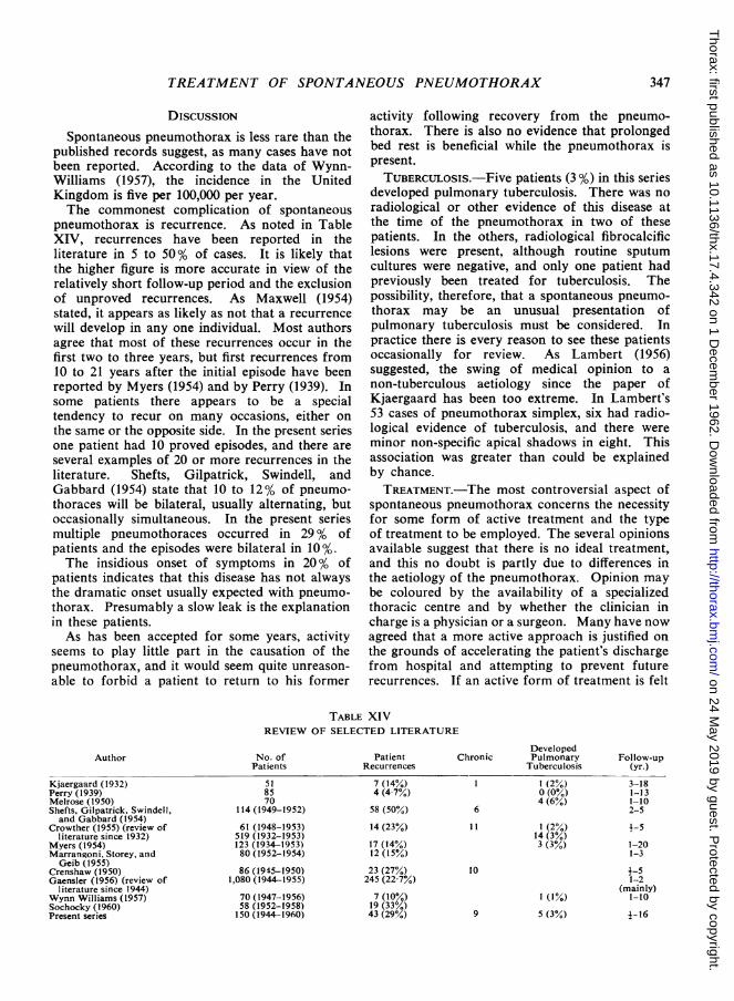
Spontaneous pneumothorax is less rare than thepublished records suggest, as many cases have notbeen reported. According to the data of Wynn-Williams (1957), the incidence in the UnitedKingdom is five per 100,000 per year.The commonest complication of spontaneous
pneumothorax is recurrence. As noted in TableXIV, recurrences have been reported in theliterature in 5 to 50% of cases. It is likely thatthe higher figure is more accurate in view of therelatively short follow-up period and the exclusionof unproved recurrences. As Maxwell (1954)stated, it appears as likely as not that a recurrencewill develop in any one individual. Most authorsagree that most of these recurrences occur in thefirst two to three years, but first recurrences from10 to 21 years after the initial episode have beenreported by Myers (1954) and by Perry (1939). Insome patients there appears to be a specialtendency to recur on many occasions, either onthe same or the opposite side. In the present seriesone patient had 10 proved episodes, and there areseveral examples of 20 or more recurrences in theliterature. Shefts, Gilpatrick, Swindell, andGabbard (1954) state that 10 to 12% of pneumo-thoraces will be bilateral, usually alternating, butoccasionally simultaneous. In the present seriesmultiple pneumothoraces occurred in 29 % ofpatients and the episodes were bilateral in 10%.The insidious onset of symptoms in 20% of
patients indicates that this disease has not alwaysthe dramatic onset usually expected with pneumo-thorax. Presumably a slow leak is the explanationin these patients.As has been accepted for some years, activity
seems to play little part in the causation of thepneumothorax, and it would seem quite unreason-able to forbid a patient to return to his former
activity following recovery from the pneumo-thorax. There is also no evidence that prolongedbed rest is beneficial while the pneumothorax ispresent.TUBERCULOSIs.-Five patients (3 %) in this series
developed pulmonary tuberculosis. There was noradiological or other evidence of this disease atthe time of the pneumothorax in two of thesepatients. In the others, radiological fibrocalcificlesions were present, although routine sputumcultures were negative, and only one patient hadpreviously been treated for tuberculosis. Thepossibility, therefore, that a spontaneous pneumo-thorax may be an unusual presentation ofpulmonary tuberculosis must be considered. Inpractice there is every reason to see these patientsoccasionally for review. As Lambert (1956)suggested, the swing of medical opinion to anon-tuberculous aetiology since the paper ofKjaergaard has been too extreme. In Lambert's53 cases of pneumothorax simplex, six had radio-logical evidence of tuberculosis, and there wereminor non-specific apical shadows in eight. Thisassociation was greater than could be explainedby chance.TREATMENT.-The most controversial aspect of
spontaneous pneumothorax concerns the necessityfor some form of active treatment and the typeof treatment to be employed. The several opinionsavailable suggest that there is no ideal treatment,and this no doubt is partly due to differences inthe aetiology of the pneumothorax. Opinion maybe coloured by the availability of a specializedthoracic centre and by whether the clinician incharge is a physician or a surgeon. Many have nowagreed that a more active approach is justified onthe grounds of accelerating the patient's dischargefrom hospital and attempting to prevent futurerecurrences. If an active form of treatment is felt
desirable, the decision will usually rest between(1) the insertion of an intercostal tube and under-water seal with or without suction; (2) thoraco-scopy and pleurodesis; or (3) thoracotomy. Airaspiration is probably of benefit only in slightlyreducing the period of bed rest in hospital. Theuse of an intercostal tube may stimulate somedegree of pleural adhesion, but on generalprinciples recurrence would seem more probableafter this procedure than after other forms ofpleurodesis or thoracotomy. In certain patientswith obvious large bullae or large bronchopleuralfistulae, thoracotomy will be clearly indicated. Inthe others it would seem reasonable that someform of artificial pleurodesis would be preferableto a major surgical procedure, provided that themethod of pleurodesis can be shown to be safeand effective.
Spengler in 1906 first attempted pleurodesiswith hypertonic glucose and later with 5% silvernitrate. Bethune (1935), after extensive experi-mental work in animals and man, advised pleuralinsufflation with iodized talc and considered thisthe most satisfactory form of pleurodesis. Singer,Jones, and Tragerman (1941) confirmed thisopinion after a large number of animal experi-ments. In a 10-year follow-up study of patientstreated with pure talc pleurodesis, the Frenchworkers Bernard and Meyer (1951) found nosignificant complications such as talc granulomataor fibrothorax. Since 1906 many substances havebeen introduced into the pleural cavity to produceartificial pleurodesis, either physically, e.g., talc, orchemically, e.g., silver nitrate. These substancesinclude talc, kaolin, silver nitrate, hypertonicglucose, oil of gomenol, iodized oil, blood,camphor, gases, killed bacteria, India ink, ether,and even rubber solution. It appears that silvernitrate and plain or iodized talc have been usedmost extensively, although many workers haveabandoned silver nitrate because of severe pleuralpain. There is no large published series ofpatients treated with iodized talc poudrage.Marrangoni, Storey, and Geib (1955) advised plaintalc poudrage for large or recurrent pneumo-thoraces and report 25 cases with a short follow-up. Crowther (1955) advised thoracoscopy andiodized talc pleurodesis and reports on 13 patients.Many thoracic surgeons feel that thoracotomy
is advisable for all patients with chronic andrecurrent episodes, as well as those with obviousbullae. Gaensler (1956) condemns pleurodesis onthe grounds that iodized talc is " useless, uncertain,or dangerous." He states that severe pain, pro-longed stay in hospital, febrile reactions, shock,
and gross residual pleural thickening are the usualresults. However, he gives few details of suchpatients. On the basis of nine cases of recurrentpneumothorax followed for periods of only one to15 months after operation, he strongly advocatesthoracotomy and parietal pleurectomy as thetreatment of choice for all such cases. Hiscomments regarding iodized talc pleurodesis arecertainly not borne out by the results of thepresent series. In our view, it appears unjustifiableto resort to major surgery with its significantcomplication rate unless an obvious indication ispresent. Shefts et al. (1954) also condemn the useof plain talc, after reporting on five patients intheir series of 114 patients. The fact that Gaenslerstates that multiple small bullae or other obviouscause can be demonstrated at operation orthoracoscopy in 90% of cases of recurrentpneumothorax is no argument against pleurodesisif, in fact, this procedure can be shown to beeffective. This we believe to be the case.Baronofsky, Warden, Kaufman, Whatley, andHanner (1957) carried the surgical view toextremes when they advised double thoracotomyat the time of first spontaneous pneumothorax onthe grounds that there is a 10 to 15% chanceof a pneumothorax appearing on the opposite side.A mortality rate of 20% has been quoted in
haemopneumothorax (Holloway, Speir, andSadler, 1952). This indicates the serious natureof this complication. Thoracotomy will often benecessary, although two out of four patients inthe present series did well without thoracotomyand in neither was there evidence of significantradiological pleural fibrosis.There were nine cases of chronic pneumothorax.
Thoracotomy was performed in only two patients,and the others did well despite the firm surgicalopinion of Gaensler (1956) that thoracotomy isnecessary. Brock (1948) felt that pleurodesis waslogical and advised silver nitrate. It is obviousthat the basic pathology of the chronic pneumo-thorax will influence the results of treatment, andit is unlikely, for example, that pleurodesis willsucceed if a large epithelialized bronchopleuralfistula is present. However, it has certainly beenunnecessary to subject the majority of our patientsto major surgery.A recent annotation in the British Medical
Journal (Annotation, 1958) discusses the indica-tions for active treatment, and these views arevery similar to our own and to those of Bernardand Meyer (1951) and Shefts et al. (1954). Con-servative therapy is probably indicated for firstepisodes and certainly if the spontaneous pneumo-
348
on 24 May 2019 by guest. P
rotected by copyright.http://thorax.bm
j.com/
Thorax: first published as 10.1136/thx.17.4.342 on 1 D
thorax is small and uncomplicated by associatedchest disease. It seems unnecessary to confinesuch patients to bed. However, as Maxwell (1954)suggested, there is a 50% chance of a recurrence,and a more active approach may well be indicated,especially if a patient is unduly apprehensiveabout the possibility of recurrence. If little re-expansion has occurred after a 10 to 14 day period,an active form of treatment should be advised,preferably in a thoracic centre. Associated chestdisease, such as emphysema and pneumoconiosis,especially in patients over 40, may provide anurgent indication for active treatment in the firstepisode of pneumothorax. Not infrequently suchpneumothoraces may be relatively small, althoughconsiderable respiratory distress may be evident.All recurrent cases should be treated actively.Few would disagree that active treatment isnecessary for simultaneous bilateral pneumo-thorax, haemopneumothorax, tension pneumo-thorax, chronic pneumothorax, and those patientswith large cysts and bullae. Our presentexperience indicates that thoracoscopy andpoudrage with iodized talc is safe and effectiveand, as far as we are aware, has not led to anyserious complications. It seems unnecessary toresort to major surgery with its inherent risksexcept where a clearly defined indication ispresent, such as large bullae or cysts, certain casesof chronic pneumothorax with large broncho-pleural fistulae or gross pleural thickening, andsome cases of haemopneumothorax. In all othercases after any necessary emergency decompres-sion with an intercostal catheter rather than theFoster-Carter needle (for which purpose it wasnever intended) a thoracoscopy and poudrage withiodized talc under general anaesthesia is a verysatisfactory form of treatment. The patient canthen look forward to an early discharge fromhospital, early return to work, and can be givena guarantee that no recurrence will develop.
It is apparent that prolonged observation of thepatients in this series is necessary to confirm ourpresent views and a further review of thesepatients is planned in approximately five years.
SUMMARY
A series of 150 patients with spontaneouspneumothorax treated between 1944 and 1960 ispresented.
Sixty-two of these patients have been treatedwith iodized talc pleurodesis combined withthoracoscopy. So far only three recurrent pneumo-thoraces have developed, of which one was very
trivial, and there have been no significant compli-cations from the treatment.A more active approach is necessary in the
treatment of spontaneous pneumothorax with theaim of reducing the period in hospital and pre-venting future recurrences which will develop inapproximately 50% of patients. Thoracotomyseems unnecessary and undesirable unless a firmindication is present.
In view of the slight possibility that aspontaneous pneumothorax may result from anearly tuberculous lesion, regular follow-up isadvisable.
We wish to thank the medical and surgical con-sultants of Sully and Llandough Hospitals for allowingus to review this series of patients under their care.The views expressed are largely personal but corre-spond fairly closely to the general opinions held bythe combined team of surgeons and physicians.We are grateful to the many chest physicians in
South Wales and elsewhere who helped to trace andhave radiographs taken of these patients, and for thecooperation and help we received from family practi-tioners and from the patients themselves.
Mr. Gumley, acting nursing tutor, rendered valuableassistance in the collection and analysis of data.We thank Miss P. Edwards and her staff at Sully
Hospital and Miss L. Smyth at Perth Chest Hospitalfor valuable secretarial assistance.
Finally, we wish to thank Dr. H. M. Foreman andMr. Dillwyn Thomas for reading this paper beforesubmitting it for publication.
REFERENCESAnnotation (1958). Brit. med. J., 1, 1347.Baronofsky, I. D., Warden, H. G., Kaufman, J. L., Whatley, J., and
Hanner, J. M. (1957). J. thorac. Surg., 34, 310.Bernard, E., and Meyer, A. (1951). Dis. Chest, 19, 641.Bethune, N. (1935). J. thorac. Surg., 4, 251.Brock, R. C. (1948). Thorax, 3, 88.Crenshaw, G. L. (1950). Dis. Chest, 17, 369.Crowther, J. S. (1955). Tubercle (Lond.), 36, 265.Gaensler, E. A. (1956). Surg. Gynec. Obstet., 102, 293.Holloway, J. B., Speir, R. C., and Sadler, R. N. (1952). Amer. Surg.,
18, 518.Itard, J. E. (1803). Thesis, Paris.Kjaergaard, H. (1932). Acta med. scand. suppl., 43.Laennec, R. T. (1826). Traite de L'Auscultation M'diate, 2nd ed.
Chaude, Paris.Lambert, H. P. (1956). Tubercle (Lond.), 37, 207.Marrangoni, A. G., Storey, C. F., and Geib, P. 0. (1955). Amer.
Rev. Tuberc., 72, 257.Maxwell, J. (1954). Thorax, 9, 10.Melrose, H. G. (1950). Glasgow med. J., 31, 263.Myers, J. A. (1954). Dis. Chest, 26, 420.Palmer, J. P., and Taft, R. B. (1931). J. Amer. med. Ass., 96, 653.Perry, K. M. A. (1939). Quart. J. Med., 8, 1.Shefts, L. M., Gilpatrick, C., Swindell, H., and Gabbard, J. G. (1954).
Dis. Chest, 26, 273.Singer, J. J., Jones, J. C., and Tragerman, L. J. (1941). J. thorac.
Surg., 10, 251.Sochocky, S. (1960). Brit. J. clin. Pract., 14, 12.Spengler, L. (1906). Bruhs' Beitr. klin. Chir., 49, 68.Wynn-Williams, N. (1957). Thorax, 12, 253.
349
on 24 May 2019 by guest. P
rotected by copyright.http://thorax.bm
j.com/
Thorax: first published as 10.1136/thx.17.4.342 on 1 D