Trehalose-Based Block Copolycations Promote Polyplex Stabilizationfor Lyophilization and in Vivo pDNA DeliveryZachary P. Tolstyka,† Haley Phillips,† Mallory Cortez,†,‡ Yaoying Wu,†,§ Nilesh Ingle,† Jason B. Bell,⊥
Perry B. Hackett,⊥ and Theresa M. Reineke*,†
†Department of Chemistry and Center for Genome Engineering, University of Minnesota, 207 Pleasant Street SE, Minneapolis,Minnesota 55455, United States⊥Department of Genetics, Cell Biology and Development, and Center for Genome Engineering, University of Minnesota,Minneapolis, Minnesota 55455, United States
*S Supporting Information
ABSTRACT: The development and thorough character-ization of nonviral delivery agents for nucleic acid and genomeediting therapies are of high interest to the field of nano-medicine. Indeed, this vehicle class offers the ability to tunechemical architecture/biological activity and readily packagenucleic acids of various sizes and morphologies for a variety ofapplications. Herein, we present the synthesis and characteriza-tion of a class of trehalose-based block copolycations designedto stabilize polyplex formulations for lyophilization and in vivoadministration. A 6-methacrylamido-6-deoxy trehalose (MAT)monomer was synthesized from trehalose and polymerized viareversible addition−fragmentation chain transfer (RAFT) polymerization to yield pMAT43. The pMAT43 macro-chain transferagent was then chain-extended with aminoethylmethacrylamide (AEMA) to yield three different pMAT-b-AEMA cationic-blockcopolymers, pMAT-b-AEMA-1 (21 AEMA repeats), -2 (44 AEMA repeats), and -3 (57 AEMA repeats). These polymers alongwith a series of controls were used to form polyplexes with plasmids encoding firefly luciferase behind a strong ubiquitouspromoter. The trehalose-coated polyplexes were characterized in detail and found to be resistant to colloidal aggregation inculture media containing salt and serum. The trehalose-polyplexes also retained colloidal stability and promoted high geneexpression following lyophilization and reconstitution. Cytotoxicity, cellular uptake, and transfection ability were assessed in vitrousing both human glioblastoma (U87) and human liver carcinoma (HepG2) cell lines wherein pMAT-b-AEMA-2 was found tohave the optimal combination of high gene expression and low toxicity. pMAT-b-AEMA-2 polyplexes were evaluated in mice viaslow tail vein infusion. The vehicle displayed minimal toxicity and discouraged nonspecific internalization in the liver, kidney,spleen, and lungs as determined by quantitative polymerase chain reaction (qPCR) and fluorescence imaging experiments.Hydrodynamic infusion of the polyplexes, however, led to very specific localization of the polyplexes to the mouse liver andpromoted excellent gene expression in vivo.
Gene therapy offers new avenues for the treatment of geneticdiseases characterized by deficiency of a protein that can betreated by the delivery of DNA encoding the required poly-peptide.1−8 Viral vectors are often used for DNA delivery;9−11
however, the most commonly used viruses have limited DNAcargo capacity and many elicit strong immunological responses.In contrast, nonviral vectors can package any length of nucleicacid; however, delivery of nonviral vectors into cells is generallyvery difficult to achieve at levels that have therapeutic efficacy.12
Cationic polymers that form an interpolyelectrolyte complexwith the negatively charged phosphodiester backbone of DNA orRNA,13 termed polyplexes, are an attractive choice for nonviralgene delivery agents as their chemical structure, functionality,
and molecular weight can be tailored in a controlled fashion.Typically, polyplexes contain an excess of polymer, which yields acomplex that has a net positive charge. However, when suchpolyplexes are introduced into the bloodstream they aggregatewith various negatively charged proteins, which hinders theirability to penetrate the plasma membranes of cells in tissues.14
Therefore, noncharged “stealth” coatings such as polyethyleneglycol (PEG) have been used to help shield these particles fromaggregation.15 PEGylated complexes generally have decreasedimmunogenicity and increased lifetimes in the blood when
Received: July 22, 2015Accepted: November 20, 2015Published: December 22, 2015
This is an open access article published under an ACS AuthorChoice License, which permitscopying and redistribution of the article or any adaptations for non-commercial purposes.
comparedwith their unmodified counterparts.16,17However, PEG isnonbiodegradable and has been implicated in antibody formation,17
hypersensitivity reactions18,19 and accelerated blood clearance uponrepeated dosage.20−22 Accordingly, we have focused on buildingnovel carbohydrate-based polymers to create a hydrophilic polyplexshell as an alternative to PEG.This study presents the first systematicinvestigation of these glycopolymers in vivo.Previously, we polymerized the glucose-based monomer,
2-deoxy-2-methacrylamido glucopyranose (MAG), to form ablock copolymer with N-(2-aminoethyl) methacrylamide(AEMA).23 Tests in vitro suggest that the cationic glycopolymershave superior solution stability and lower cell toxicity in cellculture compared to polyethylenimine (PEI) complexes. The useof trehalose has also recently been explored as a PEG alternative.24
Trehalose is an α−α-linked dimer of glucose that is synthesizedby bacteria, fungi, plants, and invertebrate animals25,26 and usedby living systems to survive in extreme conditions.27,28 Trehalosehas long been used as a stabilizing agent in drug formulations suchas Herceptin and Lucentis.29 Step-growth polymers have beensynthesized using a modified trehalose as one of the monomers.Polyplexes formed with these polymers have been found to dis-play lower toxicity, high cellular uptake, and similar gene expres-sion when compared to the commercially available gene deliverypolymer jetPEI.30−32 Polytrehalose has been shown to impartthermal stability to proteins: lysozyme enzymatic activity wasretained upon extreme heating and cooling when polytrehalosewas covalently attached.33 Our lab has reported polyplexes for-mulated with short interfering RNA (siRNA) and a trehaloseblock copolycation 6-methacrylamido-6-deoxy trehalose-co-N-(2-aminoethyl) methacrylamide (pMAT-b-AEMA).24 Polyplexesformulated with pMAT-b-AEMA polymers displayed high uptakeefficiency and powerful gene knockdown in U87 cells.24
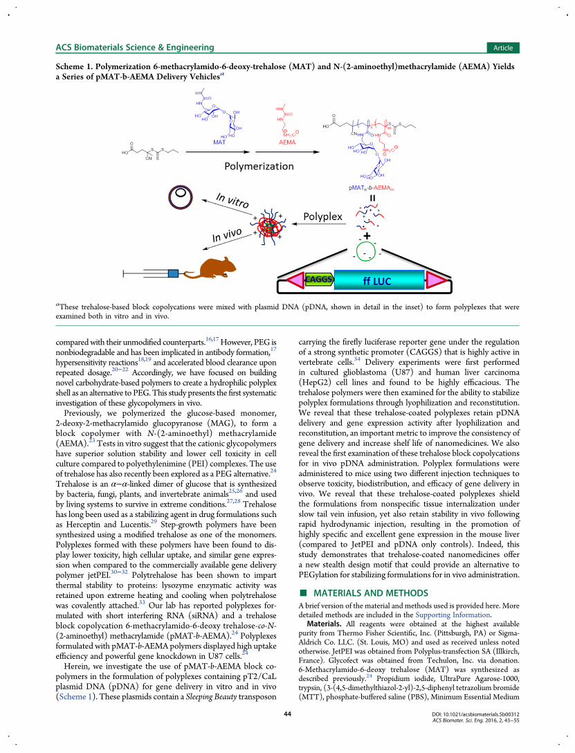
Herein, we investigate the use of pMAT-b-AEMA block co-polymers in the formulation of polyplexes containing pT2/CaLplasmid DNA (pDNA) for gene delivery in vitro and in vivo(Scheme 1). These plasmids contain a Sleeping Beauty transposon
carrying the firefly luciferase reporter gene under the regulationof a strong synthetic promoter (CAGGS) that is highly active invertebrate cells.34 Delivery experiments were first performedin cultured glioblastoma (U87) and human liver carcinoma(HepG2) cell lines and found to be highly efficacious. Thetrehalose polymers were then examined for the ability to stabilizepolyplex formulations through lyophilization and reconstitution.We reveal that these trehalose-coated polyplexes retain pDNAdelivery and gene expression activity after lyophilization andreconstitution, an important metric to improve the consistency ofgene delivery and increase shelf life of nanomedicines. We alsoreveal the first examination of these trehalose block copolycationsfor in vivo pDNA administration. Polyplex formulations wereadministered to mice using two different injection techniques toobserve toxicity, biodistribution, and efficacy of gene delivery invivo. We reveal that these trehalose-coated polyplexes shieldthe formulations from nonspecific tissue internalization underslow tail vein infusion, yet also retain stability in vivo followingrapid hydrodynamic injection, resulting in the promotion ofhighly specific and excellent gene expression in the mouse liver(compared to JetPEI and pDNA only controls). Indeed, thisstudy demonstrates that trehalose-coated nanomedicines offera new stealth design motif that could provide an alternative toPEGylation for stabilizing formulations for in vivo administration.
■ MATERIALS AND METHODSA brief version of the material and methods used is provided here. Moredetailed methods are included in the Supporting Information.
Materials. All reagents were obtained at the highest availablepurity from Thermo Fisher Scientific, Inc. (Pittsburgh, PA) or Sigma-Aldrich Co. LLC. (St. Louis, MO) and used as received unless notedotherwise. JetPEI was obtained from Polyplus-transfection SA (Illkirch,France). Glycofect was obtained from Techulon, Inc. via donation.6-Methacrylamido-6-deoxy trehalose (MAT) was synthesized asdescribed previously.24 Propidium iodide, UltraPure Agarose-1000,trypsin, (3-(4,5-dimethylthiazol-2-yl)-2,5-diphenyl tetrazolium bromide(MTT), phosphate-buffered saline (PBS), Minimum Essential Medium
Scheme 1. Polymerization 6-methacrylamido-6-deoxy-trehalose (MAT) and N-(2-aminoethyl)methacrylamide (AEMA) Yieldsa Series of pMAT-b-AEMA Delivery Vehiclesa
aThese trehalose-based block copolycations were mixed with plasmid DNA (pDNA, shown in detail in the inset) to form polyplexes that wereexamined both in vitro and in vivo.
with reduced serum (Opti-MEM) and Dulbecco’s Modified EagleMedium (DMEM) were purchased from Life Technologies − ThermoFisher Scientific (Carlsbad, CA). The pT2/CaL plasmid was prepared asdescribed previously.35 The Luciferase Assay Kit and cell lysis bufferwere obtained from Promega Corporation (Madison, WI). Bio-RadDC Protein Assay Reagent A, Reagent B and Reagent S were obtainedfrom Bio-Rad Laboratories, Inc. (Hercules, CA). HepG2 and U87 cellswere obtained from the American Type Culture Collection (ATCC)(Manassas, VA). The cells were grown in complete DMEM[supplemented with 10% (v:v) fetal bovine serum and 1% antibiotic-antimycotic solution (containing penicillin, streptomycin, and ampho-tericin B)] at 37 °C and 5% CO2 in a humidified incubator.Wild-type (WT) C57BL/6J 6wk-old mice were purchased from
Jackson Laboratories (Sacramento, CA). All mice were maintainedunder AAALAC-accredited (Association for Assessment and Accredita-tion of Laboratory Animal Care) specific pathogen-free conditions.Instrumentation. NMR spectra were recorded using a Bruker
Avance III HD 500 MHz spectrometer in D2O purchased fromCambridge Isotope Laboratories, Inc. (Andover, MA). NMR data wasanalyzed using Bruker Top Spin version 3.1. UV−vis data was collectedwith an Ocean Optics Inc. CUV 1 cm cuvette holder powered by aMikropack DH-2000 Deuterium/Halogen open-close TTC lamp, anddata was analyzed by Ocean Optics Inc. Basic Acquisition Software.Size exclusion chromatography (SEC)was conducted using an Agilent
1260 High Performance Liquid Chromatograph running 1.0 wt % aceticacid/0.1 M Na2SO4 as the eluent at a flow rate of 0.4 mL/min on sizeexclusion chromatography columns [CATSEC1000 (7 μ, 50 × 4.6),CATSEC100 (5 μ, 250 × 4.6), CATSEC300 (5 μ, 250 × 4.6), andCATSEC1000 (7 μ, 250 × 4.6)] obtained from Eprogen Inc. (DownersGrove, IL). Signals were acquired using Wyatt HELEOS II lightscattering detector (λ = 662 nm), and an Optilab rEX refractometer(λ = 658 nm). SEC trace analysis was performed using Astra VI software(version 5.3.4.18), Wyatt Technologies (Santa Barbara, CA). Thehydrodynamic diameters of the polyplexes were recorded via dynamiclight scattering measurements (DLS) with aMalvern Zetasizer Nano ZA.MTT, protein, and luciferase assay plates were analyzed using a BiotekSynergy H1 plate reader (BioTek Instruments, Inc., Winooski, VT).Cy5-uptake was measured on a FACSVerse (Becton DickinsonBiosciences, San Jose, CA) flow cytometer. TEM images were obtainedwith a FEITecnai G2 Spirit BioTWIN (FEI, Hillsboro, OR) transmissionelectron microscope, operated at 120 kV.Live animal and animal tissue imaging were performed on an
IVIS Spectrum In Vivo Imaging system and data was analyzed with theLiving Image software (PerkinElmer Inc., Waltham, MA). RT-qPCRwas performed using an Eppendorf Mastercycler (software version 2.2;Eppendorf).Polymer Synthesis and Polyplex Characterization. Synthesis
of Poly(methacrylamidotrehalose) (pMAT43). PMAT43 was synthe-sized as previously described.24 Briefly, MAT monomer waspolymerized via RAFT in acetate buffer. 4-cyano-4-(propylthiocarbo-nothioylthio)-pentanoic acid was dissolved in 645 μL of MeODH andadded to the MAT solution, followed by 4,4′-azobis(4-cyanovalericacid) (V-501). Finally, 861 μL of MeOD was added and oxygen wasremoved by bubbling nitrogen through the system for 45 min. The flaskwas heated to 70 °C for 6 h and the reaction was stopped by removingthe septum and cooling the reactionmixture on ice. The polymer solutionwas dialyzed against ultrapure water (3500 Da MWCO) and acidified topH 4−5 with HCl. After 3 d of dialysis, the polymer solution was freeze-dried on a VirTis benchtopK lyophilizer at 62 mT with the condenserat −57.4 °C to yield 454 mg of white solid. Method details andcharacterization results can be found in Figure S1.Synthesis of Cationic Diblock Copolymers pMAT-b-AEMA-1, -2,
and -3. Cationic diblock copolymers were synthesized as previouslyreported.24 Briefly, pMAT43 and V-501 were dissolved in 1.0 M acetatebuffer (pH 5.5) and added to a Schlenk tube containing amino-ethylmethacrylamide hydrochloride (AEMA). Deoxygenation wasachieved via bubbling nitrogen gas for 45 min. The tube was heatedto 70 °C. Aliquots (1.25 mL) were removed via syringe at 30 min(pMAT-b-AEMA-1) and 60 min (pMAT-b-AEMA-2). Each aliquotwas exposed to air and cooled in liquid nitrogen to stop polymerization.
After 90 min (pMAT-b-AEMA-3), the reaction was halted by septumremoval and submerging the Schlenk tube in liquid nitrogen. All threecopolymers were dialyzed (3500 Da MWCO) against 0.5 M NaClsolution, followed by 0.1 M NaCl and finally ultrapure water. All dialysismedia were acidified with HCl to pH 4−5. Polymer solutions werelyophilized as described above to yield white, flocculent powders. Allexperiments were completed using polymers pMAT-b-AEMA-1, -2, and -3.Polymer characterization details can be found in Figures S2−S5.
Synthesis of Cy7-pMAT-b-AEMA-2. PMAT-b-AEMA-2 was labeledusing an NHS-Cy7 fluorophore by dissolving it in H2O. Cy7 func-tionalized with N-hydroxysuccinamide (NHS-Cy7) in DMF was addedfollowed by DMF to target 1 fluorophore/50 amine residues. Themixturewas vortexed for 30 s and allowed to proceed at room temperature for 4 hin the dark. The labeled polymer was purified via dialysis (3500 MWCO)against H2O (acidified to pH ∼5.5 with HCl) for 2 days and lyophilizedusing the same conditions listed before to yield a blue flocculent solid.
Extent of labeling was quantified by measuring the absorbanceof a 0.2 mg/mL solution of labeled polymer in H2O at 750 nm viaUV−vis (ε = 199000 at 750 nm, Abs = 0.16), giving approximately 1 Cy7fluorophore/10 polymer chains (Figure S6).
Polyplex Formation for Gel Electrophoresis and Tissue Culture (InVitro) Experiments. Polymer solution at an appropriate concentrationin water was added to a 400 μM solution of pDNA in DNase/RNase-free distilled water in an equal volume to yield polyplexes of (nitrogen tophosphate ratios) N/P = 7, 14, and 21 at a final concentration of 200 μMpDNA. The nitrogen ratios were calculated based on the concentrationsof amines (either pendant from AEMA blocks or within the backboneof jetPEI or Glycofect). The solutions were incubated at 23 °C for 1 hbefore further use.
Gel Electrophoresis. A gel electrophoresis mobility shift assay wasrun to determine the minimal amount of polymer needed to achievecomplete binding of the pDNA. Ten μL of each polyplex solutionformulated at various N/P ratios were diluted with 10 μL of water toachieve a concentration of 100 μM and were incubated at 25 °C for 1 hto allow polymer-pDNA binding. The polyplex suspensions were runon agarose gels (0.6%) containing 6 μg ethidium bromide at 60 V for80 min. Images were obtained using 312 nm UV light (Figure S7).
Dynamic Light Scattering (DLS). To measure polyplex size in aprotein environment over time, we ran DLS experiments in DMEMcontaining 10% FBS. Polyplexes were formulated at N/P = 7, 14, and 21in H2O and incubated for 1 h at a concentration of 200 μM pDNA(to allow complexation) before being diluted with DMEM containing10% FBS by volume. For DLS studies prior to lyophilization, thepolyplexes were diluted to a final concentration of 100 μM(T = 0 h). Forthe postlyophilization DLS, polyplexes were prepared and lyophilizedas previously described, reconstituted with water for 1 h, then dilutedwith DMEM with 10% FBS to 133 μM. Size measurements weretaken at 25 °C using a 173° detection angle at times of 0, 1, 2, and 4 h(Figure S8).
Transmission Electron Microscopy (TEM) Imaging. Three μL ofpolyplex solution (formulated with pDNA and polymers pMAT-b-AEMA-1, -2, or -3) prepared at N/P = 10 were applied to a 300-meshcarbon coated copper grid (Ted Pella, Inc., Redding, CA) and negativelystained with uranyl acetate. Images were saved as TIFF files and poly-plexes sized (excluding polyplexes on image edges) by counting pixelsusing Microsoft Paint, and the sizes are plotted in Figure S9.
Examination of Polyplex Function In Vitro. MTT Assay. MTT(3-(4, 5-dimethylthiazol-2-yl)-2, 5-diphenyltetrazolium bromide) wasused to estimate the cytotoxicity of the polyplexes. HepG2 or U87 cellswere seeded at 50 000 cells/well in 24-well plates 24 h priorto transfection. Polyplexes were formulated as described previously,then mixed with transfection media (200 μL of either OptiMEM(Figure S10) or DMEM containing 10% FBS). The mixture was addedto each well. The transfection was ended 4 h later by diluting polyplexeswith 1 mL of complete DMEM containing 10% FBS. 48 h afterpolyplexes were added to the cells, 0.5 mg/mL of MTT was addedto each well and incubated for 1 h. Cells were lysed using DMSO and200 μL aliquots were transferred to a 96-well plate for analysis using theplate reader (absorbance was measured at 570 nm). Nontransfectedcells were used for normalizing the data.
Cellular Uptake Assay. Flow cytometry experiments were performedto examine the cellular uptake of Cy5-labeled pDNA 4 h post-transfection. HepG2 or U87 cells were seeded at 300 000 cells/well in6-well plates 24 h prior to transfection. Polyplexes were prepared asdescribed in the MTT assay protocol, except that the formulationwas scaled up from 50 000 cells to 300 000 cells. After 4h, the polyplexmedia was removed and cells were detached from the plate surface usingtrypsin, centrifuged to remove polyplexes and trypsin, and then washedtwice with PBS. Finally, 1 mL of PBS was added and the cellularsuspensions were kept on ice. Propidium iodide (1.0 mg/mL, 2.5 μL)was added prior to analysis. Each experiment was performed in triplicate,and OptiMEM results are plotted in Figure S11.Luciferase Gene Expression. Cells (HepG2 and U-87) were seeded
at 50 000 cells/well in 24-well plates 24 h prior to transfection.Polyplexes were prepared, diluted, and applied to the cells as describedin the cytotoxicity section above. After 48 h, the cells were washed withPBS and treated with cell lysis buffer. Aliquots (5 μL) of cell lysatecombined with 100 μL luciferase substrate were examined in 96-wellplates using a luminometer to determine relative light units (RLUs).Data were measured in triplicate and normalized for the amount ofprotein in each sample. Sample averages were plotted with error barsrepresenting standard deviations (OptiMEM results in Figure S12).Mouse Studies. Tail Vein Injections. Appropriate amounts of
polyplexes (10 μg or 25 μg of pDNA/dose at desired N/P of polymer)were prepared in a 5% by weight aqueous dextrose solution (D5W,Hospira, Inc.), and a 200 μL dose of polyplexes was injected through thetail vein of experimental animals. After injection, animals were returnedto their colony and imaged after 24 and 48 h. Mice injected withCy7-labeled polymer were imaged immediately (data not shown),euthanized, and then their organs were imaged ex vivo.Hydrodynamic Injections. Appropriate amounts of polyplexes
(10 μg or 25 μg of pDNA/dose at desired N/P of polymer) wereprepared in D5W solution and injected through the tail vein of experi-mental animals using a hydrodynamics-based procedure as previouslydescribed.36 Briefly, animals to be injectedwereweighed. A polyplex solutionwith a volume equivalent to 10% of the weight of the mouse was injected in3 to 4 s. After injection, animalswere placed on a heating pad to recover, thenreturned to their colony. Mice were either imaged for luciferase transgeneexpression about 24 and 48 h after transfection or imaged immediately afterinjection for Cy7 polymer fluorescence. After fluorescence imaging, micewere euthanized and their organs were imaged ex vivo.Bioluminescence Imaging In Vivo. One hundred microliters of
luciferin substrate solution (28.5 mg/mL) was injected intraperitoneallyto each mouse. Three to five minutes later, mice were imaged for 1 minusing a Xenogen Spectrum CCD camera system (Xenogen, Alameda,CA) according to the manufacturer’s instructions. Each experiment wasperformed in triplicate.Euthanizing and Tissue Collection/Processing. Mice were
euthanized by carbon dioxide (CO2) inhalation, perfused with saline,and selected organs were resected and preserved for analyses. Plasmaand tissues were stored at −80 °C. Frozen tissues were homogenized bymortar and pestle, and∼100mgof each tissue samplewas removed forDNAprocessing. DNA was pulled from the organs using a phenol-chloroformextraction and ethanol precipitation. DNA purity and concentrationwere determined by using a NanoDrop instrument (Thermo Scientific,Wilmington, DE).qPCR. DNA samples were normalized to 100 ng/μL. Five
hundred nanograms of DNA was used with TaqMan Gene ExpressionMaster Mix (Life Technologies − Thermo Fisher Scientific, Carlsbad,CA). TaqMan primers were designed and manufactured by LifeTechnologies − Thermo Fisher Scientific (Carlsbad, CA), Custom PlusTaqMan Assay, Assay ID− AJ6RNJ4. A standard curve was prepared byserially diluting pT2/CaL plasmid into WT C57BL/6 DNA. Sampleswere run using the impulse setting. The following conditions were usedfor the qPCR experiments: 95 °C for 10 min and 40 cycles of 90 °C for15 s then 60 °C for 1 min.Statistical Analysis. Statistical analysis was performed using JMP
Pro Software (SAS Institute, Cary, NC) through the University ofMinnesota Supercomputing Institute.
■ RESULTS AND DISCUSSIONPolymer Synthesis and Polyplex Characterization.
Trehalose is a carbohydrate with unique cryo- and lyo-protectantproperties, which have prompted the use of this carbohydrate inmaterials synthesis to both retain and enhance these protectiveabilities.24−28,37 Previously, we have shown that trehalose-containing block copolymers are effective in the formulation ofpolyplexes and efficient delivery of siRNA.24 Herein, we soughtto examine these trehalose-based polycations for delivery ofplasmids to cells in culture and in organs of living animals.Trehalose-based polymers were synthesized via reversible-addition−fragmentation chain-transfer (RAFT) polymerizationwith a 65:1 monomer: chain-transfer agent (CTA) ratio of6-methacrylamido-6-deoxymethacrylamido trehalose (MAT) and4-cyano-4-(propylthiocarbonothioylthio)-pentanoic acid (CPP)with 4,4′-azobis(4-cyanovaleric acid) (V-501) as the initiator toyield pMAT with 43 repeats. This 43-repeat unit pMAT was usedas a macro CTA and chain-extended via RAFT polymerizationwith 125 equiv of N-(2-aminoethyl) methacrylamide (AEMA)and V-501 as the initiator to yield three cationic block copolymers(Table 1),24 pMAT-b-AEMA-1 (21 repeats), -2 (44 repeats), and -3
(57 repeats), with each having the same length of MAT block butincreasing lengths of the AEMA block.These polymers were combined with pDNA encoding for the
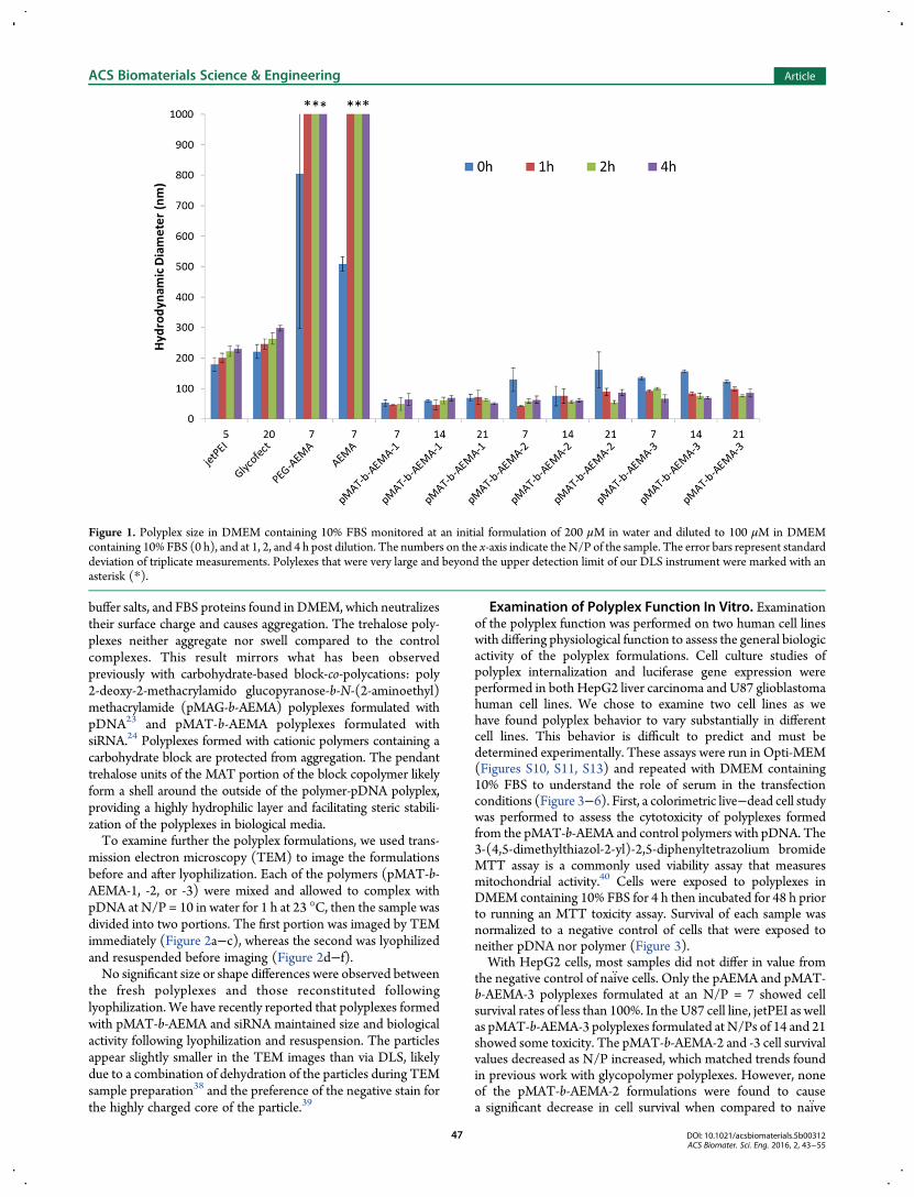
luciferase reporter gene to form polyplexes. The amount ofpolymer needed to fully bind the pDNA was first determined viagel electrophoresis. Polymers and pDNAweremixed at nitrogen-to-phosphate (N/P) ratios of 1, 2, 3, 4, 5, and 10, mixed with 1 μLof running buffer and electrophoresed through a 0.6% w/wagarose gel containing ethidium bromide in TAE buffer. The gelwas imaged under UV light to confirm binding (Figure S7).Anionic plasmid that remains uncomplexed by the cationicpolymer migrates through the gel toward the positively chargedanodewhereas fully complexedDNAdoes not. All three polymersfully bound the DNA at N/P = 3. For further studies, eachpolymer was combined with DNA at three different N/P ratios:7, 14, and 21, yielding nine polyplex formulations. Polyplex sizeand stability from aggregation in biological media containingserum (10% by volume fetal bovine serum, FBS) were examinedvia dynamic light scattering (DLS). We have previously shownthat polyplexes formed from this polymer and siRNA have apositive ζ-potential in H2O. Polyplexes were formulated at200 μM pDNA and allowed to complex for 1 h at 23 °C in water.The polyplexes were then diluted with Dulbecco’sModified EagleMedium (DMEM) containing 10% by volume fetal bovine serum(FBS) solution to a final concentration of 100 μM in pDNA, andthe polyplex size wasmonitored over 4 h (Figure 1). ζ-potential ofthese polyplexes was not collected as the presence of FBS disruptsthe measurement.Trehalose polyplex size (∼50−100 nm in diameter) remained
consistent over the course of this experiment relative to thecontrol polyplexes, jetPEI (N/P = 5) and Glycofect (N/P = 20),which increased in diameter with time. It is proposed that thejetPEI and Glycofect polyplexes aggregate with amino acids,
buffer salts, and FBS proteins found in DMEM, which neutralizestheir surface charge and causes aggregation. The trehalose poly-plexes neither aggregate nor swell compared to the controlcomplexes. This result mirrors what has been observedpreviously with carbohydrate-based block-co-polycations: poly2-deoxy-2-methacrylamido glucopyranose-b-N-(2-aminoethyl)methacrylamide (pMAG-b-AEMA) polyplexes formulated withpDNA23 and pMAT-b-AEMA polyplexes formulated withsiRNA.24 Polyplexes formed with cationic polymers containing acarbohydrate block are protected from aggregation. The pendanttrehalose units of the MAT portion of the block copolymer likelyform a shell around the outside of the polymer-pDNA polyplex,providing a highly hydrophilic layer and facilitating steric stabili-zation of the polyplexes in biological media.To examine further the polyplex formulations, we used trans-
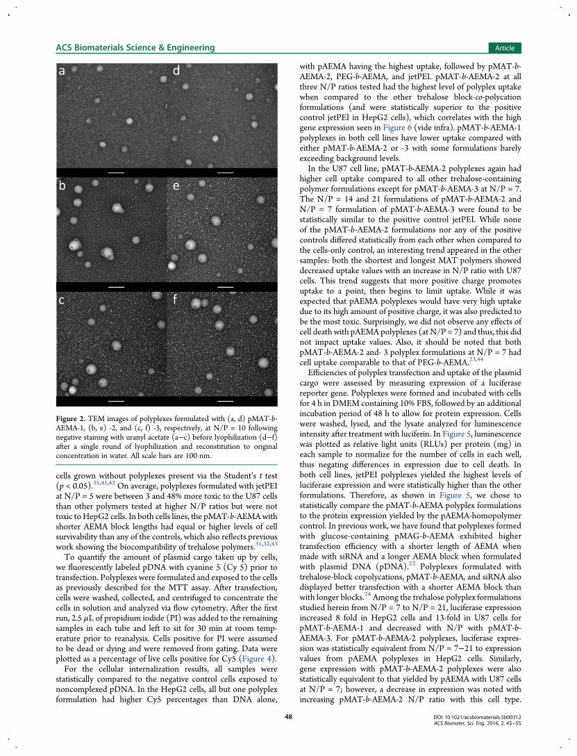
mission electron microscopy (TEM) to image the formulationsbefore and after lyophilization. Each of the polymers (pMAT-b-AEMA-1, -2, or -3) were mixed and allowed to complex withpDNA at N/P = 10 in water for 1 h at 23 °C, then the sample wasdivided into two portions. The first portion was imaged by TEMimmediately (Figure 2a−c), whereas the second was lyophilizedand resuspended before imaging (Figure 2d−f).No significant size or shape differences were observed between
the fresh polyplexes and those reconstituted followinglyophilization. We have recently reported that polyplexes formedwith pMAT-b-AEMA and siRNA maintained size and biologicalactivity following lyophilization and resuspension. The particlesappear slightly smaller in the TEM images than via DLS, likelydue to a combination of dehydration of the particles during TEMsample preparation38 and the preference of the negative stain forthe highly charged core of the particle.39
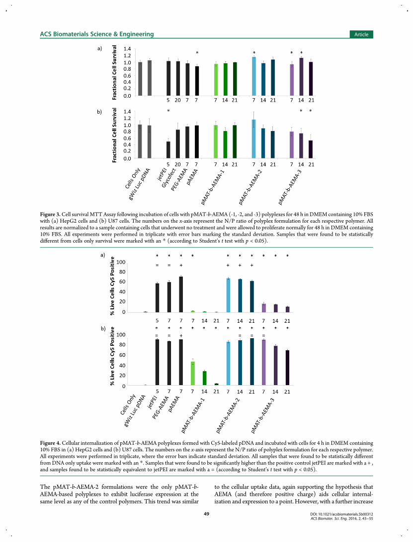
Examination of Polyplex Function In Vitro. Examinationof the polyplex function was performed on two human cell lineswith differing physiological function to assess the general biologicactivity of the polyplex formulations. Cell culture studies ofpolyplex internalization and luciferase gene expression wereperformed in both HepG2 liver carcinoma and U87 glioblastomahuman cell lines. We chose to examine two cell lines as wehave found polyplex behavior to vary substantially in differentcell lines. This behavior is difficult to predict and must bedetermined experimentally. These assays were run in Opti-MEM(Figures S10, S11, S13) and repeated with DMEM containing10% FBS to understand the role of serum in the transfectionconditions (Figure 3−6). First, a colorimetric live−dead cell studywas performed to assess the cytotoxicity of polyplexes formedfrom the pMAT-b-AEMA and control polymers with pDNA. The3-(4,5-dimethylthiazol-2-yl)-2,5-diphenyltetrazolium bromideMTT assay is a commonly used viability assay that measuresmitochondrial activity.40 Cells were exposed to polyplexes inDMEM containing 10% FBS for 4 h then incubated for 48 h priorto running an MTT toxicity assay. Survival of each sample wasnormalized to a negative control of cells that were exposed toneither pDNA nor polymer (Figure 3).With HepG2 cells, most samples did not differ in value from
the negative control of naiv̈e cells. Only the pAEMA and pMAT-b-AEMA-3 polyplexes formulated at an N/P = 7 showed cellsurvival rates of less than 100%. In the U87 cell line, jetPEI as wellas pMAT-b-AEMA-3 polyplexes formulated at N/Ps of 14 and 21showed some toxicity. The pMAT-b-AEMA-2 and -3 cell survivalvalues decreased as N/P increased, which matched trends foundin previous work with glycopolymer polyplexes. However, noneof the pMAT-b-AEMA-2 formulations were found to causea significant decrease in cell survival when compared to naiv̈e
Figure 1. Polyplex size in DMEM containing 10% FBS monitored at an initial formulation of 200 μM in water and diluted to 100 μM in DMEMcontaining 10% FBS (0 h), and at 1, 2, and 4 h post dilution. The numbers on the x-axis indicate theN/P of the sample. The error bars represent standarddeviation of triplicate measurements. Polylexes that were very large and beyond the upper detection limit of our DLS instrument were marked with anasterisk (*).
cells grown without polyplexes present via the Student’s t test(p < 0.05).31,41,42 On average, polyplexes formulated with jetPEIat N/P = 5 were between 3 and 48% more toxic to the U87 cellsthan other polymers tested at higher N/P ratios but were nottoxic toHepG2 cells. In both cells lines, the pMAT-b-AEMAwithshorter AEMA block lengths had equal or higher levels of cellsurvivability than any of the controls, which also reflects previouswork showing the biocompatibility of trehalose polymers.31,32,43
To quantify the amount of plasmid cargo taken up by cells,we fluorescently labeled pDNA with cyanine 5 (Cy 5) prior totransfection. Polyplexes were formulated and exposed to the cellsas previously described for the MTT assay. After transfection,cells were washed, collected, and centrifuged to concentrate thecells in solution and analyzed via flow cytometry. After the firstrun, 2.5 μL of propidium iodide (PI) was added to the remainingsamples in each tube and left to sit for 30 min at room temp-erature prior to reanalysis. Cells positive for PI were assumedto be dead or dying and were removed from gating. Data wereplotted as a percentage of live cells positive for Cy5 (Figure 4).For the cellular internalization results, all samples were
statistically compared to the negative control cells exposed tononcomplexed pDNA. In the HepG2 cells, all but one polyplexformulation had higher Cy5 percentages than DNA alone,
with pAEMA having the highest uptake, followed by pMAT-b-AEMA-2, PEG-b-AEMA, and jetPEI. pMAT-b-AEMA-2 at allthree N/P ratios tested had the highest level of polyplex uptakewhen compared to the other trehalose block-co-polycationformulations (and were statistically superior to the positivecontrol jetPEI in HepG2 cells), which correlates with the highgene expression seen in Figure 6 (vide infra). pMAT-b-AEMA-1polyplexes in both cell lines have lower uptake compared witheither pMAT-b-AEMA-2 or -3 with some formulations barelyexceeding background levels.In the U87 cell line, pMAT-b-AEMA-2 polyplexes again had
higher cell uptake compared to all other trehalose-containingpolymer formulations except for pMAT-b-AEMA-3 at N/P = 7.The N/P = 14 and 21 formulations of pMAT-b-AEMA-2 andN/P = 7 formulation of pMAT-b-AEMA-3 were found to bestatistically similar to the positive control jetPEI. While noneof the pMAT-b-AEMA-2 formulations nor any of the positivecontrols differed statistically from each other when compared tothe cells-only control, an interesting trend appeared in the othersamples: both the shortest and longest MAT polymers showeddecreased uptake values with an increase in N/P ratio with U87cells. This trend suggests that more positive charge promotesuptake to a point, then begins to limit uptake. While it wasexpected that pAEMA polyplexes would have very high uptakedue to its high amount of positive charge, it was also predicted tobe the most toxic. Surprisingly, we did not observe any effects ofcell death with pAEMA polyplexes (at N/P = 7) and thus, this didnot impact uptake values. Also, it should be noted that bothpMAT-b-AEMA-2 and- 3 polyplex formulations at N/P = 7 hadcell uptake comparable to that of PEG-b-AEMA.23,44
Efficiencies of polyplex transfection and uptake of the plasmidcargo were assessed by measuring expression of a luciferasereporter gene. Polyplexes were formed and incubated with cellsfor 4 h in DMEM containing 10% FBS, followed by an additionalincubation period of 48 h to allow for protein expression. Cellswere washed, lysed, and the lysate analyzed for luminescenceintensity after treatment with luciferin. In Figure 5, luminescencewas plotted as relative light units (RLUs) per protein (mg) ineach sample to normalize for the number of cells in each well,thus negating differences in expression due to cell death. Inboth cell lines, jetPEI polyplexes yielded the highest levels ofluciferase expression and were statistically higher than the otherformulations. Therefore, as shown in Figure 5, we chose tostatistically compare the pMAT-b-AEMA polyplex formulationsto the protein expression yielded by the pAEMA-homopolymercontrol. In previous work, we have found that polyplexes formedwith glucose-containing pMAG-b-AEMA exhibited highertransfection efficiency with a shorter length of AEMA whenmade with siRNA and a longer AEMA block when formulatedwith plasmid DNA (pDNA).23 Polyplexes formulated withtrehalose-block copolycations, pMAT-b-AEMA, and siRNA alsodisplayed better transfection with a shorter AEMA block thanwith longer blocks.24 Among the trehalose polyplex formulationsstudied herein from N/P = 7 to N/P = 21, luciferase expressionincreased 8 fold in HepG2 cells and 13-fold in U87 cells forpMAT-b-AEMA-1 and decreased with N/P with pMAT-b-AEMA-3. For pMAT-b-AEMA-2 polyplexes, luciferase expres-sion was statistically equivalent from N/P = 7−21 to expressionvalues from pAEMA polyplexes in HepG2 cells. Similarly,gene expression with pMAT-b-AEMA-2 polyplexes were alsostatistically equivalent to that yielded by pAEMA with U87 cellsat N/P = 7; however, a decrease in expression was noted withincreasing pMAT-b-AEMA-2 N/P ratio with this cell type.
Figure 2. TEM images of polyplexes formulated with (a, d) pMAT-b-AEMA-1, (b, e) -2, and (c, f) -3, respectively, at N/P = 10 followingnegative staining with uranyl acetate (a−c) before lyophilization (d−f)after a single round of lyophilization and reconstitution to originalconcentration in water. All scale bars are 100 nm.
The pMAT-b-AEMA-2 formulations were the only pMAT-b-AEMA-based polyplexes to exhibit luciferase expression at thesame level as any of the control polymers. This trend was similar
to the cellular uptake data, again supporting the hypothesis thatAEMA (and therefore positive charge) aids cellular internal-ization and expression to a point. However, with a further increase
Figure 3.Cell survival MTTAssay following incubation of cells with pMAT-b-AEMA (-1, -2, and -3) polyplexes for 48 h in DMEM containing 10% FBSwith (a) HepG2 cells and (b) U87 cells. The numbers on the x-axis represent the N/P ratio of polyplex formulation for each respective polymer. Allresults are normalized to a sample containing cells that underwent no treatment and were allowed to proliferate normally for 48 h in DMEM containing10% FBS. All experiments were performed in triplicate with error bars marking the standard deviation. Samples that were found to be statisticallydifferent from cells only survival were marked with an * (according to Student’s t test with p < 0.05).
Figure 4. Cellular internalization of pMAT-b-AEMA polyplexes formed with Cy5-labeled pDNA and incubated with cells for 4 h in DMEM containing10% FBS in (a) HepG2 cells and (b) U87 cells. The numbers on the x-axis represent the N/P ratio of polyplex formulation for each respective polymer.All experiments were performed in triplicate, where the error bars indicate standard deviation. All samples that were found to be statistically differentfrom DNA only uptake were marked with an *. Samples that were found to be significantly higher than the positive control jetPEI are marked with a + ,and samples found to be statistically equivalent to jetPEI are marked with a = (according to Student’s t test with p < 0.05).
in the N/P ratio, expression values start to decrease. The decreasecould be due to two factors: (i) prevention of the release andtranscription of DNA cargo23 or (ii) by increasing cell membranepermeability to the point of toxicity.40,45 Because luminescenceRLUs are normalized to the amount of cell protein in each sample,cytotoxicity is not enough to account for low expression in eithercell line. It is more likely that polyplexes with large AEMA blocksand high ratios of polymer-to-plasmid have higher chargedensities that prevent cargo release and transcription,23,46 whereascomplexes with too little AEMA or too small an N/P ratio couldprematurely release DNA prior to nuclear entry.47 We were notas concerned with this latter issue because of the stability ofthe polyplexes that was observed in the DLS data. It should benoted that differences between some samples are not statisticallysignificant, but the general trend that appears in the uptake assaysas well as gene expression studies for both cells lines correlateswith previous work in the field.23,41,44,46
The pMAT-b-AEMA polymers were next tested for theirability to act as a lyoprotectant when complexed with pDNA.pMAT-b-AEMA-2, jetPEI, Glycofect, and PEG-b-AEMA wereused to formulate polyplexes in H2O and allowed to incubate for1 h at room temperature. They were immediately frozen in liquidnitrogen and lyophilized to dryness. The resulting powder wasthen reconstituted with water, allowed to incubate for anotherhour, and either administered to cells in DMEM + 10% FBSor resubjected to the same lyophilization procedure. Luciferaseexpression assays were completed as previously described. Theexpression for each lyophilized polyplex sample was normalizedto samples made with the same polymer that had not beenlyophilized. Figure 6 shows these data as a percentage of geneexpression retained for each round of lyophilization.Following a single round of lyophilization and reconstitution, only
the two polymers containing a sugar moiety, pMAT-b-AEMA-2
and Glycofect, retained any ability to transfect U87 cells. pMAT-b-AEMA-2 exhibited expression equal to 69% of its initial levelwhile Glycofect was reduced to 4%. It is hypothesized that thepMAT-b-AEMA polyplexes form a core−shell structure with thetrehalose-containing block coating the external surface of thepolyplex, aiding polyplex stability during the lyophiliza-tion process. Neither Glycofect nor jetPEI polyplexes containstabilization layers, leaving these formulations susceptible toaggregation via the lyophilization procedure. Interestingly, thePEG-b-AEMA was expected to have a similar polyplex structure(containing a hydrophilic PEG shell coating) to the pMAT-b-AEMA-2; however, it did not retain transfection capability
Figure 5. Luminescence of cell lysate following addition of luciferin in (a) HepG2 cells and (b) U87 cells. Cells were incubated with the polyplexformulations for 4 h in DMEM containing 10% FBS followed by an additional 48 h to allow for protein expression. The numbers on the x-axis representthe N/P ratio of polyplex formulation for each respective polymer. All experiments were performed in triplicate with error bars showing standarddeviation. All samples that were found to be statistically different from cells only luciferase expression were marked with an *. Samples that were found tobe significantly higher than the positive control pAEMA aremarked with a + , and samples found to be statistically equivalent to pAEMA aremarked withan = (according to Student’s t test with p < 0.05).
Figure 6. Luciferase expression in U87 cells following transfection withlyophilized polyplexes. JetPEI polyplexes were formulated at N/P = 5,Glycofect polyplexes were formulated at N/P = 20, and PEG-b-AEMAand pMAT-b-AEMA-2 polyplexes were formulated at N/P = 7. Allexperiments were performed in triplicate with error bars showingstandard deviation.
following lyophilization. The trehalose blocks appear to impart aunique protective property to polyplexes for potential lyophiliza-tion and storage.Mouse Studies. Polyplexes formed from pMAT-b-AEMA-2
and pDNA had the highest level of gene expression in culturedcells in vitro and low toxicity at the three tested N/P ratios; thesepromising properties led to the selection and examination ofthis formulation in vivo. Murine studies with pMAT-b-AEMA-2were explored for delivering luciferase-encoding pDNAwith C57black 6 (C57BL/6) mice. Tail vein injection was used to probefor biodistribution, toxicity, and transfection of the polyplexes ina living organism.Mice treated via standard tail vein injections were adminis-
tered with one of four polyplex formulations in 5% dextrose(D5W, Table S1) and a fifth control sample of D5W only.Importantly, with all formulations, all mice survived to the 48 hend point of the study with no noticeable health deterioration.All mice were active and showed no signs of lethargy nor ill healtheffects after being dosed with up to 490 μg of polymer per mouse(the equivalent of 19.6 mg/kg of polymer/mouse). Following tailvein injection, the mice were then injected peritoneally withluciferin at 24 and 48 h after polyplex injection and imaged usinga bioluminescence Xenogen Spectrum CCD camera system.Luciferase gene expression could not be detected either 24 or 48 hafter the infusion of polymers. Following the 48 h imaging, all micewere euthanized and the heart, liver, lung, spleen, kidneys, and brainwere harvested for analysis by qPCR to determine biodistributionof the pDNA cargo (Table S1).48 QPCR analysis revealed a nearlyuniform distribution of genetic material throughout the tissues. Ona per-cell basis, pDNA was found at a range of 1.1−10 plasmidcopies per cell in each organ (values less than or equal to 1.0 copies/cell cannot be resolved from the background). Hydrodynamicinjection of 25 μg of pDNA, discussed in further detail below,typically achieves delivery of ∼100 plasmids per cell in the liver,leading to significant gene expression.36,49 The lower level ofgenetic material delivered to each of these organs following tail veininjection was insufficient to achieve observable gene expression.Multiple cyanine fluorophores, including Cy7, have been
used successfully to tag DNA cargos for in vivo imaging.50−52
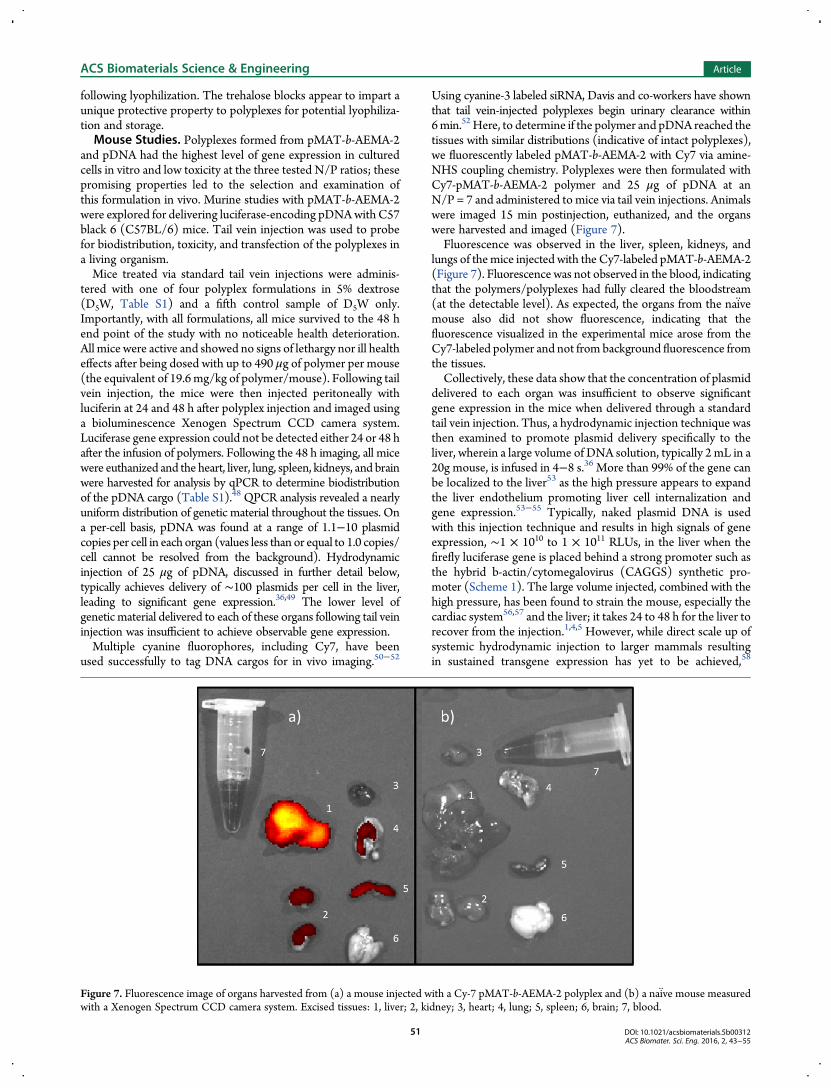
Using cyanine-3 labeled siRNA, Davis and co-workers have shownthat tail vein-injected polyplexes begin urinary clearance within6min.52Here, to determine if the polymer and pDNA reached thetissues with similar distributions (indicative of intact polyplexes),we fluorescently labeled pMAT-b-AEMA-2 with Cy7 via amine-NHS coupling chemistry. Polyplexes were then formulated withCy7-pMAT-b-AEMA-2 polymer and 25 μg of pDNA at anN/P = 7 and administered to mice via tail vein injections. Animalswere imaged 15 min postinjection, euthanized, and the organswere harvested and imaged (Figure 7).Fluorescence was observed in the liver, spleen, kidneys, and
lungs of themice injected with the Cy7-labeled pMAT-b-AEMA-2(Figure 7). Fluorescence was not observed in the blood, indicatingthat the polymers/polyplexes had fully cleared the bloodstream(at the detectable level). As expected, the organs from the naiv̈emouse also did not show fluorescence, indicating that thefluorescence visualized in the experimental mice arose from theCy7-labeled polymer and not from background fluorescence fromthe tissues.Collectively, these data show that the concentration of plasmid
delivered to each organ was insufficient to observe significantgene expression in the mice when delivered through a standardtail vein injection. Thus, a hydrodynamic injection technique wasthen examined to promote plasmid delivery specifically to theliver, wherein a large volume of DNA solution, typically 2 mL in a20g mouse, is infused in 4−8 s.36 More than 99% of the gene canbe localized to the liver53 as the high pressure appears to expandthe liver endothelium promoting liver cell internalization andgene expression.53−55 Typically, naked plasmid DNA is usedwith this injection technique and results in high signals of geneexpression, ∼1 × 10¹⁰ to 1 × 10¹¹ RLUs, in the liver when thefirefly luciferase gene is placed behind a strong promoter such asthe hybrid b-actin/cytomegalovirus (CAGGS) synthetic pro-moter (Scheme 1). The large volume injected, combined with thehigh pressure, has been found to strain the mouse, especially thecardiac system56,57 and the liver; it takes 24 to 48 h for the liver torecover from the injection.1,4,5 However, while direct scale up ofsystemic hydrodynamic injection to larger mammals resultingin sustained transgene expression has yet to be achieved,58
Figure 7. Fluorescence image of organs harvested from (a) a mouse injected with a Cy-7 pMAT-b-AEMA-2 polyplex and (b) a naiv̈e mouse measuredwith a Xenogen Spectrum CCD camera system. Excised tissues: 1, liver; 2, kidney; 3, heart; 4, lung; 5, spleen; 6, brain; 7, blood.
it may be potentially translatable if the high pressure injection isisolated only to the organ of interest and formulation of DNAinto polyplexes could improve delivery and gene expression.For example, Itaka and co-workers were able to perform ahydrodynamic injection directed to skeletal muscle by injectinginto a limb isolated with a tourniquet.59 DNA delivered as apolyplex formulated from a PEG-poly-L-lysine cationic blockcopolymer showed increased expression and DNA lifetime in thetissue when compared to similar injections performed with nakedpDNA.59 Nakamura and co-workers performed hydrodynamicinjection followed by a luciferase assay on excised liver tissueand found that polyplexes formed with jetPEI yielded higherluminescence than naked pDNA.60
Herein, Glycofect, jetPEI, and pMAT-b-AEMA-2 were usedto deliver 10 μg of pDNA via hydrodynamic injection andcompared to naked pDNA as a control (Table 2). All mice
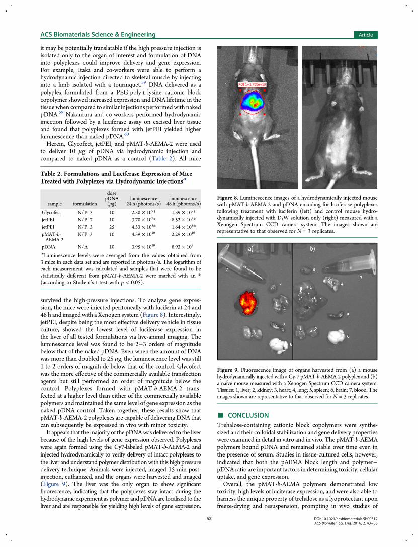
survived the high-pressure injections. To analyze gene expres-sion, the mice were injected peritoneally with luciferin at 24 and48 h and imaged with a Xenogen system (Figure 8). Interestingly,jetPEI, despite being the most effective delivery vehicle in tissueculture, showed the lowest level of luciferase expression inthe liver of all tested formulations via live-animal imaging. Theluminescence level was found to be 2−3 orders of magnitudebelow that of the naked pDNA. Even when the amount of DNAwas more than doubled to 25 μg, the luminescence level was still1 to 2 orders of magnitude below that of the control. Glycofectwas the more effective of the commercially available transfectionagents but still performed an order of magnitude below thecontrol. Polyplexes formed with pMAT-b-AEMA-2 trans-fected at a higher level than either of the commercially availablepolymers andmaintained the same level of gene expression as thenaked pDNA control. Taken together, these results show thatpMAT-b-AEMA-2 polyplexes are capable of delivering DNA thatcan subsequently be expressed in vivo with minor toxicity.It appears that themajority of the pDNAwas delivered to the liver
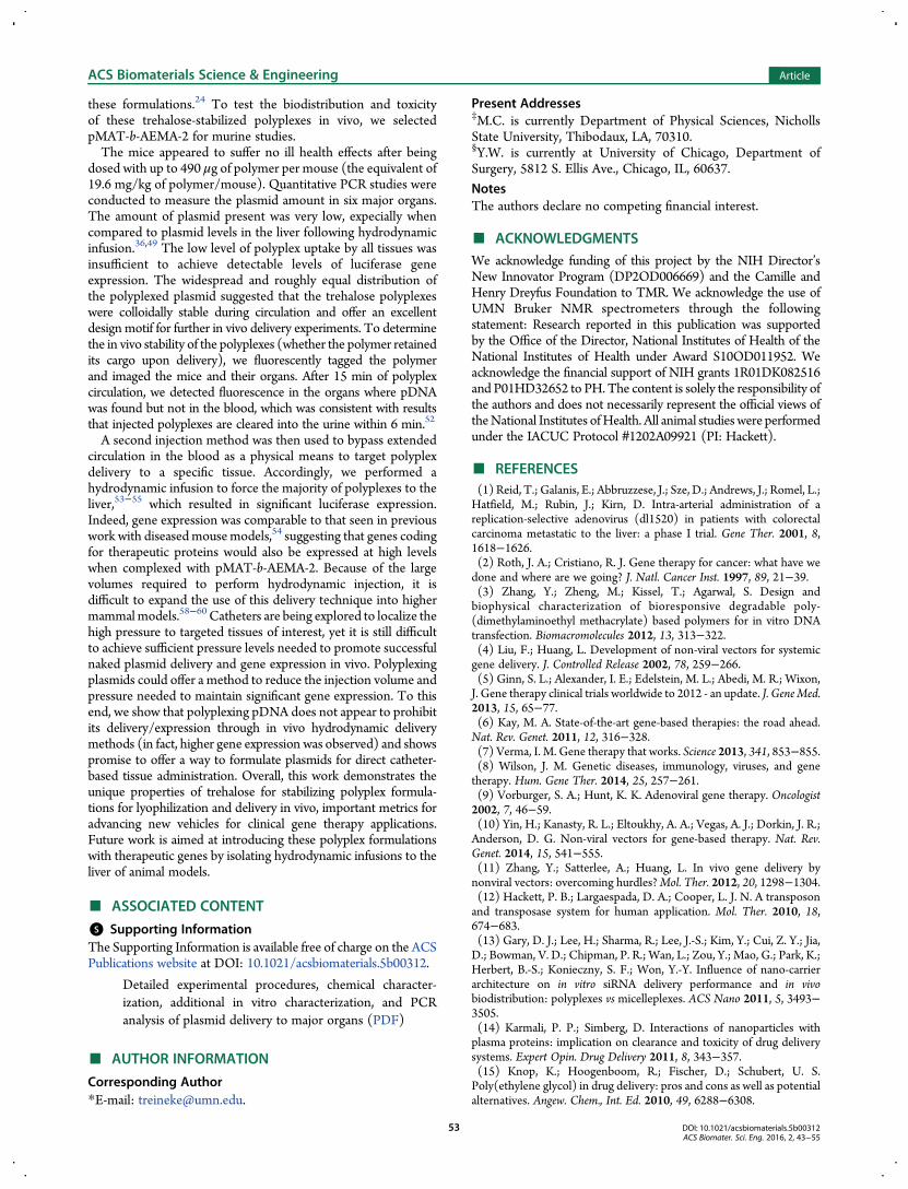
because of the high levels of gene expression observed. Polyplexeswere again formed using the Cy7-labeled pMAT-b-AEMA-2 andinjected hydrodynamically to verify delivery of intact polyplexes tothe liver and understand polymer distribution with this high pressuredelivery technique. Animals were injected, imaged 15 min post-injection, euthanized, and the organs were harvested and imaged(Figure 9). The liver was the only organ to show significantfluorescence, indicating that the polyplexes stay intact during thehydrodynamic experiment as polymer and pDNAare localized to theliver and are responsible for yielding high levels of gene expression.
■ CONCLUSIONTrehalose-containing cationic block copolymers were synthe-sized and their colloidal stabilization and gene delivery propertieswere examined in detail in vitro and in vivo. The pMAT-b-AEMApolymers bound pDNA and remained stable over time even inthe presence of serum. Studies in tissue-cultured cells, however,indicated that both the pAEMA block length and polymer−pDNA ratio are important factors in determining toxicity, cellularuptake, and gene expression.Overall, the pMAT-b-AEMA polymers demonstrated low
toxicity, high levels of luciferase expression, and were also able toharness the unique property of trehalose as a lyoprotectant uponfreeze-drying and resuspension, prompting in vivo studies of
Table 2. Formulations and Luciferase Expression of MiceTreated with Polyplexes via Hydrodynamic Injectionsa
aLuminescence levels were averaged from the values obtained from3 mice in each data set and are reported in photons/s. The logarithm ofeach measurement was calculated and samples that were found to bestatistically different from pMAT-b-AEMA-2 were marked with an *(according to Student’s t-test with p < 0.05).
Figure 8. Luminescence images of a hydrodynamically injected mousewith pMAT-b-AEMA-2 and pDNA encoding for luciferase polyplexesfollowing treatment with luciferin (left) and control mouse hydro-dynamically injected with D5W solution only (right) measured with aXenogen Spectrum CCD camera system. The images shown arerepresentative to that observed for N = 3 replicates.
Figure 9. Fluorescence image of organs harvested from (a) a mousehydrodynamically injected with a Cy-7 pMAT-b-AEMA-2 polyplex and (b)a naiv̈e mouse measured with a Xenogen Spectrum CCD camera system.Tissues: 1, liver; 2, kidney; 3, heart; 4, lung; 5, spleen; 6, brain; 7, blood. Theimages shown are representative to that observed for N = 3 replicates.
these formulations.24 To test the biodistribution and toxicityof these trehalose-stabilized polyplexes in vivo, we selectedpMAT-b-AEMA-2 for murine studies.The mice appeared to suffer no ill health effects after being
dosed with up to 490 μg of polymer per mouse (the equivalent of19.6 mg/kg of polymer/mouse). Quantitative PCR studies wereconducted to measure the plasmid amount in six major organs.The amount of plasmid present was very low, expecially whencompared to plasmid levels in the liver following hydrodynamicinfusion.36,49 The low level of polyplex uptake by all tissues wasinsufficient to achieve detectable levels of luciferase geneexpression. The widespread and roughly equal distribution ofthe polyplexed plasmid suggested that the trehalose polyplexeswere colloidally stable during circulation and offer an excellentdesign motif for further in vivo delivery experiments. To determinethe in vivo stability of the polyplexes (whether the polymer retainedits cargo upon delivery), we fluorescently tagged the polymerand imaged the mice and their organs. After 15 min of polyplexcirculation, we detected fluorescence in the organs where pDNAwas found but not in the blood, which was consistent with resultsthat injected polyplexes are cleared into the urine within 6 min.52
A second injection method was then used to bypass extendedcirculation in the blood as a physical means to target polyplexdelivery to a specific tissue. Accordingly, we performed ahydrodynamic infusion to force the majority of polyplexes to theliver,53−55 which resulted in significant luciferase expression.Indeed, gene expression was comparable to that seen in previouswork with diseasedmousemodels,54 suggesting that genes codingfor therapeutic proteins would also be expressed at high levelswhen complexed with pMAT-b-AEMA-2. Because of the largevolumes required to perform hydrodynamic injection, it isdifficult to expand the use of this delivery technique into highermammalmodels.58−60 Catheters are being explored to localize thehigh pressure to targeted tissues of interest, yet it is still difficultto achieve sufficient pressure levels needed to promote successfulnaked plasmid delivery and gene expression in vivo. Polyplexingplasmids could offer a method to reduce the injection volume andpressure needed to maintain significant gene expression. To thisend, we show that polyplexing pDNA does not appear to prohibitits delivery/expression through in vivo hydrodynamic deliverymethods (in fact, higher gene expression was observed) and showspromise to offer a way to formulate plasmids for direct catheter-based tissue administration. Overall, this work demonstrates theunique properties of trehalose for stabilizing polyplex formula-tions for lyophilization and delivery in vivo, important metrics foradvancing new vehicles for clinical gene therapy applications.Future work is aimed at introducing these polyplex formulationswith therapeutic genes by isolating hydrodynamic infusions to theliver of animal models.
■ ASSOCIATED CONTENT
*S Supporting InformationThe Supporting Information is available free of charge on the ACSPublications website at DOI: 10.1021/acsbiomaterials.5b00312.
Detailed experimental procedures, chemical character-ization, additional in vitro characterization, and PCRanalysis of plasmid delivery to major organs (PDF)
Present Addresses‡M.C. is currently Department of Physical Sciences, NichollsState University, Thibodaux, LA, 70310.§Y.W. is currently at University of Chicago, Department ofSurgery, 5812 S. Ellis Ave., Chicago, IL, 60637.
NotesThe authors declare no competing financial interest.
■ ACKNOWLEDGMENTSWe acknowledge funding of this project by the NIH Director’sNew Innovator Program (DP2OD006669) and the Camille andHenry Dreyfus Foundation to TMR. We acknowledge the use ofUMN Bruker NMR spectrometers through the followingstatement: Research reported in this publication was supportedby the Office of the Director, National Institutes of Health of theNational Institutes of Health under Award S10OD011952. Weacknowledge the financial support of NIH grants 1R01DK082516and P01HD32652 to PH. The content is solely the responsibility ofthe authors and does not necessarily represent the official views oftheNational Institutes ofHealth. All animal studies were performedunder the IACUC Protocol #1202A09921 (PI: Hackett).
■ REFERENCES(1) Reid, T.; Galanis, E.; Abbruzzese, J.; Sze, D.; Andrews, J.; Romel, L.;Hatfield, M.; Rubin, J.; Kirn, D. Intra-arterial administration of areplication-selective adenovirus (dl1520) in patients with colorectalcarcinoma metastatic to the liver: a phase I trial. Gene Ther. 2001, 8,1618−1626.(2) Roth, J. A.; Cristiano, R. J. Gene therapy for cancer: what have wedone and where are we going? J. Natl. Cancer Inst. 1997, 89, 21−39.(3) Zhang, Y.; Zheng, M.; Kissel, T.; Agarwal, S. Design andbiophysical characterization of bioresponsive degradable poly-(dimethylaminoethyl methacrylate) based polymers for in vitro DNAtransfection. Biomacromolecules 2012, 13, 313−322.(4) Liu, F.; Huang, L. Development of non-viral vectors for systemicgene delivery. J. Controlled Release 2002, 78, 259−266.(5) Ginn, S. L.; Alexander, I. E.; Edelstein, M. L.; Abedi, M. R.; Wixon,J. Gene therapy clinical trials worldwide to 2012 - an update. J. GeneMed.2013, 15, 65−77.(6) Kay, M. A. State-of-the-art gene-based therapies: the road ahead.Nat. Rev. Genet. 2011, 12, 316−328.(7) Verma, I. M. Gene therapy that works. Science 2013, 341, 853−855.(8) Wilson, J. M. Genetic diseases, immunology, viruses, and genetherapy. Hum. Gene Ther. 2014, 25, 257−261.(9) Vorburger, S. A.; Hunt, K. K. Adenoviral gene therapy. Oncologist2002, 7, 46−59.(10) Yin, H.; Kanasty, R. L.; Eltoukhy, A. A.; Vegas, A. J.; Dorkin, J. R.;Anderson, D. G. Non-viral vectors for gene-based therapy. Nat. Rev.Genet. 2014, 15, 541−555.(11) Zhang, Y.; Satterlee, A.; Huang, L. In vivo gene delivery bynonviral vectors: overcoming hurdles?Mol. Ther. 2012, 20, 1298−1304.(12) Hackett, P. B.; Largaespada, D. A.; Cooper, L. J. N. A transposonand transposase system for human application. Mol. Ther. 2010, 18,674−683.(13) Gary, D. J.; Lee, H.; Sharma, R.; Lee, J.-S.; Kim, Y.; Cui, Z. Y.; Jia,D.; Bowman, V. D.; Chipman, P. R.; Wan, L.; Zou, Y.; Mao, G.; Park, K.;Herbert, B.-S.; Konieczny, S. F.; Won, Y.-Y. Influence of nano-carrierarchitecture on in vitro siRNA delivery performance and in vivobiodistribution: polyplexes vs micelleplexes. ACS Nano 2011, 5, 3493−3505.(14) Karmali, P. P.; Simberg, D. Interactions of nanoparticles withplasma proteins: implication on clearance and toxicity of drug deliverysystems. Expert Opin. Drug Delivery 2011, 8, 343−357.(15) Knop, K.; Hoogenboom, R.; Fischer, D.; Schubert, U. S.Poly(ethylene glycol) in drug delivery: pros and cons as well as potentialalternatives. Angew. Chem., Int. Ed. 2010, 49, 6288−6308.
(16) Caliceti, P.; Veronese, F. M. Pharmacokinetic and biodistributionproperties of poly(ethylene glycol)-protein conjugates. Adv. DrugDelivery Rev. 2003, 55, 1261−1277.(17) Dewachter, P.; Mouton-Faivre, C. Anaphylaxis to macrogol 4000after a parenteral corticoid injection. Allergy 2005, 60, 705−706.(18) Chanan-Khan, A.; Szebeni, J.; Savay, S.; Liebes, L.; Rafique, N.M.;Alving, C. R.; Muggia, F. M. Complement activation following firstexposure to pegylated liposomal doxorubicin (Doxil): possible role inhypersensitivity reactions. Ann. Oncol. 2003, 14, 1430−1437.(19) Szebeni, J. Complement activation-related pseudoallergy: a newclass of drug-induced acute immune toxicity. Toxicology 2005, 216,106−121.(20) Ishida, T.; Harada, M.; Wang, X. Y.; Ichihara, M.; Irimura, K.;Kiwada, H. Accelerated blood clearance of PEGylated liposomesfollowing preceding liposome injection: Effects of lipid dose and PEGsurface-density and chain length of the first-dose liposomes. J. ControlledRelease 2005, 105, 305−317.(21) Ishida, T.; Kashima, S.; Kiwada, H. The contribution ofphagocytic activity of liver macrophages to the accelerated bloodclearance (ABC) phenomenon of PEGylated liposomes in rats. J.Controlled Release 2008, 126, 162−165.(22) Ishida, T.; Kiwada, H. Accelerated blood clearance (ABC)phenomenon upon repeated injection of PEGylated liposomes. Int. J.Pharm. 2008, 354, 56−62.(23) Smith, A. E.; Sizovs, A.; Grandinetti, G.; Xue, L.; Reineke, T. M.Diblock glycopolymers promote colloidal stability of polyplexes andeffective pDNA and siRNA delivery under physiological salt and serumconditions. Biomacromolecules 2011, 12, 3015−3022.(24) Sizovs, A.; Xue, L.; Tolstyka, Z. P.; Ingle, N. P.; Wu, Y. Y.; Cortez,M.; Reineke, T. M. Poly(trehalose): sugar-coated nanocomplexespromote stabilization and effective polyplex-mediated siRNA delivery. J.Am. Chem. Soc. 2013, 135, 15417−15424.(25) Streeter, J. G. Accumulation of alpha,alpha-trehalose byRhizobium bacteria and bacteroids. J. Bacteriol. 1985, 164, 78−84.(26) Teramoto, N.; Sachinvala, N. D.; Shibata, M. Trehalose andtrehalose-based polymers for environmentally benign, biocompatibleand bioactive materials. Molecules 2008, 13, 1773−1816.(27) Ramløv, H.; Westh, P. Survival of the cryptobiotic eutardigradeAdorybiotus coronifer during cooling to −196°C: effect of cooling rate,trehalose level, and short-term acclimation. Cryobiology 1992, 29, 125−130.(28) Somme, L. Anhydrobiosis and cold tolerance in tardigrades. Eur. J.Entomol. 1996, 93, 349−357.(29) Ohtake, S.; Wang, Y. J. Trehalose: current use and futureapplications. J. Pharm. Sci. 2011, 100, 2020−2053.(30) Anderson, K.; Sizovs, A.; Cortez, M.; Waldron, C.; Haddleton, D.M.; Reineke, T. M. Effects of trehalose polycation end-groupfunctionalization on plasmid DNA uptake and transfection. Biomacro-molecules 2012, 13, 2229−2239.(31) Srinivasachari, S.; Liu, Y.; Prevette, L. E.; Reineke, T. M. Effects oftrehalose click polymer length on pDNA complex stability and deliveryefficacy. Biomaterials 2007, 28, 2885−2898.(32) Srinivasachari, S.; Liu, Y.; Zhang, G.; Prevette, L.; Reineke, T. M.Trehalose click polymers inhibit nanoparticle aggregation and promotepDNA delivery in serum. J. Am. Chem. Soc. 2006, 128, 8176−8184.(33) Mancini, R. J.; Lee, J.; Maynard, H. D. Trehalose glycopolymersfor stabilization of protein conjugates to environmental stressors. J. Am.Chem. Soc. 2012, 134, 8474−8479.(34) Podetz-Pedersen, K. M.; Vezys, V.; Somia, N. V.; Russell, S. J.;McIvor, R. S. Cellular immune response against firefly luciferase aftersleeping beauty-mediated gene transfer in vivo. Hum. Gene Ther. 2014,25, 955−965.(35) Belur, L. R.; Podetz-Pedersen, K.; Frandsen, J.; McIvor, R. S.Lung-directed gene therapy in mice using the nonviral Sleeping Beautytransposon system. Nat. Protoc. 2007, 2, 3146−3152.(36) Bell, J. B.; Podetz-Pedersen, K. M.; Aronovich, E. L.; Belur, L. R.;McIvor, R. S.; Hackett, P. B. Preferential delivery of the Sleeping Beautytransposon system to livers of mice by hydrodynamic injection. Nat.Protoc. 2007, 2, 3153−3165.
(37) Wright, J. C.; Westh, P.; Ramløv, H. Cryptobiosis in tardigrada.Biol. Rev. 1992, 67, 1−29.(38) Li, X.; Mya, K. Y.; Ni, X.; He, C.; Leong, K. W.; Li, J. Dynamic andstatic light scattering studies on self-aggregation behavior ofbiodegradable amphiphilic poly(ethylene oxide)-poly[(R)-3-hydroxy-butyrate]-poly(ethylene oxide) triblock copolymers in aqueoussolution. J. Phys. Chem. B 2006, 110, 5920−5926.(39) Nielsen, P. E. InDNA-Protein Interactions: Principles and Protocols,third ed.; Moss, T., Leblanc, B., Eds.; Humana Press: New York, 2009; p87.(40) Grandinetti, G.; Ingle, N. P.; Reineke, T. M. Interaction ofpoly(ethylenimine)−DNA polyplexes with mitochondria: implicationsfor a mechanism of cytotoxicity. Mol. Pharmaceutics 2011, 8, 1709−1719.(41) Ahmed, M.; Narain, R. The effect of polymer architecture,composition, and molecular weight on the properties of glycopolymer-based non-viral gene delivery systems. Biomaterials 2011, 32, 5279−5290.(42) Lee, C.-C.; Liu, Y.; Reineke, T. M. A general structure-bioactivityrelationship for poly(glycoamidoamines)s: amine density and branchingaffects cytotoxicity and pDNA delivery efficiency. Bioconjugate Chem.2008, 19, 428−440.(43) Reineke, T. M.; Davis, M. E. Structural effects of carbohydrate-containing polycations on gene delivery. 1. Carbohydrate size and itsdistance from charge centers. Bioconjugate Chem. 2003, 14, 247−254.(44) Li, H.; Cortez, M. A.; Phillips, H. R.; Wu, Y.; Reineke, T. M.Poly(2-deoxy-2-methacrylamido glucopyranose)-b-Poly(methacrylateamine)s: optimization of diblock glycopolycations for nucleic aciddelivery. ACS Macro Lett. 2013, 2, 230−235.(45) Grandinetti, G.; Smith, A. E.; Reineke, T. M. Membrane andnuclear permeablization by polymeric pDNA vehicles: efficient methodfor gene delivery or mechanism of cytoxicity? Mol. Pharmaceutics 2012,9, 523−538.(46) Hwang, S. J.; Bellocq, N. C.; Davis, M. E. Effects of structure ofbeta-cyclodextrin-containing polymers on gene delivery. BioconjugateChem. 2001, 12, 280−290.(47) Buckwalter, D. J.; Sizovs, A.; Ingle, N. P.; Reineke, T. M. MAGversus PEG: incorporating a Poly(MAG) layer to promote colloidalstability of nucleic acid/“click cluster” complexes. ACSMacro Lett. 2012,1, 609−613.(48) VanGuilder, H. D.; Vrana, K. E.; Freeman, W. M. Twenty-fiveyears of quantitative PCR for gene expression analysis. BioTechniques2008, 44, 619−626.(49) Aronovich, E. L.; Bell, J. B.; Belur, L. R.; Gunther, R.; Koniar, B.;Erickson, D. C. C.; Schachern, P. A.;Matise, I.; McIvor, R. S.;Whitley, C.B.; Hackett, P. B. Prolonged expression of a lysosomal enzyme in mouseliver after Sleeping Beauty transposon-mediated gene delivery:implications for non-viral gene therapy of mucopolysaccharidoses. J.Gene Med. 2007, 9, 403−415.(50) Cieslewicz, M.; Tang, J.; Yu, J. L.; Cao, H.; Zavaljevski, M.;Motoyama, K.; Lieber, A.; Raines, E. W.; Pun, S. H. Targeted delivery ofproapoptotic peptides to tumor-associated macrophages improvessurvival. Proc. Natl. Acad. Sci. U. S. A. 2013, 110, 15919−15924.(51) Lin, X.; Zhu, H.; Luo, Z.; Hong, Y.; Zhang, H.; Liu, X.; Ding, H.;Tian, H.; Yang, Z. Near-infrared fluorescence imaging of non-Hodgkin’slymphoma CD20 expression using Cy7-conjugated obinutuzumab.Mol.Imaging Biol. 2014, 16, 877−887.(52) Zuckerman, J. E.; Choi, C. H. J.; Han, H.; Davis, M. E. Polycation-siRNA nanoparticles can disassemble at the kidney glomerular basementmembrane. Proc. Natl. Acad. Sci. U. S. A. 2012, 109, 3137−3142.(53) Podetz-Pedersen, K. M.; Bell, J. B.; Steele, T. W. J.; Wilber, A.;Shier, W. T.; Belur, L. R.; McIvor, R. S.; Hackett, P. B. Gene expressionin lung and liver after intravenous infusion of polyethyleniminecomplexes of Sleeping Beauty transposons. Hum. Gene Ther. 2010, 21,210−220.(54) Aronovich, E. L.; Bell, J. B.; Khan, S. A.; Belur, L. R.; Gunther, R.;Koniar, B.; Schachern, P. A.; Parker, J. B.; Carlson, C. S.; Whitley, C. B.;McIvor, R. S.; Gupta, P.; Hackett, P. B. Systemic correction of storage
disease in MPS I NOD/SCID mice using the sleeping beautytransposon system. Mol. Ther. 2009, 17, 1136−1144.(55) Aronovich, E. L.; McIvor, R. S.; Hackett, P. B. The SleepingBeauty transposon system: a non-viral vector for gene therapy. Hum.Mol. Genet. 2011, 20, R14−R20.(56) Sawyer, G. J.; Dong, X.; Whitehorne, M.; Grehan, A.; Seddon, M.;Shah, A. M.; Zhang, X.; Fabre, J. W. Cardiovascular function followingacute volume overload for hydrodynamic gene delivery to the liver.GeneTher. 2007, 14, 1208−1217.(57) Zhang, G.; Gao, X.; Song, Y. K.; Vollmer, R.; Stolz, D. B.;Gasiorowski, J. Z.; Dean, D. A.; Liu, D. Hydroporation as themechanismof hydrodynamic delivery. Gene Ther. 2004, 11, 675−682.(58) Hackett, P. B.; Aronovich, E. L.; Hunter, D.; Urness, M.; Bell, J. B.;Kass, S. J.; Cooper, L. J. N.; McIvor, S. Efficacy and safety of SleepingBeauty transposon-mediated gene transfer in preclinical animal studies.Curr. Gene Ther. 2011, 11, 341−349.(59) Itaka, K.; Osada, K.; Morii, K.; Kim, P.; Yun, S. H.; Kataoka, K.Polyplex nanomicelle promotes hydrodynamic gene introduction toskeletal muscle. J. Controlled Release 2010, 143, 112−119.(60) Nakamura, S.; Maehara, T.; Watanabe, S.; Ishihara, M.; Sato, M.Improvement of hydrodynamics-based gene transfer of nonviral DNAtargeted to murine hepatocytes. BioMed Res. Int. 2013, 2013, 928790.


![The Role of Trehalose 6-Phosphate in Crop Yield and … · 2020. 5. 18. · Update on Trehalose 6-Phosphate Signaling The Role of Trehalose 6-Phosphate in Crop Yield and Resilience1[OPEN]](https://static.documents.pub/doc/80x56/60a94aac2e9d0b10d12c4d11/the-role-of-trehalose-6-phosphate-in-crop-yield-and-2020-5-18-update-on-trehalose.jpg)


