86
1
| Date post: | 22-Jan-2018 |
| Category: |
Science |
| Upload: | himank-seth |
| View: | 528 times |
| Download: | 1 times |

1
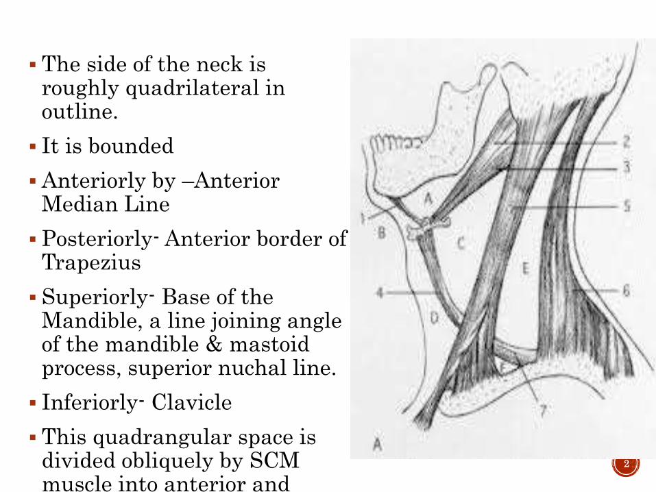
The side of the neck is roughly quadrilateral in outline.
It is bounded
Anteriorly by –Anterior Median Line
Posteriorly- Anterior border of Trapezius
Superiorly- Base of the Mandible, a line joining angle of the mandible & mastoid process, superior nuchal line.
Inferiorly- Clavicle
This quadrangular space is divided obliquely by SCM muscle into anterior and posterior triangles
2

STERNOCLEIDOMASTOID
Origin:
a) Sternal head
b) Clavicular head
Insertion:
a) Lateral process of mastoid process
b) Lateral half of the superior nuchal line of occipital bone
Nerve Supply:
Spinal Accessory Nerve
Blood Supply:
One br from sup thyroid A. and suprascapular A.
Two br from occipital A.
3
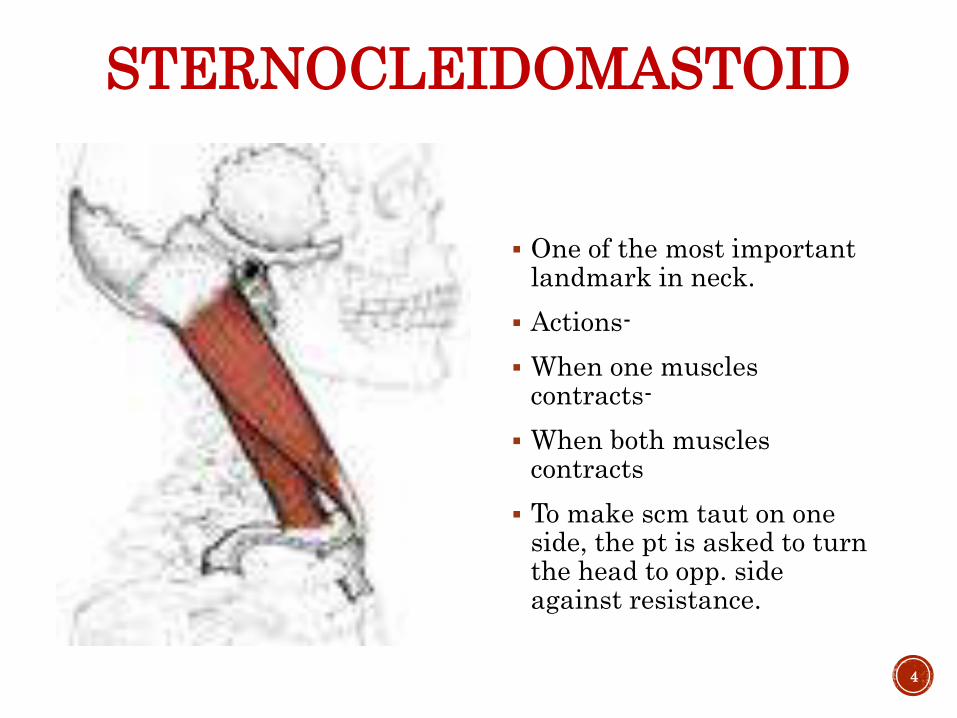
STERNOCLEIDOMASTOID
One of the most important landmark in neck.
Actions-
When one muscles contracts-
When both muscles contracts
To make scm taut on one side, the pt is asked to turn the head to opp. side against resistance.
4
PLATYSMA
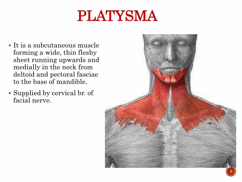
It is a subcutaneous muscle forming a wide, thin fleshy sheet running upwards and medially in the neck from deltoid and pectoral fasciae to the base of mandible.
Supplied by cervical br. of facial nerve.
5
ANTERIOR TRIANGLE OF NECK
Boundaries:
Medially: Ant median plane of neck
Laterally: SCM
Superiorly: Base of the mandible and a line joining the angle of the mandible n mastoid process
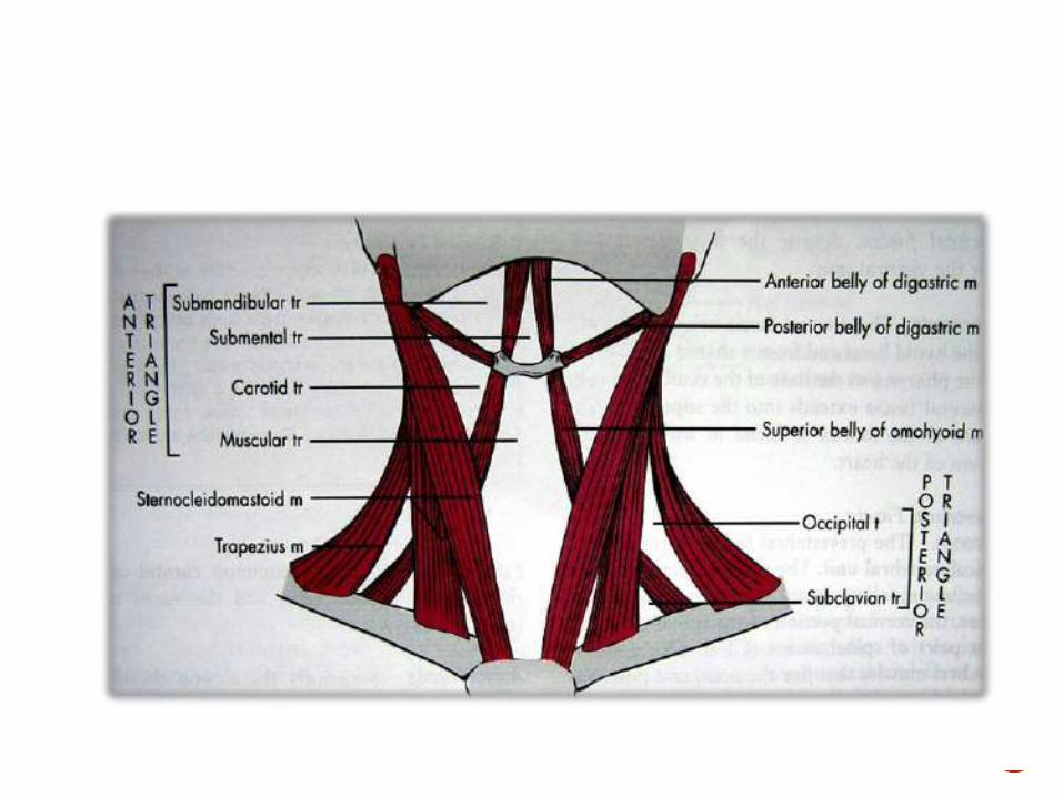
Subdivisions:
The ant triangle is subdivided by the digastric belly and sup belly of omohyoid into:
a) Submental
b) Digastric
c) Carotid and
d) Muscular Triangles
6
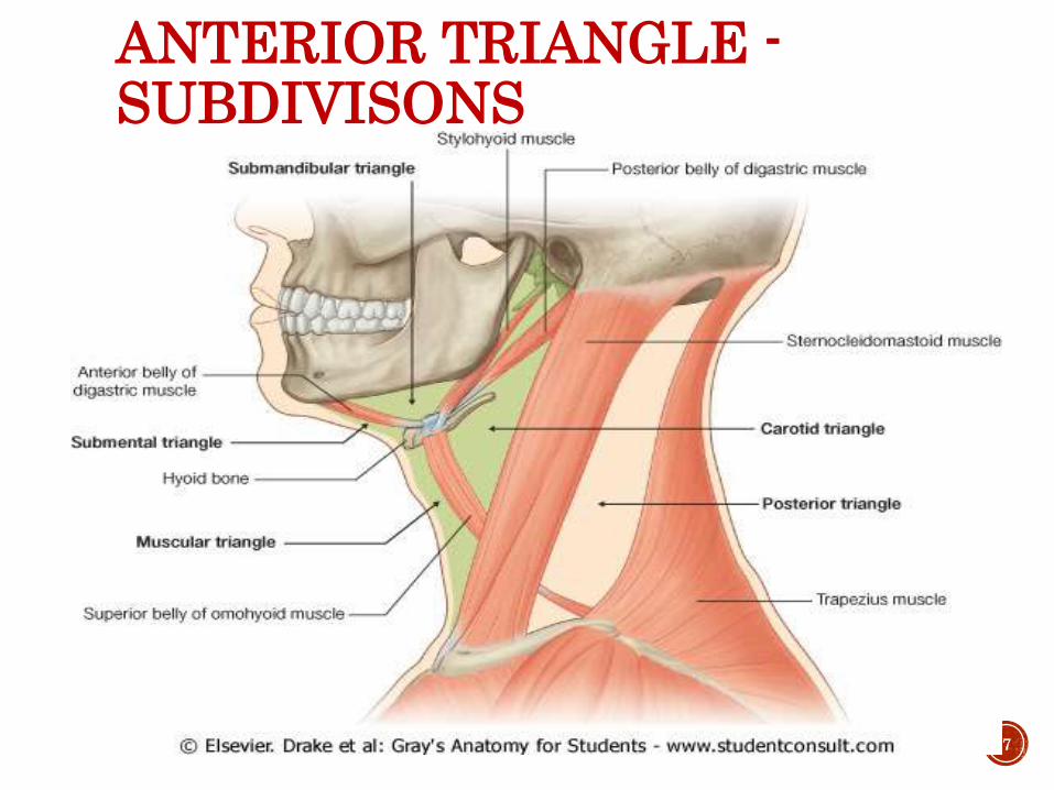
ANTERIOR TRIANGLE -SUBDIVISONS
7
8
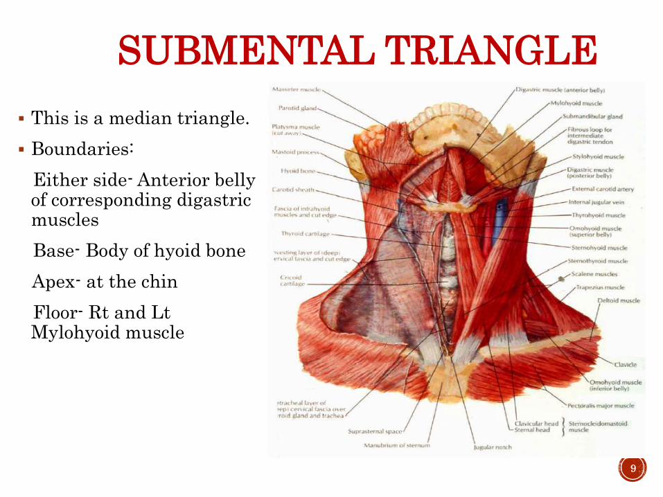
SUBMENTAL TRIANGLE
This is a median triangle.
Boundaries:
Either side- Anterior belly of corresponding digastricmuscles
Base- Body of hyoid bone
Apex- at the chin
Floor- Rt and Lt Mylohyoid muscle
9

CONTENTS OF SUBMENTAL TRIANGLE
It contains:
one or two lymph glands, the submental lymph nodes
some small veins; the latter unite to form the anterior jugular vein
The lymph nodes of the submental triangle receive lymph from the skin of the chin, the lower lip, the floor of the mouth, and the tip of the tongue. They send lymph to the submandibular and jugular chains of nodes. They belong to Level 1 gr of LNs
10

DIGASTRIC TRIANGLE The submandibular triangle
(or submaxillary or digastrictriangle) corresponds to the region of the neck immediately beneath the body of the mandible.
Boundaries:
Anteroinf: Ant belly of digastric
Posteroinf: Post belly of digastric and stylohyoid.
Superiorly: Base of the mandible.
Roof : Skin, superficial fascia with platysma muscle, deep fascia
Floor: Mylohyoid anteriorly and hyoglossus posteriorly, part of middle constrictor.
11

FLOOR OF DIGASTRICTRIANGLE
12

CONTENTS OF DIGASTRIC TRIANGLE
ANTERIOR PART OF TRIANGLE
Strucures superficial to mylohyoid are:
Superficial part of submandibular gland, Facial vein , Subman. LN (belong to Level 1 gr of LNs), Facial A.,Submental A., Mylohyoidnerve and vessels.
Structures superficial to hyoglossus: Subman.gland,Intermediatetendon of digastric and stylohyoid, Hypoglossal N.
13
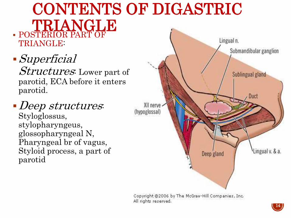
CONTENTS OF DIGASTRIC TRIANGLE
POSTERIOR PART OF TRIANGLE:
Superficial Structures: Lower part of parotid, ECA before it enters parotid.
Deep structures: Styloglossus, stylopharyngeus, glossopharyngeal N, Pharyngeal br of vagus, Styloid process, a part of parotid
14

CONTENTS OF POSTERIOR PART OF DIGASTRIC TRIANGLE
Deepest structures:
a) Internal carotid A.
b) Internal jugular vein
c) Vagus N.
• Submandibular LNs: Belong to Level 1 gr of LNs
• Clinically important because of their wide area of drainage. They are very commonly enlarged.
• They drain: a) center of forehead
b) Nose with Max. Frontal & Ethmoid sinuses
c) Inner canthus of the eye
d) Upper lip and the ant part of cheek with adjoinin gums
e)Outer part of lower lip with gums and teeth excluding incisors
f)Ant 2/3rd of tongue and floor of mouth
15

CAROTID TRIANGLE Boundaries:
Anterosup: Post belly of digastric
Anteroinf: Sup belly of omohyoid
Posteriorly: Ant border of SCM
Roof: skin, superficial fascia, investing layer of deep fascia
Floor: Thyrohyoid M, hyoglossus, Middle and Inf. constrictors of pharynx
16
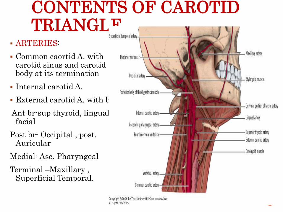
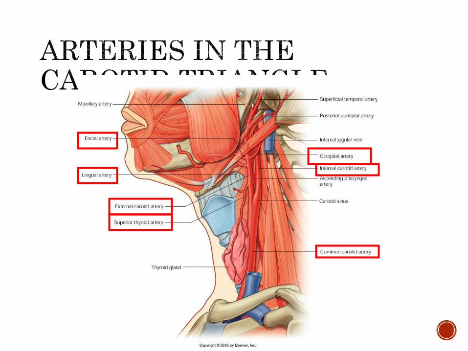
CONTENTS OF CAROTID TRIANGLE
ARTERIES:
Common caortid A. with carotid sinus and carotid body at its termination
Internal carotid A.
External carotid A. with br.-
Ant br-sup thyroid, lingual facial
Post br- Occipital , post. Auricular
Medial- Asc. Pharyngeal
Terminal –Maxillary , Superficial Temporal.
17

CONTENTS OF CAROTID TRIANGLE
VEINS:
Internal Jugular V.
Common Facial V.
Pharyngeal V
Lingual V. all draining in to internal jugular vein directly or via facial vein
19

CONTENTS OF CAROTID TRIANGLE
NERVES:
Vagus running vertically downwards
Sup L.N of vagus dividing into ext n int L.N
Spinal Accessory N
Hypoglossal N
Sympathetic chain
Carotid sheath and its contents
Lymph Nodes: Jugulo-digastric and jugulo-omohyoid
20

CONTENTS OF CAROTID TRIANGLE
ACCESSORY N.HYPOGLOSSAL N.
21

COMMON CAROTID ARTERIES
Right common carotid artery is a branch of the brachiocephalicartery.It begins in the neck behind the right sternoclavicular joint.
Left common carotid artery is a branch of the arch of aorta.Itascends to the back of the left sternoclavicular joint and enters the neck.
In the neck,each artery runs upwards within the carotid sheath,under cover of the anterior border of the sternocleidomastoidmuscle.
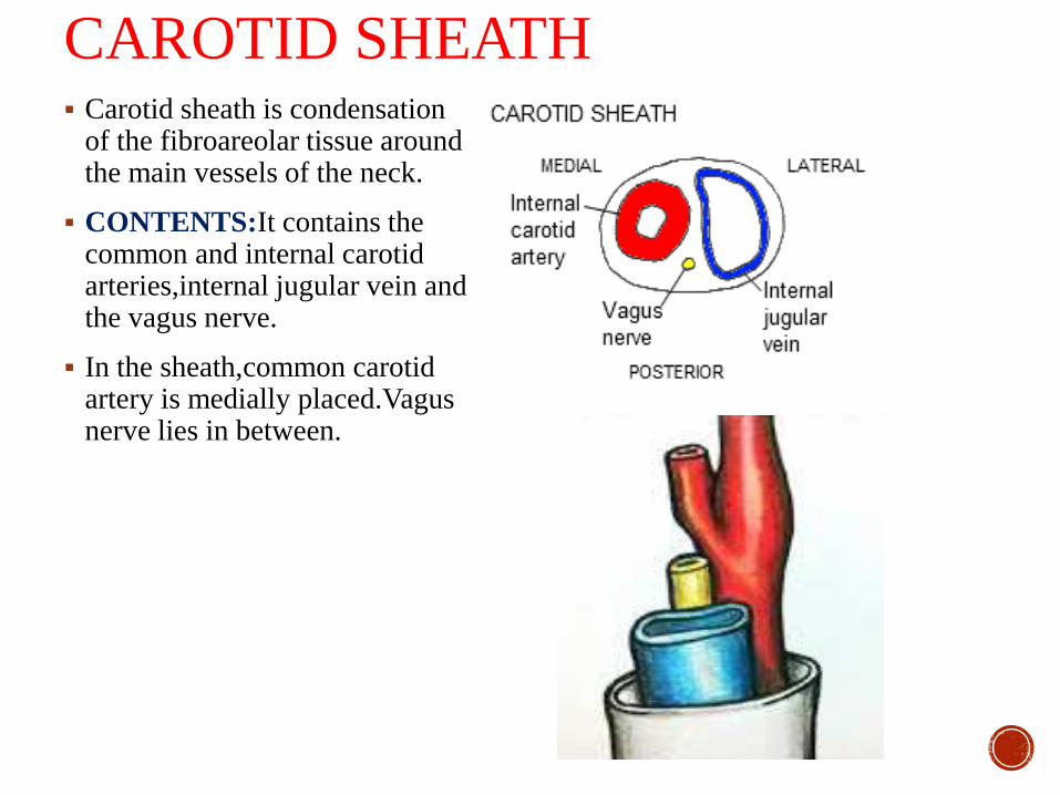
CAROTID SHEATH Carotid sheath is condensation
of the fibroareolar tissue around the main vessels of the neck.
CONTENTS:It contains the common and internal carotid arteries,internal jugular vein and the vagus nerve.
In the sheath,common carotid artery is medially placed.Vagusnerve lies in between.

RELATIONS
The ansa cervicalis lies embedded in the anterior wall of the carotid sheath.
The cervical sympathetic chain lies behind the sheath.

BIFURCATION OF COMMON CAROTID ARTERY Common carotid artery
bifurcates into external and internal carotid arteries at the level of upper border of the thyroid cartilage.
Two structures of importance at the bifurcation are
Carotid sinus
Carotid body

CAROTID SINUS AND BODY
26
CAROTID SINUS Carotid sinus is slight dilatation at the termination of the common
carotid artery or the beginning of the internal carotid artery.
It receives a rich innervation from the glossopharyngeal and sympathetic nerves.
FUNCTION:
Carotid sinus acts as a baroreceptor or pressure receptor and regulates pressure.

APPLIED ANATOMYCAROTID SINUS SYNDROME
Loss of consciousness due to simple head movements.
Hypersensitivity of the carotid sinus due to an unknown etiology.
Sudden slight pressure changes, such as that occasioned by movement of the head, may result in stimulation of the carotid sinus.
Impulses transmitted by the sinus reduce blood pressure and slow the pumping action of the heart.
Thus decreasing blood supply to the brain and resulting in sudden loss of consciousness.
While supporting the mandible care should be taken not to apply pressure on the carotid sinus.
CAROTID BODY Carotid body is a small,oval reddish-brown structure situated
behind the bifurcation.
It receives nerve supply mainly from the glossopharyngeal nerve, but also from the vagus and sympathetic nerves.
FUNCTION:
Carotid body acts as a chemoreceptor and responds to changes in the oxygen and carbon dioxide and Ph content of the blood.
EXTERNAL CAROTID ARTERY
Generally,it lies anterior to the internal carotid artery.
It is the chief artery of supply to structures in the front of the neck and in the face.

COURSE ECA begins in the carotid
triangle at the level of upper border of thyriod cartilage opposite the disc between the third and fourth cervical vertibrae.
In the carotid triangle,it lies under cover of the anterior border of the sternocleidomastiod muscle
As the artery ascends ,it passes deep to the post. Belly of digastric and stylohyoid muscle and terminates behind the neck of the mandible by dividing into the maxillary and superficial temporal arteries.

Has slightly curved course,so that it is anteromedial
to ICA in it lower part,and anterolateral to the ICA
in its upper part.

RELATIONSIN THE CAROTID TRIANGLE
Superficially—Cervical branch of facial nerve
Hypoglossal nerve
Facial,lingual,and superior
thyriod veins
Deep to the artery— Wall of pharynx
Superior laryngeal nerve
Ascending pharyngeal artery

ABOVE THE CAROTID TRIANGLE
Lies deep in the substance of the parotid triangle.
Within the gland, it is related
Superficially—Retromandibular vein
Facial nerve
Deep to the artery—ICA
Structures passing between ECA and ICA
Styloglossus
Stylopharyngeus
IXth nerve
Pharyngeal branch of
Xth nerve
Styloid process

BRANCHES OF EXT.CAROTID ARTERY
35

ANTERIOR BRANCHES
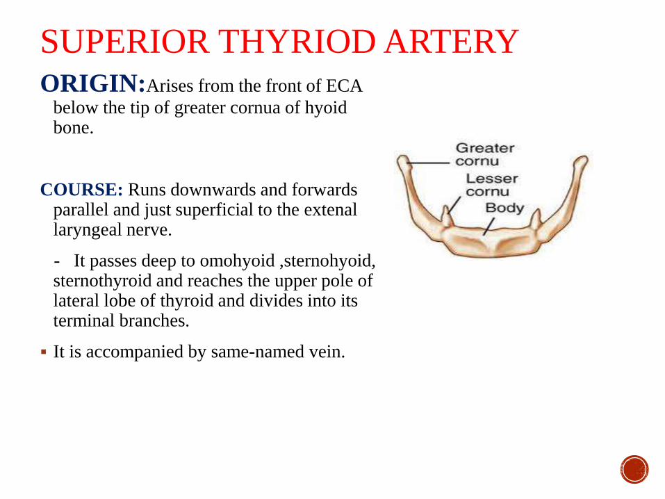
SUPERIOR THYRIOD ARTERYORIGIN:Arises from the front of ECA
below the tip of greater cornua of hyoid bone.
COURSE: Runs downwards and forwards parallel and just superficial to the extenallaryngeal nerve.
- It passes deep to omohyoid ,sternohyoid, sternothyroid and reaches the upper pole of lateral lobe of thyroid and divides into its terminal branches.
It is accompanied by same-named vein.
BRANCHES:
INFRAHYOID ARTERY
STERNOCLEIDOMASTOID ARTERY
SUPERIOR LARYNGEAL ARTERY
CRICOTHYROID ARTERY
GLANDULAR BRANCHES

APPLIED ANATOMY The arch of superior thyroid artery is characteristic – diagnostic landmark
The artery and external laryngeal nerve are close to each other higher up, but diverge slightly near the gland.
- So, ligature of superior thyroid artery in thyroid surgery should be made close to the gland in order to avoid injury of the external laryngeal nerve.
-Damage to the external laryngeal nerve causes some weakness of phonation due to loss of tightening effect of the cricothyriod on the vocal cord.
Intra-arterial infusion chemotherapy for laryngeal and hypopharyngealcancers.
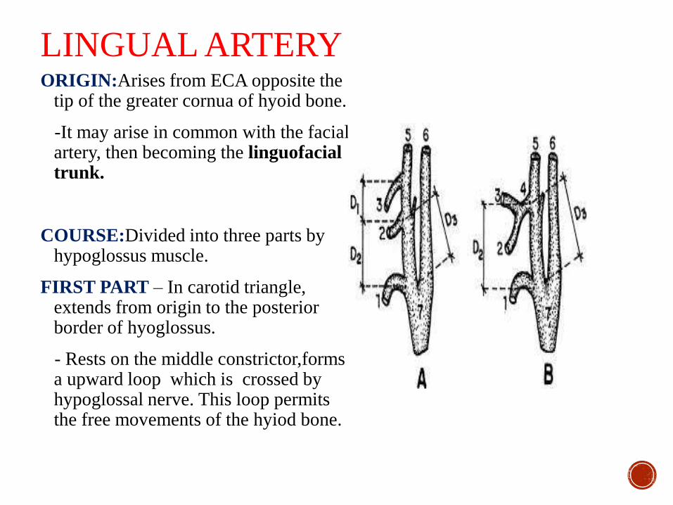
LINGUAL ARTERYORIGIN:Arises from ECA opposite the
tip of the greater cornua of hyoid bone.
-It may arise in common with the facial artery, then becoming the linguofacialtrunk.
COURSE:Divided into three parts by hypoglossus muscle.
FIRST PART – In carotid triangle, extends from origin to the posterior border of hyoglossus.
- Rests on the middle constrictor,formsa upward loop which is crossed by hypoglossal nerve. This loop permits the free movements of the hyiod bone.

SECOND PART – Deep to hyoglossus, runs horizontally forward along the upper border of hyoid bone between hyoglossuslaterally and middle constrictor, stylohyoid ligament medially.
THIRD PART [ ‘arteria profundalinguae’ ]—Also called as deep lingual artery.
-It runs upwards along the anterior Border of hyoglossus, then horizontally forwards on the undersurface of tongue on each side of frenum linguae.
-In vertical course,it lies b/t the genioglossus medially & inferior longitudinal muscle of tongue laterally. Horizontal part is accompanied by lingual nerve.

BRANCHES
SUPRAHYOID ARTERY
DORSAL LINGUAL ARTERY
SUBLINGUAL ARTERY
DEEP LINGUAL ARTERY
FACIAL ARTERYORIGIN: Arises from the ECA just above the tip of greater cornua
of hyoid bone.
COURSE: Runs upwards in -- neck as cervical part ; face -- facial part.
Tortuous course—In neck allows free movements of pharynx during deglutition,
on face -- free movements of mandible , lips, & cheek during mastication & facial expressions, escapes traction & pressure during movements.
.
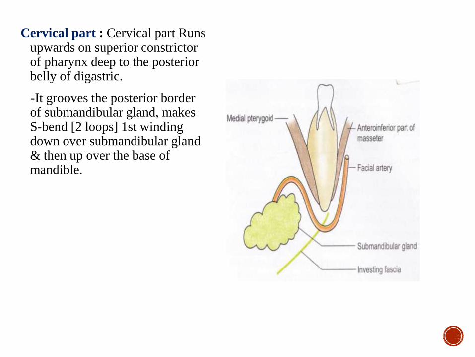
Cervical part : Cervical part Runs upwards on superior constrictor of pharynx deep to the posterior belly of digastric.
-It grooves the posterior border of submandibular gland, makes S-bend [2 loops] 1st winding down over submandibular gland & then up over the base of mandible.

Facial part:The vessel enters the face by winding around the base of the mandible, and by piercing the deep cervical fascia,at the anteroinferior angle of the masseter muscle.
It runs upwards and forwards deep to the risorus, to a point 1.25cm lateral to the angle of the mouth.
Then it ascends by the side of the nose upto the medial angle of the eye where it terminates by anastomosing with the dorsal nasal branch of the ophthalmic artery.
BRANCHES
CERVICAL PART:
ASCENDING PALATINE ARTERY
TONSILLAR A RTERY
GLANDULAR ARTERIES
SUBMENTAL ARTERY
FACIAL PART:
INFERIOR LABIAL ARTERY
SUPERIOR LABIAL ARTERY
LATERAL NASAL ARTERY
ANGULAR ARTERY
POSTERIOR BRANCHES

OCCIPITAL ARTERYORIGIN:Arises in carotid triangle
from posterior aspect of ECA ,opposite the origin of facial artery.
-It is crossed at its origin by hypoglossal nerve.
COURSE: Passes backwards and upwards along & under cover of lower border of post. Belly of diagastric , crossing carotid sheath, hypoglossal & accessory nerves.
Then it runs deep to the mastiodprocess and muscles attached to it i.e.,sternocleidomastiod,
digastric etc.

Then crosses the rectus capituslateralis,superior oblique,andsemispinalis capitus muscle at the apex of the posterior triangle.
Finally it pierces the trapezius muscle and ascends in a tortuous course in the superficial fascia of the scalp.
Its terminal portion comes to lie along the greater occipital nerve.

BRANCHES
STERNOMASTOID BRANCHES
AURICULAR BRANCH
MASTOID BRANCH
MENINGEAL BRANCH
MUSCULAR BRANCH

POSTERIOR AURICULAR ARTERYORIGIN: Arises from the posterior
aspect of the external carotid artery just above the posterior belly of the digastric.
COURSE:It runs upwards and backwards deep to parotid gland, but superficial to the styloidprocess.It crosses the base of the mastiod process and ascends behind the auricle.

BRANCHES
Stylomastoid.
Auricular
Occipital.
MEDIAL BRANCH
ASCENDING PHARYNGEAL ARTERY
ORIGIN:The smallest branch arising from the medial side of the external carotid artery, near its commencement.
COURSE: Ascends vertically between the internal carotid and the side of the pharynx, to the under surface of the base of the skull, lying on the Longuscapitis.

BRANCHES
PHARYNGEAL BRANCHES
PALATINE BRANCH
PREVERTEBRAL BRANCHES
INFERIOR TYMPANIC ARTERY
MENINGEAL BRANCHES
TERMINAL BRANCHES

MAXILLARY ARTERYORIGIN:Large terminal branch given
off behind the neck of the mandible.
COURSE: Divided into three parts by lateral pterygiod muscle.
The first or mandibular portionpasses horizontally forward, between the ramus of the mandible and the sphenomandibular ligament, where it lies parallel to and a little below the auriculotemporal nerve; it crosses the inferior alveolar nerve, and runs along the lower border of the lateral pterygiod.
The second or pterygoid portion runs obliquely forward and upward superficial to the lower head of the lateral pterygiod.
The third or pterygopalatine portion passes between the two heads of the lateral pterygiod and pterygomaxillary fissure,to enter into the pterygopalatine fossa where it lies in front of the sphenopalatine ganglion.

BRANCHESFirst or Mandibular Portion
Deep Auricular.
Anterior Tympanic.
Middle Meningeal
Accessory Meningeal
Inferior Alveolar.
Second or Pterygoid Portion
Deep Temporal.
Masseteric.
Pterygoid.
Buccinator.
Third or Pterygopalatine
Portion
•Posterior Superior
Alveolar.
•Infraorbital.
•Greater palatine artery
•Pharyngeal.
•Aretry of pterygiod canal
•Sphenopalatine.

SUPERFICIAL TEMPORAL ARTERYORIGIN: The smaller of the two terminal branches of the external
carotid, appears, to be the continuation of ECA. It begins in the substance of the parotid gland, behind the neck of the mandible.
COURSE: It runs vertically upwards crossing over the root of the zygomatic process
-about 5 cm. above this process it divides into two branches, a frontal and a parietal.

Relations.—As it crosses the zygomatic process, it is covered by the Auricularis anterior muscle, and by a dense fascia; it is crossed by the temporal and zygomaticbranches of the facial nerve and one or two veins, and is accompanied by the auriculotemporalnerve, which lies immediately behind it.

BRANCHESBesides some twigs to the parotid gland, to the
temporomandibular joint, and to the Masseter muscle,
its branches are:
Transverse Facial.
Anterior Auricular.
Middle Temporal.
Frontal.
Parietal

MUSCULAR TRIANGLE
Boundaries Anteriorly: midline of neck
Superiorly: superior belly of omohyoid.
Inferiorly: anterior border of
sternomastoid .
Contents
Sternohyoid muscle
Sternothyroidmuscle
Thyrohyoid muscle
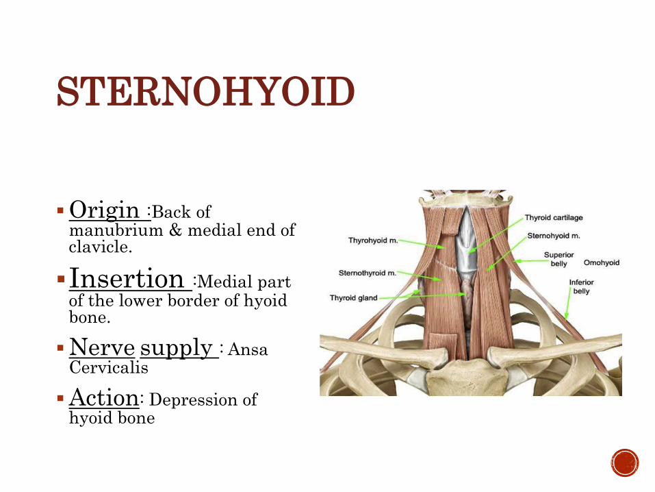
STERNOHYOID
Origin :Back of manubrium & medial end of clavicle.
Insertion :Medial part of the lower border of hyoid bone.
Nerve supply : AnsaCervicalis
Action: Depression of hyoid bone
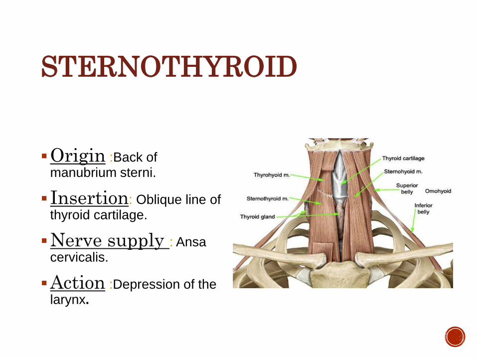
STERNOTHYROID
Origin :Back of manubrium sterni.
Insertion: Oblique line of thyroid cartilage.
Nerve supply : Ansacervicalis.
Action :Depression of the larynx.

THYROHYOID
Origin: Oblique line of the thyroid cartilage.
Insertion :Lower border of hyoid bone.
Nerve supply :hypoglossal nerve(C1fibers).
Action:Depression of hyoid or elevation of larynx.

INFRAHYOID / STRAP / RIBBON MUSCLES
70
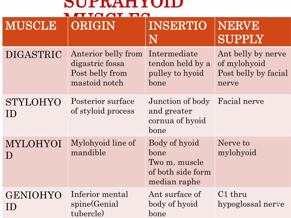
SUPRAHYOID MUSCLESMUSCLE ORIGIN INSERTIO
N
NERVE
SUPPLY
DIGASTRIC Anterior belly from
digastric fossa
Post belly from
mastoid notch
Intermediate
tendon held by a
pulley to hyoid
bone
Ant belly by nerve
of mylohyoid
Post belly by facial
nerve
STYLOHYO
ID
Posterior surface
of styloid process
Junction of body
and greater
cornua of hyoid
bone
Facial nerve
MYLOHYOI
D
Mylohyoid line of
mandible
Body of hyoid
bone
Two m. muscle
of both side form
median raphe
Nerve to
mylohyoid
GENIOHYO
ID
Inferior mental
spine(Genial
tubercle)
Ant surface of
body of hyoid
bone
C1 thru
hypoglossal nerve71

INFRAHYOID MUSCLESMuscle Origin Insertion Innervation
Sternohyoid Post surface
of manubrium
sterni
Medial part of
lower border of
hyoid bone
Ansa cervicalis
Sternothyr
oid
-Post surface
of manubrium
sterni
Oblique line on
lamina of
thyroid
cartilage
Ansa cervicalis
Thyrohyoid Oblique line
on lamina of
thyroid
cartilage
Body and greater
cornua of hyoid
bone
C1 thru
hypoglossal N
Omohyoid
Sup and inf
bellies
Upper border
of scapula
near
suprasternal
notch
Lower border of
body of hyoid
bone
Ansa cervicalis
72
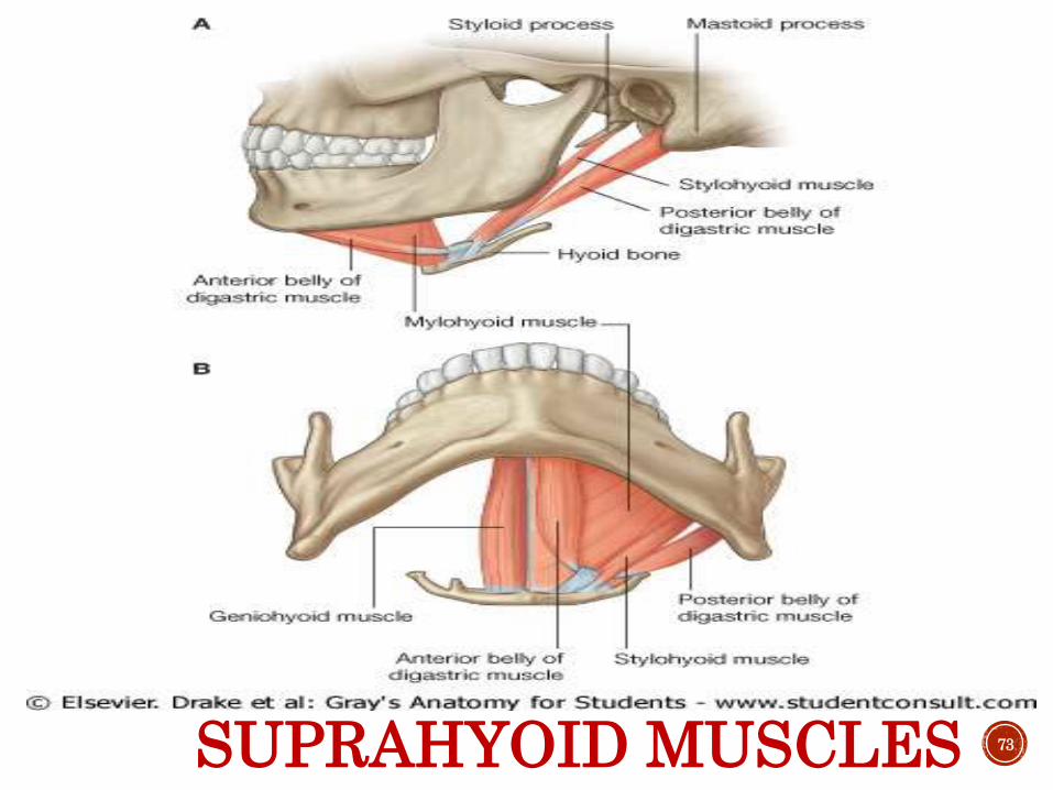
SUPRAHYOID MUSCLES 73

Table of Muscles
Muscle Origin Insertion Action Nerve Supply
Sternohyoid sternum hyoid ansa
OmohyoidSuprascapular notch
hyoid ansa
SternothyroidBelow sternohyoid on manubrium
Thyroid cartilage oblique line
ansa
ThyrohyoidThyroid cartilage oblique line
hyoid C1-C2 (ansa)
Anterior Belly Digastric
-----intermediate tendon------
Inner surface of mandile
Trigeminal nerve
Posterior BellyDigastric
Medial aspect of the mastoid process
-intermediate tendon-
Facial nerve
MylohyoidMylohyoid line of mandible
Hyoid boneTrigeminal nerve
Hyoglossus Hyoid boneLateral side of tongue
hypoglossal
Stylohyoid Styloid process hyoid Facial nerve
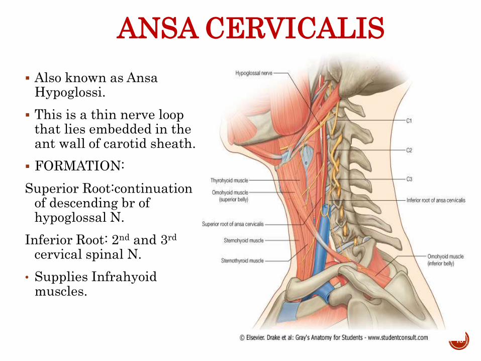
ANSA CERVICALIS
Also known as AnsaHypoglossi.
This is a thin nerve loop that lies embedded in the ant wall of carotid sheath.
FORMATION:
Superior Root:continuationof descending br of hypoglossal N.
Inferior Root: 2nd and 3rd
cervical spinal N.
• Supplies Infrahyoid muscles.
75
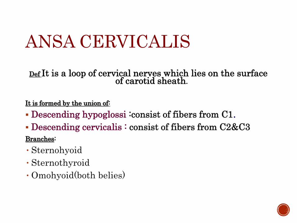
ANSA CERVICALIS
Def It is a loop of cervical nerves which lies on the surface of carotid sheath.
It is formed by the union of:
Descending hypoglossi :consist of fibers from C1.
Descending cervicalis : consist of fibers from C2&C3
Branches:
•Sternohyoid
•Sternothyroid
•Omohyoid(both belies)

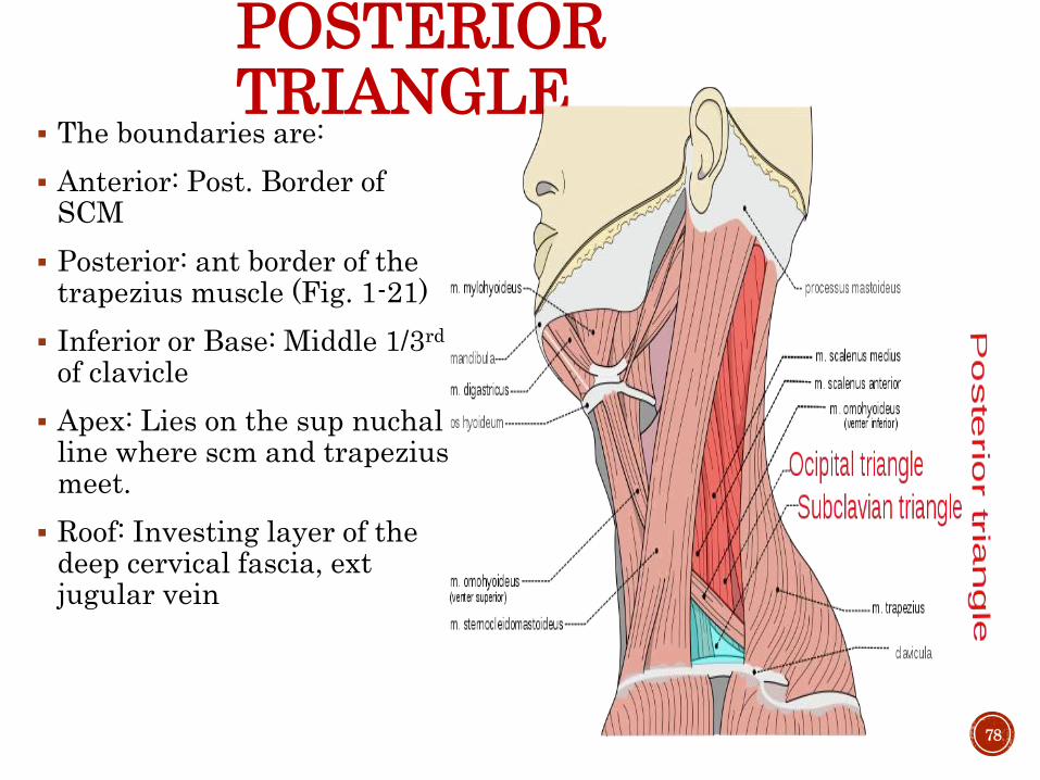
POSTERIOR TRIANGLE
The boundaries are:
Anterior: Post. Border of SCM
Posterior: ant border of the trapezius muscle (Fig. 1-21)
Inferior or Base: Middle 1/3rd
of clavicle
Apex: Lies on the sup nuchalline where scm and trapeziusmeet.
Roof: Investing layer of the deep cervical fascia, ext jugular vein
78

ROOF OF POSTERIOR TRIANGLE
79

FLOOR OF POSTERIOR TRIANGLE
Floor: prevertebral layer of deep cervical fascia, covering following muscles
a) Splenius capitus muscle,
b) Levator scapulae
c) Scalenus medius
d) Occassionally scalenusposterior
80

SUBDIVISIONS OF POSTERIOR TRIANGLE OF NECK Subdivided by inf. belly of
the omohyoid into
a) Larger upper triangle-Occipital triangle
b) Smaller lower triangle-Supraclavicular or Subclavian triangle
81

CONTENTS OF POSTERIOR TRIANGLE Nerves
• Accessory nerve
• Root, trunks of brachial plexus and their branches :
Nerves to rhomboideus(dorsal scapular n)
Nerves to serratus anterior(long thoracic n)
Nerves to subclavius
Suprascapular nerve
Cervical nerves
Greater occipital nerve
Great auriclular nerve
Lesser occipital nerve
Transverse cervical nerve of neck
Supraclavicular nerve
• 3rd and 4th cervical nerves supplying trapezius
82
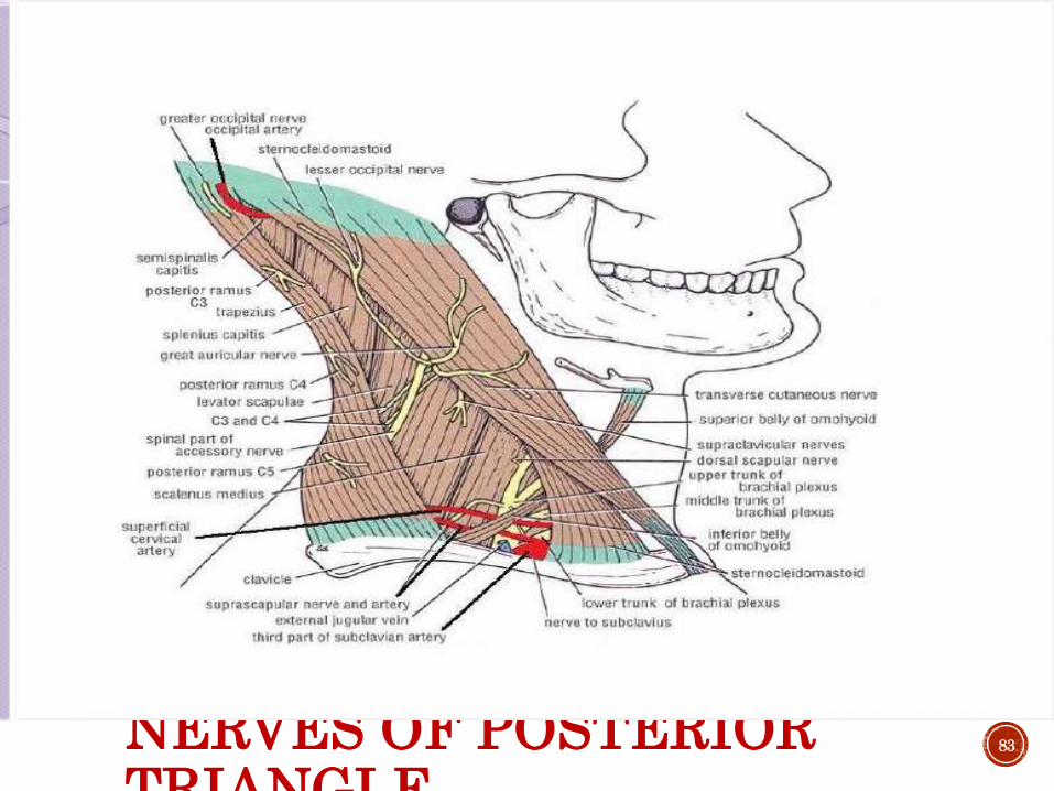
NERVES OF POSTERIOR TRIANGLE
83

ARTERIES OF POSTERIOR TRIANGLE
Arteries
Occipital artery
Third part of subclavianartery & branches of subclavianartery
Suprascapular
Transverse cervical
84
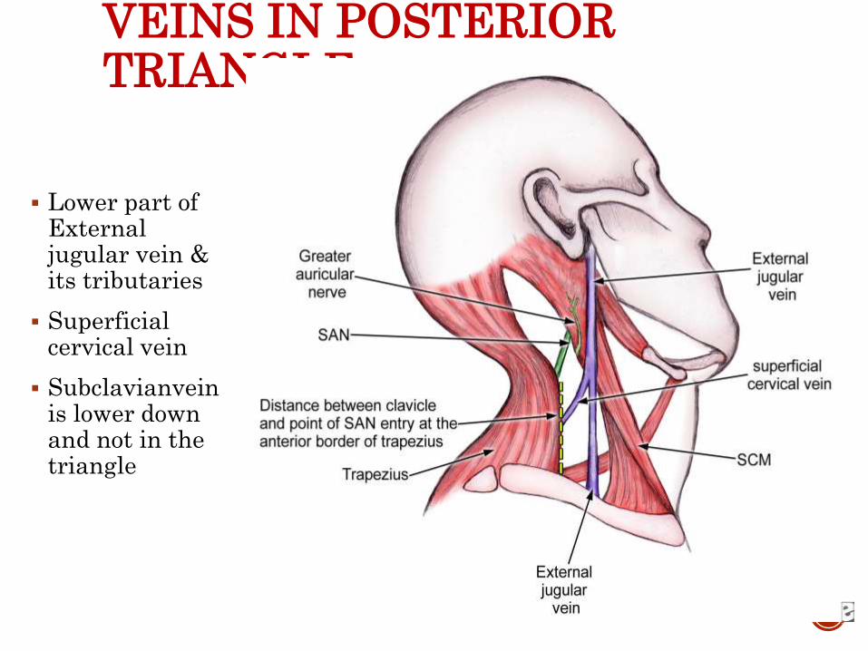
VEINS IN POSTERIOR TRIANGLE
Lower part of External jugular vein & its tributaries
Superficial cervical vein
Subclavianveinis lower down and not in the triangle
85
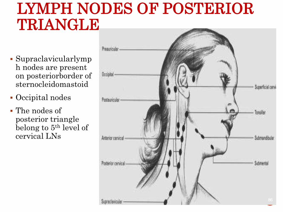
LYMPH NODES OF POSTERIOR TRIANGLE
Supraclavicularlymph nodes are present on posteriorborder of sternocleidomastoid
Occipital nodes
The nodes of posterior triangle belong to 5th level of cervical LNs
86


