TTP and BRF proteins nucleateprocessing body formation to silencemRNAs with AU-rich elementsTobias M. Franks and Jens Lykke-Andersen1
Molecular, Cellular and Developmental Biology, University of Colorado, Boulder, Colorado 80309, USA
In mammalian cells, mRNAs with AU-rich elements (AREs) are targeted for translational silencing and rapiddegradation. Here we present evidence that in human cells the proteins Tristetraprolin (TTP) and BRF-1deliver ARE-mRNAs to processing bodies (PBs), cytoplasmic assemblies of mRNAs, and associated factors thatpromote translational silencing and mRNA decay. First, depletion of endogenous TTP and BRF proteins, oroverexpression of dominant-negative mutant TTP proteins, impairs the localization of reporter ARE-mRNAsin PBs. Second, TTP and BRF-1 localize tethered mRNAs to PBs. Third, TTP can nucleate PB formation onuntranslated mRNAs even when other mRNAs are trapped in polysomes by cycloheximide treatment.ARE-mRNA localization in PBs is mediated by the TTP N- and C-terminal domains and occurs downstreamfrom mRNA polysome release, which in itself is not sufficient for mRNA PB localization. The accumulationof ARE-mRNAs in PBs is strongly enhanced when the mRNA decay machinery is rendered limiting by mRNAdecay enzyme depletion or TTP/BRF-1 overexpression. Based on these observations, we propose that the PBfunctions as a reservoir that sequesters ARE-mRNAs from polysomes, thereby silencing ARE-mRNA functioneven when mRNA decay is delayed. This function of the PB can likely be extended to other mRNA silencingpathways, such as those mediated by microRNAs, premature termination codons, and mRNA deadenylation.
Supplemental material is available at http://www.genesdev.org.
Received September 19, 2006; revised version accepted February 5, 2007.
mRNA turnover plays a key role in the regulation ofgene expression. AU-rich elements (AREs) are found inthe 3� untranslated region (UTR) of many humanmRNAs (ARE-mRNAs) that undergo translational si-lencing and rapid turnover, a number of which encodeinterleukins, cytokines, and proto-oncogenes (Chen andShyu 1995; Gueydan et al. 1999; Wilusz et al. 2001; Shimand Karin 2002; Zhang et al. 2002; Mazan-Mamczarz etal. 2006). AREs have been divided into different classes(Classes I, II, and III), depending on the occurrence of thesequence “AUUUA” (Chen and Shyu 1995). The AREsequences function as binding sites for trans-acting fac-tors that regulate ARE-mRNA translation and stability(Chen and Shyu 1995; Gueydan et al. 1999; Piecyk et al.2000; Wilusz et al. 2001; Shim and Karin 2002; Zhang etal. 2002; Lopez de Silanes et al. 2005; Mazan-Mamczarzet al. 2006).
One of the best-characterized ARE-binding proteins isTristetraprolin (TTP). TTP belongs to a family of humanARE-binding proteins that contains tandem CCCH zincfinger RNA-binding domains required for interaction
with the ARE (Lai et al. 2000; Blackshear 2002). Theother members of the TTP family are BRF-1 (also calledhsERF1/ZFP36L1/TIS11b) and BRF-2 (also calledhsERF2/ZFP36L2/TIS11d) (Lai et al. 2000; Stoecklin etal. 2002). TTP has been shown to destabilize a number ofClass II (containing overlapping AUUUA-repeats) ARE-containing mRNAs, including those that encode TumorNecrosis Factor � (TNF-�), Granulocyte Macrophage-Colony-Stimulating Factor (GM-CSF), and Interleukin-3(IL-3) proteins (Lai et al. 1999; Carballo et al. 2000;Stoecklin et al. 2000; Lai et al. 2006). The TTP paralogsBRF-1 and BRF-2 are less studied, but affect ARE-mRNAstability in a manner similar to TTP when overexpressedor depleted (Lai et al. 2000; Stoecklin et al. 2002). How-ever, whereas TTP is primarily expressed in lympho-cytes (Carballo et al. 1998), BRF-1 and BRF-2 appear to beexpressed in a wider range of tissues (Ramos et al. 2004;Stumpo et al. 2004). The mechanism by which TTP andBRF proteins destabilize ARE-mRNAs is not well under-stood. However, TTP and BRF-1 can target a tetherednon-ARE reporter mRNA for rapid decay (Lykke-Andersen and Wagner 2005). In addition, TTP and BRF-1interact with a number of mRNA decay enzymes respon-sible for deadenylation, decapping, and exonucleolyticdecay, as well as with the endonuclease Ago2, which is
1Corresponding author.E-MAIL [email protected]; FAX (303) 492-7744.Article is online at http://www.genesdev.org/cgi/doi/10.1101/gad.1494707.
involved in RNA interference (RNAi) (Chen et al. 2001;Jing et al. 2005; Lykke-Andersen and Wagner 2005).Thus, TTP and BRF-1 appear to activate decay of ARE-mRNAs by recruiting mRNA decay enzymes. OtherARE-binding proteins that influence ARE-mediatedmRNA decay have been identified and include K homol-ogy Splicing Regulatory Protein (KSRP), hnRNP D/AUF-1, and HuR (DeMaria and Brewer 1996; Fan and Steitz1998; Peng et al. 1998; Xu et al. 2001; Gherzi et al. 2004;Chou et al. 2006). However, it is not known how thedifferent ARE-binding proteins cooperate to regulate theturnover of ARE-mRNAs in the cell.
A recent exciting observation is that a number of pro-teins involved in translational silencing and mRNA de-cay concentrate in discrete cytoplasmic foci called pro-cessing bodies (PBs; also called GW or Dcp bodies) (forreview, see Anderson and Kedersha 2006). These pro-teins include the decapping enzyme Dcp2, the 5�-to-3�exonuclease Xrn1, and factors that stimulate decappingor inhibit general translation, as well as factors involvedin microRNA (miRNA)- and small interfering RNA(siRNA)-mediated mRNA silencing (Ingelfinger et al.2002; van Dijk et al. 2002; Eystathioy et al. 2003; Andreiet al. 2005; Ding et al. 2005; Fenger-Gron et al. 2005;Ferraiuolo et al. 2005; Fillman and Lykke-Andersen2005; Liu et al. 2005b; Pillai et al. 2005; Sen and Blau2005). PBs are highly dynamic structures, as evidencedby the ability of proteins and mRNAs to rapidly cycle inand out of PBs (Kedersha et al. 2005). Moreover, transla-tional inhibitors that trap mRNA in polysomes cause therapid disappearance of PBs, whereas inhibitors that trig-ger polysome disassociation cause enlarged PBs (Cougotet al. 2004; Brengues et al. 2005; Teixeira et al. 2005).This suggests that PBs only form in the presence of avail-able mRNA substrates and mRNAs exist in PBs to theexclusion of polysomes. In addition, the observation thatmRNA decay intermediates can be detected in PBs sug-gests that mRNA decay can take place in the PB (Shethand Parker 2003; Cougot et al. 2004). However, it re-mains unclear under which conditions mRNAs are se-questered in PBs and what delivers them there.
Here we present several lines of evidence that TTP andBRF-1 proteins deliver ARE-mRNAs in PBs. This isstrongly enhanced when the availability of mRNA decayenzymes is limiting, suggesting that TTP and BRF pro-teins sequester ARE-mRNAs in PBs when mRNA decayis inefficient. Based on these observations, we propose amodel for PB function in which PBs act to sequesterARE-mRNAs, as well as other PB-associated mRNAs,away from polysomes when the cellular mRNA decaymachinery is limiting, thus silencing mRNA functioneven when mRNA degradation is slow.
Results
ARE-mRNAs are observed in PBs
As a first step to test whether a link exists between PBsand the silencing of ARE-mRNAs, we asked whetherARE-mRNAs can be detected in PBs. �-globin reporter
mRNAs that contain the AREs from the 3� UTRs of GM-CSF (�-GMCSF) or TNF-� (�-TNF-�) mRNAs were ex-pressed in HeLa cells and subjected to in situ hybridiza-tion. These AREs were chosen because they are bothwell-defined targets of TTP (Taylor et al. 1996; Carballoet al. 2000), and TTP has been observed previously in PBsat low levels (Kedersha et al. 2005). Transcription of thereporter mRNAs is controlled by a tetracycline regula-tory promoter, which is activated by a tetracycline-re-pressible activator protein when tetracycline is absent(see Materials and Methods). hDcp1a fused to green fluo-rescent protein (GFP) served as a PB marker. The in situhybridization assays in Figure 1 show that both the�-GMCSF (panel 5) and �-TNF-� (panel 8) ARE-mRNAsconcentrate in PBs (observed in 75% and 64% of cells,respectively), while �-globin mRNA that contains noARE does not (�-wt; 0% of cells observed) (panel 2). We
Figure 1. ARE-mRNAs are observed in PBs. (A) In situ hybrid-ization assays showing localization in HeLa cells of exog-enously expressed wild-type �-globin mRNA (�-wt mRNA;panel 2), or �-globin mRNAs containing AREs from GM-CSF(�-GMCSF; panel 5) or TNF-� (�-TNF-�; panel 8) mRNAs. (Pan-els 1,4,7) PBs were visualized using GFP-hDcp1a. Images aremerged in panels 3, 6, and 9 (GFP-hDcp1a, green; �-globinmRNAs, red). An enlarged cell section (representing the area inthe dotted square) is shown in the top left corner for each image.The percentage of cells with reporter mRNAs in PBs is dis-played for each experiment with the total number of cellscounted indicated in parentheses. Note that the plasmid ex-pressing �-GMCSF mRNA also expressed (unfused) GFP, thussome nuclear GFP staining is observed in panels 4 and 6, as wellas in experiments using the same plasmid below. (B) In situhybridization assay showing localization of the �-GMCSFmRNA in the absence of GFP-hDcp1a.
observed that exogenously expressed GFP-hDcp1a, aswell as other tested PB factors, affect PB dynamics(Fenger-Gron et al. 2005; data not shown). However, theARE-mRNAs also localize to foci when GFP-hDcp1a isnot coexpressed (Fig. 1B), excluding the possibility thatARE-mRNA localization in PBs is an artifact of hDcp1aoverexpression. We conclude that ARE-mRNAs can beobserved in PBs.
The accumulation of ARE-mRNAs in PBs correlateswith cellular levels of TTP/BRF proteins
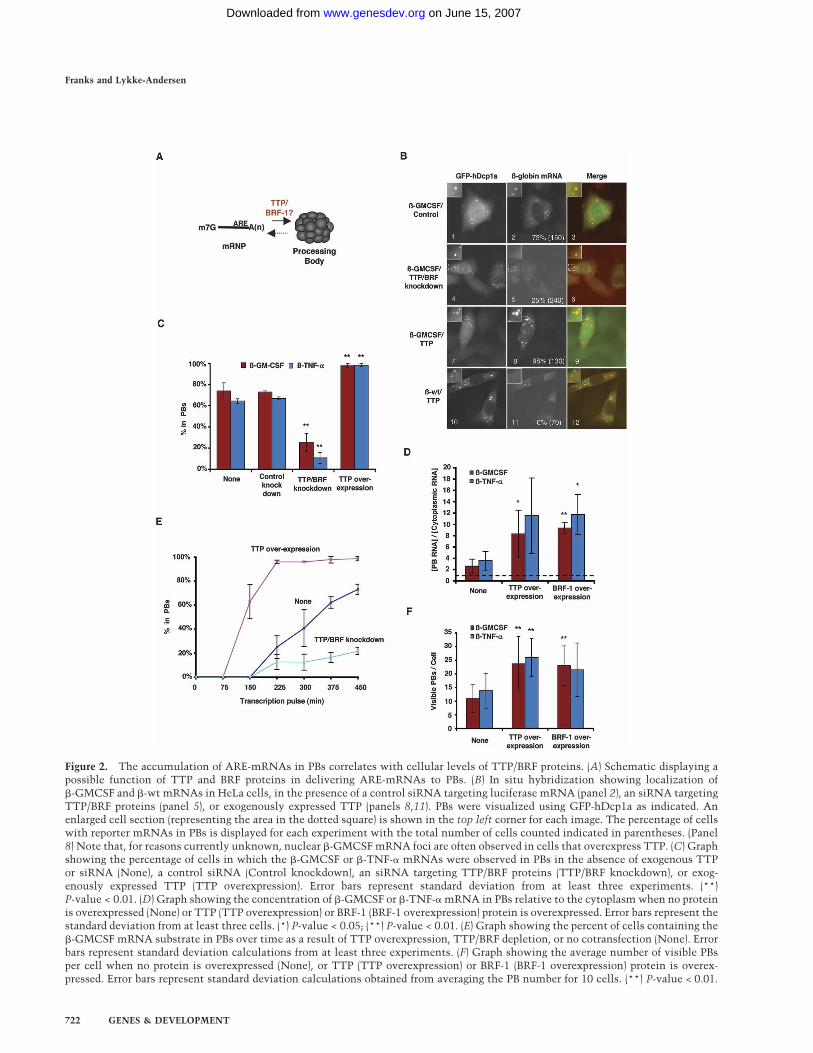
We next asked whether TTP and BRF proteins functionin the delivery of the �-GMCSF and �-TNF-� ARE-mRNAs to PBs (Fig. 2A). If TTP and BRF proteins deliversubstrate mRNAs to PBs, it is predicted that the accu-mulation of ARE-mRNA in PBs directly correlates withcellular TTP/BRF protein levels. To test this, we firstdepleted HeLa cells of TTP/BRF proteins (BRF-1 andBRF-2, but not TTP, could be detected in HeLa cells byWestern blotting) (data not shown) by using an siRNA,which targets a sequence conserved between theirmRNAs (Jing et al. 2005). Knocking down TTP/BRF pro-teins in this manner results in a twofold stabilization ofthe �-GMCSF and �-TNF-� mRNAs (Supplementary Fig.S1). As seen in Figure 2B, fewer cells concentrate�-GMCSF mRNA in PBs upon TTP/BRF protein deple-tion (panel 5; quantified in Fig. 2C). A similar effect isobserved with the �-TNF-� reporter mRNA (Supplemen-tary Fig. S2A, panel 5; quantified in Fig. 2C). Thus, areduction in TTP and BRF protein levels results in re-duced localization of ARE-mRNAs in PBs and slowermRNA decay.
It has been observed previously that overexpression ofTTP and BRF-1 results in enhancement of ARE-mediatedmRNA decay (Lai et al. 2000; Stoecklin et al. 2002;Lykke-Andersen and Wagner 2005). We therefore testedthe effect of TTP and BRF-1 overexpression on the local-ization of the reporter mRNAs. The in situ hybridizationassay in Figure 2B (panel 8) shows that overexpression ofTTP causes the �-GMCSF mRNA to accumulate in PBsat a higher concentration (approximately fourfold, quan-tified in Fig. 2D) and in an increased number of cells(quantified in Fig. 2C). Similar results were observedwith overexpressed BRF-1 (Supplementary Fig. S2B;quantified in Fig. 2D) and when TTP was coexpressedwith the �-TNF-� mRNA (Supplementary Fig. S2A,panel 8; quantified in Fig. 2C,D). Importantly, these ef-fects are specific to ARE-mRNAs because overexpres-sion of TTP does not lead to the accumulation in PBs of�-globin mRNA with no ARE (Fig. 2B, panel 11). Thetime-course experiment in Figure 2E, in which the accu-mulation of �-GMCSF mRNA in PBs was followed overtime after its transcriptional induction, shows that TTPoverexpression stimulates rapid accumulation of�-GMCSF mRNA in PBs, whereas TTP/BRF depletionhas the opposite effect. Interestingly, the number of vis-ible PBs per cell also increased ∼2.5-fold upon TTP orBRF-1 overexpression (Fig. 2F). While overexpression ofTTP has been observed previously to induce the forma-
tion of stress granules (Kedersha et al. 2005), the ARE-mRNA foci observed here correspond to PBs and notstress granules as they colocalize with GFP-hDcp1a, butnot with the stress granule marker TIA-1 (Supplemen-tary Fig. S2C).
We conclude that a strong correlation exists betweencellular TTP/BRF protein levels and the rate and extentof localization of �-GMCSF and �-TNF-� mRNAs inPBs. This suggests that TTP and BRF proteins functionto directly or indirectly mediate the localization of targetARE-mRNAs in PBs. In addition, the extent of ARE-mRNA PB localization directly correlates with the rateof ARE-mRNA decay.
TTP and BRF-1 can target a tethered mRNA to PBs
To further test the hypothesis that mRNAs that associ-ate with TTP and BRF-1 are targeted to PBs, we usedpreviously characterized tethering assays (Lykke-Andersen et al. 2000) to ask whether association withTTP and BRF-1 is sufficient to bring an mRNA to the PBin the absence of an ARE. A �-globin reporter mRNAthat contains six MS2 coat protein-binding sites in the3� UTR (�-6bs) was coexpressed with TTP or BRF-1 fusedto the MS2 coat protein (MS2-TTP and MS2-BRF-1). Thelocalization of the �-6bs mRNA was subsequently moni-tored by in situ hybridization. As seen in Figure 3, in thepresence of MS2-TTP or MS2-BRF-1, the �-6bs mRNAlocalizes in PBs in 29%–33% of cells (panels 5,8). In con-trast, the �-6bs mRNA does not significantly localize toPBs when expressed alone (Fig. 3, panel 2) or when co-expressed with unfused MS2 (Fig. 3, panel 11), TTP (Fig.3, panel 14), or BRF-1 proteins (Fig. 3, panel 17). Theenhancement of �-6bs localization in PBs correlates witha sevenfold to 7.5-fold increase in the rate of decay of themRNA in the presence of MS2-TTP or MS2-BRF-1(Lykke-Andersen and Wagner 2005). These results showthat TTP and BRF-1 are capable of targeting a tetherednon-ARE-mRNA to PBs. Interestingly, however, AREsare more efficient at targeting mRNAs to PBs than areMS2-TTP and MS2-BRF-1 (cf. panels 5,8 in Figs. 3 and 1).This suggests that tethered TTP and BRF proteins maynot completely recapitulate ARE function (see below).
The N- and C-terminal domains of TTP are PBlocalization domains
If TTP can target ARE-mRNAs to PBs, it is expected thatone or more domains of TTP are required to mediate thisprocess. We observed earlier that the N-terminal domain(NTD) and C-terminal domain (CTD) of TTP and BRF-1can each activate decay of an associated mRNA (Lykke-Andersen and Wagner 2005), whereas the RNA-bindingdomain (RBD) inhibits ARE-mediated mRNA decay in adominant-negative manner when overexpressed (Lai etal. 2000; Lykke-Andersen and Wagner 2005). We there-fore tested whether removal of the NTD or CTD of TTPreduces the ability of TTP to enhance the localization of
Figure 2. The accumulation of ARE-mRNAs in PBs correlates with cellular levels of TTP/BRF proteins. (A) Schematic displaying apossible function of TTP and BRF proteins in delivering ARE-mRNAs to PBs. (B) In situ hybridization showing localization of�-GMCSF and �-wt mRNAs in HeLa cells, in the presence of a control siRNA targeting luciferase mRNA (panel 2), an siRNA targetingTTP/BRF proteins (panel 5), or exogenously expressed TTP (panels 8,11). PBs were visualized using GFP-hDcp1a as indicated. Anenlarged cell section (representing the area in the dotted square) is shown in the top left corner for each image. The percentage of cellswith reporter mRNAs in PBs is displayed for each experiment with the total number of cells counted indicated in parentheses. (Panel8) Note that, for reasons currently unknown, nuclear �-GMCSF mRNA foci are often observed in cells that overexpress TTP. (C) Graphshowing the percentage of cells in which the �-GMCSF or �-TNF-� mRNAs were observed in PBs in the absence of exogenous TTPor siRNA (None), a control siRNA (Control knockdown), an siRNA targeting TTP/BRF proteins (TTP/BRF knockdown), or exog-enously expressed TTP (TTP overexpression). Error bars represent standard deviation from at least three experiments. (**)P-value < 0.01. (D) Graph showing the concentration of �-GMCSF or �-TNF-� mRNA in PBs relative to the cytoplasm when no proteinis overexpressed (None) or TTP (TTP overexpression) or BRF-1 (BRF-1 overexpression) protein is overexpressed. Error bars represent thestandard deviation from at least three cells. (*) P-value < 0.05; (**) P-value < 0.01. (E) Graph showing the percent of cells containing the�-GMCSF mRNA substrate in PBs over time as a result of TTP overexpression, TTP/BRF depletion, or no cotransfection (None). Errorbars represent standard deviation calculations from at least three experiments. (F) Graph showing the average number of visible PBsper cell when no protein is overexpressed (None), or TTP (TTP overexpression) or BRF-1 (BRF-1 overexpression) protein is overex-pressed. Error bars represent standard deviation calculations obtained from averaging the PB number for 10 cells. (**) P-value < 0.01.
�-GMCSF mRNA in PBs. The �-GMCSF mRNA was co-expressed with either a TTP-�NTD (TTP100–326) or aTTP-�CTD (TTP1–174) mutant protein (Fig. 4A), and PBlocalization was visualized by in situ hybridization. Theresults in Figure 4B show that neither the NTD (panel 2)nor the CTD (panel 5) of TTP is required to enhance theaccumulation of the �-GMCSF mRNA in PBs (quantifiedin Fig. 4C). This is corroborated by the time-course assayin Figure 4D. In contrast, �-GMCSF localization in PBswas strongly inhibited upon expression of the TTP RBD(TTP100–174), which lacks both the NTD and CTD (Fig.4B, panel 8, quantified in C,D). In addition, exogenousexpression of a mutant TTP protein that is incapable ofbinding the ARE (TTP-F126N) (Fig. 4A; Lai et al. 2002;
Lykke-Andersen and Wagner 2005) also inhibits accu-mulation of �-GMCSF mRNA in PBs (Fig. 4B panel 11,quantified in C,D). Similar results were observed withthe �-TNF-� ARE-mRNA substrate (Fig. 4C; Supplemen-tary Fig. S2A). These results suggest that the NTD andCTD of TTP function as redundant PB localizationdomains. Moreover, the ability of the overexpressedRNA-binding-deficient TTP-F126N protein to inhibit PBlocalization of �-GMCSF and �-TNF-� mRNAs in adominant-negative manner suggests that titratabletrans-acting factors that interact with TTP are impli-cated in the process.
If the NTD and CTD of TTP are in fact PB localizationdomains, it is expected that these protein fragments cantarget a tethered mRNA to PBs, similarly to TTP. Wetherefore tested whether the NTD (TTP1–100), CTD(TTP176–326), or RBD (TTP100–174) of TTP (Fig. 4A) aresufficient to recruit the tethered �-6bs mRNA to PBs.The in situ hybridization assays in Figure 4E demon-strate that, when fused to the MS2 coat protein, both theNTD and CTD of TTP are sufficient to target the �-6bsmRNA to PBs (panels 5,8), while the MS2 coat protein(panel 2) and the tethered RBD of TTP are not (panel 11).However, it is important to note that the NTD of TTPwas much less efficient than the CTD and the full-lengthprotein. This correlates with a previously observedweaker activation of mRNA decay by tethered NTD(Lykke-Andersen and Wagner 2005).
TTP and BRF-1 deliver nontranslating mRNAs to PBs
We next wished to gain insight into the mechanism bywhich TTP and BRF-1 shift the pool of ARE-mRNAsfrom polysomes to PBs. Previous studies have shownthat mRNAs exist in PBs to the exclusion of polysomes(Brengues et al. 2005; Coller and Parker 2005; Teixeira etal. 2005). We considered the possibilities that TTP andBRF-1 could facilitate the localization of ARE-mRNAs toPBs either directly, or indirectly by stimulating the re-lease of ARE-mRNAs from polysomes (Fig. 5A). It wasobserved previously that mRNAs that are released frompolysomes in yeast through cell starvation (Brengues etal. 2005) or in mammalian cells through miRNA-medi-ated translational silencing accumulate in PBs (Liu et al.2005b; Pillai et al. 2005). It was therefore possible thatTTP/BRF proteins mediate the localization of ARE-mRNAs in PBs solely through inhibition of polysomeformation. If this was the case, it would be predicted thatinhibition of mRNA polysome association stimulates PBlocalization even in the absence of TTP/BRF association.To test this, we asked whether insertion of a hairpin thatinhibits translation initiation (Kozak 1989) into the 5�UTR of our �-globin reporter mRNAs results in PB lo-calization. However, as seen in Figure 5B, insertion ofthe 5� UTR hairpin does not stimulate PB localization ofwild-type �-globin mRNA (�HP-wt) (panel 2) and doesnot enhance the localization in PBs of the �-GMCSF re-porter mRNA (�HP-GMCSF) (cf. panel 5 in Figs. 5B and1). Importantly, the hairpin efficiently inhibits polysomeformation, as evidenced by sucrose gradient polysome
Figure 3. TTP and BRF-1 can target a tethered mRNA to PBs.In situ hybridization assays showing localization of a �-globinmRNA with six MS2 coat protein-binding sites in the 3� UTR(�-6bs) in HeLa cells in the presence of exogenous MS2-TTP(panel 5), MS2-BRF-1 (panel 8), MS2 (panel 11), TTP (panel 14),or BRF-1 (panel 17) proteins, or no exogenous protein (panel 2).PBs were visualized by GFP-hDcp1a as indicated. An enlargedcell section (representing the area in the dotted square) is shownin the top left corner for each image. The percentage of cellswith reporter mRNAs in PBs is displayed for each experimentwith the total number of cells counted indicated in parentheses.
Figure 4. The NTD and CTD of TTP are PB localization domains. (A) Schematic showing TTP and mutants thereof. The NTD andCTD are indicated in gray, while the RBD is indicated in black. TTP-F126N contains a phenylalanine-to-asparagine mutation atposition 126. (B) In situ hybridization showing localization of �-GMCSF mRNA in HeLa cells in the presence of exogenously expressedTTP proteins lacking the NTD (TTP-�NTD; panel 2), the CTD (TTP-�CTD; panel 5), or both the NTD and CTD (TTP-RBD; panel 8),or in the presence of the TTP RNA-binding mutant protein (TTP-F126N; panel 11). PBs were visualized using GFP-hDcp1a. Anenlarged cell section (representing the area in the dotted square) is shown in the top left corner for each image. The percentage of cellswith reporter mRNAs in PBs is displayed for each experiment with the total number of cells counted indicated in parentheses. (C)Graph showing the percentage of cells in which �-GMCSF or �-TNF-� mRNAs were observed in PBs in the absence of exogenous TTPprotein (None) or TTP, TTP-�NTD, TTP-�CTD, TTP-RBD, or TTP-F126N proteins. Error bars represent standard deviation from atleast three separate experiments. (**) P-value < 0.01. (D) Graph showing the percent of cells that contain the �-GMCSF mRNAsubstrate in PBs over time as a result of the expression of wild-type and mutant TTP proteins. Error bars represent standard deviationcalculations from at least three experiments. (E) In situ hybridization showing localization of �-6bs mRNA in HeLa cells in thepresence of MS2 (panel 2), MS2-TTP-NTD (panel 5), MS2-TTP-CTD (panel 8), or MS2-TTP-RBD (panel 11). PBs were visualized usingGFP-hDcp1a. An enlarged cell section (representing the area in the dotted square) is shown in the top left corner for each image. Thepercentage of cells with reporter mRNAs in PBs is displayed for each experiment with the total number of cells counted indicated inparentheses.
Figure 5. TTP and BRF-1 deliver nontranslating mRNAs to PBs. (A) Schematic showing possible steps at which TTP/BRF proteinsmay act to facilitate the localization of ARE-mRNAs in PBs. (B) In situ hybridization assays showing localization in HeLa cells ofwild-type �-globin (�HP-wt; panel 2) or ARE-containing �-globin mRNA (�HP-GMCSF; panel 5) that each contain a stable hairpin intheir 5� UTR. PBs were visualized using GFP-hDcp1a. An enlarged cell section (representing the area in the dotted square) is shownin the top left corner for each image. The percentage of cells with reporter mRNAs in PBs is displayed for each experiment with thetotal number of cells counted indicated in parentheses. (C) Polysome fractionation analysis showing exclusion of hairpin-containingmRNAs from polysomes in HeLa tet-off cells. Polysomes were differentially sedimented by sucrose gradient and fractionated into 11fractions. Ribosomal rRNA and mRNA content was analyzed by methylene blue staining and Northern blot analysis, respectively. (D)In situ hybridization assay showing localization in HeLa cells of a �-globin mRNA with six MS2 coat protein-binding sites in the 3�
UTR and a stable hairpin in the 5� UTR (�HP-6bs) in the presence of exogenous MS2-TTP (panel 5), MS2-BRF-1 (panel 8), MS2 (panel11), TTP (panel 14), or BRF-1 (panel 17) proteins, or no exogenous protein (panel 2). PBs were visualized using GFP-hDcp1a. (E) Graphshowing the percentage of cells in which the �-GMCSF and �-6bs mRNAs localize to PBs in the absence (red bars) or presence (bluebars) of a stable hairpin in the 5� UTR. Error bars represent standard deviation calculations from at least three experiments. (*)P-value < 0.05; (**) P-value < 0.01.
fractionation assays (Fig. 5C). We conclude that inhibi-tion of polysome formation in itself is insufficient for PBlocalization of mRNA in human HeLa cells.
We next asked whether TTP and BRF-1 mediate thelocalization of nontranslating mRNAs to PBs. If this isthe case, it is expected that the translationally repressed�HP-6bs mRNA, which contains six MS2 coat protein-binding sites in the 3� UTR and a 5� UTR hairpin, willlocalize to PBs only when coexpressed with MS2-TTP orMS2-BRF-1. As seen in Figure 5D, both MS2-TTP andMS2-BRF-1 stimulate the localization of �HP-6bsmRNA in PBs (cf. panels 5,8 and 2). In contrast, unfusedMS2 (Fig. 5D, panel 11), TTP (Fig. 5D, panel 14), or BRF-1(Fig. 5D, panel 17) proteins do not stimulate PB localiza-tion of the �HP-6bs mRNA. We conclude that TTP/BRFproteins (Fig. 5D) and AREs (Fig. 5B) stimulate deliveryof mRNAs to PBs downstream from the release frompolysomes (Fig. 5A, right arrow). However, it cannot beexcluded that TTP/BRF proteins and AREs also play arole in polysome release (Fig. 5A, left arrow). In fact,AREs have been observed previously to inhibit transla-tion (Zhang et al. 2002). Interestingly, quantificationsrevealed that the translationally repressed �HP-6bsmRNA was more efficiently recruited to PBs by MS2-TTP and MS2-BRF-1 than was the corresponding trans-lated �-6bs mRNA (Fig. 5E). A similar enhancement wasobserved with the �HP-6bs mRNA as compared with the�-6bs mRNA tethered to the NTD or the CTD of TTP(data not shown). In contrast, localization of the�-GMCSF mRNA in PBs was not enhanced by transla-tional repression (Fig. 5E). This suggests that polysomerelease is a limiting step in localization of an mRNA toPBs by tethered TTP and BRF-1, but not by an ARE.Thus, either tethered TTP and BRF-1 may not fully re-capitulate endogenous TTP/BRF function, or ARE-bind-ing proteins other than TTP and BRF-1 stimulate ARE-mRNA polysome release.
The accumulation of ARE-mRNAs in PBs is stronglyenhanced upon mRNA decay enzyme depletion
The observations described above provide evidence thatTTP and BRF proteins sequester ARE-mRNAs in PBsaway from the translational machinery. To ask whethera correlation exists between the accumulation of ARE-mRNAs in PBs and the efficiency of mRNA decay, wetested the effect of depleting mRNA decay enzymes.HeLa cells were transfected with plasmids expressingthe �-GMCSF mRNA along with short hairpins targetingthe mRNAs for the 5�-to-3� exonuclease hXrn1 or thedecapping enzyme hDcp2, both catalytic components ofthe PB. As seen in Figure 6A, a dramatically enhancedaccumulation of �-GMCSF mRNA in PBs is observed inhXrn1- and hDcp2-depleted cells (panels 5,8), whereas noenhanced accumulation was observed with a controlhairpin targeting luciferase mRNA (panel 2). In contrastto the �-GMCSF mRNA, the �-wt mRNA, which lacksan ARE, accumulates in PBs in <10% of cells followingmRNA decay factor depletion (data not shown). The con-centration of the �-GMCSF mRNA in PBs was quanti-
fied relative to its concentration in the cytoplasm andrevealed a three- to fourfold increase upon hXrn1 orhDcp2 knockdown (Fig. 6B). This rules out the possibil-ity that the enhanced localization of ARE-mRNAs in PBsfollowing hXrn1 and hDcp2 knockdown is simply a re-sult of elevated ARE-mRNA levels (1.5-fold to twofoldhigher levels of �-GMCSF mRNA were observed) (datanot shown). In addition, we observed that localization ofexogenously expressed TTP (Fig. 6C) and BRF-1 (data notshown) to PBs is also strongly enhanced following hXrn1or hDcp2 depletion (Fig. 6C, cf. panels 5,8 and 2). Weconclude that TTP/BRF proteins and ARE-mRNAs accu-mulate in PBs following hDcp2 and hXrn1 depletion.
The exosome has been implicated previously in ARE-mRNA decay (Chen et al. 2001; Mukherjee et al. 2002),and knockdown of the exosome component PMScl-75was observed to impair ARE-mRNA decay (Stoecklin etal. 2006). In contrast to hXrn1 and hDcp2, it is not clearwhether the exosome associates with PBs, although exo-some subunits have been observed in cytoplasmic foci inDrosophila cells (Graham et al. 2006). As seen in Figure6A (panel 11), knockdown of PMScl-75 causes enhancedaccumulation of the �-GMCSF reporter in PBs similar towhat was observed following hDcp2 and hXrn1 deple-tion (quantified in Fig. 6B). Double knockdowns ofmRNA decay enzymes revealed even stronger accumu-lation of the �-GMCSF mRNA in PBs (Fig. 6A, panels14,17,20, quantified in B). We conclude that the�-GMCSF mRNA strongly accumulates in PBs upondepletion of mRNA decay enzymes. This suggests thatARE-mRNAs are sequestered in PBs when mRNA decayenzymes become limiting for the ARE-mRNA decaypathway. The importance of deadenylation for the local-ization of ARE-mRNAs in PBs is currently unclear, be-cause overexpression of catalytically inactive deadenyl-ases (hPan2 D1083A, hCcr4b D525A, hCaf1b D40A/E42A) showed no significant effect on the accumulationof �-GMCSF mRNA in PBs (data not shown), perhapsdue to the redundance of deadenylases in the human cell(Yamashita et al. 2005; Wagner et al. 2006).
TTP nucleates PB formation on substrate mRNAs
Based on the observations in Figure 6, we hypothesizedthat mRNAs only accumulate in visible PBs when theyare targeted for mRNA decay but their degradation isslow or absent. If this is the case, it is predicted that PBscan be re-created under conditions in which PBs are nor-mally absent (such as during cycloheximide treatment) ifthe mRNA decay machinery is rendered limiting. To testthis, we asked whether PBs containing ARE-mRNAs canbe generated in the presence of cycloheximide whenhDcp2 is knocked down or TTP is overexpressed. Asseen in Figure 7A, the �-GMCSF ARE-mRNA does notlocalize in PBs in the presence of cycloheximide (panel2). Similarly, the translationally silenced �HP-GMCSFARE-mRNA was not observed in PBs upon cyclohexi-mide treatment (Fig. 7A, panel 4), even though thismRNA is not trapped in polysomes under these condi-tions (Fig. 5C; data not shown). However, upon depletion
of the decapping enzyme hDcp2, 15% of cells form PBscontaining �HP-GMCSF mRNA in the presence of cy-cloheximide (Fig. 7A, panel 8). In contrast, little PB for-mation is observed with the translated �-GMCSF mRNA(Fig. 7A, panel 6). Thus, an untranslated ARE-mRNA cannucleate PB formation in the presence of cycloheximidewhen hDcp2 is knocked down. We also tested whetheroverexpressed TTP can nucleate PB formation in thepresence of cycloheximide. As seen in Figure 7A (panels10,12), overexpression of TTP triggers efficient PB for-
mation. This is particularly evident for the translation-ally repressed �HP-GMCSF mRNA (Fig. 7A, panel 12),but also was observed on the translated �-GMCSFmRNA (Fig. 7A, panel 10), suggesting that overexpressedTTP can localize ARE-mRNAs in PBs even before theirassociation with polysomes. When hDcp2 was depletedin combination with TTP overexpression, the accumu-lation of the �-GMCSF mRNAs in PBs was further en-hanced (Fig. 7A, panels 14,16).
We next tested the ability of tethered TTP to nucleate
Figure 6. ARE-mRNAs accumulate in PBs when mRNA decay enzymes are depleted. (A) In situ hybridization assays showingaccumulation of the �-GMCSF mRNA in PBs in HeLa cells expressing siRNAs targeting luciferase (panel 2), hXrn1 (panel 5), hDcp2(panel 8), PMScl-75 (panel 11), hXrn1 and hDcp2 (panel 14), hDcp2 and PMScl-75 (panel 17), or hXrn1 and PMScl-75 (panel 20). PBswere visualized using GFP-hDcp1a. An enlarged cell section (representing the area in the dotted square) is shown in the top left cornerfor each image. The percentage of cells with reporter mRNAs in PBs is displayed for each experiment with the total number of cellscounted indicated in parentheses. (B) Graph showing the average concentration of �-globin mRNA in PBs relative to the cytoplasm.Error bars represent standard deviation from measurements of at least three cells. (*) P-value < 0.05; (**) P-value < 0.01. (C) Indirectimmunofluorescence assays showing localization of endogenous hDcp1a (panels 1,4,7) and exogenous Myc-tagged TTP (panels 2,5,8)in untreated cells, or cells treated with siRNAs against hXrn1 or hDcp2, as indicated. The percentage of cells in which Myc-tagged TTPprotein was observed colocalizing with hDcp1a is shown with the total number of cells counted indicated in parentheses.
Figure 7. TTP can nucleate PB formation onsubstrate mRNAs in the presence of cyclohexi-mide. (A) In situ hybridization assays showinglocalization of �-GMCSF (left panels) and �HP-GMCSF (right panels) mRNAs in cycloheximide-treated HeLa cells in the presence of hDcp2 siRNAs(panels 5–8), TTP protein (panels 9–12), a combi-nation of TTP protein and hDcp2 siRNAs (panels13–16), or no exogenous siRNAs or protein (pan-els 1–4). PBs were visualized using GFP-hDcp1a.An enlarged cell section (representing the area inthe dotted square) is shown in the top left cornerfor each image. The percentage of cells with re-porter mRNAs in PBs is displayed for each ex-periment with the total number of cells countedindicated in parentheses. (B) In situ hybridizationassays showing localization of �-6bs (top left pan-els), �-GMCSF (bottom left panels), �HP-6bs (topright panels), or �HP-GMCSF (bottom right pan-els) mRNAs in cycloheximide-treated HeLa cellsin the presence of hDcp2 siRNAs (panels 5–8),MS2-TTP-F126N protein (panels 9–12,17–20), ora combination of both MS2-TTP-F126N proteinand hDcp2 siRNAs (panels 13–16), or no exog-enous siRNAs or protein (panels 1–4). PBs werevisualized using GFP-hDcp1a. The percentage ofcells with reporter mRNAs in PBs is displayed foreach experiment with the total number of cellscounted indicated in parentheses.
PB formation. As seen in Figure 7B (panel 10), in thepresence of cycloheximide the ARE-binding-deficientmutant TTP-F126N protein stimulates formation of PBswhen tethered via the MS2 coat protein to the �-6bsmRNA. This is strongly enhanced when polysome for-mation on the �-6bs mRNA is inhibited by insertion ofthe 5� UTR hairpin (Fig. 7B, panel 12) and when hDcp2 issimultaneously knocked down (Fig. 7B, panels 14,16). Incontrast, hDcp2 knockdown was insufficient to stimu-late PB localization of the �-6bs mRNAs (Fig. 7B, panels6,8). Importantly, the MS2-TTP-F126N protein likelyhas no cellular mRNA targets other than the �-6bsmRNAs, since it is mutant for ARE binding (Lai et al.2002; Lykke-Andersen and Wagner 2005) and does notstimulate PB localization of translated or untranslated�-GMCSF ARE-mRNA (Fig. 7B, panels 18,20). Thus, thePBs observed in Figure 7B are likely highly homoge-neous, containing primarily the �-6bs reporter mRNAs.We conclude that TTP can nucleate PB formation onsubstrate mRNAs under conditions in which othermRNAs are trapped in polysomes. This only occurswhen the mRNA decay machinery is rendered limitingby hDcp2 depletion or TTP overexpression.
Discussion
AREs are key elements in post-transcriptional gene regu-lation that target mRNAs in which they reside for trans-
lational silencing and decay, thereby regulating the ex-pression of multiple human genes (Chen and Shyu 1995;Wilusz et al. 2001; Shim and Karin 2002; Zhang et al.2002). It was observed recently that a number of factorsinvolved in mRNA decay and translational silencingconcentrate in cytoplasmic foci called PBs (van Dijk etal. 2002; Eystathioy et al. 2003; Sheth and Parker 2003;Cougot et al. 2004). Here we show that the ARE-bindingproteins TTP and BRF-1 nucleate PB formation on ARE-mRNAs. This silences the ARE-mRNAs by sequesteringthem from the translation machinery until they undergomRNA decay (Fig. 8A).
TTP and BRF proteins mediate the localizationof ARE-mRNAs in PBs
The mechanism by which AREs repress protein expres-sion is poorly understood, but involves both transla-tional silencing and mRNA decay (Chen and Shyu 1995;Gueydan et al. 1999; Piecyk et al. 2000; Wilusz et al.2001; Shim and Karin 2002; Lopez de Silanes et al. 2005;Mazan-Mamczarz et al. 2006). Here, we show evidencethat ARE-mRNA silencing involves delivery of ARE-mRNAs to PBs by the proteins TTP and BRF-1. First,�-GMCSF and �-TNF-� ARE-mRNAs are observed inPBs (Fig. 1). This localization is inhibited upon depletionof TTP/BRF proteins using siRNAs or upon overexpres-sion of dominant-negative mutant TTP proteins (Figs. 2,
Figure 8. Model for mRNA silencing by PB localization in human cells. (A) TTP and BRF proteins nucleate the formation of asubmicroscopic PB subcomplex on ARE-mRNAs. When decay enzymes are limiting, this PB subcomplex aggregates with othermRNA-PB subcomplexes to form PBs, which can grow large enough to be microscopically visible. (B) Under conditions in whichmRNA decay enzymes are nonlimiting, mRNAs are efficiently turned over before they can aggregate into a PB, and thus PBs remainsubmicroscopic (above the dotted line). When mRNA decay enzymes are limiting, PB subcomplexes form larger aggregates (below thedotted line) visible to the microscope that serve to silence translation until mRNA degradation (see Discussion).
4). In contrast, localization of �-GMCSF and �-TNF-�mRNAs in PBs is enhanced upon TTP or BRF-1 overex-pression (Fig. 2; Supplementary Fig. S2). Second, TTP andBRF-1, as well as the NTD and CTD of TTP, can targeta tethered non-ARE mRNA to PBs (Figs. 3, 4). However,it is important to note that targeting to PBs of �-globinmRNA by tethered TTP and BRF-1 is not as efficient asby AREs. This suggests that the function of the ARE isnot completely recapitulated by tethered TTP and BRF-1. Nevertheless, our experiments demonstrate that theTTP family of ARE-binding proteins deliver target ARE-mRNAs to PBs and, in the case of TTP, this can be me-diated by both the NTD and CTD.
TTP and BRF proteins target polysome-releasedARE-mRNAs to PBs
How do TTP/BRF proteins shift the equilibrium of ARE-mRNAs from polysomes to PBs? The following observa-tions suggest that TTP/BRF proteins act, at least in part,to target ARE-mRNAs to PBs subsequent to their poly-some release (Fig. 8A). First, the presence of a hairpinthat efficiently inhibits polysome formation does nottarget an mRNA to PBs unless the mRNA contains anARE or is tethered to TTP or BRF-1 (Fig. 5). Second, TTPcan nucleate the formation of PBs on an ARE-mRNA ora tethered mRNA even when other PB substrates aretrapped in polysomes by cycloheximide treatment (Fig.7). Thus, polysome release is insufficient for mRNA PBlocalization, and TTP and BRF-1 efficiently localize non-translated mRNAs to PBs.
This raises the question of how TTP and BRF proteinspromote localization of ARE-mRNAs in PBs. Our obser-vations suggest that this is mediated through direct in-teractions between TTP/BRF proteins and PB subunits.First, TTP and BRF-1 stimulate PB localization of mRNAalready released from polysomes (Fig. 5). Second, overex-pression of the ARE-binding-deficient TTP-F126N mu-tant protein inhibits ARE-mRNA localization in PBs(Fig. 4), suggesting that titratable trans-acting factorsthat bind to TTP are important for PB localization.Third, TTP exists in complex with several PB factors(Fenger-Gron et al. 2005). Some of these interactions ap-pear to be direct, as evidenced by interaction assays be-tween bacterially purified TTP and PB componentstranslated in rabbit reticulocyte lysates (C. Egan, T.M.Franks, and J. Lykke-Andersen, unpubl.). Taken to-gether, these observations suggest that TTP nucleatesthe formation of a PB “subcomplex” on ARE-mRNAsthat subsequently mediates the association with thePB (Fig. 8A). Once a PB subcomplex is nucleated byTTP/BRF proteins, how does it mediate the localizationof ARE-mRNAs in PBs? Future studies should revealwhether this is a result of active transport or passivediffusion followed by retention in the PB. However, wehave not observed any effect on PB formation of de-pleting actin filaments or microtubules using cytoskel-eton inhibitors (J. Dennis and J. Lykke-Andersen, un-publ.).
Do ARE-binding proteins actively release ARE-mRNAsfrom polysomes?
Experiments using translational inhibitors suggest thatmRNAs need to be released from polysomes prior totheir localization in PBs (Fig. 8A; Cougot et al. 2004;Teixeira et al. 2005). Our observation that a 5� UTR hair-pin that represses translation initiation does not enhancelocalization of the �-GMCSF ARE-mRNA in PBs (Fig. 5)suggests that ARE-binding proteins may stimulate poly-some release, thus negating the effect of the 5� UTR hair-pin. This is consistent with observations by others thatAREs can repress translation (Zhang et al. 2002). Inter-estingly, tethered TTP and BRF-1 were less efficient thanan ARE at localizing an mRNA in PBs (Figs. 1, 3), butregained activity similar to an ARE when translationwas inhibited (Fig. 5). This suggests that either TTP andBRF-1 do not efficiently release bound mRNAs frompolysomes, or the tethered proteins do not fully recapitu-late endogenous protein function. Using sucrose gradientpolysome assays, we observed no evidence that an AREor tethered TTP or BRF-1 stimulates accumulation ofpolysome-released mRNA (data not shown). However, itis possible that, under these conditions, the releasedmRNA is rapidly degraded and therefore is undetectable.Candidate factors implicated in releasing ARE-mRNAsfrom polysomes include (in addition to TTP and BRFproteins) the microRNA miR16 and the associated RNA-induced silencing complex (RISC), which has been im-plicated in the decay of mRNAs containing the AREfrom TNF-� mRNA (Jing et al. 2005), as well as the trans-lation repressor proteins TIA-1 and TIAR, which haveaffinity for AREs (Dember et al. 1996; Gueydan et al.1999; Piecyk et al. 2000; Lopez de Silanes et al. 2005;Mazan-Mamczarz et al. 2006). In addition, general trans-lation repressors that are components of PBs may stimu-late localization of ARE-mRNAs in PBs by repressingpolysome formation. Interestingly, the PB componentRck/p54 exists in complex with TTP (Fenger-Gron et al.2005), and its yeast ortholog, Dhh1p, is a translationalrepressor (Coller and Parker 2005). An important goal forfuture studies is to delineate how the multiple ARE-binding proteins and their associated factors cooperate tosilence ARE-mRNAs.
TTP and BRF proteins facilitate the localizationof ARE-mRNAs in PBs when mRNA decay enzymesare limiting
Our studies raise the important question of under whichconditions TTP and BRF proteins translocate ARE-mRNAs from the cytoplasm to PBs. The following ob-servations suggest that the sequestration of the ARE-mRNAs into microscopically visible PBs takes place un-der conditions in which mRNA decay enzymes acting onthe ARE-mRNA become limiting (Fig. 8A). First, whenlevels of hDcp2, hXrn1, or PMScl-75 are reduced byknockdown, ARE-mRNAs accumulate at higher concen-trations in PBs (Fig. 6). Second, when TTP or BRF-1 pro-teins are overexpressed, which is predicted to leave en-
dogenous mRNA decay factors limiting for TTP andBRF-1 function, the ARE-mRNAs accumulate at highlevels in the PBs (Fig. 2; Supplementary Fig. S2). Third,TTP can nucleate the formation of PBs on substratemRNAs during cycloheximide treatment only whenTTP is overexpressed or hDcp2 is depleted (Fig. 7). Thus,TTP and BRF-1 sequester ARE-mRNAs in PBs when theavailability of mRNA decay enzymes becomes limitingfor ARE-mRNA decay (Fig. 8A). This is predicted to se-quester the ARE-mRNA away from the translation ma-chinery, thereby providing an efficient means of silenc-ing ARE-mRNA function even while the mRNA isawaiting decay.
Can the sequestered ARE-mRNAs degrade in the PB,or do they have to be released from the PB prior to theirdecay? The observations that the catalytic mRNA decayenzymes hDcp2 and hXrn1 concentrate in PBs (Shethand Parker 2003; Cougot et al. 2004) and that theirknockdown results in enhanced accumulation of ARE-mRNAs in PBs (Fig. 6) suggest that these enzymes func-tion to degrade ARE-mRNAs in the PB. Moreover, anymanipulation of TTP and BRF-1 proteins that enhancesARE-mRNA decay also stimulates ARE-mRNA localiza-tion in PBs (Fig. 2), whereas those conditions that impairTTP/BRF-1 function result in decreased degradation andPB localization of the ARE-mRNAs (Figs. 2, 4). However,while these observations suggest that ARE-mRNAs candegrade in PBs, it is a formal possibility that mRNA de-cay enzymes are kept inactive in the PB and only degrademRNAs when both the mRNA and the mRNA decayenzymes are released from the PB. Even if ARE-mRNAdegrades in the PB, it remains to be determined whatfraction of the total cellular mRNA degrades there.Moreover, it is unknown whether the exosome assists inARE-mRNA decay in the general cytoplasm or in con-junction with PBs or PB subunits. Indirect immunofluo-rescence assays have revealed that several Drosophilaexosome subunits are observed in cytoplasmic foci, al-though it is unclear whether these correspond to PBs(Graham et al. 2006).
A general function of PBs in sequestering mRNAs thatundergo delayed decay
The model in Figure 8A can likely be extended to othermRNAs that interface with PBs. In this scenario, themiRISC complex would serve (via GW182) to nucleatePBs on miRNA target mRNAs (Yang et al. 2004; Ding etal. 2005; Jakymiw et al. 2005; Liu et al. 2005b; Pauley etal. 2006), whereas Upf proteins mediate PB formation onNMD substrates (Sheth and Parker 2006)—in humancells, likely via hSmg7 (Unterholzner and Izaurralde2004). In addition, it can be speculated that, at least inyeast, mRNAs that lack destabilizing cis-elements aretargeted after their deadenylation to PBs by the Lsm1-7–Pat1 complex, which localizes in PBs (Sheth and Parker2003) and specifically associates with and activates de-cay of deadenylated mRNAs (Tharun et al. 2000; Tharunand Parker 2001).
If the model in Figure 8A provides a general paradigm
for how mRNAs interface with PBs, it is predicted thatthe size of PBs in the cell directly correlates with theavailability of mRNA substrates and inversely correlateswith the availability of mRNA decay enzymes (Fig. 8B).These predictions are consistent with our observations,as well as with observations by others, of the dynamicsof PBs. For example, PBs grow large when the mRNAdecay machinery is rendered limiting by an increase inthe level of polysome-free cytoplasmic mRNA by puro-mycin treatment (Teixeira et al. 2005), or by a reductionin cellular Dcp2 and Xrn1 activity (Fig. 6; Sheth andParker 2003; Cougot et al. 2004). In contrast, PBs disap-pear when mRNA substrates become limiting upon en-trapment of translated mRNAs in polysomes by cyclo-heximide treatment (Fig. 7; Cougot et al. 2004; Teixeiraet al. 2005), or when decapping is rendered nonlimitingby hDcp2 overexpression (Fenger-Gron et al. 2005).These considerations provide a possible explanation forthe observation that PBs disappear when miRNAs aredepleted from human HeLa cells through depletion ofDrosha (Pauley et al. 2006), or when miRNA function isinhibited through depletion of the PB factor GW182(Yang et al. 2004; Jakymiw et al. 2005; Liu et al. 2005a;Rehwinkel et al. 2005; Pauley et al. 2006). In this case,depletion of a major cellular pathway that employsmRNA decay enzymes is likely to render the mRNAdecay enzymes nonlimiting for other mRNA decay path-ways, thus degrading them prior to their aggregation into(microscopically visible) PBs (Fig. 8B). In a similar man-ner, PBs disappear when deadenylases are depleted inyeast cells (Sheth and Parker 2003), even though a subsetof PB-associated mRNAs, such as NMD substrates(Sheth and Parker 2006), can undergo mRNA decay in-dependently of deadenylation (Muhlrad and Parker1994). The concept that PB factors are not inactive whenPBs are invisible is underscored by our observation thatPBs can form on TTP-associated mRNAs even whenother mRNAs are trapped in polysomes by cyclohexi-mide treatment, which normally results in the disap-pearance of PBs (Fig. 7).
Thus, PB formation may provide a mechanism for thecell to deal with situations in which mRNAs targeted fordecay become overabundant. A key purpose for the PBmay therefore be to sequester mRNAs targeted for decayaway from the translational machinery when the mRNAdecay machinery is limiting, thus effectively silencingprotein production even before the mRNA is degraded.Interestingly, some mRNAs sequestered in PBs can re-turn to the cytoplasm for translation rather than beingdegraded (Brengues et al. 2005; Bhattacharyya et al.2006). For example, Bhattacharyya et al. (2006) demon-strated that the CAT-1 mRNA, which has both miRNAtargets and an ARE in its 3� UTR, can be relieved ofmiRNA-mediated suppression in PBs by the ARE-bind-ing protein HuR. An interesting question for future stud-ies will be to determine if mRNAs that are targeted toPBs by an ARE can also be removed from PBs by HuR orother ARE-binding proteins. This could conceivably beregulated for those mRNAs by an inability of the PBlocalizing factors to recruit catalytic mRNA decay en-
zymes. Alternatively, mRNA decay enzyme functioncould be inhibited globally under certain conditions. Itcan also be speculated that aggregation into PBs providesa mechanism to effectively increase the local concentra-tion of mRNA decay enzymes. If mRNA decay can occurin the PB, it is thus predicted that the rate of mRNAdecay is higher in the PB than it is in the general cyto-plasm. Finally, the PB may act to sequester mRNA decayenzymes to prevent promiscuous mRNA decay in thecytoplasm. Thus, the aggregation of mRNAs into PBsmay provide an efficient mechanism to silence mRNAsby simultaneously shutting down translation and stimu-lating mRNA decay. The PB could therefore be thoughtof as a “buffer” for the cellular mRNA decay machinery,which ensures that mRNAs targeted for degradation arefunctionally repressed even when the cellular mRNA de-cay machinery has become limiting.
Materials and methods
Plasmids
Plasmids encoding the �-wt (pPCBwt), �-GMCSF(pPCBwtATGMCSF), �-Gap (pcB-Gap), and �-6bs (pcTet2Bwt-3MS2) reporter mRNAs have been described previously (Lykke-Andersen and Wagner 2005). The plasmid pPCBwtTNF-�,which encodes the �-TNF-� mRNA, contains the core TNF-�ARE (Carrick et al. 2004) inserted into an ApaI site of thepPCBwt plasmid (Fenger-Gron et al. 2005). To create the con-structs for �HP-wt (pcTet2BHP-wt), �HP-MS2 (pcTet2BHP-3MS2), and �HP-GMCSF (pcTet2BHP-GMCSF) mRNA expres-sion, a previously described hairpin sequence (Kozak 1989) wasinserted into the HindIII and NheI sites of the corresponding�-globin mRNA expression vectors upstream of the �-globincoding region. A plasmid encoding a tetracycline-responsive ac-tivator protein was used to activate transcription of reportermRNAs (pTet-TTA; Clontech). Plasmids encoding TTP, BRF-1,or derivatives thereof have been described previously (Lykke-Andersen and Wagner 2005). The plasmid pcNEGFP-hDcp1a,which was used as a PB marker, has two tandem copies of theEnhanced GFP (EGFP) inserted into the HindIII site of pcDNA3and the ORF of hDcp1a inserted between EcoRI and NotI sites.Plasmids used to knock down hDcp2, hXrn1, and PMScl-75expression were created by cloning an siRNA precursor se-quence (see Supplemental Material) into the BseRI and BamHIsites of the pSHAG vector (Paddison et al. 2004). Plasmid se-quences are available upon request.
Indirect immunofluorescence and in situ hybridizationassays
HeLa cells in DMEM/10% fetal bovine serum (FBS) at ∼50%confluency in 12-well plates were transfected using TransITHeLaMonster reagent according to the manufacturer’s protocols(Mirus), with a total of 1 µg of plasmid. Cells were split tochamber slides 24 h later. For indirect immunofluorescence ex-periments, cells were transfected with 50 ng of pcDNA3-Myc-TTP/BRF-1 and 0.95 µg of empty pcDNA3 vector. Forty-eighthours after transfection, cells were fixed in 4% paraformalde-hyde for 15 min, and permeabilized and blocked with PBS/1%goat serum/0.1% Triton X-100 for 30 min. Cells were then in-cubated with rabbit anti-hDcp1a (Lykke-Andersen and Wagner2005) and mouse anti-myc (9B11; Cell Signaling) antibodies at1:200 and 1:1000 dilutions, respectively, for 2 h. Following re-
moval of the primary antibody, cells were incubated for 1 h with4 µg/mL secondary anti-rabbit antibodies labeled with Alexa488 fluorophore and anti-mouse antibodies labeled with Texas-red fluorophore (Molecular Probes).
For in situ hybridization experiments, cells were transfectedin the presence of 50 ng/mL tetracycline with 300 ng of reportermRNA expression plasmid, 300 ng of pTet-TTA, and 100 ng ofpcNEGFP-hDcp1a. Plasmids expressing the following proteinswere cotransfected in specific experiments: 50 ng (Figs. 2, 7) or150 ng (Figs. 3, 5) of pcDNA3-Myc-TTP, 150 ng of pcNMS2-TTP/BRF-1 (Figs. 3, 5), 50 ng of pcDNA3-Myc-TTP-�NTD/�CTD/F126N (Fig. 4), 200 ng of pcDNA3-Myc-TTP-RBD (Fig.4), and 150 ng of pcNMS2-TTP-F126N (Fig. 7). pcDNA3 wasadded to a total of 1 µg of plasmid in each experiment. Fortyhours after transfection, transcription of reporter mRNAs wasinitiated by washing cells in phosphate-buffered saline (PBS) andplacing them in DMEM/10% FBS, containing no tetracycline.After a transcriptional pulse of 0–12 h (see Pulsed ExpressionExperiments and Cell Phenotype Quantification), cells werefixed in 4% paraformaldehyde for 15 min and permeabilizedovernight in 70% ethanol. Cells were then rehydrated for 10min in 50% formamide and 2× SSC. Next, cells were incubatedovernight at 37°C in a solution containing 50% formamide, 2×SSC, 0.02% bovine serum albumin (BSA), 2 mM vanadyl–ribo-nucleoside complexes, 1 µg/mL total yeast RNA, and 0.1 mg/mL dextran sulfate. In order to detect the localization of the�-globin mRNA, four Texas-red labeled 50-nucleotide DNAoligo probes (Invitrogen) complementary to sequences in exons1, 2, and 3 were also added to the mixture at a concentration of20 ng/mL each (sequences of oligos are given in the Supplemen-tal Material). Cells were washed twice for 30 min at 37°C in50% formamide and 2× SSC prior to visualization.
Pulsed expression experiments and cell phenotypequantification
Transcription was pulsed for 8 h prior to the observation of thelocalization of �-wt, �-GMCSF, and �-TNF-� reporter mRNAs(Figs. 1, 2, 4–7). To observe the localization of reporter mRNAsover time, transcription of individual cell samples was inducedfor the indicated time prior to fixation (Figs. 2, 4). For tetheringexperiments, transcription of reporter mRNAs was pulsed for 12h (Figs. 3, 5, 7).
To quantify the number of cells with reporter mRNAs con-centrated in PBs, transfected cells expressing detectable quan-tities of reporter mRNA were scored for reporter mRNA colo-calization with GFP-hDcp1a. The cell counts from at least threeexperiments were averaged to produce a final percentage andstandard deviation measurement.
Quantification of RNA concentration in the PBversus the cytoplasm
The intensity of the in situ hybridization signal in the back-ground (outside the cell), the cytoplasm, and the five most in-tensely staining PBs were calculated for each of three randomlychosen cells using OpenLab software. After subtraction of thebackground intensity, the average PB intensity was divided bythe cytoplasmic intensity to determine the concentration ofreporter mRNA in the PB versus the cytoplasm.
RNAi-mediated knockdown
For TTP/BRF knockdown, HeLa (Fig. 2) or HeLa tet-off (Supple-mentary Fig. S1) cells were seeded onto 3.5-cm wells at 75%density 20 h prior to siRNA transfection. On the following day,
each cell sample was incubated for 6 h in a 1-mL transfectionmixture containing 10 µL of Lipofectamine 2000 reagent (Invit-rogen) and siRNAs at a concentration of 200 nM, along with thefollowing plasmids: 200 ng pcNEGFP-hDcp1a, 600 ng reportermRNA, and 600 ng pTet-TTA (Clontech) in DMEM/10% FBScontaining tetracycline at a concentration of 50 ng/mL. On thefollowing day, cells were split to chamber slides or 12-wellplates. For in situ hybridization experiments and mRNA decayassays, expression of mRNA reporters was induced 40 h aftersiRNA transfection. For hDcp2, hXrn1, and PMScl-75 knock-downs (Fig. 6), HeLa cells were transfected as previously de-scribed under “immunofluorescence and in situ hybridization”;however, in this case, cells were also transfected with pSHAGplasmids encoding a precursor siRNA targeted against hDcp2,hXrn1, or PMScl-75 mRNAs.
Polysome profiles
HeLa tet-off cells were seeded onto 10-cm plates at ∼50% den-sity 1 d prior to transfection. On the following day, cells weretransfected in the presence of 50 ng/mL tetracyline with 3.75 µgof reporter plasmid, 1.5 µg of a plasmid encoding a desired MS2coat protein (if relevant), and 500 ng of pc�-Gap transfectioncontrol reporter and pcDNA3 to a total of 10 µg plasmid. Fortyhours after transfection, transcription of reporter mRNAs waspulsed for 8 h. Polysome profile experiments were then con-ducted as described by Johannes and Sarnow (1998).
PB nucleation experiments
Expression of mRNA reporters was pulsed for 8 h. Cyclohexi-mide was added to cells at a concentration of 10 µg/mL for theduration of the pulse, and cells were visualized as describedabove.
Acknowledgments
We thank Min Han for use of fluorescence microscope, PaulaGrissom and the laboratory of Dick McIntosh for supplies usedto conduct polysome profiles, and Christian Damgaard for insitu hybridization protocol and TIA-1 clones. This research wassupported by grants to J.L. from the National Institutes ofHealth (grant GM 066811) and the Pew Scholars Program in theBiological Sciences, sponsored by the Pew Scholar CharitableTrusts. T.F. was supported by National Institutes of HealthNRSA Institutional Training Grant no. GM-07135 from the Na-tional Institute of General Medical Sciences.
References
Anderson, P. and Kedersha, N. 2006. RNA granules. J. Cell Biol.172: 803–808.
Andrei, M.A., Ingelfinger, D., Heintzmann, R., Achsel, T.,Rivera-Pomar, R., and Luhrmann, R. 2005. A role for eIF4Eand eIF4E-transporter in targeting mRNPs to mammalianprocessing bodies. RNA 11: 717–727.
Bhattacharyya, S.N., Habermacher, R., Martine, U., Closs, E.I.,and Filipowicz, W. 2006. Relief of microRNA-mediatedtranslational repression in human cells subjected to stress.Cell 125: 1111–1124.
Blackshear, P.J. 2002. Tristetraprolin and other CCCH tandemzinc-finger proteins in the regulation of mRNA turnover.Biochem. Soc. Trans. 30: 945–952.
Brengues, M., Teixeira, D., and Parker, R. 2005. Movement ofeukaryotic mRNAs between polysomes and cytoplasmic
processing bodies. Science 310: 486–489.Carballo, E., Lai, W.S., and Blackshear, P.J. 1998. Feedback in-
Carballo, E., Lai, W.S., and Blackshear, P.J. 2000. Evidence thattristetraprolin is a physiological regulator of granulocyte-macrophage colony-stimulating factor messenger RNAdeadenylation and stability. Blood 95: 1891–1899.
Carrick, D.M., Lai, W.S., and Blackshear, P.J. 2004. The tandemCCCH zinc finger protein tristetraprolin and its relevance tocytokine mRNA turnover and arthritis. Arthritis Res. Ther.6: 248–264.
Chen, C.Y. and Shyu, A.B. 1995. AU-rich elements: Character-ization and importance in mRNA degradation. Trends Bio-chem. Sci. 20: 465–470.
Chen, C.Y., Gherzi, R., Ong, S.E., Chan, E.L., Raijmakers, R.,Pruijn, G.J., Stoecklin, G., Moroni, C., Mann, M., and Karin,M. 2001. AU binding proteins recruit the exosome to degradeARE-containing mRNAs. Cell 107: 451–464.
Chou, C.F., Mulky, A., Maitra, S., Lin, W.J., Gherzi, R., Kappes,J., and Chen, C.Y. 2006. Tethering KSRP, a decay-promotingAU-rich element-binding protein, to mRNAs elicits mRNAdecay. Mol. Cell. Biol. 26: 3695–3706.
Coller, J. and Parker, R. 2005. General translational repressionby activators of mRNA decapping. Cell 122: 875–886.
Cougot, N., Babajko, S., and Seraphin, B. 2004. Cytoplasmic fociare sites of mRNA decay in human cells. J. Cell Biol. 165:31–40.
DeMaria, C.T. and Brewer, G. 1996. AUF1 binding affinity toA + U-rich elements correlates with rapid mRNA degrada-tion. J. Biol. Chem. 271: 12179–12184.
Dember, L.M., Kim, N.D., Liu, K.Q., and Anderson, P. 1996.Individual RNA recognition motifs of TIA-1 and TIAR havedifferent RNA binding specificities. J. Biol. Chem. 271:2783–2788.
Ding, L., Spencer, A., Morita, K., and Han, M. 2005. The devel-opmental timing regulator AIN-1 interacts with miRISCsand may target the argonaute protein ALG-1 to cytoplasmicP bodies in C. elegans. Mol. Cell 19: 437–447.
Eystathioy, T., Jakymiw, A., Chan, E.K., Seraphin, B., Cougot,N., and Fritzler, M.J. 2003. The GW182 protein colocalizeswith mRNA degradation associated proteins hDcp1 andhLSm4 in cytoplasmic GW bodies. RNA 9: 1171–1173.
Fan, X.C. and Steitz, J.A. 1998. Overexpression of HuR, anuclear-cytoplasmic shuttling protein, increases the in vivostability of ARE-containing mRNAs. EMBO J. 17:3448–3460.
Fenger-Gron, M., Fillman, C., Norrild, B., and Lykke-Andersen,J. 2005. Multiple processing body factors and the ARE bind-ing protein TTP activate mRNA decapping. Mol. Cell 20:905–915.
Ferraiuolo, M.A., Basak, S., Dostie, J., Murray, E.L., Schoenberg,D.R., and Sonenberg, N. 2005. A role for the eIF4E-bindingprotein 4E-T in P-body formation and mRNA decay. J. CellBiol. 170: 913–924.
Fillman, C. and Lykke-Andersen, J. 2005. RNA decapping insideand outside of processing bodies. Curr. Opin. Cell Biol. 17:326–331.
Gherzi, R., Lee, K.Y., Briata, P., Wegmuller, D., Moroni, C.,Karin, M., and Chen, C.Y. 2004. A KH domain RNA bindingprotein, KSRP, promotes ARE-directed mRNA turnover byrecruiting the degradation machinery. Mol. Cell 14: 571–583.
Graham, A.C., Kiss, D.L., and Andrulis, E.D. 2006. Differentialdistribution of exosome subunits at the nuclear lamina andin cytoplasmic foci. Mol. Biol. Cell 17: 1399–1409.
Gueydan, C., Droogmans, L., Chalon, P., Huez, G., Caput, D.,and Kruys, V. 1999. Identification of TIAR as a protein bind-ing to the translational regulatory AU-rich element of tumornecrosis factor � mRNA. J. Biol. Chem. 274: 2322–2326.
Ingelfinger, D., Arndt-Jovin, D.J., Luhrmann, R., and Achsel, T.2002. The human LSm1-7 proteins colocalize with themRNA-degrading enzymes Dcp1/2 and Xrnl in distinct cy-toplasmic foci. RNA 8: 1489–1501.
Jakymiw, A., Lian, S., Eystathioy, T., Li, S., Satoh, M., Hamel,J.C., Fritzler, M.J., and Chan, E.K. 2005. Disruption of GWbodies impairs mammalian RNA interference. Nat. CellBiol. 7: 1267–1274.
Jing, Q., Huang, S., Guth, S., Zarubin, T., Motoyama, A., Chen,J., Di Padova, F., Lin, S.C., Gram, H., and Han, J. 2005. In-volvement of microRNA in AU-rich element-mediatedmRNA instability. Cell 120: 623–634.
Johannes, G. and Sarnow, P. 1998. Cap-independent polysomalassociation of natural mRNAs encoding c-myc, BiP, andeIF4G conferred by internal ribosome entry sites. RNA 4:1500–1513.
Kedersha, N., Stoecklin, G., Ayodele, M., Yacono, P., Lykke-Andersen, J., Fritzler, M.J., Scheuner, D., Kaufman, R.J.,Golan, D.E., and Anderson, P. 2005. Stress granules and pro-cessing bodies are dynamically linked sites of mRNP remod-eling. J. Cell Biol. 169: 871–884.
Kozak, M. 1989. Circumstances and mechanisms of inhibitionof translation by secondary structure in eucaryotic mRNAs.Mol. Cell. Biol. 9: 5134–5142.
Lai, W.S., Carballo, E., Strum, J.R., Kennington, E.A., Phillips,R.S., and Blackshear, P.J. 1999. Evidence that tristetraprolinbinds to AU-rich elements and promotes the deadenylationand destabilization of tumor necrosis factor � mRNA. Mol.Cell. Biol. 19: 4311–4323.
Lai, W.S., Carballo, E., Thorn, J.M., Kennington, E.A., andBlackshear, P.J. 2000. Interactions of CCCH zinc finger pro-teins with mRNA. Binding of tristetraprolin-related zinc fin-ger proteins to Au-rich elements and destabilization ofmRNA. J. Biol. Chem. 275: 17827–17837.
Lai, W.S., Kennington, E.A., and Blackshear, P.J. 2002. Interac-tions of CCCH zinc finger proteins with mRNA: Non-bind-ing tristetraprolin mutants exert an inhibitory effect on deg-radation of AU-rich element-containing mRNAs. J. Biol.Chem. 277: 9606–9613.
Lai, W.S., Parker, J.S., Grissom, S.F., Stumpo, D.J., and Black-shear, P.J. 2006. Novel mRNA targets for tristetraprolin(TTP) identified by global analysis of stabilized transcripts inTTP-deficient fibroblasts. Mol. Cell. Biol. 26: 9196–9208.
Liu, J., Rivas, F.V., Wohlschlegel, J., Yates III, J.R., Parker, R.,and Hannon, G.J. 2005a. A role for the P-body componentGW182 in microRNA function. Nat. Cell Biol. 7:1261–1266.
Liu, J., Valencia-Sanchez, M.A., Hannon, G.J., and Parker, R.2005b. MicroRNA-dependent localization of targetedmRNAs to mammalian P-bodies. Nat. Cell Biol. 7: 719–723.
Lopez de Silanes, I., Galban, S., Martindale, J.L., Yang, X., Ma-zan-Mamczarz, K., Indig, F.E., Falco, G., Zhan, M., andGorospe, M. 2005. Identification and functional outcome ofmRNAs associated with RNA-binding protein TIA-1. Mol.Cell. Biol. 25: 9520–9531.
Lykke-Andersen, J. and Wagner, E. 2005. Recruitment and acti-vation of mRNA decay enzymes by two ARE-mediated de-cay activation domains in the proteins TTP and BRF-1.Genes & Dev. 19: 351–361.
Lykke-Andersen, J., Shu, M.D., and Steitz, J.A. 2000. HumanUpf proteins target an mRNA for nonsense-mediated decaywhen bound downstream of a termination codon. Cell 103:
1121–1131.Mazan-Mamczarz, K., Lal, A., Martindale, J.L., Kawai, T., and
Gorospe, M. 2006. Translational repression by RNA-bindingprotein TIAR. Mol. Cell. Biol. 26: 2716–2727.
Muhlrad, D. and Parker, R. 1994. Premature translational ter-mination triggers mRNA decapping. Nature 370: 578–581.
Mukherjee, D., Gao, M., O’Connor, J.P., Raijmakers, R., Pruijn,G., Lutz, C.S., and Wilusz, J. 2002. The mammalian exosomemediates the efficient degradation of mRNAs that containAU-rich elements. EMBO J. 21: 165–174.
Paddison, P.J., Silva, J.M., Conklin, D.S., Schlabach, M., Li, M.,Aruleba, S., Balija, V., O’Shaughnessy, A., Gnoj, L., Scobie,K., et al. 2004. A resource for large-scale RNA-interference-based screens in mammals. Nature 428: 427–431.
Pauley, K.M., Eystathioy, T., Jakymiw, A., Hamel, J.C., Fritzler,M.J., and Chan, E.K. 2006. Formation of GW bodies is aconsequence of microRNA genesis. EMBO Rep. 7: 904–910.
Peng, S.S., Chen, C.Y., Xu, N., and Shyu, A.B. 1998. RNA sta-bilization by the AU-rich element binding protein, HuR, anELAV protein. EMBO J. 17: 3461–3470.
Piecyk, M., Wax, S., Beck, A.R., Kedersha, N., Gupta, M., Mar-itim, B., Chen, S., Gueydan, C., Kruys, V., Streuli, M., et al.2000. TIA-1 is a translational silencer that selectively regu-lates the expression of TNF-�. EMBO J. 19: 4154–4163.
Pillai, R.S., Bhattacharyya, S.N., Artus, C.G., Zoller, T., Cougot,N., Basyuk, E., Bertrand, E., and Filipowicz, W. 2005. Inhi-bition of translational initiation by Let-7 MicroRNA in hu-man cells. Science 309: 1573–1576.
Ramos, S.B., Stumpo, D.J., Kennington, E.A., Phillips, R.S.,Bock, C.B., Ribeiro-Neto, F., and Blackshear, P.J. 2004. TheCCCH tandem zinc-finger protein Zfp36l2 is crucial for fe-male fertility and early embryonic development. Develop-ment 131: 4883–4893.
Rehwinkel, J., Behm-Ansmant, I., Gatfield, D., and Izaurralde,E. 2005. A crucial role for GW182 and the DCP1:DCP2 de-capping complex in miRNA-mediated gene silencing. RNA11: 1640–1647.
Sen, G.L. and Blau, H.M. 2005. Argonaute 2/RISC resides insites of mammalian mRNA decay known as cytoplasmicbodies. Nat. Cell Biol. 7: 633–636.
Sheth, U. and Parker, R. 2003. Decapping and decay of messen-ger RNA occur in cytoplasmic processing bodies. Science300: 805–808.
Sheth, U. and Parker, R. 2006. Targeting of aberrant mRNAs tocytoplasmic processing bodies. Cell 125: 1095–1109.
Shim, J. and Karin, M. 2002. The control of mRNA stability inresponse to extracellular stimuli. Mol. Cells 14: 323–331.
Stoecklin, G., Ming, X.F., Looser, R., and Moroni, C. 2000. So-matic mRNA turnover mutants implicate tristetraprolin inthe interleukin-3 mRNA degradation pathway. Mol. Cell.Biol. 20: 3753–3763.
Stoecklin, G., Colombi, M., Raineri, I., Leuenberger, S., Mal-laun, M., Schmidlin, M., Gross, B., Lu, M., Kitamura, T., andMoroni, C. 2002. Functional cloning of BRF1, a regulator ofARE-dependent mRNA turnover. EMBO J. 21: 4709–4718.
Stoecklin, G., Mayo, T., and Anderson, P. 2006. ARE-mRNAdegradation requires the 5�–3� decay pathway. EMBO Rep. 7:72–77.
Stumpo, D.J., Byrd, N.A., Phillips, R.S., Ghosh, S., Maronpot,R.R., Castranio, T., Meyers, E.N., Mishina, Y., and Black-shear, P.J. 2004. Chorioallantoic fusion defects and embry-onic lethality resulting from disruption of Zfp36L1, a geneencoding a CCCH tandem zinc finger protein of the Tristet-raprolin family. Mol. Cell. Biol. 24: 6445–6455.
eyer, H.E., Haynes, B.F., et al. 1996. A pathogenetic role forTNF � in the syndrome of cachexia, arthritis, and autoim-munity resulting from tristetraprolin (TTP) deficiency. Im-munity 4: 445–454.
Teixeira, D., Sheth, U., Valencia-Sanchez, M.A., Brengues, M.,and Parker, R. 2005. Processing bodies require RNA for as-sembly and contain nontranslating mRNAs. RNA 11: 371–382.
Tharun, S. and Parker, R. 2001. Targeting an mRNA for decap-ping: Displacement of translation factors and association ofthe Lsm1p-7p complex on deadenylated yeast mRNAs. Mol.Cell 8: 1075–1083.
Tharun, S., He, W., Mayes, A.E., Lennertz, P., Beggs, J.D., andParker, R. 2000. Yeast Sm-like proteins function in mRNAdecapping and decay. Nature 404: 515–518.
Unterholzner, L. and Izaurralde, E. 2004. SMG7 acts as a mo-lecular link between mRNA surveillance and mRNA decay.Mol. Cell 16: 587–596.
van Dijk, E., Cougot, N., Meyer, S., Babajko, S., Wahle, E., andSeraphin, B. 2002. Human Dcp2: A catalytically activemRNA decapping enzyme located in specific cytoplasmicstructures. EMBO J. 21: 6915–6924.
Wagner, E., Clement, S.L., and Lykke-Andersen, J. 2006. Anunconventional human Ccr4-Caf1 deadenylase complex innuclear Cajal bodies. Mol. Cell. Biol. [Epub December 18,2006; DOI: http://mcb.asm.org/cgi/content/abstract/MCB.01483-06v1].
Wilusz, C.J., Wormington, M., and Peltz, S.W. 2001. The cap-to-tail guide to mRNA turnover. Nat. Rev. Mol. Cell Biol. 2:237–246.
Xu, N., Chen, C.Y., and Shyu, A.B. 2001. Versatile role forhnRNP D isoforms in the differential regulation of cytoplas-mic mRNA turnover. Mol. Cell. Biol. 21: 6960–6971.
Yamashita, A., Chang, T.C., Yamashita, Y., Zhu, W., Zhong, Z.,Chen, C.Y., and Shyu, A.B. 2005. Concerted action of poly(A)nucleases and decapping enzyme in mammalian mRNAturnover. Nat. Struct. Mol. Biol. 12: 1054–1063.
Yang, Z., Jakymiw, A., Wood, M.R., Eystathioy, T., Rubin, R.L.,Fritzler, M.J., and Chan, E.K. 2004. GW182 is critical for thestability of GW bodies expressed during the cell cycle andcell proliferation. J. Cell Sci. 117: 5567–5578.
Zhang, T., Kruys, V., Huez, G., and Gueydan, C. 2002. AU-richelement-mediated translational control: Complexity andmultiple activities of trans-activating factors. Biochem. Soc.Trans. 30: 952–958.