Morphological and Molecular Characterization of Potential Toxic Benthic Dinoflagellate, Gambierdiscus belizeanus (Dinophyceae) from Malaysia Tuan Nurhariani Tuan Halim (20188) This project is submitted in partial fulfilment of the requirement of the degree of Bachelor of Science with Honours (Resource Biotechnology) Supervisor: Dr Leaw Chui Pin Co-Supervisor: Dr Lim Po Teen Resource Biotechnology Programme Department of Molecular Biology Faculty of Resource Science and Technology Universiti Malaysia Sarawak 2010
Transcript
Morphological and Molecular Characterization of Potential Toxic Benthic Dinoflagellate, Gambierdiscus belizeanus (Dinophyceae) from Malaysia
Tuan Nurhariani Tuan Halim (20188)
This project is submitted in partial fulfilment of the requirement of the degree of Bachelor of Science with Honours
(Resource Biotechnology)
Supervisor: Dr Leaw Chui Pin Co-Supervisor: Dr Lim Po Teen
Resource Biotechnology Programme Department of Molecular Biology
Faculty of Resource Science and Technology
Universiti Malaysia Sarawak 2010
i
ACKNOWLEDGEMENTS
Thanks to God that I am able to finish this final year report smoothly. This project would
not have been possible or successful without the valuable contributions and help from my
lovely supervisor, Dr Leaw Chui Pin. I am heartily thankful to her whose give a fully
encouragement, guidance and support from the initial to the final levels which enable me
to develop the understanding of this project. Also I would like to thank Dr Lim Po Teen
as my co-supervisor for the help and inspiration that he extended. My deepest
appreciation goes to the seniors in the Exotoxicology Laboratory and IBEC Molecular
Laboratory who assisted me in handling the apparatus and machines.
My deepest gratitude is due to my parents for their understanding with my altitude
and their financial and emotional supports. Lastly, I offer my regards and blessings to all
of those who support me in any during the completion of this final year project.
ii
Morphological and Molecular Characterization of Potentially Toxic Benthic Dinoflagellate, Gambierdiscus belizeanus (Dinophyceae) from Malaysia
ABSTRACT
Species in the genus Gambierdiscus, particularly Gambierdiscus toxicus have been known to produce a potent neurotoxin, ciguatoxin that cause ciguatera fish poisoning (CFP), the most common illness that associated with fish consumption worldwide. The dinoflagellate is commonly found in the coral reefs of ciguatera endemic area. In this study, a detailed morphological investigation by using Scanning Electron Microscopy (SEM) was conducted to characterize the Gambierdiscus species found in the Malaysian waters. A strain of Gambierdiscus sp. from Kota Kinabalu, Sabah was cultured and maintained in SWII medium at 26oC under 12:12 hr light:dark photocycle. The SEM observation of the strain showed that the cells are areolated, ellipsoid in apical view and compressed anterioposteriorly. The plate formula observed was Po, 3´, 7´´, 5´´´, 1p and 2´´´´. Dimensions of the cells were 56.7 µm to 59.4 µm in depth and 57.8 µm to 64.4 µm in width. Detailed examination of important morphological features used to distinguish Gambierdiscus morphospecies such as thecal surface morphology, shape of apical pore plate (Po) and 1p plate as well as the morphometric information obtained in this study revealed similar morphological characteristics of G. belizeanus. Thus, the strain was identified as G. belizeanus. This represents the first report on the occurrence of G. belizeanus in the Pacific region.
Keywords: ciguatoxin; Ciguatera Fish Poisoning; dinoflagellates; Gambierdiscus; morphospecies; Gambierdiscus belizeanus;
ABSTRAK
Spesies dalam genus Gambierdiscus, terutamanya Gambierdiscus toxicus telah diketahui menghasilkan neurotoksin, ciguatoksin yang menyebabkan keracunan ikan siguatera (CFP), keracuanan yang paling biasa dikaitkan dengan pemakanan ikan sedunia. Dinoflagelat ini biasanya ditemui di terumbu karang di kawasan endemik siguatera. Dalam kajian ini, kajian mofologikal terperinci dengan menggunakan mikroskopi elektron imbasan (SEM) telah dijalankan untuk pencirian morfologi spesies Gambierdiscus yang terdapat di perairan Malaysia. Satu strain Gambierdiscus sp. dari Kota Kinabalu, Sabah telah dikultur dalam media SWII pada 26°C di bawah kitaran cahaya gelap 12:12 jam. Pemerhatian sampel melalui SEM menunjukkan sel-sel mempunyai permukaan yang areolated, elipsoid dalam pandangan apeks dan mampat secara anterioposterior. Formula plat diperhatikan ialah Po, 3´, 7´´, 5´´´, 1p dan 2´´´´. Dimensi sel-sel ialah 56.7-59.4 µm panjang dan 57.8-64.4 µm lebar. Pemeriksaan terperinci ciri-ciri mofologi penting yang digunakan untuk membezakan morfospesies Gambierdiscus seperti morfologi permukaan thecal, bentuk apeks liang plat (Po) dan plat 1p serta maklumat morphometric yang diperolehi dalam kajian ini menunjukkan sel dari Kota Kinabalu ini menunjukkan ciri-ciri morfologi yang serupa dengan G. belizeanus. Oleh yang demikian, strain ini telah dikenalpasti sebagai G. belizeanus. Ini mewakili laporan pertama kewujudan G. belizeanus di rantau Pasifik ini.
Katakunci: ciguatoxin; keracunan ikan siguatera; dinoflagellates; Gambierdiscus; morfospesies; Gambierdiscus belizeanus.
iii
TABLE OF CONTENTS
ACKNOWLEDGEMENTS i
ABSTRACT ii
ABSTRAK ii
TABLE OF CONTENTS iii
LIST OF ABBREVIATIONS v
LIST OF TABLES vi
LIST OF FIGURES vii
1.0 INTRODUCTION 2
2.0 LITERATURE REVIEW 4
2.1 Harmful Algal Blooms (HABs) 4
2.2 Characteristic of Dinoflagellates 5
2.3 Ciguatera Fish Poisoning (CFP) 6
2.4 Taxonomy of Gambierdiscus: from morphology to
phylogeography
7
3.0 MATERIALS AND METHODS 12
3.1 Algal Cultures 12
3.1.1 Preparation of Culture Vessels 12
3.1.2 Sea Water Medium (SWII) 12
3.1.3 Algal Clonal Cultures 13
3.2 Morphological Observation 14
3.2.1 Faust Methods of SEM preparation 14
3.2.2 Litaker Methods of SEM preparation 15
3.2.3 Critical Point Drying (CPD) 16
3.2.4 SEM Observation and morphometric measurement
17
3.3 Molecular Analysis 17
3.3.1 Genomic DNA Isolation 17
3.3.2 Ribosomal RNA genes amplification 18
iv
3.3.3 PCR product purification and sequencing 19
4.0 RESULTS AND DISCUSSION 21
4.1 Gambierdiscus Culture Condition 21
4.2 Morphological Observations of Gambierdiscus Culture 22
4.3 Genomic DNA Isolation 33
5.0 CONCLUSION 36
REFERENCES 37
APPENDICES
A Raw morphometric data obtained in this study
B Electrophregrams of LSU rDNA sequences
v
LIST OF ABBREVIATIONS
CFP Ciguatera Fish Poisoning
DNA Deoxyribonucleic acid
D1 Domain 1
D2 Domain 2
EtOH Ethanol
HABs Harmful Algal Blooms
LM Light Microscopy
LSU Large Subunit
PCR Polymerase Chain Reaction
RFLP Restriction fragment length polymorphism
RNA Ribonucleic acid
SEM Scanning Electron Microscopy
SSU Small Subunit
TBE Tris-borate-EDTA
UV Ultraviolet
OsO4 Osmium tetroxide
vi
LIST OF TABLES
Table Page
Table 3.1 Stock Solution used in preparing SW II medium 14
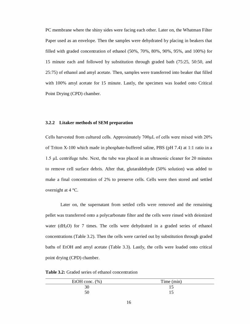
Table 3.2 Graded series of ethanol concentration 16
Table 3.3 Graded bath of EtOH and amyl acetate 17
Table 3.4 Obligonucleotide primers used in this study. 19
Table 3.5 Types of PCR reagent used in this study 20
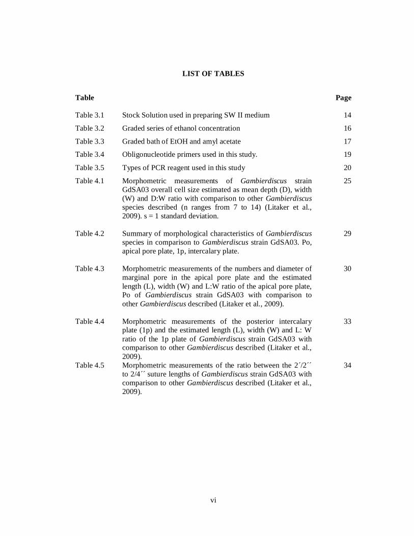
Table 4.1 Morphometric measurements of Gambierdiscus strain GdSA03 overall cell size estimated as mean depth (D), width (W) and D:W ratio with comparison to other Gambierdiscus species described (n ranges from 7 to 14) (Litaker et al., 2009). s = 1 standard deviation.
25
Table 4.2 Summary of morphological characteristics of Gambierdiscus species in comparison to Gambierdiscus strain GdSA03. Po, apical pore plate, 1p, intercalary plate.
29
Table 4.3 Morphometric measurements of the numbers and diameter of marginal pore in the apical pore plate and the estimated length (L), width (W) and L:W ratio of the apical pore plate, Po of Gambierdiscus strain GdSA03 with comparison to other Gambierdiscus described (Litaker et al., 2009).
30
Table 4.4 Morphometric measurements of the posterior intercalary plate (1p) and the estimated length (L), width (W) and L: W ratio of the 1p plate of Gambierdiscus strain GdSA03 with comparison to other Gambierdiscus described (Litaker et al., 2009).
33
Table 4.5 Morphometric measurements of the ratio between the 2´/2´´ to 2/4´´ suture lengths of Gambierdiscus strain GdSA03 with comparison to other Gambierdiscus described (Litaker et al., 2009).
34
vii
LIST OF FIGURES
Figure Page
Figure 2.1 Ciguatera fish poisoning The food web that transfers the toxin to human.
7
Figure 2.2 Line drawing of Gambierdiscus toxicus. A. Theca plate tabulations, B. ventral view (Adachi and Fukuyo, 1979).
9
Figure 2.3 Line drawing of Gambierdiscus toxicus. The ventral view of sulcal plate pattern (Adachi and Fukuyo, 1978).
10
Figure 2.4 Diagram of rRNA genes showing the positions of 5.8S rDNA, the large subunit (LSU) and small subunit (SSU) of rRNA genes.
11
Figure 4.1 Scanning electron micrographs of Gambierdiscus strain GdSA03 with comparison of two SEM preparation methods of Faust et al. (1996) (A-D) and Litaker et al. (2009) (E-F). (A-B) Specimens observed showed broken cells with empty thecal shells. (C-D) Cells covered with debris. (E-F) Cells showing well preserved shape and clean surface. Scale bar = 10µm.
26
Figure 4.2: Scanning electron micrographs of Gambierdiscus strain GdSA03. (A) Apical view of cell showing the epitheca with three apical plates (3´), seven precingular plates (7´´) and apical pore plate (Po). (B) Antapical view of cell showing the hypotheca with five postcingular plates (5´´´), one intercalary plate and two antapical plates (2´´´´). (C-D) Ventral view showing the sulcal region. S.a.r., right anterior sulcal plate; S.r., right sulcal plate and t., transitional. Scale bar = 10µm.
27
Figure 4.3 Scanning electron micrographs of Gambierdiscus strain GdSA03 featuring the ellipsoid apical pore plate with apical pore (Po) and the marginal pores (arrow). Scale bar = 1 µm.
31
Figure 4.4 SEM micrographs of featuring the morphology of posterior intercalary plate (1p). Scale bar = 10µm.
32
Figure 4.5 Gel image of the PCR products of D1/2 rDNA obtained from Gambierdiscus GdSA03.
35
Figure 4.6 Electrophoregram of D1/2 rDNA sequence obtained from Gambierdiscus GdSA03 showing multiple peaks, full sequences were attached in Appendix A.
35
2
1.0 INTRODUCTION
Algae are important component in the primary productivities of marine ecosystems. Most
of them are not harmful to others organisms. However, there are some species that
produced potent toxins that can harm the sea animals and humans. People who come in
contact with these toxic algae whether by consuming the shellfish contaminated by the
algal-origin toxins or swimming in areas infested with harmful algae, he or she may
experience neurological symptoms such as tingling fingers and toes as well as respiratory
and gastrointestinal symptoms. Certain species of benthic dinoflagellates are known to
produce potent neurotoxins that may involve in ciguatera fish poisoning (CFP). They are
several types of benthic dinoflagellates such as Gambierdiscus, Ostreopsis, Coolia,
Prorocentrum and Amphidinium.
Ciguatera fish poisoning (CFP) is a human illness that cause by eating finfish that
is contaminated by toxins produced by the marine algae, Gambierdiscus toxicus. People
who have ciguatera may experience nausea, vomiting and neurologic symptoms such as
tingling fingers or toes. Ciguatera fish poisoning has no cure. This symptom basically
goes in days or weeks but can be last for a year. This study is important because fish is
one the important source of proteins in Malaysia. People will always looking for fish as
their source of protein in their daily life.
3
In this study the morphological and molecular characteristics of potentially toxic
benthic dinoflagellate, Gambierdiscus spp. found in Malaysian waters were investigated.
A strain of Gambierdiscus from Sabah was cultured and the detailed morphological
features of this strain were examined by using advanced scanning electron microscopy
(SEM). In addition to that, molecular characterization of the strain was also conducted by
amplification of the nuclear-encoded large subunit ribosomal RNA gene in the region of
domain 1 and 2 to infer the phylogenetic relationships of Gambierdiscus.
4
2.0 LITERATURE REVIEW
2.1 Harmful Algal Blooms (HABs)
Algal blooms can be occurred in freshwater and also in marine environment. An algal
bloom is a drastic elevation of the algal density of in the aquatic system where the clear
water turn turbid, colored and covered with accumulation of floating masses at the
surfaces. Blooms can be recognized with the appearance of water discoloration result of
the high density of pigmented cells. Although there is no officially recognized threshold
level, algae can be considered bloom with cells density range changed from hundreds to
thousands of cells per milliliter. Besides, algal bloom concentration may reach millions of
cells per milliliter.
Harmful algal bloom (HAB) is a dense aggregation of harmful phytoplankton,
algae or cyanobacteria in an aquatic environment. HABs may cause negative impacts to
others organisms such as mammals via the production of natural toxins, mechanical
damage or by other means. In addition, an algal bloom can kill fish and others aquatic life
due to hypoxia/anoxic conditions in the water. Moreover, HABs will produces potent
toxins where can be associated with specific, harmful consequences of such blooms in
regions where one type is common with another, for example fish kills in Florida, fish and
shellfish kills in Japan and paralytic fish poisoning (PSP) of humans on the Altlantic and
Pacific coasts of North America (Loeblich et al., 1986). There are more than 5,000
5
species of marine phytoplankton that exist in worldwide, and 2% are known to be harmful
or toxic to environment and organisms (Richlen et al., 2008)
2.2 Characteristic of Dinoflagellates
Dinoflagellates are one of the groups of predominantly unicellular, eukaryotic, flagellated
organisms that posses of photosynthetic and non photosynthetic members. Both of
photosynthetic and non photosynthetic members are able to swim because they have
many cell walls and both botanists and zoologists have been claim that they are algae and
protozoa and both have produced classification schemes for dinoflagellates.
Other features that are common to dinoflagellates is that the presence of
chlorophyll a, c2 and c1 (usually absent), the production of starch and oils as reserves,
mitochondrial cristae with cellular cross-sections and a triple-membraned enveloped
which surrounding chloroplast. There are others unique features of dinoflagellates such as
rod-like, ejectile bodies where usually occurred in several phytoflagellates groups
(Taylor, 1987).
The habitats in which dinoflagellates may be found are also very varied. Almost
90% of them are marine planktonic or benthic form, with the greatest diversity in tropical
waters. Besides, dinoflagellates also can be found in polar waters, in sea ice and even in
snow. Others groups such as diatoms or green algae are more successful in cold
environment. The photosynthetic members are restricted to illuminated water because
many of them can survive under very dim light conditions, whereas the heterotrophs can
extend into non-illuminated depths and both in water and in sediments.
There are four genera of thecated species that is found in the warm tropical waters
such as Gambierdiscus, Ostreopsis, Coolia and Prorocentrum (Adachi and Fukuyo.,
1979; Fukuyo., 1981; Besada et al., 1982). Those thecated species belong to the toxic
6
dinoflagellates which are distributed world-wide in marine waters. Approximately 50
dinoflagellates species are known to produce toxin that will cause the death of marine
animals and birds, toxicity in bivalves and seafood poisoning in humans (Steidinger.,
1993).
2.3 Ciguatera Fish Poisoning (CFP)
Ciguatera fish poisoning (CFP) is a food borne illness that effect humans worldwide. CFP
is caused by benthic microalgae species commonly known as Gambierdiscus toxicus.
Humans get this illness by eating reef fishes which contained toxins from G. toxicus
which is also known as ciguatoxins (CTXs).
CTXs are lipid soluble polyether compounds which consist of 13 to 14 rings fused
by ether linkages into a most rigid ledder-like structure. Ciguatera is attributed to a lipid-
soluble toxin produced by several genera of benthic-associated dinoflagellates, including
those of Ostreopsis spp., Prorocentrum spp., and Gambierdiscus spp., especially G.
toxicus (Bagnis and Yasumoto, 1977).
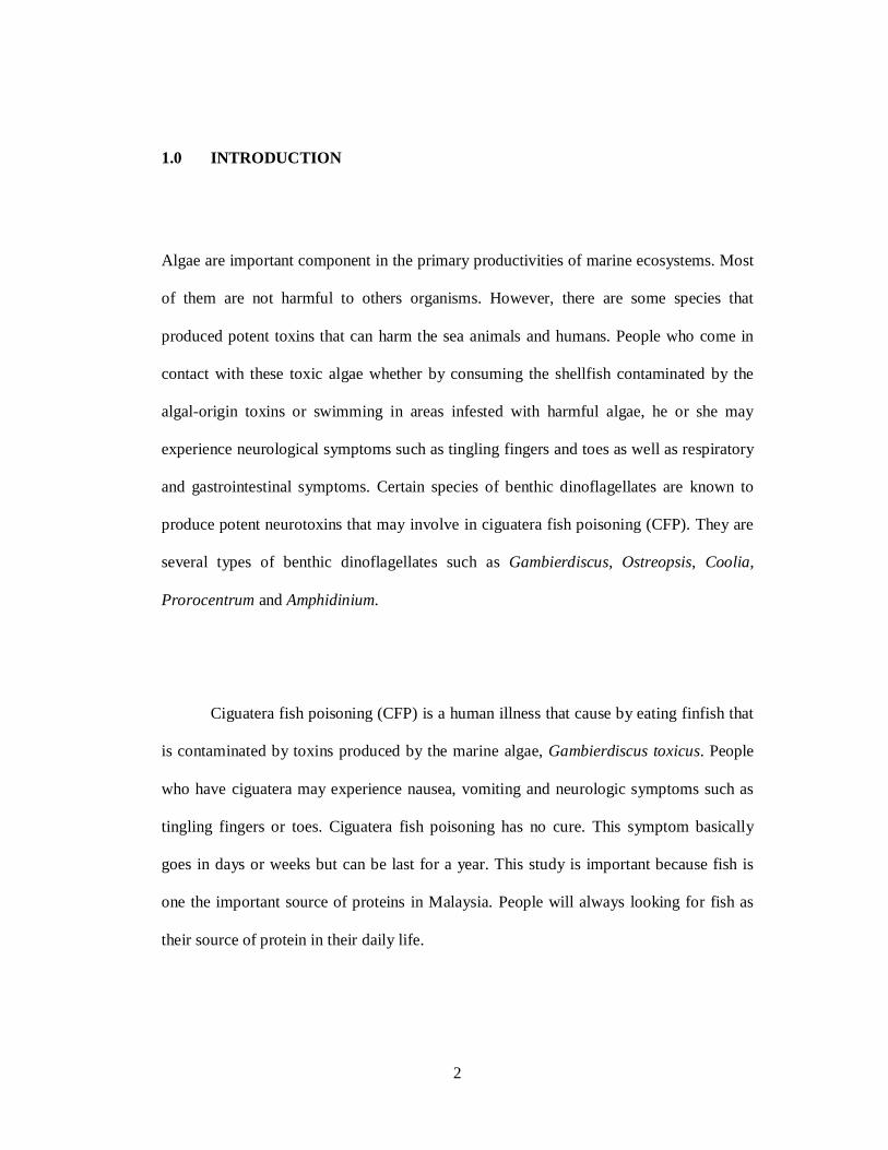
Ciguatoxins are relatively heat stable molecules that remain toxic although after
cooking and exposed to the mild acidic and basic conditions. Ciguatoxins producing
microalgae grow in association with macroalgae in coral reefs. The toxin is then
transferred through the food web (Figure 2.1). The algae are then consumed by
herbivorous fish, which are then consumed by carnivorous fish and last will be consumed
by humans with each step concentrating the toxins.
7
Figure 2.1: The food web that transfers the toxin to human.
Symptoms of CFP are gastrointestinal, neurological and cardiovascular symptoms.
Basically, the gastrointestinal symptoms occurred first such as vomiting, diarrhea and
abdominal pain. It is followed by neurological dysfunction including dizziness, anxiety,
sweating, reversal of temperature sensation and a numbness. Paralysis and death have
been documented, but symptoms are usually less severe although debilitating (Miller,
1991). There is no antidote but rapid treatment within 24 hours is reported to relieve some
symptoms. However, the recovery time is different between individuals. It may take
weeks, months or even years. The prevention of intoxication is depend upon complete
abstinence from eating any tropical reef fish due to no practical way to routinely measure
ciguatoxins in any seafood product prior to consumptions.
8
2.4 Taxonomy of Gambierdiscus species: from morphology to phylogeny
The Benthic Dinoflagellate, Gambierdiscus toxicus is the causative agent of ciguatera
which was found to produce and transmit ciguatoxin and maitotoxin to herbivorous fish
(Yasumoto et. al., 1979). The species was originally described from material collected
from the Gambier Islands in the Pacific Ocean (Adachi and Fuyuko, 1979). For the first
time, G. toxicus was thought to be the only species of the genus Gambierdiscus until the
recent description of Gambierdiscus belizeanus by Faust 1995. Unfortunately, the
production of toxins or other secondary metabolites of G. belizeanus has not been
reported because G. belizeanus was found in a sand dwelling community of benthic
dinoflagellates (Faust, 1995).
In the coral reef areas, it was found that benthic dinoflagellates were richer in both
numbers and species on epi-benthic layers than in the water. If the toxic metabolites of
this benthic species are taken up by herbivorous fish, the toxin will be contributed to the
manifestation of the complex symptoms of ciguatera. The actual occurrence of minor
toxins in the viscera of herbivorous fish has been confirmed and the toxigenicity of
several benthic species has also been demonstrated before (Yasumoto et. al., 1976:
Nakajima et. al., 1981).
The morphological characters are the primary means in describing dinoflagellate
species. Dinoflagellates morphological characters such as the shape and size of the apical
pore plate (Po) and posterior intercalary plate provide a very useful and complex
characteristic for species recognition. Species in the genus Gambierdiscus are anterio-
posteriorly compressed in the ventral view and theca plate tabulations are observed in the
apical or antapical view (Figure 2.2). The epitheca and hypotheca are not noticeably
9
different in size. A distinguishing feature is the shape and size of the apical pore complex
(Faust, 1992). The cell surface is smooth with numerous deep and dense pores. Thecal
plates are very thick. The thecate formula of the Gambierdiscus spp. is Po, 3´, 7´´, sulcus
plate, 5´´´ and 2´´´´.
Figure 2.2: Ink drawing of Gambierdiscus toxicus. A. Theca plate tabulations, B. ventral
view (Adachi and Fukuyo, 1979).
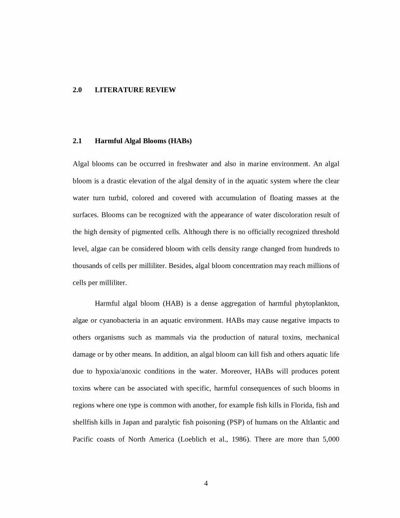
The sulcus of the Gambierdiscus spp. is very short and most parts except posterior
plate are deeply concave like a hollow. The sulcus is composed of eight plates. They are
the sulcal right anterior plate (S.a.r), the sulcal left anterior plate (S.a.l), the sulcal right
(S.r), the sulcal left (S.l), the transitional (t) plate, the sulcal posterior plate (S.p), the
sulcal medium anterior plate (S.m.a) and the sulcal medium posterior plate (S.m.p)
(Adachi and Fukuyo., 1978). The S.p plate comprises of five sided and relatively large.
Most of this part is out of the sulcal hollow. It develops to the right along the postcingular
6´´´. The S.r plate fits to the concave of the 6´´´, which is constitutes the right posterior
wall of the sulcus. The upper half of the S.r plate is ridged in the same manner of
postcingulars. In addition, the transitional (t) plate is boat-shaped, where it is located at
the extension of the cingulum and enters deeply into the sulcus. The left posterior wall of
the sulcus is occupied by S.l plate which is located between the S.m.p plate and posterior
A. B.
10
intercalary plate. The round S.m.a plate is located at the bottom and its anterior margin
contacts with the S.a.r plate and S.a.l. plate. They are subrectangular and situate between
the precingular 1´´ and the S.m.a plate (Figure 2.3).
Figure 2.3: Line drawing of Gambierdiscus toxicus. The ventral view of sulcal plate
pattern (Adachi and Fukuyo, 1978).
In the recent studies morphological approach tends to combine with molecular
analysis. For example, in the genus of Alexandrium, the morphological features used to
classify the species level of the genus showed ambiguity in their taxonomy and molecular
approach has been introduced to solve the cryptic species of the genus (Scholin and
Anderson, 1994). The molecular techniques involving DNA analyses have provided a rich
set of data for various dinoflagellate species (Chinain et. al., 1997). In addition, the
morphological approach gives particular emphasis to phenotypic characters, consistently
cited the advantage of DNA analyses is that they measure changes in the genome rather
than the phenotype (Chinain et. al., 1999). Nucleic acid sequencing has been proven to be
11
a very useful tool in systematic and phylogenetic studies in many dinoflagellate species
(Scholin and Anderson, 1994).
Studies of Gambierdiscus clones based on comparisons of small subunit (SSU)
and large subunit (LSU) rRNA genes (Figure 2.4). Based on previous study, Babinchak et
al. (1994) directly examined the genetic diversity by using restriction fragment length
polymorphism (RFLP) of the LSU ribosomal DNA gene, which showed that substantial
genetic variability does indeed exist among distributed strains, while RFLP of the SSU
rDNA recovered a different ribotypes within populations of G. toxicus. A comprehensive
examination of genetic variation among globally distributed strains using DNA
sequencing is still lacking. Furthermore, the phylogenetic of the Gambierdiscus
morphospecies are not fully understood.
Figure 2.4: A single repeat of ribosomal RNA genes precursor showing the positions of 5.8S rDNA, the large subunit (LSU) and small subunit (SSU) of rRNA genes, the internal transcript spacer (ITS1 and ITS2) and external transcript spacer (ETS).
eETS
ITS1 ITS2
ETS LSU (28S) 5.8S SSU (18S)
ETS
12
Combination of morphological and molecular analyses accounts for both
phenotypic and genotypic characters will provide a robust taxonomic classification of the
genus. Furthermore, detailed examination of the morphological variability within a
phylogenetic framework could distinguish phenotypically plastid characters and between
ancestral and derived characters used in morphological classifications. Therefore
combined morphology-molecular approach provides a reliable phylogenetic
characterization of this important species.
13
3.0 MATERIALS AND METHODS
3.1 Algal Cultures
3.1.1 Preparation of Culture Vessels
Proper care need to be employed when cleaning test tube to remove carbonate deposits
and others materials that attached to the test tubes. Test tubes were soaked in 10% dilute
acid hydrochloride (HCl) for overnight to remove diluted carbonate deposits, followed by
washing in phosphate free detergent and scrubbed with a brush to dislodged solid algal
remains. Test tubes were rinsed with distilled water several times to avoid residual
amounts of detergent that will affect the growth of culture.
3.1.2 Sea Water Medium (SWII)
Natural sea water was used as a medium base with a salinity of 30 psu. One liter of
natural sea water was added in the beaker. Salinity of natural sea water was checked by
using reflectometer. Salinity of seawater will be adjusted to 30 psu by adding natural salt
or distilled water. Medium stock solutions were added into the beaker that contained
natural sea water. The stock solutions were listed as below:
14
Table 3.1: Stock Solution used in preparing SW II medium