39
Spinal Tuberculosis Dr Veronica White MD FRCP Consultant Physician Barts and the London NHS Trust
Spinal Tuberculosis
Dr Veronica White MD FRCP
Consultant PhysicianBarts and the London NHS Trust

Introduction
Spinal TB first described by Percival Pott in 1779
“Pott’s disease”

Background
Found in the remains of Egyptian mummies andin many other human skeletons
21st Century
Spinal TB makes up a relatively small number of TB patients
Presentation can be insidious
Outcome variable and despite treatment can lead to long term disability

Epidemiology of Spinal TB
Approximately 10% of TB cases affect the skeleton and 5% are in the spine
365 number of cases in UK in 2010
167 in London

In East London
Ten year data base 1997-2006 (Our study)
Multidisciplinary clinic since 2004

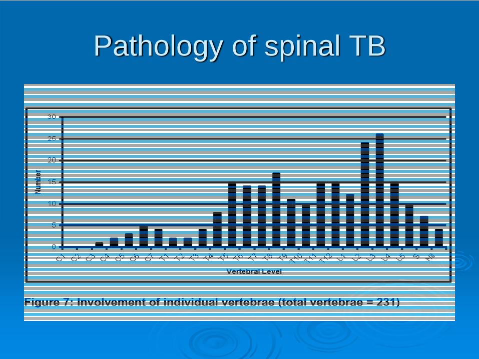
Pathology of spinal TB
Haematological seeding from lung
MTB affects vertebral body which can then spread to discs (discitis)
Occurs at any level and can be multilevel
More common in thoracic and lumbar
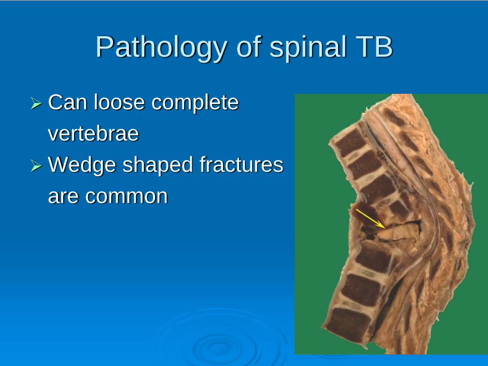
Pathology of spinal TB
Pathology of spinal TB
Can loose complete vertebrae Wedge shaped fractures are common

Pathology of spinal TB
Approx 1/3 have evidence of TB elsewhere
Approx 1/3 have associated psoas abscess
Only 25% have an abnormal CXR

Symptoms of Spinal TB
Back pain (95%)
40-50% neurological symptoms –weakness, paresthesia, bowel symptoms
40-50% with systemic symptoms – fever, night sweats, weight loss

Difficulty in diagnosis
Back pain is very common
Systemic symptoms are often ignored by patient or forgotten by health professionals

Characteristics of patients
Born in high incidence area
May have been in UK for sometime (average 9.6 years, range 0-50)
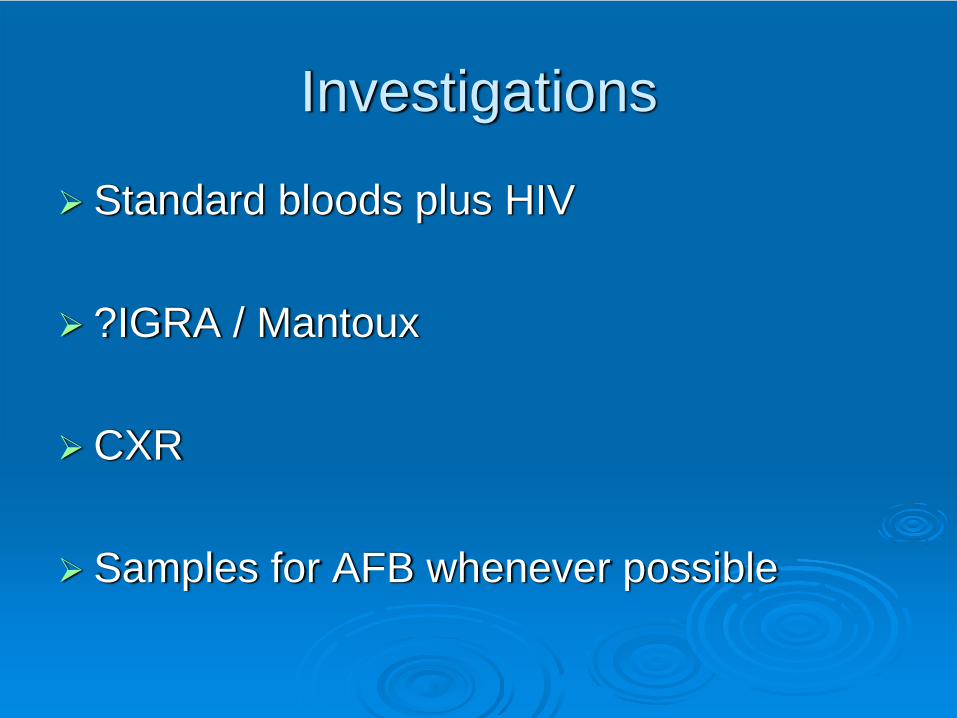
Investigations
Standard bloods plus HIV
?IGRA / Mantoux
CXR
Samples for AFB whenever possible

CXR
Often normal in non-pulmonary TB
Bloods
May be completely normal

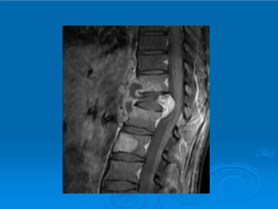
Imaging of Spinal TB
MRI
CT guided biopsy
US guided drainage of collection
ENSURE SAMPLES SENT FOR AFB

Making a diagnosis
82 percutaneous biopsies
4 open biopsies
17 intra-operative samples
2 I&D samples

Case history
31 year old Somalian patient referred byGP with interscapular pain
No systemic symptoms; tenderness over T4

Case history

Case history

Case history

Case history
Admitted and had CT guided biopsy
Started on treatment – Pharmacy DOT
Pain improving at 2 months


Psoas abscesses


Medical treatment
Standard quadruple therapy
BTS recommend 6 months
Most experts give 9-12 months
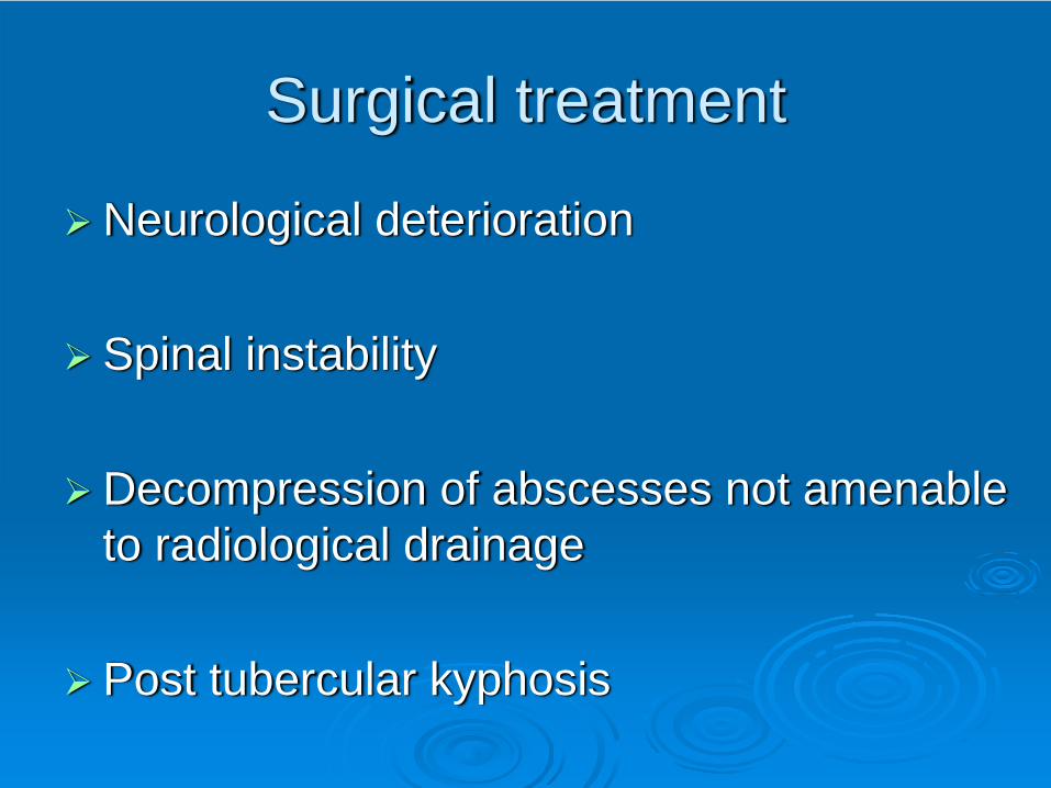
Surgical treatment
Neurological deterioration
Spinal instability
Decompression of abscesses not amenable to radiological drainage
Post tubercular kyphosis


Use of steroids
Only if evidence of cord compression
“Double dose” due to rifampicin

Outcome
Many patients may be left with chronic back pain, but not lose of function (60%)
Small number paraplegic (4%)
Some neurological deficit (15%)

Conclusion
Back pain +/- systemic symptoms
Get biopsies wherever possible AFB
Multidisciplinary care
At least 6 months treatment +/- steroids


Acknowledgements
Mr Colin Natali and Mr Siva RamanMiss Chloe CritchleyDr Robert Davidson, Northwick ParkTB team at the Barts and the LondonWellcome imagesDr John Moore-Gillon and Prof Sheila Hillier

Questions?


