submit.radiology.or.kr J Korean Soc Radiol 2011;65(3):285-288 285
INTRODUCTION
Tubulocystic renal carcinoma (TCRC) is a rare subtype of renal cell carcinoma that has not yet been included in the World Health Organization (WHO) 2004 classification of re-nal tumors (1). This tumor entity occurs predominantly in males, and has a specific macroscopic spongy appearance and microscopic characteristics presenting as cysts lined by hob-nail cells separated by a thin fibrotic stroma (2, 3).
To date, few published case reports exist on this rare tumor entity. Since most of the previously reported articles have been published in the pathologic literature, little attention has been paid to the radiological features of TCRC.
Herein, we present a case of a 22-year-old woman with TCRC which metastasized to the parasymphyseal pubic bone. We describe the imaging findings of computed tomography
(CT), ultrasonography (US), and 18-fluoro-2-deoxy-D-glu-cose positron emission tomography/computed tomography (18F-FDG PET/CT) with histopathologic correlation.
CASE REPORT
A 22-year-old woman with an incidentally discovered renal mass on check-up abdominal US in an outside hospital was admitted to our hospital for further evaluation and manage-ment. The general physical examination, laboratory tests, and urine analysis were normal.
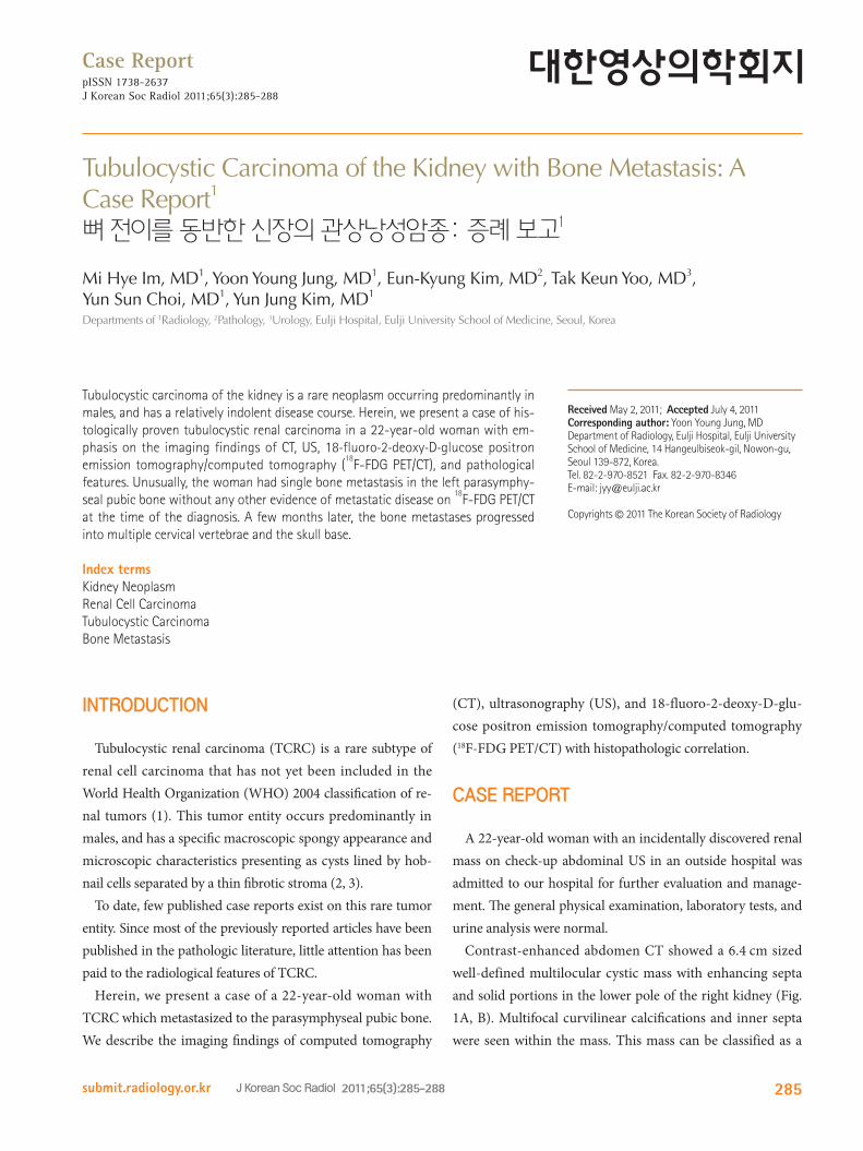
Contrast-enhanced abdomen CT showed a 6.4 cm sized well-defined multilocular cystic mass with enhancing septa and solid portions in the lower pole of the right kidney (Fig. 1A, B). Multifocal curvilinear calcifications and inner septa were seen within the mass. This mass can be classified as a
Case ReportpISSN 1738-2637J Korean Soc Radiol 2011;65(3):285-288
Received May 2, 2011; Accepted July 4, 2011Corresponding author: Yoon Young Jung, MDDepartment of Radiology, Eulji Hospital, Eulji University School of Medicine, 14 Hangeulbiseok-gil, Nowon-gu, Seoul 139-872, Korea. Tel. 82-2-970-8521 Fax. 82-2-970-8346E-mail: [email protected]
Tubulocystic carcinoma of the kidney is a rare neoplasm occurring predominantly in males, and has a relatively indolent disease course. Herein, we present a case of his-tologically proven tubulocystic renal carcinoma in a 22-year-old woman with em-phasis on the imaging findings of CT, US, 18-fluoro-2-deoxy-D-glucose positron emission tomography/computed tomography (
18F-FDG PET/CT), and pathological
features. Unusually, the woman had single bone metastasis in the left parasymphy-seal pubic bone without any other evidence of metastatic disease on
18F-FDG PET/CT
at the time of the diagnosis. A few months later, the bone metastases progressed into multiple cervical vertebrae and the skull base.
Index termsKidney NeoplasmRenal Cell CarcinomaTubulocystic CarcinomaBone Metastasis
Tubulocystic Carcinoma of the Kidney with Bone Metastasis: A Case Report1
뼈 전이를 동반한 신장의 관상낭성암종: 증례 보고1
Mi Hye Im, MD1, Yoon Young Jung, MD1, Eun-Kyung Kim, MD2, Tak Keun Yoo, MD3, Yun Sun Choi, MD1, Yun Jung Kim, MD1
Departments of 1Radiology, 2Pathology, 3Urology, Eulji Hospital, Eulji University School of Medicine, Seoul, Korea
Tubulocystic Carcinoma of the Kidney with Bone Metastasis
submit.radiology.or.krJ Korean Soc Radiol 2011;65(3):285-288286
enhancement. Additional temporal bone CT and 18F-FDG PET/CT showed multiple bone metastases in the left tempo-ral bone and the fourth and sixth cervical vertebrae, which were pathologically confirmed as metastatic lesions by follow-ing the percutaneous biopsy of the sixth cervical vertebra. Due to disease progression, the patient was treated with 13 cycles of radiotherapy for metastasis at the left temporal bone and the fourth and sixth cervical vertebrae, as well as targeted chemotherapy of sunitinib malate at 50 mg/day for two weeks with one week of rest. Five months after chemotherapy, the patient remained well without signs of any further progres-sion of the metastatic disease.
DISCUSSION
TCRC is a rare renal neoplasm and recently entitled entity that was first described in 1997 by MacLennan et al. (4) as a “low-grade collecting duct carcinoma of the kidney”, because at that time, it was thought to be collecting duct origin. However, Amin et al. (5) later renamed this tumor as “tubulocystic carci-noma of the kidney”, by showing a distinctive immunostaining that differs from classical collecting duct carcinoma.
Grossly, TCRC is a well-circumscribed tumor that often exhib-its a cystic component which may present as radiological classifi-cation of Bosniak III or IV. Microscopically, it is composed of tu-bules and cysts lined by a single layer of tumor cells with eosinophilic cytoplasm and prominent nucleoli, and separated by fibrotic septa (2, 3). Immunohistochemical studies show vari-able marker positivity with cytokeratins CK7, CK8, CK18, CK19, CD10, and sometimes with E-cadherin (5-7).
TCRC has a tendency to be seen predominantly in male pa-tients with a wide age range, mostly in the fifth and sixth de-cade, and usually shows no symptoms. Moreover, most TCRCs have taken an indolent course (4-7). In a study on 13 cases of TCRC, seven patients had tumors with pT1, four had with pT2, and two had pT3 (4). Amin et al. (5) reported 31 cases of TCRC, in which 24 patients had tumors with pT1, four patients had with pT2, and three had pT3 at presentation. Follow-up stud-ies in 22 patients showed that one patient had bone metasta-ses, and one patient had bone and liver metastases. As seen above, most TCRCs have taken an indolent course, but some reports shows aggressive metastasis observed in the liver and
cystic lesion of Bosniak category IV. Also, an osteolytic lesion with soft tissue formation was detected in the left parasym-physeal pubic bone, suggesting pubic bone metastasis (Fig. 1C). Abdominal gray-scale and color Doppler US scan were performed to evaluate a small low attenuation lesion of the liver that was detected on CT. US scan also revealed a lobulat-ing multiseptated heterogeneous cystic mass with calcifica-tions and solid portions which show some inner vascularity in the lower pole of the right kidney (Fig. 1D, E). 18F-FDG PET/CT showed peripheral intense FDG uptake (SUVmax 7.5) with central photon defect in the right renal mass, and also showed another intense FDG uptake (SUVmax 7.2) in the left parasymphyseal pubic bone (Fig. 1F, G).
The patient underwent a radical nephrectomy with dissec-tion of the lymph nodes, and was subsequently treated with a wide marginal excision and allograft for pubic bone metastasis. On pathologic report, the main mass in the right kidney was a well circumscribed, multilocular cystic and solid mass with a spongy appearance, measuring 11.3 × 10.0 × 6.5 cm (Fig. 1H). Microscopic examination revealed that the tumor was com-posed of variable-sized cysts and well-formed tubules. The epi-thelial cells had a cuboidal appearance with abundant eosino-philic cytoplasm and prominent nucleoli (Fig. 1I). The cyst lining cells had a hobnail appearance. The tumor cells were positive for epithelial membrane antigen (EMA), CD10 and vi-mentin, and negative for cytokeratin (CK) 7 and E-cadherin. As a result of these findings, the final histopathologic diagnosis was tubulocystic carcinoma of the kidney. According to the seventh edition of the American Joint Committee on Cancer (AJCC) cancer staging manual, which is based on the extent of the tumor (T), the extent of spread to the lymph nodes (N), and the presence of distant metastasis (M), the right kidney tu-mor was pT2N0M1.
After the surgery, the patient was treated with immuno-therapy, consisting of interleukin-2 (IL-2) at 10 million inter-national units (MIU)/day and interferon-alpha at 6 MIU/day for four weeks. Then, the patient presented with headache and a gadolinium-enhanced brain magnetic resonance imag-ing revealed a 1.8 × 1.6 cm sized expansile mass at the left pe-trous bone, just anterior and inferior to the left internal audi-tory canal. This mass showed low signal intensity (SI) on T1 weighted images (WI), high SI on T2 WI and homogeneous
Mi Hye Im, et al
submit.radiology.or.kr J Korean Soc Radiol 2011;65(3):285-288 287
of cases is necessary for a better prediction of prognosis. Un-usually, our patient was a young woman and initially had sin-gle bone metastasis without demonstrable lymph node metas-
bone (5). Therefore, TCRC has a low but definite possible po-tential for metastasis. However, because of the rarity of this tumor, long-term follow-up information of a larger number
A
G
D
B
H
E F
C
IFig. 1. Tubulocystic carcinoma of the right kidney and left parasymphyeal pubic bone metastasis in a 22-year-old woman.A, B. Contrast-enhanced axial and coronal reformatted CT images show a 6.4 cm sized well-defined multilocular cystic mass (arrow) with en-hancing septa, multifocal curvilinear calcifications, and solid portions in the lower pole of the right kidney. C. Contrast-enhanced axial CT image shows an osteolytic lesion (arrowhead) with soft tissue formation in the left parasymphyseal pubic bone.D, E. Gray-scale and color Doppler US images showed a lobulating multiseptated heterogeneous cystic mass with solid portions (arrows) and calcifications (arrowheads), which showed some inner vascularity in the lower pole of the right kidney.F, G. Coronal 18F-FDG PET/CT images show peripheral intense FDG uptake with central photon defect in the lower pole of the right kidney (ar-row), and another intense FDG uptake in the left parasymphyseal pubic bone (arrowhead). H. The tumor is a well circumscribed, multilocular cystic and solid mass with a spongy appearance, measuring 11.3 × 10.0 × 6.5 cm on gross specimen.I. Photomicrograph showed varying sized cysts and well-formed tubules (Hematoxylin & Eosin stain, × 40). The epithelial cells have abundant eo-sinophilic cytoplasm and prominent nucleoli (Inserted box, × 200).Note.-18F-FDG PET/CT = 18-fluoro-2-deoxy-D-glucose positron emission tomography/computed tomography
Tubulocystic Carcinoma of the Kidney with Bone Metastasis
submit.radiology.or.krJ Korean Soc Radiol 2011;65(3):285-288288
Press, 2004
2. Moch H. Cystic renal tumors: new entities and novel con-
cepts. Adv Anat Pathol 2010;17:209-214
3. Srigley JR, Delahunt B. Uncommon and recently described
renal carcinomas. Mod Pathol 2009;22 Suppl 2:S2-S23
Vieillefond A, et al. Tubulocystic carcinoma of the kidney:
clinicopathologic analysis of 31 cases of a distinctive rare
subtype of renal cell carcinoma. Am J Surg Pathol 2009;
33:384-392
6. Yang XJ, Zhou M, Hes O, Shen S, Li R, Lopez J, et al. Tubu-
locystic carcinoma of the kidney: clinicopathologic and
molecular characterization. Am J Surg Pathol 2008;32:
177-187
7. Azoulay S, Vieillefond A, Paraf F, Pasquier D, Cussenot O,
Callard P, et al. Tubulocystic carcinoma of the kidney: a
new entity among renal tumors. Virchows Arch 2007;451:
905-909
8. Hora M, Urge T, Eret V, Stránský P, Klecka J, Kreuzberg B,
et al. Tubulocystic renal carcinoma: a clinical perspective.
World J Urol 2011;29:349-354
tasis. However, despite significant surgical and medical treatment, she had an aggressive prognosis presenting as mul-tiple bone metastases.
Since TCRC frequently has cystic components, this tumor entity is often radiologically described as a cystic lesion of Bosniak category III or IV (4, 6). In a recent study of five cas-es of TCRC, there were two Bosniak III cases, one IV case and two solid tumors (8). Therefore, making a radiological differential diagnosis TCRC from other benign or malignant renal cystic lesions, such as multiloculated clear cell renal cell carcinoma, cystic nephroma, mixed epithelial and stromal tu-mor and cystic oncocytoma, may be a challenging problem. In our case, TCRC was also classified as a cystic lesion of Bos-niak category IV on contrast-enhanced CT images.
In summary, we report a case of a rare aggressive TCRC on the kidney with multiple bone metastases in a young woman that was confirmed on pathology. This tumor presented as a cystic lesion of Bosniak category IV on CT scan with intense FDG uptake on 18F-FDG PET/CT.
REFERENCES
1. Eble JN, Sauter G, Epstein JI, Sesterhenn IA. Pathology and
genetics of tumours of the urinary system and male geni-
tal organs. WHO classification of tumours . Lyon: IARC
뼈 전이를 동반한 신장의 관상낭성암종: 증례 보고1
임미혜1 · 정윤영1 · 김은경2 · 유탁근3 · 최윤선1 · 김윤정1
신장의 관상낭성암종은 주로 남자에서 발견되며 비교적 완만한 임상경과를 보이는 드문 종양이다. 저자들은 병리조직학
적으로 확진된 22세 여자 환자에서 발생한 신장의 관상낭성암종에 대해 CT, US 및 18-fluoro-2-deoxy-D-glucose