57
TUMOURS OF THE CENTRAL NERVOUS SYSTEM FM Brett MD., FRCPath
| Date post: | 23-Dec-2015 |
| Category: |
Documents |
| Upload: | hortense-cobb |
| View: | 215 times |
| Download: | 4 times |
TUMOURS OF THE CENTRAL NERVOUS SYSTEM
FM Brett MD., FRCPath
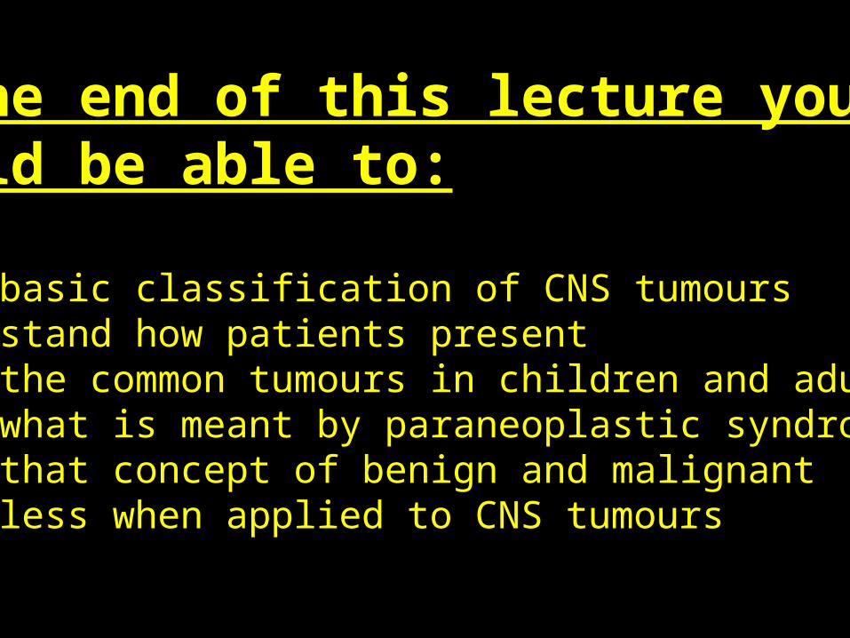
At the end of this lecture you should be able to:
1. Give basic classification of CNS tumours2. Understand how patients present3. Know the common tumours in children and adults4. Know what is meant by paraneoplastic syndromes5. Know that concept of benign and malignant meaningless when applied to CNS tumours
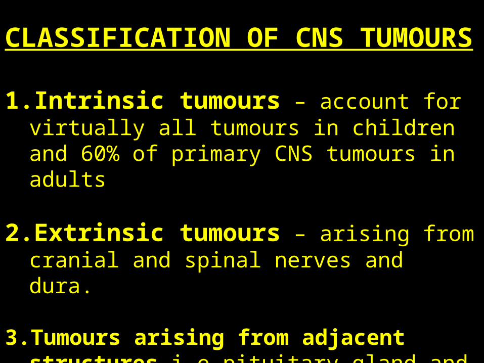
CLASSIFICATION OF CNS TUMOURS
1. Intrinsic tumours – account for virtually all tumours in children and 60% of primary CNS tumours in adults
2. Extrinsic tumours – arising from cranial and spinal nerves and dura.
3. Tumours arising from adjacent structures i.e pituitary gland and metastatic tumours.
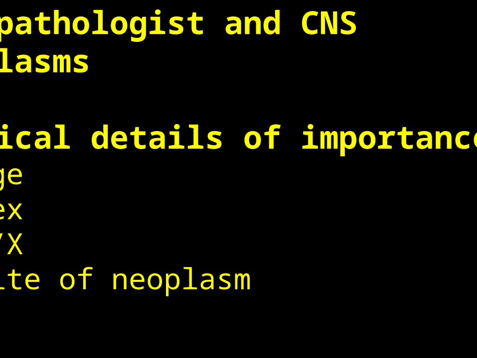
The pathologist and CNS neoplasms
Clinical details of importance~ Age~ Sex~ F/X~ Site of neoplasm
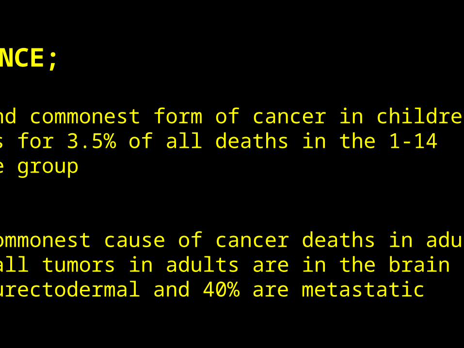
INCIDENCE;
~ Second commonest form of cancer in childrenAccounts for 3.5% of all deaths in the 1-14 year age group
Sixth commonest cause of cancer deaths in adults25% of all tumors in adults are in the brain and 35% are neurectodermal and 40% are metastatic
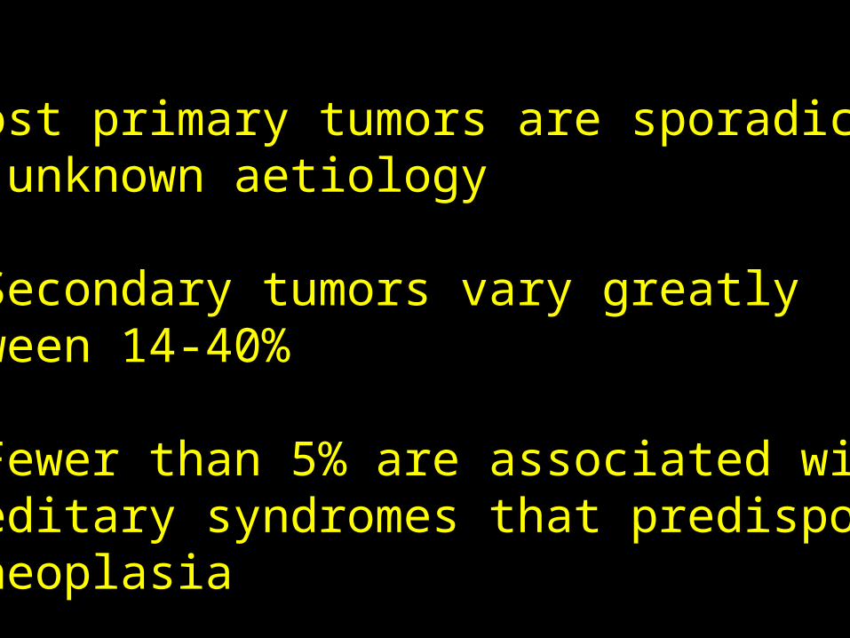
~ Most primary tumors are sporadic andof unknown aetiology
~ Secondary tumors vary greatly between 14-40%
~ Fewer than 5% are associated with hereditary syndromes that predispose to neoplasia
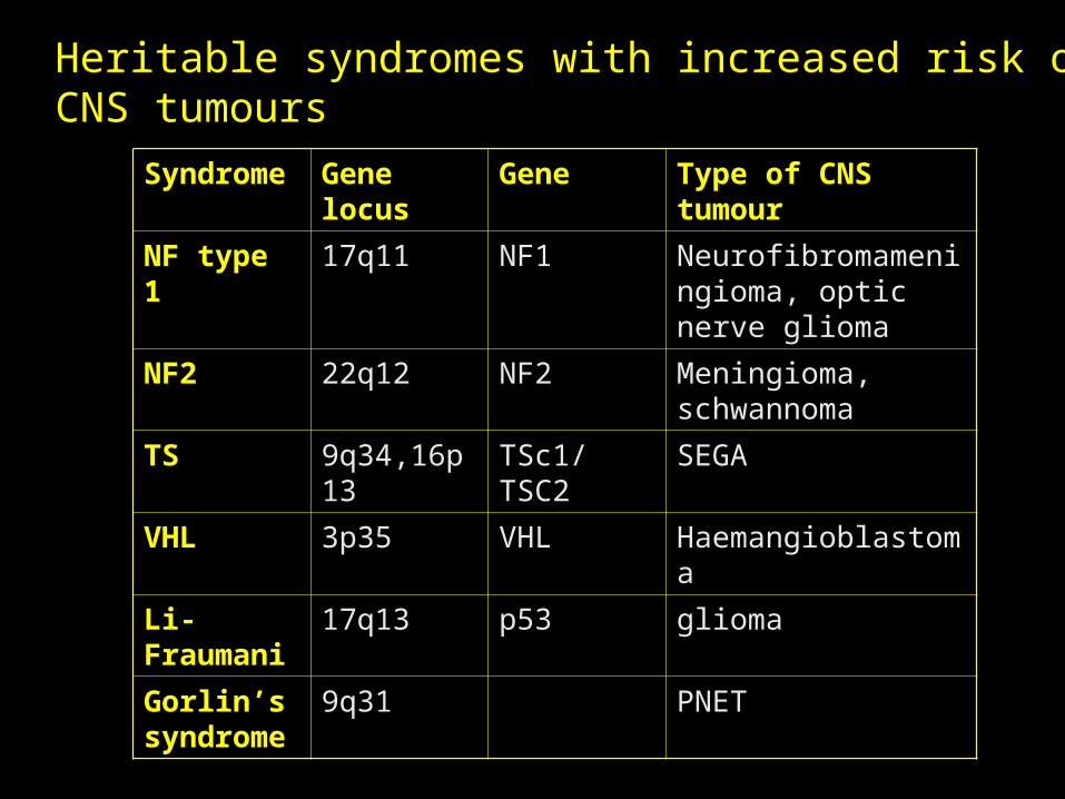
Syndrome Gene locus Gene Type of CNS tumour
NF type 1 17q11 NF1 Neurofibromameningioma, optic nerve glioma
NF2 22q12 NF2 Meningioma, schwannoma
TS 9q34,16p13 TSc1/TSC2 SEGA
VHL 3p35 VHL Haemangioblastoma
Li-Fraumani
17q13 p53 glioma
Gorlin’s syndrome
9q31 PNET
Heritable syndromes with increased risk of CNS tumours
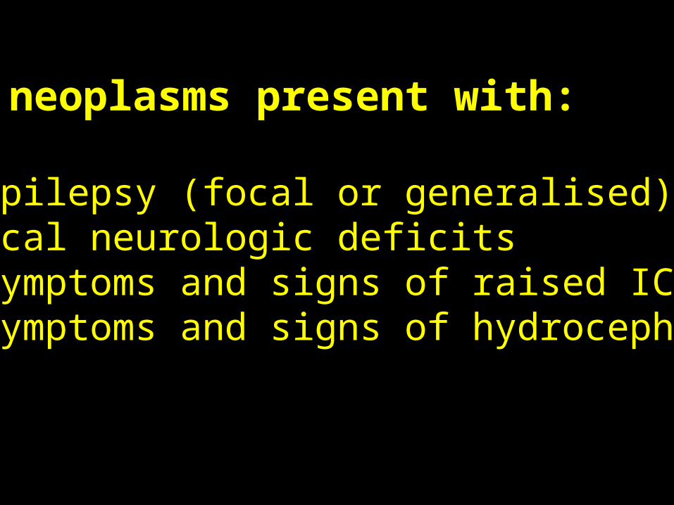
CNS neoplasms present with:
~ epilepsy (focal or generalised)~ focal neurologic deficits~ symptoms and signs of raised ICP~ symptoms and signs of hydrocephalus
SSites of cerebral tumorsSites of cerebral tumors
ADULTSSupratentorial tumors account for 90%
Therefore increased incidence of epilepsy and decreased incidence of headache
Posterior fossa tumours cause headache and vomiting as early features
CHILDRENCerebellum
PonsOptic nerve/chiasm
SUPRATENTORIAL TUMORS ARE RARE
ThereforeHeadache, vomiting, visual disturbances
commonEpilepsy - unusual
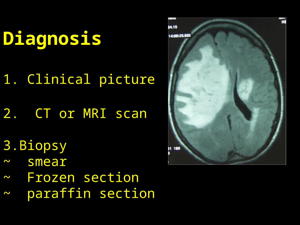
DiagnosisDiagnosis
1. Clinical picture
2. CT or MRI scan
3. Biopsy ~ smear~ Frozen section~ paraffin section
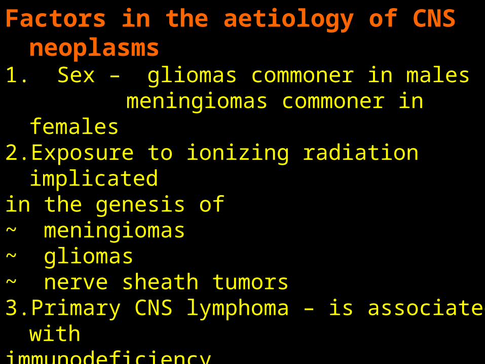
Factors in the aetiology of CNS neoplasms1. Sex – gliomas commoner in males
meningiomas commoner in females2. Exposure to ionizing radiation implicated in the genesis of~ meningiomas~ gliomas~ nerve sheath tumors3. Primary CNS lymphoma – is associated with immunodeficiency4. Nitroso compounds cause CNS neoplasms in animals5. No convincing convincing evidence has linked CNS neoplasms
with trauma, occupation, diet, electromagnetic fields
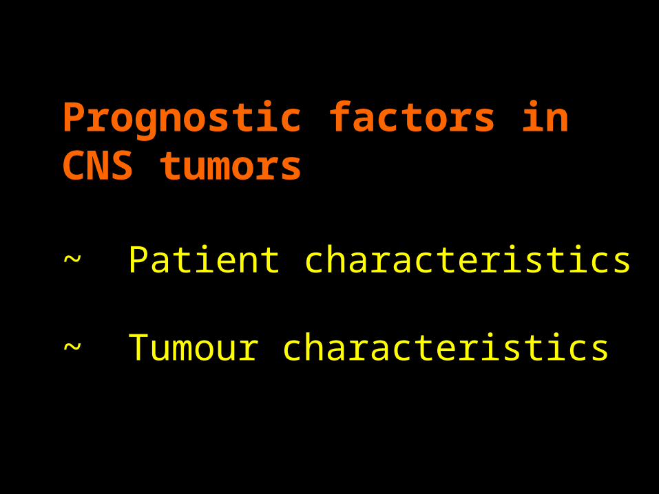
Prognostic factors in CNS tumorsPrognostic factors in CNS tumors
~ Patient characteristics
~ Tumour characteristics
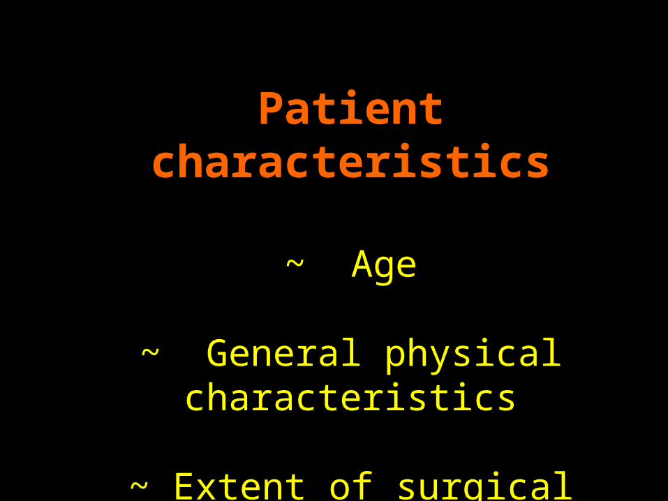
Patient characteristicsPatient characteristics
~ Age
~ General physical characteristics
~ Extent of surgical resection
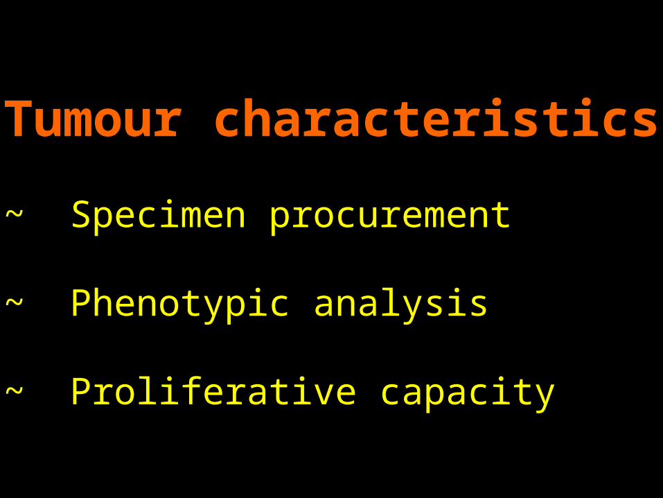
Tumour characteristicsTumour characteristics
~ Specimen procurement
~ Phenotypic analysis
~ Proliferative capacity
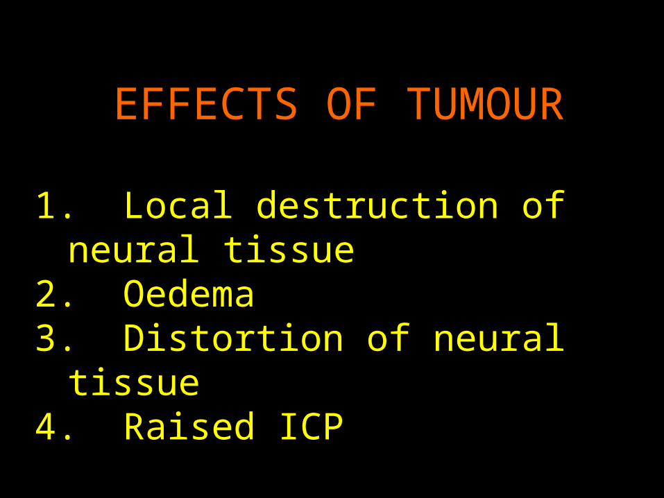
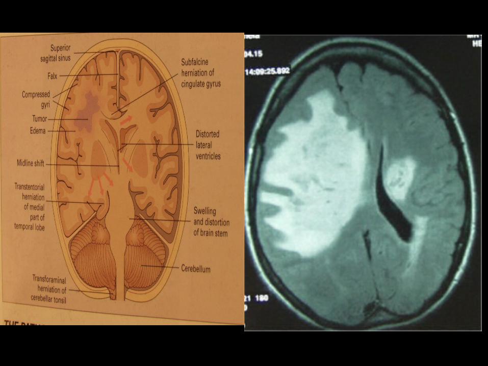
EFFECTS OF TUMOUREFFECTS OF TUMOUR
1. Local destruction of neural tissue2. Oedema3. Distortion of neural tissue4. Raised ICP
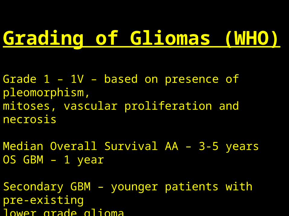
Grading of Gliomas (WHO)
Grade 1 – 1V – based on presence of pleomorphism, mitoses, vascular proliferation and necrosis
Median Overall Survival AA – 3-5 yearsOS GBM – 1 year
Secondary GBM – younger patients with pre-existing lower grade glioma
Primary – 60-70
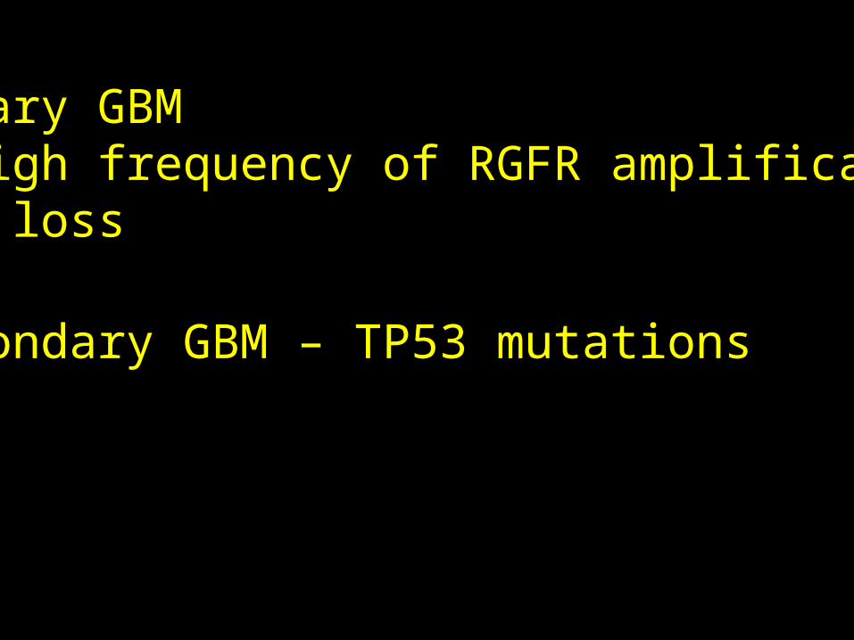
Primary GBM – high frequency of RGFR amplification-p16 loss
-Secondary GBM – TP53 mutations
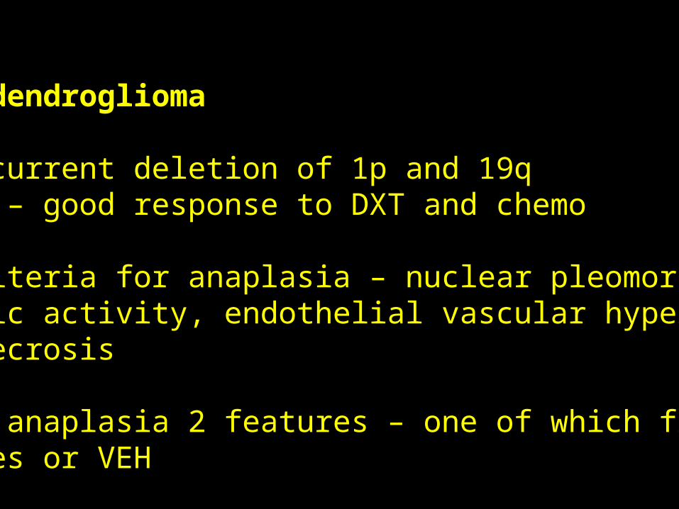
Oligodendroglioma
~ Concurrent deletion of 1p and 19q In AO – good response to DXT and chemo
~ Criteria for anaplasia – nuclear pleomorphism, mitotic activity, endothelial vascular hyperplasia and necrosis
~ For anaplasia 2 features – one of which frequent mitoses or VEH
Predictive Markers in Malignant Gliomas
~ 1p19q loss in AO associated with enhanced chemosensitivity and longer overall survival
~ MGMT status in GBM inc responsivness to temezolamide
~ EGFR – inc in GBM
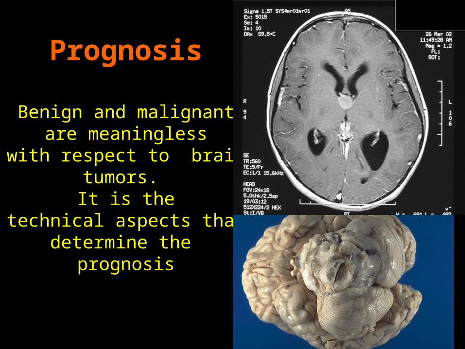
Prognosis
Benign and malignant are meaningless
with respect to brain tumors. It is the
technical aspects that determine the
prognosis
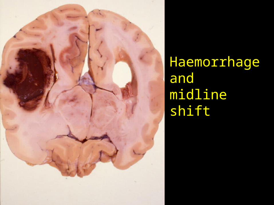
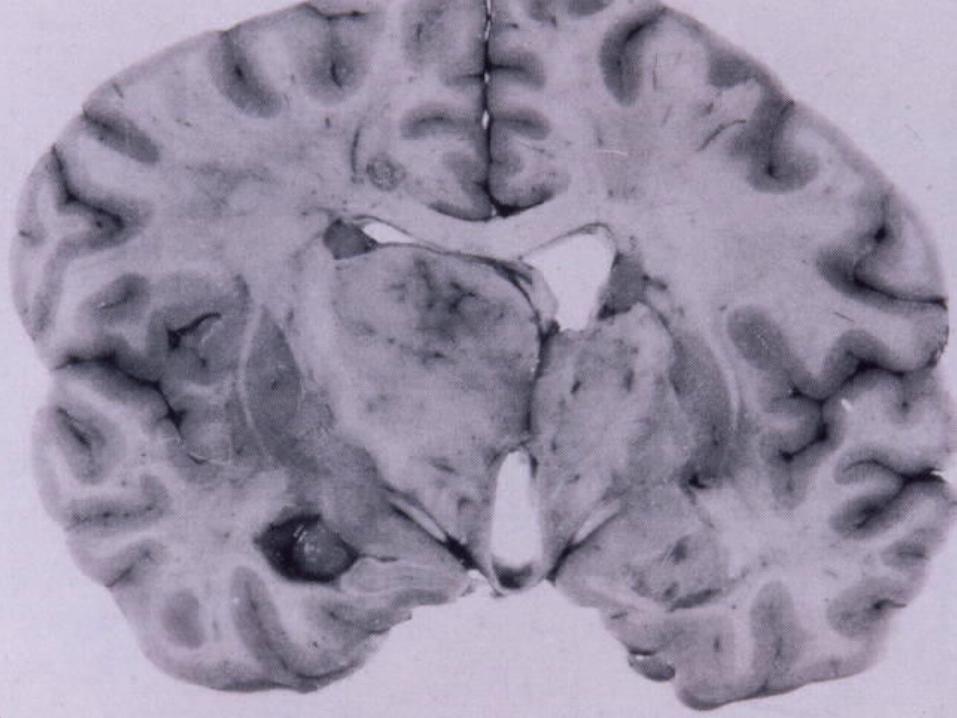
Haemorrhage andmidline shift
Raised ICP
~ As neoplasm grows – contents of the skull are compressed~ Within the skull brain occupies 1400mlsCSF 100-200mls and blood 100-150mls~ Displacement of CSF and blood compensate initially for mass effect~ Then ICP rises quickly mass effect compression vascular insufficiency
IC
ICP Herniations
~ Subfalcine herniation~ Tentorial herniation~ Tonsillar herniation
FALSE LOCALISING SIGNSFALSE LOCALISING SIGNS
~ Occulomotor nerve compression~ Abducens nerve compressed againstthe petrous ligament~ Ipsilateral hemiparesis – from compression of the cerebral peduncle against the tentorium~ PCA infarction from compression of the artery against the tentorium
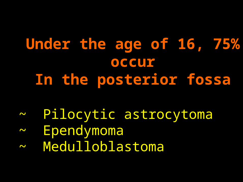
Under the age of 16, 75% occurIn the posterior fossa
~ Pilocytic astrocytoma~ Ependymoma~ Medulloblastoma
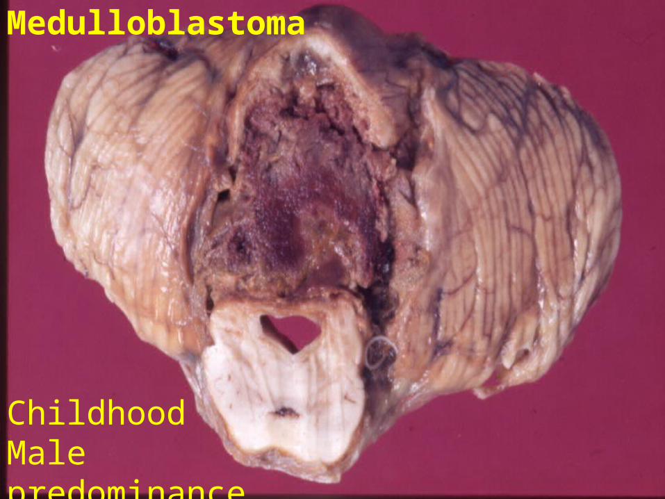
Medulloblastoma
ChildhoodMale predominance
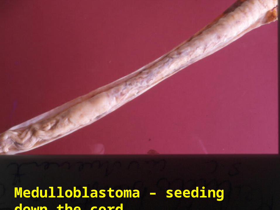
Medulloblastoma – seeding down the cord
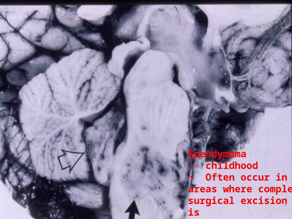
Ependymoma ~ childhood~ Often occur in areas where complete surgical excision is impossible
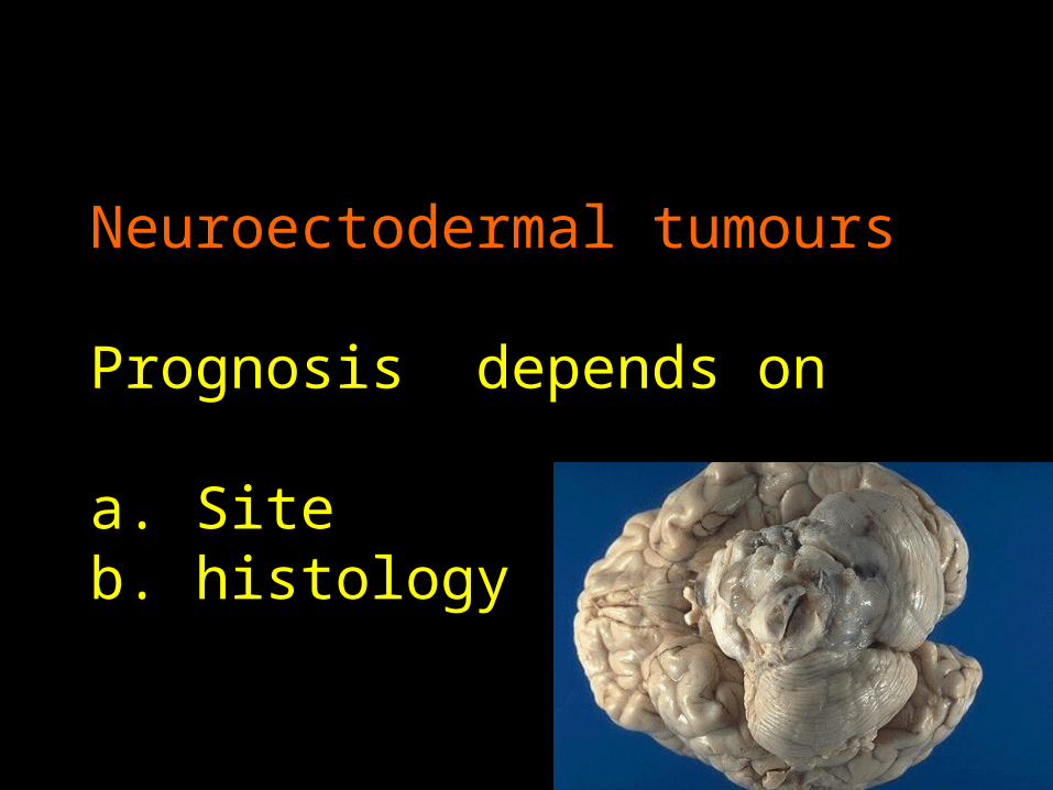
Neuroectodermal tumours
Prognosis depends on
a. Siteb. histology
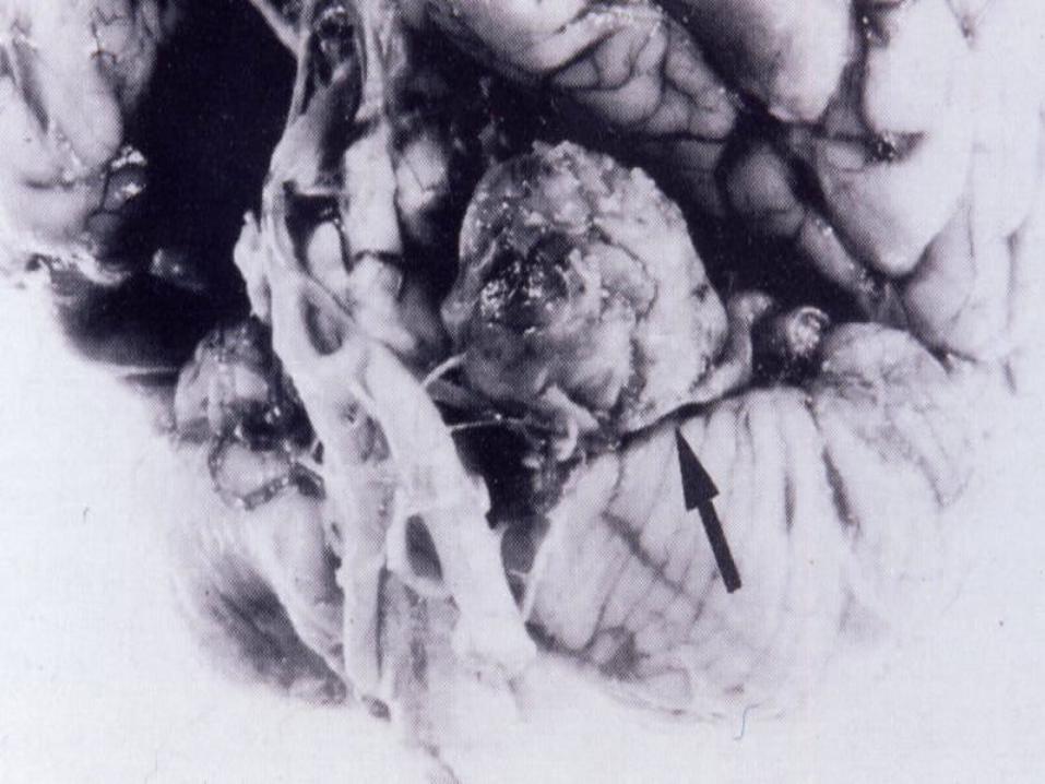
Meningiomas
~ Older adults usually female~ Increased incidence in Von Recklinhausen disease~ Association between meningiomas and breast cancer
Meningioms
Clinical presentation depends on:a)Siteb) Rapidity of growth
Prognosis – ~ benign (usually)~ slowly growing~ often can be completelyexcised
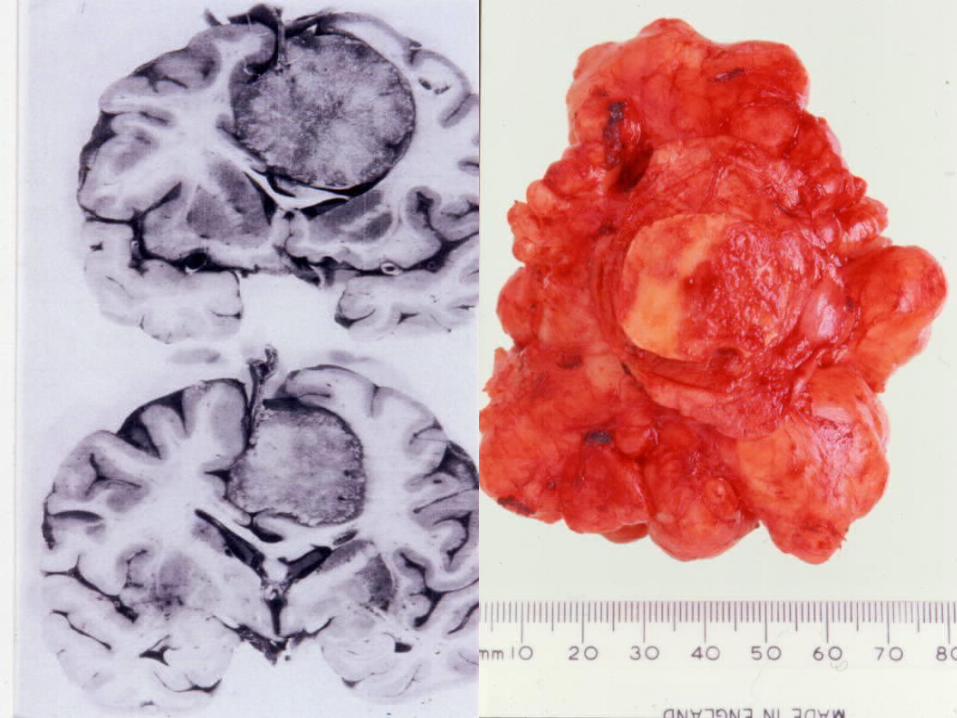
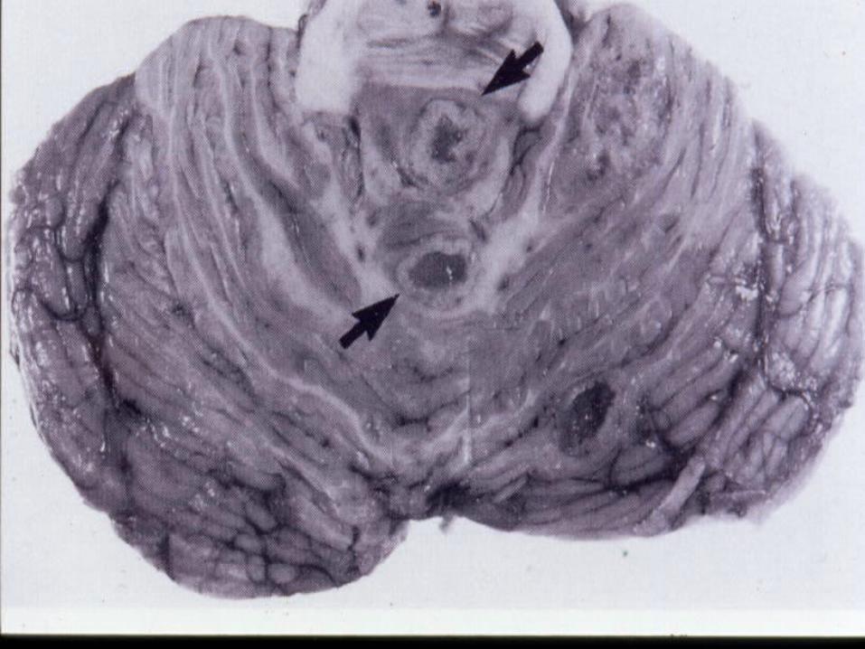
Meningiomaarising from the falxcerebri
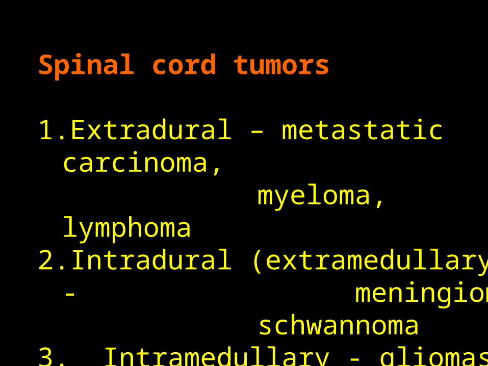
Spinal cord tumors
1. Extradural – metastatic carcinoma, myeloma, lymphoma
2. Intradural (extramedullary) - meningioma
schwannoma3. Intramedullary - gliomas
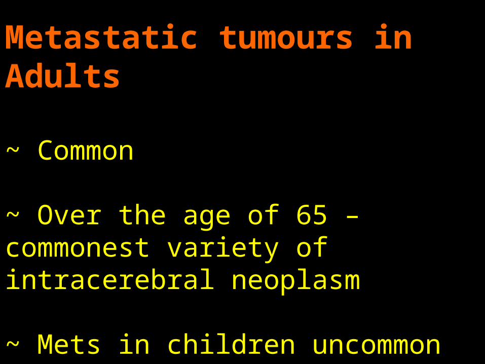
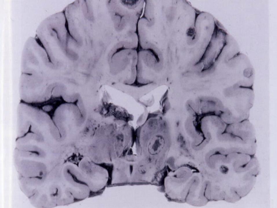
Metastatic tumours in Adults
~ Common
~ Over the age of 65 – commonest variety of intracerebral neoplasm
~ Mets in children uncommon but CNS well recognised site for relapse of ALL
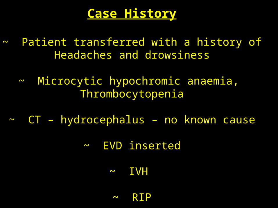
Case History
~ Patient transferred with a history ofHeadaches and drowsiness
~ Microcytic hypochromic anaemia, Thrombocytopenia
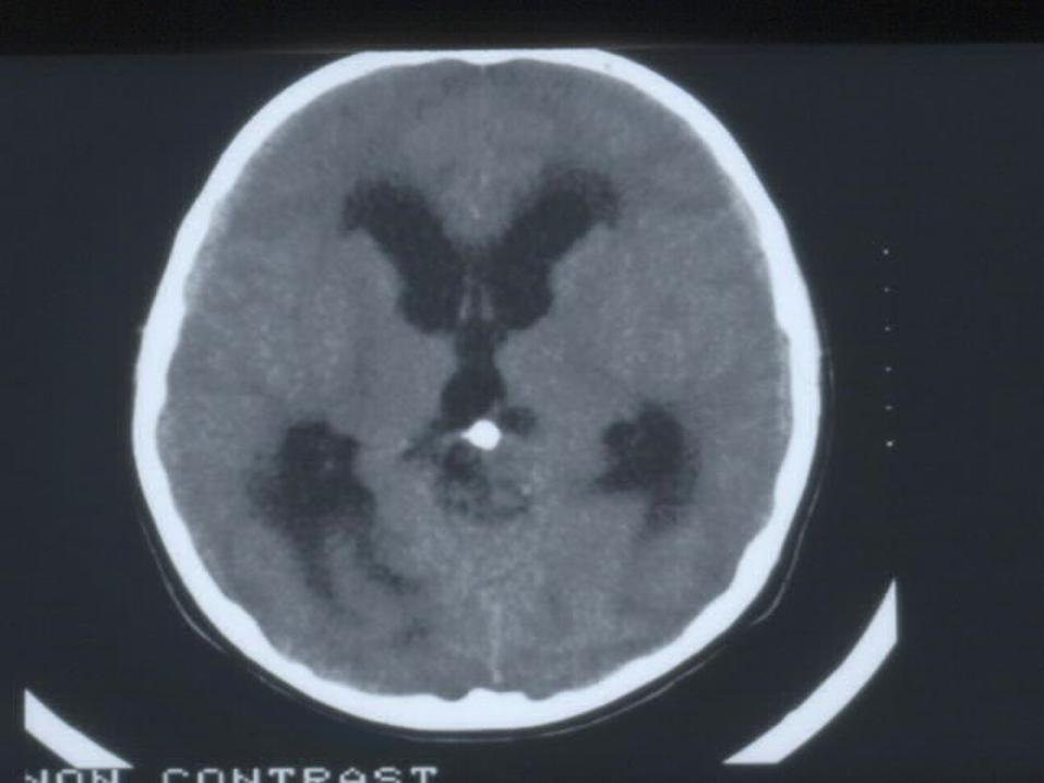
~ CT – hydrocephalus – no known cause
~ EVD inserted
~ IVH
~ RIP
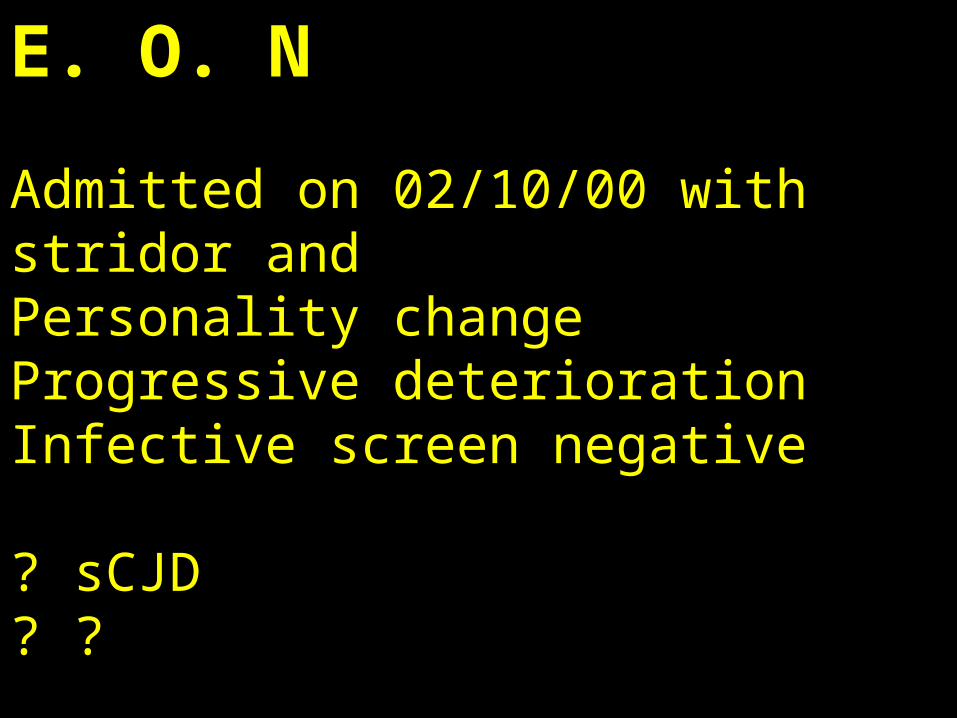
E. O. N
Admitted on 02/10/00 with stridor and Personality changeProgressive deteriorationInfective screen negative
? sCJD? ?
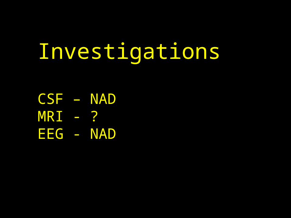
Investigations
CSF – NADMRI - ?EEG - NAD
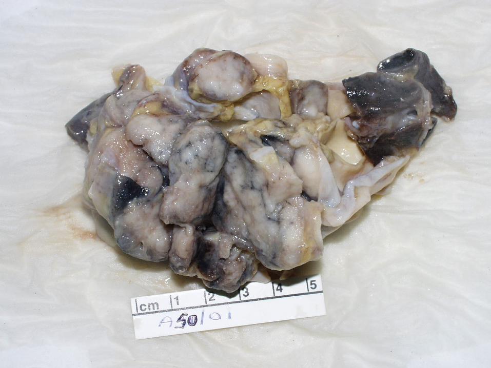
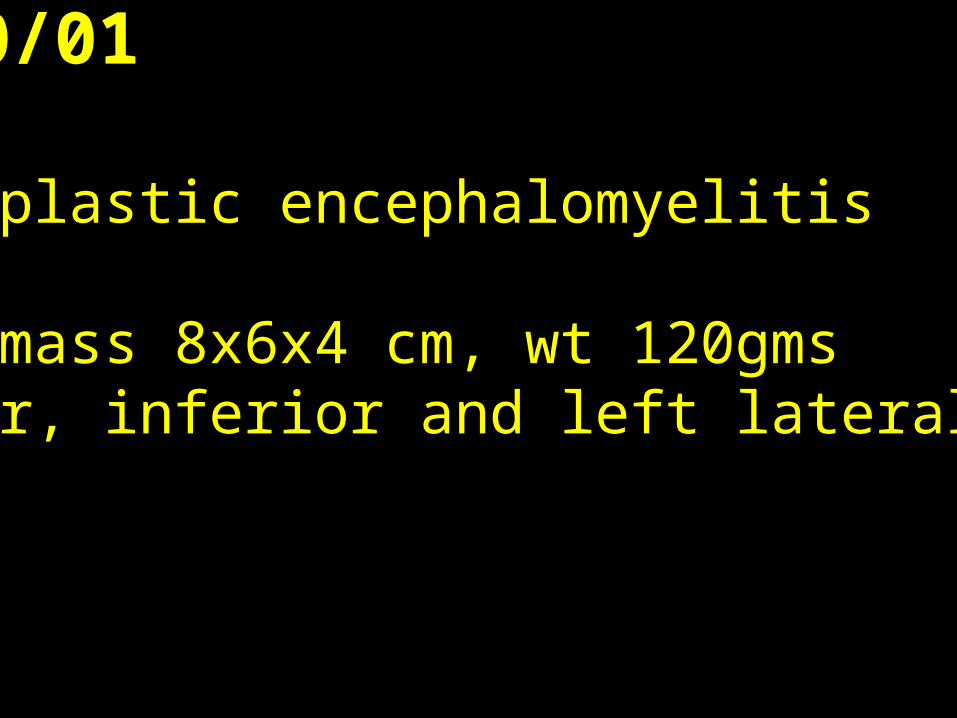
PM A50/01
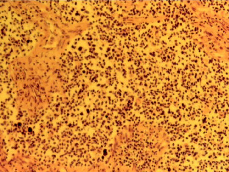
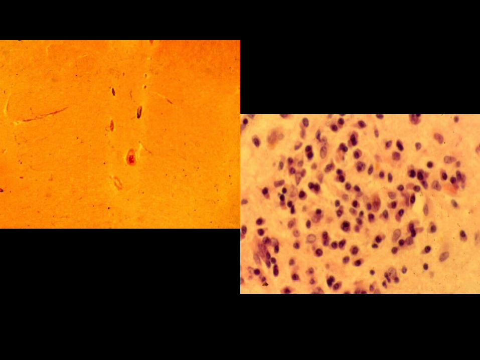
Paraneoplastic encephalomyelitis
Tumour mass 8x6x4 cm, wt 120gmsAnterior, inferior and left lateral to the Thyroid
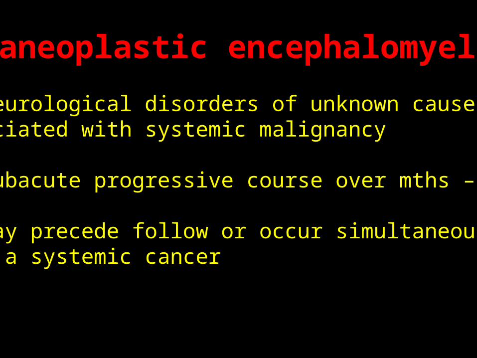
Paraneoplastic encephalomyelitisParaneoplastic encephalomyelitis
~ neurological disorders of unknown cause associated with systemic malignancy
~ Subacute progressive course over mths – years
~ May precede follow or occur simultaneously with a systemic cancer
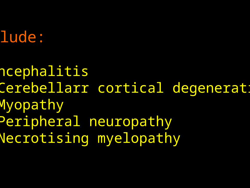
Include:
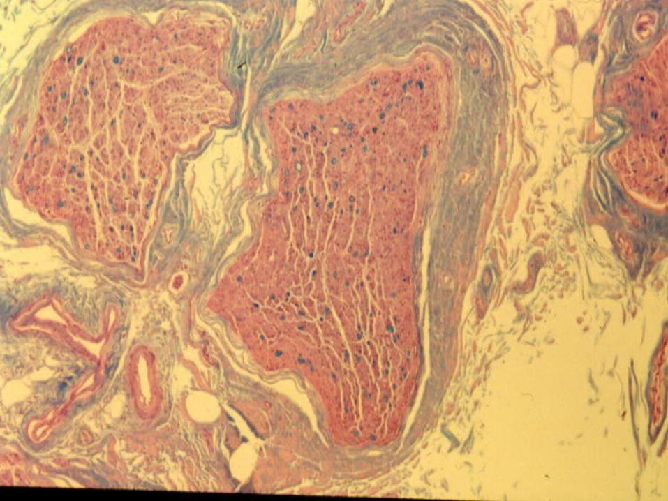
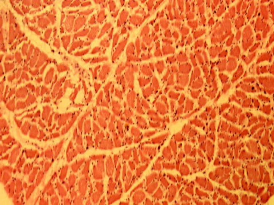
1. Encephalitis2. Cerebellarr cortical degeneration3. Myopathy4. Peripheral neuropathy5. Necrotising myelopathy
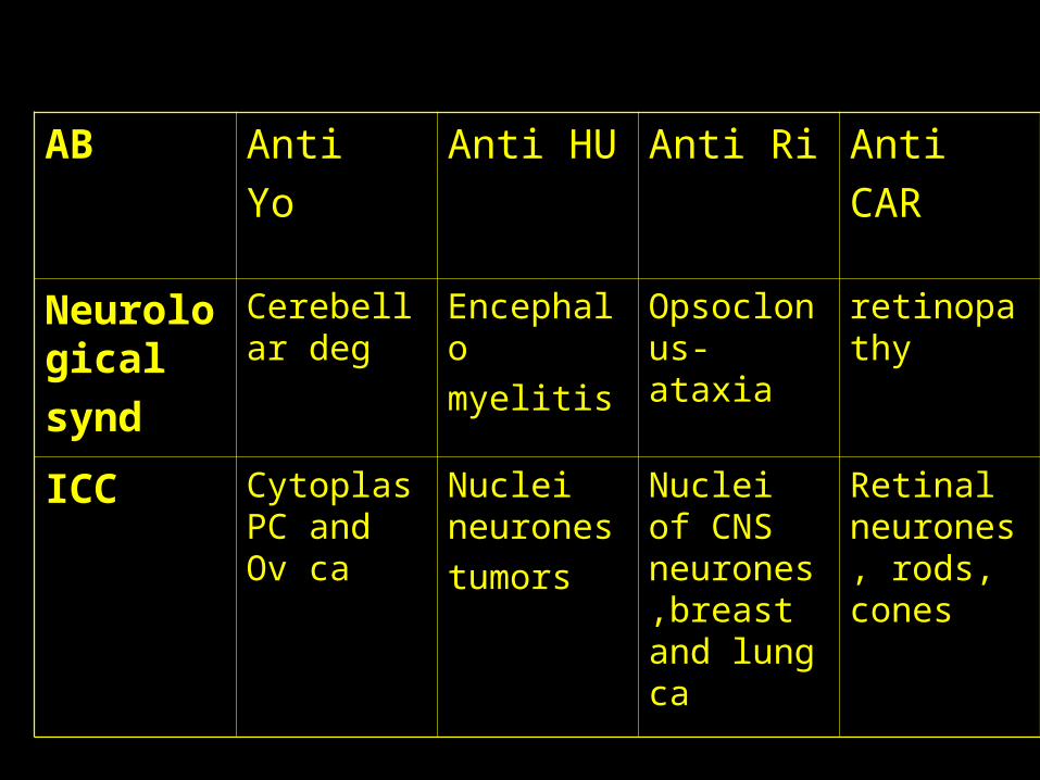
AB Anti
Yo
Anti HU Anti Ri Anti
CAR
Neurological
synd
Cerebellar deg
Encephalo
myelitis
Opsoclonus-ataxia
retinopathy
ICC Cytoplas PC and Ov ca
Nuclei neurones
tumors
Nuclei of CNS neurones,breast and lung ca
Retinal neurones, rods, cones
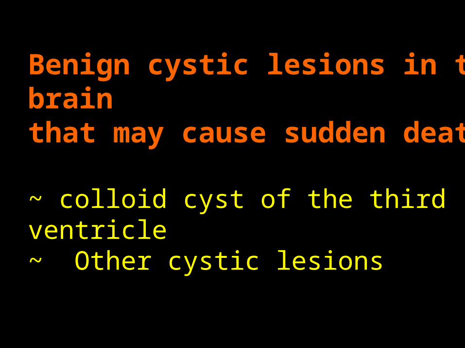
Benign cystic lesions in the brain that may cause sudden death
~ colloid cyst of the third ventricle~ Other cystic lesions
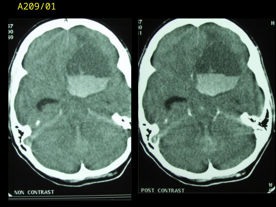
A209/01
CONCLUSION
1. Brain tumours classified into intrinsic,extrinsic and spread from adjacent structures
2. Adults usually present with supratentorial tumours3. Commonest primary tumour in adults gliomas. >65
metastatic tumours common4. Paraneoplastic syndromes – non-metastatic
complications of an underlying malignancy.


