196
Tutorials on Clinical Methods in Paediatrics Dr M R Ghuman MBBS (Pb), MSc Med (Wits), FCPaed (SA) 6 th Draft 2020

Tutorials on Clinical Methods in Paediatrics
Dr M R Ghuman
MBBS (Pb), MSc Med (Wits), FCPaed (SA)
6th
Draft 2020

Tutorials on Clinical Methods in Paediatrics: Dr M R Ghuman et al. Draft 6, 2020
2
Preface
Due to Covid-19 lockdown most of the Medical Students and Interns who are under training
in the discipline of Paediatrics and Child Health, may find it difficult to have a face to face
tutorial in their respective hospitals. Therefore, I have developed this document in such a
way that each tutorial discusses basic information related to that system first and then
explains clinical methods with photographs and lively illustrations.
It was presumed that students learning Paediatrics have had previous exposure of clinical
methods therefore in-depth description of basic clinical terms is not done in this review. It is
also not intended to be a text book or encyclopaedia, instead to serve as a guide to what
need to be done at the bedside.
Although main emphasis is made on history taking and clinical examination, students are
also guided to make logical deductions and differential diagnosis on the basis of their clinical
findings. Since my approach is basically clinical that will be confined largely to clinical signs
and symptoms in children of varying ages, it does not include pathophysiology, laboratory
investigations and treatment protocol. Readers are therefore directed to main text books of
Paediatrics and Neonatology if detailed information is required on a particular condition. In
keeping with the need of the reader I have tried to retain a problem-oriented approach
where possible, and I hope that this manual fills the need for a distant learning concise set
of tutorials on Clinical Methods in Paediatrics.
I am indebted to my team of doctors which include Dr Yajna Ramdass, Dr Riaz Bux and Dr
Irfaan Saib for assisting me for lively illustrations and photographic shots. I would also like to
thank the parents of little boy for allowing us to include him for illustrating the clinical
examination.
M R Ghuman

Tutorials on Clinical Methods in Paediatrics: Dr M R Ghuman et al. Draft 6, 2020
3
SOURCE OF INFORMATION &
ACKNOWLEDGEMENTS
1. Macleod’s Clinical Examination 11th
ed
2. Students Clinical Methods: M Ali 4th
ed
3. Clinical Examination: NJ Tally et al 5th
ed
4. Examination Paediatrics: Wayne Harris 3rd
ed
5. Coovadia’s Paediatrics and Child Health 6th ed
6. Weiner and Levitt’s Pediatric Neurology 4th ed
7. Parks Paediatric Cardiology for Practitioners 5th
ed
8. Clinical Paediatrics for post graduate students 3rd
ed
9. Paediatric Guidelines: Mc Karrow, Patrick & Stephen et al
10. Bates Guide to Clinical Examination and History Taking 4th
ed
11. Handbook of Clinical Methods Course for 4th
year UKZN Students 2018
12. The History and Clinical Examination of Children: University of Pretoria 2009

Tutorials on Clinical Methods in Paediatrics: Dr M R Ghuman et al. Draft 6, 2020
4
CONTENTS
Tutorial Topic page
Tutorial 1 History taking 5
Tutorial 2 Anthropometry 14
Tutorial 3 Micronutrients 29
Tutorial 4 Neurodevelopmental assessment 41
Tutorial 5 Dysmorphology 54
Tutorial 6 General physical examination 63
Tutorial 7 Respiratory system 83
Tutorial 8 Cardiovascular system 98
Tutorial 9 Abdomen 117
Tutorial 10 Central nervous system 134
Tutorial 11 Musculoskeletal system 176
Tutorial 12 Endocrine system 183
Tutorial 13 Renal system 185
Tutorial 14 Haematological system 186
Tutorial 14 Final assessment 189
Tutorial 15 Infant feeding 190

Tutorials on Clinical Methods in Paediatrics: Dr M R Ghuman et al. Draft 6, 2020
5
TUTORIAL 1
HISTORY TAKING OVERALL OBJECTIVES: At the end of this module, students are expected to acquire the art of appropriate history
taking by:
a. Eliciting the chief complaints i.e. the reasons for presentation
b. Evaluating each complaint through questioning
c. Assessing relevant positive and negative aspects in the history that contribute to the
most likely diagnosis
Followings need to be taken into account when taking a paediatric history History taking from both parents and the child is an important art for the interpretation of
clinical signs in children of varying ages.
SYMPTOMATOLOGY A symptom is a normal physiologic response to a harmful stimulus. Every symptom and sign
has a beginning and a course of development that may be progressive.
Symptoms and signs are products of the body that produced them. Each body creates
symptoms and signs in a unique way, and each personality adapts to them its own way.
One symptom by itself usually means very little. It is its relationship to other symptoms that
is significant. For instance, vomiting accompanied by abdominal pain in the lower right
quadrant may suggest appendicitis, while vomiting with headache and failing vision could
lead one to suspect something causing increased intracranial pressure.
Symptoms are either subjective or objective, or both. Subjective symptoms are those
perceptible only to the patient such as sensory disturbances i.e. pain, tenderness, headache,
nausea, itching and numbness. Pain and itching are pure subjective symptoms.
Objective symptoms which are evident to the observer are called physical signs like
temperature, pulse rate and rhythm, respiratory rate and character, oedema, and gait.
In order to elaborate a symptom fully, we need to ask the relevant questions and duration
of each symptom related to patients’ complaints. Paediatric patients presenting with the following symptoms should be enquired about:
1. FEVER Mode of onset of fever: sudden or gradual
Associated with rigors or sweating
High or low grade
Pattern of fever: continuous, remittent or intermittent
Associated with vomiting, diarrhoea, abdominal pain, headache or burning on
micturition

Tutorials on Clinical Methods in Paediatrics: Dr M R Ghuman et al. Draft 6, 2020
6
2. COUGH Severity and frequency
Sudden vs gradual (foreign body)
Occurs during a special time of day or night
Seasonal or associated with dust, pollen or perfumes
Abnormal sounds while coughing (croupy, whooping cough)
Colour changes while coughs (cyanosis)
Dry or productive
Colour of sputum or presence of blood
3. VOMITING Frequency of vomiting
Forceful or projectile
Relationship between food and vomiting (may be meningitis)
Special time of vomiting (early morning: may be space occupying lesion)
Amount, smell, colour and contents of vomitus: yellow, bile stained or feculent
Any other associated symptoms like heart burn or epigastric pain
4. DIARRHOEA Loose motions: intermittent or continuous
If intermittent: duration which patient is free from symptoms
Frequency of stools passed per day (count per 24hrs period)
Colour, quantity, odour and contents of stool
Any blood in stools: red or black / separate or mixed in stools
Any history or eating out / bottle feeding/ formula feeding
Any history of diarrhoea in any other family member staying with child
History of pain during defecation
When last did the child passed urine?
History of administration of ORS and if made appropriately
Check if the child is vaccinated for Rota virus vaccination
5. PAIN Site of pain
Localised or diffuse
Continuous or intermittent
If radiates to which direction
Dull, burning, colicky or stabbing
Intensity of pain: mild, moderate or severe /excruciating
Factors which aggravate or relieve pain
Does it affect sleep, play and activity (growing pains vs pathological pains)
6. FITS Generalised or localised
First episode or repeat
Previous history of fits and the time duration
Tutorials on Clinical Methods in Paediatrics: Dr M R Ghuman et al. Draft 6, 2020
7
Frequency and time duration of each fit
Type: myoclonic/tonic/clonic/ atonic/absence
Any loss of consciousness or history of aura
Weakness or hemiparesis thereafter
Any history of fall, head trauma or loss of sphincteric control
Any other associated symptoms like tongue bite, froth in the mouth, deviation of eye
balls during the attack
Family history of epilepsy or fits
7. WEAKNESS Mode of onset: sudden or gradual
Extent of weakness: generalised or localised to one part of body like one arm or one
leg (monoplegia), one half of the body (hemiplegia), both legs (paraplegia), or one
half of the face (facial palsy)
Grade of weakness:
o Grade 0: No contraction or muscle movement
o Grade 1: Trace muscle activation, such as a twitch, without achieving
full range of motion
o Grade 2: Movement at the joint with gravity eliminated
o Grade 3: Movement against gravity, but not against added resistance
o Grade 4: Movement against external resistance with less strength than usual
Any other associated symptoms like loss of consciousness, fever, headache, neck
rigidity, loss of sensations or presence of tremors
History or trauma / head injury or fever in the past
Feeling week at rest or after prolonged use of muscles (myasthenia gravis)
8. HEADACHE Character: dull, shooting or throbbing
Continuous or intermittent
If intermittent: duration and frequency of each attach
Related to any particular time of the day
Relieving or aggravating factors
Part of head involved: one half (migraine), frontal or occipital region
Any associated symptoms like vomiting, nausea, impairment of vision or
consciousness
History of head injury, hypertension, neck rigidity, or loss of consciousness
History of blurred vision or unable to see black boards in school children
9. LUMP OR MASS Site, size and number of lumps
Then note its surface, consistency, mobility, tenderness and fluctuation
Note skin over the mass: fixed or mobile
Any sinus formation over the swelling
Note lymph nodes: swollen, separate or matted together

Tutorials on Clinical Methods in Paediatrics: Dr M R Ghuman et al. Draft 6, 2020
8
HISTORY TAKING In order to have correct diagnosis we need a perfect history, good clinical examination and
laboratory investigations. Following general but important points need to be noted before
starting an interview with the child, parents or the caregivers.
1. See the patient in private room if possible and listen carefully to everything that is
said.
2. Introduce yourself and make the acquaintance of both the child and parent.
3. Light banter about the weather or how they found the travelling to hospital help to
break the ice.
4. A detailed discussion with the parents and the child is very important step towards
correct diagnosis, identification of the problems and further management.
5. Don’t jump to conclusions prematurely or you may come with wrong answers. 6. Let the child or the parents tell the story in their own way & words and make notes as
you go along. At the same time an occasional guiding question will keep the story
going in the right direction. Show interest and concern for the patient’s problems.
7. Listen to the voice, but also watch the facial impressions, body movements and hand
positions. These may provide additional information. The actions will often provide
information about the underlying emotional state as well as giving an indication as to
the reliability of information being supplied.
8. Collect the patient’s information without expressing surprise or making judgments. It is unethical to comment adversely on another practitioner’s or clinical nurse’s previous treatment. Neither you nor the patient has enough information to pass the
judgment. The information you receive may sometimes be falsified by the parents for
fear of possible accusation of neglect, for compensation or even insurance purposes.
Information can also be suppressed because of fear. Mothers may misinterpret
symptoms but rarely invent them.
9. Avoid “why did not you” questions – they may lead to hostility and consequent lack
of co-operation as the parent / guardian feels threatened. The same question can be
phrased in a less accusing form.
10. Parents experience reactions to health problems like fear and anxiety. Reassurance
must be given and fears relieved whenever possible.
11. Be sure that you understand what the patient or parents mean and that they
understand what you mean. This is of greater importance especially when
interpreters have to be used. Always use lay terms for questions and explanations.
12. When taking history, make notes of both positive and negative findings and define
the problems. From this you should be able to decide which system must be
examined in detail. This history will often produce a working diagnosis but keep an
open mind until full examination is completed.
13. After taking history and doing clinical examination define the patient’s problem. Explain each problem and its management to the parents and when they should see
you again.
14. Warn the patients / parents of possible complications or danger signs which may
occur and what to do in that event.
15. Remember there are three assessments to be made:
a. The physical diagnosis and the plan of management
b. The assessment of patients hidden but unspoken fear about the child’s problem

Tutorials on Clinical Methods in Paediatrics: Dr M R Ghuman et al. Draft 6, 2020
9
c. The assessment of parents’ capability of understanding the nature of the
problem and the likelihood of their following your advice
BACKGROUND AND BASIC INFORMATION A complete database entails the following:
1. Name & surname: for identification
2. Age & date of birth: certain conditions are common in early childhood others in
later life like Wilms’ tumour is present in childhood and malignancy and vascular disorders common in older patients
3. Gender: Certain conditions present in males others in females like Haemophilia
never occur in females and gout and ankylosing spondylitis has preponderance in
males
4. Religion & Church affiliation: to understand the problems that may arise in certain
situations like: blood transfusions are not accepted by the people practicing
Jehovah’s Witness; Muslims & Jews both practice circumcision in early childhood
5. Parents’ Name and Surname: their cell and telephone numbers at home & at work
6. Residential address: to understand social circumstances around the child and the
fact that certain diseases & conditions are common in certain areas. Secondly a
future correspondence may be required.
7. Informant: name and relation to the patient. If the informant is other than the
mother, elaborate the reason?
8. Details of Doctor who referred the patient: family physician & his/her address.
Document if patient is not a referral case – self referral?
9. Place of consultation: consulting rooms, outpatient department, causality or
paediatric ward
10. Date and time of your consultation
11. Name of the ward or the unit (if admitted)
12. Name and qualifications of admitting doctor
PRESENTING COMPLAINTS A presenting symptom (chief complaint) is that symptom, or group of symptoms, about
which the patient complains or seeks relief. Chief complaints which have brought the
patient to the doctor are arranged in chronological order i.e. complaint of longest duration
at the top and that of shortest duration in the last.
Each complaint should be written in one line and should be brief. Where possible, write
down the information offered in their own word like if the Parents say that the child has
shortness of breath or difficulty in breathing do not decide to write “dyspnoea”.
Date the commencement of symptoms precisely and completely and use adjectives with
each complain like fever with rigors for 5 days instead of fever for 5 days.
HISTORY OF PRESENT ILLNESS Details of each complaint are asked in chronological order by asking additional questions to
bring out full description of individual symptom. Avoid asking leading questions and do not
use technical terminologies, instead if possible, write down the history in the patient’s own
words. While describing the symptoms, following general points need to be noted:

Tutorials on Clinical Methods in Paediatrics: Dr M R Ghuman et al. Draft 6, 2020
10
1. Duration of symptoms: ascertain the duration by asking “were you alright before such period of time”. Date the commencement of symptoms precisely and
completely, like diarrhoea started on 25th
Dec 2019 – that is 5 days ago.
2. Health status: immediately prior to the present illness & how long ago the patient
was completely well
3. Mode of onset: to ask if the symptoms appear suddenly or gradually
4. Disease course, sequence and period separating new symptoms: to ask if the
symptoms have been present continuously since their onset or have there been
intervals of freedom – note length of these intervals.
5. Factors aggravating or relieving symptoms
6. Associated phenomena: to ask if any other symptoms associated with the main
complaints like abdominal pain due to intussusception may be associated with
vomiting, diarrhoea or blood in stools and pain in right upper quadrant may be
associated with jaundice & pale stools in obstructive jaundice.
7. Treatment received and duration - whether the symptoms are improving or getting
worse or of the same nature. History of drug allergy should be asked as well.
8. Possible exposure to infectious diseases or recent travel or visits to malaria areas
9. Relevant negative data obtained by direct questioning e.g. no visits to possible
malaria areas
10. If the symptoms occur in clusters or attacks – a typical attack should be described
with regard to frequency, duration and degree of symptoms
SYSTEMIC ENQUERY Questions related to all body systems can be asked but special emphasis should be made on
systems involved.
SKIN: Skin temperature, skin colour, pruritus, hydration, eczema and other skin
rashes – associated medication / environmental exposure etc.
EYES: Vision, glasses, infection, allergies and strabismus, swelling discharge
ENT: Hearing, pain, nose obstruction, snoring, runny nose, epistaxis, sneezing, caries
teeth, stomatitis, sore throat, tonsillitis, adenitis, mouth breathing etc.
RESPIRATORY SYSTEM: Cough, wheeze, shortness of breath, strider, cyanosis,
sputum, haemoptysis, asthma, foreign body, snoring, mouth breathing and enquiry
about precipitating factors like exercise, dust, animals. Ability to speak full sentence
with wheeze
GASTROINTESTINAL SYSTEM: Weight loss or abnormal weight gain, appetite,
vomiting, haemochezia, constipation, diarrhoea, jaundice, melena, worms in stool,
pain and thirst.
CARDIOVASCULAR SYSTEM: Shortness of breath, tires while feeding, excessive
sweating (in infants it is sign of CCF), limitation of exercise, oedema, palpitations,
haemoptysis and cyanosis.
CENTRAL NERVOUS SYSTEM: Headache, mental state, seizures, tremors, paralysis,
sight, hearing, behaviour, inattention or hyperactivity, ataxia, weakness, gait, co-
ordination, dizziness, muscle and joint swelling & pains, weakness and postural
deformity.
GENITOURINARY SYSTEM: Frequency, bladder control, dysuria, character of stream,
urine colour (be aware of the pigments resembling blood) enuresis, and vaginal
discharge.

Tutorials on Clinical Methods in Paediatrics: Dr M R Ghuman et al. Draft 6, 2020
11
ENDOCRINE SYSTEM: Growth, polyuria, polydipsia, apathy, hoarse voice,
muscularity, menstruation, breast and testes and growth of pubic and axillary hair.
HAEMOPOITIC SYSTEM: Pallor, tiredness, shortness of breath, bleeding tendency,
bruising, enlarged glands and spleen.
PAST HISTORY This should not be confused with the earlier symptoms of the present condition but this
includes attacks of a similar nature when a long interval has elapsed. Following points need
to be kept in mind:
1. Diseases of childhood such as rheumatic fever, whooping cough, mumps, measles or
primary pulmonary tuberculosis should be enquired or any history of relevant
disease in the past.
2. Any history of trauma, accident, surgery, visit abroad, intake of “herbal medications, herbal enemas or scarification from traditional healers.
3. History of any previous admission in the hospital may indicate the seriousness of the
disease like child with uncontrolled chronic persistent asthma admitted twice since
last month.
BIRTH HISTORY Birth history is particularly important in young infants which should be confirmed by looking
into the Road to Health Booklet. Salient points should include:
1. ANTENATAL HISTORY o Age of mother
o Gravida, para, abortions, perinatal deaths
o Birth weight and health of previous infants
o Gestational age at birth of the index patient
o Planned or unplanned pregnancy
o Foetal growth and wellbeing
o Complications of pregnancy e.g. eclampsia, gestational diabetes
o HIV status and CD4 count and viral load if HIV infected
o Antiretroviral prophylaxis or treatment detail
o Results of WR or RPR – treatment if syphilis was diagnosed
o Blood group of mother
o Exposure to medicines, smoking, alcohol
2. NATAL HISTORY o Date and place of delivery
o Duration and mode of delivery
o History of foetal distress
o Other problems during labour and delivery
o Medication received by mother
o Apgar score and if baby needed resuscitation
o Estimated gestational age
o Birth weight, length, head circumference
o Vitamin K and medications given to the baby
o Congenital abnormalities
3. POSTNATAL HISTORY o History of admission in nursery, NICU – duration

Tutorials on Clinical Methods in Paediatrics: Dr M R Ghuman et al. Draft 6, 2020
12
o Breathing problems
o Jaundice, anaemia, cyanosis
o Skin rashes or history of haemorrhage
4. FEEDING PROBLEMS o A 24-hour dietary history is useful when assessing a feeding problem of child
with signs and symptoms of malnutrition
o Breast feeding problems: poor latching, small nipples or cracked nipples
o Formula feeding: formula type, change of formula, volume, frequency,
preparation and dilution of feeds
o Weaning age, solid foods and supplements and appetite
o Food intolerance, food allergy, weight gain or loss
CHILD HEALTH RECORD 1. IMMUNIZATION
Check if the immunisation is up to date on road to health booklet. Following is the current
schedule of EPI South Africa
Birth OPV (0) BCG
6 W OPV (1) DTaP-IPV // Hib (1) Hep B (1) PCV (1) RV (1)
10 W DTaP-IPV // Hib (2) Hep B (2)
14 W DTaP-IPV // Hib (3) Hep B (3) PCV (2) RV (2)
9 M Measles (1) PCV (3)
18 M Measles (2) DTaP-IPV // Hib (4)
6 Y (M/F) Td vaccine
12 Y (M/F) Td vaccine
Non-EPI vaccines: MMR & Varicella: @ 15 months & Cervirex (HPV) for F @ 12 yrs.
RV vaccine is not administered after 24 months
Preterm infants: if acutely ill, vaccinations are deferred until recovered. All vaccinations are
given at chronological age and not the corrected age. BCG is aslo deferred until infant is
ready to be discharged. Side effects like skin rash and fever are not worse than term infants.
Of note use of acellular pertussis in current EPI-SA is safe and does not cause apnoea as it
was the case with cellular pertussis vaccine.
2. CLINIC VISITS: Growth monitoring, vitamin A administration and deworming
FAMILY HISTORY Ask about the number of family members, both males and females and their health. In case
of suspected heriditary or familial type e.g. haemophilia, mental retardation, and
myopathies a detailed family history is required including consanguinity, pregnancy or child
loss. Pedigrees are often used to determine the mode of inheritance of genetic diseases. A
sample pedigree is as follows:

Tutorials on Clinical Methods in Paediatrics: Dr M R Ghuman et al. Draft 6, 2020
13
Diseases like hypertension, diabetes, epilepsy, certain renal and cardiovascular diseases,
rheumatic fever also have familial background. Tuberculosis is very common in South Africa,
thus any history of tuberculosis in the family should always be enquired.
SOCIAL HISTORY A detailed history about the psycho-social circumstances of the family where child functions
must be taken as follows:
Primary care giver
Occupation and employment history of parents
Housing, sanitation and movement
Financial situation like paternal support, grants and employment
Long parental absences from home and support systems: family, friends
Marital state and stability
Psychiatric diseases, substance abuse, alcohol and smoking
Family violence, traumatic family episodes, deaths, divorce or accidents
Recreation as a family
The aspects listed above are sensitive, thus indirect questioning should be used. By
observing family interaction and body language good amount of information can be
gathered. A simple question like “what do you as a family do for recreation and leisure,” can provide sufficient information about interpersonal relationships within the family.

Tutorials on Clinical Methods in Paediatrics: Dr M R Ghuman et al. Draft 6, 2020
14
TUTORIAL 2
ANTHROPOMETRY AND NUTRITION OVERALL OBJECTIVES At the end of this module the student should be able to:
a) Take a dietary history and growth history of children
b) Use growth parameters to assess nutritional status (normal or abnormal)
c) Describe the clinical features of malnutrition
d) Recognise by appropriate history taking and examination, common and life threatening
complications of nutritional deficiencies in infants and children and investigate and
manage these complications.
e) Use and impart the significance of Road to Health Booklet to parents.
ANTHROPOMETRIC MEASUREMENTS A normal distribution pattern for weight, length and head circumference has been
established for every month of age in the first 2 years of life and for every 2 months untill
the age of 12 years. This was done by measuring hundreds of normal children in each group.
From the above range of normal measurement Z-score charts have been developed for
weight, length/height, BMI and head circumference.
Weight:
The most rapid period of growth after intrauterine period is during the first 2 years of life.
Average birth weight doubles by 5-6 months and tripples by 1 year. Ideally the weight of
every child seen at the clinic or hospital should be plotted on a Z-score chart than trying to
rely on memory. Weight is easy to measure than height and shows rapid changes.
Remember that weight, but not height, can be lost therefore weight is considered to be a
more sensitive measure. Idealy children should be undressed when weighed but light
clothing need not to be removed and weighing scale should be checked regulary.
Average weight values at certain age groups
Age Weight in Kg
Birth 3.5
5-6 months 7
1 Year 10
2 yrs 12
Formula to calculate an average weight from 1 year therefater: Age (in years) + 4 x 2 = Kg
For example: approximate weight of a 2 years old child: 2 + 4 x 2 = 12 kg
Length: Length is measured by infantometer and height by stadiometer and it follows the similar
pattern of weight.

Tutorials on Clinical Methods in Paediatrics: Dr M R Ghuman et al. Draft 6, 2020
15
Average length/height in certain age groups
Age Length/height in cm
Birth 50
6 months 65
1 year 75
2 years 87
3 years 95
4 years 100
Approximate height of a child at the age of 4 years is considered as 100 cm and therafter
every year should add 6 cm per year till the age of 15 years. For example a 5 years old child’s estimated height will be calculated as follows: 100+6 = 106 cm
Head Circumference or Occipito-Frontal Circumference (OFC):
Head circumference is of importance as increase in head size is almost entirely dependent
on brain growth. The most rapid period of brain growth is before birth and during the first
into the second year of life. More brain growth occurs during the first year after birth than
in the total period from 1 year of age untill addulthood. In fact most growth occurs during
the first 6 months of life. If severe nutritional insult or prolonged illness occurs during this
very vulnerable period “catch up” brain growth may be insufficient for the potential of that individual ever to be achieved. Measurement of OFC is also useful if some chronic brain
abnormality or abnormal growth of head is suspected.
Average head circumference in certain age groups
Age OFC in cm
Birth 35
3 months 40
6 months 44
1 year 47
2 years 50
5 yrs 52
Adult 56+
PHYSICAL GROWTH Children grow, and if they don’t, there is a problem. Their growth depends on varuis factors,
the most important being the heriditary, genetic, environmental, emotional health, chronic
diseases, endocrine and timing of puberty .
1. Heriditary and constitutional factors: this refers to the type of body build inherited from
the parents. If both parents are small and short, the children are likely to be small
whereas tall parents tend to have tall children. However, temperament, manifested as
the behavioural style of the child, is influenced by child-rearing practices as well as by
the continuous interaction between partents and children.

Tutorials on Clinical Methods in Paediatrics: Dr M R Ghuman et al. Draft 6, 2020
16
2. Intrauterine period: During intra-uterine life, maternal influences play an important part
in the growth of the fetus. Foetal growth may be affected by maternal nutritional,
socioeconomic status, habbits and medical problem.
3. Postnatal period: The major part of infancy is cheratcterized by a rapid growth rate,
which becomes increasingly related to the genetic background. This period of rapid
growth is largely determined by nutritional rather than endocrine factors.
4. Nutrition: Growth is profoundly affecetd by nutrition. Linear growth, expressed as
length or height measurement, is a sensitive indicator of the physical health of a child.
5. Health status: physical health and emotional status of a child or of his family may have
direct or indirect effect on his growth and development. Chronic illness often leads to
disability.
6. Socioeconomic status: Poverty, poor education, and social adversity increases the
possibility of having complications from pregnancy, birth, infancy and childhood.
7. Cultural factors: Child-rearing beliefs and practices vary markedly from one culture to
another and have profound effect on growth and development of children.
8. Environmental: this includes our surroundings, and factors that can influence our life
and growth. A diet which is poor in quantity or quality will prevent normal growth. This
is what can often be detected on properly completed growth charts.
Poor health includes infections, as well as any serious disturbances of physiological function
such as heart disease or severe congenital abnormalities. If the vital organs will not function
properly, the child will not grow normally. Health and nutrition influence each other. In turn,
unhealthy child may become malnourished and the poorly nourished child suffers with more
frequent infections. Thus a serious cycle is set in motion, the one factor aggravating the
other.
Sprcific factors may be affecting at specific age groups:
Infancy (upto two years): Food, nutrition and chronic diseases
Childhood: Genes, growth hormones and chronic diseases
Puberty: Sex hormones
Types of growth charts
Road-to-Health Booklet growth charts: contains Weight for age & Height for age
Longitudinal growth charts: Percentile graphs & Z-Score graphs
BMI charts
Weight for length/height charts
Growth velocity Charts
Combined prenatal and postnatal growth charts
Growth charts for special populations: Down syndrome, Turner syndroms
WHO Anthro: for personal computers – software for assessning growth and
developemnt of world’s children. www.who.int/growth

Tutorials on Clinical Methods in Paediatrics: Dr M R Ghuman et al. Draft 6, 2020
17
Uses and advantages of growth charts 1. To check whether the child falls into the normal range for his age. Many children
are found whose weight falls below -2SD line. They do not necessarily look ill or
obviously malnourished. They are just small for their ages and if their weights were
not plotted on growth charts, they would have not been noticed. These children are
underweight for age (UFA) and represent the largest group of undernutrition. Steps
must be taken to improve their nutrition because they are more likely to develop
infections and obvious malnutrition.
2. Recording growth repeatedly at intervals, gives the most valuable information as to
whether the rate of growth is satisfactory. The child’s charted weight graph should
run parallel to one of the normal growth lines given on the graph i.e parallel growth
= good (normal) growth.
Z-SCORE GROWTH CHARTS Measurement is the only way to recognise whether growth is normal or not. In order to
record child’s growth and physical development, special Z-Score growth charts are used in
South Africa.
In a graph one factor is plotted against another but related factor e.g. wight for age, weight
for height and height for age. Repeated observations are recorded at regular intervals.
Following triangle for anthropometric measurements is helpful tool for plotting the
variables on the growth charts.
o Weight for age (WFA Z score)
o Weight for height (WFH Z score)
o Height for age (HFA Z score)
Increasing age should be accompanied by increase in weight. Following graph shows this
and the rate at which it occurs. Increase in weight by itself is not enough as the time period
over which it occurred must also be taken into account.
Weight
WFH <-2SD WFA <-2SD
Wasted Under Weight for age
Height Age
HFA <-2SD Stunted
Triangle for anthropometric assessment

Tutorials on Clinical Methods in Paediatrics: Dr M R Ghuman et al. Draft 6, 2020
18
Explanation of above graph:
A graph is made indicating the baby’s growth. When seen at 2 months female baby is found to have a weight of 4kg. Following a straight
line up from 2 months and across from 4 kg a point can be plotted.
At the age of 4 months the baby weighs 5 kg and at 6 months 6 kg. Find and mark these
points on the graph and then join these points. In a similar way height and age or head
circumference and age can be plotted.
Babies grow very rapidly in the first year of life. A normal infant should double its birth
weight by 5-6 months and it should triple by the age of 1 year. In later childhood growth is
generally slower but increases again markedly at puberty.
Human beings of any given age are not all the same size even though they may be perfectly
normal and healthy. It can therefore not be said that a normal newborn should weigh 3.5
kg. There is always a range of normal and in this case, it is from 2.5 to 4 kg.
It is difficult to remember all these normal variations for all ages; therefore, growth charts
are used with normal lines showing both the normal range and the rate of growth.

Tutorials on Clinical Methods in Paediatrics: Dr M R Ghuman et al. Draft 6, 2020
19
Graph above showing abnormal growth pattern
If a child was supposed to gained 1 kg over a certain period of time but has only gained ½ kg,
this is slowing of growth, and is an early important sign of inpending malnutrition.
Following graph The example is the following graph

Tutorials on Clinical Methods in Paediatrics: Dr M R Ghuman et al. Draft 6, 2020
20
1. Abnormal growth pattern of weight loss
Graph above showing well-nourished infant showing loss of weight. The continued
recording of weight at intervals is of far greater value than any single recording. Regular
recording of weight will show progress the child is making, i.e., pattern of growth.
2. Abnormal growth pattern of poor weight gain
Graph above showing poor weight gain leading to malnutrition. Plotting once off on the
weight chart may be misleading as it gives no indication of direction whereas repeated
recordings give both the direction and adequacy of weight curve which is more important in
assessing progress. Stopping breast feeding and giving only porridge may be seen as plotted
in the above graph.
While most children with severe acute malnutrition (SAM) with oedema have weight for age
below -2SD, fluid may falsely increase the weight. A gain of 500g due to fluid retention will
however show as obvious oedema. Since SAM with oedema can develop rapidly, the child
may still be within normal z-scores but will have deviated from his previous growth curve.
3. Infection and growth:
In babies who are weighed regularly and growth charts used, the frequent relationship
between infection and poor growth is noted. Every time an infant suffers from gastro-
enteritis, a respiratory tract infection and particulaly having measles, there will be weight
loss. As poorly nourisehd children tend to get more frequent infections, so one problem
leads to other. If recovery after an illness is satisfactory and complete, there should be a
rapid regaining of the lost weight i.e. “catch up growth” so that the child continues again on his previous normal growth line.

Tutorials on Clinical Methods in Paediatrics: Dr M R Ghuman et al. Draft 6, 2020
21
Graph above indicates catch up growth
If this catch up growth doesn’t occur, e.g. after measles, suspects some complication. In this case the probability of TB, which is common after or reactivated by measles. This can be
picked up on the growth chart and early diagnosis or even permanent brain damage from TB
meningitis.

Tutorials on Clinical Methods in Paediatrics: Dr M R Ghuman et al. Draft 6, 2020
22
Graph above shows failure to regain weight and return to normal growth line.
If weight gain is inadequate, check for underlying disease, enquire about diet (type and
frequency of feeds, and advise about food supplements). If above attempts to promote
growth are unsuccessful – consider underlying infection or chronic illness.
4. Excessive growth
In infancy, particularly with artificial feeds and the early introduction of excessive amounts
of cereals, a rapid and marked weight gain is undesirable and obesity may develop. This may
persist into adult life causing problems such as hypertension, diabetes and cardiac disease
and should be avoided.

Tutorials on Clinical Methods in Paediatrics: Dr M R Ghuman et al. Draft 6, 2020
23
Above graph growth shows pattern of excessive growth
NB! Standard Road to Health Booklets should be used for all babies wether rich or poor,
state or private health care system. The same comprehensive record must be used to record
not only growth but immunization, milestones of development, and other important
information. Road to heath bookelt is an important document and parents should be
encouraged to take it with them when they go for a clinic or a hospital visit.
In summary, the growth charts in road to health booklet can be used to;
Determine and promote adequate growth
Detect children at risk of malnutrition
Introduce comprehensive health care to the family
In a busy clinic or POPD, can time be found for all this? Time must be found!
EXAMINATION OF A CHILD FOR ANTHROPOMETRIC ASSESSMENT Look for obvious clinical features
o Child with visible severe wasting
o Child with generalised oedema
o Or is this is a chubby child
Measure the child’s weight by relaible weighing scale
Small infants with baby scale
Older children with standard scale
Measure the child’s Length: supine with infantometer while head position is in vertical Frankfurt plane

Tutorials on Clinical Methods in Paediatrics: Dr M R Ghuman et al. Draft 6, 2020
24
Height: standing up with stadiometer while head position is in horizontal Frankfurt
plane
Frankfurt plane: Frankfurt plane is an imaginary line from the center of the external
auditary meatus to the lower border of the eye. It should be vertical in supine position and
horizontal in standing position while measuring the length or heigth respectively.
Measure child’s occipito-frontal circumference (OFC) with measuring tape. Plot the child’s weight, height, and head circumference on the growth charts provided. Before plotting
make sure that measurements do make sense to you and you have the correct growth
charts in hand.
Separate growth charts are available for:
VLBW infants (wt <1500g)
Turners & Downs Syndromes
Achondroplasia
Plotting
Plot completed weeks, months, years on the vertical line not in between lines like 5½
month – plot at 5 months
Plot length or height on or between horizontal lines precisely
Judge if the plotted point seems sensible
Z score lines on growth charts are numbered:
Positively +1, +2, +3 or
Negatively -1, -2, -3
Plotted point that is far from Median in either direction i.e. close to +3 or -3 Z score, may
represent growth problem, although other factors should be considered like:
Growth pattern on road to health booklet i.e. acute drop in weight, plateau pattern
or crossing of a Z score
General health of the child
Height and weight of parents in case of abnormality noted in child’s anthropometry Crossing a Z score line
If child’s line crosses a Z score line, it means there has been a significant change in the child’s growth:
o If shift is towards a higher median – it is possibly a good change
o If shift is away from the median above – this signals a problem or a risk
Growth Patterns
If child has been ill or severely undernourished, a sharp incline is expected during re-
feeding period as the child experiences catch-up-growth. Otherwise sharp incline is
not good which may indicate change in feeding practices, which may result in the
child being overweight.

Tutorials on Clinical Methods in Paediatrics: Dr M R Ghuman et al. Draft 6, 2020
25
If a child has gained weight rapidly, then look at the height. If there is no change in
height then there may be a problem of overweight
Flat growth line is called stagnation which indicates a problem
For children in age groups where the growth rate is fast as shown by steep growth
curves i.e. during 1st
6 months of life, even one month’s stagnation in growth represents a problem.
BODY MASS INDEX BMI is an index for classifying adiposity and is recommended as a screening tool for children
and adolescents to determine wether an individual is overweight. Growth charts for ploting
of BMI are also available.
Calculate BMI especially in children >5 years old as follows and plot on the growth charts:
BMI = Weight / Height2
BMI
Under weight <18.5
Normal 18.5 – 24.9
Overweight 25 – 29.9
Obese 30 – 39.9
Morbid obese >40
MUAC MUAC is measured at a mid-point between acromion and olecranon
Measurement is usually done every 3 months from 6 months of age
If MUAC is <11.5 cm, child is classified as SAM and must be referred to a hospital for
assessment & treatment
If MUAC is 11.5 - <12.5 cm and child has no oedema, this indicates moderate acute
malnutrition and child can be managed as an outpatient according to IMCI
guidelines.
MUAC REFERENCE RANGE ACCORDING TO AGE (IMCI GUIDELINES) Age of patient < -3 Z score
Sever acute malnutrition
< -2 Z score
Moderate acute malnutrition
6 – 12 months <11.0 cm <12.0 cm
1 – 5 yrs <11.0 cm <13.0 cm
6 – 9 yrs <13.5 cm < 14.5 cm
10 – 14 yrs <16.0 cm < 18.5 cm

Tutorials on Clinical Methods in Paediatrics: Dr M R Ghuman et al. Draft 6, 2020
26
Classification of malnutrition according to WHO
WHO classification
Assessment Weight for age Weight for Height MUAC Other
Moderate
acute
malnutrition
WFA Z score
< -2 SD
WFH Z score
< -2 SD
Yellow band
11– 12.5 cm
Sever acute
malnutrition
WFA Z score
< -3SD
WFH Z score
< -3 SD
Red band
< 11 cm
Visible wasting or
oedema of both feet
Under wight for
age (UWFA)
Wasting Wasting SAM with oedema
HEAD CIRCUMFERENCE
Measure the OFC of your patient and plot it on the growth chart
Comment as microcephaly (Z-score <2SD) or macrocephaly (Z-score >2 SD)
Small OFC - parents may be of smaller head size (genetic heritage). Baby’s brain may
not be growing well during early crucial period of brain growth spurit that lasts until
2 yrs of age which can lead to microcephaly due to failure of brain development.
OFC can be small in sever chronic malnutririon but increased in vitamin D deficiency
CAUSES OF MICROCEPHALY (Small head)
1. Trisomies 21, 13, 18
2. Intrauterine infections like TORCH, syphilis, HIV, varicella
3. Head injury, perinatal hypoxia, stroke
4. Post meningitis, encephalitis
5. Teratogens like radiations or alcohol (foetal alcohol syndrome)
6. Undernutrition or chronic illness (renal, cardiac, pulmonary disease)
7. Hypothyroidism or hypopituitarism
8. Agenesis of carpus callosum
9. Craniosynostosis totalis
10. Grey matter dx, White matter dx
CAUSES OF MACROCEPHALY (Big head)
1. Rickets (vitamin D deficiency)
2. Osteogenesis imperfecta
3. Galactosaemia
4. Leukodystrophy
5. Head injury, haematoma (NAI)
6. Infections
7. Perinatal infections like TORCH, syphilis, HIV
8. Post natal infections like meningitis, encephalitis
9. IVH, Stroke or intravascular haemorrhage
10. Tuberous sclerosis, Sturge Weber Syndrome

Tutorials on Clinical Methods in Paediatrics: Dr M R Ghuman et al. Draft 6, 2020
27
Organs which are obligate glucose users
Brain
Errythrocytes
Cells of renal medulla
TYPES OF HYDROCEPHALUS WITH CAUSES 1. Obstructive hydrocephalus
o Acqueduct stenosis
o Spina bifida
o Dandy-Walker malformation
o Brain tumors
2. Non-obstructive hydrocephalus
o Fibrosis secondary to haemorrhage
o Brain malformations
o Neurofibromatosis
o Tuberous sclerosis
o Papilloma of choroid plexus
ASSESSMENT OF ANTHROPOMETRY
If the child has oedema or ascities it does not reflect the true weight so oedema and
ascities need to be resolved for the assessment of true weight
If stunted or wasted: interprete as a chronic problem
If the weight and height are both normal look for other signs of chronic disease like
clubbing and say even though anthropometry is normal, child still shows signs of chronic
disease.
Now see the trend of growth in the Road to Health Booklet
o Common finding is poor weight gain
o Height of the child can be significantly small in:
1. Chronic disease
2. Hypothyroidism
3. Hypopituatarism
4. Chronic malnutrition
5. Zinc deficiency
6. Rickets
7. Achondroplasia

Tutorials on Clinical Methods in Paediatrics: Dr M R Ghuman et al. Draft 6, 2020
28
INTERPRET THE GOWTH PARAMETERS
Z-scores Growth indicators
Length/height for age
Weight for age Weight for length/height
BMI for age
Above 3 (See note 1)
(See note 2)
Obese Obese
Above 2 Overeweigth Overweight
Above 1 Possible risk of
overeweight
(See note 3)
Possible risk of
overweight
(See note 3)
0 (median)
Below -1
Below -2 Stunted
(See note 4)
Underweight Wasted Wasted
Below -3 Severely stunted
(See note 4)
Severly
underweight
(See note 5)
Severely wasted Severely wasted
Notes:
1. A child at this range is very tall. Tallness is rarely a problem, unlessnit is so
excessivetahtit may indicate an endocrine disorder such as growth hormone tumor.
Refer a child in this range for assessment if you suspect an endocrine disorder (i.e. if
parents of normal height have a child who is excessively tall for his or her age).
2. A child whose weight for age falls in this range may have a growth probplem, but this
is better assessed from weight for length/height ot BMI for age.
3. A plotted point above 1 shows possible risk. A trend towards the 2 z-score line shows
definite risk
4. It is possible for stunted or severely stunted child to become overweight.
5. This is referred to as very low wight in IMCI training modules.
Make your final assessment:
Child has normal anthrpomerty and normal growth pattern
Child has severe or moderate malnutrition or at risk of malnutrition
It is acute or chronic malnutrition

Tutorials on Clinical Methods in Paediatrics: Dr M R Ghuman et al. Draft 6, 2020
29
TUTORIAL 3
MICRONUTRIENTS
OVERAL OBJECTIVES At the end of this module the student should be able to:
a) Take a dietary history and growth history of children
b) Describe the signs and symptoms of micronutrient deficiency
c) Recognise by appropriate examination, common and life threatening complications of
micronutrient deficiencies in infants and children and investigate and manage these
complications.
Signs & symptoms of micronutrient deficiency
The specific examination of child, who grows poorly, needs full clinical examination focusing
on the cause, degree and complications of malnutrition. It is therefore important, to learn
the signs and symptoms of deficiency of individual vitamins, minerals and trace elements.
Classification of micronutrients 1. Vitamins
a. Fat soluble vitamins
b. Water soluble vitamins
2. Minerals
3. Trace elements
1. VITAMINS
Vitamins are organic dietary constituents necessary for life, health, and growth that do not
function by supplying energy. These are the substances that act as co-enzymes and are
regulators of metabolic processes.
a. Fat soluble vitamins
i. Vitamin A: Retinol It is constituent of visual pigments, necessary for foetal development and for cell
development throughout the life.
Signs of deficiency
Night blindness
Xerophthalmia – dry eyes due to inadequate production of tears
Conjuctival xerosis – dryness of eye membrane
Corneal xerosis – dryness of cornea
Keratomelacia – softening of cornea – seen in infancy
Bitots spots in school children
Follicular hyperkeratosis of skin

Tutorials on Clinical Methods in Paediatrics: Dr M R Ghuman et al. Draft 6, 2020
30
Signs of hypervitaminosis
Irritabilty & anorexia
Headache and bone pain
Hepatosplenomegaly
Scaly dermatitis & patchy loss of hair
ii. Vitamin D: Cholecalciferol It increases intestinal absorption of calcium and phosphate and promotes normal bone
formation and mineralization
Signs of deficiency
Rickets:
o Epiphyseal enlargement – painless – over 6 months of age
o Beading of ribs – rachitic rosary
o Bowed legs and knocked knees
Persistanty open anterior fontanelle after 18 months
Frontal bossing
Craniotabes < 1 yr
Delayed dentician/poor dentition
Rachitic tetany caused by hypocalcaemia
Signs of hypervitaminosis
Weight loss
Calcification of many soft tissues, and eventual renal failure
iii. Vitamin E: Tocopherol
Cholestasis and Cystic fibrosis are important causes of deficiency
Clinical deficiency does not appear until 1 year of age, even in children with
cholestasis since birth
It functions as an anti-oxidant; precise biochemical functions unknown
It is also a co-factor in electron transport in cytochrome chain
Signs of deficiency
Loss of deep tendon reflexes
Limb ataxia, intention tremor, dysdiadochokinesia
Truncal ataxia: wide based unsteady gait
Ophthalmoplegia: limitation of upward gaze
Nystagmus
Decreased proprioception: positive Romberg test
Visual field constriction: may progress to blindness
In premature infants: haemolysis of RBCs during 2nd
month of life
Haemolytic anaemia in preterm infants: oedema may also be present

Tutorials on Clinical Methods in Paediatrics: Dr M R Ghuman et al. Draft 6, 2020
31
Young children with congenital cholestasis: a genetically inherited rare disease –
familial isolated vitamin E (FIVE) deficiency. Despite normal lipid absorption they
have low undetactable level of plasma vitamin E. Reduced tendon reflexes occur at
3-4 yrs of age and disabling cerebrospinal symptoms occur by early adolescence.
iv. Vitamin K: Phytomenadion
It catalyzes gama carboxylation of glutamic acid residues on various proteins concerned with
blood clotting. It is essential for production of coagulation factors: 2, 7, 9 , 10 and other
factors like protein C, S, Z
Signs of deficiency
Easy bruisability: look for patechiae and bruising
Conjunctival haemorrhages
Mocosal bleeding
GIT haemorrhage, haematuria and epistaxis
Signs of hypervitaminosis
Gastrointestinal disturbances
Anaemia
b. Water soluble vitamins
i. Vitamin B1: Thiamine It is cofactor for decarboxylations.
Signs of deficiency
Beriberi & neuritis
Dry beriberi
o Pain - tingling or loss of sensation in hands and feet – peripheral neuropathy
o Muscle wasting – loss of function and paralysis of lower extremities
Wet beriberi
o Oedema - lung congestion
o Cardiomegaly – Congestive cardiac failure (CCF)
Infantile beriberi
o Convulsions and acute cardiac failure in early months of life
ii. Vitamin B2: Riboflavin It is constituent of flavoprotein enzymes which are important in oxidation reduction
reactions.
It act as co-enzyme in carbohydtrate metabolism
It is also needed to process fats and amino acids
It activates vitamin B6 and folic acid

Tutorials on Clinical Methods in Paediatrics: Dr M R Ghuman et al. Draft 6, 2020
32
Signs of deficiency
Cataracts – corneal vascularization – blurred vision
Burning and itching of eyes
Reddening of lips – cheilosis
Glossitis – magenta tongue
Delayed mental response
iii. Vitamin B3: Niacine It is the constituent of NAD & NADP: coenzymes in numerous oxidative reduction reactions.
Signs of deficiency
Pallegra
3D: diarrhoea, dementia, dermatitis
Tongue fissuring
Malar and supraorbital pigmentation
Hyperpigmented skin rash in sun exposed areas
iv. Vitamin B5: Pantothenic acid It is an important component of co-enzyme A and Acyle carrier protein involved in fatty acid
metabolism.
Signs of deficiency
Dermatitis
Alopecia
Numbness
Paresthesias
Muscle cramps
Adrenal insuffficiency
v. Vitamin B6: pyridoxine It forms prosthetic group of certain decarboxylases and transaminases and gets converted in
body into pyridoxal phosphate & pyridoxamine phosphate. It is also constituent of co-
enzymes for fatty acid metabolism, haem synthesis and is essential for homocystein
metabolism.
Signs of deficiency
Peripheral neuritis in patients receiving INH
Convulsions in infants
Hyperirritability
Hypochromic anaemia

Tutorials on Clinical Methods in Paediatrics: Dr M R Ghuman et al. Draft 6, 2020
33
vi. Vitamin B9: Folic acid
It is important for synthesis of DNA, RNA and nuclear proteins
It is used in the metabolism of homocysteine so its deficiency leads to accumulation
of homocystein in the body
Signs of deficiency
Sprue
Pale conjunctiva: megaloblastic anaemia
Congenital abnormalities in the new born: spina bifida
Coronary artery disease and stroke: due to accumulation of homocysteine
vii. Vitamin B12: Cyanocobalamine It act as coenzyme in amino acid metabolism & stimulates erythropoiesis. It plays an
important role in DNA synthesis and neurological function.
Signs of deficiency
Pale conjunctiva: megaloblastic anaemia
Loss of appetite intermittent diarrhoea or constipation
Numbness and tingling sensation in hands
Optic neuritis: optic nerve inflammation
Romberg’s sign Jaundice
B1, B6, B12 deficiency: main features: peripheral neuropathy and decreased knee & ankle
jerks
viii. Biotin It catalyzes carbon dioxide “fixation” (in fatty acid synthesis).
Signs of deficiency
Dermatitis
Enteritis
Seborrhoea
Alopecia
Conjunctivitis
ix. Vitamin C: Ascorbic acid
It maintains prosthetic metal ions in their reduced form and scavenges free radicals.
It protect folic acid reductase, which converts folic acid to folinic acid

Tutorials on Clinical Methods in Paediatrics: Dr M R Ghuman et al. Draft 6, 2020
34
It maintains non-haem Fe absorption and Fe transfer from transferrin to ferritin
Signs of deficiency
Scurvy: haemorrhagic manifestations and abnormal bone and dentine formation
Spongy and bleeding gums, infection, gangrene, losening of teeth at later stage
Slow healing of wounds
Bowing of long bones epiphyseal enlargement (painful)
Haemorrhages under the skin intramuscular and subperiosteal haematoma
Follicular hyperkeratosis
Infentile scurvy
Haematoma formation
Painful epiphyseal enlargement especially at costochondral junction
Spongy bleeding gums: does not occure in the absence of teeth
2. MINERALS i. Calcium
It has important role in cell permeability, bone and teeth formation
It is essential for blood co-agulation, transmission of nerve impulses and normal
muscle contraction
Signs of deficiency
Irritability – reptitive muscle spasms
Twitching corpopedal spasm tetany
Cardiac arrythemias
Chvostek’s sign / Troussea’s sign Decreased muscle tone – weakness – lethargy
ii. Phosphorous
It forms structure of teeth, bones and cell membrane
It is an important component of ATP and forms RNA and DNA
Deficiency of Phosphate can cause reduction of 2,3 DPG levels in RBCs which
decreases the oxygen delivery to tissues. It can also cause Haemolysis & dysfunction
of white blood cells
Signs of deficiency
Rickets
Delayed dentition and tooth abscesses
Short stature without clinical evidence of bone disease
Rhabdomyolysis

Tutorials on Clinical Methods in Paediatrics: Dr M R Ghuman et al. Draft 6, 2020
35
Proximal muscle weakness and atrophy
Cardiomegally / cardiac failure
iii. Magnesium Hypomagnesaemia causes secondary hypocalcaemia by imparing release of PTH by
parathyroid gland and through blunting the tissue response to PTH.
Signs of deficiency
Tetany
Positive Chvostek and Trousseu signs
Seizures
iv. Sodium Most of the body sodium is located in blood and in the fluid round cells. It helps the body
keep fluids in a normal balance, plays a key role in normal nerve and muscle function.
Signs of deficiency
Brain cell swelling is responsible for most symptoms
Anorexia, nausea and vomiting
Confusion and lethargy
Headach, seizures and coma and decreased reflexes
Cheyn Stokes respiration
Muscle cramps and weakness
Chronic hyponatraemia may be asymptomatic: owing to a compensatory decrease in
brain cell osmolality which limits brain swelling
v. Potassium It helps regulate fluid balance, muscle contractions and nerve signals.
Signs of deficiency
Ventricular fibrillation
Skeletal muscle weakness and cramps
Paralysis: if potassium < 2.5 – starts with legs followed by arms
Respiratory paralysis: some may develop rhabdomyolysis
Constipation: ilius, due to slow GIT motility
May cause polyuria by producing secondary nephrogenic diabetes insipidus
3. TRACE ELEMENTS By definition a trace element is the one which is required in <0.01% of the body weight to
maintain normal body growth

Tutorials on Clinical Methods in Paediatrics: Dr M R Ghuman et al. Draft 6, 2020
36
i. Copper It is the component of enzymes involved in:
Energy production through cytochrome oxidase
Protection of cells from free redical damage through supperoxide dismutase
Brain neurotransmitters through dopamine hydroxylase
Also involved in absorption, storage and metabolism of iron
Signs of deficiency
Microcytic anaemia
Neutropenia
Osteoporosis
Depigmentation of hair and skin
ii. Iron It is the main component of haemoglobin and myoglobin.
Signs of deficiency
Pale conjunctiva: Fe deficiency anaemia
Kiolonychia: spoon shaped nails – in older children and adults
Toungue – atrophic lingula papillae
Angular stomatitis
Decreased alertness
Impaired learning
iii. Zinc It is essential for body’s immune system. It plays a role in cell devision, cell growth, wound
healing and breakdown of carbohydrates. Its supplementation is beneficial in diarrhoea and
to improve neurodevelopmental outcome.
Signs of deficiency
Hypogonadism: delayed puberty
Skin changes: particularly associated with a rash – dermititis of extremities and
around orifices
Impaired immunity, poor wound healing
Diarrhoea
Mental lethargy
iv. Manganese It is an enzyme co-factor. It is essential for connective tissue, bones, clotting factors and sex
hormones. It is also necessary for normal brain & nerve function and blood sugar regulation.

Tutorials on Clinical Methods in Paediatrics: Dr M R Ghuman et al. Draft 6, 2020
37
Signs of deficiency
Hypercholesterolaemia
Decreased clotting proteins
Memory disturbance
Tiredness: mysthenia gravis
Ataxia: fainting
Hearing loss
v. Iodine Component of thyroid hormone
Signs of deficiency
Goiter
Coarse facies, dry skin
Lethargy
Jaundice
Large tongue
Bradycardia
Wide posterior fontanelle
Hypothermia
Constipation
vi. Fluroide It is the main constituent of bone and forms an active componenet of teeth enamel.
Signs of deficiency
Brittle bones
Dental caries
[enamel defects (vit D), looseness (vit C)]
vii. Selenium It plays a role in metabolism & thyroid function and prevents oxidative damage
Signs of deficiency
Myopathy
Fatigue
Hyperthyroidism
Lack of mental dexterity
Keshan disease: it is congestive cardiomyopathy (myocardial necrosis which leads to
weakneing of the heart muscle) caused by a combination of dietary deficiency of
selenium and the presence of a mutated strain of Coxsackievirus.
Tutorials on Clinical Methods in Paediatrics: Dr M R Ghuman et al. Draft 6, 2020
38
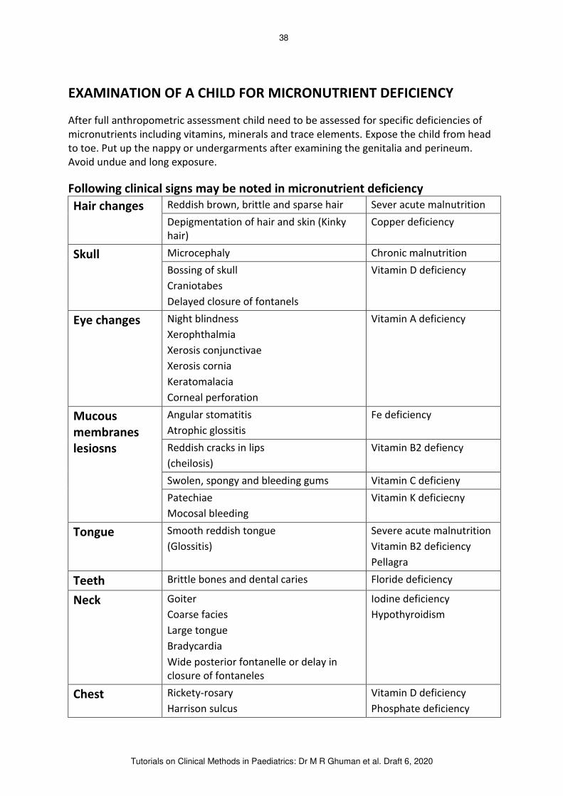
EXAMINATION OF A CHILD FOR MICRONUTRIENT DEFICIENCY
After full anthropometric assessment child need to be assessed for specific deficiencies of
micronutrients including vitamins, minerals and trace elements. Expose the child from head
to toe. Put up the nappy or undergarments after examining the genitalia and perineum.
Avoid undue and long exposure.
Following clinical signs may be noted in micronutrient deficiency
Hair changes Reddish brown, brittle and sparse hair Sever acute malnutrition
Depigmentation of hair and skin (Kinky
hair)
Copper deficiency
Skull Microcephaly Chronic malnutrition
Bossing of skull
Craniotabes
Delayed closure of fontanels
Vitamin D deficiency
Eye changes Night blindness
Xerophthalmia
Xerosis conjunctivae
Xerosis cornia
Keratomalacia
Corneal perforation
Vitamin A deficiency
Mucous membranes lesiosns
Angular stomatitis
Atrophic glossitis
Fe deficiency
Reddish cracks in lips
(cheilosis)
Vitamin B2 defiency
Swolen, spongy and bleeding gums Vitamin C deficieny
Patechiae
Mocosal bleeding
Vitamin K deficiecny
Tongue Smooth reddish tongue
(Glossitis)
Severe acute malnutrition
Vitamin B2 deficiency
Pellagra
Teeth Brittle bones and dental caries Floride deficiency
Neck Goiter
Coarse facies
Large tongue
Bradycardia
Wide posterior fontanelle or delay in
closure of fontaneles
Iodine deficiency
Hypothyroidism
Chest Rickety-rosary
Harrison sulcus
Vitamin D deficiency
Phosphate deficiency

Tutorials on Clinical Methods in Paediatrics: Dr M R Ghuman et al. Draft 6, 2020
39
Pectus carinatum or
Pectus excavatum
Skeletal abnormalities
Broadening of wrists & ankles
Kyphosis, scoliosis or lordosis
Legs: X or O or laterlised deformaity
Tetanic spams
Vitamin D deficiency
Phosphate deficiency
Loss of deep tendon reflexes
Limb ataxia
Truncal ataxia
Vitamin E deficiency
Peripheral neuropathy and decreased
knee & ankle jerks
Numbness and tingling sensation of
hands
Vit B1, B6 and B12
Deficiency
Abnormal bone and dentine formation
Pseudoparalysis
Tender epiphyseal enlargement
Vitamin C deficieny
Twitching & corpopedal spasm (tetany)
Chvostek’s sign
Troussea’s sign
Calcium and or magnisium
deficiency
Skeletal muscle weakness and muscle
cramps
Potassium deficiency
Skin Follicular hyperkeratosis (shouldrs,
gluteal area and extensor surfaces of
extremities
Vitamin A deficiency
Hyperpigmentation of sun exposed parts Nicotinic acid deficiency:
Pellegra
Depigmentation or hyperpigmentation of
flanks with wet and raw lesions in diaper
area
Sever acute malnutrition
Easy bruisability
Patechiae and bruising
Conjunctival haemorrhages
Mocosal bleeding
Vitamin K deficiecny
Poor wound healing
Hypogonadism
Delayed puberty
Impaired immunity
Mental lethargy
Zinc deficiency
Palmer pallor Pale palms
Pale conjunctivae
Fe, folate or B12
deficiency anaemia
Nails Brittleness of nails Fe deficiency

Tutorials on Clinical Methods in Paediatrics: Dr M R Ghuman et al. Draft 6, 2020
40
Kiolonychia: spoon shaped nails (in older
children and adults)
Organomegaly Hepatomegaly Severe acute malnutrition
History of pica &
Spleenomegaly
Fe deficiency
Non specific symptoms
Irratability
Loss of appetite
Easy fatiguability
Decreased alertness
Impaired learning
Fe deficiency
Vitamin D deficiency
Vitamin C deficiency
Sever acute malnutrition
Lastley make summary of your assessment and exclude Refeeding Syndrome (RFS) in the
child if he is already receiving treatment.
Refeeding Syndrome (RFS)
There is no agreed international definition of RFS.
It is a syndrome of severe electrolyte and fluid shifts associated with metabolic
abnormalities in malnourished patients undergoing refeeding, whether orally,
enterally, or parenterally
Hypophosphataemia is the adopted surrogate marker for diagnosing RFS though low
phosphate is not pathognomic as hypophosphataemia is uncommon in hospitalised
patient population
Symptoms occur because changes in serum electrolytes affect the cell membrane
potential impairing the function in nerve, cardiac and skeletal cells
Spectrum of symptoms ranges from simple nausea, vomiting, and lethargy to
respiratory insufficiency, cardiac failure, hypotension, arrythmias, delirium, coma
and death
Prevention is the key to successful management: early identification of high risk
individuals, monitoring during refeeding, and an appropriate feeding regimen
Principles of management are to correct biochemical abnormalities and fluid
imbalances returning levels to normal where possible

Tutorials on Clinical Methods in Paediatrics: Dr M R Ghuman et al. Draft 6, 2020
41
TUTORIAL 4
NEURO DEVELOPMENTAL ASSESSMENT Overall objective
1. The student should be able to perform a neuro-developmental assessment on all
children i.e hearing and vision, gross motor & fine motor, language & speech,
performance assessment, personal & social assessments.
2. At the end of this examination student should be able to make an assessment about
the aproximate age at which the child is functioning and if that is equal or less than the
other children of the same age.
Ask the following questions to caregiver when doing neurodevelopmental assessment and proceed with the examination
Is your child able to see?
Is your child able to hear and communicate as other children of the same age?
Is your child doing the same things as the other children of the same age?
PHYSICAL Vision
Optical blink
Menace response
Follow light , tracking
Can count fingers
Hearing
Accoustic blink
Turn to sound
Rinnie’s & Webber’s tests in children >4 yrs old
Gross motor
Sitting and crawling
Standing, and walking
Running – up and down stairs, tendom walking (forward, backward), kicking a ball
Fine motor
Palmer grasp, reaching for objects
Pincer grip, handedness, scribbles, writing, geomatrical figures
Feeding / dressing

Tutorials on Clinical Methods in Paediatrics: Dr M R Ghuman et al. Draft 6, 2020
42
INTELLECTUAL Language
Expresive: being able to produce speech and communicate a message
Comprehension
Expression
Articulation
1 yr, 1 word
2 yrs, 2 words sentence
3 yrs, 3 words sentence
Receptive: being able to follow a series of commands (listening and understanding
what is communicated) i.e. responds to name like give me a toy, give me a brush
Speech: Babbles, coos, do distraction test, check if say words, phrases or sentences and ask
for name, age, color etc.
Performance assessment Basic concepts: objective performance 6-12 months
Take a cup and marble – retreive marble from under the cup
Motor constructive: bricks and blocks building >1 yr
Perceptual motor: geomatric designs > 2 yrs
± 2 yrs stright line
± 2.5 yrs – vertical line
± 3 yrs – round circle
± 4 yrs – cross
± 4 ½ yrs – rectangle
± 5 yrs - cross like multiplication
± 5 ½ yrs - triangle
± 6 yrs – diamond
Draw a man test – good enough test
Total of five items or body parts where each scores 3/12. Add this total score to a
basic age of 3yrs to obtain developmental age as follows:
Head 3/12
Neck 3/12
Arms 3/12
Spine 3/12
Legs 3/12
Developmental age: 15/12 + 3 = 5/4 + 3 = 17/4 = 4 3/12
PERSONNEL / SOCIAL
Face regard, responsive smile, response to images, stranger anxiety
Feeding, drinking by holding a cup, exchanging
Dressing up with help, alone
Plays peak a boo
Co-operative play - plays together
Parallel play – plays alone – along side each other

Tutorials on Clinical Methods in Paediatrics: Dr M R Ghuman et al. Draft 6, 2020
43
NEURODEVELOPMENTAL MILESTONES ACCORDING TO THE AGE AGE VISION &
HEARING GROSS MOTOR
CONGNITIVE
FINE MOTOR SPEECH & LANGUAGE
PERSONAL / SOCIAL
WARNING SIGNS
New Born Closes eyes
to sudden
bright light
Still to
sounds
Ventral
suspension
Head droops
Hips flaxed
Limbs hang
Moro +
Palmer grasp +
Fisted
hands
Startles to
sudden
loud sound
Alternates
between
drowsiness and
alert
wakefulness
Increased or
decreased
tone
Asymetry
Sever head
lag
Poor suck
Screening 6 weeks
Follows
Stares
Red reflex
Pupillary
reactions
Rattle or
bell
Some head
control
Prone (pelvis
up), Moro +
Ventral
suspension
(head up, hips
extension)
Keeps
hands open
50% of the
time
Startles -
on history
Smiles
Turn to face
Not fixing or
following Asymetry
Floppy
Not smiling
Poor sucking
No response
3 months Follows
through 180
degree
Turns head
towards
sounds
Pull to sit – no
head lag
Prone: rise on
elbows
Rolls over
Holds rattle
Hands open
Watches
hands
Pulls at
clothes
Coos (sound
like a
pigeon)
Chuckles
Laugh
quitely
Responds to
bottle
Excited when
fed
6 months Prone (extends
arms, lifts chest)
Pull to sit
(braces
shoulders)
Sits with
support
Supine (plays
with feet)
Reaches
Transfers
Mouths
Initiates
conversation
Responds to
mirror
Starts to hold
bottle
Shows likes and
dislikes
Screening at 9 months
No squint
Normal eye
movements
Near vision
Follows
dropped
toy
Rolls over
Weight bears
Sits without
support
Crawls
Pulls to stand
Distraction
Hearing test
Holds cubes
in both
hands
Immediately
reaches out
Vocalises
delibrately
Bebbles
Responds
to name
Stranger anxiety
Holds cup and
bottle
Plays:
‘peek – a boo’ with mother
Not sitting
Hand
preference
Fisting
Squint
Primitive
reflexes +
No resonse
to sound
10 months Pulls to stand Picks up Waves Plays peek-a-

Tutorials on Clinical Methods in Paediatrics: Dr M R Ghuman et al. Draft 6, 2020
44
Walks with
assistance
small object
b/w thumb
and index
finger
bye – bye
Shakes
head for no
boo with
mother
12 months Bear walks
Walks holding
on
Pincer grip
Releases
object on
request
Knows
name
Simple
words:
come, go
Finger feeds
Arm into sleave
Unresponsive
to sound Abnormal
grasp
15 months Walks alone 2 cubes
tower
Jabbers Holds & drinks
from cup
Attempts
feeding with
spoon
Spills most
Tests screening 18 months
Near vision
Far vision
Pulls and carries
toy
Climbs into
chair
Walks well
3 cube
tower
Pincer grip
Scribbles
3 or more
words –
excluding
mama dada
Obeys
simple
commonds
Indicates toilet
needs – wet
nappy
Failure to
walk
No pincer
grip
Inability to
understand
simple
commonds
No
spontaneous
vocalisation
Mouthing
Drooling
2 years Near vision
test Runs
Stairs up and
down- 2 feet
per step
Kicks ball
6 cube
tower
Train with
cubes
Immitates
vertical line
Hand
preference
Short
phrases
Uses
pronouns
Clean and dry in
day
Spoon feeds -
no spilling
Not
understnding
Tremor
Incordination
3 years Rides bicycle
Stairs:
Up 1 foot step
Down 2 feet
step
9 cubes
tower
Bridge
Copies
circle
Sentences
Knows
name and
sex
Talks
incessantly
Toilet trained
Dresses without
supervision
Ataxia
Using simple
words only
4 years Hops on
preferred foot
Builds gate
with cubes
Knows full
name, age,
Eats with spoon
and fork
Poor
articulation

Tutorials on Clinical Methods in Paediatrics: Dr M R Ghuman et al. Draft 6, 2020
45
Stairs: up and
down 1 foot
step
Copies
cross
home
address
Matches
colours
Washes and
dries hands
Make-believe
play
Dresses and
undresses
Screening 5 years
Near and
far vision
test
Hearing test
history
Walks easily
along narrow
line - heel to toe
Hops on either
foot
Bounces ball
6-10 cube
steps
Copies
square and
triangle
Draws a
man with
full features
(6 parts)
Knows
birthday
Fluent
speech
Uses knife and
fork well
competently
Chooses own
friends
Dresses and
undresses alone
Emotional
immaturity
6 years Walks backward
on straight line
(10 paces)
10 cube
steps
Copies
diamond
Word
definition
Composition
Co-operative
play
Clumsy
Poor posture
7 years Adds
Substratcs 2
digit numbers
Counts 2’s, 100’s
Draw a man
Diamond
Defines
words &
nouns
Similarities
&
differences
One special
friend
Poor pencil
grip
8 years Counts 5’s, 100’s Simple
multiplication
Cursive
writing
Knows days
of the week
Talks
sentences
of 10
syllables
Dresses,
undresses
completely withot help
9 years Simple division
2 digit
multiplication
fraction
Cylinder Understands
obsurdity
10 years Division
Runs downstairs
Writes
three word
sentences
Complex
meaning
Produces all
speech
sounds
including
‘s’, ‘z’ and
‘ng’
Takes full
responsibility
for personal
care
Speech
sound
difficulty

Tutorials on Clinical Methods in Paediatrics: Dr M R Ghuman et al. Draft 6, 2020
46
AN APPROACH TO A HANDICAP CHILD
Parents may visit with one or more of the following complaints:
The child is delayed in sitting or walking i.e. delay in motor development
The child is slow in all aspects of development (motor and social)
The child is slow to talk (language delay)
The child repeatedly falls in the early grades at school
The combination of all above – global developmental delay
Past history Take full past history.
Look for incidents that may have caused neurological damage.
Note the following in particular:
Maternal: Age of mother (Down syndrome in older women)
Illness of mother during pregnancy – eclampsia, rubella, drugs, alcohol
Delivery: Abnormal means of delivery – forceps, vacuum etc.
Condition of the baby at birth, was there a prolonged period of cyanosis, or did he/she take
long time to cry and breath normally.
Prematurity: The degree can be assessed by the duration of stay in the hospital after birth
Postnatal: Jaundice/kernicterus - any history of exchange transfusion
Fits in newborn period have a close association with later handicap
Past illnesses:
Ask for history of severe diarrhoea & dehydration. This may have caused a cerebral
cortical vein thrombosis or electrolyte abnormalities more specifically hypo or
hypernatraemia.
Has there been a serious illness needing hospital admission such as pneumonia (hypoxic
episode) or meningitis either bacterial or TB meningitis?
Has there been a head injury?
Family history: Is there a history of developmental delays or hearing loss or other handicapping disorder?
If so, there may be a genetic basis
Developmental history: Most parents do not remember minor milestones such as rolling over or holding head clear
of the bed. It is best to enquire about the major milestones such as smiling, sitting unaided,
standing, walking alone and saying single intelligible words. Remember there is never a fixed
normal age for reaching a particular milestone. There is always range of normality.

Tutorials on Clinical Methods in Paediatrics: Dr M R Ghuman et al. Draft 6, 2020
47
Upper limits of normality – if abnormal refer for assessment
Not smiling by 8 weeks
Not sitting unaided by 9 months
Not standing unsupported by 12 months
Not walking alone by 18 months
Only single word with meaning at 36 months
Delay in sitting or walking: the main causes of motor delay are:
1. Severe illness: a child recovering from severe acute malnutrition or measles may be
static in his motor progress.
2. Mental retardation: these children are usually delayed in all aspects of development
3. Cerebral palsy: this is a group of motor disorders caused by an insult to the brain at
or near the time of birth. Three important types are:
a. Spastic (increased tone)
b. Athetoid (abnormal movements)
c. Hypotonic (decreased tone)
4. Rare neuromuscular disorders: cause severe hypotonia like spinal muscular atrophy
EXAMINATION OF MOTOR SYSTEM 1. When the child is lying on his/her back
a. Observe the child’s position
In spastic cerebral palsy (CP) the limbs are usually extended
In hypotonic states the limbs lie flat on the bed
b. Perform passive abduction with knees flexed and straight to test tone
In spastic CP there is usually increased tone especially on abducting the knees and
supinating the forearms (see diagram)
In hypotonic states tone is reduced and the limbs are floppy

Tutorials on Clinical Methods in Paediatrics: Dr M R Ghuman et al. Draft 6, 2020
48
2. Pull-to-sit:
Start with the child lying on his back.
Take his hands and gradually pull up to a sitting position.
Observe (diagram)
a. Head: does it fall back or stay in line with the baby or even come forward ahead
of the baby (as is normal after 6 months).
b. The curve of the back: is it very rounded like a newborn baby or straight (which
is normal after 6 months of age)
In both spastic and hypotonic cerebral palsy, the back is abnormally rounded

Tutorials on Clinical Methods in Paediatrics: Dr M R Ghuman et al. Draft 6, 2020
49
c. Position of the hips and knees: the normal position after 6 months of age is as
seen in the Diagram
In spastic CP the hips are at less than a right angle and the knees remain flexed.
Head may be in normal position, forward or hanging back in the young infant
d. Can the child sit unaided? This is normal after 6-9 months of age.
3. Suspension Hold the child under the armpits and lift him clear of the floor or couch.
Observe: a) Does he slip through your hands like a rag doll? This is typical of hypotonic states.
b) The position of the legs: in spastic CP the legs and feet extend abnormally and tend
to cross over each other. This is known as scissoring (see diagram).

Tutorials on Clinical Methods in Paediatrics: Dr M R Ghuman et al. Draft 6, 2020
50
4. Walking: if the child can walk get him to walk up and down your consulting room or the
corridor and observe:
a. Movements of arms: do they swing freely as is normal or does one arm not moving.
In hemiplegia one arm may be held at the side or comes up and is held in the
position without swinging
b. The legs: in spastic CP the walk is on the toes with a flexed hip and knee (see
diagram). Take very small steps and legs are internally rotated.

Tutorials on Clinical Methods in Paediatrics: Dr M R Ghuman et al. Draft 6, 2020
51
Management of children with motor disorders If your assessment suggests motor disorder, the child should be referred to specialist
centre for diagnosis and treatment purpose.
In the case of CP, it is vital to begin expert physiotherapy as soon as possible.
Intensive therapy can make a great difference to the child’s final degree of handicap.
The therapist usually sees the mother and child weekly for a prolonged period.
The mother must be taught the techniques and exercises so that therapy can continue at
home.
In many remote areas it is quite impossible for the pair to come regularly. In these cases,
it is useful to admit the child together with mother to hospital for a week or two several
times a year.
During these times the mother is taught the correct positioning and exercises for the
child which she then carries out when they return home.
The therapist can also supply them with simple aids such as positioning wedges and
chairs made out of cardboard or other disposable material.
Children who are unable to walk eventually become too heavy for the mother to carry
and need a wheel chair.
The collapsible very light ‘Buggy-Major’ or similar model is particularly suitable as it can be folded up for easy storage, fits in the boot of a car or can be taken on a bus.

Tutorials on Clinical Methods in Paediatrics: Dr M R Ghuman et al. Draft 6, 2020
52
AN APPROACH TO A CHILD WITH SPEECH DELAY Take a full history as above and try to establish if there is really a significant speech delay;
Its normal development if the child is: 10 months: uses one word with meaning
1 year: uses 2-3 words with meaning
21 months: joins 2 words, repeat things said, ask for food, drinks
2 years: uses I, me, you and talks incessantly
3 years: knows name and sex, uses 3 words sentences
There is quite a wide range around these normals but it is always abnormal to be using only
single words at 3 years. Always ask for a speech development of siblings and parents as
benign delay in speech tends to run in families.
Carry out full physical examination. This may reveal an associated handicap such as CP. If
delay is present, consider the possible common causes. Speech is learned by imitation of
sounds the child hears. It is not part of normal natural development such as walking.
Deafness or hearing loss: It is essential to assess the hearing of any child with speech
delay. Details of simple screening tests are given below. If your screening test suggests
hearing loss, the child must be referred for formal audiometry at the ENT department of a
hospital with the necessary staff and equipment.
Mental retardation: in this case there will be evidence of major delay in most other
aspects of development such as motor and social.
Environmental deprivation: a child who is not frequently exposed to the speech of
adults and children will be delayed in speech development.
Isolated delay in speech development: in this disorder other aspects of child’s development including hearing are normal. It is an isolated disability, akin to dyslexia.
MANAGEMENT This depends upon the cause. With deafness or hearing loss, it is important to place the
child in therapy as soon as the deafness has been detected. If this is delayed the child may
subsequently have greater difficulty in learning to speak.
Babies attend group parent sessions with the speech therapists who teach how the baby
can be stimulated. From 3 years the child may attend a special nursery school and later a
special school for the deaf. These schools are equipped with special apparatus and the staffs
are specially trained in teaching the deaf. The child with isolated speech delay needs
intensive speech therapy. The mentally retarded deaf child is largely managed as discussed
in the section of mental retardation.

Tutorials on Clinical Methods in Paediatrics: Dr M R Ghuman et al. Draft 6, 2020
53
Screening tests for hearing loss: (see in ENT examination section) 0-6 months: Observe if the young infant quietens to the sound of his mother’s voice or if
he turns his eyes towards a sound.
6-12 months
The rattle test: it is essential to use the proper acoustic rattle which is obtainable from
the ENT department, Tygerberg Hospital, Parow, Cape Town.
Method: the child sits on the mother’s lap facing forward. The room must be quiet. The
examiner stands behind the mother out of sight of the child. The rattle is held about 5 cm
from the ear. Then turn the rattle through 90 degrees. This will produce a soft high
frequency sound. If the child can hear it, he will usually turn his head to look at the source of
the sound. The other ear is then tested. If the result is doubtful refer the child for normal
audiometry at the ENT Department of a suitably equipped hospital.
Older children: the easiest screening method is to get the child to repeat the words that
have been spoken (not whispered) into the ear at close range (10 cm). The STYCAR
(Screening Test for Young Children and Retardates) screening tests are very useful when
screening is done on a large scale. These require some simple special equipment. Further
advice and help about deaf or hard to hearing children can be obtained from the local
branch of The National Council for the Deaf.
The child who is slow in all aspects of Development (mental retardation) This situation usually involves mental retardation either alone or in combination with other
forms of handicap. Causes may include Down’s syndrome, perinatal hypoxia, hypoglycaemia
or kernicterus
CAUSES OF SPEECH DELAY 1. Bilingualism
2. Hearing loss
3. Mental retardation
4. Expressive language disorder
5. Psychosocial deprivation
6. Autism or elective autism
7. Receptive aphasia 8. Cerebral palsy
CAUSES OF DEAFNESS Sensorineural hearing loss
1. Waardenburg syndrome
2. Klipple Feil syndrome
3. Alport’s syndrome Conductive hearing loss
1. Achondroplasia
2. Treacher-Collin syndrome
3. Craniosynostosis
NB! Deafness has aslo been associated with severe hypothyroidism and diabetes mellitus

Tutorials on Clinical Methods in Paediatrics: Dr M R Ghuman et al. Draft 6, 2020
54
TUTORIAL 5
DYSMORPHOLOGY
Dysmorphology is the branch of clinical genetics in which clinicians and researchers study
and attempt to interprete the patterns of human growth and structural defetcs.
OVERALL OBJECTIVES At the end of this module the student should be able to:
Recognise major malformations and describe dysmorphic features
Interprete the patterns of human growth and structural defects
To reach an acceptable diagnosis for chidren with dysmorphic features and describe
this in keeping with a particular
o Syndrome
o Sequence
o Association
In order to start examination of a child with dysmorphic features you need to have
relevevant family history and ages of both parents.
Expose the child in steps as you go along the clinical examination and inspect: CRANIOFACIAL Head
Hair: quality, texture, distribution
Shape: symmetrical or asymmetrical
OFC: normo, macro or micocephalic, brachycephalic
Is there craniotabes? Painful- be gentle. Not of significance under 3 months
Craniotabes: It is thinning of skull outer table. When pressed firmly over occiput or parietal
bones, it gives-way as if pressing on Ping-Pong ball. It can be a normal variant or present in
newborns, resolves by 1 month of age or may persist normally at suture lines
Differential Diagnosis includes: Rickets, osteogenesis imperfecta, hydrocephalus.
Fontanelle
Anterior: diamond shaped
Posterior: triangular
Third fontanelle: may only be barely perceptable widening of the sagital suture
between anterior and posterior fontanelles. It may be a normal varient or clue to
Down syndrome
Closed or open: If open – is it normal, under tension or sunken
Fontanelles usually close around 1 year of age. Delayed closure > 18 months may
suggest:

Tutorials on Clinical Methods in Paediatrics: Dr M R Ghuman et al. Draft 6, 2020
55
o Hydrocephalus
o Down syndrome
o Hypothyroidism
Abnormally wide fontanelle at any age may suggest:
o Rickets
o Hypothyroidism
o Cranial synostosis
o Rare syndromes like:
Smith-Lemli-Opitz syndrome
Zelweger’s syndrome Rubinstein-Taybi syndrome
Sutures: palpate with the finbers and palm of your hand
Coronal
Sagital
Lambdoid
Metopic: in the midline of the forehead due to early synostosis between the frontal
bones (cosmatic problem only)
FACIAL FEATURES Divide face into 4 regions
Forehead
Overt prominance: achondoplasia
Bitemporal narrowing: high forehead – occasional vertical ridges on forehead
Miller Dieker syndrome
Sloping appearance - primary microcephaly
Mid face From eye brows to the upper lip & from outer canthi to the commissures of mouth
Look at inner canthal distance
Measure interpupillary distance if suspecting abnormal – ask for charts to plot

Tutorials on Clinical Methods in Paediatrics: Dr M R Ghuman et al. Draft 6, 2020
56
Check for Hypotelorism: may be associated with defect in midline brain formation
Hypotelorism is narrowness of the intraorbital distance which may occur as a
morphogenetic varient or in association with other anomalies like epicanthic folds (vertical
folds of skin on either side of the nose), or secondary to cranial dystrophy.
Check for Hypertelorism (roughly check if 3rd
eye can fit in)
Hypertelorism is wide separation of eyes or increased intraorbital distance (> +2 SD). It may
be morphogenetic varient, a primary deformity or secondary phenomenon in association
with developmental abnormalities like: frontal meningocele, encephalocele and persistance
of facial cleft. It may be associated with strabismus, extropia or optic atrophy.
Exophthalmos
o It is protrusion of eyes commonly due to orbital disease
o Shallowness of orbits due to craniofacial malformations
o Increased tissue mass in orbit like neoplasia, vascular, inflammatory disorders
Occular complications due to exophthalmos include:
Exposure keratopathy
Occular motor disturbances
Optic atrophy – loss of vision
Enophthalmos
o It is shrinking of the eye back into the orbit that may be due to
Orbital fracture
Atrophy of orbital tissue
Thyroid related ophthalmopathy
o It is believed to be secondary to immune mechanisms leading to inflammations
and deposition of mucopolysaccharoids and collegen in extraoccular muscles and
orbital fat.
o Involvement of extra occular muscles may lead to restrictive strabismus
o Complications include corneal exposure leading to inflammation and perforation
and compression of optic nerve.
If Protuberant upper lip with thin vermian border, consider Miller Dieker syndrome
If short length of pelpebral fissure consider foetal alcohol syndrome
If excesive length of pelpebral fissure consider Kabuki make up syndrome (with short
stature and mental retardation)
EYES A direct ophthalmoscopic examination helps assess the status of optic discs and macula.
Complete examination of retina requires dilating the pupil and using an indirect ophthalmoscope.
Abnormalities noted
Coloboma
Conjunctival telangiectasia
Upward stant: consider Downs syndrome
Downwards slant: consider Treacher Collins syndrome

Tutorials on Clinical Methods in Paediatrics: Dr M R Ghuman et al. Draft 6, 2020
57
Prominent epicanthic folds: consider Downs or Foetal alcohol syndrome
Squint: consider Fanconi’s syndrome or 6th nerve palsy with intracranial bleed
Proptosis: consider Fanconi’s syndrome or 3rd nerve palsy or myasthenia gravis
Synophrys: eyebrows that meet in the midline
Cataract: consider diabetes mellitus, congenital rubella, galactosaemia
NOSE
Nasal bridge
o Flattened: Down or Foetal alcohol syndrome
o Prominent: Velocardiofacial syndrome
MALAR REGION It extends from ears to the midline
Note:
Size and shape
Low set ears
Posterior rotation
Pre-auricular skin tag
Ear lobe creases (Beckwith-Wiedemann syndrome)
Note if the child has a hearing aid. Deafness can occure in Treacher Collin, CHARGE,
foetal rubella and Waardenburg syndrome
MANDIBULAR REGION It extends from lower portion of ears bounded out to the chin by the mandible
o Small chin: consider Pierre Robin syndrome
NECK o Webbing of neck: consider Turner or Noonan syndrome
o Short neck: consder Klippel-Feil syndrome
o Position of posterior hair line is low: consider Klipple-Feil or Noonan syndrome
TRUNK
Sheild like chest: consdier Turner or Noonan syndrome
Pectus deformity: consider Marfan syndrome
Scoliosis: consider Marfan syndrome
EXTREMITIES
Check knee joints: look for patella (missing patella in “Nail Patella Syndrome”) Check range of movements of all joints and screen for carrying angle
o Increased (cubitus valgus)
Turner syndrome

Tutorials on Clinical Methods in Paediatrics: Dr M R Ghuman et al. Draft 6, 2020
58
Noonan syndrome
Check if joint laxity is present:
o Hypermobility
Osteogenesis imperfacta
Marfan syndrome (wrist sign, thumb sign)
Ehler-Danlos syndrome
o Multiple contractures
Arthrogryposis multiplex congenita
o An inability to pronate and supinate elbow
radioulner synostosis
X chromosome aneuploidy
Foetal alcohol syndrome
HANDS
Syndactyly: two or more digits at least partially or completely fused: consider Apert
syndrome, Smith-Lemli-Opitz syndrome
Polydactyly (extra digit): consider Ellis-van Creveld syndrome
Brachydactyly (short digits): consider Aarskog syndrome, Downs syndrome
Camptodactyly (digit is bent or fixed in direction of flexion): can be associated with
other skeletal anomalies and number of congenital syndromes like Jacobson
syndrome and Beals syndrome.
Clinodactyly (digit is crooked, curves towards or away from the other digits):
consider Russell-Silver syndrome
DERMATOGLYPHICS
Single transverse palmer crease and hypotonia during early foetal life: consider
Downs syndrome in 50% cases but can be normal in 10% cases.
Characteristic palmer pattern is seen in Foetal alcohol syndrome
GENITALIA
Disorder of sexual differentiation may indicate congenital adrenal hyperplasia
Hernia; especially in neonate and hypospadias; if associated with cryptorchidism,
there is strong possibility of a syndrome
MAKE AN ASSESSMENT
Try to recognize if it is Syndrome, Sequence or Association, otherwise say you don’t recognize a particular syndrome but would either refer or try to look it up. Chromosome
studies are indicated when 3 or more than 3 dismorphic features are present.

Tutorials on Clinical Methods in Paediatrics: Dr M R Ghuman et al. Draft 6, 2020
59
Common Syndromes Down syndrome
Trisomy 21: non-dysjunction (95%), translocation (4%), mosaicism (1%) Flat facial profile, Up slanting eyes, Epicanthic folds, Flat nasal bridge, Small and low
set eras, Protruding tongue Short stature, Brachydactyly, 5
th finger clinodactyly, Single palmer crease, Sandal-gap
between 1st
and 2nd
toes Congenital heart defect (40%), Duodenal atresia, Hypotonia, Brachycephaly Complications: Hypothyroidism, Acute leukaemia, Alzheimer’s disease, Mental
retardation Diagnosis: QF-PCR aneuploidy test, Karyotyping Treatment: growth and neurodevelopment monitoring. Medical treatment of
cardiac failure and hypothyroidism, neurodevelopmental interventions Prevention: Female education, Family planning, Pregnancy screening for advanced
maternal age (AMA), Sonar and maternal screening, Screening for hypothyroidism
Turner syndrome
Appearance: short neck with a webbed appearance
Low hairline at the back of the neck
Low-set ears
Hands and feet that is swollen or puffy at birth
Soft nails that turns upward.
Broad chest with widely spaced nipples
High, narrow roof of the mouth (palate)
Arms that turn outward at the elbows
Fingernails and toenails that are narrow and turned upward
Swelling of the hands and feet, especially at birth
Slightly smaller than average height at birth
Slowed growth
Cardiac defects
Receding or small lower jaw
Short fingers and toes
CATCH 22 Cardiac defects – Interrupted aortic arch, truncus arteriosus, VSD, PDA, TOF
Abnormal facial features – wide set eyes, narrow small groove in upper lip, small jaw
Thymus underdevelopment
Cleft palate
Hypercalcaemia
CHARGE association Coloboma and cranial nerve abnormalities – defects of the eye ball
Heart anomalies – TOF
Atresia of choanae – bony and or membranous
Retardation of growth – both cognitive and somatic growth
Genital anomalies and or hypoplasia in males

Tutorials on Clinical Methods in Paediatrics: Dr M R Ghuman et al. Draft 6, 2020
60
Ear anomalies and or deafness
CHILD syndrome Congenital Hemidysplasia
Ichthyosiform erythroderma
Limb Defects
LEOPARD syndrome Lentigines (brownish pigmented spots on skin due to increased deposition of
melanin & increased number of melanocytes)
ECG abnormalities: axis deviation, unilateral or bilateral ventricular hypertrophy,
conduction abnormalities
Ocular hypertelorism
Pulmonary stenosis
Abnormal genitalia
Retardation of growth, deafness
PHASE Syndrome (Frieden et al 1996 – Neuron 2011) Posterior fossa anomalies
Haemangioma
Arterial lesions
Cardiac abnormalities – aortic co-arctation
Eye abnormalities
VACTERL / VATER association Vertebral anomalies
Anal atresia with or without fistula
Cardiac anomalies – VSD
Tracheo-Esophageal fistula with oesophageal atresia
Renal anomalies – urethral atresia, with hydronephrosis
Limb anomalies – polydactyly, humeral hypoplasia, radial aplasia and proximally
placed thumb
CRANIOSYNOSTOSIS (Nelson p2456)
Premature closure of cranial sutures
Primary – closure of one or more sutures due to abnormality of skull development
Secondary – due to failure of brain growth
Crouzen Syndrome (Ziai page 21)
Craniofacial dysostosis, Shape of head depends upon timing, often is compressed
back to front diameter, Orbits are underdeveloped & Ocular proptosis is prominent
Hypoplasia of maxilla & orbital hypertelorism are typical facial features
Beak type nose, Exophthalmos, Strabismus, Short upper and protruding lower lip
Apert syndrome Syndactyly of 2
nd, 3
rd & 4
th fingers which may be joined to thumb & the 5
th finger
Similar abnormalities often occur in feet
Progressive calcification & fusion of bones of hands, feet and cervical spine
Mental retardation, Asymmetric facies – eyes are less proptotic than Crouzen
Carpenter Syndrome Many fusions of suture, tend to produce the Kleeblattschadel skull deformity
Soft tissue syndactyly of hands & feet is always present

Tutorials on Clinical Methods in Paediatrics: Dr M R Ghuman et al. Draft 6, 2020
61
Mental retardation, Obesity, Hypogonadism (Ziai page 22)
Heart disease, Corneal opacities, Coax valga and genu varum
Pfeiffer syndrome Commonly associated with turricephaly, Eyes are prominent (widely spaced)
Thumbs and great toes are short and broad, Partial soft tissue syndactyly
Each of the above genetic syndromes poses additional anomalies including Hydrocephalus and increased intracranial pressure
Optic atrophy – due to abnormalities of optic foramina
Respiratory problems secondary to a deviated nasal septum or choanal atresia
Disorders of speech and deafness
Craniotomy is mandatory for management of increased intracranial pressure
Hunter’s Syndrome (Forfar p983)
Usually between 2-5 years but milder types
Slowly deteriorating MR, Stunted growth – claw hands
Hepatosplenomegaly & Cardiac defects due to infiltration into valves & arterial walls
Papilledema (may lead to blindness)
Hernias, Progressive deafness, Carpel Tunnel Syndrome
Differs from Hurler’s – no gibbous formation
Kabuki Makeup Syndrome Mental retardation, Short stature
Unusual skin ridges patterns on the fingers, toes, palms of hands & soles of feet
Klipple-Feil Syndrome Short neck, low hair line, Sensorineural hearing loss
Paralysis of external rectus muscles – one or both eyes squint
Miller Dieker Syndrome Lissencephaly, Sever hypotonia at birth
Seizures with corticospinal tract signs
Pierre Robin Syndrome (Nelson p1534) Triad of micrognathia + glossoptosis (results in) airway obstruction
Micrognathia is usually associated with high arched palate & cleft palate. Tongue
usually of normal size but floor of the mouth foreshortened. Obstruction of air
passage may occur, on inspiration – usually requiring treatment to prevent
suffocation
Tracheostomy or endotracheal intubation may be required
Mandibular distraction procedures in neonates can improve mandibular size –
enhance respiration – facilitate oral feeding
Sufficient mandibular growth may take place to relieve obstruction – growth often
achieves normal profile in 4-6 yrs
Dental anomalies require individual treatment
50% of children with PRS have Stickler Syndrome (AD) that includes other findings –
early arthritis and ocular problems
DD: Treacher Collin (mandibulo-facial dysostosis), Hemifacial microsomia, Goldenhar
Syndrome
Treatment: Address airway, Prone position, do U/E (look for Bicarb retention)

Tutorials on Clinical Methods in Paediatrics: Dr M R Ghuman et al. Draft 6, 2020
62
Pradder Willi Syndrome Major criteria: Neonatal hypotonia, Excessive weight gain, Feeding problems, Facial
features, Hypogonadism, Developmental delay, Hyperphagia
Minor criteria: Decreased foetal activity, Behaviour problems, Sleep disturbances /
sleep apnoea, Short stature, Hypopigmentation, Small hands & feet, Narrow hands &
straight ulnar border, Eye abnormalities, Thick viscous saliva, Articular defects and
Skin picking
Diagnosis: Chromosomal analysis for methylation patterns in PWS, PCR or Southern
Blot hybridization and FISH analysis – parental
Rubinstein-Taybi Syndrome (Forfar)
Broad thumbs, great toes
Severe mental retardation
Characteristic facies: Antimongoloid slants, Mid face Hypoplasia, High arched palate
Narrow beaked nose
Russell Silver Syndrome Growth delays, increased risk of learning disabilities
Before birth is IUGR and after birth will be growth deficiency
Clinodactyly, Hemihypertrophy – unequal asymmetrical growth
Smith-Lemli-Opitz Syndrome (Nelson p2401) Prenatal and post-natal growth retardation
Ptosis
Anteverted nares
Broad alveolar ridges
Syndactyly of 2nd
and 3rd
toes
Microcephaly
Severe mental retardation
Types: Classical type 1 (associated with pyloric stenosis) & type 2 (associated with
Hirschsprung disease, Cleft palate, Skeletal abnormalities
Treacher Collin Syndrome Malformation of external ears
Hearing loss
Breathing difficulties
Abnormal down ward slant of palpebral fissures
Craniofacial abnormalities
Hypoplasia of certain bones of head
Zygomatic complex and the jaw & cheek bones
Zelweger Syndrome – Cerebro-hepato-renal syndrome (B Nelson p 263) High forehead, Flat orbital ridges, Widely opened fontanels
Hepatomegaly, Hypotonia, FTT, Seizures & nystagmus

Tutorials on Clinical Methods in Paediatrics: Dr M R Ghuman et al. Draft 6, 2020
63
TUTORIAL 6
GENERAL PHYSICAL EXAMINATION OVERALL OBJECTIVES At the end of this module the student should be able to do:
A full general physical examination
Able to differentiate abnormal from normal findings in different age groups children
By careful observation more information can be obtained than by any single form of
examination in children. Each system must be fully observed before it is examined. You need
to have the followings in your possession before starting a clinical examination.
Pupil torch
Stethoscope
Tape measure
Patella hammer
Blood pressure apparatus
ENT set with ophthalmoscope
Growth charts (Z-Score) and a ruler
Pen and exam pad to document your findings
Weighing scale, Stadiometer and Infantometer (these are usually provided in exams)
SEQUESNCE OF GENERAL PHYSICAL EXAMINATION There is no hard and fast rule but you may follow the following sequance when doing a
General Physical Examination
Scene & Surroundings
General out look & facies
Posture & attitude
General impression and vital signs
J Jaundice
A Anaemia
C Cyanosis
C Clubbing
O Oedema
L Lymph nodes
S Splinter haemorrhages
T Thyroid gland
E Eyes
E Ear Nose Throat
S Skin
T Tanner staging

Tutorials on Clinical Methods in Paediatrics: Dr M R Ghuman et al. Draft 6, 2020
64
1. SCENE & SURROUNDINGS Look out for clues around the bedside
Pale stool in a nappy: think obstructive jaundice
Cola colored urine in a container: think post streptococcal glomerulonephritis
Position for examination
Patient should be approached from right hand side!
Under 6 months: on the couch or the bed
6 months to 3-4 yrs: start with child on mother’s lap
Over 4 yrs: standing up, lying down or sitting up
2. GENERAL OUTOOK AND FACIES Ill looking, in severe respiratory distress or comfortable at room air
Moon-like face: Cushing’s syndrome
Idiotic face: Mental retardation
Pale faec: Anaemia
Reddish face: Polycythemia, mitral stenosis, high grade fever
Dusky face: Uraemia
Yellow face: Jaundice
Bluish face: Cyanosis
3. POSTURE AND ATTITUDE Look for adopted posture by the patient on the bed.
Patient sitting upright with back rest: heart failure (congestion of lungs)
Patient with extended neck, head bore into pillow: photophobia; meningitis
Patient with fever and peculiar aspect of helplessness, the limbs lying motionless,
the joints being swollen, stiff and painful: rheumatic fever

Tutorials on Clinical Methods in Paediatrics: Dr M R Ghuman et al. Draft 6, 2020
65
4. GENERAL IMPRESSION AND VITAL SIGNS Does the child has any obvious abnormality i.e. a child with a big head
Does this suggest some syndrome; strange looking child
Does the child look mentally retarded
Is he co-operative or un-coperatice, abnormally irritable
Note if the child is well cared for or not
Note if the child is on iv fluids, nasogastric feeds or nasal prongs oxygen or getting
nebulised.
Then note the vital signs
Temperature ( normal 36.5 to 37.5 OC)
Pulse: rate, rhythm, character and volume
Blood Pressure
Respiratory Rate
GCS
Check GM (Glucose) if necessary

Tutorials on Clinical Methods in Paediatrics: Dr M R Ghuman et al. Draft 6, 2020
66
b. Perfusion & hydration
Comment if the child is ill, distressed or toxic. If the child is ill, comment if critical or stable.
Remove clothing gradually but undress fully, after asking permission from the mother
and/or the examiners. Do up the nappy once you have checked the perineum and genitalia
5. JAUNDICE
When serum bilirubin level rises more than 45 µmol/L, jaundice appears clinically.
Method of examination Retract upper eye lid upward with the help of your thumb. Now ask the patient to look
down towards his/her feet without tilting head (difficult in small children). See the colour of
sclera which will be yellow in case of jaundice. In addition, examine the under surface of
tongue and mucous membranes of the mouth. Also check stool colour, urine and size of the
liver (hepatomegaly).
NB! Patient should be examined in sunlight and never in artificial light especially when
looking for jaundice.
CAUSES OF JAUNDICE IN CHILDHOOD
Conjugated Jaundice 1. Obstructive: Biliary attresia, bile duct stenosis and choledochal cyst
2. Infectious: TORCH, EBV, measles and varicella, hepatitis A, B, C, UTI especially gram
negative, cholecystitis
3. Metabolic: Galactosaemia, tyrosinaemia, cystic fibrosis
4. Toxic: TPN, salicylates, Iron & INH, Valproic acid & Phenytoin sodium
5. Autoimmune: Autoimmune chronic hepatitis, Graft versus host disease
Unconjugated jaundice 1. No haemolysis
Physiological jaundice of the new born
Breast milk jaundice & breast-feeding jaundice
Crigler-Najjar Syndrome: deficiency of glucuronide transferase
Gilbert syndrome: Decreased hepatic levels of glucuronide transferase
Hypothyroidism
Pyloric stenosis
2. Haemolysis and reticulosis
Positive Coombs test
o ABO and Rh incompatibility
o Auto immune (SLE)
Negative Coomb’s test o RBC Enzyme defect: G6PDD
o Haemoglobinopathy: sickle cell anaemia
o RBC membrane defect: hereditary spherocytosis
o Haemolytic uraemic syndrome

Tutorials on Clinical Methods in Paediatrics: Dr M R Ghuman et al. Draft 6, 2020
67
6. PALLOR (ANAEMIA) A condition of having a lower than normal number of red blood cells and quantity of
haemoglobin resulting in pallor and weariness. It is exmined from the palms, nails,
conjuctiva of lower eye lids, undersurface of tongue, mucous membrane of cheeks and hard
palate.
Method 1: Examine the palms and nails of the patient and compare them with your own
palms and nails. In anaemia there will be pale nails and palmer pallor.
Method 2: Ask the patient to look upward and pull his lower eye lid down (be gentle). See
the anterior of the eyelids. Normally this is pink but in anaemia it will become pale.
Method 3: Ask the patient to open the mouth and examine the colour of the tongue and
mucous membranes of the cheeks and hard palate. In anaemia it will look pale.
In order to investigate anaemia we need to check haemoglobin, indices, retics count and red
cell morphology to classify as follows:
CAUSES OF ANAEMIA 1. Hypochromic, microcytic anaemia
Iron dfeiciency (chronic blood loss, poor diet, cow’s milk protein intollerence) Thalassemia (alpha & beta)
2. Normochromic, normocytic anaemia
Chronic inflammatory disease

Tutorials on Clinical Methods in Paediatrics: Dr M R Ghuman et al. Draft 6, 2020
68
Recent blood loss
Malignancy
Chronic renal failure
3. Macrocytic anaemia
Vitamin B12 and folic acid deficiency
Hypothyroidism
Chronic liver disease
4. Haemlyitc anaemia
Sickle cell disease
G6PD deficiency
Heriditary spherocytosis
Haemolyitc uraemic syndrome
7. CYANOSIS When amount of reduced Hb reaches up to 5g % or more, cyanosis appears clinically. It can
be peripheral or central cyanosis depending upon the underlying cause. A bluish tinge can
be found in the nails, lips, tongue, and tip of the nose and mucous membrane of mouth.
Peripheral cyanosis: occurs due to vasoconstriction and other conditions leading to the
stasis of blood in the blood vessels. The peripheries will be cold, and cyanosis will disappear
on making the patient warm. Common causes are:
Exposure to cold
Poor perfusion
Venous congestion
Raynand’s disease (excessive vasomotor stimulation)
Central cyanosis: all the circulating blood is blue therefore extremities, tongue, mucous
membranes are blue due to mixing of venous and arterial blood or impaired diffusion of
oxygen due to pulmonary disease.
COMMON CAUSES OF CENTRAL CYANOSIS 1. Respiratory
Consolidation (pneumonia)
Cor pulmonale
Chronic obstructive pulmonary disease
Bronchial Asthma
Persistent pulmonary hypertension
2. Haematological
Methemoglobinaemia
Sulph-heamoglobinaemia
Sever sepsis
3. Cardiac
Transposition of great arteries
Truncus arteriosis

Tutorials on Clinical Methods in Paediatrics: Dr M R Ghuman et al. Draft 6, 2020
69
Tricuspid atresia
Tetralogy of Fallot
Total anomalous pulmonary venous return (TAPVR)
8. CLUBBING In finger clubbing, the soft tissues at the base of the nails are thickend and the angle
between the base of the nail and the adjacent skin of the finger (Lovibond angle) is
obliterated.
Schamroth’s window test: Originally demonstrated by South African Cardiologist Dr Leo
Schamroth on himself is a popular test for clubbing.
When the distal phalanges of corresponding fingers of opposite hands (preferably thumb
nails) are directly apposed (placed against each other back to back), a small diamond shaped
window is normally apparent between the nail beds. If this window is obliterated, the test is
positive and clubbing is present.
Long standing arterial desaturation (usually longer than 6 months in duration) even if too
mild to be detected by an inexperienced person results in clubbing of finger nails and toes. It
appears earliest and most noticeably in thumb.
Clubbing develops in 5 steps: 1. Fluctuation and softening of the nail bed
2. Loss of normal Lovibond angle (165o)
3. Increased convexity of the nail fold
4. Thickening of the whole distal finger (resembling drum stick)

Tutorials on Clinical Methods in Paediatrics: Dr M R Ghuman et al. Draft 6, 2020
70
5. Shiny aspect and striation of the nail and
skin

Tutorials on Clinical Methods in Paediatrics: Dr M R Ghuman et al. Draft 6, 2020
71
Stages of finger clubbing: 1
st stage: obliteration of angle between nailbed and forefinger
2nd
stage: fluctuation at the base of nail
3rd
stage: drumstick appearance
CAUSES OF FINGER CLUBBING Idiopathic or primary: Familial, hypertrophic osteoarthropathy
1. Lungs:
Interstitial lung disease
Pulmonary HTN
Cystic fibrosis
Bronchiectasis
Lung abscess
Empyema
2. Heart (any disease featuring chronic hypoxia)
Cyanotic disaeses of the heart like TOF etc.
Subacute bacterial endocarditis
3. GIT and endocrine
1. Malabsorption syndrome
2. Crohn’s disaese
3. Ulcerative colitis
4. Hyperthyroidism
4. Liver
Hepatopulomonary syndrome
Biliary cirrhosis (primary)
Hepatic cirrhosis
Intrahepatic biliary atresia
Axillary artery aneurysm (unilateral clubbing)
9. KOILONYCHIA The nails become soft, brittle and spoonshaped
CAUSES
1. Iron deficiency anaemia
2. Plummer-Vinson’s syndrome
3. Familial
4. Excessive use of detergents and soap
5. Idiopathic
10. LEUKONYCHIA Abnormal whiteness of nails, either total or in spots or streaks
Cause may be chronic liver disease

Tutorials on Clinical Methods in Paediatrics: Dr M R Ghuman et al. Draft 6, 2020
72
11. SPLINTER HEAMORRHAGES IN THE NAILS These are small, reddish, dark brown, vertical lines in the nails.
CAUSES
1. Subacute bacterial endocarditis
2. Haemorrhagic disorder
3. Trichurus trichura
4. Sickle cell anaemia
5. Atrial myxoma
12. OEDEMA It is an excess of fluid present in the interstitial tissues. It can be pitting type or non-pitting.
Pitting oedema The swollen part is pressed with thumb for 15 to 20 seconds. If it leaves a pit which stays for
half a minute the oedema is called as pitting oedema. Oedema is demonstrated in the
dependent parts of the body like sacrum and tibial aspect of lower legs.
Non pitting oedema In the above method if pit doesn’t form, this is called as non-pitting oedema.
CAUSES GENERALIZED OEDEMA
1. Hypoproteineamia (inadequate protein intake/absorption)
Severe malnutrition with oedema
Crohn’s disease
Coeliac disease
2. Inadequate protein production
Chronic liver disease causing hypoalbuminaemia (cirrhosis) 3. Excessive protein loss
Nephrotic syndrome (>5g/day)
Sepiated removal of ascitic fluid
Protein losing enteropathy (coeliac disease)
Thiamine deficiency (wet beriberi) 4. Fluid overload
Congestive cardiac failure, cor-pulmonale, constrictive pericarditis
Secondary hyperaldosteronism
Acute glomerulonephritis
Excessive fluid replacement (iatrogenic)
5. Hypothyroidism

Tutorials on Clinical Methods in Paediatrics: Dr M R Ghuman et al. Draft 6, 2020
73
LOCALISED OEDEMA 1. Venous causes – (pitting – unilateral lower limb oedema)
Post-surgical
Venous thrombosis
Pressure from neighbouring tumour or lymph node
Valvular incompetence (varices)
2. Lymphatic causes – (nonpitting lower limb oedema) +
Lymphoedema
Filariasis (lymphatic obstruction by filarial worms)
Milroy’s disease (unexplained lymphoedema at puberty common in females)
Congenital lymphoedema (lymphatic agenesis or hypoplasia of lymphatics of
legs)
3. Inflammatory causes
Infection
Injury or ischaemia (histamine, bradykinin, cytokinin) causing inflammation,
redness, heat, pain
4. Allergic causes
Angio-oedema
11. LYMPH NODES Palpate various groups of lymph nodes.
They are significantly enlarged if following groups are:
More than 1 cm
o postrior cervical
o submandibular
o axillary
o femoral
More than 1.5 cm
o Inguinal
Any size
o Supraclavicular
o Epitrochlear (mention if no peripheral inflammation)

Tutorials on Clinical Methods in Paediatrics: Dr M R Ghuman et al. Draft 6, 2020
74

Tutorials on Clinical Methods in Paediatrics: Dr M R Ghuman et al. Draft 6, 2020
75
CAUSES OF GENERALIZED LYMPHADEMOPATHY (LAD)
1. Viral: Infectious mononucleoisis, CMV, measles, rubella, viral hepatitis and HIV
2. Bacterial: Syphilis (lymph nodes shotty painless discrete), tuberculosis, bacterial
endocarditis
3. Fungal: Histoplasmosis
4. Protozoal: Toxoplasmosis
5. Non infectious inflammatory diseases; Rheumatoid arthritis, SLE

Tutorials on Clinical Methods in Paediatrics: Dr M R Ghuman et al. Draft 6, 2020
76
6. Malignant: Leukaemias: Chronic lymphocytic , acute lymphocytic leukaemia, non-
Hodgkin’s lymphoma, Hodgkin’s disease
CAUSESS OF LOCALISED LYMPHADEMOPATHY 1. Local pyogenic infections like pharyngitis, dental abscess, otitis media
2. Viral infection: cat scratch fever, lyphogranuloma venereum
3. TB (lymph nodes matted and there may be sinus formation)
4. Non- Hodgkin’s lymphoma
5. Secondary carcinoma – hard regular and fixed lymph nodes
6. Virchow’s gland (supraclavicular nodes, left sided enlarged in carcinoma of stomach
– uncommon in children
12. THYROID Inspect the neck for local or general enlargement of thyroid gland and observe its
movement with the larynx as the patient swallows. The thyroid gland will move upward.
Examination of thyroid gland Stand behind the patient and put your fingers of both hands in front of the neck to palpate
the gland.
If the gland is enlarged, note its:
Size, shape, surface, tenderness
Movement of skin over it
Pulsation
Systolic bruit (toxic goiter)
van Graef’s sign - in thyrotoxicocis
Von Graefe's sign is the lagging of the upper eyelid on downward rotation of the eyes,
indicating exophthalmic goiter (Graves’ disease). It is a dynamic sign, whereas lid lag is a
static sign which may also be present in cicatricle eyelid retraction or congenital ptosis.
Tutorials on Clinical Methods in Paediatrics: Dr M R Ghuman et al. Draft 6, 2020
77
13. TANNER STAGING Tanner staging, also known as Sexual Maturity Rating (SMR) is an objective classification
system that is used to document and track the development and sequence of secondary sex
characteristics of children during their puberty. It was developed by Marshal & Tanner,
based on their observational data on development of external genitalia:
Phallus, scrotum, and testes volume in males;
Breasts in females;
Pubic hair in both males and females.
Tanner stages are utilized in paediatric and adolescent practice to counsel patients about
the timing of anticipated body changes, perform appropriate medical screenings, and
monitor for deviations in normal timing and sequence of physical signs of puberty that may
represent physiological problems.
Changes that are associated but are not directly measured by Tanner Staging include bone
growth and fusion, body composition and linear growth and haematocrit values. In males,
the onset of puberty ranges from 9 to 14 years of age. In females, the normal onset of
puberty ranges from 8 to 13 years old averaging 10 years in White Americans and age 8.9
years in African Americans.
TANNER STAGING
Girls Boys
BREAST STAGING GENITALIA STAGING
B1 Prepubertal
B2 Breast budding
B3 Development of actual breast
mound
B4 Areola projects at an angle to breast
mound
B5 Adult configuration
G1 Prepubertal penis, unstretched length
2.5-6 cm, scrotum and testes volume <3 ml
G2 Testes > 4 ml and scrotal laxity but no
penile enlargement
G3 Penile lengthening and further
dvelopment of testes and scrotum
G4 Penile lengthening and broadening
further development of testes ( volume 10-
12 ml)
G5 Adult genitalia
PUBIC HAIR STAGING
P1 No pubic hair
P2 Fine hair over mons pubis / scrotum / labia
P3 Adult type hair (curly coarse) but distribution confined to pubis
P4 Extension to near adult distribution
P5 Adult
AXILLARY HAIR STAGING
A1 No axillary hair
A2 Hair present not adult amount
A3 Adult

Tutorials on Clinical Methods in Paediatrics: Dr M R Ghuman et al. Draft 6, 2020
78
14. ENT
Ears: Inspection of external ear
Pinna in general
Auditory cannal: ask and check if tragus is painful
Draw outer part of the ear up & back in the older child and down & back in the
infant
Examine auditaory cannal for inflamation, foreign body, wax or discharge
Do otoscopy for redness, discharge, perforations of tympanic membrane
Nose Notice any deformity - nasal septum deviation
Look for signs of allergic rhinitis
Nasal polyps, masses, turbinate hypertrophy
Percussion of sinuses (acute/acute on chronic if tender)
Mouth open Inspect buccal mucosa for
Ulceration
Oral thrush
Kaposi’s sarcoma
Koplik spots
Dental caries
Tongue tie
Thrush
Leukoplakia
Inspect gums for
Gum hyperplasia – acute myeloid leukaemia (AML)
Hypertrophied and firm – treatment with phenytoin
Soft haemorrhagic – scurvy

Tutorials on Clinical Methods in Paediatrics: Dr M R Ghuman et al. Draft 6, 2020
79
Spongy and haemorrhagic – congenital cyanotic heart disease
Punctate blue line – lead poisoning
Teeth
Primary dentition: 3 yrs – 20 teeth
Secondary start at 6 years: 32 teeth
Look for caries or missing teeth
Floating teeth – Burkitts lymphoma

Tutorials on Clinical Methods in Paediatrics: Dr M R Ghuman et al. Draft 6, 2020
80
Salivary glands
Protids
Submandibular
Sublingual
Throat - pharynx / larynx
Look hard palate – abnormal arch
Cleft palate
Talangiectasia
Tonsills – size, color, discharge
Gag reflex
EYES Conjunctivae: look for purpura, jaundice, anaemia, cyanosis
Cornea: normal or look for scars or ulcers
Cataract: consider diabetes mellitus, congenital rubella, trauma to the eye, galactosaemia
Hypopyon: It is the presence of pus (accumulation of WBCs) in anterior chamber of the eye
In leukaemia there may be infiltration of the iris
Leukocoria: white pupilary reflex – cat eye reflex.
The D/D can be:
Cataract: galactosaemia, rubella
Cicatrical retinopathy of prematurity
Retinal detachment
Retinoblastoma
15. SKIN BCG scar: Look for
BCGioma
BCG lymphadenitis
Disseminated BCG disease
Regarding BCG scar:
There is no associateion between the presence of a BCG scar and immunogenicity or
effectiveness of the vaccine.
Prolonged ulceration at BCG site may occur with lymphadenitis in 1-10% of cases
and is more common in HIV infected children
Mantoux test: if present measure and interprete
Two strains of PPD are available in South Africa
PPD-RT 23 strain: dose 2 TU (currently used in KZN)
BCG Danish 1331 strain: dose 5 TU
Surgical scars: count and measure – work out what procedure might have been done
Traditional markings: if present ask for any use of traditional medication or herbal enema

Tutorials on Clinical Methods in Paediatrics: Dr M R Ghuman et al. Draft 6, 2020
81
Bleeding diathesis: look over the skin, inside mouth (wet petechie - thrombocytopenia)
Scratch marks: haemolysis, jaundice
Trophic changes: occur in soft tissue (skin, fascia, muscle), resulting from interruption of
nerve supply
Rash: it is an acute manifestation of disease. When describing a rash assess the following:
The type of rash
The distributon i.e. parts of the body involved
Progression of rash
How it has regressed
How it has healed
Types of rash
Erythema marginatum: pale centered ringlets with pink margins, present over trunk
and flexor surfaces of joints i.e., Rheumatic Fever
Erythema nodosum: conical rounded nodules present just under skin , present on
extensor surfaces of the joints – elbows shins and dorsum of the hands and feet
Common causes Less common causes
Rheumatic fever
TB
GIT infections (yersinia, salmonella,
shigella, or systemic fungal infections)
Ulcerative collitis
Crohn’s disease
Drug sensitivity – sulpha, salisylates
Tissue defect: Maceration, Erosion, Ulcer
Deposition of skin: Scale formation, Crust on scab formation
Macule: alteration in colour
Papule: hemispherical elevation of epidermis <5mm
Vesicles bulla: accumulation of serous fluid
Pustule: accumulation of pus
Nodule: involvement of whole thickness of skin by inflammatory infilterate or some new
growth of cells – it is harder than papule >5mm
Heberdens nodules: small nodules present at the terminal phalangeal joints in case of
osteo-arthrosis
Wheel formation: oedema formation in dermis due to allergy and associated with severe
itching – uricaria
Erythematous lesion: capillary dilatation – will blench on pressure
Haemorrhagic spots: these do not blench on pressure: D/D telengiectasia
Petechial: small pin point lesions in the skin
Purpura spots: haemorrhagic spots which are larger than petechiae
Ecchymosis: like large bruises
Kaposi’s sarcoma (Human Herpes Virus 8): multipe firm, purple blue or reddish-brown
plaques and nodules typically appear initially on the hands and feet and progress up the
arms and legs over a period of years and decades, eventually involving the viscera or
mucosa in about 10% of patients. Untreated lesions evolve from flat discolourations or

Tutorials on Clinical Methods in Paediatrics: Dr M R Ghuman et al. Draft 6, 2020
82
patches to plaques and then to raised nodules that become confluent. Lymphaedema may
preced or follow the appearance of lesions.
Neurocutaneous lesions – café a lait spots
Café a lait spots are flat hyperpigmented lesions on the skin due to increased deposition of
melanin and an increased number of melanocytes. Differentiate from motherspots (birth
marks). Considered significant if:
5 or more and >5 mm in prebubertal
6 or more and >15 mm in post pubertal
SYNDROMES ASSOCIATED WITH THE PRESENCE OF CAFÉ A LAIT SPOTS
(Acronym: Nandi And Fharnisa Have To Go To Buy Me My New Ring Very Soon)
1. Neurofibromatosis
2. Ataxia telangiectasia
3. Fanconi’s anaemia 4. Hurler syndrome
5. Tuberous sclerosis
6. Gaucher’s disease
7. Turner syndrome
8. Bloom syndrome
9. Marfan syndrome
10. McCun Albright syndrome
11. Noonan syndrome
12. Russel silver syndrome
13. Von Hipple-Lindau disease
14. Sturg Weber syndrome

Tutorials on Clinical Methods in Paediatrics: Dr M R Ghuman et al. Draft 6, 2020
83
TUTORIAL 7
THE EXAMINATION OF RESPIRATORY SYSTEM
OVERALL OBJECTIVES At the end of this module the student should be able to differentiate, diagnose and manage
common respiratory problems like:
1. Upper airway obstruction [laryngotracheobronchitis (LTB), foreign body (FB) in
airway] and
2. Lower airway obstruction [pneumonia and its complications (pleural effusion,
pneumothorax, cysts, collapse), chronic chest disease (pulmonary TB,
bronchiectasis)]
To look for extra general features: 1. Cough:
Productive: results from an inflammatory or infective exudate on the bronchial
mucosa: Cystic fibrosis, Pulmonary Tuberculosis (PTB)
Non-productive nocturnal: post nasal drip, asthma
On exercise: asthma
Dry intermittent or persistent: may imply irritation of the upper respiratory
tract or of the bronchial wall by a foreign body, or an enlarged lymph node.
Productive more than 2 weeks: Consider PTB
Croupy: barking associated with strider and hoarseness – Croup, acute
laryngotracheobronchitis (LTB)
Whooping: inspiratory gasp, prolonged distressing cough, ending in a whoop,
may follow vomiting – pertussis
During or after feed: inhalation, aspiration
Bovine brassy: Tracheitis
Absent during sleep – psychogenic
2. Hallitosis and a productive cough – Chronic suppurative lung disease
3. Sputum:
Clear mucoid or tacky tenacious – indicative of asthma
Green yellow, grey (dirty) – infection most common is PTB
Haemoptysis: rare in children – may indicate cystic fibrosis or PTB
4. Parotid enlargement in lymphocytic interstitial pneumonia (LIP)
5. Delerium, pleural pain, rusty sputum and herpes labilais – suspect pneumococcal
pneumonia
6. Look for plethora

Tutorials on Clinical Methods in Paediatrics: Dr M R Ghuman et al. Draft 6, 2020
84
1. INSPECTION
Comment on general inspection To avoid anxiety in children, mother should hold the child on her lap during the initial
examination. Inspection should be done first before doing anything else which may disturb
the child. Clothing need to be removed and chest exposed as the examination proceeds.
Scene: look around if there is a specimen container and note its contents like sputum,
blood, mucous or pus
Expose the chest and look for:
Scars: post op, post chest drain insertation
Pigemntation: Addisons disease
Rashes: on the chest wall
Features of allergic facies Note tired looking face, open mouth breathing, double folds of lower eye lids (Denne-
Morgan lines), allergic shiners, allergic salute (nasal crease) and nasal discharge.
Shape of the chest Normal chest is elliptical i.e. its anterioposterior diameter is lesser than its transverse
diameter that is 3:5 respectively. Abnormal forms can be as follows:
1. Asymmetry: Retraction of chest wall on one side may be due to;
o lung collapse, fibrosis, spinal deformity
2. Asymmetry: Bulging of chest wall on one side may be due to:
o Pleural effusion, pneumothorax, massive cardiomegaly, kyphosis or
intrathoracic tumour.
3. Deformity: Check for scoliosis or kyphosis and ask older children to touch their toes
to determine whether the deformity is fixed or postural
4. Barrel shaped chest: Increased AP diameter and angle of Louis becomes more
prominent: consider air trapping 5. Harrison’s sulcus: This is transverse constriction which begins at the level of the
xiphisternum, passes outward and slightly downwards. The sulcus is formed along
the line of diaphragm (normal diaphragm pulls on soft ribs). Causes:
o Chronic airway obstruction
o Left to right cardiac shunt
o Rickets
6. Hoover sign: it is isolated subcostal recession associated with hyperinflation of the
lungs in a patient with peripheral airway disease. It causes diaphragm to flatten and
to contract laterally against the lower chest wall resulting in indrawing below the rib
cage.
7. Pectus carinatum (pigeon chest)
8. Pectus excavatum (funnel chest)
9. Rachitic rosary: Rickets
10. Absent clavicles: Cleido-cranial dysostosis
11. Absent pectoralis: Poland’s syndrome

Tutorials on Clinical Methods in Paediatrics: Dr M R Ghuman et al. Draft 6, 2020
85
Movements of chest: 1. Stand at the end of the bed and compare two sides of the chest – pathology is
usually on the side with lesser movement
2. Note the signs on one side of chest and compare with the other side of chest on the
respective point
3. Count respiratory rate for a full minute – say if its normal or abnormal
4. Note nasal flaring, difficulty in breathing or shotness of breath
5. Note respiratory movements – normal or abnormal
6. State if patient is in respiratory distress. Ist it mild, moderate or severe
7. Look for intercostal & subcostal recession. It is also due to airway obstruction or
decreased lung compliance and by using accessary muscles, especially
sternocleidomastoids.
Common causes of fast breathing 1. Airway obstruction
2. Bronchopneumonia
3. Metabolic acidosis
4. Atelectasis
5. Pneumothorax
6. Fever and anxiety
7. Drugs like salicylates
8. Note if tracheal tug is present: Tracheal tug is downward pull of the trachea,
manifested by a downward movement of the thyroid cartilage. In children it is due to
lower airway obstruction probably due to lung compliance problems. This recession
is due to greater negative intrathoracic pressure which draws in the soft tissue. 9. Audible sounds:
Stridor, stertor, snoring, hoarseness, wheeze
Grunting (breathing out against a closed glottis) increases pressure in the lungs
and small airways to prevent collapse and improve oxygen transfer.
Acute laryngotracheobronchitis (stridor) Common between 6 months to 2 years of age
Grading:
Grade 1: inspiratory strider
Grade 2: Inspiratory and expiratory strider
Grade 3: Inspiratory and expiratory strider and pulsus paradoxus
Grade 4: Impending apnoea, cyanosis, apathy and marked retractions
Type of breathing patterns 1. Cheyne- Stokes breathing: It is terminal pattern of respiration which dependent on CO2
drive. It is characterised by progressively deeper and faster breathing followed by a
gradual decrease in breathing until apnoea. The pattern repeats, with each cycle
and usually takes 30 seconds to 2 minutes.
o First there is rapid and deep breathing which causes fall in PCO2 in pulmonary
blood. When this blood reaches the brain, it inhibits the respiratory centre
and causes apnoea.

Tutorials on Clinical Methods in Paediatrics: Dr M R Ghuman et al. Draft 6, 2020
86
o As a result of apnoea, alveolar pulmonary PCO2 gradually builds up. When
this blood carrying high CO2 reaches in the brain, it causes stimulation of
respiratory centre and in this way respiration begins again. Thus, initiating
the new cycle.
o Causes include left cardiac failure, increased intracranial pressure (trauma,
cerebral haemorrhage), and acute kidney injury or sever pneumonia.
2. Kussmal respiration: It is deep and rapid breathing pattern that indicates that the
body or organs have become too acidotic. The respiratory centre is stimulated and
body works constantly to maintain a normal temperature and neutral blood pH.
Causes are the conditions which lead to metabolic acidosis like diabetic
ketoacidosis, chronic kidney injury etc.
3. Sleep apnoea: cessation of air flow for more than 10 seconds, more than 10 times at
night during sleep
4. Shallow and painful breathing: pleuritic
5. Paradoxical respiration: it is paradoxical inward movement of abdomen during
inspiration. Causes: diaphragmatic paralysis, COPD etc.

Tutorials on Clinical Methods in Paediatrics: Dr M R Ghuman et al. Draft 6, 2020
87
2. PALPATION Feel for any tendeness, to exclude any local inflammatory condition like Teitze’s disease (costochondritis) or fractured rib. Crepitations are felt in case of surgical emphysema. Note
the position of mediastinum:
Palpation of trachea (to assess the upper mediastinum) Stand in front of the patient and place your 3 fingers (ring, middle, and ring finger) in a way
that index and ring fingers rest on the right and left sternoclavicular joints respectively while
the middle finger is free to feel the gap between the sternomastoid muscle (of either side)
and trachea. Reduced gap indicates the shifting of trachea toward that side. A tracheal
position 1-2 mm to the right is considered as normal due to aortic arch curving over from
right to left displacing the trachea
Conditions which pull the trachea
1. Atelactasis or collapsed upper lobe of lung
2. Fibrosis of lungs
3. Pneumonectomy
Conditions which push the trachea
1. Pneumothorax
2. Pleural effusion
3. Upper mediastinal tumour
4. Retrosternal goiter
5. Bochdalek hernia
When trachea is not displaced to the side of fibrosis or colapse of lung or to the opposit side
in case of pleural effusion, this is called as splinting of trachea.

Tutorials on Clinical Methods in Paediatrics: Dr M R Ghuman et al. Draft 6, 2020
88
Palpation of apex beat (to assess the lower mediastinum) Apex beat is localised at the most lateral and inferior point at which palpating fingers are
raised with each heart beat. Place the palm of your hand over the pericardium and feel the
apex beat. If not palpable, then try to feel it by turning the patient to his/her left side.
Following may be the cause when it is not palpable:
1. Thick muscular chest wall
2. Pneumothorax of left side
3. Pleural effusion of left side
4. Pericardial effusion
5. Emphysema
6. Dextrocardia
If apex beat is palpable then localise it by placing the index finger over it vertically. The apex
beat is normally situated in the 5th
intercostal space in midclavicular line in small children &
1 cm medial to left midclavicular line in older children & adolescents. Now assess if it is at
the right position or displaced. This may be displaced “pushed” outward only or outward
and downward. Apex beat may also be “pulled” mean displaced inward.
Causes which push the apex beat – outward/downward
1. Left ventricular enlargement
2. Right ventricular enlargement (outwards only)
3. Pleural effusion of right side
4. Pneumothorax of right side
5. Pectus excavatum
Causes of displacement of apex beat inward (pulled)
1. Collapse of right lung
2. Fibrosis of right lung
3. Dextrocardia – apex beat on the right hand side
Vocal fremitis
This is feeling of voice vibration with the flat hand on chest wall. Its not easy to demonstrate
in small children.
Method: Place the palm of your hand flat on the identical points on either side of the chest
wall and ask the patient to say “One, one, one” or “ninty nine” and feel the vibration. The
vocal fremitus may be nornal or
Increased vocal fremitus: in cavitation, collapse with patent bronchus and
consolidation (better conduction to the surface)
Diminished vocal fremitus: in asthma, bronchiolitis, pneumothorax, pleural
effusion, fibrosis and collapse with obstructed bronchus
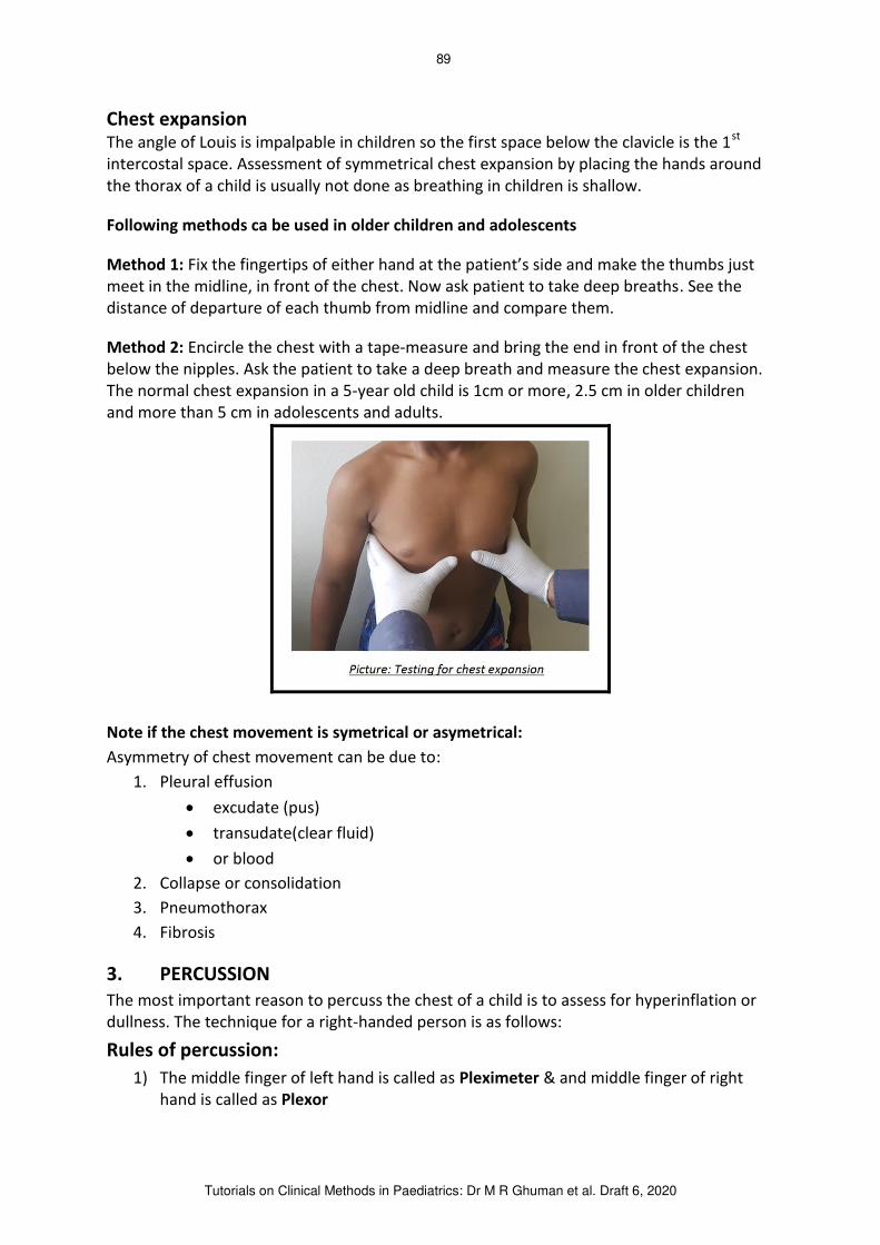
Tutorials on Clinical Methods in Paediatrics: Dr M R Ghuman et al. Draft 6, 2020
89
Chest expansion The angle of Louis is impalpable in children so the first space below the clavicle is the 1
st
intercostal space. Assessment of symmetrical chest expansion by placing the hands around
the thorax of a child is usually not done as breathing in children is shallow.
Following methods ca be used in older children and adolescents
Method 1: Fix the fingertips of either hand at the patient’s side and make the thumbs just
meet in the midline, in front of the chest. Now ask patient to take deep breaths. See the
distance of departure of each thumb from midline and compare them.
Method 2: Encircle the chest with a tape-measure and bring the end in front of the chest
below the nipples. Ask the patient to take a deep breath and measure the chest expansion.
The normal chest expansion in a 5-year old child is 1cm or more, 2.5 cm in older children
and more than 5 cm in adolescents and adults.
Note if the chest movement is symetrical or asymetrical:
Asymmetry of chest movement can be due to:
1. Pleural effusion
excudate (pus)
transudate(clear fluid)
or blood
2. Collapse or consolidation
3. Pneumothorax
4. Fibrosis
3. PERCUSSION The most important reason to percuss the chest of a child is to assess for hyperinflation or
dullness. The technique for a right-handed person is as follows:
Rules of percussion:
1) The middle finger of left hand is called as Pleximeter & and middle finger of right
hand is called as Plexor

Tutorials on Clinical Methods in Paediatrics: Dr M R Ghuman et al. Draft 6, 2020
90
2) Percussion is done from resonant to dull area and comparison is done with
respective point on the other sides.
3) Pleximeter should be placed parallel to the border of organ to be percussed in close
contact with the body surface and Plexor should strike the middle phalanx of
Pleximeter perpendicularly
4) Movements should take place at the wrist and finger joints
5) Strike the Plexor twice and then lift it off the Pleximeter i.e. it should not remain in
contact with Pleximeter after striking it because it stops the overtones and hence
changes the character of the sound produced.
6) The blow should be no heavier than is necessary to elicit the resonance of the part
being examined, and the wrist joint must move loosely.
7) The character of the sound varies quantitatively and qualitatively producing different
types of percussion notes.

Tutorials on Clinical Methods in Paediatrics: Dr M R Ghuman et al. Draft 6, 2020
91
Methods of percussion: Chest is percussed anteriorly, laterally and posteriorly.
Apical percussion Rotate the head of the patient to the opposit side and place the pleximeter above the
medial third of clavicle and percuss it.
Clavicular percusson Place a finger above and below the clavicle and stabilise it. Now percuss the clavicle directly
without pleximeter.
Anterior percussion After apical and clavicular percussions move in front of the chest
Precaution should be taken to keep the pleximeter away from the cardiac dullness
Upper border of the liver must be percussed and marked before starting to percuss for the
anterior of the chest. Look for obvious differences between left and right chest
Posterior percussion Patient sits up on the bed and crosses his hands in fron of the chest places on the shoulders
and bends forward. Now start percussion and look for obvious difference between left and
right chest.

Tutorials on Clinical Methods in Paediatrics: Dr M R Ghuman et al. Draft 6, 2020
92
Lateral percussion Patient sits up on the bed and place his/her hands on the head. Now percuss the lateral
sides of chest from above downward and look for obvious difference between left and right
chest.
Types of percussion notes
Normal resonance A percussion note heard over a normal air-filled lung will be resonant
Hyperresonant: Hyperresonance on percussion indicates too much air within the
lung tissue note, that may be due to: o Pneumothorax
o Air trapping in bronchospasm
o Bronchiolitis
o Emphysema
Decreased resonance – may be due to:
o Consolidation
o Pleural thickening
o Fibrosis
o Collapse of lung
Dull note o Percussion note over a solid organ like liver, spleen and heart
Stony dull - percussion note over the fluid filled cavity for example:
o Pleural effusion
o Ascities
Tympanitic note Percussuin note over a cavity containing air. A best example to percuss over an
empty stomach.
Signs of hyperinflation 1. Loss of cardiac dullness
2. Upper border of liver pushed down (usually in 4th
to 5th
intercostal space)
3. Barrel shaped chest – increased AP diameter
Causes of hyperinflation 1. Chronic asthma
2. Emphysema
3. Cystic fibrosis
4. Bronchiolitis
5. Chronic lung disease

Tutorials on Clinical Methods in Paediatrics: Dr M R Ghuman et al. Draft 6, 2020
93
4. AUSCULTATION Patient should sit or lie down supine on the bed with upper garments removed. Now ask the
patient to relax, open the mouth and take deep breaths.
Small children can be examined in their mothers’ lap and auscultation can just be done
while they are breathing normally.
Warm the chest piece of stethoscope by rubbing it on the palm of your hand and out it on
the chest wall in a way that it is in full contact with the chest and should not move on the
skin while patient is breathing.
First auscultate anteriorly and then posteriorly. When auscultating, first note character of
breath sounds, second character of vocal resonance, and third the presence or absence of
other sounds.
Auscultate:
Anteriorly from clavical down to 6th
rib
Laterally from axilla to the 8th
rib
Posteriorly down to the 11th
rib
Breath sounds are two types: vesicular and bronchial
Vesicular breath sound This is produced by the passage of air in and out of normal lung tissue. It is heard all over
the healthy chest and most typically in the axillary and intrascapular regions. Throughout
inspiration the sound is fairly intense and of low pitch with a characteristic rustle.

Tutorials on Clinical Methods in Paediatrics: Dr M R Ghuman et al. Draft 6, 2020
94
Bronchial breathing This is pathological sound. It is blowing and hollow in character. Expiration may be
prolonged and is harsher and more intense than inspiration. There is pause between
inspiration and expiration. When auscultating the chest, the abnormal side is usually the
side with decreased breath sounds.
Broncheal breathing may also be produced by the passage of air through the trachea and
large bronchi. It is very intense when heard over the trachea but far less intense and of
same quality when heard over a diseased lung. The inspiratory sound of bronchial breathing
is harsh and becomes inaudable shortly before the end of inspiration. Not to say the child
has bronchial breathing if listening near the trachea. Always look for Cor Pulmonale in
Chronic Lung Disease
Causes of bronchial breathing Consolidation of lung
Cavitation
Collapse (with patent bronchus)
Top of pleural effusion
Vocal resonance It is difficult to assess in small children but easy in older children and adolescents.
Method: Auscultate when patient say “one, one, one” or “ninety-nine”. The ear perceives,
not the distinct syllables, but a resonant sound, the intensity of which depends on the
loudness and depth of the patient’s voice and the conductivity of his lungs; the nearer the
stethoscope is to a large bronchus, the more intense the sound. Each point examined on
one side of the chest should be at once compared with the corresponding point on the
other side. Vocal resonance of normal intensity generally conveys the impression of being
produced just at the chest-piece of the stethoscope. If it seems to be nearer the ear than
this, the resonance is increased. Increased resonance occurs when the lung substance
conducts the sound waves set up by the voice more clearly than usual from the bronchi.
Decreased vocal resonance may be due to: Reduced conduction
o Pleural effusion
o Marked pleural thickening
o Pneumothorax
Reduced air flow
o Generalised COPD
o Localised lung collapse
Increased vocal resonance may be due to: o Consolidation
o Cavitation
o Collapse with patent bronchus

Tutorials on Clinical Methods in Paediatrics: Dr M R Ghuman et al. Draft 6, 2020
95
Crackles These are discontinuous crackling or bubbling sounds produced due to the presence of
mucous or fluid in bronchi or alveoli. These may be fine (due to the presence of fluid in the
alveoli as occurs in heart failure or early stages of pneumonia when there is exudate in
alveoli) or coarse (due to secretions in the bronchi in bronchitis or bronchiectasis).
Early inspiratory: small airway disease – bronchiolitis
Mid inspiratory: pulmonary oedema
Late inspiratory: pulmonary fibrosis
Crackles in the inspiration and expiration (biphasic) are present in bronchiectasis
Wheeze These are prolonged uninterrupted noises, arising in the bronchi due to the partial
obstruction of lumen, by swelling of mucosae, by viscous secretion or by constriction of
bronchial smooth muscle often associated with turbulent air flow within the lumen. These
are high pitched and can be heard without the use of stethoscope.
Asthma (high pitched and expiratory)
Bronchiolitis
COPD
A pleural rub/friction rub: It is described as grating or creaking sound made when roughened inflamed surfaces of the
pleura move over one another during respiration. It has a superficial course, grating,
creaking quality surfaces from just beneath the bell/diaphragm of the stethoscope. It is a
sign of pleurisy (pleuritic).
Transmitted sounds These are sounds transmitted from oropharynx to the chest and are common in infants and
toddlers with lots of mucous and secretions in their upper respiratory tract. They are rough,
sometimes leathery sounds often mistaken for a pleural rub and first hearing.
At end summarise your findings and make a reasonable provisional diagnosis in the light of
history and your clinical examination.
Summarise your findings and make a provisional diagnosis: Identify the anatomical section of the system affected
Work out possible aetiology and pathology of the disease process
Is the organ function is compromised and to what degree?
Look for complications of the disease process
For chronic symptoms note some of the clues on general examination Anthropometry
Finger clubbing
Chest deformity
Generalised lymphadenopathy

Tutorials on Clinical Methods in Paediatrics: Dr M R Ghuman et al. Draft 6, 2020
96
Upper respiratory tract infection
Middle respiratory tract infection Lower respiratory tract infection
Rhinitis
Otitis media
Sinusitis
Tonsillitis
Mastoiditis
Pharyngitis
Spasmodic laryngitis
Laryngotracheobronchitis
Epiglottis
Tracheitis
Tracheobronchitis Bronchitis Bronchiolitis Pneumonia Empyema
Differential diagnosis of airway obstruction according to signs & symptoms Acute upper airway obstruction Persistent upper airway
obstruction Lower airway obstruction
Laryngotracheobronchitis
Acute bacterial epiglottitis
Foreign body aspiration
Retropharyngeal abscess
Bacterial tracheitis
Subglottic oedema after
intubation Angioneurotic oedema
Laryngomalacia
Vocal card paralysis
Subglottic stenosis
Laryngeal
papilomatosis
Obstructive sleep
apnoea
Acute viral
bronchiolitis Asthma Cystic fibrosis Foreign body
inhalation
Foreign body inhalation should be considered in DD if: Strider not responding to therapy
Asthma not responding to bronchodilators
Pneumonia not responding to treatment
Repeated episodes of pneumonia occurring in the same lobe
Unexplained chronic cough
Unexplained lobar collapse
Localised bronchiectasis

Tutorials on Clinical Methods in Paediatrics: Dr M R Ghuman et al. Draft 6, 2020
97
Differential diagnosis of pneumonia according to signs and symptoms
Condition Symptoms Signs Bronchiolitis Cough
Wheeze
Rapid breathing
Poor feeding
Oral frothing
Respiratory difficulty
Hyperinflated chest
Diffuse crackles
Bilateral rhonchi
Bronchopneumonia Cough
Wheeze
Irritability
Fever
Poor feeding
Flaring of alar nasi
Grunting
Respiratory difficulty
Unilateral or bilateral crackles
Occasional rhonchi
Wheezy bronchitis Cough
Wheeze
Low grade fever
Irritability
Tachypnoea
Recessions
Audible wheeze
Bilateral rhonchi
Tuberculosis Contact with a PTB patient
Persistent cough may be
associated with wheeze or
strider
Failure to thrive
Weight loss and night sweats
Mild fever and tiredness
Erythema nodosum
Pleural effusion
Phlyctenular conjunctivitis
Reactive (Porcet) arthritis
Hepatosplenomegaly
Generalised Lymphadenopathy
Pneumocystis
Jirovecii
Fever
Non-productive cough
Tachypnoea
Intercostal & subcostal recession
Prominent hypoxia
Respiratory distress with hypoxia
out of proportion to the chest
findings
Fine crackles
Asthma Family history of asthma or
allergies
Coughing specially at night
Shortness of breath
Whistling sound when
breathing out
Chest congestion and tightness
Breathing difficulty
Intercostal and subcostal recessions
Hyperinflated chest
Wheezing especially during
expiration
If an atopic child has chronic cough, who is otherwise thriving, asthma should be considered
in differential diagnosis. But if such a child is losing weight or failing to thrive then
tuberculosis, cystic fibrosis and HIV related lung disease should be considered.

Tutorials on Clinical Methods in Paediatrics: Dr M R Ghuman et al. Draft 6, 2020
98
TUTORIAL 8
EXAMINATION OF CARDIOVASCULAR SYSTEM
OVERALL OBJECTIVE By the end of this module the student should be able to:
Differentiate between the child with normal heart and the one with a heart disease
If there is heart disease; is it congenital or acquired
Recognise and manage common and life-threatening cardiovascular problems in
infants and children
Recognise by appropriate history taking and clinical examination and by doing
appropriate investigations and be able to discuss principles of management of:
o Cardiac failure (acute cardiac failure, pulmonary oedema)
o Rheumatic fever (active carditis vs rheumatic heart disease)
o Congenital heart disease (including but not limited to VSD, ASD, PDA,
tetralogy of Fallot, transposition of great arteries)
o Myocarditis, pericarditis, pericardial effusion
o Cardiomyopathy
BORDERS OF THE HEART Left border: from superior to inferior is formed by: Arch of aorta, pulmonary artery, left
arterial appendix and left ventricle.
Right border: from superior to inferior is formed by: Ascending aorta, superior vena cava,
right atrium and inferior vena cava.
When examining cardiovascular system, specially look for
Plethora: may be due to cyanotic congenital heart defect
Jaundice: may be due to sever CCF, hepatic congestion or prosthetic heart valve
induced
Mitral facies: rosy cheeks with bluish ting due to dilatation of malar capillaries
associated with pulmonary haemorrhage or severe mitral stenosis due to reduced
cardiac out put
Malar rash may be due to mitral stenosis or pulmonary stenosis
High arched palate: as a feature of Marfan’s syndrome consider
o Aortic regurgitation due to aortic root dilatation
o Mitral regurgitation due to mitral valve prolapse
Teeth: can be source of infective endocarditis
Tongue, lips, and finger tips: look for central or peripheral cyanosis. If room is
artificially lit, look again in natural day light. Avoid saying “pink or blue”: say
‘cyanosed’ or ‘not cyanosed’. Tongue has same colour in all races with good blood supply therefore, it presents as an important sign for central cyanosis. If the patient
is cyanosed – state whether they are peripherally or centrally cyanosed.
Mucosa: for petechiae, signs of IEC
Neck
o JVP (not commonly practical in small children due to short neck).
o Central venous pressure height
o Wave forms

Tutorials on Clinical Methods in Paediatrics: Dr M R Ghuman et al. Draft 6, 2020
99
o Abdominojugular reflex test
o Carotid pulse character
Signs of Infective Endocarditis, Acute Rheumatic Fever and Cardiac Failure:
Infective Endocarditis Acute Rheumatic Fever Cardiac Failure
1. Fever
2. New or changing
murmur
3. Pallor
4. Splenomegaly
5. Clubbing
6. Haematuria
7. Splinter hemorrhages
8. Osler’s nodes
9. Janeway lesions
10. Arrhythmia
11. Tachycardia
12. Heart failure
Revised Jones criteria
Two Major (PECCS)
1. Polyarthritis
2. Erythema marginatum
3. Carditis
4. Chorea
5. Subcutaneous nodules
Or 1 major and 2 Minor
criteria (FAPPLE)
1. Fever
2. Arthralgia
3. Previous Rh fever
4. Prolonged P-R interval
5. Leucocytosis
6. Elevated CRP & ESR
Two ↑ in number
1. RR – tachypnoea
2. HR – tachycardia
may lead to gallop
rhythm
Two ↑ in size
1. Cardiomegaly
2. Hepatomegaly
An approach to cardiovascular examination Start your examination at the periphery and work towards the heart.
Look for cyanosis, clubbing, respiratory difficulty, pallor or plethora or
polycythaemia.
Jugular venous pulse and pressure are difficult to appreciate in infancy due to
relative shortness of neck.
1. PULSES Pulse is a wave of increased pressure which passes along the arteries with each beat of
heart. Following points need to be noted while examining the pulse:
Feel radial, bracheal, axilary, facial, temporal, carotid, dorsalis pedis, posterior tibial,
popliteal and femoral pulses - are all pulses present?

Tutorials on Clinical Methods in Paediatrics: Dr M R Ghuman et al. Draft 6, 2020
100
Radio-femoral delay (RFD) only relevant in the older child (>6yrs) once collaterals have
had time to develop. Reduction of one femoral only is not due to coarctation but due to
local trauma/cardiac catheterisation. Looking for RFD is important if there is history of
hypertension.
Pulse rate: pulse rate is counted by placing the finger tips on the radial artery while the
forearm is pronated and wrist slightly flexed. It is counted for 1 minute. Normal pulse
rate ranges between:

Tutorials on Clinical Methods in Paediatrics: Dr M R Ghuman et al. Draft 6, 2020
101
Age Beats per minute
Birth to 3 months 120-160
1 yr 80-140
2 yrs 80-130
3 yrs 80-130
Older 70-115
In febrile condition each 1 degree oC rise in temp
above normal increases pulse rate by 10 per min
Note:
o Tachycardia:
Causess: anxious child, fever, anaemia, shock , heart failure,
hyperthyroidism and in emotional state.
o Bradycardia:
Causes: junior athelete, drugs (beta blockers, digoxin), complete heart
block, increased intracranial pressure, and hypothyroidism.
Rhythm:
Here the time interval between the beats is noted which is equal in most individuals.
However in some patients there may be some disturbance during respiration. During
inspiration, pulse becomes rapid while during expiration this becomes slow. This is a normal
phenomenon which is called Sinus Arrythmia. There are other forms of arrythmias like:
o Regularly irregular heart rate: here the pulse is irregular but its irregularity is
manifested after regular intervals like premature beat which comes after fixed
number of beats; sinus arrythmia and 2nd
and 3rd
degree heart blocks.
o Irregularly irregular heart rate: here beats are non-equidistant seen in atrial
fibrillitaion, multiple ectopic beats, and atrial flutter with variant block
(uncommon in small children)
Volume (small volume, large volume or absent)
This is amplitude of the wave passing through the blood vessel during the ventricular
contraction. In other words this is the force by which the palpating fingers are lifted up.
Conditions causing increased cardiac out put are associated with increased volume of
pulse.
Small volume (narrow pulse
pressure)
Cadiac failure
Shock: circulatory failure due
to hypovolumia
Out flow obstruction:
o aortic stenosis
o pericardial effusion
Full volume (increased/wide pulse
pressure)
Anaemia
Carbon dioxide retention
Thyrotoxicosis
PDA & truncus arteriosus
Fever – sepsis (vasodilatation)
A-V malformation

Tutorials on Clinical Methods in Paediatrics: Dr M R Ghuman et al. Draft 6, 2020
102
Character
o Normal
o Slow rising - aortic stenosis
Collapsing or water hammer pulse
This is a rapid, high volume nonsustained pulse.
Method: Hold the wrist of the patient with your hand. Feel for radial pulse and then
suddenly raise the arm of the patient straight up. The volume of the pulse will increase if it
is water hammer pulse. Sudden rise is due to increased stroke volume and collapsing
character is due to backward leakage (seen in aortic incompetance) or due to low peripheral
resistance which enhances the blood flow to peripheral vasculature during diastolic phase.
This is seen in conditions which lead to increase in pulse pressure like:
o Aortic incompetence
o Thyrotoxicosis
o Fever
o Arteriovenous fistula
o Severe anaemia
o PDA (large volume – rapid collapse – often a neonate)
o Truncus arteriosus
o In adults: Beri-Beri and females; pregnancy
Pulsus deficit There is difference in the rate of pulse and the apex beat. This is seen in arterial fibrillation
and multiple ectopic beats. This is due to the fact that certain beats are not strong enough
to push the blood in the peripheral blood vessels. This is also called as missing beat.

Tutorials on Clinical Methods in Paediatrics: Dr M R Ghuman et al. Draft 6, 2020
103
Pulsus paradoxus In normal circumstances there is decrease in the volume of pulse during inspiration due to
decrease (5-10 mm of Hg) in cardiac output and the blood pressure. If there is exaggeration
of normal response i.e., the volume of pulse decreases markedly or even become
impalpable this is called as pulsus paradoxus. On auscultation the heart sounds are still
audible even when the pulse becomes impalpable. This is present in the following
conditions:
Constrictive pericarditis
Pericardial effusion
Severe asthma and severe strider
Pulsus bisference This is a combination of anacrotic and collapsing pulses. This is felt twice in one beat and is
seen in combined lesions of aortic stenosis and aortic incompetence.
Delayed pulse Normally, the radial and the femoral pulses can be felt simultaneously but in case of
coarctation of aorta, the femoral pulse is delayed.
2 BLOOD PRESSURE Types of blood pressure (BP)
Systolic blood pressure is maintained by the elasticity of blood vessels and stroke
volume
Diastolic blood pressure is regulated by peripheral resistance
Pulse pressure is the difference between systolic & diastolic blood pressure.
Method of checking the blood pressure There are many types of sphygmomanometers like Mercury, Aneroid, and Electronic.
Mercury sphygmomanometer is preferably used and students are also examined on this BP
apparatus specifically in adolescents and adults as electronic sphygmomanometers are
routinely used in infants and small children.
Patient should be sitting at ease or lying down on the bed comfortably (especially if a small
child). Now place the sphygmomanometer at the level of heart and of doctor’s eye. Choose the correct size of cuff which should cover 2/3
rd of the upper arm. A range of cuff widths –
7cm, 9cm, 11cm and 13cm – will be required. The largest cuff which fits comfortably around
the arm should be applied. There are two methods of taking blood pressure i.e., palpatory
and the auscultatory.
Krotokof sounds (KS) KS are blood flow sounds which are observed while taking blood pressure with a
sphygmomanometer over an artery (most commonly brachial artery in the antecubital
fossa). These sounds appear and disappear as the BP cuff is inflated and deflated. Following
are phases of KS and a diagramatic presentation of reading a blood pressure of an
adolescent.

Tutorials on Clinical Methods in Paediatrics: Dr M R Ghuman et al. Draft 6, 2020
104
Phases of Krotokof Sounds
Systolic Diastolic
Silence Phase 1
Clear tapping
sound
Phase 2
Softens and
becomes
swishing sound
Phase 3
Sharper sounds
become criper
sound
Phase 4
Muffling
and Blowing
sound
Phase 5
Silence
..˄..
120 110 100 90 80
Palpatory method Palpate the radial pulse at the wrist and inflate the armlet. The mercury column will rise in
the manometer. Now note the level of mercury column when the pulsation in the radial
artery stops. Inflate further so that the column of mercury should further rise. Then deflate
the armlet by opening the screw slowly. The level of mercury column at which the first pulse
is felt, is noted. Mean of these two is systolic blood pressure.
Auscultatory method Inflate the armlet and make the mercury column to rise above the level of systolic BP which
was determined by the palpatory method. Now put the chest piece of stethoscope gently on
the brachial artery in antecubital fossa and deflate the armlet slowly. Note the column of
mercury when the sound is heard. This is systolic BP. Now gradually keep on deflating the
cuff. The sound will come louder and louder and suddenly this will change its character, i.e.,
it becomes soft and finally inaudible. Note this point when it becomes soft, which is diastolic
BP.
Check four limb BP if the child has primarily a CVS problem. For this patient should lie down
on the bed with face downward. The cuff is applied on the lower part of the thigh and the
stethoscope is applied on the popliteal artery to note the BP. Systolic is where sound is first
heard. Diastolic is where sound softens and not where it disappears
Normal BP
Normal systolic blood pressure = 2 x age in yrs + 65
Mean diastolic blood pressure = 55 + age in yrs
Mean systolic blood pressure = 90 + age in yrs
Upper limits of normal blood pressure
o Mean + 20 for diastolic
o Mean + 18 for systolic
Single raised values are of no significance; they must be repeated several times. Blood
pressures recorded on admission to hospital may be unreliable because of combination of
anxiety plus obesity in the child.

Tutorials on Clinical Methods in Paediatrics: Dr M R Ghuman et al. Draft 6, 2020
105
Age
Years
Systolic Blood pressure Diastolic Blood Pressure
Systolic BP Upper limit of
normal
Diastolic Bp Upper limit of
normal
2-4 90 110 64 80
6 100 120 66 82
8 105 125 70 86
10 110 130 72 88
12 115 135 74 90
14 120 140 76 92
Mean systolic BP = 90+age in yrs
BP rises by aproximately 2.5mmHg per
year thereafter.
Mean diastolic BP = 55+age in yrs
Up to the age of 12 years there is no
appreciable differences between boys’ and girls’ BP
3. JUGULAR VENOUS PRESSURE (JVP) JVP is unhelpful in the infant and small child because of their short necks but is useful in
older children and adolescents.
Technique Patient should recline at an angle of 45 degrees. This is the angle between the bed and the
back of the patient. Now look for prominent jugular veins. The prominence of jugular veins
in this position indicates some pathology. The basis of this test is that in healthy individuals
in upright posture the upper level of venous column corresponds with manubrium sterni.
This remains behind or slightly above the level of clavicle when the person is reclining at the
angle of 45 degree so we cannot see the prominent veins. But in certain conditions like
congestive heart failure (other conditions listed below), the column of blood in jugular veins
rises up. So even when patient reclines at an angle of 45 degrees, the veins become
prominent and visible.
Hepatojugular reflex (HJR) When pressure is sustained over hepa for 10 seconds and there is a rise of >2-3 cm in
children (4 cm in adults), it is considered significant which is called as HJR also known as
abdominal jugular reflex.
The causes of prominent neck veins
Congestive cardiac failure
Constrictive pericarditis
Pericardial effusion
Cardiac temponade
Enlarged lymphnode or mass compressing superior vena cava
Tricuspid stenosis or incompitance
Volume over load

Tutorials on Clinical Methods in Paediatrics: Dr M R Ghuman et al. Draft 6, 2020
106
4. PRECORDIUM
INSPECTION Look for congenital chest deformity or pericardial bulge due to bowing forward of
sternum and the ribs, giving the chest an overblown appearance.
Look for visible ventricular impulse:
The right ventricular impulse may be visible under the xiphisternum.
The left ventricular impulse or apex beat is frequently visible in children with
hyperdynamic circulation (due to fever or excitement) and in children with true left
ventricular enlargement.
Look for any scars on precordium, do not miss thoracotomy or sternostomy scars
(complex cardiac surgery), scar at groin (cardiac catheterisation) etc.
PALPATION Palpation includes localisation of apex beat, appreciation of palpable sounds or murmur and
search for right or left venricular enlargement
1. Apex beat:
At the most lateral and inferior point, left hand side of precordium
Method for localising the apex beat
Make the child to lay down and place the palm of your right hand over the left precardium.
Feel the apex of heart beating under your palm. Now localise it by placing the index finger
over it vertically and count the intercostal space.
The apex beat is normally found in the 4th intercostal space (ICS) in mid clavicular line (MCL)
in infants and toddlers. In school children it is at 4-5th
ICS in MCL and 1 cm medial to the left
MCL in adolescents and adults. Followings may be the causes if it is not palpable:
Plump, healthy infant and toddler
Pleural effusion of left side
Pneumothorax of left side
Pericardial effusion
Dextrocardia

Tutorials on Clinical Methods in Paediatrics: Dr M R Ghuman et al. Draft 6, 2020
107
If apex beat is displaced, check position of the trachea to make sure that this is due to
cardiomegaly and is not displaced due to a mediastinal shift.
Thrill: a palpable murmur is referred to as a thrill. A thrill in the suprasternal notch may
suggest coarctation or aortic stenosis
2. Search for right or left venricular enlargement
Right ventricular enlargement (RVE) is outwards and downwards.
Method of assessing ventricular enlargement
Right ventricular enlargement: Place fingertips of your right hand between 2nd
, 3rd
and 4th
ribs along the left sternal border or use ulner border of your right hand and feel for tap of
lift. A slight ventricular tap in a thin child may be a normal finding. An epigastric pulsation
and a left parasternal lift is indicative of right ventricular hypertrophy (RVH).
Left ventricular enlargement: See method of localising apex beat (above). Apex beat
displacement towards left may be due to left ventricular enlargement.
Causes of apex beat displacement Apex beat displaced to left side
o LV enlargement
o Scoliosis
o Pectus excavatum
o Pneumothorax on the right side
o Pleural effusion of the right side
Apex beat displaced to the right side
o Congenital dextrocardia - feel for the liver (Kartagener’s syndrome) o Acquired dextroposition – heart pushed or pulled to the right
o Left diaphragmatic hernia
o Collapsed lung in the right side
o Percussion is really only used if a large pericardial effusion is suspected and then
the dullness will extend beyond the apex beat
Character of the apex beat Character of apex beat is of two types:
1. Heaving: Place index finger of your right hand over the apex beat and note if it gently
lifts the palpating finger. If the tip of finger is moving significant upward with apex beat
during systole and downward during diastole this is called as heaving of apex beat. This
is present in left ventricular hypertrophy.
Causes of left ventricle enlargement
Mitral incompetence
Aortic incompetence
Aortic stenosis
Hypertension

Tutorials on Clinical Methods in Paediatrics: Dr M R Ghuman et al. Draft 6, 2020
108
Coarctation of aorta – older children
Tricuspid atresia
AVSD
2. Tapping: This is palpable first heart sound. In this the apex strikes the palpating finger
and goes back nicely. This is present in case of mitral stenosis or tricuspid stenosis
leading to RVH
Causes of right ventricular enlargement
Secondary to failure of LV
Mitral stenosis
Pulmonary stenosis
Cor-pulmonale
Tricuspid incompetence
Pulmonary incompetence
Primary pulmonary hypertension
Coarctation in infants
Palpable P2 can also be felt in pulmonary area in patients with pulmonary hypertension
PERCUSSION Percussion has got limited value in examination of cardiovascular system especially in
children but can be of value in an academic clinical exam. Therefore, technique should be
leaned which is same as for adults.
Percussion of right border of heart: first percuss the upper border of the heart and then
percuss in vertical direction, from lateral to medial side in 4th
intercostal space. Mark where
percussion note becomes dull this will be the right border of heart.
Percussion of left border of heart: left border of the heart runs from above downward and
laterally, so percussion is done in an oblique fashion. Now do the percussion in 4th
intercostal space, on left chest from left to medial side till the percussion note becomes dull.
Same way percuss the 3rd
and 5th
intercostal spaces and mark the left border of the heart.

Tutorials on Clinical Methods in Paediatrics: Dr M R Ghuman et al. Draft 6, 2020
109
The area of dullness is decreased in case of pneumothorax and increased in pericardial
effusion.
AUSCULTATION By the time auscultation is performed a short list of possibilities should have been compiled:
Peripheral findings – pulse, blood pressure, JVP
Cyanotic or acyanotic
Heart failure or no heart failure
Precordium findings
Auscultation is done in all four areas of the heart which are as follows:
1) Mitral area
2) Tricuspid area
3) Aortic area
4) Pulmonary area
SURFACE MARKING OF AREAS OF THE HEART
Mitral area: it corresponds with the apex beat which is over the 5th
intercostal space slightly
medial to left mid clavicular line
Tricuspid area: this is near the lower end of left border of sternum in the 4th
and 5th
intercostal spaces
Pulmonary area: this is over the left second intercostal space near the left border of
sternum.
Aortic area: there are 2 aortic areas. First is over the right second intercostal space near the
sternum (A1) and the second is over the left third intercostal space near the sternum.

Tutorials on Clinical Methods in Paediatrics: Dr M R Ghuman et al. Draft 6, 2020
110
POSTURES DURING AUSCULTATION
When listening child should be calm, use both diaphragm and bell (Paediatric stethoscope is
preferable), and note any variation with respiration. Patient may need to resume following
three postures if possible, to best hear the different types of murmurs.
1. While patient lies flat on the bed, auscultate all the 4 areas
2. Turn the patient on his left side and auscultate at the apex with bell of stethoscope.
In this posture murmur of mitral area becomes more audible.
3. Then let patient sit up and bend forward. Auscultate over aortic and pulmonary
areas. Early diastolic murmur of aortic area is best heard in this posture.
Keep in mind the old adage: ‘sounds first, murmurs second’. During auscultation note the
followings:
Heart rate and rhythm
Intensity of heart sounds
Splitting of heart sounds
Pericardial friction rub
Clicks and opening snaps
Murmurs
1. HEART RATE AND RHYTHM Two heart sounds are normally heard:

Tutorials on Clinical Methods in Paediatrics: Dr M R Ghuman et al. Draft 6, 2020
111
First heart sound is produced by the closure of atrioventricular valves (mitral &
tricuspid)
Second heart sound is produced by the closure of pulmonary and aortic valves.
These two heart sounds correspond, the words “Lub” and “Dup”. Now count the Lub and Dup for one minute. This will be the heart rate. Note whether rhythm is regular or irregular.
Rhythm becomes irregular in the following conditions:
Extra systole
Atrial flutter
Atrial fibrillation
Multiple ectopic beats
2. INTENSITY OF HEART SOUNDS First heart sound The intensity of first heart sound is increased in:
Mitral stenosis
Systemic hypertension
Tachycardia
The intensity of first heart sound is decreased in:
Rheumatic carditis
Mitral incompetence
1st
degree heart block
Second heart sound Second heart sound has two components: aortic and pulmonary
Increased intensity of aortic component is seen in: Hypertension
Atheroma of aorta (uncommon in children)
Aortic aneurysm (uncommon in children)
Decreased intensity of aortic component is seen in: Aortic stenosis
Aortic incompetence
Increased intensity of pulmonary component: Feel for palpable P2 at 2
nd ICS. PA gets dilated and becomes palpable.
Palpable pulsation on the epigastric region may be due to pulmonary hypertension.
o Increased pulmonary blood flow may be due to PDA, ASD, and large VSD.
o Other causes may be mitral stenosis and cor-pulmonale.
Decreased intensity of pulmonary component is seen in: Pulmonary stenosis
Pulmonary incompetence
Fallot’s tetralogy

Tutorials on Clinical Methods in Paediatrics: Dr M R Ghuman et al. Draft 6, 2020
112
3. SPLITING OF HEART SOUNDS Splitting of first heart sound: First heart sound is produced by the closure of atrioventricular valves. This sound may
normally split as mitral valve closes earlier than tricuspid valve.
Wide splitting may also occur in:
Mitral stenosis
Atrial septal defect.
Splitting of second heart sound: Second heart sound is produced by closure of aortic & pulmonary valves therefore it has
two components:
Aortic (A2)
Pulmonary (P2)
A2 is audible over whole of the precordium while P2 is audible over pulmonary area.
Normally aortic valve closes earlier than pulmonary valve. A widely split and fixed S2 is
found in conditions that prolong the RV ejection time or that shorten the LV ejection.
Pathological causes of splitting of 2nd heart sound are:
Pulmonary hypertension
ASD or partial anomalous pulmonary venous return (PAPVR) – volume overload
Pulmonary stenosis (pressure overload)
RBBB (a delay in electrical activation of the RV) delays the completion of RV ejection
Mitral regurgitation (a decreased forward output seen in this condition shortens the
LV ejection time, making aortic closure occur earlier than normal).
A single S2 is found in the following situations:
When only one semilunar valve is present (aortic or pulmonary atresia)
When the P2 is not audible (Transposition of Great Arteries (TGA), Tetralogy of Fallot
(TOF), severe Pulmonary Stenosis (PS).
When aortic closure is delayed (sever Aortic Stenosis [AS]
When P2 occurs early (sever Pulmonary Hypertension)
Keep in mind physiological splitting of second heart sound
Third heart sound: Rapid ventricular filling (normal in healthy children). Childrens’ hearts go faster than adults
so you may not be able to listen it normally.
4. GALLOP RHYTHM Third heart sound occurs in early diastole at the time of maximal ventricle filling. This may
occur in any healthy young adult but in any other clinical setting its presence indicates
abnormal ventricular filling. The most important causes of this are left ventricular failure
and mitral regurgitation.
Fourth heart sound occurs when bolus of blood is delivered into the ventricle from atrial
contraction. It can be caused by an increased stiffness or non-compliance of the ventricles.

Tutorials on Clinical Methods in Paediatrics: Dr M R Ghuman et al. Draft 6, 2020
113
When heart rate is rapid, diastole is shortened and the third and the fourth heart sounds
may coincide. When this occurs the amplitude of the sound increases and is more easily
detected, giving rise to a summation gallop rhythm, so called because it gives the auditory
impression of a galloping horse. Third and fourth heart sounds may originate from either
right or left ventricles. These diastolic sounds are best heard with the bell of a stethoscope
and auscultation should routinely include a search of these sounds with the patient turned
slightly onto the left side.
5. MURMURS Murmurs are due to turbulence in the blood flow at or near a valve or an abnormal
communication within the heart. It follows that a loud murmur may originate from a rather
small defect such as ventricular septal defect. Equally a soft murmur may originate from a
large abnormal orifice as in very sever aortic regurgitation.
Therefore, while it is important to note the intensity of a murmur, one should not make
immediate deductions about its importance solely from its loudness. Not all murmurs are
produced by a structural disorder of the heart; they may be due to abnormally rapid flow of
blood through a normal valve. Such murmurs are called as flow murmurs.
When examining a murmur following points must be noted 1) Decide time of occurrence: note if it is systolic or diastolic or both. This can be made out
by synchronising the murmur with carotid pulse or by apex beat.
2) Pitch and quality: high or low, harsh or blowing
3) Grade them according to the intensity of murmur:
a. Grading of systolic murmurs:
Grade I: very faint
Grade II: medium intensity
Grade III: loud without thrill
Grade IV: loud with thrill
Grade V: very loud (stethoscope must be on the chest wall to listen it)
Grade VI: Murmur audible with stethoscope off the chest wall
b. Grading of diastolic murmurs
Grade I: barely audible
Grade II: Faint but audible
Grade III: Easy to hear
Grade IV: loud
4) Note point of maximum intensity
5) Note the direction of propagation beyond the precordial area
6) Note the character of murmur
7) Note changes with respiration and posture
8) Then feel for a big liver (CCF) and spleen (IE)
9) Is there a thrill?
Thrill: It is purring sensation which is felt by the palpatory hand which is present in a palpable murmur at suprasternal & supraclavicular areas. It is of two types (systolic or
diastolic) which can be made out by synchronising it with apex beat or carotid pulse.

Tutorials on Clinical Methods in Paediatrics: Dr M R Ghuman et al. Draft 6, 2020
114
Systolic: It corresponds with systolic murmurs. When apex beat strikes the palpating hand,
thrill felt during this time is systolic
Diastolic: When apex beat is away from the hand, thrill felt during this time is diastolic
Same rule applies in case of carotid pulse: when you feel the pulse, it is systolic & when you
do not feel, it is diastolic
Causes of systolic murmurs
Ejection systolic murmur
Innocent flow murmur Left sternal edge or
Pulmonary area
Anaemia Left sternal edge or
Aortic area
ASD Pulmonary area - (mid diastolic)
Left 2nd
intercostal space
Pulmonary stenosis Pulmonary area – (can be mid
diastolic in peripheral PS)
Left 2nd
intercostal space
Aortic stenosis or Aortic area,
Right 2nd
intercostal space to
carotids in AS Bicuspid aortic valve
Mitral valve prolapse Apex
Pansystolic (holosystolic) murmurs
VSD Left sternal edge, 4th
intercostal
space
Co-arctation of aorta Left sternal edge and between
scapulae
Mitral regurgitation Mitral area - radiates to axilla
Tricuspid regurgitation Tricuspid area
Causes of diastolic murmurs ASD
VSD
Mitral stenosis
Aortic regurgitation
Causes of continuous or machinery murmurs PDA
Coarctation of aorta
CONGENITAL DISORDERS ASSOCIATED WITH HEART DISEASE 1. Major syndromes with cardiovascular abnormalities
Downs: (Trisomy 21): AVSD (30%), VSD, ECD

Tutorials on Clinical Methods in Paediatrics: Dr M R Ghuman et al. Draft 6, 2020
115
Turner’s: (XO): Coarctation of aorta, bicuspid aortic valve, aortic stenosis,
hypertension, aortic dissection later in life,
Alagille: peripheral pulmonary artery stenosis
CHARG association: TOF, truncus arteriosus, aortic arch anomalies
Di-George: interrupted aortic arch, truncus arteriosus, VSD, PDA, TOF
Pierre Robin: occasional VSD, PDA
2. Intrauterine infection Rubella (esp. in the 1
st trimester): PDA, septal defects, peripheral pulmonary valve
stenosis
3. Maternal disease Diabetes: Infant of diabetic mother: TGA, VSD, COA, cardiomyopathy, PPHN
Systemic lupus erythematosis: congenital heart block
4. Drugs in pregnancy Anticonvulsants: aortic stenosis, pulmonary stenosis, coarctation of aorta
Foetal warfarin syndrome: TOF, VSD
Foetal alcohol syndrome: VSD, PDA, ASD, TOF
AT THE END OF CVS EXAMINATION MAKE A REASONABLE ASSESSMENT
1. What is the lesion?
Decide if
Congenital (usually a young child) or
Acquired (usually an older child)
2. If congenital, decide if ACHD or CCHD and offer the most likely lesion with a
differential diagnosis
3. Is there Congestive cardiac failure (CCF)? (big heart/big liver/fast heart/fast
breathing)
4. Is there pulmonary hypertension (PHT)? (loud P2 and evidence of RVH)
5. Is there cor-pulmonale
6. Is there infective endocarditis (IE)?
7. If congenital HD, is there a system associated?
8. If acquired HD (usually Rheumatic HD, sometimes CMO) is there Acute Rheumatic
Fever at present?
9. Is there growth/development failure?

Tutorials on Clinical Methods in Paediatrics: Dr M R Ghuman et al. Draft 6, 2020
116
A PRACTICAL APPROACH TO AUSCULTATE A PRECORDIUM OF A CHILD (KZN DOH)
Commence auscultation at apex, with diaphragm of stethoscope then bell (for diastolic)
Work across to and up the sternal border – tricuspid, pulmonary and aortic areas
1. Listen to each component of cardiac cycle carefully
2. Note the intensities of S1, S2
3. Whether S2 splits normally with respiration
4. Listen for added sounds
5. Then for systolic and diastolic murmurs
6. Note radiation of any murmurs to axillae or carotids
7. Next sit the child up and listen to any murmur variation with this change in position
8. Listen with the child in full expiration for the subtle early diastolic murmur of aortic
incompetence
9. Listen at the back for radiation of murmur and for any pulmonary adventitious
sounds
10. Inspiratory crackles with left ventricular failure
11. Variable findings with coexistent chest infection in Kartagener’s syndrome 12. Lay the child down and examine the abdomen for:
a. Hepatomegaly – CCF
b. Pulsatile liver – tricuspid incompetence
c. Splenomegaly – Subacute Bacterial Endocarditis (SBE)
13. Then feel for ankle oedema
14. Request urine analysis for blood – SBE
15. Then temperature chart – SBE
16. If SBE does appear likely, request for an ophthalmoscope to detect Roth spots
17. Give your succinct diagnosis based on your clinical findings and only after this you
request the CXR and ECG.

Tutorials on Clinical Methods in Paediatrics: Dr M R Ghuman et al. Draft 6, 2020
117
TUTORIAL 9
THE EXAMINATION OF ABDOMEN
OVERALL OBJECTIVE At the end of this module, the student should be able to:
1. Do a full clinical examination of abdomen and able to make a reasonable provisional
and differential diagnosis of vomiting, gastroenteritis, failure to thrive,
hepatomegaly, splenomegaly and jaundice in infants and children.
2. To list the complications and principles of management of chronic diarrhoea,
malnutrition, chronic liver disease and portal hypertension.
EXTRA GENERAL FEATURES INCLUDE
Signs of Chronic Liver Disease Signs of Chronic Liver Failure
Jaundice
Ascites
Spider telangiectasia
Spider navi
Palmer erythema
Finger clubbing
Pigmentation
Itching
Foetor hepaticus, scratch marks, pruritus
Malabsorption of vitamin D: Rickets
Malabsorption of Vit K: Bleeding/bruising/
haemorrhage/ epitasis/ purpura
Splenomegaly - Portal hypertension
Hepatic encephalopathy
Vit A deficiency: Xerophthalmia, corneal
xerosis, follicular hyperkeratosis
Spider telangiectasia It is a branched group of dilated capillary blood vessels forming a spiderlike image on the
skin. It is compressible and blanchable with adequate pressure (glass slide). Generally, as the
pressure is released the central feeding vessel can be seen and which often pulsates.
Spider navi These are spider shaped small reddish marks formed by the dilatation of central arterioles
from which small vessels radiate. They are found on the chest above the nipples, face
forearm, and sometimes dorsum of the hands. These are present in case of liver cirrhosis.
Complications of cirrhosis (HEPATIC)
Hepatic encephalopathy: hepatorenal syndrome, hepatopulmonary syndrome
Esophageal varices
Portal hypertension
Protein energy malnutrition(PEM)
Ascites
Thrombosis of portal vein
Infection/peritonitis
Coagulopathy

Tutorials on Clinical Methods in Paediatrics: Dr M R Ghuman et al. Draft 6, 2020
118
EXAMINATION OF ABDOMEN After taking permission from the parents, talk to the child and explain what you are going to
do. Expose the abdomen and lower chest with child laying on his her back.
In small children, examination is done on the mother’s lap if they are anxious, otherwise mother can stand at the upper end next to you, holding the hand of child to make him her
more comfortable and relaxed. Leave the genitalia to examine at the end.
The abdomen is divided into 9 areas as described in the following diagram.
The examination of abdomen will include inspection, palpation, percussion, auscultation,
measurement of abdomen and rectal examination.
1. INSPECTION Bladder should be emptied and nappy changed before the abdominal examination
Child should lie flat, comfortable and fully relaxed, hands by his/her side
No pillow under the head, good light and good exposure: from below nipples to include
inguinal area.
Do inspection and note the followings:
In new born baby abdomen is symmetric, slightly rounded and moves simultaneously
with the chest during respiration
In older infants, abdomen is more prominent
In babies, infants and small children, respiratory movements are mostly abdominal

Tutorials on Clinical Methods in Paediatrics: Dr M R Ghuman et al. Draft 6, 2020
119
In older children, adolescents and adults, respiration is mainly thoracic in character
Absence of abdominal movements may be due to peritonitis
Paradoxical movements i.e., recession with inspiration on the paralysed side of
abdomen can be seen with phrenic nerve palsy.
Scaphoid abdomen (depressed in the centre or flat) is normal in older children but
indicates a diaphragmatic hernia in the newborns
Abdominal distension may be due to:
1) Intraperitoneal air or fluid i.e. ascites, blood, lymph, pus, tumours (Wilm’s), distended bladder or organomegaly.
2) Flatus (gas), faeces (constipation), intestinal obstruction, ileus or
Hurschsprung’s disease
3) Hypothyroidism and hypotonic musculature i.e., rickets, Downs syndrome
etc.
4) Enlarged abdominal organs or any other masses usually give rise to a
localised swelling.
5) Oedema is noted in nephrotic syndrome etc.
Prominent veins: Dilated superficial veins also called as caput medusae may be
present in the following conditions.
1) Portal hypertension: obstruction in the portal circulation. Blood will be
shunted in to the para umbilical veins, umbilical veins, superior and inferior
umbilical veins – so these veins will become prominent called as “caput
medusae”. The flow in these veins is away from the umbilicus to the
surroundings.
2) Obstruction to the inferior vena cava: there is venous anastomosis of
inferior epigastric vein tributary of femoral vein with the axillary vein. Due to
the obstruction of inferior vena cava the connecting veins of the two will
become prominent in the antero-lateral part of the trunk – blood flow is
from below upward
Scars of previous surgery or biopsies: stomas
Pinkish stria may be seen in Cushing’s syndrome Peristaltic waves may at times be seen in preterm babies but in term babies if
present may indicate pyloric stenosis or obstruction to ileocaecal valve
If secondary hair absent, consider hypopituitarism or hypogonadism
Examine hernial orifices and look for hernias like: epigastric, inguinal, umbilical,
femoral and assess if it’s reducible or irreducible hernia
Suprapubic bulge may be due to full urinary bladder
Examine the genitalia
o Males: look for Disorder of Sexual Differentiation (DSD), micropenis,
hypospadias, undescended testes or urine voiding stream
o Females: Clitoris: shape and size. Is it unusually enlarged, consider
masculinisation, Labia: fused, check for DSD, Vagina: check for discharge
o Anus: normal, patent or patulous
2. PALPATION Child should lie flat on the bed without pillow and should be relaxed if possible. Lower limbs
should be flexed at the hips and the knees. Now you should stand on the right hand side of

Tutorials on Clinical Methods in Paediatrics: Dr M R Ghuman et al. Draft 6, 2020
120
the patient and rub your hands to warm them up. Start palpation with your right hand; first
superficial then deep palpation.
Superficial palpation First do light supperficial palpation. If ther e is a possible tender or painful area, this should
be palpated last. Child need to be made comfortable either by giving a soft toy or asking the
mom to stand next to you at the upper end, holding the childs hand. Even if the child is
crying, his abdomen can be examined by dipping your fingders during inspirtaion.
In calm and co-operative children start examination from left iliac fossa, moving in
anticlockwise direction to find out any tenderness, rigidity or mass.
Guarding: it is voluntary protective contraction of abdominal wall muscles
Rigidity: it is involuntary, check if its generalised or localised
Young children cannot precisely indicate painful area and often just point to the umbilicus.
The facial impression of the child often helps to determine the degree of tenderness. Babies
seldom manifest abdominal rigidity even in the presence of peritonitis.
In case there is tenderness it should be graded as follows:
Grade 1: when patient complains there is pain on touching
Grade 2: when patient shows facial expressions for feeling pain
Grade 3: when patient moves away his part being touched
Grade 4: when patient does not allow the examiner to touch the affected part
In case there is inflammatory process in abdomen, there may be rebound tenderness
(appendicitis) or rigidity (peritonitis). Rebound tenderness is a painful procedure with
limited validity in young children.

Tutorials on Clinical Methods in Paediatrics: Dr M R Ghuman et al. Draft 6, 2020
121
Other signs of acute abdomen: Rovsing’s sign: is elicited by palpating the left lower quadrant of a patient’s abdomen. If it increases the pain felt in the right quadrant, the patients is said to have a Rovsing’s sign and may have appendicitis.
Psoas sign: is elicited by asking the patient to lie down on his or her left side while the right
thigh if flexed backward. Pain may indicate an in inflamed appendix overlying the psoas
muscle.
Obturator sign: is elicited by asking the patient to lie down supine. Now flex the right hip
and knee at 90 degrees and rotate it internally and externally. Pain felt by patient may
indicate inflamed appendix.
Murphy’s sign: is elicited by asking the patient to take in and hold a deep breath while
palpating the right subcostal area. If pain occurs on inspiration, when inflamed gallbladder
comes into contact with examiner’s hand, Murphy’s sign is positive. Discomfort is more likely to be nonspecific in paediatric cholecystitis and patients of this age often present with
irritability, jaundice and acholic stools.
Deep palpation Deep palpation is done by placing the whole right hand on the abdominal wall in a way that
whole hand should touch the abdomen in a relaxed form. Keeping the fingers straight and
flexing them slightly at the metacarpophalangeal joints. The abdomen is pressed firmly but
gently. The forearm should be in horizontal position in line with the wrist joint. Following
scheme can be followed while palpation is done:
1) Left iliac fossa
2) Left lumber area (for left kidney)
3) Left hypochondrium (for spleen)
4) Right lumber area (for right kidney)
5) Right hypochondrium (for liver & gall bladder) and epigastrium
6) Right iliac fossa and hypochondrium (for urinary bladder and uterus in females)
7) Umbilical area for aortic, para-aortic, and mesentric lymph nodes
8) Palpate both groin areas for lymph nodes and femoral arteries
9) Palpate the hernial orfices: size, contents, reducibility, tenderness etc.
10) Palpate the external genitalia (see next section p 131).
11) Do rectal examination (see the next section p 130)
If mass is palpable, note the following points:
1) Area in which mass is palpable
2) Number of masses
3) Size, shape, consistency, pulsatile, tenderness
4) Mobility during respiration and examination
5) Whether the mass is intra or extra abdominal

Tutorials on Clinical Methods in Paediatrics: Dr M R Ghuman et al. Draft 6, 2020
122

Tutorials on Clinical Methods in Paediatrics: Dr M R Ghuman et al. Draft 6, 2020
123
METHOD OF PALPATION OF SPLEEN Spleen is a flat disc which lies in the left upper part of the abdomen between 9
th, 10
th and
11th
ribs. Patient should lie supine with legs flexed and abdomen exposed as stated above.
For palpation of spleen with right hand, start palpating from right iliac fossa and go up
obliquely to the left hypochondrium as spleen usually enlarges in this direction – infero-
medially. In infants, spleen enlarges inferiorly so palpate from left iliac fossa to left
hypochondrium.
Older children can be asked to take a deep inspiration. With this, the diaphragm moves
downwards so the spleen is pushed down. In order to become palpable, spleen has to be
enlarged by 2-3 times of its normal size.
While palpating the spleen in older children, it is sometimes helpful to let them turn slightly
on the right side, with legs a little flexed. By putting your left hand in the left loin of the
patient and pressing the spleen forward can be helpful at times.
When spleen is palpable describe its:
1) Size: take longest measurement. Spleen usually enlarges downward and to the right
but can enlarge in any direction – measure it from the costal margin to the longest
direction
2) Tenderness
3) Texture
4) Notch
Tutorials on Clinical Methods in Paediatrics: Dr M R Ghuman et al. Draft 6, 2020
124
Common causes of splenomegaly 1) Cardiac: Subacute bacterial endocarditis, constrictive pericarditis, CCF (Rt)
2) Connective tissue disease: Systemic onset juvenile chronic arthritis , serum sickness
3) Haematological: Polycythemia, Sickle cell disease, hereditary spherocytosis, G6PD
deficiency,thalassaemias, haemolytic anaemia, purpura
4) Infections/infestations: Infectious mononeucliosis, sub acute bacterial endocarditis,
malaria, CMV, kalazar, hydated cyst, typhoid fever
5) Malignancy: Leukaemia (myeloid), Hodgkins disease
6) Portal hypertension: Post UV line, Budd-Chiari sydrome
7) Storage diseases: Gaucher’s disease
8) Liver cirrhosis
METHOD OF PALPATION OF LIVER Liver lies in the right hypochondrium. Its upper border lies approximately in the 4
th
intercostal space or behind the 5th
rib in mid clavicular line. Patient lies flat on the bed
(pillow removed) with knees flexed. You stand on right hand side. Place your right hand over
the right iliac fossa to the outer side of rectus muscle with index finger’s edge towards the costal margin. Now palpate the liver by going vertically upward. If liver is palpable, note the
following points.
Feel of edge: is it smooth, sharp, rounded, irregular or thickenend
Texture: check if it is soft, firm or hard:
o Soft – non pathological
o Firm – pathological
o Hard – more pathological
Surface of liver: whether smooth or nodular
Size: measure in cm below costal margin in the midclavicular line
o Upto 3cm Hepa is normally palpable till 5 yrs of age and 1-2cm till 10 yrs. So if
its 1-2cm palpable, say its 1-2cm palpable hepa and not to say
hepatomegally.
o Percuss the upper border, it should be in 4th
to 5th
ICS
o Mark the upper border and measure the liver span
Tenderness: common in liver congestion (CCF) or infective hepatitis
Pulsation: this is felt by pressing in the right hypochondrium with right hand, while
supporting the right lower chest posteriorly by left hand. Pulsatile liver can be seen
in tricuspid incompitance.
Look for the presence of bruit
Look for sub hepatic masses
Common causes of hepatomagaly 1) Structural: extrahepatic billiary atresia, choledochal cyst
2) Storage and metabolic: glycogen storage diseases, sever malnutrition
3) Haematological: Leukaemia, thalassaemia, sickle cell disease
4) Heart: Congestive cardiac failure, Constrictive pericarditis

Tutorials on Clinical Methods in Paediatrics: Dr M R Ghuman et al. Draft 6, 2020
125
5) Infections/parasites: TORCH infections, neonatal septicaemia, malaria, tuberculosis
6) Inflamatory: Chronic active hepatitis, chronic perisistant hepatitis
7) Reticuloendothelial: Non-Hodgkin’s lymphoma, Hodgkin’s disease
8) Rheumatological: Systemic onset juvenile idiopathic arthritis, Systemic Lupus
Erythematosis
9) Tumours: Neuroblastoma/Willim’s tumour
METHOD OF PALPATING THE KIDNEYS Kidneys lie in the lumbar region. The left being 2 inches higher than the right kidney. Both
kidneys are palpated bimanually. In newborn and premature babies, they are usually
palpable, but as a rule not so in older children.
The patient lies on his or her back without pillow with knees flexed. Place the left hand in
the lumber region posteriorly and the right hand anteriorly on respective sides.
First palpate the left kidney and then right. The left hand presses the loin forwards while the
right hand pushes the anterior abdominal wall backwards, upwards and inwards. Older
children can be asked to take a deep breath and relax abdominal muscles while trying to
palpate the kidney between the two hands. NB! Kidneys don’t move with respiration.
If kidney enlarged it will be felt between the two hands. In very lean lower pole of right
kidney may be felt normally.
Common causes of renal enlargement 1) Hydronephrosis
2) Multicystic kidney (unilateral)
3) Posterior uretheral valve
4) Perinephric abscess

Tutorials on Clinical Methods in Paediatrics: Dr M R Ghuman et al. Draft 6, 2020
126
5) Obstructive uropathy
6) Polycystic kidney disase
7) Urolethiasis
8) Wilm’s tumour
9) Neuroblastoma
10) Hydronephrosis
11) Renal cysts
12) Renal vein thrombosis
13) Tuberous sclerosis
Differentiating between enlarged left kidney and enlarged spleen Enlarged kidney Enlarged spleen
Bean shaped with round edge Flat disc with sharp edge
Notch is not present Nocth is present
Kidney enlarges downwards only Spleen enlarges downward and medially
Moves very little if at all with respiration Moves down with respiration
Bimanually palpable Cab be palpable with one hand
Can get above the mass/margin Cannot get above the mass
Resonant note is present in front of the
kidney
Dull note is present in front of spleen
PALPATION OF ABDOMINAL LYMPH NODES AND TUMOURS
Method of palpating aortic and para-aortic lymph nodes In majority of children, aorta can be felt readily. For its palpation finger tips of both hands
(in a line from above downward) are pressed deeply in the abdomen at a site which is little
above on the left side of umbilicus. After feeling the aortic pulsation, the fingers are
displaced to the right so to have an idea about the width of aorta.

Tutorials on Clinical Methods in Paediatrics: Dr M R Ghuman et al. Draft 6, 2020
127
When enlarged, aortic and para-aortic lymph nodes can be palpated in the umbilical and
epigastric regions, along the left aortic wall as firm and rounded masses. They may be
separate or matted together. In most of the cases they are fixed to the posterior abdominal
wall.
Mesenteric lymph nodes can be palpated along the medial 2/3rd
of a line which joins right
iliac spine to the umbilicus and extends about 2-3 cm to the left.
Common causes of abdominal lymph node enlargement 1) Abdominal TB
2) Bacterial infections: E-Coli, Yersinia and Staph species
3) Giardia lamblia and non Slamonella typhoid
4) Viruses like Adeno, Coxsackie, Rubiola, EBV, HIV and Catchscratch disease
5) Lymphoma
Following palpable lymph nodes of any size may indicate significant pathology
1) Femoral
2) Mediastinal
3) Abdominal
4) Epitrochlear
5) Supraclavicular
Hepatospleenomegaly with lymphadenopathy in HIV positive may indicate presence of
Kaposi’s sarcoma even if there are no skin lesions
PALPATION OF URINARY BLADDER Normal urinary bladder cannot be palpated. If there is urinary retention and the patient is
unable to empty the bladder, it is felt in the supra pubic area as a symetrical oval shaped
swelling, firm in consistancy, with smooth surface and regular margins.
Method of palpation: stand on the right hand side of patient and with left hand horizontley
from ziphisternum towards the pubic symphysis. By gentle palpation its upper border
(which may be as high as umbilicus) and lateral margins can be felt but not the lower
border. Now percuss the distended bladder and see if the dullness is present or absent.
Pressure over it makes the patient desire to pass urine.
3. PERCUSSION Percussion is done to confirm the visceral enlargement, fluid accumulation or gas in the gut.
Normal percussion note in abdomen is tympanic (like a drum). If the fluid is present in the
peritoneal cavity, the percussion note will be stony dull and fluid thrill will be present. When
fluid is comparatively less in amount, shifting dullness may be present.
The ability to demonstrate ascites is usually assessed by examiners in clinical exams
therefore students should acquire this skill correctly. You should expect gross ascites when
abdomen is distended, umbilicus is everted, flanks are full, skin looks oedematous, pressure
marks on the skin are obvious and vulva or scrotum are full. Presence of following three
signs usually confirms presence of ascites.

Tutorials on Clinical Methods in Paediatrics: Dr M R Ghuman et al. Draft 6, 2020
128
Fluid thrill A fluid thrill is an unreliable sign and can easily be elicited as false positive in very obese
children. To find out whether fluid thrill is present or not, patient should lie flat on the bed
with knees flexed (to relax abdominal muscles). Two hands method is used i.e. palmer
surface of one hand is placed on the flank which is away from examiner and with the finger
of the other hand flick the proximal flank. A sensation will be felt by the palpating hand.
Thrill can also be conducted through the fat along the skin surface so to rule this out,
another person is asked to place the ulnar side of his hand in the middle of the abdomen
vertically and same procedure is repeated. If thrill is felt this is due to the fluid, as
conduction by fat is checked by the third hand.
Shifting dullness This is more reliable sign than fluid thrill. This is done to detect a small amount of fluid
which is not enough to produce fluid thrill. The child is instructed to lie flat on his/her back
and percussion is done from the centre of the abdomen toward the flank till the percussion
note becomes dull. Keep the finger there at the point of dullness and ask the child to turn
on the other side for 30 seconds and then percuss again. Now the percussion note will be
resonant as under the effect of gravity the fluid has moved to the lower flank. Positive
shifting dullness indicates free fluid in the peritoneal cavity. If it remains dull, there is no
shifting dullness.
Distribution of ascites dullness may be noted as horseshoe shaped especially if small
amount of fluid is present. In this case child is asked to stand dullness can be felt in the
flanks. Free mobile fluid in the abdomen can be felt in children which is called as ‘jelly belly’.
Puddle sign This can be elicited in older and co-operative children, which may indicate presence of as
little fluid as 120 ml. The child is instructed to lie down on his/her abdomen for few minutes
and then is asked to be on the hands and knees. Chest piece of stethoscope is applied over
the lower part of the suspended abdomen. Now flick the proximal flank repeatedly with the
finger and listen with stethoscope. The sound will be dull and soft. As you move the
stethoscope away from the examiner toward the opposite flank, the sound will get louder.
For further confirmation place the chest piece of stethoscope back to the lower most
suspended part of abdomen, flick the finger in the flank and keeping the stethoscope there,
ask the patient to sit up. Flicking is done again. Now the sound will be louder and clearer as
compared to the first.
Now make your assessment: 1) Is abdomen resonant or dull
2) Fluid thrill present of absent
3) Shifting dullness present or absent
4) Dullness over distended urinary bladder, present or absent

Tutorials on Clinical Methods in Paediatrics: Dr M R Ghuman et al. Draft 6, 2020
129
If signs of ascites are present or the abdomen is distended, do horizontal abdominal
measurement at the level of the umbilicus and below it. Circumference of abdomen is taken
by measuring tape and is recorded. This can be used for prognostic purpose.
Ascitic fluid in newborn may be:
A transudate as in hydrops and heart failure
An exudate in peritonitis
Biliary – rupture of common bile duct
Chylous – rupture of lymphatic duct
Common causes of ascities 1) Hepatic: Cirrhosis, portal hypertension
2) Renal: Nephrotic syndrome
3) Gastro-intestinal
Protein loosing enteropathy
Caeliac disease
Inflammatory bowel disease
Neutritional: Beri Beri , PEM
4) Lymphatic: Acquired chylous ascities (thoracic duct obstruction)
5) Infection: Chronic tuberculous peritonitis
6) Cardiovascular
Right ventricular failure

Tutorials on Clinical Methods in Paediatrics: Dr M R Ghuman et al. Draft 6, 2020
130
Constrictive pericarditis
Inferior vena cava obstruction
Hepatic vein obstruction (Budd-Chiari syndrome)
4. AUSCULTATION Place the chest piece over the abdominal wall and auscultate for peristaltic sounds, called
borborygmi. The sounds may increase in case of intestinal obstruction and intestinal hurry
i.e. diarrhoea or may be absent in later stages. If the sound is absent even after auscultation
for 2-3 minutes at various points, this may indicate paralytic ileus (silent abdomen).
Also listen for bruits
over the liver – hepatoma
over the aorta – aneurysm
over the kidneys – renal artery stenosis (bruit is present in cases of incomplete
stenosis but no bruit if complete stenosis)
Succussion splash Make the patient to lie down supine on the bed. Now place hands on both sides of the
abdomen and shake it/move vigorously horizontally. Splash usually is present in stomach
but sound may indicate presence of intestinal obstruction.
Groin Examine the groins and genitalia for inguinal hernia, strangulated hernia, undescended
testes, sexual abuse etc.
Anus and rectum Examine the anus and if possible, stool in the nappy. Imperforate anus is usually missed.
Note volume, colour and consistency of stools as well as the presence of blood – bright red,
dark red or black – each one has a particular significance. Stool mcs can be sent for bilharzia,
amoebiasis or worm infestation.

Tutorials on Clinical Methods in Paediatrics: Dr M R Ghuman et al. Draft 6, 2020
131
Rectal examination is usually not done as a routine in children but in case of acute
abdomen, chronic constipation and rectal bleeding. Ask permission from parents and older
children and explain the procedure and the reason why you are doing it. Ask the mothers or
the nurse to be present during this procedure.
Ask the child to lie down on his left lateral position with left leg straight and right flexed. Use
the little finger in small children and index finger for older children and lubricate properly
with K-Y Jelly. Relax the child as best as you can and approach the rectum from behind by
inspecting the perianal area.
When finger is inserted assess for anal tone. A tight anus resisting your finger is suggestive
of anal stenosis. A loose patulous anus usually is usually present in myelomeningocele or
other lower spinal lesions.
Common abnormalities found are: 1) Tenderness in retrocaecal appendix
2) Rectal prolapse and rectal polyps
3) Fissure-in-ano at 6 and 12 O’ clock or sentinel tags 4) Rectal foreign bodies
5) Roundworms and tapeworms (usually noted by mothers)
6) Condyloma acuminata or genital warts – exclude sexual abuse
7) Constipation with overflow incontinance
8) Explosive release of flatus may indicate Hirschprung’s disease
Back Evaluate the back as well as the dorsal, lumber and saceral areas to rule out intraspinal
abnormalities like:
Myelomeningocele
Pilonidal tract or sinus
Hair tufts
Dimples
Anal wink: Also called as anal reflex, perineal reflex or anocutaneous reflex and is usually
done in infants with spina bifida. In order to elicit apply a tactile stimulus around the anus
and note a wink contraction of anal sphincter muscles and also flexion. This stimulus is
detected by pudendal nerve and response is integrated by the spinal cord sacral segments
S2-S4. Absence of this reflex indicates interruption of reflux arc or damage to the lowest
sacral segments of the spinal cord.
Genitalia Familiarise yourself with normal genital appearance and age appropriate size and shape of
its parts so that when you examine the child you can differentiate between normal and
abnormal genitalia in male and female infants, toddlers and school children. Keep in mind
the disorders of sexual differentiation.

Tutorials on Clinical Methods in Paediatrics: Dr M R Ghuman et al. Draft 6, 2020
132
Inspection and examination of perineum in girls and penis and testes in boys is part of
routine examination but should always be performed in the presence of mother or a nurse.
Female genitalia Vaginal palpation is not usually performed unless it is clinically indicated for example foreign
body, suspected sexual abuse or vaginal discharge.
In premature baby girl’s clitoris and labia minora is usually prominent, dark coloured, and swollen soon after birth. It is sometimes fused by a transparent membrane. The clitoris may
also appear prominent. If it is abnormally large, virilisation should be considered.
During first week after birth, a vaginal discharge often occurs due to transplacental maternal
hormones. The colour could be white, grey or blood stained.
A discharge is abnormal in older children and could be due to bacterial infection, irritating
clothing, lack of hygiene, bubble baths, threadworms and foreign body. In children with
diabetes mellitus, candidiasis may be a common cause of vaginal discharge.
In some children, hymen is imperforated and there may be accumulation of secretions
causing development of hydrocolpos. At puberty haematocolpos may develop. Around the
genitalia, also note presence of blisters, or ulceration due to herpes, condylomata due to
syphilis tumours like sarcoma botryoides.
Epispadias may also occur in females. There is midline split in the clitoris and mons.
If baby urinates during examination take note of the volume and stream. A weak dribbling
stream is abnormal. Uterus and ovaries are not usually palpable in infants and children.
Male genitalia Examine the size of penis. Enlarged penis may be seen in certain endocrine and neurological
conditions. For example, in congenital adrenal hyperplasia the penis is large but the
testicular volume is normal. The explanation of usually reported small penis is a normal
penis buried in fat. True micropenis is rare.
In small children, prepuce is usually adherent to the glans. Sometime you may see small
harmless white spots on distal prepuce which usually disappear in few days. Also, some
children have small foreskin opening which does not obstruct the urine flow. We don’t need to separate the foreskin from the glans as it occurs naturally with time.
On inspection of scrotum, a normal rugosity and testes should be visible.
Enlargement of scrotum can be seen due to: 1) Enlarged testes
2) A hydrocele
3) An inguinal hernia
If there is any apparent abnormality in the size of testes, an orchidometer can be used to
measure the testicular volume. The examination of testes is done in three positions:
Standing position: toddlers and small children

Tutorials on Clinical Methods in Paediatrics: Dr M R Ghuman et al. Draft 6, 2020
133
Lying flat on the couch
Squatting position
Following are the common abnormalities seen in children:
Epispadias: the meatus is situated dorsally on the penile shaft or the glans.
Hypospadias: the penis is abnormally shaped and the meatus is on the ventral side
accompanies by ventral curving called as chordee.
Undescended testes: examine the scrotum cautiously and you may find one or both testes
missing from the scrotum. In order to confirm the presence of testes, put a slight pressure
on the superficial inguinal ring and feel the testes in the scrotum.
In normal full-term neonate the testes are usually hiding in the inguinal canal, but can be
brought into scrotum. In preterm babies the testes are in the inguinal canals and cannot be
moved into scrotum soon after birth.
Torsion of testes: suspect if the testes are swollen, tender and painful. This is an emergency
situation since necrosis of testes may ensue. This needs to be differentiated from orchitis
and epididymitis, as both have the similar features. Check for cremasteric reflex which is
absent in case of torsion but present in epididymitis. Pain relief with lifting the affected
testicle points towards epididymitis also called as Prehn’s sign.
Inguinal hernia: look for the bulge in the groin. This may only become visible when the child
is crying, coughing, or staining during a bowel movement or it may appear larger during
these times.
Hydrocele: a soft and non-tender swelling where the testes cannot usually be felt. Presence
of fluid is demonstrated by trans-illumination test.

Tutorials on Clinical Methods in Paediatrics: Dr M R Ghuman et al. Draft 6, 2020
134
TUTORIAL 10
THE EXAMINATION OF CENTRAL NERVOUS SYSTEM
OVERALL OBJECTIVES At the end of this module, students should to be able to:
Recognise by appropriate history and examination:
o The anatomical site of a neurological lesion i.e. to distinguish between upper
and lower motor neuron lesion
o The general pathology of the lesion i.e. to distinguish between vascular,
inflammatory, metabolic, neoplastic or degenerative lesions
Investigate appropriately and manage common and life threatening neurological
problems in infants and children
Describe common indications for lumber puncture, EEG, cranial ultrasonography, CT
and MRI scans
Extra General Features for CNS examination Dysmorphology
Neurocutaneous lesions – café au lait spots etc.
Meningomyelocele, gibbus and kyphoscoliosis
Head and abdomen for VP shunt scars
Hypertrophied calf muscles, wasting, fasciculations and trophic signs
PARTS OF CENTRAL NERVOUS SYSTEM Cerebral cortex: cortical functions Frontal lobe (precentral gyrus is motor strip)
Controls
o Voluntary movements
o Personality
o Social behaviour
Labile mood possible frontal lobe involvement
Temporal lobe
Responsible for memory and perception of smell
Contains lower fibers of optic radiations
Parietal lobe (post central gyrus is sensory strip)
Responsible for conscious sensations
Contains upper fibers of optic radiations
Occipital lobe
Responsible for analysis of visual information
Broca’s area
Responsible for speech production and articulation Wernick’s area
Responsible for language comprehension

Tutorials on Clinical Methods in Paediatrics: Dr M R Ghuman et al. Draft 6, 2020
135
Motor system Upper motor neuron (UMN)
1) UMNs pass down from cortex to corona radiata, internal capsule, midbrain, pons,
medulla oblongata and spinal cord
2) Axons of UMN arborize around the LMN in the anterior horn
3) Axons of UMN while passing through the internal capsule occupy the genu and
anterior 2/3rd
of posterior limb
4) In midbrain these fibers occupy the middle 3/5th
of the peduncles
5) In pons these fibers are split by the nuclei of the pons and pontine fibers
6) In medulla, these fibers are condensed anteriorly to form a bulge called pyramid
7) Majority of these fibers (90%) docusate in the lower part of medulla (and some
uncrossed fibers) forming lateral corticospinal tract
8) While the rest (10%) pass uncrossed to the same side forming anterior corticospinal
tract
Lower motor neurons (LMN) consist of:
1) Anterior horn cells & homologous cell in the brain stem
2) Fibers passing through anterior spinal nerve roots and peripheral nerves to muscles
3) Motor nuclei of cranial nerves
Extra-pyramidal system This includes all the higher centres in the Nervous System
1) Basal ganglia
2) Substantia nigra
3) Red nucleus
4) Sub-thalamic nuclei
5) Reticular nuclei
6) Olivary nuclei
Five major extrapyramidal tracts 1) Reticulo-spinal tract
2) Rubrospinal tract
3) Olivo-spinal tract
4) Tectospinal tract
5) Vestibulo-spinal tract
All 5 tracts run in the anterior column of spinal cord & end around the anterior horn cells.
Their functions include: movements, posture and tone in the voluntary muscles.
Lesions in these fibers produce three clinical features:
1) Change in the muscle tone
2) Involuntary movements
3) Slowing of the movements

Tutorials on Clinical Methods in Paediatrics: Dr M R Ghuman et al. Draft 6, 2020
136
SENSORY SYSTEM
There are 3 neurons:
1) The cyton (cell body) of the first neurons lies in the root ganglia of spinal cord and
cranial nerve. It conveys 2 types of sensations:
Superficial sensation (from the skin):
1) Sensation of touch
2) Temperature
3) Pain
Deep sensations include:
1) Sense of position, movements and vibration
2) Sense of size and shape of an object
3) Sensations of muscle and joint movements
Central axon of the first neurone arborises around the cells situated at the base of grey
horn of the spinal cord. The fibers which convey deep sensations go up in posterior
white column and end in the nucleus gracilis and cuneatus. From these nuclei second
axons arise and cross to the opposite side.
2) Cyton of the second neuron lies in the nucleus gracilis and nucleus cuneatus in the
medulla. They cross over to the opposite side and form two tracts:
Anterior spino-thalmic tract – crude touch and pressure signals
Lateral spino-thalmic tract – pain and temperature signals.
Spinothalmic fibers go upward through the spinal cord, the brain stem and finally into
thalamus
3) Cyton of third neuron lies in the thalamus. From here fibers arise and go to the sensory
cortex (by way of the internal capsule) lying in the parietal lobes behind the fissures of
Rolando. Sensory tracts passing through the brain stem receive fibers from sensory
nucleus of trigeminal nerve.
SPINAL CORD Extends from foramen magnum down till upper border of the 2
nd lumber vertebra
Membranes of spinal cord end in the second sacral vertebra.
Spinal cord terminates at L1 in adults and L3 in infants and children
In upper cervical regions, spinal nerve roots are short and horizontal but lumber and
sacral roots form vertical leash of nerves called as cauda equina
There are 7 cervical vertebrae but 8 cervical roots
There are 4 coccygeal vertebrae but one coccygeal spinal root
Physiologically spinal cord is composed of supper-imposed segments:
Segments: C 8 T 12 L 5 S 5 C 1-2 (coccygeal) = 31
Vertebrae: C7 T 12 L 5 S 5 C4 (Three fused) = 30

Tutorials on Clinical Methods in Paediatrics: Dr M R Ghuman et al. Draft 6, 2020
137
Summary of Neurologic examination of a child 1) Mental state assessment: Assesses various zones of cerebral cortex
2) Cranial nerves assessment: Evaluate integrity of brain stem
3) Motor examination: Evaluates upper and lower motor neuron function
4) Sensory examination: Assesses peripheral sensory receptors & their central
reflections
5) Deep tendon reflexes: Assess upper and lower motor neuron connections
6) Gait assessment: Puts motor system into a dynamic state for functional assessment.
CLINICAL EXAMINATION Here the process differs from the other systems:
First just look at the child for abnormal posture and movements
Let the child stand, walk, sit & then get up from the floor, this will give you important
information
1. LEVEL OF CONSCIOUSNESS Consciousness has two components
State of consciousness: how awake the child is – this determines the integrity of
ascending reticular activating system
Content of consciousness: how aware the child is – this determines integrity of
cerebral cortex, thalamus and their connections
Mental state evaluation: consists of alertness and intellectual abilities
Alertness is assessed: In infants by
Observing spontaneous activities
Feeding behaviour
Fixing and following
Response to tactile, visual and auditory stimuli
In older children
Orientation to time, place, space and person
Intellectual abilities are assessed by language skills Receptive (Wernicke) aphasia: It is the inability to understand language i.e., fluent speech
but nonsensical. For example, Tina had a stroke which involved Wernicke’s area. She has
difficulty understanding when spoken to.
Expressive (Broca) aphasia: It is the partial loss of the ability to produce language (spoken,
manual or written) although comprehension generally remains intact. For example, Nandi
had a stroke which involved Broca’s area. She knows what she wants to say but has difficulty
expressing herself. This should not be described as confused.
Receptive and expressive (Global) aphasia: It is severe form of non-fluent aphasia caused
by damage to the left side of brain, that affects receptive and expressive language skills
(needed for both written and oral language) as well as auditory and visual comprehension.

Tutorials on Clinical Methods in Paediatrics: Dr M R Ghuman et al. Draft 6, 2020
138
Glasgow coma scale (GCS) EYE OPENING VERBAL MOTOR
Spontaneous 4 Oriented 5 Obeys 6
Voice 3 Confused 4 Localises pain 5
Pain 2 Inappropriate words 3 Withdaws 4
None 1 Vocalises sounds – no words 2 Flexion 3
No vocalization 1 Extension 2
None 1
Severity of head injury:
GCS <8: Severe
GCS 9 to 12: Moderate
GCS >13: Mild injury
AVPU score: Alert
Responsive to Voice
Responsive to Pain
Unresponsive
P & U scores indicate coma and correspond to GCS of 8 or less
Signs of upper and lower motor neuron lesions UMNL LMNL
Paralysis affects group of muscles Individual muscles are paralysed
Muscle tone increases Muscle tone decreases
Muscle wasting absent Muscle wasting present
Babinski sign positive Babinski sign negative
Deep reflexes exagerated Deep reflexes diminished
Clonus present Clonus absent
Trophic changes absent Trophic changes present
Reaction of degeneration is not present Reaction of degeneration is present
UMNL appears as stiffness LMNL is detected by weakness
Involuntary movements (IM) absent IM present: fasciculations
Muscle fasciculations indicate denervation from disease of anterior horn cells or
peripheral nerve
Bulbar palsy Bulbar palsy is LMNL
Bulbar refers to the lower brain stem (medulla oblongata) which is the control centre
for cranial nerves 9 to 12
Cranial nerves 9, 10 & 12 are involved in bulbar palsy; palsy means weakness

Tutorials on Clinical Methods in Paediatrics: Dr M R Ghuman et al. Draft 6, 2020
139
If muscles supplied by these cranial nerves are weak then it referrs to as bulbar palsy
There is no emotional lability.
Pseudobulbar palsy Pseudobulbar palsy is UMNL which affects the corticobulbar fibers
Corticobulbar fibers are the neurons which connect cerebral cortex to cranial nerve
nuclei in the medulla oblongata
In pseudobulbar palsy these corticobulbar fibers get disrupted causing mess up of
voluntary control but there is no intrinsic damage to the cranial nerves or brain stem
Examples are
o Stroke involving both hemispheres of brain
o Degenerative disorders
2. SIGNS OF MENINGEAL IRRITATION Neck stiffness: There is an increased resistance to passive flexion of neck due to
inflammation of meninges. Patient feels discomfort or pain when trying to turn, move or
flex the neck.
Kerning’s sign: When patient is in supine position with hips flexed at 90 degree, note pain
and inability to fully extend the knees.
Brudzinski’s sign: When patient is in supine position, do flexion of neck. There will be
involuntary flexion at the knees and hips.
Jolt sign: Tell the patient to shake his head. Patient will experience severe headache or will
not be able to shake his head due to inflamed meninges.
Meningism is not a sign of meningitis or meningeal irritation. It is neck stiffness due to non-
meningitic causes; for example, neck muscle sprain, or soft tissue injury. It can also be due
to an acute febrile illness in children which involves triad of nuchal rigidity, photophobia and
headache. It therefore requires differentiation from other CNS problems with similar
symptoms, including meningitis and some types of intracranial haemorrhage.

Tutorials on Clinical Methods in Paediatrics: Dr M R Ghuman et al. Draft 6, 2020
140
Signs of raised intracranial pressure 1) Irritability
2) Headache
3) Vomiting
4) High pitched cry
5) Full or buldging fontanelle in infants
6) Confusion
7) Somnolence
8) Coma
9) Cushing’s triad (Bradycardia, Hypertension, Irregular breathing)
Important to note that:
Raised ICP produces confusion which may mimic global encephalopathy
Signs of compression of cranial nerve III: ptosis, anisocoria (unequal pupil size)
Signs of compression of cranial nerve VI: lateral rectus weakness
Causes of raised ICP
CNS causes
1) CNS infections 2) Transverse myelitis 3) Guillain Bare Syndrome (GBS) 4) Tuberous sclerosis
5) Blood in subarachnoid space
6) Neurofibromatosis type 1
Other causes 1) Trauma / haemorrhage 2) Hyperaemia 3) Hydrocephalus 4) Tumors and abscesses 5) Metabolic abrasions 6) Hypoxic ischaemia
3. STANCE
Patient should stand upright with feet closed together and eyes opened
Observe for swaying or lurching
If present consider cerebellar ataxia
Patient should stand upright with feet closed together and eyes closed
Observe for swaying/lurching or loss of balance.
If present consider sensory ataxia. This can be due to proprioceptive
(somatosensory) deficit due to dorsal column dysfunction and is not primarily a test
for cerebellar dysfunction.
This is also called as positive Romberg sign.

Tutorials on Clinical Methods in Paediatrics: Dr M R Ghuman et al. Draft 6, 2020
141
Romberg’s Sign: It is also called as Romberg’s Test or Romberg’s Manoeuvre which is used in an examination
of neurological function for balance. This sign is also used to test for driving under the
influence of an intoxicant.
The examination is based on the premise that a person requires at least two of the three
following senses to maintain the balance while standing.
Proprioception (the ability to know one’s body position in space) Vestibular function (the ability to know one’s head position in space) Vision (to monitor and adjust for changes in body position)
A patient who has a problem with proprioception can still maintain balance by using
vestibular function and vision.
Procedure: patient should stand upright with slightly opened legs and you observe:
If already ataxic – will get worse on closing eyes
If not already ataxic – will become slightly ataxic on closing eyes
If there is sensory ataxia (dorsal column lesion), patient will need to use eyes to position
feet and stand upright. These patients on closing eyes rely only on dorsal column to keep
balance.
4. GAIT Advise the opatient to:
Walk normally for 10 meters & turn through 180o – watch width of steps tendancy to
veer away
Toe walk/heel walk: if there is distal weakness, consider peripheral neuropathy

Tutorials on Clinical Methods in Paediatrics: Dr M R Ghuman et al. Draft 6, 2020
142
Heel to toe walk (tendom gait) in straight line: If there is gait instability (broad based
unsteady gait) consider gait ataxia that may be due to cerebellar dysfunction
Lie down and get up quickly. Patient will role over prone position, get on knees and
get up this is called as positive Gower’s sign seen in proximal muscle weakness
Stand and walk normally:
Scissors like stance and gait consider bilateral UMNL
Hemiplegic gait consider unilateral UMNL
Waddling gait may be due to myopathic proximal muscle weakness
NB! Access how motor & co-ordination systems are functioning:
A 6 years old child can tendom walk and walk high on toes and heels
An infant can creep, crawl or cruise or small child can walk or crawl
5. CRANIAL NERVES Folowing is the list of Cranial Nerves and their place of origion
1, 2 out of the brain
3, 4 mid brain
5, 6, 7 ,8 pons
9, 10, 11, 12 medulla
Motor neuclei of Cranial Nerves are supplied by the pyramidal tract of both sides except the
two:
Hypoglossal and
Part of the 7th
cranial nerve which supplies the lower half of the face
EXAMINATION OF CRANIAL NERVES
1. Olfactory
It is difficult to examine this nerve in small children but we can test it in older & co-operative
children.
Method: Close one nostril of the patient and bring some vinegar or mint closer to the other
nostril and ask the patient to smell:
If the patient has loss of smell, this is called as Anosmia
If the patient has perversion of smell this is called as Parosmia
When a person smells something which is no there, it’s called as Phantosmia
Causes of Anosmia
Severe head injury
Upper respiratory tract infection
Meningitis
Invasion of cancer i.e., ethmoid tumors etc.
Causes of Parosmia
Severe head injury
Sinus infection

Tutorials on Clinical Methods in Paediatrics: Dr M R Ghuman et al. Draft 6, 2020
143
Chemical exposure
Side effects of certain drugs
Certain conditions of Psychosis (especially elderly)
Brain tumors
2. Optic nerve
This nerve is examined for: Visual acuity, visual field, colour vision and fundus
Visual acuity: This is the sharpness with which the details and contour of the object are
perceived. Test it for near vision and far vision.
For near vision: ask patient to read a book at a distance for about 10 inches
For far vision: Use standard Snellen’s Chart
Visual acuity of the newborn is 20/200 and for an infant at 6 months is 20/20
Best method of finding out if a child can see is to check if the child fixes and follows
your smiling face. Babies should rapidly fix a large object and follow it
The examiners face is the best visual target. In infants, the visual acuity can be
assessed by the ability to follow rolling white balls of varying sizes
From the age of above 3 years, a more accurate estimate of visual acuity can be
made using the Sheridan-Gardiner Test.
Procedure: Mother holds a card with a number or letter on it
The examiner then shows the child one of the letters on an identical card and asks
the child to point to the same letter on his own card
When the child has understood the test, the examiner moves to a distance of 6
meters and shows the child a series of letters of descending size until the child fails
to make a match
This test is also useful in assessing visual acuity in patients with whom the examiner
has no common language.
Causes of blindness
Sudden blindness in both eyes
o Bilateral occipital lobe infarction
o Bilateral occipital lobe trauma
o Bilateral optic nerve damage
Sudden blindness in one eye
o Retinal artery or vein occlusion
o Temporal arteritis
o Optic neuritis
o Migrain
Gradual blindness bilateral
o Cataract
o Acute glaucoma
o Diabetic retinopathy

Tutorials on Clinical Methods in Paediatrics: Dr M R Ghuman et al. Draft 6, 2020
144
o Bilateral optic nerve/optic chiasma compression
o Vitamin A deficiency
Visual fields The full extent of this vision is called as the visual field. Forexample: when looking at an
object, we not only see that object but also a number of other objects in the
neighbourhood, more or less distinctly.
The simplest method of assessning the visual field is comparing the extent of the patient’s visual field with your own; testing the field of confruntation. Both eyes must be tested
together first and then each must be tested separately.
Confrontation test Sit opposite the patient, at a distance of about one meter from him.
If his right eye is to be tested, ask him to cover his left eye with his hand and look
stadily at your left eye.
Cover your right eye with your right hand and gaze at the patient’s right eye. Hold up your left hand in a plane midway betweeen the patient’s face and your own,
at almost a full arm’s length to the side. Keep moving the fingers of the hand and
bring it near until you can just perceive the movements of the fingers ‘with the tail of
your eye’. Ask the patient whether he/she can see the movements, telling him/her meanwhile
to be sure not to take his own eye off yours. If he/she fails to see the fingers, keep
bringing them closer untill he/she can see them.
Test the field in this fasion in every direction; upwards, downwards, to the right and
to the left; using the extent of your own visual field for the purpose of comparison.
Followings may be noted:
o Central vision defects: due to disease of macula
o Concentric contraction of the field: due to optic arophy
o Hemianopias: loss of sight in one half of the visual field
Homonymous hemianopia: same halves of both the visual fields are affected
Cerebral haemorrhage
Neurosurgical procedure
Hetronymous hemianopia: opposite halves of both the visual fields are
affected
Bitemporal hemianopia: outer halves of both (temporal or lateral), the right
and the left visual field affected
Pituitary adenoma
Craniopharyngioma
Meningioma
Colour vision Colour vision is most easily tested by the use of pseudo-isochromatis plates, the best known
being those of Ishihara. People with defective colour vison confuse certain colours. These
plates are constructed in a way that a person with abnormal vision will read a different
number to a normal person on the same plate.

Tutorials on Clinical Methods in Paediatrics: Dr M R Ghuman et al. Draft 6, 2020
145
Ophthalmoscopy Examination of the fundus of the eye, i.e. the retina and its associated structures, with an
ophthalmoscope is an essential part of every complete medical examination. Valuable
infromation can be obtained about the state of the optic nerve head and of the arteries and
veins of the retina, in addition to the detection of local ophthalmic disorders.
Method of doing ophthalmoscopy: Check that the ophthalmoscope actually works: check batteries
When switched on, the emitted light should be:
o Bright: turn it to maximum
o White: ignore all other colours
o Circular: ignore all slits and crosses; turn the dial untill you get a round circle
Ask the patient to remove his/her glasses but you can keep your or remove
Switch the room lights off or make them dim
Explain the procedeure and warn the patient that bright light can hurt a little
Use 1% tropicamide in each eye and wait for 15 minutes to dilate the pupil
Sit opposite the patient; confrontation position
Tell patient to fixate on a precise area; corner of the room or any apecific object.
Instruct the patient to look at this point no matter what; even if someone come in
their way
This spot should be located so that they are looking slightly away from you when
they are examined i.e., to the left when you examine the right eye and to the right
when you examin the left eye
Its better that you examine the patient’s left eye with your left eye and right eye with your right eye
Now place your hand on the patient’s forehead so that your fingers are splayed but
your thumb is on the upper lid
Look at the structures from front to back using the following ophthalmoscope
settings:
o Cornea +20
o Iris +15
o Lense +12
o Vitreous +4
o Fundus 0 (rotate the dial of the ophthalmoscope untill the vessels or the optic
nerve is clearly seen.
Followings need to be looked for during Fundoscopy 1. Red reflex: media opacities obscure the red reflex like corneal scars, cataract, viterious
haemorrhage
2. Optic disc: look for optic disc size, colour (pallor or congestion), cup disc ratio. Margins,
haemorrhages, new vessels, collaterals. Pale and clearly demarcated disc indicates optic
atrophy; yellow grey disc with blurred margins/haemorrhages indicates bilateral
papilloedema and new vessels on the disc may indicate diabetic retinopathy etc.

Tutorials on Clinical Methods in Paediatrics: Dr M R Ghuman et al. Draft 6, 2020
146
3. Vessels: Follow the retinal vessels out from disc to periphery. Look for haemorrhages,
excudates, abnormal vessels and pigmented lesions. Also look for microaneurysms, blot
haemorrhages, hard exudates, cotton wool spots etc.
4. Macula: this is a structure temporal to the disc. To examine the macula, ask the patient to
look at your ophthalmoscope’s light and note the white foveal reflex in the middle of the
macula. A circinate ring of hard excudate, haemorrhage or pigment deposition are the most
common findings.
5. The ophthalmoscope can also be used for examining the anterior part of the eye by turning
the lens dial to +10.
6. Record any abnormalities in a diagram using the disc diameter as a reference measure
7. Explain the patient that he/she may have blurred vision for 2-3 hrs after dilatation
Following may be noted during ophthalmoscopy Optic atrophy
Papillooedema
Chorioretinitis
Retinitis pigmentosa
3. Occulomotor, 4. Trochlear, 6. Abduscens Look for ptosis:
If ptosis is present, consider 3rd
nerve involvement. Then see if the eyes move in all
directions. If they do then these nerves are intact. A 6th
nerve lesion means that the eye on
the affected side cannot look laterally.
Check if the pupils are equal and reacting to light (PEARL) Size of the pupils depends upon a balance of parasympathetic & sympathetic innervation
Parasympathetic stimulation will cause pupillary constriction
Sympathetic stimulation will cause pupillary dilatation
CHECK FOR NORMAL PUPILLARY REFLEXES Direct light reflex:
Examine each eye separately with the patient in an indirectly illuminated place
Ask the patient to look at a distance (to relax his/her accomodation)
Shine a bright light into the eye to be tested
The pupil should contract immediately and then dilate again a little and after
undergoing a few slight oscillations, settle down to a smaller size.
When the light is switched off, the pupil should rapidly dilate to its previous diamater
Consensual light reflex:
When the bright light is shone into one eye, both pupils contract; and if the light is shut off
from one eye both pupils dilate slightly. The former is the consensual light reflex.
Method:

Tutorials on Clinical Methods in Paediatrics: Dr M R Ghuman et al. Draft 6, 2020
147
It is tested by keeping one eye in the shade while shining a bright light into the other
one.
The effect on the pupil of the unilluminated eye is then observed. This is due to the
decussation of some of the fibers in the optic nerves at the optic chiasma.
Accommodation reaction:
The pupils become smaller on accomodation for a near object (miosis). Accomodation is
only rarely lost in brain stem lesions, but may be impaired with lesions of the oculomotor
nerve and in certain neuropathies in which there is autonomic involvement.
Method:
Hold one finger close to the patient’s nose.
Ask him to look away at a distant object.
Then ask him to look quickly at your finger.
As the eyes converge to accomplish this pupils become smaller.
If the patient is blind, the test may still be carried out by getting him to hold up his
own finger about a foot in front of his face, and then asking him to ‘look at the finger’.
CHECK FOR ABNORMAL PUPILLARY REFLEXES
Argyle Robertson pupils
This is a classical pupillary abnormality in neuro-syphilis. The pupil is small and irregular,
reacts briskly to acommodation, but does not react to light. There is loss of direct and
consensual light reflexes due to mid brain lesion which is usually due to neuro-syphilis. Near
convergence reflex is preserved.
Marcus Gun Pupil (MGP):
When light is swung over the abnormal eye, both pupils dilate inappropriately. This is called
as afferent pupillary defect or MGP. This may be due to optic nerve lesion. This is
particularly useful sign in retrobulber neuritis and in ischemic or compressive lesions of
optic nerve.
Constricted pupils
Sympathetic nerve activity causes pupillary dilatation and elevation of the upper eyelid; the
later through the contraction of smooth muscle fibers in the levator palpebrae superioris.
Interruption of the cervical sympathetic nerve supply to the eye causes Horner’s synrome (oculosympathetic paresis) which is characterised by the classic triad of:
miosis (contricted pupil)
partial ptosis and
loss of hemifacial sweating (anhydrosis)
o Anhydrosis is less commonly seen. It is the absence of sweating on the
corresponding half of the face and neck both in front and behind, extending
as low as the third rib and third cervical spine and over the whole of the
upper limb on the same side.

Tutorials on Clinical Methods in Paediatrics: Dr M R Ghuman et al. Draft 6, 2020
148
An apparent enophthalmos is often present, and is useful clue to the diagnosis when
the patient is first seen.
Congenital Horner’s syndrome is associated with neuroblastoma and urine VMA should be measured.
Drugs like opiates and cholinesterase inhibitors can also cause constricted pupils and
should be considered in the differential dignosis of Horner’s syndrome.
Ciliospinal reflex (pupillary-skin reflex)
It is the dilatation of the ipsilateral pupil in response to pain applied to the neck, face and
upper trunk. If the right side of the neck is subjected to a painful stimulus, the right pupil
dilates (increase in size 1-2 mm from base line). This is due to reflex excitation of the pupil-
dilating cervical sympathetic fibers. This reflex is abolished by the lesions of these nerves
and by some medullary, cervical and upper thoracic cord lesions.
Dilated pupils: Causes
Mydriatic drugs like atropine, tropicamide etc.
Blindness due to damage to optic nerve
Unreactive pupils: Causes
Third nerve palsy
Cataracts
Corneal opacities
Vitreous/retinal haemorrhage
Prosthetic eye
Eye movements in different nerve lesions
3rd nerve lesion may result in:
o Ptosis, dilated pupils and loss of light and accomodation reflexes
4th nerve lesion may result in:
o Inability to look down and medially
6th nerve lesion may result in:
o Inability of eye ball to move laterally
Red reflex
Transparancy of the media of the eye may be observed by observing the red reflex .
Method to elicit red reflex:
Set the ophthalmoscope at the zero and stand about 40 cm or an arm’s length away from the patient.
Whilst looking through the ophthalmoscope shine the light into each pupil.
The vessels and the colour of choroid should elicit a red reflection.
Red reflex may be dark or absent when cataract or viterous opacity is present

Tutorials on Clinical Methods in Paediatrics: Dr M R Ghuman et al. Draft 6, 2020
149
A white reflex may be seen in case of retinoblastoma.
Abnormalities of these three nerves may cause diplopia.
Since 6th
cranial nerve within subarachnoid space has very long route, raised intracranial
pressure (ICP) may cause failure of abduction of one or both eyes; a nonspecific sign of
raised ICP.
5. Trigeminal Major branches: ophthalmic, maxillary and mandibular divisions
Ophthalmic division
The lesions of ophthalmic division result in loss of cutaneous and corneal sensation. Trophic
changes in the cornea may occur. Corneal reflex is abolished. Wisp of cotton or an
esthesiometer can be used to measure sensation.
Method of testing corneal reflex
Aske patient to gaze at the distance or onto the ceiling.
Twist a light wisp of cotton into a fine hair and lightly touch the lateral edge of
the cornea at its conjunctival margin with the wisp.
If the reflex is present, the patient will blink.
The two sides should be compared.
Cornea should not be wiped with cotton and central part of cornea should not be
touched to avoid the risk of corneal ulceration.
Maxillary division
The lesions of maxillary division, lead to loss of palatal reflex and loss of sensation at:
the cheek
the front of the temple
the lower eye lid and its conjunctival surface
Mandibular division
The lesions of mandibular divison, lead to loss of sensations at:
lower part of the face
lower lip
ear
tongue
lower teeth
Whole trigeminal nerve
Lesions of the whole trigeminal nerve:
will lead to loss of sensations in skin & mucous membrane of the face &
nasopharynx.
may develop diminished, salivary, buccal and lacrimal secretations leading to the
development of trophic ulcers in the mouth, nose and cornea.

Tutorials on Clinical Methods in Paediatrics: Dr M R Ghuman et al. Draft 6, 2020
150
may spare the taste but lack of oral secretions may result in its subjective
impairment.
will result in weakness of the muscles of mastication.
7. Facial Examine the child and look if the face is symmetrical when crying or smiling?
Can the child wrinkle the forehead and close eyes tightly?
o A LMN lesion will affect both upper and lower halves of the face whereas an
UMN lesion will affect only the lower half.
Check blood pressure in a child with facial palsy; this may be the first presentation of
systemic hypertension.
Check the sense of taste
Motor function: facial nerve supplies all the muscles of facial expression
Any asymetry of the face
o Opens eyes Occulomotor - sympathetic nerves

Tutorials on Clinical Methods in Paediatrics: Dr M R Ghuman et al. Draft 6, 2020
151
o Closes eyes facial nerve
Spontaneous movements
Involuntary movements
Wrinkle forehead
Screw your eyes
Show me your teeth
Blow out cheeks
Common lesions of facial nerve
Bells palsy and Bells phenomenon
Ramsay Hunt Syndrome
Unilateral LMNL: weakness of both upper and lower facial muscles
Unilateral UMNL: facial paresis is marked in the lower facial muscles – sparing of
upper face – because there is bilateral cortical innervation of the upper facial
muscles
D/d of bilateral facial weakness, palsy or paralysis
Lower motor neuron lesions of facial nerve
Unilarteral or bilateral
Bell’s palsy: idiopathic acute paralysis of facial nerve (most commonly due to herpes
simplex infection) affecting all the muscles on ipsilateral side of the lesion
Ramsay Hunt syndrome: acute facial paralysis (most commonly) due to vericella
zoster infection of lateral geniculate body
Vascular lesions
Bilateral parotid disease
Myasthenia gravis
Miller- Fisher varient of GBS
Upper motor neuron lesions of facial nerve
Stroke, haemorrhagic/thrombotic vascular accidents
Brain tumours causing raised ICP may present with buldging fontanelle & increased
OFC
Infections: herpes/ vericella
Moebius syndrome: Palsy of 6 and 7th
cranial nerve usually bilateral + TEV (Talipes
Equinovarus) & micrognathia. It is a sporadic condition and is the result of a number
of different etiologies including developmental abnormalities of brain, peripheral
nerve or myopathy
Metabolic (seizure, myopathy):
o MELAS (Mitochondrial Encephalopathy, Lactic acidosis, and Stroke like
episodes)
o MERF (Myoclonic Epilepsy with Ragged-red Fibers)
Di-George syndrome: associated with facial palsy and congenital heart disease
versus cardiofacial syndrome

Tutorials on Clinical Methods in Paediatrics: Dr M R Ghuman et al. Draft 6, 2020
152
8. Vestibulocochlear Nerve
Vestibulocochlear division comprises of:
Auditory fibers which arise from cochlea
Vestibular fibers which arise from otolith organs & semicircular cannals
Vestibulocochlear nerve runs with facial nerve in internal auditary meatus and both enter
the brain stem at the cerebellopontine angle. Both may be affected by posterior fossa
tumors or an accoustic neuroma
Lesions of cochlear component of 8th
cranial nerve will produce deafness &
Lesions of vestibular component of 8th
cranial nerve will produce symptoms of
o Vertigo
o Nausea
o Vomiting
o Diaphoresis
o Nystagmus
For examination ask mom if the child can hear?
Jangle the keys away from the vision of the child – see if he/she turns to the sound
Clap hands and observe if he/she stares or blinks
Alert neonates blink in response to bell
Four months old infant turns his head and eyes to lacalise a sound
Cochlear division: to test the hearing loss
The simplest method of testing the hearing is a conversational voice that should be heard at
3.6 m in each ear separately and if this facility extends to 6 m it can be presumed that the
patient has normal hearing.
Tuning fork tests: Tuning forks 256 or 512Hz are usually used
Rinne test:
It compares the hearing through air conduction with the bone conduction (with base
of the fork on the mastoid process).
In the normal ear, air conduction is better than bone conduction (Rinne positive).
This also applies to patients who have a sensorineural deafness, where as patients
with a conductive deafness will show the opposite response i.e., bone conduction
better than air conduction (Rinne negative).
Webber’s test:
The base of tuning fork is placed anywhere on the midline of the skull or mandible.
In conductive deafness patient will perceive in the deafer ear whilst, in the cases of
sensorineural loss, it is referred to the better ear.
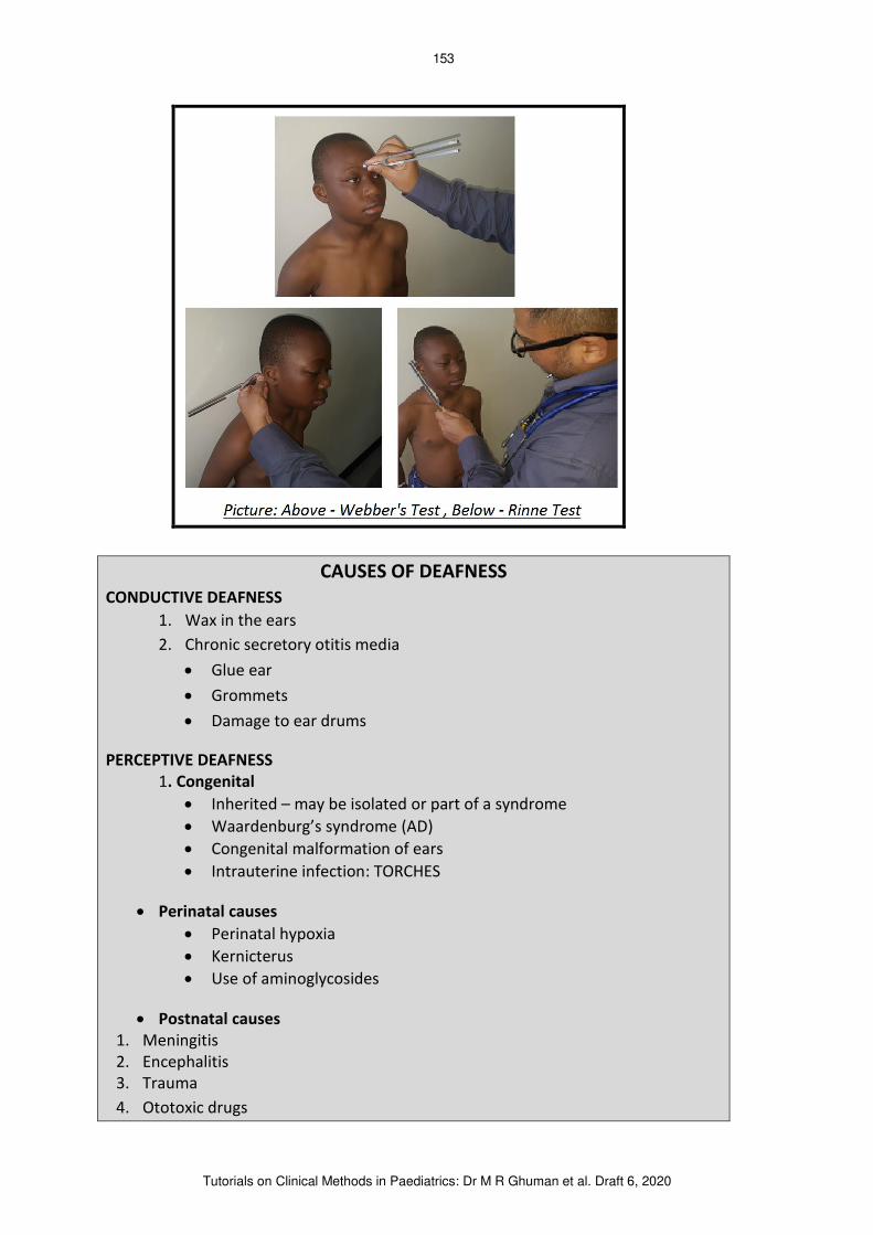
Tutorials on Clinical Methods in Paediatrics: Dr M R Ghuman et al. Draft 6, 2020
153
CAUSES OF DEAFNESS CONDUCTIVE DEAFNESS
1. Wax in the ears
2. Chronic secretory otitis media
Glue ear
Grommets
Damage to ear drums
PERCEPTIVE DEAFNESS 1. Congenital
Inherited – may be isolated or part of a syndrome
Waardenburg’s syndrome (AD)
Congenital malformation of ears
Intrauterine infection: TORCHES
Perinatal causes
Perinatal hypoxia
Kernicterus
Use of aminoglycosides
Postnatal causes 1. Meningitis
2. Encephalitis
3. Trauma
4. Ototoxic drugs

Tutorials on Clinical Methods in Paediatrics: Dr M R Ghuman et al. Draft 6, 2020
154
Vestibular division To assess for sense of balance and spacial orientation for the purpose of coordinationg
movement with balance i.e., vertigo & nystagmus
Abnormality of posture or gait
Induction of positional nystagmus
Doll’s eye reflex
Oculovestibular reflex
Dix-Hallpike maneuver
Nystagmus is caused by the lesion of cerebellum, brainstem, cervical cord or inner ear -
always offer to examine hearing, cerebellar functions and the occular fundi
Nystagmus Involuntary & repetitive oscillations of the eye or eyes is called as nystagmus. It is defind by
the direction of the fast phase but it is the slow phase which is pathological. Nystagmus may
suggest end-organ or intrinsic disease of the nervous system.
The most common cause of nystagmus in children is caused by the centrally acting
medications like anticonvulsants
Central nystagmus:
Nystagmus in any direction results from:
Brain stem lesions
Vertebrobasilar ischaemia
Phenytoin toxicity
Pendular nystagmus
This refers to the waveform of involuntary eye movement in which eye moves in sinusoidal
trajectory, similar to that of a pendulum. Rhythmic abnormalities that are equal in all planes
of gaze; associated with either cerebellar or retinal disease
Jerk nystagmus Consists of a slow and fast phase and refers to abnormalities of vestibular system
Vertical nystagmus
Primarily seen in abnormalities of the brain stem
Ocular nystagmus
Due to poor macular vision which impairs retinal fixation
9. Glassopharyngeal Nerve Ask simple questions to the care giver and observe like:
Can the child swallow?
Is the child drooling?
Then:
Assess patient’s speech for dysarthria or dysphonia

Tutorials on Clinical Methods in Paediatrics: Dr M R Ghuman et al. Draft 6, 2020
155
Check for:
Palatal reflex: tickle the back of the pharynx, and note if contraction occurs. This is
also a test for vagus nerve.
Ask the child to puff out cheeks with lips closed: air leak from nose will indicate
lesion
Examine the taste in the posterior 1/3rd
of tongue: Loss of taste at this part of the
tongue may occur with a lesion of the trunk of the glossopharyngeal nerve.
10. Vagus Nerve Following test can be performed:
Ask the patient to say Aah and look at the palate and note position of uvula. If it is in
the center and moving in either side than that’s normal and if it is deviating on one
side that side is considered normal because of pull by the stronger muscles
Note nasal twang
Note regurgitation of food
Note hoarsenes of voice; due to paralysis of laryngeal muscles
Common causes of bilateral vagus nerve injury
Progressive bulber palsy
Bilateral supranuclear lesions
Cerebrovascular disease
Multiple sclerosis
9th
through 12 cranial nerves are called bulbar cranial nerves. Gag reflex is caused by
touching the posterior pharyngeal mucosa. Both 9th
and 10th
cranial nerves are responsible
for gag reflex.
11. Accessary Nerve It is purely a motor in function. Check bulk and test power of sternocleidomastoid and
trapezius muscles.
Method
Testing the trapizius muscle
o Tell the patient to shrug shoulders while the examiner presses downward on them.
Paralysis of the upper part of trapizius muscle is demonstrated in this way.
Testing the right sternocleidomastoid muscle
o Face the patient and place your right palm laterally on the patient’s left cheek. Tell the
patient to turn head to the left, resisting the pressure you are excerting in the
opposite direction. Paralysis of the sternomastoid causes weakness of rotation
towards the opposite side.
Testing the left sternocleidomastoid muscle
o Face the patient and place your left palm laterally on the patient’s right cheek. Tell the patient to turn head to the right, resisting the pressure you are excerting in the

Tutorials on Clinical Methods in Paediatrics: Dr M R Ghuman et al. Draft 6, 2020
156
opposite direction. Paralysis of the sternomastoid causes weakness of rotation
towards the opposite side.
12. Hypoglossal Nerve It is purely a motor nerve. It supplies the tongue and the depressors of the hyoid bone.
It is tested by examining the tongue and its movements as follows:
If the nerve is injured, at rest the tongue may appear to have the appearance of a
“bag of worms” (fasciculations) or wasting (atrophy). Fasciculations must be assessed
with the tongue relaxed in the mouth, not when protruded.
Ask the patient to stick the tongue out as far as possible. If the hypoglossal nerve is
paralysed, the tongue, instead of being protruded straight, will be pushed over to the
paralysed side.
Ask the patient to move the tongue from side to side and lick each cheek with it;
observe whether patient can do so freely.
Strength may aslo be assessed by pressing against the tongue with a finger as the
patient protrudes it into each cheek in turn.
Patients with hypoglossal nerve disorder have difficulty speaking, chewing and
swallowing.
Assess speech by asking the patient to say “red lorry and yellow lorry” etc.
SUMMARY: IF ALL CRANIAL NERVES ARE NORMAL - SAY
1. Pupils equal and reactive to light
2. Fixes and follows
3. No ophthamoplegia
4. No facial asymetry
5. Hearing appears intact
6. Tongue central
7. Positive gag reflex
8. No drooling
NB! Cavernous sinus thrombosis causes problems in 3 cranial nerves; Occulomotor,
Trochlear and Abducent (OTA) nerves.
6. EXAMINATION OF MOTOR SYSTEM
Achronym: BPTR: Bulk, Power, Tone, Reflexes
EXAMINE THE MUSCLE BULK
Inspection
Examine after full exposure and look for deformity
o Bony or soft tissue
o Pes cavis/Pes planus or claw of hands
Pes mean foot
Pes cavis: foot with abnormally high longitudinal arch

Tutorials on Clinical Methods in Paediatrics: Dr M R Ghuman et al. Draft 6, 2020
157
Pes planus or flat foot: when entire sole of the foot touches the floor
on standing
o Ankle valgus: an insidious deformity that results in pronation of the foot and
medial malleolar prominence
Look for asymmetry: Proximal/distal
Look for muscle wasting or hypertrophy
o Loss of muscle bulk may be due to a lower motor neuron lesion, disuse
atrophy or generalised wasting
o Excessive muscle bulk may be due to Duchenne Muscular Dystrophy or
Myotonia Congenita, may look like baby Hercules
Examine the posture:
Shoulder adducted, elbow flexed, hand clinched may be due to pyramidal tract
problem
Erb’s palsy (policeman tip hand deformity)
Claw hand (atrophy and distortion of hand)
Arthrogryposis (persistant flexion of a joint, tetnoid spasm)
Palpate the muscles to assess bulk
Common abnormalities
Athetosis: repetitive involuntary, slow gross movements which involve distal parts of
the extremities
Peripheral neuropathy: distal wasting (inverted champaign bottle)
Check thenar/hypothenar for muscle wasting
EXAMINE THE POWER Grade the power according to Medical Research Council Scale
Grade 0: No muscle movement (complete paralysis)
Grade 1: Muscle flicker, no joint movement
Grade 2: Full movement is possible when gravity is illiminated
Grade 3: Movement against gravity but not against resistance
Grade 4: Power against resistance but can be overcome by resistance
Grade 5: Normal power against gravity and full resistance is present
Check proximal/distal power
Upper limbs
o Check thumb for flexion/extension
o Check abduction/adduction of fingers
o Squeez hands and check grip/strength
o Examine wrists – stablise at elbow – extend/flex
o Examine elbows – stabilize upper arm – extend/flex
o Check shoulders for abduction/adduction – shrug shoulders

Tutorials on Clinical Methods in Paediatrics: Dr M R Ghuman et al. Draft 6, 2020
158
o Elicit pronator drift: when child extends his or her arms out in front with
palms upword and eyes closed. Hands on weak side cup and pronate slowly.
This is scalled as apronator drift.
Examine neck: patient to lie flat, check for flexion/extension of neck
Examine trunk for muscle weekness: note for pot belly/lumber lordosis
Check for proximal weakness: elicit Gower’s sign
Lower limbs
o Big toe: extend & check for muscle wasting at the dorsum of the foot
o Foot: invert/evert, dorsiflex/planter flex
o Hip, knee – extend and flex
Compare
o UL/LL
o Right to left
o Proximal to distal

Tutorials on Clinical Methods in Paediatrics: Dr M R Ghuman et al. Draft 6, 2020
159
EXAMINE THE TONE Muscular tone is a state of tension or contraction found in healthy muscles. It represents
the dynamic resistance of muscles to movement across a joint (stretch or to gravity). An
increase in the tone is called as hypertonia and a diminution as hypotonia. The degree of
tone is estimated by handling the limbs and moving them passively at their various joints.
Passive tone is the resistance to stretch felt when limb is flexed and extended
manually
o In LMNL, passive tone is reduced
o In chronic UMNL, passive tone is increased
Active tone is the posture that an infant adopts when placed in a particular position
Hypertonia Causes of hypertonia may include upper motor neuron disease, lesion in basal ganglia, and
anxiety. Hypertonia following lesions of the corticospinal system (UMNL) is termed as
spasticity. The term spasticity is used to describe a state of increased tone which is of claps
knife type.
Claps knife type hypertonia is when the limb is fairly rapidly flexed or extended i.e.
when limb is flexed, initially it is very rigid but after certain degree of flexion the
resistance suddenly disappears & same thing happens when extending it. Clasped
knife response or spasticity is seen in upper motor neuron lesions.
Cogwheel rigidity hypertonia results from disease of the basal ganglia also termed as
extrapyramidal rigidity. In this type, the resistance to passive movements is fluctuant
i.e. if you try to flex the elbow, it will flex in a jerky manner described as like a lever
rubbing on the teeth of a cogwheel (cogwheel rigidity). This cogwheel rigidity is seen
in extrapyramidal disease.
Lead pipe rigidity, the rigidity is uniformly present throughout the passive
movements (flexion or extension). This is present in cases in which patient’s state of consciousness is impaired.
Hypotonia In hypotonia, there is little or no resistance to passive movement of the limb and when
handled or shaken the unsupported part flops about inertly. Hypotonic muscles are
abnormally soft to palpation. Causes of hypotonia may include lower motor neuron disease,
unconsciousness, and neural shock i.e. in early stages of UMNL.
Do passive movements of flexion and extension in all the four limbs, slowly and then quickly
and note abnormalities of tone:
Hypotonia
Spasticity
Rigidity

Tutorials on Clinical Methods in Paediatrics: Dr M R Ghuman et al. Draft 6, 2020
160
EXAMINE THE REFLEXES There are superficial & deep tendon reflexes.
Superficial reflexes Reflex How elicited Result Level of cord
concerned
Anal Stroking or scratching
the skin near the anus
Contraction of anal
sphincter
S3, S4
Planter Stroking sole of foot Flexion of toes, foot or leg L5, S1
Cremasteric Stroking skin at upper
and inner part of thigh*
Upward movement of
testicle
L1, L2
Lost in UMNL
Abdominal Stroking abdominal wall
below costal margin, at
the level of umbilicus
and in iliac fossa
Contraction of abdominal
muscles
T7 to T12
Lost in UMNL &
LMNL
Scapular Stroking skin in the
intrascapular region
Contraction of scapular
muscles
C5 to T1
*the cremasteric reflex can often be more easily elicited by pressing over the sartorius in
the lower third of Hunter’s canal (an aponeurotic tunnel in the middle third of the thigh,
extending from the apex of the femoral triangle to the opening in the adductor magnus,
the adductor hiatus)
Babinski’s sign: Scratch the outer border of the sole of foot with edge of a key or the stick of a patella
hammer. Normally the greater toe goes up and there is flexion and adduction of all other
toes and foot is dorsiflexed and inverted. This is called as negative Babinski sign or planter
reflex (planters are flexors). In positive Babinski the big toe bends up and back to the top of
the foot and other toes fan out. Nerve roots involved are L5, S1.
Positive Babinski is present in:
Upper motor neuron lesion
During sleep
In infants – untill they start walking (about 1 yr)
Post ictal or coma

Tutorials on Clinical Methods in Paediatrics: Dr M R Ghuman et al. Draft 6, 2020
161
Deep tendon reflexes If the tendon of a slightly stretched muscle is struck (a single, sharp blow) with a soft rubber
hammer, the muscle contracts breifly. This is the monosynaptic stretch reflex. It is the test
of integrity of the afferent and efferent pathways and of the excitability of the anterior horn
cells in the spinal segment of the stretched muscle. When eliciting the tendone reflexes
grade responses as follows:
Grade 0: Absent reflex
Grade 1: Present normal
Grade 2: Brisk (as normal knee jerk)
Grade 3: Very brisk
Grade 4: Sustained clonus
When eliciting a tendon reflex following need to be kept in mind:
There should be no joint disease of the limb on which reflex is going to be elicited
Patient should be fully relaxed and the part of the body to be tested should be fully
exposed.
Concentration should be on the movements of the muscles whose tendon has been
struck by the hammer
BICEPS JERK (C5, C6) Method 1:
Flex the arm at the elbow and place forearm on the abdomen.
Put your index finger over the tendon of biceps muscle and press it lightly.
Now strike the hammer on your finger and note the Biceps jerk.
Method 2:
Hold the patient’s elbow in your left hand in a manner that your thumb should be on
the tendon of biceps muscle.
The patient’s upper limb should be fixed at elbow and it should rest on your left
forearm relaxed.

Tutorials on Clinical Methods in Paediatrics: Dr M R Ghuman et al. Draft 6, 2020
162
Now press the tendon of biceps muscle lightly with your thumb and strike the
hammer over your thumb.
Note intensity of reflex by seeing the contraction of the muscle.
TRICEPS JERK (C6, C7) Keep the forearm on the abdomen with elbow flexed.
Strike the hammer on the tendon of triceps muscle just above the olecranon
process.
Note intensity of reflexion by seeing the contraction of the triceps muscle.
SUPPINATOR JERK (C5, C6) Keep the forearm placed on the abdomen in semi pronated position with flexed
elbow.
The hand should slightly deviate towards the ulnar side.
Now strike with patella hammer on the styloid process of the radius (the tendon of
brachio-radialis muscle).
Note intensity of reflex by seeing the contraction of Brachio-radialis muscle.
KNEE JERK (L3, L4) There are many methods to elicit the knee jerk. Following three are commonly used:
Method 1:
Make the patient to sit at the edge of the bed with his legs hanging down.
Remove trousers from the thigh’s and locate the tendon of the quadriceps femoris
Give a stroke on the tendon with patella hammer and note the grade of reflex.
Method 2:
Make the patient to lie on the bed with both knee joints flexed.
Pass your hand under the knee to be tested and bring it to rest upon the opposite
knee.
In this way the knee to be tested will hang on the dorsum of your wrist.
Now divert the attention of the patient in some way and strike the hammer on the
tendon of the quadriceps midway between the tibial tuberosity and the patella.

Tutorials on Clinical Methods in Paediatrics: Dr M R Ghuman et al. Draft 6, 2020
163
Method 3:
Make the patient to lie supine on the bed with lower limbs slightly flexed at hip and
knee joints with femur externally rotated in a manner that the heels of both sides
are near to each other.
Now strike the tendon of quadriceps femoris muscle and note intensity of reflex by
seeing the contraction of the muscle.
JENDRASSIK MANEUVER Jendrassik manoeuvre is used to elicit the deep tendon reflexes. In this manoeuvre the
patient clenches the teeth, flexes both sets of fingers into a hook-like form, and interlocks
those sets of fingers together. The tendon below the patient’s knee is then hit with the patella hammer. The elicited response is compared with the reflex result of the same action
when the manoeuvre is not in use.
The amplitude of the response is increased because a larger number of motor units have
been activated. Clinically some brain stem lesions are manifested by exaggerated spinal cord
reflexes. Isometric contractions in another muscle group enhance the response of the
reflex. Often this is used to distract the patient and relax the muscles in the quadriceps.
ANKLE JERK (S1, S2): Method:
Ask the patient to lie on the bed with legs everted and slightly flexed at the knee.
Now, with one hand slightly dorsiflex the foot and strike the tendon Achilles.
There will be contraction of the calf muscles and the foot will undergo planter
flexion.

Tutorials on Clinical Methods in Paediatrics: Dr M R Ghuman et al. Draft 6, 2020
164
CLONUS This is elicited when the tendon reflexes are exagerated.
Clonus ordinarily occurs when the stretch reflex is highly sensitised by facilitatory impulses
from the brain. For instance in a decerabrate animal in which stretch reflexes are highly
facilitated, clonus develops readily. It is done to determine the degree of facilitation of
spinal cord.
Clonus may be sustained or unsustained. Sustained clonus is indicative of hyper-reflexia
(UMNL), while unsustaiend clonus is present in nervous and anxious persons. This is of two
types: Patellar and Ankle clonus
Patellar clonus
Method:
Ask the patient to lie down supine on the bed and you hold the tendon of quadriceps
by upper border of the patella.
Now give 2 to 3 tendon jerks downwards. In positive cases, there will be series of
clonic contractions of the quadriceps and the patella will move with it.
Ankle clonus
Method:
Ask the patient to lie down supine on the bed.
Flex the knee along with slight flexion of hip joint and support the leg with your
hand.

Tutorials on Clinical Methods in Paediatrics: Dr M R Ghuman et al. Draft 6, 2020
165
Now grasp the forepart of the foot with other hand and give 2 to 3 sudden
dorsiflexion movement to the foot.
The pressure on the sole of the foot is sustained by keeping it in dorsiflexed position.
There will be series of contractions of calf muscles in positive cases. So there will be
repeated vibrations at the ankle joint.
Hoffman’s reflex The test involves tapping the nail or flickering the terminal phalanx of the middle finger.
Hoffman’s test is positive if you see adduction of thumb and flexion of index finger which may indicate an upper motor neuron lesion or a pyramidal sign. Hoffman’s sign may be seen in multiple sclerosis and conditions which cause spinal cord compression such as cervical
myelopathy, cervical spondylitis, tumors or degenerative arthritis.
Wartenburg’s sign It is a neurological sign consisting of involuntary abduction of the little finger, caused by
injury of the ulner nerve which supplies intrinsic hand muscles (particularly palmer
interosseous muscle) which results in unopposed action of the extensor digiti minimi. This is
commonly seen in ulner nerve neuropathy and cervical neuropathy.
Pendular knee reflex This is one of the cerebelalr sign where leg keeps swinging after knee jerk more than 4 times
(4 or less is normal), which ma be due to cerebellar damage.
Crossed adductor reflex Contraction of both hip adductors when either knee jerk is elicited. The cross adductor
response usually disappears by 7 to 8 months. Its persistance beyond 8 months is a sign of
pyramidal tract dysfunction.
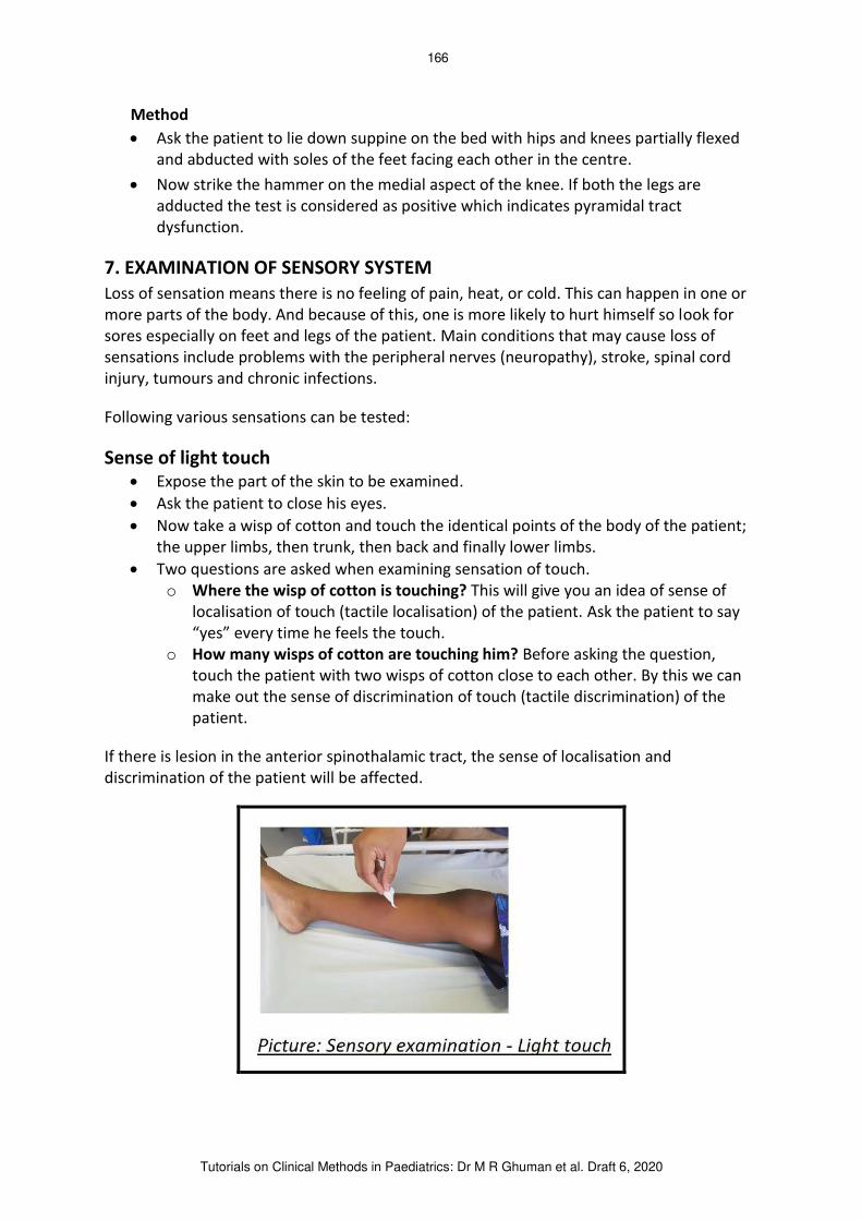
Tutorials on Clinical Methods in Paediatrics: Dr M R Ghuman et al. Draft 6, 2020
166
Method
Ask the patient to lie down suppine on the bed with hips and knees partially flexed
and abducted with soles of the feet facing each other in the centre.
Now strike the hammer on the medial aspect of the knee. If both the legs are
adducted the test is considered as positive which indicates pyramidal tract
dysfunction.
7. EXAMINATION OF SENSORY SYSTEM Loss of sensation means there is no feeling of pain, heat, or cold. This can happen in one or
more parts of the body. And because of this, one is more likely to hurt himself so look for
sores especially on feet and legs of the patient. Main conditions that may cause loss of
sensations include problems with the peripheral nerves (neuropathy), stroke, spinal cord
injury, tumours and chronic infections.
Following various sensations can be tested:
Sense of light touch Expose the part of the skin to be examined.
Ask the patient to close his eyes.
Now take a wisp of cotton and touch the identical points of the body of the patient;
the upper limbs, then trunk, then back and finally lower limbs.
Two questions are asked when examining sensation of touch.
o Where the wisp of cotton is touching? This will give you an idea of sense of
localisation of touch (tactile localisation) of the patient. Ask the patient to say
“yes” every time he feels the touch. o How many wisps of cotton are touching him? Before asking the question,
touch the patient with two wisps of cotton close to each other. By this we can
make out the sense of discrimination of touch (tactile discrimination) of the
patient.
If there is lesion in the anterior spinothalamic tract, the sense of localisation and
discrimination of the patient will be affected.

Tutorials on Clinical Methods in Paediatrics: Dr M R Ghuman et al. Draft 6, 2020
167
Sense of course touch Method
Press the limb with finger or blunt probe on identical points.
Now ask the patient whether he appreciates or not.
Sense of pain Pain is carried by lateral spinothalamic tract; therefore, its lesion will impair the pain
sensation.
Method
Take a steel pin or sterile needle (rarely done in children).
The pin is pricked on the identical points and the patient is asked whether he feels
pain or not.
Sense of deep pain: Two tests are done
Squeeze the calf muscles and apply pressure in between the thumb and the index
finger. Note if the patient feels pain.
Deep pain is absent in tabes dorsalis (a form of neurosyphilis in which there is loss of
both axons and the myelin in the dorsal roots, with pallor and atrophy in the dorsal
columns of the spinal cord).
Sense of temperature
This is tested by hot and cold water contained in two copper test tubes.
The naked area under the examination is touched with each, in turn, and the patient
is asked whether he feels it hot or cold.
Lesion in the lateral spinothalamic tract will impair or even abolish this sensation.
Sense of position and passive movements (posterior column integrity): Method 1 Here the movements of thumb and big toe are tested.
The patient is made to understand the movements of big toe or thumb which is
moved up, down and sideways.
Ask the patient to close his eyes.
Now hold the big toe with your thumb and index finger.
Flex it at the distal interphalangeal joint, slightly without touching the other toes and
ask the patient whether it is directed upward or downward.
If unable to make final assessment test can be repeated number of times.
Same procedure can be used for upper limb by the movement of thumb.
Method 2
Ask the patient to close his eyes.
Hold the thumb of the patient and move it in various directions through the air.
Now place the thumb of the patient in an abnormal position and ask the patient to
place his other thumb in the same position.
In the case of hand, move the fingers in various directions and ask the patient to
imitate with other hand while eyes are closed.

Tutorials on Clinical Methods in Paediatrics: Dr M R Ghuman et al. Draft 6, 2020
168
Testing limb should not touch any other skin surface; otherwise patient will be able
to appreciate its position in spite of the lesion.
Sense of vibration Tuning fork (frequancy 128/sec) is used to test sense of vibration.
Strike the tuning fork on the side of your knee and then place its base over the bony
prominances, i.e. medial and lateral maleoli, tubercles of tibia, anterior superior iliac
spines and olecranon processes.
Normal individual will feel the buzzing sensation.
If unable to feel the vibration sense, consider polyneuropathy or disease of posterior
column.
Brown-Sequard Syndrome
It is a rare neurological condition cheracterised by an incomplete lesion in the spinal cord
(often in the cervical cord region) which results in weakness or paralysis (hemiparaplegia),
spascity and ataxia on one side of the body (upper motor neuron paralysis) and loss of pain
and temperature sensation (hemianaesthesia) on the opposite side. There may also be loss
of bladder and bowel control.
Papilloedema is observed in few patients usually in association with markedly elevated CSF
protein levels that presumably interfere with normal CSF flow dynamics.
Focus mainly need to be on the underlying cause of the disorder. Early treatment with high
dose steroids may be beneficial in many cases. Physical, occupational and recreational
therapy is important aspects of patient rehabilitation.

Tutorials on Clinical Methods in Paediatrics: Dr M R Ghuman et al. Draft 6, 2020
169
8. CO-ORDINATION: CEREBELLAR SIGNS The cerebellum receives information from the sensory system, the spinal cord and other
parts of the brain and then regulates motor movements such as posture, balance,
coordination, speech and rate, range, rhythm and force of muscle contraction. Following
signs should be lookd for in the lesions of cerebellum:
Ataxia
Ataxia is impaired balance or coordination of movement. Patients with cerebellar lesion will
not be able to walk in straight line with the eyes open. Cerebellum and associated pathways
modulate volitional movement.
If either afferent cerebellar connections (joint position sense) or efferent cerebellar
connections (cerebellum through thalamus through cerebral cortex) are disturbed, the
patient experiences ataxia.
Adiadochokinesia (inability to conduct rapid alternating hand movements)

Tutorials on Clinical Methods in Paediatrics: Dr M R Ghuman et al. Draft 6, 2020
170
Method 1
To elicit loss of rapid alternate movements.
Ask the patient to flex his elbows at the right angle and to pronate and supinate his
forearm by placing one hand over the other.
The patient will not be able to do so and his movements will be slow and incomplete.
Method 2
Ask the patient to tap your palm with his finger tips as fast as possible.
Patient will not be able to do so.
Asynergia: This means inco-ordination. This is tested in both limbs:
UPPER LIMB
Finger-Nose test (finger nose pointing or dysmetria)
Ask the patient to touch tip of your index finger which is at adistance of about ½
meter from the patient) with his index finger and then to touch his nose.
This should be done rapidly. Patient will fail to do so.
Finger-Finger test
Ask the patient to touch the tips of his both index fingers with extended arms
bringing them from a distance rapidly.
Patient will not be able to do so.
Rebound phenomenon (loss of rebound control):
Ask the patient to flex his elbow slightly and flex it further firmly against resistance
(i.e. you hold his hand) then release it suddenly.
The patient will slap his chest due to rebound phenomenon but normal individual
will be able to prevent it to happen.
If the results of above tests are doubtful, then patient should repeat the above tests
while his eyes closed - irregularity in the movements will become more marked
LOWER LIMB
Heel-Knee Test
Ask the patient to lie in the bed in supine position

Tutorials on Clinical Methods in Paediatrics: Dr M R Ghuman et al. Draft 6, 2020
171
Then to put the heel of his foot on the knee of other leg and rub it down along the
shin
Then raise the leg in the air and again put the heel on the knee.
He will fail to do so.
Cerebellar gait
It is a staggering, wide-based, unsteady gait which has lurching movements with a
breakdown of normal synergy of walking.
In the older child one can test ataxic gait by asking the patient to walk by heel to toe
(like walking on a tight rope).
Pendulous knee jerk
Ask the patient to sit in the bed with legs hanging down and elicit the knee jerk.
The leg after extension will fall down in a pendulous fashion
Charcot’s triad [SIN]
Scanning speech
o Ask the patient, to speak few words
o Note slow, slurring or explosive dysarthria, poor or no cordination
Intension tremor
o Ask the patient to reach out and touch your finger
o Note the fine tremor increasing as it approaches the finger
Nystagmus
o Ask the patient to look straight ahead at you
o Note the abnormal movement of eye from slow to rapid phase - nystagmus
9. SPINAL CORD (CERVICAL, THORACIC AND LUMBER SPINE) Do full examination of the spine
Look for hairy patch, lipoma or spina bifida
Palpate the spinous processes and paraspinal tissues
Note overall alignment and focal tenderness

Tutorials on Clinical Methods in Paediatrics: Dr M R Ghuman et al. Draft 6, 2020
172
Lightly percuss the spine with your closed fist – note any tenderness
Move and observe flexion, extension, and lateral flexion
Note clinical signs in the case of “Hemisection of Spinal Cord”
Below the level of lesion
o Features ipsilateral to the lesion
Impaired light touch
Impaired vibration touch
Corticospinal tract signs
o Features contralateral to the lesion
Impaired pain sensation
Impaired temperature sensation
At the level of lesion
o Features ipsilateral to the lesion
Segmental zone of hyperpathia (pain)
Signs of lower motor neuron at this level
Lesion in the anterior horn cells Lesion in the nerve
Disease process is gradual in onset Disease process is sudden in onset
Fibrillation and fasciculations are
present
Fibrillation and fasciculations are not
present
Only motor system is involved Both motor and sensory systems are
involved
There is no sphinteric disturbance Sphinteric loss is present
10. AUTONOMIC NERVOUS SYSTEM
Sympathetic Nervous System
Horner’s syndrome: This is due to the lesions in the cervical sympathetic trunk which leads to:
Ptosis of the eye lid
Enophthalmos (eye ball moves inward)
Miosis (constriction of the pupil)
Loss of sweating (anhydrosis)
Loss of cilio-spinal reflex:
On pinching the lateral side of the neck, sympathetic trunk gets stimulated which results in
dilatation of the pupil. This reflex is lost in the diseases involving cervical sympathetic trunk.

Tutorials on Clinical Methods in Paediatrics: Dr M R Ghuman et al. Draft 6, 2020
173
Parasympathetic nervous system
Cranial nerve III (ciliary ganglion) supply cilliary muscles and the sphincter
pupillae
Cranial nerve VIII (Pteropalatine ganglion + submandibular ganglion) axons projet
to the lacrimal glands, mucous membrane and nasal mucosa
Cranial nerve IX (otic ganglion) scretomotor fibers then pass to parotid gland
Cranial nerve X (ganglions close to organ) supplies the:
o Heart
o Larynx/trachea/bronchi
o Oesophagus, stomach
o Abdominal blood vessels
o Liver & bile duct
o Pancrease
o Adrenal gland
o Small intestine
o Large intestine
S1 to S4 Colon, rectum, kidney, bladder, penis, clitoris
11. EXTRAPYRAMIDAL SYSTEM Lesions of the basal ganglia cause involuntary movements
Chorea (of hands and fingers)
Cheracterised by involuntary, irregular and purposeless movements of the hands and fingers
like piano-playing movements
Huntington’s chorea: absent caudate shadow on CT brain
Signs
Protruded toungue
Milkmaid sign
Shelving sign
Sydenham chorea
Post streptococcal
Increased ASOT
Cardiac murmur
Athetosis
Slower and writhing movements
Dystonia
Abnormal posture due to periodic sustained contraction of opposing muscle group

Tutorials on Clinical Methods in Paediatrics: Dr M R Ghuman et al. Draft 6, 2020
174
Ballism
Sudden massive jerky flinging movements
Basal nuclei
Lesions of basal nuclei do cause cog wheel or lead pipe regidity
12. PERIPHERAL NERVE DAMAGE Lesions in bracheal plexus
Upper lesion – Erb-Douchenne’s paralysis
Deformity:
o upper limb hangs by the side
o internally rotated
o and forearm pronated
o also called as policeman’s receiving tip hand
Lesion C5 and C6
Lesion C7 in 50% cases
Paralysis of biceps brachialis, brachioradialis and supinator muscles
Lower lesion – Klumpke’s paralysis: here lower nerve roots are injured
This deformity is also called as “Claw Hand” in which there is: o Weakness of the flexor muscles of the wrist
o Weakness of the small muscles of the hand
o Lesion C8, T1
o Up to 1/3rd
of these patients have Horner’s syndrome.
Median nerve lesion: carple tunnel syndrome
Radial nerve lesion: wrist drop
Ulner nerve lesion: claw hand
Common peronial nerve lesion: foot drop
Lateral cutaneous nerve of thigh lesion: loss of sensation over lateral aspect of thigh
GENERAL PRINCIPLE
When hemiplegia is crossed i.e. hemiplegia on the one side and involvement of cranial
nerves on the opposite side, the lesion is in the brainstem which may be in the:
Midbrain
Pons
Medulla oblongata
But if the hemiplegia is uncrossed i.e. hemiplegia and the involvement of cranial nerves are
on the same side, the lesion is above the brainstem which may be in:
1) Internal capsule
2) Corona radiata

Tutorials on Clinical Methods in Paediatrics: Dr M R Ghuman et al. Draft 6, 2020
175
3) Cerebral cortex
AT THE END OF THE EMANIBATION MAKE A REASONABLE ASSESSMENT
What is the dysfunction like spastic quadriplegia with profound developmental
delay
Where is the lesion like in cerebral cortex
What is the aetiology like infarct due to hypoxic ischaemic encephalopathy
WHAT IS THE DIAGNOSTIC EVALUATION LIKE 1) Normal or abnormal
2) UMNL or LMNL
3) Site of lesion
4) Nature of lesion
5) Complications
6) Developmentally normal or abnormal
7) Static or progressive
8) Acute or chronic
If it is lower motor neuron lesion, then describe as: It is lower motor neuron lesion due to
Hypotonia
Weakness
Absent reflexes
Wasting
And conclude with summary of your findings and make a provisional diagnosis on the basis
of your clinical examination.

Tutorials on Clinical Methods in Paediatrics: Dr M R Ghuman et al. Draft 6, 2020
176
TUTORIAL 11
THE EXAMINATION OF MUSCULO- SKELETAL SYSTEM
OVERALL OBJECTIVE At the end of this module, the student should be able to do a full clinical examination of
musculoskeletal system and to be able to make a reasonable differential diagnosis of
swollen joints, rheumatic fever, systemic lupus erythematosis, dermatomyositis, gait
problems, muscular atrophy or hypertrophy in infants and children.
Extra General features include:
Malar rash of SLE
Supraorbital rash of dermatomyositis
Muscular hypertrophy or pseudohypertrophy
Muscle atrophy
Muscle fasciculations
Signs of acute rheumatic fever
Rickety rosery, craniotabes, widening of wrists – rickets
Screening questions 1) Do you have pain or stiffness in your muscles, joints, back?
2) Can you dress up yourself without difficulty?
3) Can you walk up and down stairs without difficulty?
EXAMINATION OF A JOINT Introduce yourself to the patient and the caregiver
Sit in front of the patient: good position is confruntation position
Do a good observation
o Ask the patient to walk a short distance and watch gait
o Ask the patient to take off a jumper or shirt and observe upper limb
function
o Ask the patient to write his name and observe his hand function
o Ask the patient to undress and observe the upper limb function
General examinatin will include:
Sequence: Look, Feel, Move, Measure
GALS screen: Gait, Arms, Legs, Spine
SEQUENCE
1. Look
General inspection, see if the patient is:
o Thriving, has any bandages or is on a wheelchair.
o Having any prosthesis e.g. calipers or special shoes

Tutorials on Clinical Methods in Paediatrics: Dr M R Ghuman et al. Draft 6, 2020
177
o Has any spine deformaties e.g. kyphosis, scoliosis or kyphoscoliosis
o Has any limb deformaties like:
Amelia (birth defect of lacking one or more limbs)
Hemiamelia (upper part of limb well developed but the lower
part is rudementary or absent)
Phocomelia (the upper part of limb is extremely
underdeveloped or missing and lower part is directly attached to
the trunk” Contracture
Joint swelling
Also look for any:
o Skin changes or rash like erythema or trophic skin changes
o Muscle wasting
2. Feel Enquire first if any part is painfull and what exacerbates pain.
Then feel for
1) Move to assess Temperature
2) Tenderness (watch expression)
3) Enlargement
4) Effusion
5) Synovial thickening
6) Bony enlargment
3. Move to assess Move to assess active before passive movements
1) Range of active movements (normal or limited)
a. Range of passive movements (normal or limited)
2) Is movement painful
3) Is movement accompanied by crepitus
4) Is there any fixed deformaity: when joint cannot be placed in the neutral
anatomical position due to inability of movement or laxity of joint ligaments.
5) State of peripheral circulation
4. Measure Limb circumference for muscle wasting
5-10 cm above tibial tuberosity
5-10 cm below tibial tuberosity
Limb length for length discrepancy
True leg length: from anterior superior iliac spine to medial maleolus
Apparent leg length: from umbilicus (or pubics symphysis) to medial
maleolus

Tutorials on Clinical Methods in Paediatrics: Dr M R Ghuman et al. Draft 6, 2020
178
Causes of true leg length discrepancy 1) Undetected CDH
2) Previous trauma/bone surgery
3) Increased limb growth – arthritis of the knee
4) Ollier’s disease 5) Osteogenesis imperfecta
o Blue sclera
o Hyperextensible joints
6) Ployostotic fibrous dysplasia
o Café a lait spots
o Sexual precosity
7) Causes of apparent leg length discrepancy
o Severe hemiparesis
o Adductor spam causing pelvic tilt
GALS SCREENING
1. Gait
Watch patient walking in straight line
o Bare foot
o In shoes
Walking fast or running will unmask any abnormality
Evaluate from, behind, front and the side
o Foot, Ankle, Knee, Hip, Pelvis, Spine
Note for limp or abnormal gait
o Pain – antalgic gait – dot dash
o Structural – limb length discrepancy
o Weakness
o Trendlengurg gait – Duchenne sign
o Drop foot – common peronial nerve palsy – the gait is high stepping to lift
the drop foot
Note for increased tone
o UMNL – Crouch gait
o CP
o CVA
2. Arms Advise the patient to:
1) Put your hands behind your head. Note elbows going backward. In frozen
shoulder or rotators cuff problems there will be painful abduction
2) Bend arms & touch shoulders: note elbow flexion
3) Straight arms: extension at 180 degree
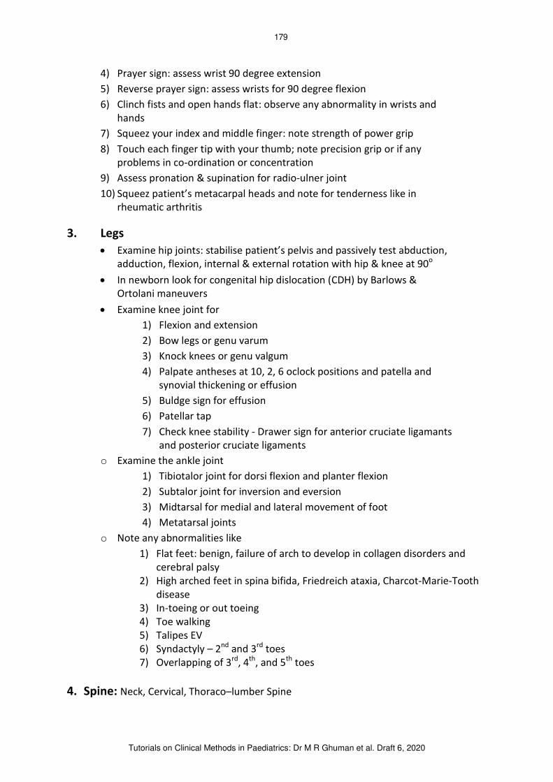
Tutorials on Clinical Methods in Paediatrics: Dr M R Ghuman et al. Draft 6, 2020
179
4) Prayer sign: assess wrist 90 degree extension
5) Reverse prayer sign: assess wrists for 90 degree flexion
6) Clinch fists and open hands flat: observe any abnormality in wrists and
hands
7) Squeez your index and middle finger: note strength of power grip
8) Touch each finger tip with your thumb; note precision grip or if any
problems in co-ordination or concentration
9) Assess pronation & supination for radio-ulner joint
10) Squeez patient’s metacarpal heads and note for tenderness like in
rheumatic arthritis
3. Legs
Examine hip joints: stabilise patient’s pelvis and passively test abduction,
adduction, flexion, internal & external rotation with hip & knee at 90o
In newborn look for congenital hip dislocation (CDH) by Barlows &
Ortolani maneuvers
Examine knee joint for
1) Flexion and extension
2) Bow legs or genu varum
3) Knock knees or genu valgum
4) Palpate antheses at 10, 2, 6 oclock positions and patella and
synovial thickening or effusion
5) Buldge sign for effusion
6) Patellar tap
7) Check knee stability - Drawer sign for anterior cruciate ligamants
and posterior cruciate ligaments
o Examine the ankle joint
1) Tibiotalor joint for dorsi flexion and planter flexion
2) Subtalor joint for inversion and eversion
3) Midtarsal for medial and lateral movement of foot
4) Metatarsal joints
o Note any abnormalities like
1) Flat feet: benign, failure of arch to develop in collagen disorders and
cerebral palsy
2) High arched feet in spina bifida, Friedreich ataxia, Charcot-Marie-Tooth
disease
3) In-toeing or out toeing
4) Toe walking
5) Talipes EV
6) Syndactyly – 2nd
and 3rd
toes
7) Overlapping of 3rd
, 4th
, and 5th
toes
4. Spine: Neck, Cervical, Thoraco–lumber Spine

Tutorials on Clinical Methods in Paediatrics: Dr M R Ghuman et al. Draft 6, 2020
180
Watch child standing and then bending forward: note for scoliosis, kyphosis or
kyphoscoliosis. Palpate for tendeness
Neck 1) Flexion, extension, lateral rotation, lateral flexion
2) Common abnormality noted is Klippel Feil syndrome in which there is low
hair line and fusion of cervical spine vertibrae or hemivertibrae resulting in
short neck
3) Webbing of skin of neck seen in Turner’s syndrome 4) Torticollis
5) Cervical spina bifida
6) Thoracic hemivertebra
7) Hypoplasia of shoulder girdle muscles
8) Cervical rib
9) Temporomandibular joint: open mouth as wide as possible and palpate over
joint for crepitus
10) Sprengel’s shoulder: one scapula fixed high due to failure to descent during
fetal development resulting in reduced abduction and bracheal plexus
compression
Cervical spine: Check range of movements (ROM)
1) Flexion – chin touches chest
2) Extension – head touches back
3) Rotation – turning chin to be in line with shoulders 80 degree
4) Lateral flexion – era to shoulder 40 degree
Thoracolumber spine: check for 1) Flexion – able to touch toes
2) Extension – arching back 30 degree
3) Lateral bending – 50 degree to each side
4) Lateral rotation with child sitting – 30 degree at each side

Tutorials on Clinical Methods in Paediatrics: Dr M R Ghuman et al. Draft 6, 2020
181
NORMAL RANGE OF MOVEMENTS
Wrist
Flexion 80 O Extension 70 O
Radial deviation 20 O Ulnar deviation 30 O
MCP
Flexion 90 O Extension 30 O
PIP
Flexion 100 O
DIP
Flexion 90 O Extension 10 O
Elbows
Flexion 135 O Extension 0-10 O
Supination 90 O Pronation 90 O
Knees
Flexion 135 O Extension up to 10 O
Hips
Internal rotation 35 O Extension 35O (hips flexed 9O
)
Abduction 50 O Adduction 30 O
Ankles
Planter flexion 50 O Ext dorsiflexion 20 O
Subtalar joint
Inversion 50 O Eversion 5 O
Mid tarsal joints
Abduction 10 O Adduction 20 O
First metatarsophalangeal joint
Planter flexion 45 O Ext dorsiflexion 70 O
Note any crepitus: pain on moving the 1st
MTJ may be selectively involved in
spondyloarthropathies

Tutorials on Clinical Methods in Paediatrics: Dr M R Ghuman et al. Draft 6, 2020
182
TESTS TO ASSESS CONGENITAL HIP DISLOCATION IN NEONATES
Be gentle in performing these tests as the undue force can cause avascular necrosis of the
head. Following are high risk babies with congenital dysplasia
1. Breech deliveries
2. Family history of hip dysplasia
3. Preterm babies
Ortolani test
Baby should lie supine. Now flex the hips and knees at 90 degrees and abduct the legs until
each knee touches the examining couch. If there is congenital hip dislocation, a clunk can be
felt and it can be audible as the femoral head slips in the acetabulum. This test is difficult to
elicit after neonatal period.
Barlow’s test
Is the hip is not fully dislocated but is noted o be unstable then this can be diagnosed
by pressing the femur (90 degree flexed at the hip join) backwards and outwards.
Keep the thumb over the lesser trochanter medially and it is possible to feel the
femoral head sliding over the posterior acetabular rim. When the pressure is exerted
with the middle finger over the greater trochanter, it can be felt how the femoral
head slips back into the acetabulum.
Work out your differential diagnosis for mono & polyarthritis in children
At completion of examination summarise positive findings
1) No of joints involved
2) Symmetry: symmetrical in JRA and asymmetrical in psoriatic
3) Activity of disease: active with pain and redness
4) The functional severity
5) Differential diagnosis
CAUSES OF WEAKNESS AND HYPOTONIA
1) Muscles (myopathy, myositis, muscle dystrophy, steroids use)
2) Neuromuscular junction disorders (Myasthenia, organophosphate poisoning,
botulism)
3) Central nervous system (Poliomyelitis, Guillain-Barre Syndrome)
4) Spinal cord: tumours, gibbus formation
5) Electrolyte disturbances: Hypokalaemia, hyponatraemia, hypoglycaemia,
hypocalcaemia
6) Hypothyroidism or lax ligaments

Tutorials on Clinical Methods in Paediatrics: Dr M R Ghuman et al. Draft 6, 2020
183
TUTORIAL 12
EXAMINATION OF ENDOCRINE SYSTEM
OVERALL OBJECTIVES
At the end of this module, the student should be able to do a full clinical examination of
endocrine system and to be able to make a reasonable differential diagnosis of children with
common endocrine problems like short or tall stature, and hypo or hyper functioning of
pituitary and thyroid glands.
Extra general features
Hyperpigmented skin
Child with short or tall stature
Pot bally, protrude tongue
Coarse skin
Polyuria
CHECK IF THE CHILD IS OF ABNORMAL STATURE
1. Short (achondroplasia/ hypothyroidism)
2. Tall (Marfan’s syndrome)
EXAMINE THE CHILD
Measure child’s height Measure lower segment (LS) pubic symphysis to ground
Calculate upper segment (US) by subtracting the LS from the total height
Calculate US/LS ratio
Normal values
o at birth 1.7
o 3 yrs 1.3
o 8 yrs 1
o 18 yrs 0.9
IF THE CHILD IS SHORT
If:
US/LS ratio Short lower limbs skeletal dysplasia, hypothyroidism
US/LS ratio Short trunk vertebral radiation, scoliosis
Short neck Klipple-Feil sequence

Tutorials on Clinical Methods in Paediatrics: Dr M R Ghuman et al. Draft 6, 2020
184
Measure arm span and subtract from it the total height
Calculate AS–H
Normal values are as follows:
From birth to 7 yrs - 3 cm
From 8-12 yrs 0 cm
At 14 yrs
o Boys +4 cm
o Girls +1 cm
If:
AS – H = < N and US/LS Short limbs / normal trunk
AS – H = > N and US/LS Normal limbs / short trunk
AS – H = < N and US/LS or N Short arms / short trunk
IF THE CHILD IS TALL
If:
US/LS Lower limbs are disproportionately long
Marfanoid habitus, eunuchoid habitus
US/LS N This is more in keeping with pituitary gigantism
Familial short stature
AS – H Upper limbs are disproportionately long
Marfanoid habitus, eunuchoid habitus
AS/H > 1.05 Suggestive of Marfan syndrome
Tutorials on Clinical Methods in Paediatrics: Dr M R Ghuman et al. Draft 6, 2020
185
TUTORIAL 13
EXAMINATION OF RENAL SYSTEM
OVERALL OBJECTIVE
At the end of this module, the student should be able to:
1) Recognise by appropriate history and examination and investigate and manage common
and life-threatening problems of the kidney and urinary tract in infants and children
2) Formulate a likely differential diagnosis and treat appropriately the oedema,
oligo/anuria, haematuria, proteinuria, dysuria and hypertension
3) List complications and principles of management of:
o Glomerulonephritis
o Nephrotic syndrome
o Urinary tract infection
o Acute kidney disease
o Chronic kidney disease
o Obstructive uropathy
o Hypertension
IMPORTANT ADDITIONAL POINTS TO ADD WHEN EXAMINING A CHILD WITH RENAL PROBLEM
1) Assess patient’s mental status: for encephalopathy, due to uraemia, or aluminium
toxicity or depression due to chronic illness
2) Assess for sever malformation syndromes: check for abnormalities in genitourinary
system
3) Look for evidence of rickets, hypertension, ascites
4) Hemihypertrophy may indicated Wilim’s tumour
5) Pallor is a sign of anaemia that can be due to chronic kidney disease or haemolytic
uraemic syndrome
6) Note periorbital or peripheral oedema in cases of nephrotic syndrome or post
streptococcal glomerulonephritis
7) Presence of jaundice may indicate diagnosis of hepatorenal syndrome
8) Bruising, uremic frost, scratch marks (pruritus) may indicate chronic kidney disease
9) Presence of hirsutism may indicate use of steroids or cyclosporine
10) Cushingoid features may indicate use of steroids for transplants or nephrotic
syndrome

Tutorials on Clinical Methods in Paediatrics: Dr M R Ghuman et al. Draft 6, 2020
186
TUTORIAL 14
EXAMINATION OF HAEMATOLOGICAL SYSTEM OVERALL OBJECTIVES
At the end of this module, student should be able to
1) Recognise and manage the common haematological problems of neonates, infancy
and childhood.
2) Interpret the FBC and state the changes that occur from birth to puberty
3) Recognise by appropriate history taking, clinical examination and relevant laboratory
investigations, and approach to the following anaemias:
Iron deficiency
Folate deficiency
B12 deficiency
Anaemia of chronic disease
Anaemia infection
Aplastic anaemia
Haemolytic anaemias
4) Tabulate important aspects in prevention and management of above anaemias.
5) Bleeding disorders / purpura
o State and recognise clinically the difference in the clinical presentation of
vascular vs platelet vs co-agulation defects
o Identify important aspects in the history (including patterns of inheritance) of
a patient with a bleeding disorder
o Tabulate important aspects in the aetiology, pathology and management of
thrombocytopenia (ITP), haemophilia, A, B, DIC, Henoch Schonlein purpura
and haemorrhagic disease of the newborn.
o Interpret the basic screening tests and their application in the approach to a
patient with a bleeding disorder.
6) Malignancy
o Recognise the presenting features (clinical + laboratory) of leukaemia +
lymphoma in childhood
o State the differential diagnosis of above
o State the steps in the management of these patients
o Counsel the parents on effects of malignancy (including prognosis) and of
cytotoxic therapy
7) Component therapy
List indications for and complications of blood component therapy
8) Ethics
o Malignancy
o Cytotoxic use

Tutorials on Clinical Methods in Paediatrics: Dr M R Ghuman et al. Draft 6, 2020
187
CONSIDER HAEMATOLOGICAL SYSTEM INVOLVEMENT IF FOLLOWING HAS BEEN FOUND IN ABOVE EXAMINATION
Pallor: long list of anaemias: microcytic, macrocytic, normocytic (low Retics, high
Retics)
Petechiae: purpura, ecchymosis: ALL, AML, aplastic anaemia, ITP
Pigmentation: thalassaemia
Eczema: Wiskott-Aldrich (with ear discharge)
Chronic lung disease stigmata
Joint swelling:
o Haemophilia
o Henoch-Schonlein purpura
o Sickle cell anaemia, leukaemia
o Irritable bowel disease
o Juvenile idiopathic arthritis
Squint, nystagmus: Fanconi’s anaemia, 6th
nerve palsy with IC bleed
Ptosis: Fanconi’s anaemia
Ataxic gait: Vit B 12 def
Hemiplegia: haemophilia with IC bleed, sickle cell anaemia with cerebral sickling
Splenomegaly
o Haemoglobinopathies
o Malignancy
o Infection – SBE
o Osteopetrosis
o Storage diseases
o Congenital spherocytosis
Hepatomegaly: as above, plus
o Hepatitis
o Wilson’s disease (decrease in clotting factors 1, 2, 5) Delay in Tanner staging: Thalassaemia, sickle cell anaemia
Posterior iliac crest tenderness, scar: bone marrow aspiration
THEN THINK SPECIFICALLY ABOUT SEX, RACE AND SYNDROMES
Blackfan-Diamond red cell aplasia
Fanconi’s anaemia
Thrombocytopenia absent radius
Wiskott-Aldrich syndrome
Dyskeratosis congenita

Tutorials on Clinical Methods in Paediatrics: Dr M R Ghuman et al. Draft 6, 2020
188
Triade may become apparent in 1st
10 yrs of life (XLR 85%, AD & AR 15%)
Reticulate lacy pigmentation of upper body
Mucosal leukoplakia
Nail dystrophy
Swaschman-Diamond syndrome
Pearson syndrome
o Refractory sideroblastic anaemia
o Cytoplasmic vacuolization bone marrow precursors
o Metabolic acidosis
o Mitochondrial DNA mutation
o Exocrine pancreatic insufficiency

Tutorials on Clinical Methods in Paediatrics: Dr M R Ghuman et al. Draft 6, 2020
189
THE ASSESSMENT
THIS IS VITAL PART OF YOUR EXAMINATION SO PRESENT IT PROPERLY Include all problems that have been identified but do not simply list all positive findings.
Assess the child and do not only summarize
For example:
“My assessment of Jacob is that he is a 4-year-old little boy who has severe acute
malnutrition with oedema. He has chronic suppurative lung disease as evidenced by
abnormally shaped chest, clubbing and cough productive of purulent sputum. He also has
evidence of Cor-pulmonale on the basis of having epigastric pulsation, a LPSH and a loud P2.
He also has generalized lymphadenopathy”.
“Most likely causes of these problems in children are HIV infection with CLD following
recurrent or severe infections. He does not have parotid enlargement but I think it is still
important to consider LIP. Other important considerations would include TB as it is
important to remember that not everything is due to HIV and to consider other causes like
cystic fibrosis, which although uncommon, do occur in black children.”
“My approach to manage this child would be”:
First, relevant history including the HIV status, TB contacts, any severe/recurrent ARI’s and previous pulmonary TB. The relevant investigations would include:
1. HIV status
2. CXR – “I would look for changes suggestive of TB or LIP and most importantly it
would be useful to review old CXRs”. Lateral view CXR – look for Donut Sign (Cape
Town Sign)
3. Mantoux and AFB’s
4. CD4% if HIV infected and a review of social circumstances to start ARVs
5. FBC looking for evidence of acute infection (WBC) and the Hb to determine if
plethoric from long term hypoxia or anaemia
6. ESR nonspecific but helpful for diagnostic and prognostic purpose
7. Procalcitonin, c reactive protein and albumin will give one an idea of the
inflammation
MANAGEMENT
Depends on the diagnosis made
The longer you talk sensibly, the better you will do. To do well, you need to efficiently
examine the child so that there is time for your assessment and discussion!
Time is of essence. Good luck and all the best!

Tutorials on Clinical Methods in Paediatrics: Dr M R Ghuman et al. Draft 6, 2020
190
TUTORIAL 15
INFANT FEEDING
1. BREAST FEEDING Physiology: During pregnancy the glandular tissue in the breasts increases under the influence of the
pituitary hormone prolactin as well as oestrogen and progesterone. They prepare the
breasts for later work of lactation
Colostrum Before birth a very small quantity of colostrum is secreted. For the first few days after
delivery it alone is secreted and gradually replaced by mature milk. It contains a very high
concentration of Immunoglobulins and Immunologically active cells such as macrophages
and lymphocytes. It is an important means of providing the newborn baby with passive
immunity. The small volume corresponds with the neonate’s low fluid requirements in the first few days of life.
Mature milk Consists of two main portions; the Foremilk which is produced in the early part of a feed. It
is relatively dilute containing less protein and fat than the creamy Hindmilk produced later
in the feed. Babies are able to regulate the dilution of the milk they drink by varying the
proportions of Fore-and Hindmilk. It is incorrect to think that the Foremilk which looks
rather bluish and watery is not suitable for feeding.
Prolactin Between feeds milk is secreted by the milk producing glands in the breasts in preparation
for the next feed. The quality of milk produced depends on the amount of prolactin in the
circulation. Prolactin secretion is increased by:
Sucking by the by the baby: the more minutes per day of sucking the more prolactin
is produced. This is why it is important not to reduce suckling by complementary
bottles.
Sleep: rest is important
Lack of Mental Stress: A worried, tense mother will secrete less prolactin than a
comfortable and relaxed mother.
Oxytocin The release to the baby from the storage ducts of milk that has been secreted is controlled
by another pituitary hormone called Oxytocin.
Sucking is the most powerful stimulus to its secretion but even the sight or sound of the
baby is effective. The action of oxytocin on the breast is to cause contraction myoepithelial
cells (resembling muscle fibres) surrounding the breast ducts resulting in ejection of milk.
Oxytocin does not cause milk to be made; it only affects its release.
Nipples:
For satisfactory breast feeding it is essential that the baby is able to get the nipple and
surrounding areola into its mouth. The nipples should be checked during the antenatal

Tutorials on Clinical Methods in Paediatrics: Dr M R Ghuman et al. Draft 6, 2020
191
period and if found inverted or flat, Hoffman’s exercises should be taught to the mother. In these exercises the nipples are drawn out and rolled between the forefinger and the thumb.
The wearing of a nipple shield which helps to protrude nipple is often used.
THE ADVANTAGES OF BREAST FEEDING
1) Protection against infection: as the breast milk is sterile and contains both
immunoglobulins and immune cells, it provides protection against viral and bacterial
infections, which enter the intestine via oral route as in gastroenteritis.
2) Protection against allergy: the fact that human milk protein is less ‘foreign’ than cow’s milk protein accounts for the much lower incidence of allergic disorders in
breast fed babies.
3) Protection against Malnutrition: Breast milk is always available when the baby
needs it, even when the family finances are low. It cannot be diluted as is the case so
often with the artificial milk. Breast feeding remains a protection even in the second
year of life as it will provide an important protein supplement to a staple
carbohydrate food such as mealie meal or samp. 4) Promotion of mother-infant bonding: Breast feeding brings a mother and the baby
very close together, especially in the early weeks when the process of bonding is at
its most delicate. 5) Protection against breast cancer: women who have breast fed babies for a good
length of time have a reduced incidence of breast cancer.
6) Uterine resolution: the oxytocin secreted during breast feeding has a powerful
effect on the uterus causing it to contract and undergo involution more rapidly. 7) Convenience: Breast milk is always available at the right strength and temperature
not needing preparation or sterilisation.
MEANS OF PROMOTING BREAST FEEDING 1) Early contact: newborn babies and their mothers are alert and seek each other in
the first hour after a normal delivery. They should be allowed to have maximum
contact in that period. Routine procedures that keep them apart (such as bathing)
should be delayed for at least one hour. It has been shown that increased contact in
this period leads to increased length of breast feeding as well as superior mother-
infant relationships.
2) Avoid supplementary/complementary feeds: during the first few days the normal
term newborn has a low fluid requirement. This means that the rather small volumes
of colostrum/milk that are produced are enough. Providing extra artificial milk or
water is usually not necessary and serves to reduce the production of breast milk.
Also, it seems that it is easier for a baby to obtain milk by sucking on rubber teat
than from the mother’s nipple. So once a baby has experienced a teat it may object to the nipple. It is thus best to keep teats way from breast feeding babies.
3) Mother’s nutrition: for good lactation it is essential that the mother takes an
adequate balanced diet and extra fluids.
4) Rest and relaxation: it is very helpful if a mother can take a regular rest period
during the day and get adequate sleep at night. Many mothers find it more restful
and satisfying to have the baby sleep in bed with them. The dangers of overlying
have been greatly over emphasised in the past.

Tutorials on Clinical Methods in Paediatrics: Dr M R Ghuman et al. Draft 6, 2020
192
5) Prolactin stimulators: certain medications have the effect of stimulating the
hypothalamus to secrete prolactin. These may be used to re-stimulate a failing milk
supply. Their use must always be accompanied by good diet, extra fluids, and as
much sucking on the nipple by the baby as possible. These medications are
Maxolone (metoclopramide), Eglonyl (sulpiride) and must be prescribed by the
Doctor.
COMMON MISCONCEPTIONS ABOUT BREAST FEEDING 1) The working mother – cannot breast feed a baby: This is not so. If the baby is
artificially fed by a caretaker during the working day, the mother can breast feed
during the evening, night and early morning. Lactation is especially well maintained if
the baby sleeps with the mother. Many nurses use this scheme very successfully
with their own babies.
2) Small breasts – cannot produce enough milk: This is not so as much of the contour
of a breast is made up of adipose tissue. Small breasts produce quite as much as
larger ones.
3) A short period of breastfeeding is not worth the trouble: this is not so. Many of the
positive advantages of breastfeeding are of particular benefit to the very young
infant. These include the anti-infective and antiallergic effects. Even a period as short
as 2-3 weeks can be a lasting benefit to the baby.
COMMON PROBLEMS OF BREAST FEEDING 1) Breast engorgement: the breast become enlarged, painful and even oedematous on
the 2nd
-5th
day after delivery. The milk flow is poor and mastitis is often wrongly
diagnosed. The correct management is to empty the breasts by manual expression
or with a breast pump and then to encourage the infant to suck as much as possible
2) Mastitis: Staphylococci may enter the breast either via the milk ducts,
Montgomery’s tubercles or an abrasion in the nipple. The incomplete emptying of the breasts always makes infection more likely and should be avoided. Mastitis is not
a reason to stop breastfeeding. The baby should feed from unaffected breast whilst
the infected breast is expressed. If the mastitis is at an early stage treatment with
Penicillin or Cloxacillin may be adequate. If an abscess has already formed the
patient needs hospital referral for incision and drainage. In either case once mastitis
is improved, feeding from that breast may be resumed.
3) Painful or cracked nipples: this condition may be due to a variety of causes
including; flat nipples, engorged breasts, and tugging by the baby before fixing to the
nipple has occurred. Do not advise stopping breast feeding. The mother may need to
express her milk for 24-48 hours to give them a chance to recover. An emollient
cream such as Vandol or lanolin cream short spells of exposure of sunlight and the
treatment of candida infection if present may all help.
Inadequate lactation: the main causes of this are;
1) Use of ‘sup’ or ‘cump’ artificial feeds
2) Missing out feeds
3) Anxiety
4) Fatigue and other emotional problems

Tutorials on Clinical Methods in Paediatrics: Dr M R Ghuman et al. Draft 6, 2020
193
5) Certain contraceptive pills
MANAGEMENT INCLUDES
1) Ensuing adequate fluid intake and diet
2) Frequent suckling
3) Prolactin stimulators such as Maxolon and Eglonyl
2. ARTIFICIAL FEEDING
Choice of Milk:
0-3 months
Use a humanise formula such as NAN or S26. The main advantage of these milks is that they
present a lower solute load to the immature kidneys. Unlike breast milk however, they do
not provide protection against infection or allergy
4-6 months
Use a full cream or dried milk e.g. Lactogen, Nespray, Klim, Babymilk Plus.
6-12 Months
a) Skim milk: this is prepared from milk that has had most of the cream (fat) removed. This
results in a powder that is high in protein and lactose. If used to provide the same
number of calories as full cream milk, the high lactose content may lead to lose stools in
some infants – especially in infants with reduced lactose tolerance, such as those with
sever acute malnutrition or chronic diarrhoea. It should not be used as routine milk feed
for infants less than 9 months of age. Its main use is a high protein supplement to solid
foods. For example: skim milk powder sprinkled onto porridge makes a well-balanced
energy and protein food.
b) Soya based milk: like Isomil, Infasoya, Sobes, and Mullsoy. These milks all contain soya
protein and are lactose free. They are prescribed in cases of lactose intolerance or
allergy to cow’s milk and have sucrose as their sugar. c) Special milks for chronic Diarrhoea or Malabsorption e.g. Nutramigen, AL110, Alfare.
These are used for specific conditions and should only be prescribed at specialist level.
d) Bottle hygiene in all cases. The bottle should be thoroughly washed with soap or
detergent. A bottle brush is of great assistance, teats should be washed and rubbed with
salt and both the bottle and the teats are boiled in a pot of water for at least 15 minutes
and allow cooling down before filling. Or prepare a solution of diluted Jik and leave the
bottles and teats submerged for 10 minutes or until the next feed. It is not necessary to
rinse the bottle before filling it with milk. Teats perish very quickly when boiled so rather
use this method.
e) To prepare the Jik solution: place 2 litres of clean water in a suitable container and add
1 table spoon of un-perfumed Jik. This solution should be changed daily.
f) Note:
o If a fridge is available for storage it is fine to make up a whole day’s feeds at
once.

Tutorials on Clinical Methods in Paediatrics: Dr M R Ghuman et al. Draft 6, 2020
194
o Otherwise make only one feed at a time as the milk is breeding ground for
bacteria.
o Throw away the unfinished feed for the same reason.
3. MIXING FEEDS
Use clean cooled water
o Use 1 scoop (as provided by the manufacturer) per 25 ml of water. Avoid heaping or
packing the powder as this will produce an over concentrated feed and may lead to salt
overload and hypernatraemia.
o The most dangerous error is to prepare a dilute feed as the baby then receives
insufficient energy and protein with the result that growth is slowed down leading
before long to sever acute malnutrition.
o Volume of feeds: after first 5 days of life 150 ml/kg/24 hrs is a rough guide. The best
guide is the baby’s weight gain. Adequate weight gain is about 25g per day in the first 4 months and 500g per month from 5-12 months of age.
How many feeds per day?
Under 6 months: not less than 5
6-12 months: not less than 3
Feeding of solids to older infants and preschool children
Food is needed in the body for three main purposes:
The energy requirements of the baby which include the maintaining of body temperature,
the work done by muscles including the heart, the carrying out of the many, different
chemical reactions involved in the body’s metabolism and for many other purposes. The foods providing most of the body’s energy are carbohydrates, (4Kcal/g), and fats (9 Kcal/g): 1 kilojoule = 4.2 Kcal). Proteins are able to provide energy if no other source is
available.
The repair of tissues and cells that normally “wear out” i.e. the hair, skin, blood cells, and cells of small intestinal villi etc. this process requires both energy and protein.
The growth of the child and adolescent: this requires both energy and protein. If a child is
not getting enough to eat, the first of these processes to suffer will be growth which slows
down and in severe cases even stops completely. If the food intake is even more decreased
the processes of repair will cease. The skin will become thin, the number of red cells will
decline and the lining of the small intestine will become flattened. This stage is typically
seen sever acute malnutrition with oedema. The most severe case of all is when the energy
supply falls. In this situation the child’s activity is reduced, the body temperature falls and death is very close.
Food value of some common foods:
Energy containing foods:
Carbohydrates containing foods:
Mealie meal Samp
Rice Sugar
Potatoes

Tutorials on Clinical Methods in Paediatrics: Dr M R Ghuman et al. Draft 6, 2020
195
Fat containing foods such as:
Margarine Cooking oil
Lard Dripping
Peanut butter
Protein containing foods More expensive
Milk Skim powder
Meat Fish
Eggs
Less expensive
Beans Nuts
Protective foods
These contain vitamins and minerals but generally very little in the way of protein or energy.
Some examples:
Green vegetables yellow vegetables
Spinach Squash
Cabbage Carrots
Cauliflower Pumpkin
Points about individual food stuffs
A mealie seed consists of a large upper sac below which is a small protein rich germ bud.
When the seeds are ground coarsely and the meal is left unsifted a useful quantity of
protein remains in the meal. The highly relined and sifted meal usually sold in the shops has
led almost all the germ bud removed and thus has very low protein content.
Samp:
This is almost pure starch. It requires prolonged cooking, the fuel for which must be added
to its cost.
Beans
These are rich in both energy and protein and are one of the most economical sources of
protein.
Instant cereals
Nestum, Pronutro, Cerelac etc. are all scientifically prepared cooked foods of excellent
nutrients. Their major drawback is cost.
An outline of ideal feeding of preschool children
0-4 months
Breast milk is the ideal
If necessary artificial milk is used
Vitamins: for breast fed babies and babies fed on artificial milks, which do not contain added
vitamins, it is important to recommend a multivitamin preparation which contains vitamin D
400 IU, Vitamin C 50 mg, Vitamin A 5000 IU in daily doses.
4-9 months

Tutorials on Clinical Methods in Paediatrics: Dr M R Ghuman et al. Draft 6, 2020
196
Start at 4 months with a simple porridge. Later add vegetables one at a time giving the baby
time to get used to new tastes and textures. The quantity is gradually increased. The milk
feeds continue as before.
9 months to 4 year
600 ml of breast, artificial or fresh cow’s milk per day is required throughout this period. In
addition, 3-4 good sized solid food meals are given. The small stomach size of the young
child makes it necessary to give feeds frequently in this way. The most economical way to
provide balanced meals at this age is basic porridge such as mealie meal with small amounts
of beans or skim milk powder which are rich in protein, added. Finally add a small quantity
of fat or oil.
Some examples are:
o Mealie meal + beans + cooking oil
o Samp + skim milk
The six golden rules of good nutrition for young children are:
1. Breast feed for 2 years of age
2. Start porridge at 6 months, not sooner
3. Add protein and fat/oil to the porridge
4. Give infants and toddlers 4 good-sized meals a day
5. Give protective foods to children over 6 months
6. A sick child needs to be fed as well as any other child
THE END
For comments & suggestions: [email protected]


