Two-stage mechanism for activationof the DNA replication checkpointkinase Cds1 in fission yeastYong-jie Xu,1,2 Matthew Davenport,1 and Thomas J. Kelly1,3
1Program in Molecular Biology, Sloan-Kettering Institute, New York, New York 10021, USA; 2Biochemistry, Cellularand Molecular Biology Program, Johns Hopkins School of Medicine, Baltimore, Maryland 21205, USA
The DNA replication checkpoint is a complex signal transduction pathway, present in all eukaryotic cells,that functions to maintain genomic integrity and cell viability when DNA replication is perturbed. InSchizosaccharomyces pombe the major effector of the replication checkpoint is the protein kinase Cds1.Activation of Cds1 is known to require the upstream kinase Rad3 and the mediator Mrc1, but the biochemicalmechanism of activation is not well understood. We report that the replication checkpoint is activated in twostages. In the first stage, Mrc1 recruits Cds1 to stalled replication forks by interactions between the FHAdomain of Cds1 and specific phosphorylated Rad3 consensus sites in Mrc1. Cds1 is then primed for activationby Rad3-dependent phosphorylation. In the second stage, primed Cds1 molecules dimerize viaphospho-specific interactions mediated by the FHA domains and are activated by autophosphorylation. Thistwo-stage activation mechanism for the replication checkpoint allows for rapid activation with a highsignal-to-noise ratio.
Supplemental material is available at http://www.genesdev.org.
Received December 30, 2005; revised version accepted February 9, 2006.
The genome of a eukaryotic cell is duplicated with re-markable fidelity during each cell cycle (Kelly and Brown2000; Bell and Dutta 2002). Several factors account forthe extraordinary precision of this process: the intrinsicaccuracy of the polymerization machinery, the efficientcorrection of rare polymerization errors by post-replica-tion repair processes, and the operation of a surveillancemechanism, referred to as the replication checkpoint,that can detect and deal with perturbations that impedethe normal progression of replication forks. The replica-tion checkpoint is a complex signal transduction path-way that is activated when nucleotide pools become lim-iting for DNA synthesis or replication forks encounterunrepaired lesions in the template (Nyberg et al. 2002;Osborn et al. 2002; Ahn et al. 2004). Under these circum-stances, checkpoint activation is crucial for mainte-nance of genomic integrity and cell survival. The check-point triggers downstream pathways that prevent futileinitiation events, stimulate the repair or bypass of DNAdamage, delay mitosis, and stabilize the replisome, al-lowing replication to resume when normal conditions
are restored. It has been suggested that defects in thereplication checkpoint contribute to genomic instabilityand cancer (Nyberg et al. 2002; Bartkova et al. 2005; Gor-goulis et al. 2005).
Genetic studies, particularly in Schizosaccharomycespombe and Saccharomyces cerevisiae, have identifiedseveral components of the replication checkpoint (Boddyand Russell 2001; Nyberg et al. 2002). In S. pombe, Rad3,a protein kinase related to the phosphoinositide 3-ki-nases (PIKKs), forms a complex with Rad26 and binds toDNA structures (possibly single-stranded regions) asso-ciated with stalled replication forks (Wolkow and Enoch2002; Zou and Elledge 2003). Rad3 phosphorylates keytargets to activate the replication checkpoint, includingthe effector kinase Cds1 (see below). A heterotrimericring-like complex that contains Rad9, Hus1, and Rad1 isalso loaded onto chromatin at sites of stalled replicationforks (Osborn et al. 2002). This so-called 9–1–1 complexis related to the replication processivity factor PCNA,and its association with DNA is dependent on the activ-ity of a specific loader complex, containing Rad17 andRfc2-5, that binds to single- to double-strand transitionsin DNA (Venclovas and Thelen 2000; Majka and Burgers2003). The recruitment of 9–1–1 to stalled forks is inde-pendent of Rad3–Rad26 (Kondo et al. 2001; Melo et al.2001). While it is clear that 9–1–1 is essential for activa-
3Corresponding author.E-MAIL [email protected]; FAX (646) 422-2189.Article and publication are at http://www.genesdev.org/cgi/doi/10.1101/gad.1406706.
tion of the replication checkpoint (Lindsay et al. 1998),its precise role in the process remains unknown.
The major effector responsible for most of the biologi-cal effects of the replication checkpoint is the proteinkinase Cds1, which is related to S. cerevisiae Rad53 andmammalian Chk2 (Ahn et al. 2004). Activation of Cds1requires all of the checkpoint components describedabove, and the kinase is a target of Rad3-dependent phos-phorylation (Tanaka et al. 2001). Work in S. pombe andS. cerevisiae has shown that the phosphorylation andactivation of Cds1 (and Rad53) is dependent on an addi-tional factor, Mrc1 (for mediator of the replicationcheckpoint). Mrc1 was discovered by genetic screens andshown to be required for viability when replication isblocked by hydroxyurea (HU) (Alcasabas et al. 2001;Tanaka and Russell 2001). It appears that Mrc1 is a com-ponent of the replisome and travels with replicationforks (Katou et al. 2003; Osborn and Elledge 2003). Whenfork progression is blocked, Mrc1 is phosphorylated, pre-sumably by Rad3 and Tel1, and the phosphorylation ofMrc1 is essential for activation of Cds1 (Tanaka and Rus-sell 2004). Recent evidence strongly suggests that Mrc1phosphorylation also contributes directly to the stabili-zation of stalled replication forks (Katou et al. 2003; Os-born and Elledge 2003).
Although many of the players have been identified,little is currently known about the biochemical mecha-nisms that lead to activation of the replication check-point or the role of Mrc1 in this process. S. pombe rep-resents an excellent model system to study these mecha-nisms because the replication checkpoint is activated bya linear pathway that converges on a single effector ki-nase, Cds1 (Furuya and Carr 2003). We report here thatthe effector of the replication checkpoint, Cds1, is acti-vated in two stages. In the priming stage, Cds1 is re-cruited to stalled forks by an interaction that is depen-dent on phosphorylation of one of two critical Rad3 con-sensus sites in Mrc1. The bound Cds1 then undergoesRad3-dependent phosphorylation. In the autoactivationstage, phospho-Cds1 is activated via dimerization andautophosphorylation by a mechanism that probably doesnot require the further participation of Mrc1 or Rad3.This two-stage activation mechanism for the replicationcheckpoint allows for rapid activation with a high signal-to-noise ratio.
Results
Two clusters of potential Rad3/Tel1 phosphorylationsites in Mrc1 are required for survival in HU
Mrc1 has been identified by two laboratories as an S-phase-specific gene required for activation of the repli-cation checkpoint pathway in response to the inhibitorof ribonucleotide reductase, HU (Alcasabas et al. 2001;Tanaka and Russell 2001). We independently identifiedMrc1 in a bioinformatics search of the S. pombe genomefor novel genes regulated by Cdc10, a transcription factorrequired for the G1–S transition (Lowndes et al. 1992)
and observed that Mrc1 is essential for the replicationcheckpoint (see Supplementary Fig. S1 for details).
Previous work has shown that Mrc1 is hyperphos-phorylated when cells are treated with HU (Tanaka andRussell 2001; Zhao et al. 2003). We made use of SDS-PAGE to monitor the effects of mutations in variousprotein kinase genes on Mrc1 hyperphosphorylation.Mrc1 in extracts from wild-type cells treated with HUexhibited several bands of lower mobility that were notpresent in untreated cells (Fig. 1A). The shifted bandswere sensitive to phosphatase treatment, indicating thatthey are the result of phosphorylation (data not shown).The pattern observed in cells deficient in Rad3 was simi-lar to that of wild-type cells except for the absence of aband of intermediate mobility (marked with asterisks inFig. 1A). Deletion of both the rad3 and tel1 genes com-pletely abolished Mrc1 hyperphosphorylation. Thesedata suggest that either Tel1 or Rad3 can phosphorylateMrc1, but that there are likely to be some differences intheir sites of phosphorylation. Because cells lacking Tel1are not sensitive to HU (Lindsay et al. 1998), the Rad3phosphorylation sites must be critical for activation ofthe replication checkpoint. We obtained direct evidencethat this is the case in experiments to be described later.Interestingly, Mrc1 was hyperphosphorylated in cells de-ficient in the effector kinase, Cds1, even in the absenceof HU (Fig. 1A). This finding suggests that perturbationsof DNA replication capable of activating the replicationcheckpoint pathway may occur in normal cell cycles andcause persistent checkpoint signaling when the effectorkinase is absent. These data are consistent with thosereported by others (Zhao et al. 2003).
Rad3 and Tel1 preferentially phosphorylate target pro-teins at SQ and TQ sites. S. pombe Mrc1 contains 14such sites, including a dense cluster of eight sites locatednear the middle of the protein (Fig. 1B). To examine thepotential function(s) of the phosphorylation sites, wechanged the S and T residues to alanines in three groupsand assayed the resulting strains for sensitivity to HU.The N-terminal group contained four SQ/TQ residues(N4), the middle group contained seven SQ/TQ residues(Mid7), and the C-terminal group contained three SQ/TQresidues (C3). Elimination of the N-terminal and C-ter-minal SQ/TQ sites had little, if any, effect on sensitivityto HU (Fig. 1C, upper panel). In contrast, the eliminationof the Mid7 group of SQ/TQ sites resulted in a sensitiv-ity to HU comparable to that observed when Mrc1 wasdeleted. To determine the relative importance of theseven SQ/TQ sites in the Mid7 group, we tested the HUsensitivity of various combinations of single mutations(Fig. 1C, lower panel). We observed that simultaneousmutation of two TQ sites, T645A and T653A, resulted inHU sensitivity identical to that of the Mrc1 deletion.The single mutations reduced HU sensitivity much lessthan the double mutation, indicating that the functionsof T645 and T653 are redundant. In this regard, it isinteresting to note that T645 and T653 are located inadjacent short tandem repeats (Fig. 1B, roman numerals).Mutation of T634 or S637 had no effect on HU sensitiv-ity (data not shown). However, simultaneous mutation
Two-stage activation of Cds1
GENES & DEVELOPMENT 991
Cold Spring Harbor Laboratory Press on June 16, 2020 - Published by genesdev.cshlp.orgDownloaded from
of the four sites in the SQ cluster (S572, S599, S604,and S614) resulted in sensitivity to HU almost as greatas that of the Mrc1 deletion. Analysis of various combi-nations of SQ mutations demonstrated that S604 is mostcritical for Mrc1 function, but that the other SQ sitescontribute as well (Fig. 1C, lower panel; data not shown).These results differ from those reported previously (Zhaoet al. 2003), particularly the observation that T645 andT653 play critical, but redundant, roles in activationof the replication checkpoint. Taken together, the dataindicate that the SQ/TQ sites in Mrc1 have two sepa-rable, nonredundant functions that are both required forMrc1 activity, one mediated by the T645 and T653 re-peats and the other by the several SQ sites clustered inthe middle region. For simplicity, we refer to the twogroups as the TQ repeats and the SQ cluster, respectively(Fig. 1B).
The Mrc1 TQ repeats are required for activationof Cds1 and phosphorylation of Cds1 on T11
To further explore the functions of the TQ repeats andSQ cluster, we monitored the phosphorylation and acti-
vation of the Cds1 checkpoint kinase. We observed thatthe T645A–T653A double mutation completely elimi-nated the phosphorylation and activation of Cds1 in thepresence of HU (Fig. 2A). Mrc1 was hyperphosphorylatedin this genetic background, consistent with continuoussignaling through the checkpoint pathway in the ab-sence of the effector kinase. The single mutants T645Aand T653A exhibited only slightly reduced Cds1 activ-ity, consistent with the redundant functions of T645 andT653 in controlling HU sensitivity. Mutations in the SQcluster appeared to have cumulative effects on Cds1 ac-tivation. The triple mutation S599A–S604A–S614Agreatly reduced, but did not completely eliminate, Cds1activity in the presence of HU. The single mutationS604A reduced Cds1 activation only slightly, while adouble mutant (S599A–S604A) exhibited intermediatelevels of Cds1 activation.
The N terminus of Cds1 contains three TQ/SQ sites,one of which, T11Q12, has been shown to be a target ofphosphorylation by Rad3 and to be required for activa-tion of Cds1 in the presence of HU (Tanaka and Russell2004). The effect of Mrc1 TQ/SQ mutations on T11phosphorylation was assessed by Western blotting with a
Figure 1. Phosphorylation of Mrc1 in two dis-tinct domains regulates the replication check-point. (A) Phosphorylation of Mrc1 is depen-dent on Rad3 and Tel1. S. pombe wild-typeand �rad3 or �tel1 cells were treated with 25mM HU. Samples were removed every hour,and the mobility of Mrc1 was determined bySDS-PAGE and Western blotting. The bands oflower mobility represent phosphorylated Mrc1(data not shown). The bands of intermediatemobility marked with asterisks are Rad3-spe-cific (position indicated by the arrow in �rad3lanes). (B) Locations of Rad3 and Tel1 consen-sus phosphorylation sites in Mrc1. TQ sitesare labeled in red and SQ sites in yellow. The14 SQ/TQ sites were initially changed to AQsites in three groups: N4, Mid7, and C3, asindicated in the top panel of C. Three tandemrepeats of eight amino acids are indicated byroman numerals. Repeat I, which contains anSQ (S637), is cryptic and does not ordinarilycontribute to checkpoint activation (see Fig.3D). Repeats II and III, which contain TQ mo-tifs (T645 and T653), are functionally redun-dant and are referred to in the text as the TQrepeats. The phosphorylation sites from S572to S614 are referred to as the SQ cluster. (C)The TQ repeats and the SQ cluster of Mrc1 areindependently required for checkpoint activa-tion. Wild-type Mrc1 and various mutantswere expressed on plasmids under the controlof the mrc1+ promoter in �mrc1 cells. Fivefolddilutions of logarithmically growing cells werespotted on YE6S plates containing increasingconcentrations of HU and incubated at 30°Cfor 3 d.
Xu et al.
992 GENES & DEVELOPMENT
Cold Spring Harbor Laboratory Press on June 16, 2020 - Published by genesdev.cshlp.orgDownloaded from
T11 phospho-specific antibody (Fig. 2C). Deletion ofMrc1 eliminated T11 phosphorylation, as recently re-ported (Tanaka and Russell 2004). The T645A–T653Adouble mutation also reduced T11 phosphorylation tobackground. Mutation of the SQ cluster greatly reduced,but did not eliminate, T11 phosphorylation. Taken to-gether, the data of Figure 2 indicate that the TQ repeatsare absolutely required for T11 phosphorylation and sub-sequent activation of Cds1. The SQ cluster strongly fa-cilitates phosphorylation and activation of Cds1 but doesnot appear to be absolutely essential.
Phosphorylated Mrc1 TQ repeats directly bindthe FHA domain of Cds1
Cds1 contains a forkhead-associated (FHA) domain thatis located just C-terminal to T11 (see Fig. 4A, below).The FHA domain of Cds1 has been shown to bind tophosphothreonine-containing peptides, but its naturaltargets are largely unknown (Durocher et al. 2000; Du-rocher and Jackson 2002). We made use of surface plas-mon resonance (SPR) to determine whether the FHA do-main of Cds1 is capable of interacting with the phos-phorylated TQ repeats of Mrc1. For this purpose, a 15-amino-acid peptide from Mrc1 (residues 645–659)containing a phospho-T653 repeat was immobilized on aBIAcore sensor chip via a biotin residue at the N termi-nus. The interaction of the peptide with the GST-taggedFHA domain from Cds1 in the mobile fluid phase wasmonitored by SPR. We observed that the Cds1 FHA do-main bound to the T653 phosphopeptide with high af-finity (Fig. 3A, left) and that the interaction was depen-dent on the phosphoryl group (Fig. 3A, middle). Muta-tions of the FHA domain that abolished activation ofCds1 (see Fig. 4) also eliminated the interaction with the
T653 phosphopeptide (Fig. 3A, right). We also tested theability of Cds1 FHA domain to bind to a 20-amino-acidpeptide from Mrc1 (residues 593–612) containing phos-pho-S604, which appears to be the most important resi-due in the SQ cluster (Fig. 1C). We did not observe anysignificant binding of the S604 phosphopeptide to theCds1 FHA domain or the full-length Cds1 (data notshown). In addition, we observed that an SQ motif couldnot functionally replace a TQ motif (T645) in one of theTQ repeats (Fig. 3D). These data are consistent with pre-vious observations indicating that FHA domains interactpreferentially with motifs containing phosphothreonine(Durocher et al. 1999; Durocher and Jackson 2002) andstrongly suggest that phosphorylated TQ repeats in Mrc1are required to recruit Cds1 during checkpoint activa-tion. The data further suggest that phosphorylated SQmotifs are not directly involved in binding Cds1 and pre-sumably play a different role in checkpoint activation.
The two TQ repeats II and III have the consensus se-quence I/V-P-T-Q-I/L-D-S. While a phosphorylatedthreonine in this sequence is clearly essential for theinteraction of Mrc1 with the FHA domain of Cds1, it islikely that other residues are also involved in the inter-action. Studies of the binding of the Rad53 and Cds1FHA domains to synthetic peptide libraries have sug-gested that an aspartate in the +3 position relative to thephosphothreonine may be a critical contact (Durocher etal. 1999). Both TQ repeats contain an aspartate at the +3position. Mutation of these residues to alanines (D648A,D656A) resulted in HU sensitivity comparable to that ofthe double mutant lacking both threonines (T645A–T653A) (Fig. 3D). Immediately adjacent to the TQ re-peats II and III is a homologous repeat (I) that containsSQ in place of TQ and an aspartate residue in the +3position relative to the S637 (Fig. 3D). Interestingly, con-
Figure 2. Phosphorylation of Mrc1 TQ repeats isrequired for phosphorylation of Cds1 on T11 andactivation of Cds1 protein kinase activity. (A) Mu-tation of both Mrc1 TQ repeats abolishes activationof Cds1. S. pombe wild-type or mutant cells wereincubated in the presence (+) or absence (−) of 25mM HU for 3 h at 30°C. The phosphorylation statesof Mrc1 (top panel) and Cds1 (middle panel) wereanalyzed by SDS-PAGE and Western blotting. (Bot-tom panel) Cds1 kinase activity was measured withGST-Wee1 as substrate (Boddy et al. 1998). (B)Specificity of anti-Cds1 T11 phospho-specific anti-body. Various amounts of phosphorylated and non-phosphorylated peptides (residues 2–20) containingCds1 T11 were spotted on a nitrocellulose mem-brane and subjected to Western blotting with theaffinity-purified T11 phospho-specific antibody. (C)Mutation of both TQ repeats abolishes Cds1 phos-phorylation on T11. Wild-type and mutant Mrc1were expressed in cds1-2HA6his�mrc1 cells. AfterHU treatment, Cds1 was purified from cell extractsby immunoprecipitation with anti-HA antibodybeads. The phosphorylation state of Cds1 T11 wasdetermined by Western blotting with the purifiedT11 phospho-specific antibody.
Two-stage activation of Cds1
GENES & DEVELOPMENT 993
Cold Spring Harbor Laboratory Press on June 16, 2020 - Published by genesdev.cshlp.orgDownloaded from
version of this SQ motif to a TQ motif partially rescuedmutation of both TQ repeats (Fig. 3D; cf. Mrc1-S637T–T645A–T653A and Mrc1-T645A–T653A). These datasuggest that the phosphorylated TQ consensus sequenceis sufficient to bind the FHA domain of Cds1. Consistentwith this hypothesis, insertion of a module containingtwo TQ-repeat consensus sequences at a second site inMrc1 partially rescued the HU sensitivity of the T645A–T653A mutant (Fig. 3D).
As a more direct test of the hypothesis that the Mrc1TQ repeats recruit Cds1 in vivo following replicationblocks, we carried out a Far Western experiment withthe GST-tagged FHA domain from Cds1 as probe. Forthis purpose, Mrc1 was purified from S. pombe extract,separated by SDS-PAGE, and transferred to a nitrocellu-lose membrane. The GST-FHA probe recognized a bandwith the mobility of phospho-Mrc1 that was specificallypresent in extracts from HU-treated S. pombe (Fig. 3B).This band was not present in cells lacking Mrc1 or incells expressing Mrc1 in which all SQ and TQ motifswere eliminated by mutation (all SQ/TQ). Importantly,GST-FHA failed to bind to Mrc1-T645A–T653A, whichlacks the TQ repeats. The single mutation T645A did
not eliminate GST-FHA binding, consistent with the re-dundancy of the two sites in checkpoint activation.When all sites in the SQ cluster were mutated, binding ofGST-FHA was reduced, but not completely eliminated.The single SQ mutation, S604A, did not reduce bindingof GST-FHA to Mrc1, suggesting that the effects of theSQ motifs are partially redundant. We conclude fromthese experiments that the TQ repeats of Mrc1 play acritical role in recruiting Cds1 following an HU block.The SQ cluster of Mrc1 is not absolutely required forrecruitment, but plays a facilitatory role.
We also made use of the Far Western method to deter-mine which protein kinase is required for phosphoryla-tion of the TQ repeats in Mrc1 (Fig. 3C). We were able todetect binding of the GST-FHA probe to Mrc1 in cellslacking Tel1, but not in cells lacking Rad3. Since Rad3and Tel1 appear to be responsible for all detectable phos-phorylation of Mrc1 when replication is blocked by HU(Fig. 1A), this result strongly suggests that Rad3 specifi-cally phosphorylates the Mrc1 TQ repeats, although wecannot rule out the unlikely possibility that Rad3 con-trols the activity of some other TQ-specific protein ki-nase. Our data provide an explanation for the observa-
Figure 3. The Mrc1 TQ repeats are phosphorylated byRad3 and bind to the FHA domain of Cds1. (A) Deter-mination of the binding affinity of a phosphorylatedMrc1 TQ repeat for the FHA domain of Cds1 by SPR.Peptides containing Mrc1 T653 (biotin-TQLDSTIPT-QIDSVQ, residues 645–659), with (left panel) or without(middle panel) a phosphoryl group on T653 (underlined)were adsorbed onto the surface of a streptavidin SA sen-sor chip to ∼10 RU. Various concentrations of purifiedGST-FHA (residues 1–179 of Cds1) were allowed to flowover the chip, and binding to the peptide was measuredby SPR. The concentrations of GST-FHA in the fluidphase were 15, 7.5, 3.75, 1.88, 0.94, 0.47, 0.23, 0.12, 0.06,0.03, 0.02, and 0 µM. (Right panel) The binding of GST-FHA containing R64A and N107A mutations, which areknown to abolish checkpoint activation, was also stud-ied. (B) The Mrc1 TQ repeats are phosphorylated in vivo.S. pombe cells expressing Mrc1-6his1HA were incu-bated in the presence (+) or absence of (−) of HU. Mrc1was affinity-purified from cell extracts with TALONresin in the presence of 6 M GnHCl. (Top panel) Thepurified protein was subjected to SDS-PAGE, transferredto a nitrocellulose membrane, and probed with GST-FHA. The bound GST-FHA was detected by HRP-conju-gated goat anti-GST antibody. The SQ cluster mutantcontained S572A, S599A, S604A, and S614A. (Lowerpanel) Mrc1 was detected by Western blotting with anti-HA antibody after the GST-FHA was stripped off. (C)Phosphorylation of the TQ repeats is dependent onRad3. Mrc1 was purified from HU-treated wild-type,�mrc1, �rad3, or �tel1 cells and analyzed as in B. TheMrc1(T645A + T653A) mutant served as a negative control. Loading of Mrc1 was normalized to wild type. (D) Effects of mutations inthe three Mrc1 tandem repeats. The primary sequence of the three repeats and HU sensitivity of the mutants are shown in the top andlower panels, respectively. Serine substitution of T645 abolished the ability of TQ repeat II to activate the replication checkpoint (cf.T645S + T653A and T645A + T653A). Threonine substitution of S637 converted the nonfunctional repeat (I) to a partially functionalrepeat (cf. S637T + T645A + T653A and T645A + T653A), while threonine substitution of S604 did not. Mutation of the aspartate ofthe TQ repeats II and III caused HU sensitivity slightly less than that by the mutation of TQ motifs (cf. D648A, D656A, andD648A + D656A with T645A, T653A, and T645A + T653A). Insertion of two TQ repeats in a second locus between amino acids 517and 518 partially rescued the double TQ mutation (cf. T645A + T653A and T645A + T653A + 2× rpt).
Xu et al.
994 GENES & DEVELOPMENT
Cold Spring Harbor Laboratory Press on June 16, 2020 - Published by genesdev.cshlp.orgDownloaded from
tion that activation of Cds1 is dependent on Rad3, butnot Tel1 (Lindsay et al. 1998).
Three domains in Cds1 are essentialfor Mrc1-dependent activation
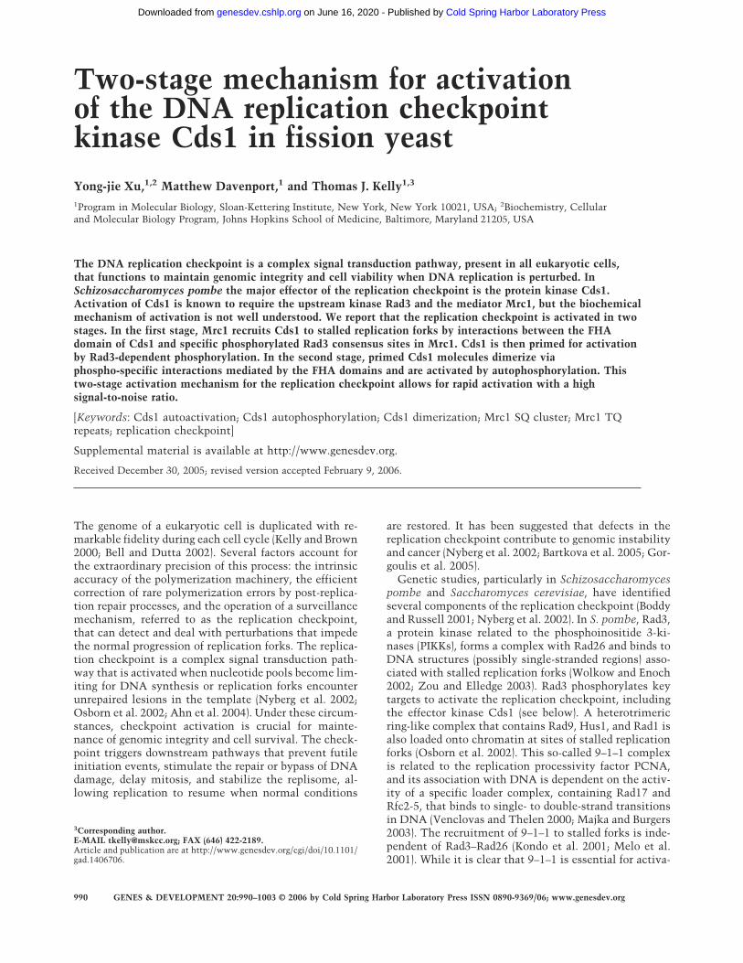
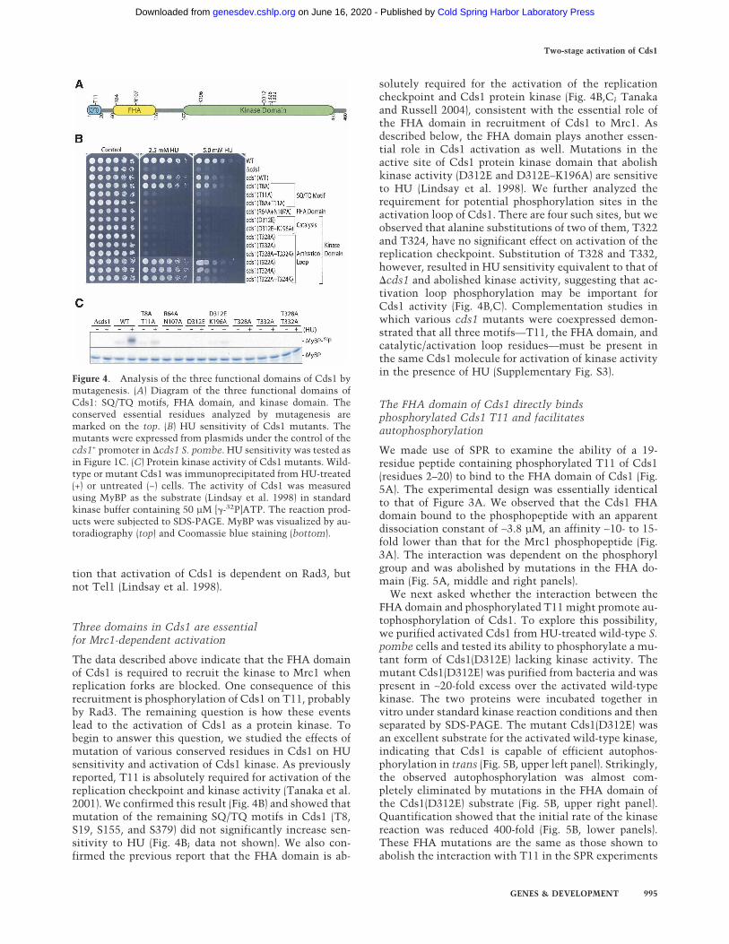
The data described above indicate that the FHA domainof Cds1 is required to recruit the kinase to Mrc1 whenreplication forks are blocked. One consequence of thisrecruitment is phosphorylation of Cds1 on T11, probablyby Rad3. The remaining question is how these eventslead to the activation of Cds1 as a protein kinase. Tobegin to answer this question, we studied the effects ofmutation of various conserved residues in Cds1 on HUsensitivity and activation of Cds1 kinase. As previouslyreported, T11 is absolutely required for activation of thereplication checkpoint and kinase activity (Tanaka et al.2001). We confirmed this result (Fig. 4B) and showed thatmutation of the remaining SQ/TQ motifs in Cds1 (T8,S19, S155, and S379) did not significantly increase sen-sitivity to HU (Fig. 4B; data not shown). We also con-firmed the previous report that the FHA domain is ab-
solutely required for the activation of the replicationcheckpoint and Cds1 protein kinase (Fig. 4B,C; Tanakaand Russell 2004), consistent with the essential role ofthe FHA domain in recruitment of Cds1 to Mrc1. Asdescribed below, the FHA domain plays another essen-tial role in Cds1 activation as well. Mutations in theactive site of Cds1 protein kinase domain that abolishkinase activity (D312E and D312E–K196A) are sensitiveto HU (Lindsay et al. 1998). We further analyzed therequirement for potential phosphorylation sites in theactivation loop of Cds1. There are four such sites, but weobserved that alanine substitutions of two of them, T322and T324, have no significant effect on activation of thereplication checkpoint. Substitution of T328 and T332,however, resulted in HU sensitivity equivalent to that of�cds1 and abolished kinase activity, suggesting that ac-tivation loop phosphorylation may be important forCds1 activity (Fig. 4B,C). Complementation studies inwhich various cds1 mutants were coexpressed demon-strated that all three motifs—T11, the FHA domain, andcatalytic/activation loop residues—must be present inthe same Cds1 molecule for activation of kinase activityin the presence of HU (Supplementary Fig. S3).
The FHA domain of Cds1 directly bindsphosphorylated Cds1 T11 and facilitatesautophosphorylation
We made use of SPR to examine the ability of a 19-residue peptide containing phosphorylated T11 of Cds1(residues 2–20) to bind to the FHA domain of Cds1 (Fig.5A). The experimental design was essentially identicalto that of Figure 3A. We observed that the Cds1 FHAdomain bound to the phosphopeptide with an apparentdissociation constant of ∼3.8 µM, an affinity ∼10- to 15-fold lower than that for the Mrc1 phosphopeptide (Fig.3A). The interaction was dependent on the phosphorylgroup and was abolished by mutations in the FHA do-main (Fig. 5A, middle and right panels).
We next asked whether the interaction between theFHA domain and phosphorylated T11 might promote au-tophosphorylation of Cds1. To explore this possibility,we purified activated Cds1 from HU-treated wild-type S.pombe cells and tested its ability to phosphorylate a mu-tant form of Cds1(D312E) lacking kinase activity. Themutant Cds1(D312E) was purified from bacteria and waspresent in ∼20-fold excess over the activated wild-typekinase. The two proteins were incubated together invitro under standard kinase reaction conditions and thenseparated by SDS-PAGE. The mutant Cds1(D312E) wasan excellent substrate for the activated wild-type kinase,indicating that Cds1 is capable of efficient autophos-phorylation in trans (Fig. 5B, upper left panel). Strikingly,the observed autophosphorylation was almost com-pletely eliminated by mutations in the FHA domain ofthe Cds1(D312E) substrate (Fig. 5B, upper right panel).Quantification showed that the initial rate of the kinasereaction was reduced 400-fold (Fig. 5B, lower panels).These FHA mutations are the same as those shown toabolish the interaction with T11 in the SPR experiments
Figure 4. Analysis of the three functional domains of Cds1 bymutagenesis. (A) Diagram of the three functional domains ofCds1: SQ/TQ motifs, FHA domain, and kinase domain. Theconserved essential residues analyzed by mutagenesis aremarked on the top. (B) HU sensitivity of Cds1 mutants. Themutants were expressed from plasmids under the control of thecds1+ promoter in �cds1 S. pombe. HU sensitivity was tested asin Figure 1C. (C) Protein kinase activity of Cds1 mutants. Wild-type or mutant Cds1 was immunoprecipitated from HU-treated(+) or untreated (−) cells. The activity of Cds1 was measuredusing MyBP as the substrate (Lindsay et al. 1998) in standardkinase buffer containing 50 µM [�-32P]ATP. The reaction prod-ucts were subjected to SDS-PAGE. MyBP was visualized by au-toradiography (top) and Coomassie blue staining (bottom).
Two-stage activation of Cds1
GENES & DEVELOPMENT 995
Cold Spring Harbor Laboratory Press on June 16, 2020 - Published by genesdev.cshlp.orgDownloaded from
(Fig. 5A). Thus, the data indicate that autophosphoryla-tion is strongly facilitated by the intermolecular inter-action between phosphorylated T11 and the FHA do-main.
Since Cds1 can undergo autophosphorylation in trans,an interesting question is whether Cds1 itself can medi-ate T11 phosphorylation. If this were the case, it wouldprovide a possible autoamplification mechanism. How-ever, this possibility seems unlikely since it has beenshown that the kinase-dead Cds1(D312E) mutant under-goes efficient T11 phosphorylation in vivo when cells aretreated with HU (Tanaka et al. 2001), an observation thatwe have confirmed (data not shown). These data indicatethat T11 phosphorylation in vivo is not dependent onCds1 kinase activity and support the view that T11 phos-phorylation is a function of Rad3. We show below thatthe critical targets of autophosphorylation for Cds1 ac-tivation as a protein kinase appear to lie in the catalyticdomain.
Concentration-dependent autoactivation of Cds1in vitro and in vivo
To further understand the mechanism of Cds1 activa-tion, we purified the enzyme in its inactive form from�rad3�tel1 cells. Increasing concentrations of the puri-fied enzyme were incubated with ATP and a constantamount of myelin basic protein (MyBP) as substrate (Fig.6A). Little Cds1 kinase activity was observed when theenzyme concentration was <100 nM. Above this level,kinase activity increased dramatically with concentra-tion and was associated with autophosphorylation (datanot shown). The data were closely fit by a function inwhich kinase activity is proportional to the 1.97 power
of the Cds1 concentration (R2 = 0.996). The striking non-linearity of the rate of substrate phosphorylation withenzyme concentration suggested that autoactivation ofCds1 occurred during the reaction. One possibility, con-firmed below, is that autoactivation was the result ofCds1 autophosphorylation in trans. We have estimatedthe intracellular concentration of Cds1 to be in theneighborhood of 80 nM (Supplementary Fig. S4C). Atthis concentration, the activity of the enzyme would beexpected to be quite low based on the in vitro data (Fig.6A). Dimerization of Cds1 via the interaction betweenphosphorylated T11 and the FHA domain, describedabove, could drive Cds1 activation by increasing the rateof autophosphorylation. The results in Figure 6A suggestthat the normal mechanism of activation can be by-passed by raising the enzyme concentration to nonphysi-ological levels.
These data are consistent with the observation thatexpression of Cds1 at high levels in Escherichia coliyielded active Cds1 (Fig. 6B,D). The activity of Cds1 ki-nase expressed in bacteria was independent of the FHAdomain and T11, but was eliminated by mutations in theactive site (e.g., D312E) or in activation loop residuesT328 or T332. Cds1 purified from E. coli was phosphory-lated (Fig. 7B; data not shown), indicating that the highconcentration of enzyme in bacteria was sufficient todrive autophosphorylation. As shown in Figure 6B, ac-tive Cds1 kinase is lethal to E. coli.
Similarly, active kinase was recovered when Cds1 wasexpressed at very high levels under the control of thenmt1+ promoter in �rad3�mrc1 S. pombe (Supplemen-tary Fig. S4). The presence of active protein kinase in-duced a significant elongation of the cells, indicative ofcell cycle delay (Supplementary Fig. S4A). As in the case
Figure 5. Intermolecular interactions between the FHAdomain and phosphorylated T11 of Cds1 molecules pro-mote autophosphorylation in trans. (A) Analysis of thebinding of the Cds1 FHA domain to a Cds1 peptide con-taining phosphorylated T11 by SPR. SPR measurementswere carried out as in Figure 3A. The immobile phasecontained the Cds1 T11 peptide (biotin-EEPEE-ATQATQEAPLHVSQ, residues 2–20) with or without aphosphate at T11 (underlined). The fluid phase containedGST-FHA of Cds1 at the same concentrations used in theexperiment of Figure 3A. (Left panel) Peptide with aphosphoryl group at T11. (Middle panel) Peptide withoutthe phosphoryl group. (Right panel) Mutant GST-FHA(R64A + N107A). (B) Autophosphorylation of Cds1is dependent on a functional FHA domain. ActivatedCds1 kinase (90 nM) purified from HU-treated S. pombewas mixed with kinase-dead Cds1(D312E, 1875 nM) pu-rified from E. coli as substrate in standard kinase buffercontaining 100 µM [�-32P]ATP. The reaction was incu-bated at 30°C, and aliquots were removed at various in-tervals. (Top panel) The substrate Cds1(D312E) was sepa-rated from the activated Cds1 kinase by SDS-PAGE on an8% gel. (Lower panel) The incorporation of 32P was quan-tified with PhosphorImager. (Right half) A second reac-tion was carried out under identical conditions except that the kinase-dead Cds1 substrate contained R46A and N107A mutations inthe FHA domain.
Xu et al.
996 GENES & DEVELOPMENT
Cold Spring Harbor Laboratory Press on June 16, 2020 - Published by genesdev.cshlp.orgDownloaded from
of E. coli, the observed in vitro kinase activity was inde-pendent of the FHA domain and T11, but was eliminatedby mutations in the active site (e.g., D312E) or in acti-vation loop residues T328 or T332 (Supplementary Fig.S4B).
To verify that the activity of Cds1 activated in vivo isdependent on phosphorylation, we tested the effect ofphosphatase treatment on Cds1 kinase activity (Fig. 6C).Wild-type S. pombe cells were treated with HU, and theactivated Cds1 was purified by binding to HA antibodybeads. Cds1 activity was greatly reduced in samplestreated with phosphatase, but not in mock-treatedsamples or samples treated with phosphatase in the pres-ence of the inhibitor NaVO4.
Induced dimerization of the Cds1 catalytic domainpromotes autophosphorylation and kinase activationin vitro
To directly test the role of dimerization and autophos-phorylation in the activation of Cds1, we made a con-struct encoding a chimeric enzyme consisting of anFK506-binding protein (FKBP) fused to the Cds1 catalyticdomain (residues 150–460, Cds1cat). This fusion con-struct lacks both T11 and the FHA domain, but can bedimerized by addition of the small bifunctional moleculeAP20187 (Ariad) (Fig. 7A; Spencer et al. 1993). The fusionprotein was expressed in E. coli and analyzed by SDS-PAGE after induction for various times. After 30 min ofinduction, the protein migrated as a single band of theexpected mobility (Fig. 7B, left panel). Most of the pro-tein migrated in bands of lower mobility after 90 min ofinduction. These bands were the result of autophos-
phorylation of the fusion protein in E. coli as demon-strated by (1) the absence of shifted bands when a kinase-dead fusion protein (D312E) was expressed under thesame conditions (Fig. 7B, right panel) and (2) the sensi-tivity of the shifted bands to phosphatase (Fig. 7C). Toobtain the unphosphorylated form of the fusion proteinfor in vitro studies, we purified it from E. coli after only30 min of induction and treated with phosphatase toremove any residual phosphoryl groups. When the puri-fied protein was incubated with increasing concentra-tions of AP20187 and analyzed on native PAGE gels, aslower moving band, presumably consisting of the in-duced dimers, was observed (Fig. 7D). This band reachedmaximal intensity at a molar ratio of AP20187 to proteinof ∼0.63 and then decreased in intensity at higher con-centrations of AP20187. In the presence of ATP andMyBP as substrate, both autophosphorylation and en-zyme activity dramatically increased with AP20187 con-centration, also reaching a maximum at a molar ratio of∼0.63 (Fig. 7E, left panels). At concentrations of AP20187>0.63, autophosphorylation and kinase activity rapidlydecreased as expected (Fig. 7A). As a control, the sameexperiments were carried out with a fusion protein con-taining a mutant (D312E) catalytic domain (Fig. 7E, rightpanels). The mutant protein formed dimers, but did notundergo autophosphorylation or activation.
To rule out the unlikely possibility that activation ofthe FKBP-Cds1cat fusion protein was due to a conforma-tional change induced by dimerization rather than auto-phosphorylation, we examined the ATP dependence ofthe activation step in a two-stage reaction (Supplemen-tary Fig. S5). In this experiment the purified fusion pro-tein was first incubated in the presence or absence of
Figure 6. The weak intrinsic kinase ac-tivity of Cds1 is greatly enhanced by au-tophosphorylation. (A) Concentration-de-pendent autoactivation of Cds1 in vitro.Unactivated Cds1 and the kinase-deadCds1 mutant (D312E) were purified from�rad3�tel1 S. pombe and incubated instandard kinase buffer containing MyBPas the substrate. Cds1 kinase activity wasmeasured by 32P incorporation into MyBPand plotted against the concentrations ofCds1. (Solid line, WT) Wild-type Cds1;(dashed line, KD) kinase-dead Cds1 mu-tant D312E. (B) Suppression of growth ofE. coli by Cds1 kinase activity. (Left plate)E. coli strains harboring vectors express-ing wild-type or mutant Cds1 under thecontrol of the lac promoter were grown on
an LB plate in the absence of IPTG. The cells were replicated onto a plate containing IPTG to induce Cds1 expression. (C) The activityof activated Cds1 is dependent on phosphorylation. Cds1 was purified from HU-treated S. pombe by immunoprecipitation withanti-HA antibody beads. The beads were then split into three aliquots; the first aliquot received phosphatase buffer alone, the secondreceived 400 U of �-phosphatase, and the third received the same amount of phosphatase plus 20 mM NaVO4. After an incubation at30°C for 40 min, the reactions were stopped by adding 1.5 mL of ice-cold TBS-T containing 10 mM EDTA and 1 mM NaVO4. Equalamounts of phosphatase and NaVO4 were added to the first and the second aliquots. After washing the beads five times with TBS-Tcontaining 1 mM NaVO4, the kinase activity was examined in standard kinase buffer containing 1 mM NaVO4 and MyBP as thesubstrate. (D) Activities of wild-type and mutant Cds1 expressed in E. coli. Extracts were prepared from the E. coli strains describedin B. Wild-type or mutant Cds1 was purified by immunoprecipitation with anti-HA antibody beads. Cds1 kinase activity was measuredin standard kinase buffer using MyBP as the substrate.
Two-stage activation of Cds1
GENES & DEVELOPMENT 997
Cold Spring Harbor Laboratory Press on June 16, 2020 - Published by genesdev.cshlp.orgDownloaded from
ATP with AP20187 at a molar ratio of 0.63 to inducedimerization. Following the first incubation, a high con-centration of AP20187 was added to promote dissocia-tion of dimers, and enzyme activity was then assayedwith MyBP as substrate. We observed that the activity ofthe enzyme preincubated with ATP (Supplementary Fig.S5, lane 4) was much greater than that of the enzymepreincubated in the absence of ATP (Supplementary Fig.S5, lane 2). Control experiments in which the AP20187concentration was maintained at a molar ratio of 0.63during the second incubation demonstrated that a highconcentration of the compound was effective in prevent-ing dimerization, autophosphorylation, and activation(Supplementary Fig. 5, cf. lanes 1 and 2). Taken as awhole, the data presented in Figures 6 and 7 and Supple-mentary Figure S5 indicate that induced dimerization issufficient to activate Cds1 and that activation is depen-dent on autophosphorylation.
Dimerization activates Cds1 in vivo and causes cellcycle delay
To examine the effects of induced dimerization in vivo,we made plasmid constructs encoding full-length Cds1
with two FKBPs at the N terminus (2xFKBP-Cds1). (TwoFKBP modules were used in order to increase the effi-ciency of induced dimerization.) The fusion protein wasexpressed in �rad3�cds1 S. pombe under the control ofthe cds1+ promoter. In the presence of 1 µM AP20187,cells expressing the fusion construct exhibited an elon-gated phenotype indicative of cell cycle delay (Fig. 8A). Asimilar phenotype has been observed when wild-typeCds1 (Supplementary Fig. S4A) or GST-tagged Cds1(Boddy et al. 1998) is expressed at a very high level in S.pombe. Cell elongation was not observed in the absenceof AP20187 or with untagged Cds1 or with a kinase-deadfusion construct containing the D312E mutation[2xFKBP-Cds1(D312E)]. The 2xFKBP-Cds1 fusion proteinwas immunoprecipitated from cell extracts and assayedfor activity with MyBP as substrate (Fig. 8B). Significantkinase activity was observed in immunoprecipitates of2xFKBP-Cds1 prepared from cells treated with AP20187,but not in those prepared from mock-treated cells northose in immunoprecipitates of 2xFKBP-Cds1(D312E).To ensure that the observed kinase activity was indepen-dent of T11 and the FHA domain of Cds1, we made afusion construct with two FKBP domains fused to the Nterminus of the catalytic domain of the enzyme
Figure 7. Induced homodimerization of the catalyticdomain of Cds1 promotes autophosphorylation and ac-tivation of kinase activity in vitro. (A) Diagram illustrat-ing the induction of dimers of FKBP-Cds1cat fusion pro-tein by the bifunctional dimerizer AP20187. The fusionprotein contains an FKBP domain (black) followed by theCds1 catalytic domain (Cds1cat, residues 150–460,green). AP20187 is shown in blue. Maximum dimeriza-tion occurs at a molar ratio of AP20187 to fusion proteinof 1:2. At significantly lower or higher concentrations ofAP20187, the fusion protein is predominantly mono-meric. (B) Expression of the FKBP-Cds1cat containingwild-type (WT) Cds1 or kinase-dead Cds1 (D312E) cata-lytic domains. Extracts of E. coli growing at 20°C wereprepared at the indicated times after induction and ana-lyzed by SDS-PAGE and Western blotting. (C) The FKBP-Cds1cat fusion protein is phosphorylated in E. coli. Thefusion protein was purified from E. coli extracts after 60min of induction and treated with buffer alone, �-phos-phatase, or �-phosphatase plus 3.3 mM NaVO4 at 30°C.(D) Purified FKBP-Cds1cat (400 nM) containing the wild-type or kinase-dead Cds1(D312E) catalytic domain wasincubated for 30 min on ice with various concentrationsof AP20187 in 10 µL of buffer containing 20 mMTris:HCl (pH 8.0) and 50 mM NaCl. Samples were ana-lyzed by electrophoresis on an 8% native polyacryl-amide gel and stained with silver. (E) Autophosphoryla-tion and activation of Cds1 are stimulated by induceddimerization. Purified FKBP-Cds1cat fusion protein (50nM) was incubated with the same concentrations ofAP20187 as in D in 20 µL of kinase buffer containing 50µM [�-32P]ATP and 5 µg of MyBP. After incubation at30°C for 20 min, the reactions were stopped by adding 5µL of 5× SDS gel loading buffer. The MyBP substrate and the FKBP-Cds1cat fusion protein were separated by a 12% SDS-PAGE. Thetop half of the gel, containing the FKBP-Cds1cat fusion protein, was transferred onto a nitrocellulose membrane for Western blottingand autoradiography. The lower half of the gel, containing the substrate MyBP, was stained with Coomassie blue to visualize MyBP.Incorporation of 32P into MyBP was measured by PhosphorImager. (Right half of figure) As a control, the same experiment wasperformed with a fusion protein containing the Cds1(D312E) mutation in the catalytic domain.
Xu et al.
998 GENES & DEVELOPMENT
Cold Spring Harbor Laboratory Press on June 16, 2020 - Published by genesdev.cshlp.orgDownloaded from
(2xFKBP-Cds1cat). This construct could also be activatedin vivo by treatment with AP20187 (Fig. 8B).
Efficient dimerization and activation of Cds1 in vivorequires Rad3-dependent phosphorylation of bothmonomers
The results described above demonstrate that dimeriza-tion of Cds1 can promote autophosphorylation and acti-vation of the kinase. We have also shown that Cds1dimerization can be driven by the interaction of phos-phorylated T11 of one molecule with the FHA domain ofanother molecule. In principle, a Cds1 dimer could beheld together by either one or two such interactions,
depending on whether one or both partners have under-gone Rad3-dependent phosphorylation on T11. Thesetwo possibilities have different implications for check-point activation in vivo. If a single phosphoT11–FHAinteraction is sufficient to drive dimerization and auto-activation under in vivo conditions, it follows that Rad3-dependent phosphorylation of a few Cds1 moleculesmight be sufficient to activate many unphosphorylatedCds1 molecules, resulting in autoamplification of thecheckpoint signal. On the other hand, if two phos-phoT11–FHA interactions are required for dimerizationand autoactivation, then only Cds1 molecules that havebeen phosphorylated by Rad3 can be activated, so au-toamplification would not be possible. To distinguish
Figure 8. Dimerization of Cds1 activates Cds1 and bypasses the requirement for Rad3 in vivo. (A) Induced dimerization of Cds1causes cell mitotic delay (cdc phenotype). S. pombe �rad3�cds1 cells expressing wild-type Cds1, the full-length Cds1 fused with twotandem FKBP domains at the N terminus (2xFKBP-Cds1), or the 2xFKBP-Cds1(D312E) fusion protein containing an inactivatingmutation in the catalytic domain were grown for 9 h at 30°C in the presence of 1 µM AP20187. The cells were photographed in a phase-contrast microscope. Bar, 30 µm. (B) Induced dimerization activates Cds1 in vivo. S. pombe �rad3�cds1 cells carrying plasmidsexpressing the fusion proteins were incubated with (+) or without (−) 1 µM AP20187 for 9 h at 30°C. The proteins were immunopre-cipitated from cell extracts, and kinase activities were measured in a standard kinase assay using MyBP as substrate. 2xFKBP-cds1 and2xFKBP-cds1(D312E) are described in A. 2xFKBP-cds1cat contains the Cds1 kinase domain (159–460 amino acids) fused to two FKBPsat its N terminus. 1xFKBP-cds1 contains the full-length Cds1 fused to one FKBP at the N terminus. (C) Activated wild-type Cds1kinase cannot activate mutant Cds1 molecules lacking Rad3 phosphorylation sites. Wild-type and various mutant Cds1 molecules(indicated at the top of the figure) were tagged with 6his3myc and coexpressed with HA-tagged wild-type Cds1 in cds1-6his2HA S.pombe. After incubation in the presence (+) or absence (−) of HU, the differentially tagged Cds1 molecules were separately purified byimmunoprecipitation with anti-HA or anti-myc antibody beads. After extensive washing, Cds1 kinase activity retained on the beadswas determined in standard kinase assays with MyBP as the substrate. Samples were analyzed by SDS-PAGE in a 12% gel, and theincorporated 32P was detected by autoradiography (MyBP-32P). The upper half of the figure shows the kinase activity of the myc-taggedCds1, while the lower half shows the kinase activity of the endogenous HA-tagged Cds1. The immunoprecipitated Cds1 protein ineach sample was detected by Western blotting (Cds1-3myc or Cds1-2HA), and the MyBP substrate was detected by Coomassie bluestaining (MyBP). (D) Model for the two-stage activation of Cds1. Mrc1 moves along with the replisome under normal conditions (Katouet al. 2003; Osborn and Elledge 2003; Zhao and Russell 2004). When replication forks are stalled, Rad3 is recruited to the stalled forksand phosphorylates the TQ repeats of Mrc1. Either one of the two phosphorylated TQ repeats of Mrc1 can recruit an inactive Cds1 bybinding to its FHA domain. Rad3 then phosphorylates T11 of the recruited Cds1, priming Cds1 for autoactivation. The phosphorylatedT11 of the primed Cds1 binds to another primed Cds1 molecule, bringing the two inactive kinase domains in close proximity. In thepresence of ATP, the dimerized kinase domains can autophosphorylate each other in trans, thus activating Cds1.
Two-stage activation of Cds1
GENES & DEVELOPMENT 999
Cold Spring Harbor Laboratory Press on June 16, 2020 - Published by genesdev.cshlp.orgDownloaded from
between these possibilities, we examined the ability ofwild-type Cds1 to activate a mutant form of Cds1 (T8A–T11A) that cannot be phosphorylated by Rad3 (Fig. 8C).For this purpose, wild-type or mutant Cds1 genes (taggedwith myc) were coexpressed with an endogenous wild-type Cds1 gene (tagged with HA). The cells were treatedwith HU and the differentially tagged Cds1 moleculeswere separately purified by immunoprecipitation withthe appropriate antibodies. The kinase activities of thepurified proteins were measured in the standard assaywith MyBP as substrate. In all experiments the endog-enous Cds1-HA exhibited robust activation followingHU treatment (Fig. 8C, lower half). When wild-typeCds1-myc was coexpressed in the same cell, it too ex-hibited HU-dependent activation (Fig. 8C, upper panel,lanes 1,2). However, when the coexpressed Cds1-myccontained mutations (T8A–T11A) that prevent Rad3-de-pendent phosphorylation, only slight activation of themutant kinase was observed (Fig. 8C, upper panel, lanes3,4). As expected, Cds1 with inactivating mutations inthe FHA domain (R64A–N107A) or the catalytic domain(D312E) was not activated following HU treatment (Fig.8C, upper panel, lanes 5–8). These data indicate that ac-tive Cds1 that has been phosphorylated by Rad3 cannotefficiently activate unphosphorylated Cds1. Thus, underphysiological conditions, it appears that Cds1 dimeriza-tion that leads to kinase activation requires that bothpartners have undergone Rad3-dependent phosphoryla-tion on T11. This requirement for two independentphosphorylation events may serve to increase the noiseimmunity of the replication checkpoint and is consis-tent with the observation that Rad3 is required for ini-tiation and maintenance of the checkpoint signaling dur-ing HU block (Martinho et al. 1998).
Discussion
We have shown that activation of the replication check-point in S. pombe occurs in two stages (Fig. 8D). In thefirst stage, the effector kinase Cds1 is primed for auto-activation. A key step in this process is the Rad3-depen-dent phosphorylation of the adaptor Mrc1 at sites in theTQ repeats and the SQ cluster. The phosphorylated TQrepeats function as specific docking sites for the FHAdomain of Cds1, recruiting the latter to the vicinity ofRad3. This interaction is facilitated in an unknown fash-ion by the phosphorylated SQ motifs. Once bound tophospho-Mrc1, Cds1 undergoes Rad3-dependent phos-phorylation at T11. In the second stage, Cds1 is activatedvia autophosphorylation, probably in the activation loop.This process is dependent on dimerization of Cds1 viainteractions between phosphorylated T11 motifs and theFHA domains.
In our model, Mrc1 functions only in the first stage ofcheckpoint activation and is not required for Cds1 auto-activation. In this view, autophosphorylation and acti-vation of Cds1 occur after dissociation from Mrc1. Whilethis is the simplest version of the model and consistentwith all the data, we cannot rule out the possibility that
Mrc1 may facilitate autoactivation via additional inter-actions with Cds1.
The model in Figure 8D is consistent with the speci-ficity and noise immunity required for the replicationcheckpoint. Specificity is provided by the requirementfor two different protein–protein interactions to activateCds1: the phospho-Mrc1–Cds1 interaction in the firststage and the phospho-Cds1–phospho-Cds1 interactionin the second stage. The noise immunity of the activa-tion reaction derives from the fact that autophosphory-lation of Cds1 proceeds at a very low rate in the absenceof dimerization under physiological conditions (Fig. 5).Thus, little autophosphorylation/activation can takeplace without the priming reaction.
We have shown that the priming reaction depends onan interaction between the FHA domain of Cds1 and oneof two redundant phosphopeptide repeats in Mrc1. Thespecificity of binding likely depends on additional inter-actions besides the FHA–phosphothreonine contact.Consistent with previous studies using synthetic peptidelibraries (Durocher et al. 2000), we have demonstratedthe importance of the aspartate at the +3 position of theMrc1 TQ repeats in the activation of the replicationcheckpoint. The Cds1-docking repeats on Mrc1 de-scribed here have not been identified for Mrc1 or Rad9 ofbudding yeast and are clearly different from those ofXenopus Claspin. The Chk1-binding motifs on XenopusClaspin are probably not phosphorylated by the Rad3-like kinases ATR and ATM (Kumagai and Dunphy 2003).We have also shown that phosphorylation of the SQ clus-ter in Mrc1 facilitates the recruitment of Cds1. Since aphosphorylated SQ motif does not have significant affin-ity for the FHA domain of Cds1 or full-length Cds1, theeffect of the SQ motifs is likely indirect. One possibilityis that phosphorylation of the SQ cluster induces a con-formational change in Mrc1 that permits more efficientbinding or phosphorylation of Cds1 by Rad3.
Our data provide strong evidence that primed Cds1 isactivated via dimerization and autophosphorylation ofresidues in the catalytic domain. First, the activity ofCds1 purified from HU-treated S. pombe is completelydependent on phosphorylation. Second, at high proteinconcentration (micromolar or greater), autoactivation(and autophosphorylation) of highly purified Cds1 occursin vitro in the absence of other factors. Third, autoacti-vation of Cds1 can be induced in vitro and in vivo byenforced dimerization of a fusion construct lacking bothT11 and the FHA domain. Fourth, autoactivation of theCds1 fusion construct requires both dimerization andthe presence of ATP. It is likely that the critical targetsof autophosphorylation reside in the activation loop ofCds1 (Nolen et al. 2004). Cds1 and HsChk2 have se-quence similarities to protein kinases that require phos-phorylation of the activation loop for full activity. Mu-tations that eliminate potential phosphorylation sites inthe activation loop of Cds1 (T328 and T332) prevent ac-tivation of the kinase. These two sites correspond to resi-dues T383 and T387 in HsChk2 that have been shown tobe essential for kinase activity (Lee and Chung 2001).
The adaptor Mrc1 is the key factor in the S. pombe
Xu et al.
1000 GENES & DEVELOPMENT
Cold Spring Harbor Laboratory Press on June 16, 2020 - Published by genesdev.cshlp.orgDownloaded from
replication checkpoint pathway, ensuring that cells re-spond to replication blocks and activate the correctdownstream effector, Cds1. There is evidence in S. cer-evisiae that Mrc1 may be a component of the replisomeand travel along DNA with the chain elongation ma-chinery (Katou et al. 2003; Osborn and Elledge 2003).However, this has not yet been confirmed in other sys-tems. How cells recognize stalled replication forks is notclear. One possibility is that uncoupling of DNA un-winding from DNA synthesis may occur when nucleo-tides are scarce or the fork encounters an unrepaired le-sion. Uncoupling may result in the generation of long-lived structures containing single- to double-strandtransitions that can bind Rad3–Rad26 and trigger theloading of the 9–1–1 complex (Bermudez et al. 2003; El-lison and Stillman 2003; Majka and Burgers 2003; Zouand Elledge 2003; Byun et al. 2005). In this scenario, thesimple proximity of Rad3 to Mrc1 at stalled forks resultsin Mrc1 phosphorylation and initiation of the check-point cascade. Alternatively, Mrc1 may play some moredirect role in recognition of stalled replication forks assuggested by studies in S. cerevisiae. Further work willbe required to distinguish these possibilities.
Besides serving as an adaptor in the replication check-point, Mrc1 may have additional roles in DNA replica-tion. In S. cerevisiae (Osborn and Elledge 2003), deletionof Mrc1 results in greater sensitivity to acute replicationblocks and DNA damage than elimination of the Mrc1TQ and SQ motifs. We have observed a similar phenom-enon in S. pombe (Supplementary Fig. S2). Thus, activa-tion of the effector kinase Cds1 may not be the onlymeans by which Mrc1 protects cells from the effects ofperturbations of DNA replication.
The role of Rad9 in the activation of S. cerevisiaeRad53 in response to DNA damage may be similar to therole of Mrc1 in the replication checkpoint describedhere. Earlier work had suggested that multimeric Rad9complexes recruit Rad53 molecules, thus increasing thelocal concentration and allowing Rad53 autophosphory-lation in trans (Gilbert et al. 2001). This model is quitedifferent from our model for Mrc1 function. Recentwork, however, is consistent with the alternative hy-pothesis that a major function of Rad9 is to recruitRad53, enabling its phosphorylation and activation byMec1 (Sweeney et al. 2005). The activity of Rad9, likethat of Mrc1, appears to require an interaction withRad53 that is dependent on phosphorylation and theRad53 FHA domains. It is not yet understood in detailhow Mec1 phosphorylation of Rad53 leads to its activa-tion. In the case of Cds1 it is clear that Rad3 phosphory-lation on T11 is not sufficient for activation, since asecond Rad3-independent phosphorylation of the cata-lytic domain is also required. Our data show that Mrc1-dependent phosphorylation of T11 by Rad3 acts indi-rectly by promoting dimerization and autophosphoryla-tion of Cds1. The same may be true for Rad9-mediatedMec1 phosphorylation of Rad53.
As noted above, there are significant similarities be-tween the activation of Cds1 and the activation of Hs-Chk2. There is good evidence that HsChk2 is activated
via autophosphorylation of activation loop residues andthat this process is mediated via HsChk2 oligomeriza-tion (Ahn and Prives 2002; Ahn et al. 2002; Xu et al.2002). Like Cds1, both N-terminal TQ residues and theFHA domain of Chk2 are required for activation, but atpresent the adaptor mediating activation is unknown.
A major unsolved problem is the identification of thecritical targets of the replication checkpoint. Checkpointactivation has several effects during S phase, includinginhibition of initiation of DNA replication, stabilizationof stalled replication forks, and activation of repair/tol-erance pathways. It is not yet completely clear whatCds1 substrates mediate these effects. In S. pombe, Hsk1and Dfp1, the homologs of S. cerevisiae Cdc7 and itsregulatory subunit Dbf4, are substrates for Cds1 in vitroand in vivo (Snaith et al. 2000; Duncker and Brown2003). Since Cdc7 kinase is absolutely required for ini-tiation of DNA replication, its phosphorylation maycontribute to the suppression of futile initiation events.The nuclease Mus81 and the recombinational repair fac-tor Rad60 have also been identified as targets of Cds1-dependent phosphorylation, and both have been shownto play a role in tolerance to perturbations of DNA rep-lication (Boddy et al. 2001, 2003). It seems likely thatadditional targets of Cds1 that contribute to stabilizationof the replisome remain to be identified.
Materials and methods
Growth of S. pombe strains and preparation of growth mediafollowed standard methods (Moreno et al. 1991). The followingstrains were generated for this study: YJ15, �mrc1�ura4+; YJ66,�mrc1�ura4+ cds1-2HA6his�ura4+; YJ293, �mrc1�ura4+
�rad3�ura4+; YJ294, �mrc1�ura4+ �tel1�ura4+; YJ374, cds1-6his2HA�ura4+. All strains contain the auxotrophic markersleu1-32, ura4-D18, ade6-M210, or ade6-M216.
Point mutations of Mrc1 and Cds1 were made by Quick-Change mutagenesis PCR. To examine drug sensitivity, 2 × 10⁷
cells/mL of logarithmically growing S. pombe were diluted infivefold steps and spotted onto YE6S or EMM6S plates that con-tained HU or MMS. The plates were incubated at 30°C for 3 d.
Cds1 was purified as described in the Supplemental Material,and Cds1 kinase assays were performed in standard kinasebuffer (20 mM Tris:HCl at pH 7.5, 5 mM MgCl2, 1 mM DTT, 75mM KCl, 50 µM [�-32P]ATP) with GST-Wee1(1–152 amino ac-ids) (Boddy et al. 1998) or MyBP (Lindsay et al. 1998) as sub-strates.
Other methods used in the study such as SPR, Western, Far-Western, and purification of Cds1 are described in detail in theSupplemental Material.
Acknowledgments
We thank the members of the Kelly laboratory for many usefulsuggestions and J. Petrini and T. Usui for sharing preliminaryresults. We also thank K. Marians and B. Lang for advice onBIAcore analysis; M. Pletcher for help with RT–PCR analysis;and J. Boeke, C. Greider, and J. Corden for advice and sugges-tions. The Micro-Chemistry, DNA Sequencing, and Flow Cy-tometry, core facilities of Sloan-Kettering Institute, providedoutstanding technical support for this work. We gratefully ac-knowledge T. Carr, P. Russell, G. Brown, S. Forsburg, and F.
Two-stage activation of Cds1
GENES & DEVELOPMENT 1001
Cold Spring Harbor Laboratory Press on June 16, 2020 - Published by genesdev.cshlp.orgDownloaded from
Ishikawa for yeast strains and plasmids and Ariad Pharmaceu-ticals, Inc., for the ARGENT Regulated Homodimerization Kit(http://www.ariad.com/regulationkits).
References
Ahn, J. and Prives, C. 2002. Checkpoint kinase 2 (Chk2) mono-mers or dimers phosphorylate Cdc25C after DNA damageregardless of threonine 68 phosphorylation. J. Biol. Chem.277: 48418–48426.
Ahn, J.Y., Li, X., Davis, H.L., and Canman, C.E. 2002. Phos-phorylation of threonine 68 promotes oligomerization andautophosphorylation of the Chk2 protein kinase via the fork-head-associated domain. J. Biol. Chem. 277: 19389–19395.
Ahn, J., Urist, M., and Prives, C. 2004. The Chk2 protein kinase.DNA Repair (Amst.) 3: 1039–1047.
Alcasabas, A.A., Osborn, A.J., Bachant, J., Hu, F., Werler, P.J.,Bousset, K., Furuya, K., Diffley, J.F., Carr, A.M., and Elledge,S.J. 2001. Mrc1 transduces signals of DNA replication stressto activate Rad53. Nat. Cell Biol. 3: 958–965.
Bartkova, J., Horejsi, Z., Koed, K., Kramer, A., Tort, F., Zieger,K., Guldberg, P., Sehested, M., Nesland, J.M., Lukas, C., et al.2005. DNA damage response as a candidate anti-cancer bar-rier in early human tumorigenesis. Nature 434: 864–870.
Bell, S.P. and Dutta, A. 2002. DNA replication in eukaryoticcells. Annu. Rev. Biochem. 71: 333–374.
Bermudez, V.P., Lindsey-Boltz, L.A., Cesare, A.J., Maniwa, Y.,Griffith, J.D., Hurwitz, J., and Sancar, A. 2003. Loading of thehuman 9–1–1 checkpoint complex onto DNA by the check-point clamp loader hRad17-replication factor C complex invitro. Proc. Natl. Acad. Sci. 100: 1633–1638.
Boddy, M.N. and Russell, P. 2001. DNA replication checkpoint.Curr. Biol. 11: R953–R956.
Boddy, M.N., Furnari, B., Mondesert, O., and Russell, P. 1998.Replication checkpoint enforced by kinases Cds1 and Chk1.Science 280: 909–912.
Boddy, M.N., Gaillard, P.H., McDonald, W.H., Shanahan, P.,Yates III, J.R., and Russell, P. 2001. Mus81-Eme1 are essen-tial components of a Holliday junction resolvase. Cell 107:537–548.
Boddy, M.N., Shanahan, P., McDonald, W.H., Lopez-Girona, A.,Noguchi, E., Yates, I.J., and Russell, P. 2003. Replicationcheckpoint kinase Cds1 regulates recombinational repairprotein Rad60. Mol. Cell. Biol. 23: 5939–5946.
Byun, T.S., Pacek, M., Yee, M.C., Walter, J.C., and Cimprich,K.A. 2005. Functional uncoupling of MCM helicase andDNA polymerase activities activates the ATR-dependentcheckpoint. Genes & Dev. 19: 1040–1052.
Duncker, B.P. and Brown, G.W. 2003. Cdc7 kinases (DDKs) andcheckpoint responses: Lessons from two yeasts. Mutat. Res.532: 21–27.
Durocher, D. and Jackson, S.P. 2002. The FHA domain. FEBSLett. 513: 58–66.
Durocher, D., Henckel, J., Fersht, A.R., and Jackson, S.P. 1999.The FHA domain is a modular phosphopeptide recognitionmotif. Mol. Cell 4: 387–394.
Durocher, D., Taylor, I.A., Sarbassova, D., Haire, L.F., Westcott,S.L., Jackson, S.P., Smerdon, S.J., and Yaffe, M.B. 2000. Themolecular basis of FHA domain:phosphopeptide bindingspecificity and implications for phospho-dependent signal-ing mechanisms. Mol. Cell 6: 1169–1182.
Ellison, V. and Stillman, B. 2003. Biochemical characterizationof DNA damage checkpoint complexes: Clamp loader andclamp complexes with specificity for 5� recessed DNA. PLoSBiol. 1: E33.
Furuya, K. and Carr, A.M. 2003. DNA checkpoints in fissionyeast. J. Cell Sci. 116: 3847–3848.
Gilbert, C.S., Green, C.M., and Lowndes, N.F. 2001. Buddingyeast Rad9 is an ATP-dependent Rad53 activating machine.Mol. Cell 8: 129–136.
Gorgoulis, V.G., Vassiliou, L.V., Karakaidos, P., Zacharatos, P.,Kotsinas, A., Liloglou, T., Venere, M., Ditullio, R.A., Kastri-nakis, N.G., Levy, B., et al. 2005. Activation of the DNAdamage checkpoint and genomic instability in human pre-cancerous lesions. Nature 434: 907–913.
Katou, Y., Kanoh, Y., Bando, M., Noguchi, H., Tanaka, H., Ashi-kari, T., Sugimoto, K., and Shirahige, K. 2003. S-phase check-point proteins Tof1 and Mrc1 form a stable replication-paus-ing complex. Nature 424: 1078–1083.
Kelly, T.J. and Brown, G.W. 2000. Regulation of chromosomereplication. Annu. Rev. Biochem. 69: 829–880.
Kondo, T., Wakayama, T., Naiki, T., Matsumoto, K., and Sugi-moto, K. 2001. Recruitment of Mec1 and Ddc1 checkpointproteins to double-strand breaks through distinct mecha-nisms. Science 294: 867–870.
Kumagai, A. and Dunphy, W.G. 2003. Repeated phosphopeptidemotifs in Claspin mediate the regulated binding of Chk1.Nat. Cell Biol. 5: 161–165.
Lee, C.H. and Chung, J.H. 2001. The hCds1 (Chk2)-FHA domainis essential for a chain of phosphorylation events on hCds1that is induced by ionizing radiation. J. Biol. Chem. 276:30537–30541.
Lindsay, H.D., Griffiths, D.J., Edwards, R.J., Christensen, P.U.,Murray, J.M., Osman, F., Walworth, N., and Carr, A.M.1998. S-phase-specific activation of Cds1 kinase defines asubpathway of the checkpoint response in Schizosaccharo-myces pombe. Genes & Dev. 12: 382–395.
Lowndes, N.F., McInerny, C.J., Johnson, A.L., Fantes, P.A., andJohnston, L.H. 1992. Control of DNA synthesis genes in fis-sion yeast by the cell-cycle gene cdc10+. Nature 355: 449–453.
Majka, J. and Burgers, P.M. 2003. Yeast Rad17/Mec3/Ddc1: Asliding clamp for the DNA damage checkpoint. Proc. Natl.Acad. Sci. 100: 2249–2254.
Martinho, R.G., Lindsay, H.D., Flaggs, G., DeMaggio, A.J.,Hoekstra, M.F., Carr, A.M., and Bentley, N.J. 1998. Analysisof Rad3 and Chk1 protein kinases defines different check-point responses. EMBO J. 17: 7239–7249.
Melo, J.A., Cohen, J., and Toczyski, D.P. 2001. Two checkpointcomplexes are independently recruited to sites of DNA dam-age in vivo. Genes & Dev. 15: 2809–2821.
Moreno, S., Klar, A., and Nurse, P. 1991. Molecular geneticanalysis of fission yeast Schizosaccharomyces pombe. Meth-ods Enzymol. 194: 795–823.
Nolen, B., Taylor, S., and Ghosh, G. 2004. Regulation of proteinkinases; Controlling activity through activation segmentconformation. Mol. Cell 15: 661–675.
Nyberg, K.A., Michelson, R.J., Putnam, C.W., and Weinert, T.A.2002. Toward maintaining the genome: DNA damage andreplication checkpoints. Annu. Rev. Genet. 36: 617–656.
Osborn, A.J. and Elledge, S.J. 2003. Mrc1 is a replication forkcomponent whose phosphorylation in response to DNA rep-lication stress activates Rad53. Genes & Dev. 17: 1755–1767.
Osborn, A.J., Elledge, S.J., and Zou, L. 2002. Checking the fork:The DNA-replication stress-response pathway. Trends CellBiol. 12: 509–516.
Snaith, H.A., Brown, G.W., and Forsburg, S.L. 2000. Schizosac-charomyces pombe Hsk1p is a potential cds1p target re-quired for genome integrity. Mol. Cell. Biol. 20: 7922–7932.
Spencer, D.M., Wandless, T.J., Schreiber, S.L., and Crabtree,
Xu et al.
1002 GENES & DEVELOPMENT
Cold Spring Harbor Laboratory Press on June 16, 2020 - Published by genesdev.cshlp.orgDownloaded from
D.R. 1993. Controlling signal transduction with syntheticligands. Science 262: 1019–1024.
Sweeney, F.D., Yang, F., Chi, A., Shabanowitz, J., Hunt, D.F.,and Durocher, D. 2005. Saccharomyces cerevisiae Rad9 actsas a Mec1 adaptor to allow Rad53 activation. Curr. Biol. 15:1364–1375.
Tanaka, K. and Russell, P. 2001. Mrc1 channels the DNA rep-lication arrest signal to checkpoint kinase Cds1. Nat. CellBiol. 3: 966–972.
———. 2004. Cds1 phosphorylation by Rad3–Rad26 kinase ismediated by Forkhead-associated domain interaction withMrc1. J. Biol. Chem. 279: 32079–32086.
Tanaka, K., Boddy, M.N., Chen, X.B., McGowan, C.H., and Rus-sell, P. 2001. Threonine-11, phosphorylated by Rad3 and atmin vitro, is required for activation of fission yeast checkpointkinase Cds1. Mol. Cell. Biol. 21: 3398–3404.
Venclovas, C. and Thelen, M.P. 2000. Structure-based predic-tions of Rad1, Rad9, Hus1 and Rad17 participation in slidingclamp and clamp-loading complexes. Nucleic Acids Res. 28:2481–2493.
Wolkow, T.D. and Enoch, T. 2002. Fission yeast Rad26 is aregulatory subunit of the Rad3 checkpoint kinase. Mol. Biol.Cell 13: 480–492.
Zhao, H. and Russell, P. 2004. DNA binding domain in thereplication checkpoint protein Mrc1 of Schizosaccharomy-ces pombe. J. Biol. Chem. 279: 53023–53027.
Zhao, H., Tanaka, K., Nogochi, E., Nogochi, C., and Russell, P.2003. Replication checkpoint protein Mrc1 is regulated byRad3 and Tel1 in fission yeast. Mol. Cell. Biol. 23: 8395–8403.
Zou, L. and Elledge, S.J. 2003. Sensing DNA damage throughATRIP recognition of RPA–ssDNA complexes. Science 300:1542–1548.
Two-stage activation of Cds1
GENES & DEVELOPMENT 1003
Cold Spring Harbor Laboratory Press on June 16, 2020 - Published by genesdev.cshlp.orgDownloaded from


![Supporting Information Hydrogen-Activation Mechanism of [Fe ... · Supporting Information Hydrogen-Activation Mechanism of [Fe] Hydrogenase Revealed by Multi-Scale Modeling Arndt](https://static.documents.pub/doc/80x56/5e3f9350c529c40668584cef/supporting-information-hydrogen-activation-mechanism-of-fe-supporting-information.jpg)


