Type II DNA Topoisomerases: Enzymes That Can Unknot a Topologically Knotted DNA Molecule via a Reversible Double-Strand Break
Leroy F. Liu, Chung-Cheng Liu and Bruce M. Alberts Department of Biochemistry and Biophysics University of California, San Francisco San Francisco, California 94143
Summary
The T4 DNA topoisomerase is a recently discovered multisubunit protein that appears to have an essen- tial role in the initiation of T4 bacteriophage DNA replication. Treatment of double-stranded circular DNA with large amounts of this topoisomerase in the absence of ATP yields new DNA species which are knotted topological isomers of the double- stranded DNA circle. These knotted DNA circles, whether covalenUy closed or nicked, are converted to unknotted circles by treatment with trace amounts of the T4 topoisomerase in the presence of ATP. Very similar ATP-dependent enzyme activi- ties capable of unknotting DNA are present in ex- tracts of Drosophila eggs, Xenopus laevis eggs and mammalian tissue culture cells. The procaryotic enzyme, DNA gyrase, is also capable of unknotting DNA. We propose that these unknotUng enzymes constitute a new general class of DNA topoisomer- ases (type II DNA topoisomerases). These enzymes must act via mechanisms that involve the concerted cleavage and rejoining of two opposite DNA strands, such that the DNA double helix is tran- siently broken. The passage of a second double- stranded DNA segment through this reversible dou- ble-strand break results in a variety of DNA topoiso- merization reactions, including relaxation:super- coiling; knotting:unknotting and catenaUon:de- catenation. In support of this type of mechanism, we demonstrate that the T4 DNA topoisomerase changes the linking number of a covalenUy closed double-stranded circular DNA molecule only by mul- tiples of two. We discuss the possible roles of such enzymes in a variety of biological functions, along with their probable molecular mechanisms.
Introduction
DNA toposiomerases are enzymes that catalyze the concerted breaking and rejoining of DNA backbone bonds (Wang and Liu, 1979); these reactions can interconvert the different topological isomers of cir- cular DNA (Wang, 1971 ; Liu, Depew and Wang, 1976; Champoux, 1977; Kirkegaard and Wang, 1978). A variety of different DNA topoisomerases have been isolated from procaryotic and eucaryotic organisms (for review see Wang and Liu, 1979). Examples of such DNA topoisomerases are the E. coli ~ protein (Eco DNA topoisomerase I) (Wang, 1971 ), eucaryotic
nicking-closing enzymes (Champoux and Dulbecco, 1972; Baase and Wang, 1974; Keller and Wendel, 1975), procaryotic DNA gyrases (procaryotic DNA topoisomerase II) (Gellert et al., 1976a; Liu and Wang, 1978a; Peebles et al., 1979) and the int gene product of bacteriophage lambda (Kikuchi and Nash, 1979). It has been suggested that DNA topoisomerases par- ticipate in a variety of biological processes involving DNA metabolism. These processes include DNA rep- lication (Wang, 1971; Champoux and Dulbecco, 1972; Gellert et al., 1976b; Sumida-Yasumoto and Hurwitz, 1977), RNA transcription (Wang, 1973; Smith, Kubo and Imamoto, 1978), genetic recombi- nation (Champoux, 1977; Kirkegaard and Wang, 1978; Kikuchi and Nash, 1979), chromosome con- densation and decondensation (Baase and Wang, 1974; Bauer et al., 1977), nucleosome assembly (Germond et al., 1979), virus encapsidation (Bauer et al., 1977) and DNA transposition (Shapiro, 1979).
A DNA topoisomerase from bacteriophage T4-in- fected E. coli cells has recently been isolated and characterized (Liu, Liu and Alberts, 1979; Stetler, King and Huang, 1979). The T4 DNA topoisomerase catalyzes the relaxation of superhelical DNAs (whether positively or negatively coiled) in a reaction requiring ATP hydrolysis. The purified enzyme con- tains multiple subunits, which are apparently coded for by T4 genes 39, 52 and 60. Genetic and biochem- ical studies of mutants in these genes indicate that the T4 DNA topoisomerase may be involved in the initia- tion of DNA replication forks (reviewed by Liu et al., 1979). Interestingly, some host reaction involving E. coli DNA gyrase can substitute partially for the T4 DNA topoisomerase function in vivo (McCarthy, 1979). For this and other reasons, we have tentatively proposed that the T4 DNA topoisomerase is an origin- specific DNA gyrase which catalyzes the ATP-de- pendent negative supercoiling of DNA at T4 replica- tion origins (Liu et alo, 1979).
In this communication, we first provide detailed evidence for our previous report (Liu et al., 1979) that the T4 DNA topoisomerase acts by catalyzing the concerted breaking and rejoining of both strands of the DNA double helix simultaneously. We then dem- onstrate that enzymes capable of completely breaking the double helix in a reversible way are widespread throughout nature, and propose that these enzymes be denoted as "type II DNA topoisomerases."
Results
Formation of a New DNA Species by T4 DNA Topoisomerase Treatment The T4 DNA topoisomerase has been shown to require ATP in order to relax superhelical DNA circles (Liu et al., 1979; Stetler et al., 1979). In the absence of ATP, a very low level of relaxation activity can be detected
Cell 698
when an equal weight of the T4 DNA topoisomerase is incubated with superhelical DNAs. The DNA mole- cules produced in this case, however, include some new DNA species quite different from the expected partially relaxed, covalently closed DNA product. For example, in Figure 1, the time course of such a reaction with pBR322 DNA in the absence of ATP has been monitored by electrophoresis of the DNA prod- ucts through an agarose gel; unexpectedly, some of the covalently closed (form I) DNA substrate in lane A is converted initially into DNA molecules that migrate slightly faster than the form I DNA in the agarose gel (lanes B, C and D). This means that these DNA mole- cules must be either smaller or more compact than the original supercoiled DNA circle used as substrate. Although this unusually fast-migrating DNA can sub- sequently be converted into a group of slower moving DNA bands by the relaxation of supercoils (lane E and F; see below), even these contain minor species that migrate at a rate different from any of the DNA bands present in a partially relaxed pBR322 DNA sample run in parallel as a control (lane G).
The electrophoretic mobilities of the fast-migrating new DNA species generated by the T4 DNA topoiso- merase treatment (Figure 1, lanes B, C and D; more
A B C D E F G
Dimer II
Figure 1. Electrophoretic Analy§is of the Formation of New DNA Species by Treatment of Superhelical DNA Circles with T4 DNA Topoisomerase in the Absence of ATP A reaction mixture (175 FI) containing 50 mM Tris-HCI (pH 7.4), 45 mM KCI, 7.5 mM MgCI2, 0.4 mM dithiothreitol, 0.5 mM Na3EDTA, 25 /~g/ml human serum albumin, 16/~g/ml superhelical (form I) pBR322 DNA (a 4362 bp, covalently closed, double-stranded DNA circle) and 22 Fg/ml T4 DNA topoisomerase was incubated at 30°C. Aliquots of 20 /d were withdrawn at various times and each was stopped by addition of 5 /d of 5% sodium dodecylsulfate (SDS), 50% glycerol plus 1 mg/ml bromophenol blue, followed by electrophoresis through a 0.8% agarose gel in MgTBE buffer as described previously (Liu et al., 1979). (A) Zero time; (B) 50 sec; (C) 1.5 min; (D) 5 rain; (E) 15 min; (F) 30 min; (G) partially relaxed normal form I pBR322 DNA control. After staining with ethidium bromide, DNA was visualized by photographing the wet gel fluorescence under ultraviolet illumination (Liu et al,, 1979). Note that this particular gel did not run evenly: the form I DNA migrated faster on the left than on the right.
clearly visible in Figure 8A, lane B) are unaffected by phenol extraction, 1% SDS treatment, or a combined 30 min proteinase K (100 #g/ml) and 1% SDS treat- ment at 60°C (data not shown). Moreover, analytical ultracentrifugation revealed no difference between the buoyant densities of the topoisomerase product DNAs and the original DNA substrate (_+0.001 g cm-3). It therefore seemed highly improbable that residual pro- tein binding was involved in creating any of the new fast-migrating DNA species shown in Figure 1. In addition, the unusual migration rates observed for these new DNA species are unchanged when the DNA is annealed at temperatures just below the Tm. Thus the apparent conformational change that has oc- curred must involve more than just hydrogen bonding rearrangements.
Several other possibilities could explain the peculiar mobilities of the new DNA species: - - t he superhelical DNA molecules could have be- come intramolecularly cross-linked due to an un- known type of covalent linkage across each DNA circle; - - t he superhelical DNA molecules might have be- come intramolecularly base-paired, which would then allow partially homologous paired strands to become intertwined topologically via a type of topoisomerase reaction known to be carried out by both the E. coli
protein (Kirkegaard and Wang, 1978) and the rat liver nicking-closing enzyme (Champoux, 1977); - - t he superhetical DNA could have become topolog- ically knotted, for example, into a form resembling a pretzel).
Each of these three possibilities could result in a further compaction of superhelical DNA circles and thus explain the increased mobility of this DNA ob- served by agarose gel electrophoresis. To help distin- guish among them, the early topoisomerase product DNA containing the fast-migrating DNA species (Fig- ure 2A) was cut with several different restriction en- donucleases. If these DNA molecules were either in- tramolecularly cross-linked (first possibility, above) or intertwined topologically through a partial DNA se- quence homology (second possibility, above), restric- tion nuclease treatment should generate linear DNA fragments which migrate differently upon agarose gel electrophoresis than the same restriction fragments derived from the original DNA substrate. When Eco RI restriction nuclease was used to digest the new to- poisomerase-generated DNA species, however, all of the DNA product migrated in an agarose gel with exactly the same mobility as the unit-length linear DNA molecules produced by digestion of the control DNA (compare Figures 2C and 2D). Consequently, this information rules out the first two possibilities mentioned above. Furthermore, Hinc II restriction en- donuclease treatment produced comparable yields of the same two DNA fragments from both topoisomer- ase-treated and control DNA samples (compare Fig-
Type II DNA Topoisomerases 699
A B C D E F A B C D
II III
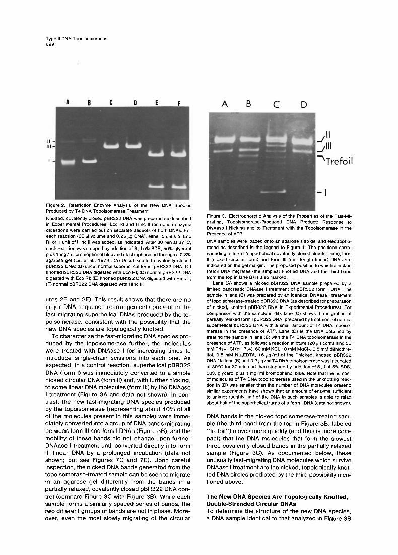
Figure 2. Restriction Enzyme Analysis of the New DNA Species Produced by T4 DNA Topoisomerase Treatment Knotted, covalently closed pBR322 DNA was prepared as described in Experimental Procedures. Eco RI and Hinc II restriction enzyme digestions were carried out on separate aliquots of both DNAs. For each reaction (25/LI volume and 0.25/~g DNA), either 5 units of Eco RI or 1 unit of Hinc II was added, as indicated. After 30 min at 37°C, each reaction was stopped by addition of 6/~15% SDS, 50% glycerol plus 1 mg/ml bromophenol blue and electrophoresed through a 0.8% agarose gel (Liu et al., 1979). (A) Uncut knotted covalently closed pBR322 DNA; (B) uncut normal superhelical form I pBR322 DNA; (C) knotted pBR322 DNA digested with Eco RI; (D) normal pBR322 DNA digested with Eco RI; (E) knotted pBR322 DNA digested with Hinc II; (F) normal pBR322 DNA digested with Hinc II.
ures 2E and 2F). This result shows that there are no major DNA sequence rearrangements present in the fast-migrating superhelical DNAs produced by the to- poisomerase, consistent with the possibility that the new DNA species are topologically knotted.
To characterize the fast-migrating DNA species pro- duced by the topoisomerase further, the molecules were treated with DNAase I for increasing times to introduce single-chain scissions into each one. As expected, in a control reaction, superhelical pBR322 DNA (form I) was immediately converted to a simple nicked circular DNA (form II) and, with further nicking, to some linear DNA molecules (form III) by the DNAase I treatment (Figure 3A and data not shown). In con- trast, the new fast-migrating DNA species produced by the topoisomerase (representing about 40% of all of the molecules present in this sample) were imme- diately converted into a group of DNA bands migrating between form III and form I DNAs (Figure 3B), and the mobility of these bands did not change upon further DNAase I treatment until converted directly into form III linear DNA by a prolonged incubation (data not shown; but see Figures 7C and 7E). Upon careful inspection, the nicked DNA bands generated from the topoisomerase-treated sample can be seen to migrate in an agarose gel differently from the bands in a partially relaxed, covalently closed pBR322 DNA con- trol (compare Figure 3C with Figure 3B). While each sample forms a similarly spaced series of bands, the two different groups ofbands are not in phase. More- over, even the most slowly migrating of the circular
j I I j i l l - Trefoil
- I
Figure 3. Electrophoretic Analysis of the Properties of the Fast-Mi- grating, Topoisomerase-Produced DNA Product: Response to DNAase I Nicking and to Treatment with the Topoisomerase in the Presence of ATP DNA samples were loaded onto an agarose slab gel and electropho- resed as described in the legend to Figure 1. The positions corre- sponding to form I (superhelical covalently closed circular form), form II (nicked circular form) and form Ill (unit length linear) DNAs are indicated on the gel margin. The proposed position to which a nicked trefoil DNA migrates (the simplest knotted DNA and the third band from the top in lane B) is also marked.
Lane (A) shows a nicked pBR322 DNA sample prepared by a limited pancreatic DNAase I treatment of pBR322 form I DNA. The sample in lane (B) was prepared by an identical DNAasel treatment of topoisomerase-treated pBR322 DNA (as described for preparation of nicked, knotted pBR322 DNA in Experimental Procedures). For comparison with the sample in (B), lane (C) shows the migration of partially relaxed form I pBR322 DNA, prepared by treatment of normal superhelical pBR322 DNA with a small amount of T4 DNA topoiso- merase in the presence of ATP. Lane (D) is the DNA obtained by treating the sample in lane (B) with the T4 DNA topoisomerase in the presence of ATP, as follows: a reaction mixture (20 #1) containing 50 mM Tris-HCI (pH 7.4), 60 mM KCI, 10 mM MgCI2, 0.5 mM dithiothre- itol, 0.5 mM Na3EDTA, 16 /~g/ml of the "nicked, knotted pBR322 DNA" in lane (B) and 0.3/~g/ml T4 DNA topoisomerase was incubated at 30°C for 30 min and then stopped by addition of 5/~1 of 5% SDS, 50% glycerol plus 1 mg/ml bromophenol blue. Note that the number of molecules of T4 DNA topoisomerase used in the unknotting reac- tion in (D) was smaller than the number of DNA molecules present; similar experiments have shown that an amount of enzyme sufficient to unknot roughly half of the DNA in such samples is able to relax about half of the superhelical turns of a form I DNA (data not shown).
DNA bands in the nicked topoisomerase-treated sam- ple (the third band from the top in Figure 3B, labeled "trefoi l") moves more quickly (and thus is more com- pact) that the DNA molecules that form the slowest three covalently closed bands in the partially relaxed sample (Figure 3C). As documented below, these unusually fast-migrating DNA molecules which survive DNAase I treatment are the nicked, topologically knot- ted DNA circles predicted by the third possibility men- tioned above.
The New DNA Species Are Topologically Knotted, Double-Stranded Circular DNAs To determine the structure of the new DNA species, a DNA sample identical to that analyzed in Figure 3B
Cell 700
was examined with the electron microscope. Some DNA structures typical of those observed are shown in Figure 4. Figure 4B shows a DNA structure resem- bling a trefoil, which is the simplest possible knot. This species would be expected to have a conformation about as compact as that of an unknotted DNA circle with three supercoils, and we believe that it corre- sponds to the slowest migrating of the bands of nicked, topoisomerase-treated DNA circles (labeled "trefoi l" in Figure 3B). Figures 4C and 4D show topoisomerase-treated DNA structures which could represent knots of successively higher complexities; we believed that these correspond to some of the faster migrating DNA bands in Figure 3B.
To quantitate the electron microscopy results, we compared the number of crossovers in topoisomer- ase-treated and untreated pBR322 DNA circles, both of which had been DNAase I-nicked to remove their supercoiling (Table 1 ). For the untreated control DNA, 80% of the molecules scored had fewer than two crossovers and thus could not be topologically knot- ted (the simplest trefoil knot should exhibit a minimum of three crossovers when projected onto a plane). In contrast, for the topoisomerase-treated DNA sample, only 36% of the molecules were found to exhibit fewer than two crossovers, while DNA molecules with more than four crossovers represented 40% of those scored (compared with only 10% in the control sam- ple). The crossovers visible in the control sample are believed to arise frorna fortuitous overlapping of DNA strands which occurs when unknotted circular mole- cules are flattened onto a plane. Considering our other data as well, we conclude that the excess crossovers in the topoisomerase-treated sample are due to a reaction which introduces topological knots into about half of the pBR322 DNA molecules present.
Efficient UnknotUng of the Knotted Double- Stranded DNA Circles by the T4 DNA Topoisomerase When knotted superhelical pBR322 DNA is treated with small amounts of the T4 DNA topoisomerase in the presence of ATP, the knotted superhelical pPB322 DNA is unknotted rapidly, with both relaxation and unknotting proceeding simultaneously. To study this unknotting reaction separately from the relaxation re- action, the knotted superhelical pBR322 DNA was converted into the nicked, knotted form by DNAase I treatment. This nicked, knotted pBR322 DNA (Figure 3B) was then treated with a trace amount of the T4 DNA topoisomerase in the presence of ATP. Electro- phoretic analysis showed that the group of bands in Figure 3B was quantitatively converted into normal form II DNA (Figure 3D). This conversion of the nicked, knotted DNA to normal nicked DNA circles was con- firmed by electron microscopy. Most importantly, the rate of such an unknotting reaction was found to be comparable to the rate at which a superhelical DNA is relaxed (see legend to Figure 3). The fact that the T4 DNA topoisomerase catalyzes the unknotting of either covalently closed or nicked, knotted DNA about as efficiently as it relaxes the superhelical turns in a closed circular DNA molecule (providing that ATP is present) suggests that the same reaction that unknots a DNA circle removes its superhelical turns.
How Unknotting and Relaxation Reactions Can Occur via the Same Reversible Double-Chain Cleavage of the DNA Helix Topologically, the knotting or unknotting of a double- stranded circular DNA requires that each of the two DNA strands in the DNA helix be interrupted at least once. Although from a purely topological point of view
Figure 4. Sample Electron Micrographsofthe pBR322 DNA Molecules Observed after T4 DNA Topoisomerase Treatment in the Ab- sence of ATP Knotted, covalently closed pBR322 DNA was prepared and then nicked with pancreatic DNAase I as described in Experimental Pro- cedures. Electron microscopic analysis of this nicked, knotted pBR322 DNA gave the results presented in Table 1 ; this figure shows typical molecules with (A) no crossover points; (B) three crossover points; (C) six crossover points; (D) higher topological complexities.
Type II DNA Topoisomerases 701
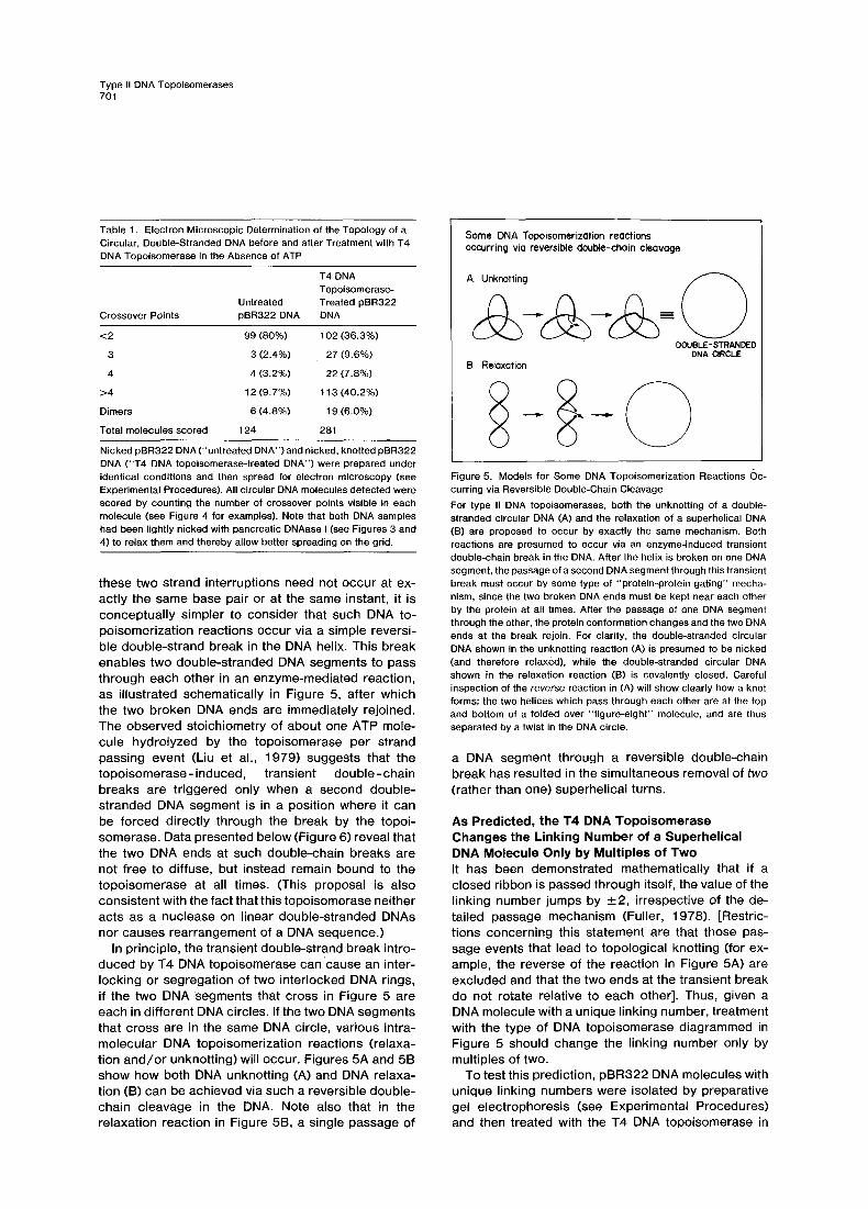
Table 1. Electron Microscopic Determination of the Topology of a Circular, Double-Stranded DNA before and after Treatment with T4 DNA Topoisomerase in the Absence of ATP
T4 DNA Topoisomerase-
Untreated Treated pBR322 Crossover Points pBR322 DNA DNA
<2 99 (80%) 102 (36.3%)
3 3 (2.4%) 27 (9.6%)
4 4 (3.2%) 22 (7.8%)
>4 12 (9.7%) 113 (40.2%)
Dimers 6 (4.8%) 19 (6.0%)
Total molecules scored 124 281
Nicked pBR322 DNA (' 'untreated DNA") and nicked, knotted pBR322 DNA ("T4 DNA topoisomerase-treated DNA") were prepared under identical conditions and then spread for electron microscopy (see Experimental Procedures). All circular DNA molecules detected were scored by counting the number of crossover points visible in each molecule (see Figure 4 for examples). Note that both DNA samples had been lightly nicked with pancreatic DNAase I (see Figures 3 and 4) to relax them and thereby allow better spreading on the grid.
these two strand interruptions need not occur at ex- actly the same base pair or at the same instant, it is conceptually simpler to consider that such DNA to- poisomerization reactions occur via a simple reversi- ble double-strand break in the DNA helix. This break enables two double-stranded DNA segments to pass through each other in an enzyme-mediated reaction, as illustrated schematically in Figure 5, after which the two broken DNA ends are immediately rejoined. The observed stoichiometry of about one ATP mole- cule hydroiyzed by the topoisomerase per strand passing event (Liu et al., 1979) suggests that the topoisomerase- induced, transient double- chain breaks are triggered only when a second double- stranded DNA segment is in a position where it can be forced directly through the break by the topoi- somerase. Data presented below (Figure 6) reveal that the two DNA ends at such double-chain breaks are not free to diffuse, but instead remain bound to the topoisomerase at all times. (This proposal is also consistent with the fact that this topoisomerase neither acts as a nuclease on linear double-stranded DNAs nor causes rearrangement of a DNA sequence.)
In principle, the transient double-strand break intro- duced by T4 DNA topoisomerase can cause an inter- locking or segregation of two interlocked DNA rings, if the two DNA segments that cross in Figure 5 are each in different DNA circles. If the two DNA segments that cross are in the same DNA circle, various intra- molecular DNA topoisomerization reactions (relaxa- tion and/or unknotting) will occur. Figures 5A and 5B show how both DNA unknotting (A) and DNA relaxa- tion (B) can be achieved via such a reversible double- chain cleavage in the DNA. Note also that in the relaxation reaction in Figure 5B, a single passage of
Some DNA Topoisomerizotion reactions occurring via reversible double-chain cleavage
A. Unknotting ( ~ )
DOUBLE-STRANDED DNA CIRCLE
B. Relaxation
Figure 5. Models for Some DNA Topoisomerization Reactions (Oc- curring via Reversible Double-Chain Cleavage For type II DNA topoisomerases, both the unknotting of a double- stranded circular DNA (A) and the relaxation of a superhelical DNA (B) are proposed to occur by exactly the same mechanism. Both reactions are presumed to occur via an enzyme-induced transient double-chain break in the DNA. After the helix is broken on one DNA segment, the passage of a second DNA segment through this transient break must occur by some type of "protein-protein gating" mecha- nism, since the two broken DNA ends must be kept near each other by the protein at all times. After the passage of one DNA segment through the other, the protein conformation changes and the two DNA ends at the break rejoin. For clarity, the double-stranded circular ONA shown in the unknotting reaction (A) is presumed to be nicked (and therefore relaxed), while the double-stranded circular DNA shown in the relaxation reaction (B) is covalently closed. Careful inspection of the r e v e r s e reaction in (A) will show clearly how a knot forms: the two helices which pass through each other are at the top and bottom of a folded over "figure-eight" molecule, and are thus separated by a twist in the DNA circle.
a DNA segment through a reversible double-chain break has resulted in the simultaneous removal of two
(rather than one) superhelical turns.
As Predicted, the T4 DNA Topoisomerase Changes the Linking Number of a Superhelical DNA Molecule Only by Multiples of Two It has been demonstrated mathematically that if a closed ribbon is passed through itself, the value of the linking number jumps by +2, irrespective of the de- tailed passage mechanism (Fuller, 1978). [Restric- tions concerning this statement are that those pas- sage events that lead to topological knotting (for ex- ample, the reverse of the reaction in Figure 5A) are excluded and that the two ends at the transient break do not rotate relative to each other]. Thus, given a DNA molecule with a unique linking number, treatment with the type of DNA topoisomerase diagrammed in Figure 5 should change the linking number only by multiples of two.
To test this prediction, pBR322 DNA molecules with unique linking numbers were isolated by preparative gel electrophoresis (see Experimental Procedures) and then treated with the T4 DNA topoisomerase in
Cell 702
the presence of ATP. As shown in Figure 6, when a pBR322 DNA sample containing an equal mixture of species with even and odd linking numbers was used as substrate, the typical Gaussian distribution of pBR322 DNA topoisomer$ with linking numbers dif- fering by one was generated (Figure 6E --> 6F). How- ever, when pBR322 DNA molecules with either a unique even or a unique odd linking number served as substrate, only the even or odd linking number species in the Gaussian distribution were obtained, respectively (Figure 6A --> 6B, and Figure 6C --> 6D). Furthermore, within each population, each species is present in the relative proportions expected at ther- modynamic equilibrium (see the legend to Figure 6).
These observations provide strong independent confirmation of the model proposed for the unknotting and relaxation reactions in Figure 5. They also reveal that the two DNA ends at the transient DNA break illustrated in Figure 5 must be constrained rigidly, because even an occasional rotation by 360 ° between a breakage and rejoining event would change the linking number by one, and thus interconvert the odd and even linking number species.
Bacterial DNA Gyrases Also Break the DNA Double Helix Reversibly We have proposed previously that the T4 DNA topo- isomerase functions as a DNA gyrase at the T4 repli-
A B C D E F
Figure 6. When It Catalyzes DNA Relaxation Reactions, the T4 DNA Topoisomerase Changes the DNA Linking Number by Multiples of Two Covalently closed pBR322 DNA molecules with different linking num- bers were electrophoretically purified from a fully relaxed pBR322 DNA sample on an agarose gel, as described in Experimental Pro- cedures. Each group of DNA molecules was then treated for 30 min at 30°C with 100 units of the T4 DNA topoisomerase in a standard assay mixture with ATP present (Liu et al., 1979). The reactions were stopped by addition of 5/~1 of 5% SDS, 50% glycerol plus 1 mg/ml bromophenol blue and electrophoresed on a 0.8% agarose gel in TBE buffer. Lanes (A), (C) and (E) are untreated controls, and contain only half the amount of DNA present in the T4 topoisomerase-treated samples analyzed in lanes (B), (D) and (F). With the most 13opulated topoisomer in the Gaussian distribution of fully relaxed pBR322 DNA topoisomers assigned a linking number of n, the linking numbers of the gel-purified DNA topoisomers used as substrate in each reaction were n for lanes (A) and (B), n + 1 for lanes (C) and (D), and a mixture of n + I plus n + 2 for lanes (E) and (F). The Gaussian distribution of the various topoisomers shown in lane (F) is indistinguishable from that in a fully relaxed pBR322 DNA sample produced by treatment of pBR322 form I DNA with excess T4 DNA topoisomerase plus ATP under identical conditions (data not shown). Since excess T4 DNA topoisomerase was present in each reaction, the product pBR322 DNA molecules in (B) and (D) were relaxed to equilibrium and there- fore can be seen to fit the same Gaussian distribution observed in a fully relaxed pBR322 DNA sample.
cation origin(s) (Liu et al., 1979). We therefore ex- pected that bacterial DNA gyrases also might be able to catalyze the unknotting of knotted, covalently closed DNA circles. When nicked, knotted pBR322 DNA was used as the substrate, neither E. coil DNA gyrase nor M. luteus DNA gyrase was capable of changing the knotted DNA topology efficiently. As shown in Figure 7, however, these DNA gyrases can efficiently catalyze the conversion of a covalently closed, knotted DNA molecule to unknotted form I DNA (Figure 7A --> 7B).
To demonstrate that the superhelical form I pBR322 DNA produced by the DNA gyrase reaction in Figure 7B is indeed in an unknotted topology, DNAase I was used to digest this reaction product. As expected, a slight DNAase I treatment converted this gyrase prod- uct first to the nicked form (form II) (Figure 7D) and then to a mixture of form II in a small amount of form III DNA (Figure 7F). As a control, the knotted mole- cules present in the original covalently closed pBR322 DNA used as substrate were converted by DNAase I to the distinct group of intermediately migrating DNA bands discussed previously (see Figure 3B) using the same treatment (Figure 7A produces 7C and 7E). We
A B C D E F
I rain 4 rain DNase I
Figure 7. Electrophoretic Analysis of the Unknotting of Knotted, Co- valently Closed pBR322 DNA by Bacterial DNA Gyrases The knotted, covalently closed pBR322 DNA shown in lane (A) was prepared as described in Experimental Procedures. The gyrase re- action was carried out to produce the product in lane (B), using conditions described in the legend to Figure 3, except that knotted, covalently closed DNA (16 /~g/ml) and 5 units of M. luteus DNA gyrase served as substrate and enzyme, respectively. (E. coil DNA gyrase gave the same results; data not shown). For analysis, a 50/d aliquot of the gyrase reaction product in lane (B) was nicked by treatment with 27 ng/ml pancreatic DNAase I at 30°C. Aliquots (25 /d) were withdrawn after 1 min (lane D) and 4 min (lane F). As a control, the knotted, covalently closed pBR322 DNA substrate (lane A) was treated identically with DNAase I for 1 min (lane C) and 4 min (lane E). Note that only after DNA gyrase treatment can the knotted DNA Sample be converted to form II DNA by a limited DNAase I nicking; this constitutes proof that it has been unknotted by gyrase.
Type II DNA Topoisomerases 703
there fore conc lude that bacter ial DNA gyrase resem- bles the T4 DNA topo isomerase, insofar as both en- zymes act by making reversib le double-s t rand breaks in the DNA doub le helix, through which a second DNA hel ix can pass.
Unknotting of Knotted pBR322 DNA by Other DNA Topoisomerases Since the superhel ic i ty observed in isolated eucar- yot ic DNA can be ful ly accounted for by the changes in l inking number created by the wrapp ing of DNA around nuc leosomes (Germond et al., 1975), there is no need for a genera l eucaryot ic DNA gyrase of the bacter ia l t ype to maintain the superhel ic i ty of eucar- yot ic DNA. On the~other hand, init iat ion of eucaryo t ic DNA repl icat ion might well require a DNA gyrase to open up a repl icat ion bubble. Since repl icat ion bub- bles in eucaryo tes seem to resemble those created dur ing T4 bac te r iophage DNA repl icat ion (Huberman and Riggs, 1968; Delius, Howe and Kozinski , 1971), we used an assay for the unknot t ing of knot ted DNA circ les to test for a eucaryot ic enzyme of the T4 DNA topo isomerase type in 1 hr embryos of Drosophi la melanogaster , where the rate of repl icat ion bubble ini t iat ion is unusual ly high (Kreigstein and Hogness, 1974).
As shown in Figure 8A, we have been able to detec t an ATP-dependent unknot t ing act iv i ty in crude ex- t racts made from such ear ly Drosophi la embryos. When the knot ted, cova lent ly c losed pBR322 DNA in lane B was incubated with the extract , all the DNA molecu les present were eventual ly conver ted to the g roup of s lower migrat ing DNA bands in lane C. These bands are character is t ic of part ia l ly re laxed pBR322 DNA molecu les wi thout topologica l knots. The product DNA molecu les in lane C were shown more d i rect ly to have lost their topolog ica l knots by the DNAase I sensi t iv i ty assay used prev iously (Figure 7) (data not shown).
We learned subsequent ly that an ATP-dependent enzyme act iv i ty that could cata lyze the re laxat ion of superhel ica l DNA circles and decatenat ion of inter- locked DNA rings had a l ready been part ia l ly pur i f ied from Drosophi la embryos by T. Hsieh and D. Brut lag at Stanford Univers i ty ' (personal communicat ion) . The exper imen t shown in Figure 8B demonst ra tes that thei r pur i f ied Drosophi la enzyme is also capab le of unknot t ing a knot ted pBR322 DNA molecule, and that this unknot t ing react ion occurs at a rate comparab le to the rate of its re laxat ion of DNA supercoi ls. L ike the T4 DNA topo isomerase, their Drosophi la enzyme can unknot both n icked and cova lent ly closed, knot ted molecules, and the unknot t ing is ATP-dependent (data not shown).
Using the same assay, we have recent ly been able to demons t ra te the ex is tence of similar ATP-de- penden t unknot t ing act iv i t ies in ext racts of Xenopus laevis eggs and in ext racts of nuclei isolated from a
/},
II- Trefoil-
I - (Knotted) I f
A B C B
A B
FigureS. Electrophoretic Test for the Unknotting of Knotted pBR322 DNA by an ATP-Dependent DNA Topoisomerase from D. melanogas- ter (A).Reaction of crude extract enzyme. Nicked, knotted pBR322 DNA (lane A) and knotted, covalently closed pBR322 DNA (lane B) were electrophoresed as standards. To obtain the DNA product in lane (C), 20/d of a reaction mixture containing 50 mM Tris-HCI (pH 7.4), 60 mM KCI, 10 mM MgCI2, 0.5 mM ATP, 0.5 mM dithiothreitol, 0.5 mM Na3EDTA, 20 #g/ml of knotted, covalently closed pBR322 DNA (see lane B) and 1 /~1 of Drosophila embryo extract (see Experimental Procedures) were incubated at 30°C for 30 rain and then analyzed by gel electrophoresis as described in the legend to Figure 1. The unknotting activity of this extract is at least partly ATP-dependent. In the absence of ATP, the superhelicity in the knotted, covalently closed pBR322 DNA is relaxed by the extract (probably due to the nicking- closing enzyme of Drosophila), but the product DNA still contains a prominent DNA band at the trefoil DNA position, indicating that most of the topological knots have not been removed (data not shown). (B) Reaction of purified enzyme. The nicked, knotted pBR322 DNA in lane A (prepared as described in Experimental Procedures) was treated with an ATP-dependent DNA topoisomerase purified from 1 hr Drosophila embryos (a gift from T. Hsieh and D. Brutlag) in a reaction mixture (20 /~1) containing 50 mM Tris-HCI (pH 7.4), 200 mM KCI, 10 mM MgCI2, 0.5 mM dithiothreitol, 0.5 mM Na3EDTA, 0.5 mM ATP, 30 #g/ml human serum albumin, 16 #g/ml of the DNA and 1 unit of the enzyme (arbitrarily defined as the amount of activity required to remove half of the superhelical turns in pBR322 under exactly the same conditions). After 30 min at 30°C, the reaction was stopped and the DNA was electrophoretically analyzed in lane (B), as described in the legend to Figure 2. Note that most but not all of the knots have been removed by this treatment.
Chinese hamster ovary (CHO) cell l ine (data not shown). It is there fore c lear that enzymes of this type are w idespread th roughout nature.
Procaryot ic ~ proteins and eucaryo t ic n ick ing-c los- ing enzymes are known to in t roduce reversib le single- chain breaks, rather than doub le-cha in breaks, into a DNA hel ix (Champoux, 1978 ; Wang and Liu, 1979). We would therefore expec t that nei ther type of enzyme would be capab le of cata lyz ing the unknot t ing of doub le-s t randed DNA rings. Indeed, when puri f ied E. coli ~ protein (Eco DNA topo isomerase I)' and a Dro- sophi la n ick ing-c losing enzyme preparat ion we re tested in our assay, nei ther enzyme showed unknot° t ing act iv i ty. The unknot t ing enzymes repor ted above thus const i tute a new class of DNA topo isomerases
Cell 704
(proposed to be designated as type II DNA topoiso- merases), whose mechanism is fundamentally differ- ent from that of both the procaryotic ~ proteins and the eucaryotic nicking-closing enzymes (proposed to be designated as type I DNA topoisomerases).
Discussion
Mechanisms of DNA Knotting by T4 DNA Topoisomerase Any random cyclization of a linear polymer can result in the formation of a knotted circle. The first such knotted polymer was reported by Liu et al. (1976), who demonstrated that knotted single-stranded DNA circles were formed as a result of treating a circular single-stranded DNA with E. coil ~ protein (Eco DNA topoisomerase I).The distribution of the various knot- ted species was shown to reach an equilibrium in the presence of the enzyme. A statistical-mechanical treatment of such random cyclization of a polymer chain has been developed by Frank-Kamenetskii, Lu- kashin and Vologodskii (1975). Using computer sim- ulation, the probability of knot formation during DNA cyclization has been shown to be a linear function of the number of DNA statistical segments (Kuhn seg- ments). For example, random cyclization of bacterio- phage lambda DNA (molecular weight 32 x 106 dal- tons, containing about 150 Kuhn segments) is pre- dicted to produce 40% knotted molecules. Since most plasmid DNAs are much shorter than lambda DNA, random cyclization of a linearized plasmid DNA is not expected to yield a significant proportion of knotted molecules. Indeed, when linearized double-stranded ~X174 DNA (molecular weight 3.6 x 10⁶ daltons, containing about 17 Kuhn segments) was cyclized and ligated, no knotted DNA circles were detected (U. Hibner, personal communication).
A knotted DNA of average plasmid size (<10 kb) is thus at a higher free energy state than its unknotted form. How then does one explain the formation of knotted DNA when negatively superhelical DNA is treated with excess T4 DNA topoisomerase ~n the absence of ATP? A clue comes from the study of bacteriophage P2 DNA in the phage head. Using several tailless P2 phage mutants, it was shown that when P2 DNA cyclizes (via its sticky ends) inside the phage head, the formation of highly knotted P2 DNA results (unpublished results of J. C. Wang, L. F. Liu and R. Calendar). It seems intuitively reasonable that the restriction of the volume of a rigid chain, such as DNA, should be equivalent to reducing the length of its statistical segments (thus increasing the number of statistical segments per molecule). Such a restriction in volume should therefore greatly favor knot forma- tion. Our present views of the knotting reaction are that both the superhelical structure of the DNA mole- cules and the excess topoisomerase protein binding
serve to reduce the effective statistical segment length of the DNA, thus favoring knot formation.
The trace amount of topoisomerase activity required to make the transient double-strand breaks needed for knotting (for example, see reverse of reaction in Figure 5A) presumably comes from a charged form of some of the T4 DNA topoisomerase molecules, each of which may drive a single cycle of relaxation in the absence of ATP (Liu et al., 1979). In this view, the absence of ATP is required merely to limit the reaction rate so that the superhelical structure of the DNA molecules (which is important for knot formation) is maintained.
If the above view is correct, addition of small amounts of the T4 DNA topoisomerase with ATP pres- ent should be able to cause the knotting reaction (or a related DNA topoisomerization reaction such as catenation) in the presence of DNA condensing agents
such as synthetic polycations, polyamines (Gosule and Schellman, 1976) or histone H1 (Cole, Lawson and Hsiang, 1978). Our preliminary study has shown that T4 DNA topoisomerase can indeed induce cate- nation of DNA rings in the presence of ATP and a trace amount of the polycation polymin P or histone H1. Furthermore, T4 DNA topoisomerase is also ca- pable of decatenating interlocked DNA rings in the presence of ATP (T. Hsieh and D. Brutlag, personal communication; L. Liu, unpublished result). As we would expect, the unknotting activities detected in extracts of Xenopus eggs and Chinese hamster ovary cells can catalyze an ATP-dependent catenation and decatenation of DNA as well (L. Liu, unpublished results).
Mechanism of ATP Utilization by Type II DNA Topoisomerases Since bacterial DNA gyrases are also able to unknot the topologically knotted, covalently closed pBR322 DNA (Figure 7), the ATP-dependent negative super- coiling reaction that these enzymes catalyze is likely to involve a transiently broken DNA double helix as an intermediate. P. Brown, K. Kreuzer and N. Cozzarelli have obtained independent evidence for such a mech- anism for the E. coil DNA gyrase (personal communi- cation). Figure 9 shows a possible model for this type of DNA gyration.
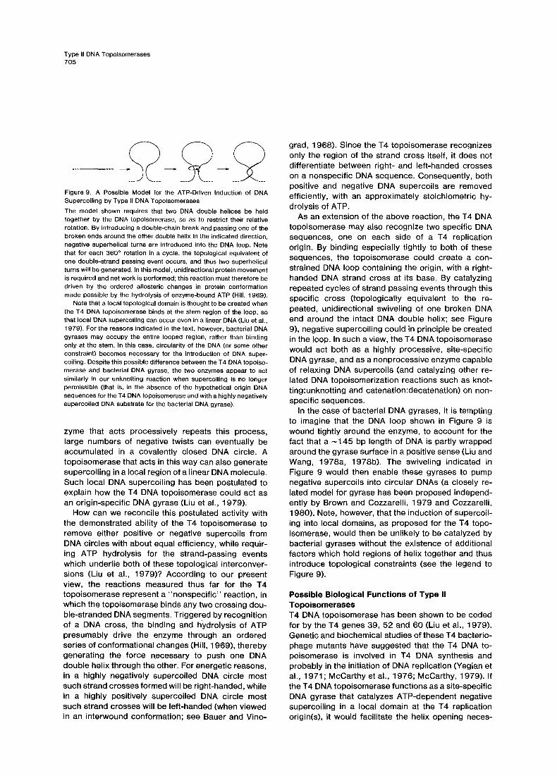
In our view, DNA gyration requires that two DNA double helices be brought together, with a double- chain break made in one of the two DNA helices by the topoisomerase. An ATP-driven, unidirectional 360 ° swiveling of one of the two broken ends around the unbroken double-stranded DNA is topologically equivalent to the passage of a DNA segment through the transient double-strand break, and thus can gen- erate two superhelical turns into the DNA loop created between the two separate helical regions bound by the topoisomerase (see discussion above). If an en-
Type II DNA Topoisomerases 705
Figure 9. A Possible Model for the ATP-Driven Induction of DNA Supercoiling by Type II DNA Topoisomerases The model shown requires that two DNA double helices be held together by the DNA topoisomerase, so as to restrict their relative rotation. By introducing a double-chain break and passing one of the broken ends around the other double helix in the indicated direction, negative superhelical turns are introduced into the DNA loop. Note that for each 360 ° rotation in a cycle, the topological equivalent of one double-strand passing event occurs, and thus two superhelical turns will be generated. In this model, unidirectional protein movement is required and net work is performed; this reaction must therefore be driven by the ordered allosteric changes in protein conformation made possible by the hydrolysis of enzyme-bound ATP (Hill, 1969).
Note that a local topological domain is thought to be created when the T4 DNA topoisomerase binds at the stem region of the loop, so that local DNA supercoiling can occur even in a linear DNA (Liu et al., 1979). For the reasons indicated in the text, however, bacterial DNA gyrases may occupy the entire looped region, rather than binding only at the stem. In this case, circularity of the DNA (or some other constraint) becomes necessary for the introduction of DNA super- coiling. Despite this possible difference between the T4 DNA topoiso- merase and bacterial DNA gyrase, the two enzymes appear to act similarly in our unknotting reaction when supercoiling is no longer permissible (that is, in the absence of the hypothetical origin DNA sequences for the T4 DNA topoisomerase and with a highly negatively supercoiled DNA substrate for the bacterial DNA gyrase).
zyme that acts processively repeats this process, large numbers of negative twists can eventually be accumulated in a covalently closed DNA circle. A topoisomerase that acts in this way can also generate supercoiling in a local region of a linear DNA molecule. Such local DNA supercoiling has been postulated to explain how the T4 DNA topoisomerase could act as an origin-specific DNA gyrase (Liu et al., 1979).
How can we reconcile this postulated activity with the demonstrated ability of the T4 topoisomerase to remove either positive or negative supercoils from DNA circles with about equal efficiency, while requir- ing ATP hydrolysis for the strand-passing events which underlie both of these topological interconver- sions (Liu et al., 1979)? According to our present view, the reactions measured thus far for the T4 topoisomerase represent a "nonspecif ic" reaction, in which the topoisomerase binds any two crossing dou- ble-stranded DNA segments. Triggered by recognition of a DNA cross, the binding and hydrolysis of ATP presumably drive the enzyme through an ordered series of conformational changes (Hill, 1969), thereby generating the force necessary to push one DNA double helix through the other. For energetic reasons, in a highly negatively supercoiled DNA circle most such strand crosses formed will be right-handed, while in a highly positively supercoiled DNA circle most such strand crosses will be left-handed (when viewed in an interwound conformation; see Bauer and Vino-
grad, 1968). Since the T4 topoisomerase recognizes only the region of the strand cross itself, it does not differentiate between right- and left-handed crosses on a nonspecific DNA sequence. Consequently, both positive and negative DNA supercoils are removed efficiently, with an approximately stoichiometric hy- drolysis of ATP.
As an extension of the above reaction, the T4 DNA topoisomerase may also recognize two specific DNA sequences, one on each side of a T4 replication origin. By binding especially tightly to both of these sequences, the topoisomerase could create a con- strained DNA loop containing the origin, with a right- handed DNA strand cross at its base. By catalyzing repeated cycles of strand passing events through this specific cross (topologically equivalent to the re- peated, unidirectional swiveling of one broken DNA end around the intact DNA double helix; see Figure 9), negative supercoiling could in principle be created in the loop. In such a view, the T4 DNA topoisomerase would act both as a highly processive, site-specific DNA gyrase, and as a nonprocessive enzyme capable of relaxing DNA supercoils (and catalyzing other re- lated DNA topoisomerization reactions such as knot- ting:unknotting and catenation:decatenation) on non- specific sequences.
In the case of bacterial DNA gyrases, it is tempting to imagine that the DNA loop shown in Figure 9 is wound tightly around the enzyme, to account for the fact that a ~145 bp length of DNA is partly wrapped around the gyrase surface in a positive sense (Liu and Wang, 1978a, 1978b). The swiveling indicated in Figure 9 would then enable these gyrases to pump negative supercoils into circular DNAs (a closely re- lated model for gyrase has been proposed independ- ently by Brown and Cozzarelli, 1979 and Cozzarelli, 1980). Note, however, that the induction of supercoil- ing into local domains, as proposed for the T4 topo- isomerase, would then be unlikely to be catalyzed by bacterial gyrases without the existence of additional factors which hold regions of helix together and thus introduce topological constraints (see the legend to Figure 9).
Possible Biological Functions of Type II Topoisomerases T4 DNA topoisomerase has been shown to be coded for by the T4 genes 39, 52 and 60 (Liu et al., 1979). Genetic and biochemical studies of these T4 bacterio- phage mutants have suggested that the T4 DNA to- poisomerase is involved in T4 DNA synthesis and probably in the initiation of DNA replication (Yegian et al., 1971 ; McCarthy et al., 1976; McCarthy, 1979). If the T4 DNA topoisomerase functions as a site-specific DNA gyrase that catalyzes ATP-dependent negative supercoiling in a local domain at the T4 replication origin(s), it would facilitate the helix opening neces-
Cell 706
sa ry fo r f o rma t i on o f a rep l i ca t i on bubb le (Liu et al., 1979 ) .
It is tempt ing to p r o p o s e that the in i t iat ion o f DNA rep l i ca t ion in a e u c a r y o t i c o r g a n i s m fo l l ows a s imi lar mechan i sm to tha t p r o p o s e d f o r b a c t e r i o p h a g e T4. Bo th the T4 DNA t o p o i s o m e r a s e and the unkno t t i ng ac t iv i ty d e t e c t e d in the nuc lea r e x t r a c t o f C H O ce l ls a re inh ib i ted by h igh n o v o b i o c i n c o n c e n t r a t i o n s (un- pub l i shed resu l ts o f L. L iu)~. ln terest ingly , Ma t t e rn and Pa in te r ( 1 9 7 9 ) have s h o w n that n o v o b i o c i n at a b o u t th is c o n c e n t r a t i o n ( 2 0 0 / ~ g / m l ) inh ib i ts the in i t iat ion o f C H O cel l DNA rep l i ca t ion w i thou t b l ock ing rep l i ca t ion f o r k movemen t . It thus s e e m s qu i te poss ib le that the type II DNA t o p o i s o m e r a s e we have de tec ted in CHO ce l ls is the ta rge t p ro te in f o r ' t h e drug, and that the e u c a r y o t i c t ype II DNA t o p o i s o m e r a s e s d e s c r i b e d in th is r epo r t f unc t i on in in i t ia t ing rep l i ca t ion fo rks , as s u s p e c t e d by a n a l o g y wi th b a c t e r i o p h a g e T4.
M a n y o the r b io log ica l p r o c e s s e s , inc lud ing the ter - m ina t ion o f DNA rep l i ca t ion ( fo r e x a m p l e , s e g r e g a - t ions o f i n t e r l ocked DNA r ings), p h a g e packag ing , s i te -spec i f i c gene t i c r ecomb ina t i on , DNA t ranspos i - t ion and c h r o m o s o m e c o n d e n s a t i o n and d e c o n d e n - sa t ion might a lso requ i re DNA d o u b l e - c h a i n b r e a k a g e and re jo in ing events . In add i t i on , i t is impor tan t to r e c o g n i z e that the e x i s t e n c e o f suCh t ype II DNA t o p o i s o m e r a s e s m e a n s t h a t a l l l a rge D N A mo lecu les (such as t hose in bac te r ia l and e u c a r y o t i c c h r o m o - somes ) wou ld be e x p e c t e d t o b e h igh ly kno t ted , es - pec ia l l y in the i r c o n d e n s e d f o r m s ( F r a n k - K a m e n e t s k i i et al., 1975) . S ince any DNA kno t can be read i l y t ied and unt ied by d o u b l e - s t r a n d pass ing events , howeve r , such k n o t s need not have a harmfu l e f fec t on gene t i c p r o c e s s e s . It is in te res t ing to s p e c u l a t e that an o r g a - n ized pa t te rn o f DNA k n o t s might even be usefu l i n he lp ing to ho ld t o g e t h e r c o m p l e x s t ruc tu res such as e u c a r y o t i c c h r o m o s o m e s .
W e p r o p o s e that the ab i l i ty to ca ta l yze a d o u b l e - s t rand pass ing reac t i on (such as tha t i l lus t ra ted sche - mat ica l l y in F igu re 5) be used to de f i ne a t o p o i s o m e r - ase o f t ype II, t hus d i s t i ngu i sh ing such e n z y m e s f rom the more c lass ica l t ype I t O p o i s o m e r a s e s wh ich re- ve rs ib l y b r e a k and re jo in on ly o n e DNA s t rand at a t ime ( C h a m p o u x , 1978 ; W a n g and Liu, 1979) . Th is p rev i ous l y u n r e c o g n i z e d c lass o f t ype II t o p o i s o m e r - ases is u n d o u b t e d l y w i d e s p r e a d in na ture , and is l ike ly to be o f g rea t i m p o r t a n c e f o r m a n y , fundamenta l ge - net ic p r o c e s s e s .
Experimental Procedures
Enzymes and Nucleic Acids~ T4 DNA topoisomerase was purified from T4 regA, amN55,, amH39- infected E. coil D110 cells as'described previously (Liu et al., 1979). The topoisomerase preparation' used in this study had a specific activity of ~106 U/mg (Liu et al., 1979). The enzyme had been stored at -20°C in 50% (w/v) glycerol; 30 m M potassium phosphate {pH 7.2), 10 mM/~-mercaptoethanol and 0.5 mM Na3EDTA for more than six months without loss of activity. Purified E. coil DNA gyrase,
subunits of M. luteus DNA gyrase and E. coil ~ protein were obtained from the laboratory of J. C. Wang. Eco RI restriction enzyme was purified in this laboratory by P. Bedinger. Restriction enzymes Hinc II and Hae II were purchased from New England Biolabs and Bethesda Research Inc., respectively. Drosophila nicking-closing enzyme pu- rified from tissue culture (Kc) cells and the ATP-dependent DNA topoisomerase from Drosophila were obtained from T. Hsieh and D. Brutlag. Proteinase K was purchased from E. M. Laboratory. Plasmid pBR322 DNA was purified by phenol deproteinization of a clear lysate followed by CsCI/ethidium bromide equilibrium centrifugation.
Electron Microscopy The formamide spreading technique described by Davis, Simon and Davidson (1971) was used. The grids were shadowed with either tungsten or platinum and viewed with a Philips EM201 or EM301 electron microscope.
Analytical Ultracentrifugation CsCI density gradient equilibrium centrifugation was performed in 12 mm double-sector cells by spinning at 40,000 rpm for 48 hr at 20°C in a Spinco Model E analytical ultracentrifuge.
Preparation of Knotted, CovalenUy Closed pBR322 DNA Knotted pBR322 DNA was typically prepared as follows. 1 ml o fa reaction mixture containing 50 mM Tris-HCI (pH 7.5), 60 mM KCI, 10 mM MgCI2, 0.5 mM Na3EDTA, 0.5 mM dithiothreitol, 30 /~g/ml human serum albumin (Worthington), 20 #g/ml pBR322 DNA (form I) and 33/Lg/ml T4 DNA topoisomerase was incubated at 30°C for 3 rain and then stopped by extracting twice with neutralized phenol. After four extractions of the aqueous layer with ether, the mixture was dialyzed into 10 mM Tris-HCI (pH 7.8), 0.1 mM Na3EDTA and stored at -20°C.
Normally, about half the pBR322 DNA (form I) can be converted to the knotted form under the above conditions. The amount of the knotted form in each preparation varies with the DNA and/or enzyme concentration. At higher DNA concentration and/or lower enzyme concentration, the amount of knotting decreases and pBR322 DNA (form I) is relaxed without being knotted, even at the initial stages of reaction.
Preparation of Nicked, Knotted pBR322 DNA The knotted, covalently closed pBR322 DNA described above was converted into nicked, knotted pBR322 DNA by a limited DNAase I treatment as follows. 1 ml of knotted, covalently closed pBR322 DNA (~20/Lg/ml) was treated with 16 ng/ml pancreatic DNAase I (Worth- ington) in a reaction mixture containing 10 mM Tris-HCI (pH 7.8), 4 mM MgCI2, 1 mM Na3EDTA and 40 /Lg/ml human serum albumin. After incubation at 30°C for i 0 min, the reaction was quenched by phenol extractions. After removing the phenol from'the aqueous phase with ether extractions, the DNA was dialyzed into 10 mM Tris- HCI (pH 7.8), 0.1 mM Na3EDTA for storage at -20°C.
Isolation of pBR322 DNA with a Unique Linking Number 7/Lg of pBR322 DNA (form i) were relaxed to completion by treatment with T4 DNA topoisomerase (500 units) in the presence of ATP. The relaxed, covalently closed pBR322 DNA was then electrophoresed through a preparative 0.8% agarose gel at room temperature in a buffer consisting of 40 mM Tris base, 90 mM boric acid (pH 8.3) and 3 mM Na3EDTA (TBE). After electrophoresis, the agarose gel was sliced into small horizontal strips, each of which contained an individ- ual DNA band (as judged by staining vertical gel slices with ethidium bromide). Each ge! strip was transferred to a dialysis bag containing 200/~1 of the electrophores!s buffer diluted 4 fold, plus 14/~g/ml of yeast tRNA as carrier. The DNA molecules were eluted from the gel by placing the dialysis tubing into the dilute electrophoresis buffer and electrophoresing for 1 hr at 100 V and 100 mA in a 20 x 10 cm electrophoresis box. The polarity of the electrodes was reversed for 4Q sec just before removing the dialysis bags. The solid contents of the dialysis bags were removed by centrifugation, and the superna- tants were precipitated by adding 10 /~g of yeast tRNA carrier and
Type II DNA Topoisomerases 707
67% ethanol in the presence of 0.1 M sodium acetate for 15 min in a dry ice bath. After ethanol precipitation, the DNA precipitates were each dissolved in 30/~1 of the buffer used for the T4 DNA topoiso- merase reactions (see above).
Preparation of Drosophila Embryo Extract Freshly deposited D. melanogaster eggs were collected from popu- lation cages at 1 hr. Embryos were rinsed extensively in a saline- detergent solution (0.7% NaCI, 0.02% Triton X-IO0), dechorionated in half-strength Chlorox for about 60 sec and then rinsed thoroughly with saline-detergent solution on a 116 mesh nylon screen (Nitex). About 1 g of embryos was immediately resuspended in 5 ml of 50 mM potassium phosphate (pH 7.3) and broken by 10 strokes of a Dounce homogenizer. The homogenate was filtered through Nitex and centrifuged at 10,000 x g for 10 rain. The supernatant was used immediately for the assay.
Acknowledgments
We wish to thank C. Chun and M, L. Wong for taking the electron micrographs; Dr. G. Wiesehahn for performing the analytical ultra- centrifugation; and Dr. J. Hearst, in whose laboratory the centrifuga- tion and part of the electron microscopy were performed. We are also most grateful to Drs. T. Hsieh and D. Brutlag for providing us with their purified DrosophUa DNA topoisomerases and for communicating unpublished results. We appreciate the advice received on the prep- aration of this manuscript from Drs. N. Brown, P. O'Farrell, F. Heffren, R. Sheridan, R. Sterngianz and K. R. Yamamote. This work was supported by a USPHS grant from the National Institute of General Medical Sciences to B. M. A. and an American Cancer Society postdoctoral fellowship to L. F, L.
The costs of publication of this article were defrayed in part by the payment of page charges. This article must therefore be hereby marked "advertisement" in accordance with 18 U.S.C. Section 1734 solely to indicate this fact.
Received November 6, 1979; revised December 21, 1979
References
Baase, W. A. and Wang, J. C. (1974). Biochemistry 13, 4299-4303. Bauer, W. R. and Vinograd, J. (1968). J. Mol. Biol. 33, 141-172, Bauer, W. R., Ressner, E. C., Kates, J. and Patzke, J. V. (1977). Prec. Nat. Acad. Sci. USA 74, 1841-1845. Drown, P. O. and Cozzarelli, N. R. (1979). Science 206, 1081-1083. Champoux, J. J. (1977). Prec. Nat. Acad. Sci. USA 74, 3800-3804. Champoux, J. J. (1978). Ann. Rev, Biochem. 47, 449-479. Champoux, J. J. and Dulbecco, R. (1972). Proc. Nat. Acad. Sci. USA 69, 143-146. Cole, R. D., Lawson, G. M. and Hsiang, M. W, (1978). Cold Spring Harbor Symp. Quant. Biol. 42, 253-263. Cozzarelli, N. R. (1980). Science, in press. Davis, R. W., Simon, M. and Davidsen, N. (1971). In Methods in Enzymoiegy, 21, part D, L. Grossman and K. Moldave, eds. (New York: Academic Press), pp. 413-428. Delius, H., Howe, C. and Kozinski, A. W. (1971). Proc. Nat. Acad. Sci. USA 68, 3049-3053. Frank-Kamenetskii, M. D., Lukashin, A. V. and Vologodskii, A. V. (1975). Nature 258, 398-402. Fuller, F. B. (1978). Proc. Nat. Acad. Sci. USA 75, 3557-3561. Gellert, M., Mizuuchi, K., O'Dea, M. H. and Nash, H. A. (1976a). Proc. Nat. Acad. Sci. USA 73, 3872-3876. Genert, M., O'Dea, M. H., Itoh, T. and Tomizawa, J. (1976b). Proc. Nat. Acad. Sci. USA 73, 4474-4478. Germond, J.-E., Hirt, B., Oudet, P., Gross-Bellard, M. and Chambon, P. (1975). Proc. Nat. Acad. Sci. USA 72, 1843-1847. Germond, J.-E., Rouviere-Yaniv, J., Yaniv, M. and Brutlag, D. (1979).
Proc. Nat. Acad. Sci. USA 76, 3779-3783. Gosule, L C. and Schellman, J. A. (1976). Nature 259, 333-335. Hill, T. L. (1969). Proc. Nat. Acad. Sci. USA 64, 267-274. Huberman, J. A. and Riggs, A. D. (1968). J. Mol. Biol. 32, 327-341. Keller, W. and Wendel, I. (1975). Cold Spring Harbor Syrup. Quant. Biol. 39, 199-208. Kikuchi, Y. and Nash, H. A. (1979). Proc. Nat. Acad. Sci. USA 76, 3760-3764. Kirkegaard, K. and Wang, J. C. (1978). Nucl. Acids Res. 5, 3811- 3820. Kreigstein, H. J. and Hogness, D. S. (1974). Proc. Nat. Acad. Sci. USA 71, 135-139. Liu, L F. and Wang, J. C. (1978a). Proc. Nat. Acad. Sci. USA 75, 2098-2102. Liu, L. F. and Wang, J. C. (1978b). Cell 15, 979-984. Liu, L. F., Depew, R. E. and Wang, J. C. (1976). J. Mol. Biol. 106, 439-452. Liu, L. F., Liu, C.-C. and Alberts, B. M. (1979). Nature 281, 456- 461. McCarthy, D. (1979). J. Mol. Biol. •27, 265-283. McCarthy, D., Minner, C., Bernstein, H. and Bernstein, C. (1976). J. Mol. Biol. 106, 963-981. Mattern, M. R. and Painter, R. B. (1979). Biochim. Biophys. Acta 563, 306-312. Peebles, C. L., Higgins, N. P., Kreuzer, K. N., Morrison, A., Brown, P. 0., Sugino, A. and Cozzarelli, N. R. (1979). Cold Spring Harbor Syrup. Quant. Biol. 43, 41-52. Shapiro, J. A. (1979). Proc. Nat. Acad. Sci. USA 76, 1933-1937. Smith, C. L., Kubo, K. and Imamoto, F. (1978). Nature 275, 420- 423. Stetler, G. L., King, G. J. and Huang, W. M. (1979). Proc. Nat. Acad. Sci. USA 76, 3737-3741. Sumida-Yasumoto, C. and Hurwitz, J. (1977). Proc. Nat. Acad. Sci. USA 74, 4195-4199. Wang, J. C. (1971). J. Mol. Biol. 55, 523-533. Wang, J. C. (1973). In DNA Synthesis in Vitro, R. D. Wells and R. B. Inman, eds. (Baltimore: University Park Press), pp. 163-174. Wang, J. C. and Liu, L. F. (1979). In Molecular Genetics, Part III, J. H. Taylor, ed. (New York: Academic Press), pp. 65-88. Yegian, C. D., Mueller, M., Seizer, G., Russo, V. and Stahl, F. W. (1971 ). Virology 46, 900-919.