Proc. Nati. Acad. Sci. USA Vol. 91, pp. 10340-10344, October 1994 Biochemistry Split ubiquitin as a sensor of protein interactions in vivo NILS JOHNSSON AND ALEXANDER VARSHAVSKY Division of Biology, California Institute of Technology, Pasadena, CA 91125 Communicated by John Abelson, July 11, 1994 ABSTRACT We describe an assay for in Wivo protein interactions. Protein fusions containing ubiquitin, a 76- residue, single-donain protein, are rapidly cleaved in vivo by ubiquitin-specific proteases, which recognize the folded con- formation of ubiquitin. When a C-te l fragment of ubiq- ultin (Cub) is expressed as a fusion to a reporter protein, the fusion is cleaved only If an N-terminal frgment of ubiquin (Nub) is ao expressed in the same cell. This reconstitution of native ubiquitin from Its rag ts, detectable by the in vivo cleavage assay, is not observed with a mutat ly altered Nub. However, if Cub and the altered Nub are each linked to polypeptides that interact in vivo, the cleavage of the fusion cotaining Cub is restored, yielding a generally applicable assay for kinetic and equilibrium aspects of in mvo protein interac- tions. This method, termed USPS (ubiquitin-based split- protein sensor), makes it possible to monitor a protein-protein interaction as a function of time, at the natural sites of this interaction in a living cell. Multiprotein complexes mediate the bulk of biological pro- cesses (1, 2). The knowledge of these complexes is extensive for oligomeric proteins whose subunit interactions are strong enough to withstand in vitro conditions. However, many oligomeric assemblies, while relevant physiologically, are transient in vivo or unstable in vitro. The understanding of in vivo protein interactions (and especially of their temporal aspects) is still fragmentary and largely qualitative, the lim- itations of existing in vivo methods being a major reason. Assays for in vivo protein interactions include crosslinking of proteins with cell-penetrating reagents (2) and use of reso- nance energy transfer between dye-coupled proteins micro- injected into cells (3). Genetic analyses of protein interactions include searches for synthetic lethal or extragenic suppressor mutations (4) which occur in genes whose products are at least functionally (and often physically) associated with a protein of interest. Another approach, the two-hybrid tech- nique (5-8), is based on expressing one protein as a fusion to a DNA-binding domain of a transcriptional activator and expressing another protein as a fusion to a transcriptional activation domain. If the test proteins interact in vivo, a transcriptional activator is reconstituted, resulting in the induction of a reporter gene. This otherwise powerful method cannot address temporal aspects of a protein-protein inter- action. In addition, the two-hybrid technique limits the set of detectable protein interactions to those that occur (or can be "reproduced") in the nucleus, in proximity to the reporter gene (5-8). We describe a ubiquitin (Ub)-based split-protein sensor (USPS; Fig. 1) that makes it possible to examine kinetic and equilibrium aspects of a protein-protein interaction at its natural sites in a living cell. MATERIALS AND METHODS Strains, Media, Pue-Chase, and Immunobiotting. All ex- periments used the YPH500 strain of the yeast Saccharomy- ces cerevisiae (MATa ura3 lys2 trpl ade2 his3 leu2) (11, 12) grown at 300C to an OD600 of =1 in a synthetic (SD) medium (13, 14) containing 0.1 mM CuSO4. Pulse-chase experiments, including the preparation of cell extracts in the presence of N-ethylmaleimide (to inhibit UBPs), immunoprecipitation with a monoclonal antibody to the "ha" epitope (14), SDS/ 12% PAGE, and fluorography, were carried out as described (11, 14), except that zero-time samples were withdrawn and processed 1 min after the addition of a chase medium (11). Immunoblotting with anti-ha antibody (11) was performed with the ECL detection system (Amersham). Test Proteins. Detailed protocols are available upon re- quest. The final constructs [verified by sequencing (15)] resided in plasmid pRS314 or pRS316 (12) and were ex- pressed from the induced PcupI promoter. The S. cerevisiae Ub gene was amplified by PCR (15) from the previously engineered Sal I site immediately upstream of the Ub start codon (11) to the first cytosine of codon 37, and from codon 35 to codon 76. The primers were constructed in a way that yielded, after ligation of the two amplified fragments, a BamHI site between codons 35 and 37 in the Ub open reading frame (ORF). Ligation of this ORF to a fragment encoding mouse dihydrofolate reductase with an ha epitope tag (DHFR-ha, dha) (11) yielded an ORF encoding Ub-dha (Fig. 2, construct I), which contained the sequence Met-His-Arg- Ser-Gly-ile-Met between Gly-76 of Ub and Val-1 of DHFR. Constructs II-IV (Fig. 2) were produced by replacing the Sal I-BstXI fragment in construct I with appropriately designed double-stranded oligonucleotides. Construct V (Fig. 2) was produced by PCR using S. cerevisiae genomic DNA and primers designed to amplify the region of STE6 (16, 17) from codon 1% to codon 262. The resulting fragment was inserted into the BamHI site between the Ub codons 35 and 37 in constructs I-IV, yielding constructs V-VIII. In construct IX (Fig. 2), residue 35 of Ub was preceded by a 32-residue linker all of whose residues except the N-terminal Met-Gly-Gly were specified by codons 234 to 262 of STE6. The zl-Cub portion of construct XIV encoded the above Ste6-derived sequence preceding the Cub moiety, the leucine zipper region of S. cerevisiae Gcn4 (residues 235-281, denoted as zi) (18-21), the construction-generated N-terminal Met, and the sequence Gly-Glu-Ile-Ser-Thr. Constructs X-XIII (Fig. 2), derived from construct XIV and constructs V-VIII, encoded Gly-Glu-Ile-Ser-Thr-Leu-Glu C-terminally to zj, with Gly- Gly-Ser-Thr-Met between z1 and Nub. The z1 motif in N1-zj and its derivatives but not in z1 Cub-dha bore a Met-250 Thr-250 replacement (residue numbers of Gcn4), which oc- curred during construction; this replacement would be ex- pected to weaken the interaction between z1 domains (18- 21). RESULTS In Vivo Folding of Ub Contin an Insertion and/or a Single-Residue Replacement. Ub is a 76-residue, single- Abbreviations: Ub, ubiquitin; USPS, Ub-based split-protein sensor; UBP, Ub-specific protease; Nub, N-terminal fragment of Ub; Cub, C-terminal fragment of Ub; DHFR, dihydrofolate reductase. 10340 The publication costs of this article were defrayed in part by page charge payment. This article must therefore be hereby marked "advertisement" in accordance with 18 U.S.C. §1734 solely to indicate this fact. Downloaded by guest on May 30, 2020
Transcript
Proc. Nati. Acad. Sci. USAVol. 91, pp. 10340-10344, October 1994Biochemistry
Split ubiquitin as a sensor of protein interactions in vivoNILS JOHNSSON AND ALEXANDER VARSHAVSKYDivision of Biology, California Institute of Technology, Pasadena, CA 91125
Communicated by John Abelson, July 11, 1994
ABSTRACT We describe an assay for in Wivo proteininteractions. Protein fusions containing ubiquitin, a 76-residue, single-donain protein, are rapidly cleaved in vivo byubiquitin-specific proteases, which recognize the folded con-formation of ubiquitin. When a C-te l fragment of ubiq-ultin (Cub) is expressed as a fusion to a reporter protein, thefusion is cleaved only If an N-terminal frgment of ubiquin(Nub) is ao expressed in the same cell. This reconstitution ofnative ubiquitin from Its rag ts, detectable by the in vivocleavage assay, is not observed with a mutat ly altered Nub.However, if Cub and the altered Nub are each linked topolypeptides that interact in vivo, the cleavage of the fusioncotaining Cub is restored, yielding a generally applicable assayfor kinetic and equilibrium aspects of in mvo protein interac-tions. This method, termed USPS (ubiquitin-based split-protein sensor), makes it possible to monitor a protein-proteininteraction as a function of time, at the natural sites of thisinteraction in a living cell.
Multiprotein complexes mediate the bulk of biological pro-cesses (1, 2). The knowledge of these complexes is extensivefor oligomeric proteins whose subunit interactions are strongenough to withstand in vitro conditions. However, manyoligomeric assemblies, while relevant physiologically, aretransient in vivo or unstable in vitro. The understanding of invivo protein interactions (and especially of their temporalaspects) is still fragmentary and largely qualitative, the lim-itations of existing in vivo methods being a major reason.Assays for in vivo protein interactions include crosslinking ofproteins with cell-penetrating reagents (2) and use of reso-nance energy transfer between dye-coupled proteins micro-injected into cells (3). Genetic analyses ofprotein interactionsinclude searches for synthetic lethal or extragenic suppressormutations (4) which occur in genes whose products are atleast functionally (and often physically) associated with aprotein of interest. Another approach, the two-hybrid tech-nique (5-8), is based on expressing one protein as a fusion toa DNA-binding domain of a transcriptional activator andexpressing another protein as a fusion to a transcriptionalactivation domain. If the test proteins interact in vivo, atranscriptional activator is reconstituted, resulting in theinduction ofa reporter gene. This otherwise powerful methodcannot address temporal aspects of a protein-protein inter-action. In addition, the two-hybrid technique limits the set ofdetectable protein interactions to those that occur (or can be"reproduced") in the nucleus, in proximity to the reportergene (5-8).We describe a ubiquitin (Ub)-based split-protein sensor
(USPS; Fig. 1) that makes it possible to examine kinetic andequilibrium aspects of a protein-protein interaction at itsnatural sites in a living cell.
MATERIALS AND METHODSStrains, Media, Pue-Chase, and Immunobiotting. All ex-
periments used the YPH500 strain of the yeast Saccharomy-
ces cerevisiae (MATa ura3 lys2 trpl ade2 his3 leu2) (11, 12)grown at 300C to an OD600 of =1 in a synthetic (SD) medium(13, 14) containing 0.1 mM CuSO4. Pulse-chase experiments,including the preparation of cell extracts in the presence ofN-ethylmaleimide (to inhibit UBPs), immunoprecipitationwith a monoclonal antibody to the "ha" epitope (14), SDS/12% PAGE, and fluorography, were carried out as described(11, 14), except that zero-time samples were withdrawn andprocessed 1 min after the addition of a chase medium (11).Immunoblotting with anti-ha antibody (11) was performedwith the ECL detection system (Amersham).
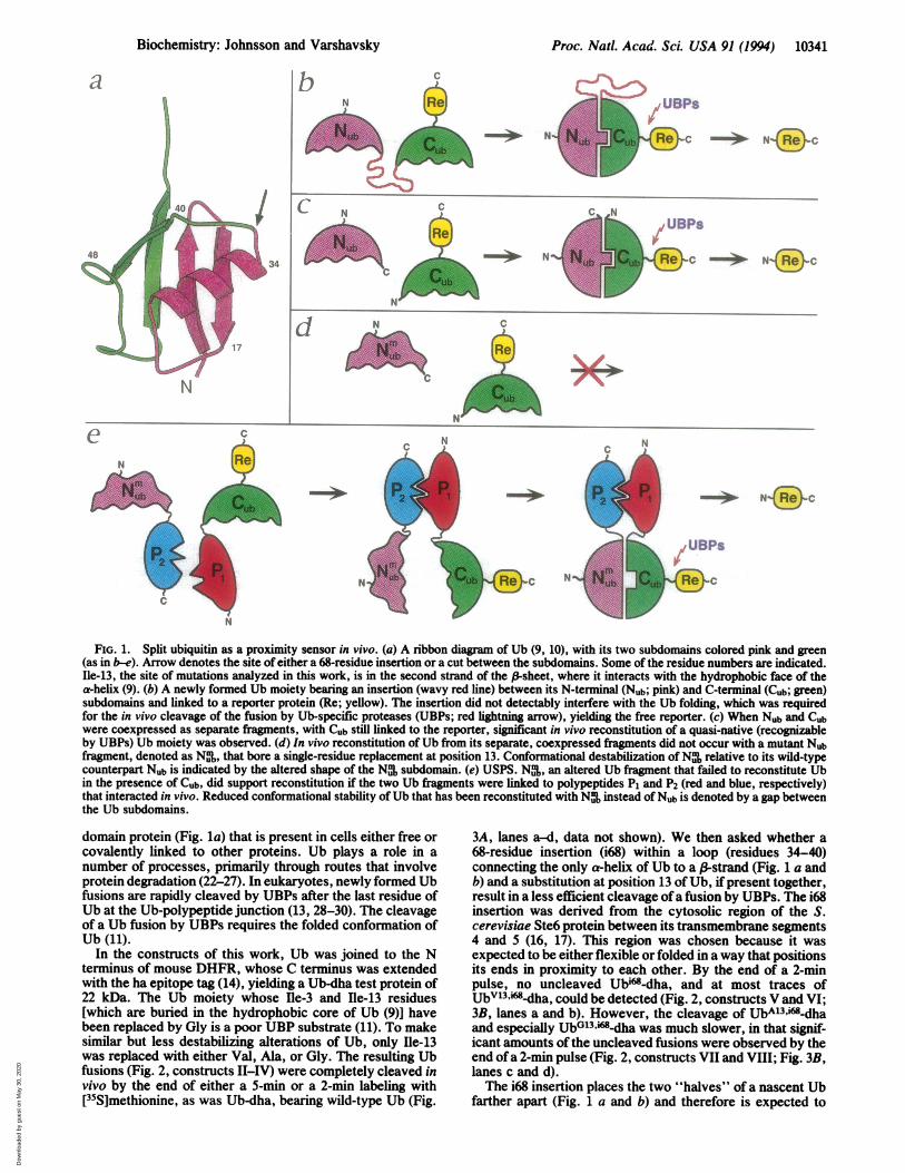
Test Proteins. Detailed protocols are available upon re-quest. The final constructs [verified by sequencing (15)]resided in plasmid pRS314 or pRS316 (12) and were ex-pressed from the induced PcupI promoter. The S. cerevisiaeUb gene was amplified by PCR (15) from the previouslyengineered Sal I site immediately upstream of the Ub startcodon (11) to the first cytosine of codon 37, and from codon35 to codon 76. The primers were constructed in a way thatyielded, after ligation of the two amplified fragments, aBamHI site between codons 35 and 37 in the Ub open readingframe (ORF). Ligation of this ORF to a fragment encodingmouse dihydrofolate reductase with an ha epitope tag(DHFR-ha, dha) (11) yielded an ORF encoding Ub-dha (Fig.2, construct I), which contained the sequence Met-His-Arg-Ser-Gly-ile-Met between Gly-76 of Ub and Val-1 of DHFR.Constructs II-IV (Fig. 2) were produced by replacing the SalI-BstXI fragment in construct I with appropriately designeddouble-stranded oligonucleotides. Construct V (Fig. 2) wasproduced by PCR using S. cerevisiae genomic DNA andprimers designed to amplify the region of STE6 (16, 17) fromcodon 1% to codon 262. The resulting fragment was insertedinto the BamHI site between the Ub codons 35 and 37 inconstructs I-IV, yielding constructs V-VIII. In construct IX(Fig. 2), residue 35 ofUb was preceded by a 32-residue linkerall of whose residues except the N-terminal Met-Gly-Glywere specified by codons 234 to 262 of STE6. The zl-Cubportion of construct XIV encoded the above Ste6-derivedsequence preceding the Cub moiety, the leucine zipper regionof S. cerevisiae Gcn4 (residues 235-281, denoted as zi)(18-21), the construction-generated N-terminal Met, and thesequence Gly-Glu-Ile-Ser-Thr. Constructs X-XIII (Fig. 2),derived from construct XIV and constructs V-VIII, encodedGly-Glu-Ile-Ser-Thr-Leu-Glu C-terminally to zj, with Gly-Gly-Ser-Thr-Met between z1 and Nub. The z1 motif in N1-zjand its derivatives but not in z1 Cub-dha bore a Met-250Thr-250 replacement (residue numbers of Gcn4), which oc-curred during construction; this replacement would be ex-pected to weaken the interaction between z1 domains (18-21).
RESULTSIn Vivo Folding of Ub Contin an Insertion and/or a
Single-Residue Replacement. Ub is a 76-residue, single-
Abbreviations: Ub, ubiquitin; USPS, Ub-based split-protein sensor;UBP, Ub-specific protease; Nub, N-terminal fragment of Ub; Cub,C-terminal fragment of Ub; DHFR, dihydrofolate reductase.
10340
The publication costs of this article were defrayed in part by page chargepayment. This article must therefore be hereby marked "advertisement"in accordance with 18 U.S.C. §1734 solely to indicate this fact.
Dow
nloa
ded
by g
uest
on
May
30,
202
0
Proc. Natl. Acad. Sci. USA 91 (1994) 10341
b
- NNo
UVBPS
4'c
C%. mN
- + N"'_N
N
,! UBPs
4Cc
-*+ N jc
- -*-- N C
N
t̂ N t t ~~~ ~~~~~~~~~~~CN C
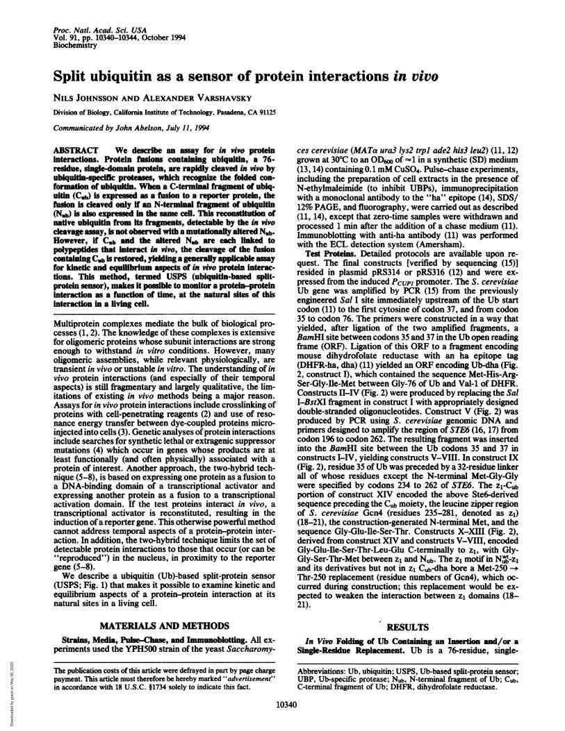
FIG. 1. Split ubiquitin as a proximity sensor in vivo. (a) A ribbon diagram of Ub (9, 10), with its two subdomains colored pink and green(as in b-e). Arrow denotes the site of either a 68-residue insertion or a cut between the subdomains. Some of the residue numbers are indicated.Ile-13, the site of mutations analyzed in this work, is in the second strand of the a-sheet, where it interacts with the hydrophobic face of thea-helix (9). (b) A newly formed Ub moiety bearing an insertion (wavy red line) between its N-terminal (Nub; pink) and C-terminal (Cub; green)subdomains and linked to a reporter protein (Re; yellow). The insertion did not detectably interfere with the Ub folding, which was requiredfor the in vivo cleavage of the fusion by Ub-specific proteases (UBPs; red lightning arrow), yielding the free reporter. (c) When Nub and Cubwere coexpressed as separate fragments, with Cub still linked to the reporter, significant in vivo reconstitution of a quasi-native (recognizableby UBPs) Ub moiety was observed. (d) In vivo reconstitution of Ub from its separate, coexpressed fragments did not occur with a mutant Nubfragment, denoted as Nm,, that bore a single-residue replacement at position 13. Conformational destabilization ofN% relative to its wild-typecounterpart Nub is indicated by the altered shape of the N% subdomain. (e) USPS. Nu%, an altered Ub fragment that failed to reconstitute Ubin the presence of Cub, did support reconstitution if the two Ub fragments were linked to polypeptides Pi and P2 (red and blue, respectively)that interacted in vivo. Reduced conformational stability of Ub that has been reconstituted with N% instead of Nub is denoted by a gap betweenthe Ub subdomains.
N
domain protein (Fig. la) that is present in cells either free orcovalently linked to other proteins. Ub plays a role in anumber of processes, primarily through routes that involveprotein degradation (22-27). In eukaryotes, newly formed Ubfusions are rapidly cleaved by UBPs after the last residue ofUb at the Ub-polypeptide junction (13, 28-30). The cleavageof a Ub fusion by UBPs requires the folded conformation ofUb (11).
In the constructs of this work, Ub was joined to the Nterminus of mouse DHFR, whose C terminus was extendedwith the ha epitope tag (14), yielding a Ub-dha test protein of22 kDa. The Ub moiety whose Hle-3 and Ile-13 residues[which are buried in the hydrophobic core of Ub (9)] havebeen replaced by Gly is a poor UBP substrate (11). To makesimilar but less destabilizing alterations of Ub, only Hle-13was replaced with either Val, Ala, or Gly. The resulting Ubfusions (Fig. 2, constructs II-IV) were completely cleaved invivo by the end of either a 5-min or a 2-min labeling with[35S]methionine, as was Ub-dha, bearing wild-type Ub (Fig.
3A, lanes a-d, data not shown). We then asked whether a68-residue insertion (i68) within a loop (residues 34-40)connecting the only a-helix of Ub to a (-strand (Fig. 1 a andb) and a substitution at position 13 of Ub, ifpresent together,result in a less efficient cleavage ofa fusion by UBPs. The i68insertion was derived from the cytosolic region of the S.cerevisiae Ste6 protein between its transmembrane segments4 and 5 (16, 17). This region was chosen because it wasexpected to be either flexible or folded in a way that positionsits ends in proximity to each other. By the end of a 2-minpulse, no uncleaved Ubi"-dha, and at most traces ofUbV3.i68m-dha, could be detected (Fig. 2, constructs V and VI;3B, lanes a and b). However, the cleavage of UbAl3,i68-dhaand especially UbGl3,i".dha was much slower, in that signif-icant amounts of the uncleaved fusions were observed by theend ofa 2-min pulse (Fig. 2, constructs VII and VIII; Fig. 3B,lanes c and d).The i68 insertion places the two "halves" of a nascent Ub
farther apart (Fig. 1 a and b) and therefore is expected to
C]
a c
N
Biochemistry: Johnsson and Varshavsky
Dow
nloa
ded
by g
uest
on
May
30,
202
0
10342 Biochemistry: Johnsson and Varshavsky
1 76
Ub-DHFR
II Ubv"DHFRA13I11 Ub -DHFR
IV UbG -DHFR
1 36 195 262 35 76
N~~b" Cub
234 262 35 76
M%,Gem
wtIX CCub-DHFR
1 37 235 281j-GGSTMI Z1 GEISTLE
x
Xi
XIl
N ub-ZN b-zlNV13-zA13N.,Z
A BI
-- _-
i ha -
; )itX11 N _-z235 281
234 262 35 76Z 1 GEIST DFt I XIV Z1-C~b-Ste6
wtI I ~~XIV IZi-Cub'DHFR
FIG. 2. Fusion constructs. Fusions used in this work containedsome of the following elements: (i) a Ub moiety, either wild-type(construct I) or bearing single-residue replacements at position 13(constructs II-IV). (ii) A Ub moiety containing the 68-residueinsertion (denoted as Ste6) derived from the cytosolic region of S.
cerevisiae Ste6 between its transmembrane segments 4 and 5 (16, 17).The insertion was positioned after residue 36 ofUb (construct V). (iii)A Ub moiety bearing both the above insertion and a single-residuereplacement at position 13 (constructs VI-VIII). (iv) A C-terminalfragment of Ub (Cub, residues 35-76) bearing the 32-residue, Ste6-derived sequence at its N terminus (construct IX). (v) The samefusion whose N terminus was extended, via the linker sequenceGly-Glu-Ile-Ser-Thr, with the 47-residue homodimerization motif("leucine zipper", or Zi) of S. cerevisiae Gcn4 (18-21) (residues235-281 ofGcn4) (construct XIV). (vi) An N-terminal fragment ofUb(Nub, residues 1-37) bearing the wild-type Ub sequence or a single-residue replacement at position 13 and a C-terminal extensioncontaining the linker sequence Gly-Gly-Ser-Thr-Met followed by theZi leucine zipper of Gcn4 (constructs X-XIII). (vii) Mouse DHFRbearing a C-terminal ha epitope (14) (denoted as DHFR in thisdiagram and as dha in the text and in Figs. 3 and 4).
retard the folding of Ub; this effect could be detected by the2-min pulse-cleavage assay if another destabilizing Ub alter-ation such as Ble-13 -- Gly-13 (which by itself is insufficientto cause a detectable retardation of Ub folding) was presentas well (Fig. 3 A and B). These results can be interpretedwithin the diffusion-collision model of protein folding (2, 31,32), in which marginally stable units of isolated secondarystructure form early and then coalesce into the native con-formation. Indeed, in native Ub, its first 34 residues arefolded into an a-helix interacting with a double-strandedantiparallel -3-sheet (Fig. la) (9). Thus, the i68 insertionretards Ub folding primarily through a reduction in thefrequency of collisions between the N-terminal and C-termi-nal subdomains of Ub, whereas the effect of substitutions atposition 13 ofUb is a decreased conformational stability of itsN-terminal subdomain (Fig. la). This in vivo evidence for Ubsubdomains (see also below) is in agreement with the resultsof circular dichroism and NMR analyses of Ub and itsfragments in a methanol/water solvent at low pH (33, 34).In Vivo Reconstitution of Ub from Its Fragments. The
relative insensitivity ofUb folding to a large insertion withinthe 34-40 loop (Fig. 1 a and b) suggested that separate,coexpressed fragments of Ub (produced by a cut within the34-40 loop) may be able to reconstitute native Ub. In a testof this conjecture, a C-terminal fragment of wild-type Ub(residues 35-76, denoted as Cub) was expressed as a fusion tothe dha reporter (Cub-dha), while an N-terminal fragment ofwild-type Ub (residues 1-37, denoted as Nub) was expressedas a fusion to the "leucine zipper" homodimerization domainof the yeast Gcn4 protein (denoted as z1) (Fig. 2, constructsIX and X; refs. 18-21). (The reason for linking N to z1 will
(.
C .,! ki I -_ A40M40w40"_
d1idk - -_-
I)
/ It. ALiMd _OW_m -- -..
cilia-
E---- (- :.Li],,!
_ -(wFj1
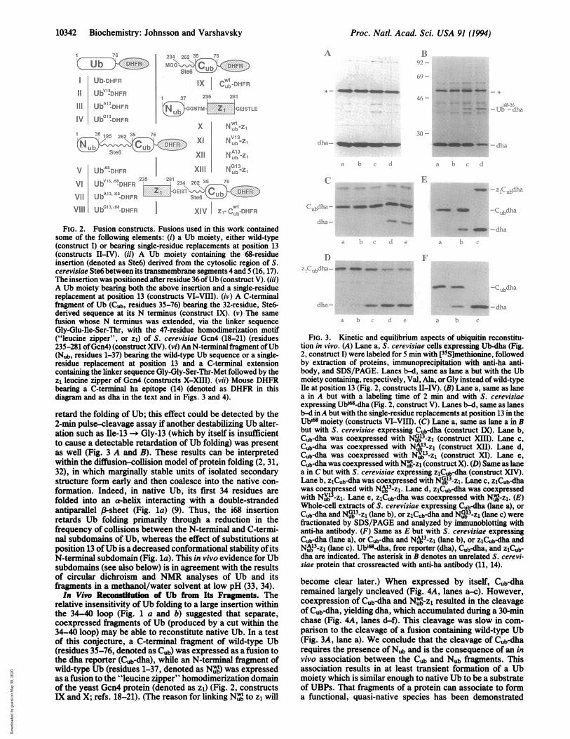
FIG. 3. Kinetic and equilibrium aspects of ubiquitin reconstitu-tion in vivo. (A) Lane a, S. cerevisiae cells expressing Ub-dha (Fig.2, construct I) were labeled for 5 min with [35S]methionine, followedby extraction of proteins, immunoprecipitation with anti-ha anti-body, and SDS/PAGE. Lanes b-d, same as lane a but with the Ubmoiety containing, respectively, Val, Ala, or Gly instead ofwild-typeIle at position 13 (Fig. 2, constructs II-IV). (B) Lane a, same as lanea in A but with a labeling time of 2 min and with S. cerevisiaeexpressing Ubi"-dha (Fig. 2, construct V). Lanes b-d, same as lanesb-d inA but with the single-residue replacements at position 13 in theUbi" moiety (constructs VI-VIII). (C) Lane a, same as lane a in Bbut with S. cerevisiae expressing Cub-dha (construct IX). Lane b,Cub-dha was coexpressed with NGU~b3-z1 (construct XIII). Lane c,Cub-dha was coexpressed with N 13-zl (construct XII). Lane d,Cub-dha was coexpressed with Nuj3Zi (construct XI). Lane e,
Cub-dha was coexpressed with NS-z1 (construct X). (D) Same as lane
a in C but with S. cerevisiae expressing zlCub-dha (construct XIV).Lane b, ZlCub-dha was coexpressed with N$Ij3-Z1. Lane c, ziCub-dhawas coexpressed with NuV3-zl. Lane d, zlCub-dha was coexpressedwith Nuj3-zl. Lane e, zCub-dha was coexpressed with Nrb-zl. (E)Whole-cell extracts of S. cerevisiae expressing Cub-dha (lane a), orCub-dha and N _3-z1 (lane b), or ZlCub-dha and N$,3-zl (lane c) werefractionated by SDS/PAGE and analyzed by immunoblotting withanti-ha antibody. (F) Same as E but with S. cerevisiae expressingCub-dha (lane a), or Cub-dha and N.13-z1 (lane b), or zlCub-dha andN._3-z1 (lane c). Ub'"-dha, free reporter (dha), Cub-dha, and ZiCub-dha are indicated. The asterisk in B denotes an unrelated S. cerevi-siae protein that crossreacted with anti-ha antibody (11, 14).
become clear later.) When expressed by itself, Cub-dharemained largely uncleaved (Fig. 4A, lanes a-c). However,coexpression of Cub-dha and Nubt-z1 resulted in the cleavageof Cub-dha, yielding dha, which accumulated during a 30-minchase (Fig. 4A, lanes d-f). This cleavage was slow in com-parison to the cleavage of a fusion containing wild-type Ub(Fig. 3A, lane a). We conclude that the cleavage of Cub-dharequires the presence of Nub and is the consequence of an invivo association between the Cub and Nub fragments. Thisassociation results in at least transient formation of a Ubmoiety which is similar enough to native Ub to be a substrateof UBPs. That fragments of a protein can associate to forma functional, quasi-native species has been demonstrated
V Ub -DHFR
G13 68VII UbA3 .68-DHFRVIl I Us13~1 68L"DHFR
Proc. Natl. Acad Sci. USA 91 (1994)
X. m mmm
E.1
4WOMOMMOM 40- ..l ji,
-.1 ;, ;l
i
-(. 1.i
.-4m m-Ci lia
Dow
nloa
ded
by g
uest
on
May
30,
202
0
Proc. Nadl. Acad. Sci. USA 91 (1994) 10343
X inNub _ I V A Gchase (mmy: 0 10 30 0 10 30 0 10 30 0 10 30 0 10 30
A
Cubdha _ _ 4_1 am _a_ Om qmw moo___>m
dhi - ____ _.__
BZ~tcdha-wm _m
'iLI -
- _ --at _ __
am am -_ _W_ _m
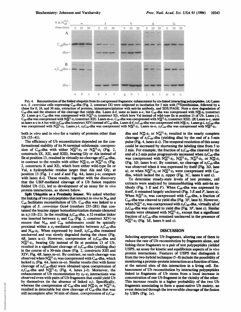
," b c ] 2 hIIIn k i IIa
FiG. 4. Reconstitution ofthe folded ubiquitin from its coexpressed firgments: enhancement by cis-linked interacting polypeptides. (A) Lanesa-c, S. cerevisiae cells expressing Cub-dha (Fig. 2, construct IX) were subjected to incubation for 5 min with [35S]methionine, followed by achase for 0, 10, and 30 min, extraction of proteins, immunoprecipitation with anti-ha antibody, and SDS/PAGE. Note a slow degradation ofCub-dha and the absence of the cleavage that yields dha. Lanes d-f, same as lanes a-c, but Cub-dha was coexpressed with Nv%-zI (constructX). Lanes g-i, Cub-dha was coexpressed with NIj3-z1 (construct XI), which bore Val instead of wild-type Ile at position 13 of Ub. Lanes ji-,Cub-dha was coexpressed with Nu_3-Z1 (construct XII). Lanes m-o, Cub-dha was coexpressed with N.9Ij3-zl (construct XIM). (B) Lanes a-c, sameas lanes a-c inA but with zCub-dha (construct XIV) instead ofCub-dha. Lanes d-f, zlCub-dha was coexpressed with Nug-zl. Lanes gi, zlCub-dhawas coexpressed with N .3-z1 Lanes j-l, z1Cub-dha was coexpressed with Nu_3-z1. Lanes n-o, z1Cub-dha was coexpressed with N -z1.
both in vitro and in vivo for a variety of proteins other thanUb (35-41).The efficiency of Ub reconstitution depended on the con-
formational stability of its N-terminal subdomain: coexpres-sion of Cub-dha with either N G13-zi or N A13-z1 (Fig. 2,constructs IX, XII, and XIII), bearing Gly or Ala instead ofIle at position 13, resulted in virtually no cleavage of Cub-dha,in contrast to the results with either N -z1 or N V13-zl (Fig.2, constructs X and XI), which bore either wild-type Ile orVal, a hydrophobic residue larger than Ala and Gly, atposition 13 (Fig. 1 c and d and Fig. 4A, lanes j-o; comparewith lanes d-i). These results, together with the discoverythat the UBP-mediated cleavage of a Ub fusion requiresfolded Ub (11), led to development of an assay for in vivoprotein interactions, as shown below.
Split Ubiquitin as a Proximity Sensor. We asked whetherthe linking oftwo polypeptides that interact in vivo to Nub andCub facilitates reconstitution of Ub. Cub-dha was linked to aregion of S. cerevisiae Gcn4 (residues 235-281) that con-tained the leucine zipper homodimerization domain (denotedas z1) (18-21). In the resulting zlCub-dha, a 32-residue linkerwas inserted between z1 and Cub (Fig. 2, construct XIV) toensure that Nub and Cub subdomains could be spatiallyproximal within a z1-mediated complex between zlCub-dhaand Nub-zi. When expressed by itself, z1Cub-dha remaineduncleaved and was slowly degraded during the chase (Fig.4B, lanes a-c). However, coexpression of zCub-dha andNG13-z1, bearing Gly instead of Ile at position 13 of Ub,resulted in a significant cleavage of zlCub-dha (yielding dha)in the course of a 30-min chase (Fig. 2, constructs XIII andXIV; Fig. 4B, lanes m-o). By contrast, no such cleavage wasobserved when Nu 3-z1 was coexpressed with Cub-dha, whichlacked z1 (Fig. 4A, lanes m-o). Similar results (but with fastercleavage of zCub-dha) were obtained upon coexpression ofzCub-dha and NuA3-z1 (Fig. 4, lanes j-l). Moreover, theenhancement of Ub reconstitution by z1-z1 interactions wasobserved even with pairs ofUb fragments that could yield Ubby themselves (in the absence of linked z1). Specifically,whereas the coexpression of Cub-dha and N -z1 or Nv13_zresulted in detectable but slow cleavage of Cub-dha that wasstill incomplete after 30 min of chase, coexpression of Z1Cub-
dha and NU-zl or Nub3-zj resulted in the nearly completecleavage of zlCub-dha (yielding dha) by the end of a 5-minpulse (Fig. 4, lanes d-i). The temporal resolution ofthis assaycould be increased by shortening the labeling time from 5 to2 min. For example, the fraction of z1Cub-dha cleaved by theend of a 2-min pulse progressively increased when z1Cub-dhawas coexpressed with NG1j3-z1, NA3-zl, Nv'3-z1, or N:-zi(Fig. 3D, lanes b-e). By contrast, no cleavage of zlCub-dhawas observed when it was expressed by itself (Fig. 3D, lanea), or when N j3-z1 or NGj3-z1 were coexpressed with Cab-dha, which lacked the z1 zipper (Fig. 3C, lanes b and c).To determine steady-state levels of test proteins, cell
extracts were analyzed by immunoblotting with anti-ha an-tibody (Fig. 3 E and F). When Cub-dha was expressed byitself, it remained largely uncleaved (Fig. 3 E and F, lanes a).When Nub3-zj was coexpressed with Cub-dha, a fraction ofCub-dha was cleaved to yield dha (Fig. 3F, lane b). However,whenNA3-z1 was coexpressed with zCub-dha, virtually all ofzlCub-dha was cleaved to yield dha (Fig. 3F, lane c). Similarresults were obtained with N13_-zl, except that a significantfraction of zlCub-dha remained uncleaved in the presence ofNG13-zl (Fig. 3E, lanes b and c).
DISCUSSIONSelecting appropriate Ub fragments, altering one of them toreduce the rate ofUb reconstitution by fragments alone, andlinking these fiagments to a pair of test polypeptides yieldedUSPS, an assay for kinetic and equilibrium aspects of in vivoprotein interactions. Features of USPS that distinguish itfrom the two-hybrid technique (5-8) include the possibility ofmonitoring a protein-protein interaction as a function oftime,at the natural sites of this interaction in a living cell. En-hancement of Ub reconstitution by interacting polypeptideslinked to fragments of Ub stems from a local increase inconcentration ofone Ub fiagment in the vicinity ofthe other.This increase results in a higher probability of the two Ubfragments associating to form a quasi-native Ub moiety, anevent detected through the irreversible cleavage ofthe fusionby UBPs (Fig. le).
Biochemistry: Johnsson and Varshavsky
Dow
nloa
ded
by g
uest
on
May
30,
202
0
10344 Biochemistry: Johnsson and Varshavsky
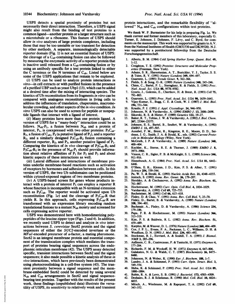
USPS detects a spatial proximity of proteins but notnecessarily their direct interaction. Therefore, a USPS signalmight also result from the binding of test proteins to acommon ligand-another protein or a larger structure such asa microtubule or a ribosome. This feature of USPS shouldallow in vivo analyses of large protein complexes, includingthose that may be too unstable or too transient for detectionby other methods. A separate, immunologically detectablereporter domain (Fig. 1) is not an essential feature of USPS:the cleavage of a Cub-containing fusion can also be followedby measuring the enzymatic activity ofa reporter protein thatis inactive until released from a Cub-containing fusion or byusing an antibody specific for a test protein linked to eitherthe C terminus or the N terminus of Cub. Listed below aresome of the USPS applications that remain to be explored.
(i) USPS can be used to analyze protein interactions invitro-in cell extracts or with purified fusions in the presenceofa purified UBP such as yeast Ubpl (13), which can be addedat a desired time after the mixing of interacting species. Thekinetics ofUb reconstitution from its fragments in vitro can becompared with analogous reconstitutions in living cells toaddress the influences of translation, chaperonins, macromo-lecular crowding, and other aspects ofthe in vivo condition. Invitro USPS can also be used to screen for peptide or nonpep-tide ligands that interact with a ligand of interest.
(ii) Many proteins have more than one protein ligand. Aversion of USPS for a "many-body" interaction could be asfollows. Nub-Pl, a fusion between NUb and a protein ofinterest, P1, is coexpressed with two other proteins: P2Cub-R1, a fusion of Cub, P2 (a putative ligand of P1), and a reporterR1, and a similarly designed P3Cub-R2 fusion containing adifferent reporter, R2, and P3, another putative ligand of P1.Comparing the kinetics of in vivo cleavage of P2Cub-Rl andP3Cub-R2 in the presence of Nub-Pi should provide informa-tion about relative affinities of P1 for P2 and P3 and aboutkinetic aspects of these interactions as well.
(iii) Lateral diffusion and interactions of membrane pro-teins underlie membrane-based reactions such as activationof receptors by hormones and adhesion between cells. In thisversion of USPS, the two Ub subdomains can be positionedwithin cytosol-exposed regions of two membrane proteins.
(iv) A USPS-based screen for genes whose products in-teract with a protein of interest P1 can employ a reporter Rwhose function is incompatible with an N-terminal extensionsuch as PjCub. The reporter would be activated upon Ubreconstitution (Fig. le) and the cleavage of PlCub-R thatyields R. In this approach, cells expressing PlCub-R aretransformed with an expression library encoding randomtranslational fusions to a mutant Nub moiety and screened forcells expressing active reporter.USPS was demonstrated here with homodimerizing poly-
peptides ofthe leucine zipper type (Figs. 2 and 4). In addition,we recently used USPS to detect and analyze in vivo inter-actions between S. cerevisiae Sec62 protein and the signalsequences of either the SUC2-encoded invertase or theMFal-encoded precursor of a-factor, a mating pheromone.Sec62 is an integral membrane protein and essential compo-nent of the translocation complex which mediates the trans-port of proteins bearing signal sequences across the endo-plasmic reticulum membrane (42). The USPS assay detectedspecific, transient interactions between Sec62 and the signalsequences; it also made possible a kinetic analysis of these invivo interactions, which have previously been demonstratedusing photocrosslinking in a cell-free system (43). The tran-sient proximity between a signal sequence and the mem-brane-embedded Sec62 could be detected by using severalNub and Cub arrangements in Sec62 and signal sequence-bearing test proteins. Together with the results of the presentwork, these findings (unpublished data) illustrate the versa-tility of USPS, its sensitivity to relatively weak and transient
protein interactions, and the remarkable flexibility of "al-lowed" Nub and Cub configurations within test proteins.We thank W. P. Burmeister for his help in preparing Fig. la. We
thank current and former members of this laboratory, especially G.Turner, E. Johnson, J. Dohmen, F. Ldvy, and C. Byrd, for com-ments on the manuscript. This work was supported by grants to A.V.from the National Institutes ofHealth (GM31530 and DK39520). N.J.was supported by a postdoctoral fellowship from the DeutscheForschungsgemeinschaft.1. Alberts, B. M. (1984) Cold Spring Harbor Symp. Quant. Biol. 49,
1-12.2. Creighton, T. E. (1992) Proteins: Structures and Molecular Prop-
erties (Freeman, New York).3. Adams, S. R., Harootunian, A. T., Buetchler, Y. J., Taylor, S. S.
& Tsien, R. Y. (1991) Nature (London) 349, 694-697.4. Guarente, L. (1993) Trends Genet. 9, 362-366.5. Fields, S. & Song, O.-K. (1989) Nature (London) 340, 245-246.6. Chien, C., Bartel, P. L., Sternglanz, R. & Fields, S. (1991) Proc.
Natl. Acad. Sci. USA 88, 9578-9582.7. Gyuris, J., Golemis, E., Chertkov, H. & Brent, R. (1993) Cell 75,
791-803.8. Guarente, L. (1993) Proc. Natd. Acad. Sci. USA 90, 1639-1641.9. Vjay-Kumar, S., Bugg, C. E. & Cook, W. J. (1987) J. Mol. Biol.
194, 531-544.10. Kraulis, P. J. (1991) J. Appl. Crystallogr. 24, 946-950.11. Johnsson, N. & Varshavsky, A. (1994) EMBO J. 13, 2686-2698.12. Sikorski, R. S. & Hieter, P. (1989) Genetics 122, 19-27.13. Baker, R. T., Tobias, J. W. & Varshavsky, A. (1992)J. Biol. Chem.
267, 23364-23375.14. Johnson, E. S., Bartel, B., Seufert, W. & Varshavsky, A. (1992)
EMBO J. 11, 497-505.15. Ausubel, F. M., Brent, R., Kingston, R. E., Moore, D. D., Sei-
dman, J. G., Smith, J. A. & Struhl, K., eds. (1992) Current Proto-cols in Molecular Biology (Wiley, New York).
16. McGrath, J. P. & Varshavsky, A. (1989) Nature (London) 340,400-404.
17. Kuchler, K., Sterne, R. E. & Thorner, J. (1989) EMBO J. 8,3973-3984.
18. Vinson, C. R., Sigler, P. B. & McKnight, S. L. (1989) Science 246,911-916.
19. Hinnebusch, A. G. (1984) Proc. Natd. Acad. Sci. USA 81, 6442-6446.
20. O'Shea, E. K., Klemm, J. D., Kim, P. S. & Alber, T. (1991)Science 254, 539-544.
21. Pu, W. T. & Struhl, K. (1993) Nucleic Acids Res. 21, 4348-4355.22. Jentsch, S. (1992) Annu. Rev. Genet. 26, 179-207.23. Hershko, A. & Ciechanover, A. (1992) Annu. Rev. Biochem. 61,
761-807.24. Hochstrasser, M. (1992) Curr. Opin. Cell Biol. 4, 1024-1029.25. Varshavsky, A. (1992) Cell 69, 725-735.26. Rechsteiner, M. (1991) Cell 66, 615-618.27. Finley, D. & Chau, V. (1991) Annu. Rev. Cell Biol. 7, 25-70.28. Finley, D., Bartel, B. & Varshavsky, A. (1989) Nature (London)
338, 394-401.29. Bachmair, A., Finley, D. & Varshavsky, A. (1986) Science 234,
179-186.30. Papa, F. R. & Hochstrasser, M. (1993) Nature (London) 366,
313-319.31. Kim, P. S. & Baldwin, R. L. (1982) Annu. Rev. Biochem. 51,
459-489.32. Karplus, M. & Weaver, D. L. (1976) Nature (London) 260,404-409.33. Cox, J. P. L., Evans, P. A., Packman, L. C., Williams, D. H. &
Woolfson, D. N. (1993) J. Mol. Biol. 234, 483-492.34. Stockman, B. J., Euvrard, A. & Scahill, T. A. (1993) J. Biomol.
NMR 3, 285-296.35. Anfinsen, C. B., Cuatrecasas, P. & Taniuchi, H. (1971) Enzymes 4,
177-204.36. Richards, F. M. & Wyckoff, H. W. (1971) Enzymes 4, 647-806.37. Galakatos, N. G. & Walsh, C. T. (1987) Biochemistry 26, 8475-
8480.38. Johnsson, N. & Weber, K. (1990) Eur. J. Biochem. 188, 1-7.39. Landro, J. A. & Schimmel, P. (1993) Curr. Opin. Struct. Biol. 3,
549-554.40. Shiba, K. & Schimmel, P. (1992) Proc. Natl. Acad. Sci. USA 89,
1880-1884.41. Rubin, R. A. & Levy, S. B. (1991) J. Bacteriol. 173, 4503-4509.42. Deshaies, R. J. & Schekman, R. (1990) Mol. Cell. Biol. 10, 6024-
6035.43. Miksch, A., Wiedmann, M. & Rapoport, T. A. (1992) Cell 69,