Published online 4 December 2008 Nucleic Acids Research, 2009, Vol. 37, No. 2 493–505 doi:10.1093/nar/gkn961 UHRF1 binds G9a and participates in p21 transcriptional regulation in mammalian cells Jong Kyong Kim 1 , Pierre-Olivier Este ` ve 1 , Steven E. Jacobsen 2 and Sriharsa Pradhan 1, * 1 New England Biolabs, Ipswich, MA 01938-2723 and 2 Department of Molecular Cell and Developmental Biology, Howard Hughes Medical Institute, University of California, Los Angeles, CA 90095, USA Received September 29, 2008; Revised October 27, 2008; Accepted November 12, 2008 ABSTRACT UHRF1 (ubiquitin-like, containing PHD and RING finger domains 1) is a multi-domain protein asso- ciated with cellular proliferation and epigenetic regulation. The UHRF1 binds to methylated CpG dinucleotides and recruits transcriptional repres- sors DNA methyltransferase 1 (DNMT1) and histone deacetylase 1 (HDAC1) through its distinct domains. However, the molecular basis of UHRF1-mediated transcriptional regulation via chromatin modifica- tions is yet to be fully understood. Here we show that UHRF1 binds histone lysine methyltransferase G9a, and both are co-localized in the nucleus in a cell-cycle-dependent manner. Concurrent with the cell-cycle progression, gradual deposition of UHRF1 and G9a was observed, which mirrored H3K9me2 accumulation on chromatin. Murine Uhrf1-null embryonic stem (ES) cells displayed a reduced amount of G9a and H3K9me2 on chromatin. UHRF1 recruited and cooperated with G9a to inhibit the p21 promoter activity, which correlated with the elevated p21 protein level in both human UHRF1 siRNA-transfected HeLa cells and murine Uhrf1- null ES cells. Furthermore, endogenous p21 promo- ter remained bound to UHRF1, G9a, DNMT1 and HDAC1, and knockdown of UHRF1 impaired the association of all three chromatin modifiers with the promoter. Thus, our results suggest that UHRF1 may serve as a focal point of transcriptional regulation mediated by G9a and other chromatin modification enzymes. INTRODUCTION The Uhrf1 gene encodes a member of RING-finger E3 ubiquitin ligase that is overexpressed in cancers (1). Mammalian UHRF1, previously known as ICBP90 (inverted CCAAT box binding protein of 90 kDa) in human (2) and Np95 (nuclear protein of 95 kDa) in mouse (3), possesses a UBI (ubiquitin-like), PHD (plant homeodomain), SRA (SET and RING associated) and RING (really interesting new gene) domains. These four distinct domains of the protein serve different functions. The UBI domain exhibits a typical a/b ubiquitin fold along with surface lysine residues similar to those of ubi- quitin molecule. The PHD domain is placed between the UBI and SRA domains. Both PHD and SRA domains participate in di- and trimethyl histone H3K9 binding (4). Although the PHD domain determines the binding specificity, SRA domain promotes binding activity. Furthermore, both domains are essential for heterochro- matic localization of human UHRF1, and down- regulation of UHRF1 in both human and mouse cells resulted in disrupted distribution of H3K9me3 and Hp1a, two known heterochromatic marks on the mamma- lian genome (4). The SRA domain of mouse UHRF1 was also shown to bind histones (5), and depletion of UHRF1 in murine cells resulted in hyperacetylated histone H4 and increased transcription of major satellites, demonstrating a role of UHRF1 in pericentromeric heterochromatin formation (6). In relevance to this observation, a recent study demonstrated that the PHD domain of mouse UHRF1 plays a role in large-scale reorganization of peri- centromeric heterochromatin (7). Apart from binding to histones, the SRA domain of UHRF1 can bind to methyl- CpG dinucleotides with a preference for hemimethylated CpG sites (8,9). Similarly, in Arabidopsis, a UHRF1 homolog VIM1 has methyl cytosine binding properties via its SRA domain and plays a crucial role in mainte- nance of heterochromatin (10). The RING domain of UHRF1 was found to be essential for its E3 ubiquitin ligase activity for histones (4,5). Deletion of the RING domain was shown to sensitize cells to the effects of che- motherapeutics such as etoposide and cis-platinum (11). Chromatin modifications exert a significant impact on gene expression. Several chromatin-modifying enzymes have been identified and known to catalyze specific *To whom correspondence should be addressed. Tel: +1 978 380 7227; Fax: +1 978 921 1350; Email: [email protected]ß 2008 The Author(s) This is an Open Access article distributed under the terms of the Creative Commons Attribution Non-Commercial License (http://creativecommons.org/licenses/ by-nc/2.0/uk/) which permits unrestricted non-commercial use, distribution, and reproduction in any medium, provided the original work is properly cited. Downloaded from https://academic.oup.com/nar/article/37/2/493/2409968 by guest on 01 January 2022
Transcript
Published online 4 December 2008 Nucleic Acids Research, 2009, Vol. 37, No. 2 493–505doi:10.1093/nar/gkn961
UHRF1 binds G9a and participates in p21transcriptional regulation in mammalian cellsJong Kyong Kim1, Pierre-Olivier Esteve1, Steven E. Jacobsen2 and Sriharsa Pradhan1,*
1New England Biolabs, Ipswich, MA 01938-2723 and 2Department of Molecular Cell and Developmental Biology,Howard Hughes Medical Institute, University of California, Los Angeles, CA 90095, USA
Received September 29, 2008; Revised October 27, 2008; Accepted November 12, 2008
ABSTRACT
UHRF1 (ubiquitin-like, containing PHD and RINGfinger domains 1) is a multi-domain protein asso-ciated with cellular proliferation and epigeneticregulation. The UHRF1 binds to methylated CpGdinucleotides and recruits transcriptional repres-sors DNA methyltransferase 1 (DNMT1) and histonedeacetylase 1 (HDAC1) through its distinct domains.However, the molecular basis of UHRF1-mediatedtranscriptional regulation via chromatin modifica-tions is yet to be fully understood. Here we showthat UHRF1 binds histone lysine methyltransferaseG9a, and both are co-localized in the nucleus ina cell-cycle-dependent manner. Concurrent withthe cell-cycle progression, gradual deposition ofUHRF1 and G9a was observed, which mirroredH3K9me2 accumulation on chromatin. MurineUhrf1-null embryonic stem (ES) cells displayed areduced amount of G9a and H3K9me2 on chromatin.UHRF1 recruited and cooperated with G9a to inhibitthe p21 promoter activity, which correlated with theelevated p21 protein level in both human UHRF1siRNA-transfected HeLa cells and murine Uhrf1-null ES cells. Furthermore, endogenous p21 promo-ter remained bound to UHRF1, G9a, DNMT1 andHDAC1, and knockdown of UHRF1 impaired theassociation of all three chromatin modifiers withthe promoter. Thus, our results suggest thatUHRF1 may serve as a focal point of transcriptionalregulation mediated by G9a and other chromatinmodification enzymes.
INTRODUCTION
The Uhrf1 gene encodes a member of RING-finger E3ubiquitin ligase that is overexpressed in cancers (1).Mammalian UHRF1, previously known as ICBP90
(inverted CCAAT box binding protein of 90 kDa) inhuman (2) and Np95 (nuclear protein of 95 kDa) inmouse (3), possesses a UBI (ubiquitin-like), PHD (planthomeodomain), SRA (SET and RING associated) andRING (really interesting new gene) domains. These fourdistinct domains of the protein serve different functions.The UBI domain exhibits a typical a/b ubiquitin foldalong with surface lysine residues similar to those of ubi-quitin molecule. The PHD domain is placed between theUBI and SRA domains. Both PHD and SRA domainsparticipate in di- and trimethyl histone H3K9 binding(4). Although the PHD domain determines the bindingspecificity, SRA domain promotes binding activity.Furthermore, both domains are essential for heterochro-matic localization of human UHRF1, and down-regulation of UHRF1 in both human and mouse cellsresulted in disrupted distribution of H3K9me3 andHp1a, two known heterochromatic marks on the mamma-lian genome (4). The SRA domain of mouse UHRF1 wasalso shown to bind histones (5), and depletion of UHRF1in murine cells resulted in hyperacetylated histone H4 andincreased transcription of major satellites, demonstratinga role of UHRF1 in pericentromeric heterochromatinformation (6). In relevance to this observation, a recentstudy demonstrated that the PHD domain of mouseUHRF1 plays a role in large-scale reorganization of peri-centromeric heterochromatin (7). Apart from binding tohistones, the SRA domain of UHRF1 can bind to methyl-CpG dinucleotides with a preference for hemimethylatedCpG sites (8,9). Similarly, in Arabidopsis, a UHRF1homolog VIM1 has methyl cytosine binding propertiesvia its SRA domain and plays a crucial role in mainte-nance of heterochromatin (10). The RING domain ofUHRF1 was found to be essential for its E3 ubiquitinligase activity for histones (4,5). Deletion of the RINGdomain was shown to sensitize cells to the effects of che-motherapeutics such as etoposide and cis-platinum (11).Chromatin modifications exert a significant impact on
gene expression. Several chromatin-modifying enzymeshave been identified and known to catalyze specific
*To whom correspondence should be addressed. Tel: +1 978 380 7227; Fax: +1 978 921 1350; Email: [email protected]
� 2008 The Author(s)This is an Open Access article distributed under the terms of the Creative Commons Attribution Non-Commercial License (http://creativecommons.org/licenses/by-nc/2.0/uk/) which permits unrestricted non-commercial use, distribution, and reproduction in any medium, provided the original work is properly cited.
Dow
nloaded from https://academ
ic.oup.com/nar/article/37/2/493/2409968 by guest on 01 January 2022
modifications including methylation, acetylation, phos-phorylation and ubiquitination (12). Enzymes thatremove the modifications have been also identified, reflect-ing the dynamic situations in chromatin structure andfunction. Most of these enzymes have an intrinsic affinityto specific target substrates; however, such preferenceis not considered to be sufficient to carry out efficientand specific chromatin modifications to accommodatedynamic changes in chromatin structure occurringduring normal cell growth. In fact, additional nuclear pro-teins have been identified and shown to direct these var-ious chromatin-modifying enzymes to specific targets tofacilitate timely and efficient modifications on chromatin.Among these additional factors, UHRF1 was found toform a complex with HDAC1 and bind to methylatedpromoter regions of tumor suppressor genes such as p16and p14 in cancer cells (8). Recently, UHRF1 was alsoshown to bind mammalian DNA methyltransferaseDNMT1 (9,13,14). The interaction between DNMT1and UHRF1 facilitates and maintains DNA methylationin both human and mouse genomes (9,14). This interac-tion was shown to maintain global and local DNAmethylation and demonstrated to repress the transcriptionof retrotransposons and imprinted genes. The loss ofUHRF1 resulted in 75% reduction in genomic methyla-tion in mouse embryonic stem (ES) cells (15).The UHRF1, a known cell-cycle regulator and tran-
scriptional activator of topoisomerase IIa expression,was also shown to be a regulator for retinoblastoma(Rb) expression (2,16). Overexpression of UHRF1resulted in down-regulation of pRb, and the UHRF1can form complexes with pRb in human cells, indicatingthat the protein complexes may affect pRb-regulated pro-moters. Indeed, the presence of a macromolecular com-plex of pRb2/p130 on estrogen receptor-a (ER-a)promoter correlated with DNA methylation status of thegene, and the same study suggested that pRb2/p130 couldcooperate with UHRF1 and DNA methyltransferasesin maintaining a specific methylation pattern of ER-agene (17). Further experimental evidence of UHRF1-associated nuclear proteins involved in DNA repair andchromatin modification was recently documented bymass-spectometric analyses of UHRF1 pull-down com-plexes (14). The UHRF1-associated protein complexessuggest that UHRF1 is involved in regulation of localand global epigenome. Therefore, UHRF1 appears to bea transcriptional regulator via regulating DNA methyl-ation and/or recruitment of transcriptional repressors.Despite significant progress toward understanding the
role of UHRF1 in gene expression, the precise role ofUHRF1 in transcriptional gene regulation is not fullyunderstood. There are both speculations and evidencesof UHRF1 being a focal point of chromatin modificationdue to its close association with DNMT1 and HDAC1.Here, we demonstrate that UHRF1 recruits G9a, anessential component of histone H3K9 modification. Thiscooperative binding of UHRF1 and G9a was observed onthe endogenous p21 promoter to repress the gene in cancercells. Therefore, a new mechanism of UHRF1-based genesilencing via histone methyltransferase recruitment isdiscussed.
MATERIALS AND METHODS
Cell culture, transfections and RNA interference
HeLa, COS-7 and HEK293 cells were obtained fromAmerican Type Culture Collection (ATCC) and main-tained in Dulbecco’s modified Eagle’s medium containing10% fetal bovine serum (FBS) (Hyclone). Mouse ES cells(a generous gift from Haruhiko Koseki and MasahiroMuto), E14 and mUhrf1�/� (19-4), were cultured on0.1% gelatin (StemCell Technologies Inc) in Glasgow’sMinimal Essential Medium supplemented with 50 U/mlmouse Leukemia Inhibitory Factor (LIF) (Chemicon),10% FBS, 1X non-essential amino acids (Invitrogen),1 mM sodium pyruvate and 55 mM b-mercaptoethanol.All media (Invitrogen) were supplemented with 2mML-glutamine and 1% antibiotic solution (ATCC). DNAtransfections for luciferase assays were performed usingFuGENE 6 (Roche Applied Science) according to themanufacturer’s instructions. For siRNA transfections,hUHRF1 (Invitrogen), G9a (New England Biolabs),DNMT1 (New England Biolabs) and control Litmus(New England Biolabs) siRNAs were transfected intoHeLa cells twice to a final concentration of 20–100 nMfor 4 days using RNAiFECT reagent (Qiagen). Cell syn-chronization at G1/S was performed essentially followingthe double thymidine block protocol (18) except that aphi-dicolin (3 mg/ml) instead of thymidine (2mM) was used forthe first block.
Plasmid construction and antibodies
GFP fusions of full-length human UHRF1 (GFP-hUHRF1) and mouse UHRF1 (GFP-mUHRF1) weredescribed previously (9). All GST-hUHRF1 constructswere generated by PCR-based cloning procedures, usingthe GFP-hUHRF1 as a template and cloning the resultingproducts into EcoRI/XhoI sites of pGEX5X-1 (GEHealthcare Life Sciences). DsRed-G9a and all GST-G9aconstructs were previously described (19). To obtain pur-ified hUHRF1 for GST pull-down assays, the full-lengthhUHRF1 fragment was cloned into NdeI/XhoI sites ofpET-28a (Novagen). A 5X Gal4-cdc2 luciferase reporter(pG5-cdc2-luc) was constructed by inserting the cdc2 pro-moter fragment (�912 to +33) from pcdc2-luc (20) intoNheI/BglII sites of pG5luc (Promega). To generate a Gal4DNA-binding domain (Gal4DBD) fusion of hUHRF1(pG4-hUHRF1), the full-length hUHRF1 fragment wascloned into SalI/XbaI sites of pBIND (Promega). TheEGFP-fused N-terminal deletion mutant of G9a (EGFP-N�G9a) was previously described (21). The DsRed-N�G9a was generated by PCR amplification of codingsequence for G9a lacking the N-terminal 394 aminoacids and subcloning the PCR product into EcoRI/BamHI sites of pDsRed2-C1 (Clontech). Antibodies(Ab) used for immunoprecipitation and western analyseswere as follows: anti-GFP Ab (Roche Applied Science),anti-hUHRF1 Ab (BD Biosciences), anti-G9a Ab(Sigma), anti-human p21 Ab (Cell SignalingTechnology), anti-mouse p21 Ab (Abcam), anti-dimethylhistone H3 (Lys9) Ab (Millipore), anti-histone H3 Ab(Cell Signaling Technology), anti-phospho-histone H3
494 Nucleic Acids Research, 2009, Vol. 37, No. 2
Dow
nloaded from https://academ
ic.oup.com/nar/article/37/2/493/2409968 by guest on 01 January 2022
(Ser10) Ab (Cell Signaling Technology), anti-actin Ab(Sigma) and anti-DNMT1 Ab (New England Biolabs).
Coimmunoprecipitation and western blot analysis
After 48 h of transfection, COS-7 cells were washed withPBS once and lysed in ice-cold RIPA buffer with protei-nase inhibitor cocktail (Sigma). Cleared cell lysates(1.2mg) were pre-incubated with BSA-blocked proteinG-magnetic beads (New England Biolabs) for 1 h at 48Cto reduce non-specific binding of proteins to the beads.After brief spin, the precleared cell lysates were incubatedwith 2 mg of indicated antibodies for 2 h at 48C beforeprecipitation of the immune complexes with proteinG-magnetic beads for 1 h. Immunoprecipitates were ana-lyzed using western blot as described previously (19). Forcoimmunoprecipitation of endogenous proteins, HEK293cells were synchronized by serum starvation for 20 h andthe subsequent release into 10% FBS-containing mediumfor 15 h before cell harvest. Immunoprecipitation was per-formed following the same procedure described earlier.
Purification of GST-fusion proteins and GSTpull-down assays
Purification of GST-fusion proteins and pull-down assayswere described previously (22). Purified hUHRF1 proteinwas obtained by bacterial expression of 6xHis-taggedhUHRF1 and Ni-sepharose chromatography. G9a wasexpressed and purified from baculovirus-infected Sf9cells (New England Biolabs).
Cytochemistry
COS-7 cells were cotransfected with DsRed-G9a andGFP-hUHRF1 or GFP-mUHRF1 plasmids withTransPass D2 reagent (New England Biolabs) for 48 h.Fluorescence microscopy was performed as described pre-viously (19). Cells were visualized with a Zeiss 200Mmicroscope with a 63� oil objective lens at 488 nm forGFP-hUHRF1 and GFP-mUHRF1 proteins, 568 nm forDsRed-G9a detection and 460 nm for DNA staining withHoechst 33342.
Chromatin isolation and chromatinimmunoprecipitation (ChIP)
Chromatin isolation procedure was described previously(9). The chromatin in buffer C (1% SDS, 10 mM EDTA,50 mM Tris–HCl, pH 8.0) was used for either western-blotanalyses or ChIP. For ChIP, the chromatin fractions werediluted 10-fold in ChIP dilution buffer (16.7 mM Tris–HCl, pH 8.0, 167 mM NaCl, 1.2 mM EDTA, 1.1%Triton X-100, 0.01% SDS) and subjected to brief sonica-tion for 20 s followed by centrifugation at 4000� g for5min. The cleared chromatin fractions (30–40 mg basedon DNA per IP) were used for immunoprecipitationwith 2 mg of indicated antibodies overnight at 48C. Theimmune complexes were precipitated by pre-blocked pro-tein G-magnetic beads (New England Biolabs) for 2 h andsubjected to sequential washes with low salt buffer (50 mMTris–HCl, pH 8.0, 150 mM NaCl, 5 mM EDTA, 1%Triton X-100, 0.1% deoxycholate, 0.1% SDS) three
times and high salt buffer (50mM Tris–HCl, pH 8.0,500mM NaCl, 5mM EDTA, 1% Triton X-100, 0.1%deoxycholate, 0.1% SDS) once. DNA was eluted in 1%SDS elution buffer (1% SDS, 10mM EDTA, 50mMTris–HCl, pH 8.0) and de-crosslinked at 658C overnight.DNA was purified by using QIAquick PCR purificationkit (Qiagen) and eluted in 50 ml of TE buffer. The recov-ered DNA was analyzed by conventional PCR or quanti-tative PCR (Q-PCR) by using specific primers to theproximal and distal regions of p21 promoter (23). Beforeeach IP, 5% input chromatin was taken and used to nor-malize the Q-PCR data as % input. The relative promoteroccupancy after knockdown (KD) experiments was calcu-lated by dividing the % input of each experimental groupby that of control KD (CTL KD).
Luciferase assay
COS-7 cells were cotransfected with pG5-cdc2-luc repor-ter plus various combinations of pG4-hUHRF1 andGFP-G9a plasmids in six-well plates as indicated inthe figure legend (Figures 4 and 6). Similarly, the p21promoter–luciferase construct [pGL2–p21 (24), a kindgift from Jane B. Trepel] was cotransfected with differentcombinations of EGFP-hUHRF1 and EGFP-G9a plas-mids. Empty vector DNA was included in the transfec-tions to ensure that the same amounts of the DNA areintroduced to the cells, and the pCMV-Gluc control plas-mid (New England Biolabs) was used to normalize thetransfection efficiency. After 48 h transfection, firefly luci-ferase activity was determined by Luciferase Assay System(Promega) and normalized by Gaussia luciferase activitymeasured by Gaussia Luciferase Assay Kit (New EnglandBiolabs). Each transfection was performed in duplicateand repeated at least three times.
Quantitative RT–PCR
Total RNA was isolated from siRNA-transfected HeLacells with RNeasy Micro Kit (Qiagen) and used forcDNA synthesis by DyNAmo SYBR Green 2-stepqRT-PCR Kit (Finnzymes). The Q-PCR was performedby a Bio-Rad iCycler using iQ SYBR Green Supermix(Bio-Rad). The amount of p21 mRNA from eachsample was normalized by the amount of GAPDH (gly-ceraldehyde-3-phosphate dehydrogenase) as an internalcontrol. The primer sequences for p21 are 50-ATGGAACTTCGACTTTGTCACC-30 and 50-AGGCACAAGGGTACAAGACAGT-30 (220 bp). The primer sequencesfor GAPDH were described previously (20). Each quanti-tative RT-PCR was performed in triplicate using fourindependent sets of cDNA.
Cell-proliferation assay
Wild-type (+/+) and mUhrf1-null (�/�) ES cells wereseeded on six-well plates at 1� 105 cells per well, andcell growth was monitored for 3 days by counting cellsfrom six replicates for each group. For BrdU (bromo-deoxyuridine)-incorporation assay, cells were plated in a96-well format at 1000 cells/well. After 24 h incubation,BrdU labeling was performed for 2 h and determined by
Nucleic Acids Research, 2009, Vol. 37, No. 2 495
Dow
nloaded from https://academ
ic.oup.com/nar/article/37/2/493/2409968 by guest on 01 January 2022
using cell proliferation ELISA kit (Roche) according tothe manufacturer’s instruction.
Bisulfite sequencing
Bisulfite conversion of genomic DNA (2mg) was per-formed by using EpiTect Bisulfite kit (Qiagen) follow-ing the manufacturer’s instruction. The primers forp21 promoter region (�398 to +11) are as follows:50-TTTTTGTTTGTTAGAGTGGGTTAG-30 and 50- ACAACTACTCACACCTCAACTAA-30. The PCR productswere ligated into pCR2.1-TOPO by using the TOPO TAcloning system (Invitrogen), and at least 20 separate clonesper group were sequenced.
RESULTS
hUHRF1 interacts with G9a in vivo and in vitro
Human UHRF1 (hUHRF1) was shown to form a com-plex with HDAC1 (8). Both hUHRF1 and mUHRF1(mouse UHRF1) were found to interact with DNMT1and contribute to maintenance DNA methylation (9,14).These observations led to a possibility that UHRF1 mayhave an extended role for recruiting a broad range ofchromatin modification enzymes during epigenetic regula-tion. To establish possible interactions between hUHRF1and G9a, COS-7 cells were transfected with an expressionvector encoding GFP-fused full-length G9a (GFP-G9a) toovercome a low abundance of G9a relative to hUHRF1.The cell extracts were used for immunoprecipitation witheither anti-GFP antibody or anti-hUHRF1 antibody, andthe immunoprecipitates were analyzed by western blotwith specific antibodies to detect the in vivo associationof the GFP-G9a and endogenous hUHRF1 proteins.Both proteins were detected by reciprocal immunoprecipi-tation when cells were transfected with GFP-G9a asopposed to transfection with GFP alone, indicating thepresence of specific association between G9a andhUHRF1 (Figure 1A). In a similar experiment, we wereable to detect the coimmunoprecipitation of the endogen-ous G9a and hUHRF1 in synchronized HEK293 cells(Figure 1B). We also performed GST pull-down assaysto determine whether there are direct physical interactionsbetween the two proteins in vitro and to map the regionson the proteins involved in the interactions. Various GST-fusion fragments of hUHRF1 and G9a were generatedand incubated with purified G9a or hUHRF1. ThehUHRF1 was found to directly interact with G9a predo-minantly through its C-terminus covering the SRA andRING domains with a stronger binding affinity of theRING domain-containing fragment to G9a (Figure 1C).A similar approach was taken to identify the region of theG9a involved in a direct interaction with hUHRF1, andrevealed that G9a interacts with hUHRF1 through its N-terminus (Figure 1D). Taken together, we demonstratedthat hUHRF1 interacts with G9a in vivo and in vitro.
UHRF1 colocalizes with G9a in vivo
The observation that hUHRF1 interacts with G9aprompted us to examine the subnuclear localization of
the two proteins by cotransfecting COS-7 cells withGFP-hUHRF1 and DsRed-G9a. The transfected cellswere synchronized by treatment of aphidicolin, releasedfrom G1/S border and followed for the indicated timepoints to monitor the localization of the two proteins.Although both proteins were present in a diffused patternthroughout the nucleus at 0 h time point (G1/S border),they formed more distinct foci along the progression ofthe S phase, which is more obvious for G9a representedby red spots (Figure 2A). Most of major distinct fociof the hUHRF1 and G9a were found to be colocalizedas revealed by yellow spots in the merged images(Figure 2A). This colocalization study was repeated withGFP-mUHRF1 (mouse UHRF1) and DsRed-G9a, essen-tially giving similar results and confirming that bothhuman and mouse UHRF1 can associate with G9a inthe nucleus (Figure 2B). Furthermore, deletion of N-term-inal hUHRF1-interacting region on G9a (DsRed-NiG9a) substantially impaired colocalization withGFP-fused hUHRF1 in COS-7 cells (Figure S1).
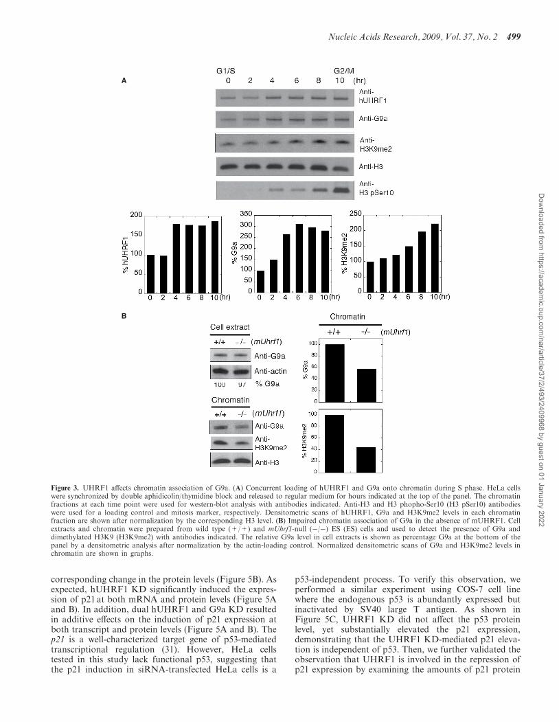
UHRF1 plays a role in chromatin association of G9a
We have previously shown that UHRF1 recruits DNMT1to chromatin (9). This finding led us to speculate thatUHRF1 may assist in the recruitment of G9a onto chro-matin by direct physical interaction. To validate thishypothesis, we first examined the chromatin-loading pat-terns of hUHRF1 and G9a in HeLa cells after synchro-nization at G1/S border and release from the arrest forgiven hours. Consistent with the colocalization data(Figure 2A), hUHRF1 and G9a displayed an increase inchromatin association over time (Figure 3A). The progres-sive chromatin loading patterns of hUHRF1 and G9awere followed by the gradual increase in histone H3-K9dimethylation (H3K9me2) that is the reaction product ofG9a. Next, we investigated if the absence of UHRF1 pro-tein would affect the overall chromatin association of G9aby examining the chromatin fractions isolated from mousewild-type and mUhrf1-null (�/�) ES cells. The G9a pro-tein levels in cell extracts were similar in both ES cell lines,whereas the chromatin fractions from mUhrf1-null EScells contained a significantly lower amount of G9a thanthose from wild-type cells, along with the correspondingdecrease in H3K9me2 (Figure 3B). These data suggest thatUHRF1 plays a role in chromatin association of G9a.
hUHRF1 cooperates with G9a to enhance thetranscriptional repression
G9a has been shown to be involved in transcriptionalrepression in euchromatin (23,25–29). We hypothesizedthat hUHRF1 might recruit G9a to specific promotersin euchromatin to suppress the transcription. To test thispossibility, reporter assays were devised using a Gal4-driven luciferase construct (pG5-cdc2-luc) with variedcombinations of Gal4DBD-fused hUHRF1 (G4-hUHRF1) and GFP-G9a plasmids. The low basal activityof pG5luc reporter containing a minimal TATA box madeit difficult to detect any distinct decrease in luciferaseactivity possibly mediated by hUHRF1. To allow for aclear detection of transcriptional repression mediated by
496 Nucleic Acids Research, 2009, Vol. 37, No. 2
Dow
nloaded from https://academ
ic.oup.com/nar/article/37/2/493/2409968 by guest on 01 January 2022
hUHRF1 and/or G9a, we engineered the pG5luc reporterby replacing the intrinsic minimal promoter with a partialcdc2 promoter fragment containing several Sp1 sites (20),creating the pG5-cdc2-luc plasmid. Transfection withincreasing amounts of G4-hUHRF1 alone resulted in pro-gressive repression of the reporter gene expression(Figure 4A). To determine if hUHRF1 can cooperatewith G9a to enhance the transcriptional inhibition of thereporter, increasing amounts of GFP-G9a were
cotransfected to COS-7 cells with or without a constantamount of G4-hUHRF1. In the absence of G4-hUHRF1,exogenous GFP-G9a had little effects on the repression ofthe reporter gene transcription (Figure 4B, lanes 6–8)while it significantly suppressed the transcription of thereporter in a dose-dependent manner in the presence ofG4-hUHRF1 (Figure 4B, lanes 3–5), suggesting that theG9a-mediated transcriptional inhibition is dependent onthe presence of hUHRF1. To determine whether
A B
C
D
Figure 1. Physical interaction of hUHRF1 and G9a. (A) Coimmunoprecipitation of hUHRF1 and G9a in cell extracts from COS-7 cells transfectedwith GFP control or GFP-G9a. The antibodies used for immunoprecipitation (IP) are indicated at the top of the panel. Western-blot analysis ofimmunoprecipitates was performed with antibodies as indicated. The input shows 2% of each lysate. (B) Coimmunoprecipitation of endogenoushUHRF1 and G9a in HEK293 cell extracts. HEK293 cells were synchronized to late S phase by serum starvation for 20 h and the subsequent releaseinto 10% FBS-containing medium for 15 h before cell harvest. Antibodies used for immunoprecipitation and western blot are shown. The inputrepresents 2% of each lysate. (C) Direct binding of hUHRF1 to G9a and mapping of the G9a-binding region on hUHRF1 using GST fusions ofhUHRF1 fragments. Various domains of hUHRF1 are indicated along with a schematic presentation of the GST fusion constructs marked withamino acid numbers. The blot from GST pull-down assay was probed with anti-G9a antibody and stained with Ponceau solution to visualize thetransferred proteins. Positions of fusion proteins are marked with asterisks. (D) Mapping of hUHRF1-binding region on G9a. Schematic diagram ofthe various GST-fusion constructs of G9a is shown with amino acid numbers. Western blot with anti-hUHRF1 antibody is shown along with thecorresponding Ponceau-stained membrane.
Nucleic Acids Research, 2009, Vol. 37, No. 2 497
Dow
nloaded from https://academ
ic.oup.com/nar/article/37/2/493/2409968 by guest on 01 January 2022
hUHRF1 affects the G9a-mediated transcriptional repres-sion by additional mechanisms other than physical recruit-ment of G9a to the promoter, we further examined thepossibility that hUHRF1 may modulate G9a methyltrans-ferase activity by its interaction with G9a. Using the pur-ified hUHRF1 and G9a that were used for GST pull-downassays (Figure 1C and D), G9a methyltransferase activitywas measured in vitro in the presence or absence ofhUHRF1, and no significant difference in activity wasobserved (data not shown). These results provide the evi-dence supporting that hUHRF1 cooperates with G9a toenhance the transcriptional repression primarily byrecruiting G9a to the target promoters.
hUHRF1 suppresses p21 expression
Several studies demonstrated that hUHRF1 protein levelsare elevated in tumor tissues (8,11,30), and the protein isrequired for proliferation in cancer cells (2,8,11). ThehUHRF1 was also shown to bind to the methylated pro-moter of tumor suppressors such as p14 and p16 (8),
possibly to suppress the expression of these genes incancer cells. However, the same study showed thathUHRF1 expression level did not have any correlationwith the expression of these cell-cycle regulators in prolif-erating cells, suggesting that there might be additionalhUHRF1 targets involved in cell-cycle regulation to pro-mote proliferation in cancer cells. Previously, it was shownthat G9a cooperates with CDP/cut and Gfi1 transcrip-tional regulators to suppress p21 expression (23,28). Onthe basis of these prior observations, we hypothesized thatp21 promoter may be one of the in vivo targets to whichhUHRF1 directs G9a to enhance the repression of thetumor suppressor, thus promoting proliferation incancer cells. To test this hypothesis, we examined thep21 mRNA and protein levels after the individual KDof hUHRF1 and G9a in HeLa cells by siRNA transfec-tion. The G9a KD resulted in �60% increase in p21mRNA level determined by quantitative RT–PCR, com-pared to the CTL KD (Figure 5A). Although the foldincrease is relatively small (<2-fold), it was a statisticallysignificant change that was reflected by the roughly
Figure 2. Colocalization of UHRF1 and G9a. (A) Subnuclear localization of GFP-hUHRF1 and DsRed-G9a transiently expressed in COS-7 cells.Cells were synchronized with aphidicolin and released from G1 arrest for a given number of hours through S phase. Nuclei were visualized withHoechst stain. (B) Colocalization of GFP-mUHRF1 and DsRed-G9a in COS-7 cells at given hours of release from synchronization.
498 Nucleic Acids Research, 2009, Vol. 37, No. 2
Dow
nloaded from https://academ
ic.oup.com/nar/article/37/2/493/2409968 by guest on 01 January 2022
corresponding change in the protein levels (Figure 5B). Asexpected, hUHRF1 KD significantly induced the expres-sion of p21 at both mRNA and protein levels (Figure 5Aand B). In addition, dual hUHRF1 and G9a KD resultedin additive effects on the induction of p21 expression atboth transcript and protein levels (Figure 5A and B). Thep21 is a well-characterized target gene of p53-mediatedtranscriptional regulation (31). However, HeLa cellstested in this study lack functional p53, suggesting thatthe p21 induction in siRNA-transfected HeLa cells is a
p53-independent process. To verify this observation, weperformed a similar experiment using COS-7 cell linewhere the endogenous p53 is abundantly expressed butinactivated by SV40 large T antigen. As shown inFigure 5C, UHRF1 KD did not affect the p53 proteinlevel, yet substantially elevated the p21 expression,demonstrating that the UHRF1 KD-mediated p21 eleva-tion is independent of p53. Then, we further validated theobservation that UHRF1 is involved in the repression ofp21 expression by examining the amounts of p21 protein
A
B
− −
− −
− −
Figure 3. UHRF1 affects chromatin association of G9a. (A) Concurrent loading of hUHRF1 and G9a onto chromatin during S phase. HeLa cellswere synchronized by double aphidicolin/thymidine block and released to regular medium for hours indicated at the top of the panel. The chromatinfractions at each time point were used for western-blot analysis with antibodies indicated. Anti-H3 and H3 phopho-Ser10 (H3 pSer10) antibodieswere used for a loading control and mitosis marker, respectively. Densitometric scans of hUHRF1, G9a and H3K9me2 levels in each chromatinfraction are shown after normalization by the corresponding H3 level. (B) Impaired chromatin association of G9a in the absence of mUHRF1. Cellextracts and chromatin were prepared from wild type (+/+) and mUhrf1-null (�/�) ES (ES) cells and used to detect the presence of G9a anddimethylated H3K9 (H3K9me2) with antibodies indicated. The relative G9a level in cell extracts is shown as percentage G9a at the bottom of thepanel by a densitometric analysis after normalization by the actin-loading control. Normalized densitometric scans of G9a and H3K9me2 levels inchromatin are shown in graphs.
Nucleic Acids Research, 2009, Vol. 37, No. 2 499
Dow
nloaded from https://academ
ic.oup.com/nar/article/37/2/493/2409968 by guest on 01 January 2022
in cell extracts of mouse wild-type and mUhrf1-null (�/�)ES cells. As shown in Figure 5D, the p21 protein levelwas found to be elevated in the mUhrf1(�/�) cells.Furthermore, mUhrf1(�/�) ES cells displayed retarded
growth and less BrdU (bromodeoxyuridine) incorporationcompared to the wild-type counterpart although it is notclear whether these effects directly resulted from theincreased p21 expression in mUhrf1(�/�) ES cells(Figure 5E and F).
hUHRF1 cooperates with G9a to enhance the transcriptionalrepression of p21 promoter
We also examined whether hUHRF1 is recruited to thep21 promoter by reporter assays using pGL2–p21. If thep21 promoter is one of the natural promoters targeted byhUHRF1, it should be recruited to the naive promoterwithout using the Gal4 reporter system that was used inFigure 4. Indeed, exogenous expression of hUHRF1inhibited the p21 promoter activity in a dose-dependentmanner, possibly by recruiting the endogenous epigeneticregulators including G9a to the p21 promoter (Figure 6A).Cotransfection of increasing amounts of G9a expressionvector with a constant amount of hUHRF1 plasmidresulted in further repression of the reporter (Figure 6B,lanes 3–5), compared to the case without the exogenoushUHRF1 (Figure 6B, lanes 6–8). The dose-dependentdecrease in p21 promoter activity in the absence of exo-genous hUHRF1 may have been caused by the recruit-ment of endogenous hUHRF1 to the promoter(Figure 6B, lanes 6–8). To validate that the UHRF1-mediated p21 promoter repression resulted from thedirect interaction between G9a and UHRF1, a G9amutant lacking the N-terminal UHRF1-interactingregion (21) was used for the identical reporter assay inparallel with wild-type G9a. As shown in Figure 6C, thedeletion of UHRF1-interaction region on G9a (NiG9a)impaired UHRF1/G9a-mediated repression of p21 pro-moter. Taken together, p21 appears to be one of thein vivo targets whose expression is repressed by coordi-nated actions of UHRF1/G9a.
hUHRF1 recruits G9a and other chromatin-modifyingenzymes to p21 promoter
To demonstrate that hUHRF1-mediated G9a recruitmentto the endogenous p21 promoter is one of the mechanismsunderlying p21 repression in HeLa cells, we transfectedHeLa cells with hUHRF1 or control siRNAs and per-formed ChIP with selected antibodies. The hUHRF1and G9a were found to associate with the proximalregion of the p21 promoter in CTL KD (transfectionwith control siRNA), suggesting that these proteins arerecruited to the p21 promoter under the native conditions(Figure 7B). In agreement with G9a association with thepromoter, dimethylation at histone H3K9 (H3K9me2)was detected in CTL KD. Furthermore, DNMT1 wasalso found to associate with the proximal region of thepromoter. As demonstrated by the previous studies,the proximal region of the p21 promoter also displayedthe association with HDAC1 (32). None of these enzymeswere found on the distal region of the promoter. Then, weexamined the effects of hUHRF1 on p21 promoter asso-ciation of G9a and other chromatin modifiers describedearlier. As expected, hUHRF1 KD significantly reducedthe amount of hUHRF1 bound to the proximal region of
A
B
Figure 4. hUHRF1 cooperates with G9a to enhance transcriptionalrepression. (A) Transcriptional repression of luciferase reporter geneby hUHRF1. COS-7 cells were cotransfected with a Gal4-driven luci-ferase reporter (2mg) and increasing amounts of Gal4DBD-hUHRF1(G4-hUHRF1). Luciferase activity was measured 48 h post-transfectionand represented by the means� SD of duplicate determinations fromthree independent experiments. (B) Enhanced transcriptional repressionby hUHRF1-mediated G9a recruitment. Luciferase activities were mea-sured after 48 h of transfection using COS-7 cells cotransfected with thesame reporter in (A), increasing amounts of EGFP-G9a (0.1–1 mg) andwith or without a constant amount (0.1 mg) of G4-hUHRF1. The datarepresent the means� SD of duplicate determinations from four sepa-rate experiments.
500 Nucleic Acids Research, 2009, Vol. 37, No. 2
Dow
nloaded from https://academ
ic.oup.com/nar/article/37/2/493/2409968 by guest on 01 January 2022
the p21 promoter compared to the CTL KD. ThehUHRF1 KD also decreased G9a association with thep21 promoter resulting in reduced H3K9me2 modificationon it, suggesting that G9a loading to the promoter isdependent on hUHRF1. We have previously shown that
hUHRF1 KD impairs the recruitment of DNMT1 ontochromatin at global levels (9). Consistent with thisfinding, hUHRF1 KD reduced the DNMT1 associationwith the p21 promoter. In addition, HDAC1 associationwas also decreased upon hUHRF1 KD (Figure 7B).
A
B
C
D
E
F
Figure 5. hUHRF1 suppresses p21 expression in cooperation with G9a. (A) Quantitative RT–PCR analysis of p21 expression. Total RNA wasisolated from HeLa cells transfected with siRNAs as indicated. The Q-PCR data normalized by GAPDH control are shown as the means� SD oftriplicate determinations from four independent experiments. Statistical significance of the differences among the groups was determined by Student’st-test. �P< 0.05; ��P< 0.01. (B) Western blot analysis of p21 expression in siRNA-mediated KD HeLa cells. After each KD, as indicated at the topof the panel, cell extracts were used for detection of p21. The densitometric scan of p21 expression is shown at the right. (C) Western blot analysis ofp53 and p21 expression in COS-7 cells after siRNA-mediated KD of hUHRF1. The densitometric scan of p21 expression is shown at the right. (D)Enhanced p21 expression in mUhrf1�/� ES cells. The blot for p21 is shown along with the densitometric analysis. (E) Growth of wild type (+/+)and mUhrf1-null (�/�) ES cells. Cell growth was monitored over 3 days after plating, and the data represent the means�SD of six replicates. (F)BrdU incorporation of wild-type (+/+) and mUhrf1-null (�/�) ES cells. After 24 h incubation, BrdU labeling was performed for 2 h and determinedby a colorimetric assay. The data represent the means � SD of three separate experiments. Statistical significance of the difference between thegroups was determined by Student’s t-test. �P< 0.05.
Nucleic Acids Research, 2009, Vol. 37, No. 2 501
Dow
nloaded from https://academ
ic.oup.com/nar/article/37/2/493/2409968 by guest on 01 January 2022
Next, we investigated whether G9a and its histone methy-lation can affect the loading of hUHRF1 to the promoter,because hUHRF1 was recently identified as a methyl K9-specific histone H3-binding protein (4). Therefore, G9aprotein level was reduced by siRNA transfection and therelative p21 promoter occupancy of hUHRF1 was exam-ined by quantitative ChIP analysis. As shown inFigure 7C, G9a KD moderately impaired the hUHRF1association with the promoter (�20% compared to theCTL KD), whereas hUHRF1 KD more profoundly
(�55%) disrupted the G9a binding to the promoter andreduced the H3K9me2 to a comparable level to that ofG9a KD. Previously, we have also shown that siRNA-mediated KD of DNMT1 impairs G9a loading ontochromatin (19). Moreover, there are several studiesdemonstrating the interdependency between histone andDNA methylation (33–35). To test whether DNMT1 KDcan negatively affect G9a recruitment onto the p21 pro-moter, HeLa cells were transfected with DNMT1 or con-trol siRNAs. After DNMT1 KD, the promoter occupancy
A
C
B
Figure 6. hUHRF1 cooperates with G9a to enhance the transcriptional repression of p21 promoter. (A) hUHRF1-mediated transcriptional repres-sion of the p21 promoter-luciferase reporter. COS-7 cells were cotransfected with the reporter (2 mg) and increasing amounts of EGFP-hUHRF1 asindicated at the bottom of the panel. (B) Enhanced transcriptional repression by G9a in the presence of exogenous hUHRF1. Luciferase activitieswere measured from COS-7 cells cotransfected with the same reporter described earlier and increasing amounts of EGFP-G9a (0.1–1 mg) with orwithout a constant amount (0.4 mg) of EGFP-hUHRF1. The luciferase assays were performed as described in Figure 4, and the data represent themeans � SD of duplicate determinations from three separate experiments. Western blot analyses of hUHRF1 and G9a expression by anti-GFPantibody are shown for each cotransfection group. (C) Loss of interaction between UHRF1 and G9a abolishes the UHRF1/G9a-mediated repressionof p21 promoter. Reporter assays were performed as described in (B), using the wild-type G9a plasmid (EGFP-G9a) and its deletion mutant lackingthe N-terminal UHRF1-interacting region (EGFP-N�G9a). Expression of hUHRF1 and G9a/N�G9a is shown by western blot analyses with anti-GFP antibody.
502 Nucleic Acids Research, 2009, Vol. 37, No. 2
Dow
nloaded from https://academ
ic.oup.com/nar/article/37/2/493/2409968 by guest on 01 January 2022
of DNMT1 was substantially decreased, whereas G9aassociation and H3K9me2 modification were not affectedmuch compared to CTL KD (Figure 7D). These resultsindicate that DNMT1 makes little contribution to thephysical recruitment of G9a to p21 promoter. In addition,DNMT1 KD did not appear to affect UHRF1 loadingonto the promoter significantly (Figure 7D). Consistentwith this observation, bisulfite sequencing of the p21 pro-moter region (�398 to +11) revealed that the CpGswithin the sequence is infrequently methylated except for
one CpG dinucleotide at �371 position which was methy-lated by 50% (Figure S2). Although the functional signif-icance of DNMT1 recruitment onto the p21 promoter isnot clear given the infrequent methylation patterns on thep21 promoter, these results suggest that UHRF1 recruit-ment onto p21 promoter may use additional mechanismsother than CpG methylation.
DISCUSSION
In mammalian cells, gene expression is regulated via epi-genetic mechanisms. Some mechanisms involve covalentmodifications on DNA and histone molecules on the chro-matin to create either a permissive or a repressive tran-scriptional environment. Given the emerging evidencesupporting the role of UHRF1 as a transcriptional regu-lator (2,16,17) and its interaction with a histone methyl-transferase G9a in this study, we hypothesized thatUHRF1 might serve as a transcriptional corepressoralong with G9a that has been shown to be involved intranscriptional repression in euchromatin (25). Indeed,UHRF1 alone was capable of repressing reporter genetranscription in a dose-dependent manner, possibly byrecruiting the endogenous epigenetic regulators to thetarget promoters. Consistent with this notion, coexpres-sion of UHRF1 and G9a enhanced the transcriptionalrepression of the reporter genes, suggesting that thedirect physical interaction of UHRF1 and G9a constitutesan effective silencer complex. Among many possibleendogenous target genes regulated by the cooperation ofUHRF1/G9a, we examined the expression of p21 in HeLacells where it is poorly expressed in a p53-independentmanner. The p21 is a cell-cycle regulator by inhibitingcyclin-dependent kinases and a modulator of apoptosisby interacting with other proteins involved in the regula-tion of apoptosis (36). Transcription of p21 gene relies onthe control of multiple different regulators, of which manyare yet to be identified. In this study, UHRF1 appears tobe one of the intrinsic regulators of p21 gene expression.We have demonstrated that the endogenous p21 pro-moter displayed the presence of UHRF1, G9a and otherchromatin-modifying enzymes such as DNMT1 andHDAC1 in HeLa cells by ChIP assays. The siRNA-mediated UHRF1 KD significantly reduced the p21promoter occupancy of all three chromatin-modifyingenzymes, placing UHRF1 as a focal point of recruitmentof these enzymes and also suggesting possible coordinatedefforts of UHRF1, G9a, DNMT1 andHDAC1 for efficientrepression of p21. This observation raises an intriguingquestion on whether all these proteins form a single macro-molecular complex or various distinct complexes depend-ing on the specific promoter architecture and cellularcontext. Gel-filtration chromatography of Jurkat celllysates containing the endogenously expressed proteinsmentioned earlier has revealed that UHRF1 cofractionateswith G9a, DNMT1 and HDAC1 to a various extent, indi-cating the possible formation of the common complexesthat are assembled by UHRF1 (data not shown).A similar coordinated epigenetic repression of p21 was
previously reported, involving a transcriptional regulator,
A
B
C
D
Figure 7. hUHRF1 recruits G9a and other chromatin modificationenzymes to p21 promoter. (A) Linearity of PCR amplification usingprimer sets for proximal (�385 to �240) and distal (�4164 to�3959) regions of p21 promoter with increasing amount of inputDNA. (B) ChIP analysis of p21 promoter after KD of hUHRF1.HeLa cells were transfected with either control siRNA (CTL KD) orhUHRF1 siRNA (hUHRF1 KD). Using the chromatin isolated fromthe KD cells, ChIP was performed to detect the proteins or histonemodification as indicated at the top of the panel. 5% input is shown.(C) Quantitative ChIP analysis for relative p21 promoter occupancy ofhUHRF1, G9a and dimethylated H3K9 (H3K9me2) after KD ofhUHRF1 or G9a. Q-PCR data of each group were normalized to itsinput as % input. The relative p21 promoter occupancy of hUHRF1 orG9a KD samples represents the fold change in percentage input overthat of the CTL KD. Error bars indicate standard deviation of threeindependent experiments. (D) ChIP analysis of p21 promoter after KDof DNMT1. HeLa cells were transfected with either control siRNA(CTL KD) or DNMT1 siRNA (DNMT1 KD). CHIP was performedas described in (B) and 5% input is shown.
Nucleic Acids Research, 2009, Vol. 37, No. 2 503
Dow
nloaded from https://academ
ic.oup.com/nar/article/37/2/493/2409968 by guest on 01 January 2022
Gfi1 (28). The Gfi1 recruits G9a and HDAC1 to p21 pro-moter and represses its expression in HL-60 cells. BothHDAC1 and G9a were found in the repressive complexassembled by Gfi1, and the KD of Gfi1 elevated the p21expression by 2–3-fold. We also observed a similar level ofp21 induction after UHRF1 KD in HeLa and COS-7 cells.Other repressor proteins can also modulate p21 expressionby epigenetic mechanisms. For example, a transcriptionfactor CDP/cut was shown to recruit G9a to the humanp21 promoter, and the transcriptional repression functionof CDP/cut is mediated through the activity of its asso-ciated G9a (23). Polycomb group (PcG) proteins are chro-matin modifiers that can transcriptionally silence theirtarget genes and maintain them in the repressed statethrough cell divisions during development (37). Recently,NSPc1, a transcriptional repressor homologous with PcGprotein Bmi-1, was demonstrated to repress p21 expres-sion via direct binding to the retinoid acid response ele-ment of its promoter (38).Furthermore, the murine Uhrf1-null ES cells also dis-
played a higher level of p21 protein, implicating the role ofUHRF1 in transcriptional repression in ES cell environ-ment. An orphan nuclear receptor TLX recruits a set ofHDACs to its target genes for transcriptional repression inneural stem cells (39). One of the genes targeted by thisrepressive complex is p21, suggesting a role very similar tothat of UHRF1-G9a repressive complex. Both histoneH3K9me2 accumulation via G9a and deacetylation byHDACs are found on many repressed genes in cell lines.As siRNA-mediated KD of G9a or inhibition of HDACscan result in p21 derepression, it is also plausible thatHDACs and G9a crosstalk during p21 repression. Infact, a recent study on SHP-mediated regulation ofCYP7A1 promoter revealed the functional interplaybetween SHP-recruited HDACs and G9a in altering thechromatin structure of CYP7A1 promoter (29). Thesefindings suggest multiple mechanisms of p21 gene expres-sion that is modulated by both histone methylation anddeacetylation. Moreover, as exemplified earlier, the pre-sence of multiple functional equivalents of UHRF1 thatcan recruit similar effectors such as G9a and HDAC1appears to ensure tight repression of p21 in cancer cells.Perhaps, this may constitute the basis for the finding thatfunctional disruption of a single factor in p21 repressiononly results in relatively minor p21 derepression (�2–3-fold) as we and other investigators observed.The UHRF1-mediated p21 repression presents another
intriguing idea of reciprocal regulation occurring betweenUHRF1 and p21 expression in response to different extra-cellular stimuli, because hUHRF1 expression was shownto be down-regulated by p53/p21-dependent DNAdamage check point signal (40). This observation maylead to a hypothesis that, in the presence of DNAdamage, the p53/p21-dependent checkpoint responsemay outweigh the transcriptional repression effects ofUHRF1 against p21 resulting in down-regulation ofUHRF1 expression, whereas the absence of DNAdamage signal allows UHRF1 to keep p21 in a repressedstate promoting cell proliferation. The reciprocal down-regulation, involving UHRF1, can be observed in anothercase where overexpression of hUHRF1 negatively
regulates pRb expression (16), whereas pRb interactswith E2F-1 transcription factor and thereby inhibits theexpression of the E2F-1 target genes including UHRF1(8,30). These findings point to the dynamic regulationmode for UHRF1 in cancer cells.
In summary, our data demonstrate a new role ofUHRF1 as a transcriptional corepressor in recruitmentof histone methyltransferase G9a and other chromatin-modifying enzymes to target promoters, and suggest thatUHRF1 acts as a focal point of gene repression mediatedby various chromatin modifiers.
SUPPLEMENTARY DATA
Supplementary Data are available at NAR Online.
ACKNOWLEDGEMENTS
We thank Dr Jane B. Trepel for kindly providing thepGL2–p21 plasmid. We also thank George R. Feeheryand other colleagues of our laboratory for technical assis-tance and support. We are grateful to Drs D.G. Comb andRich Roberts at New England Biolabs, Inc. for their sup-port and encouragement. S. E. J. is an Investigator of theHoward Hughes Medical Institute.
FUNDING
Funding for open access charge: New England Biolabs, Inc.
Conflict of interest statement. None declared.
REFERENCES
1. Bronner,C., Achour,M., Arima,Y., Chataigneau,T., Saya,H. andSchini-Kerth,V.B. (2007) The UHRF family: oncogenes that aredrugable targets for cancer therapy in the near future? Pharmacol.Ther., 115, 419–434.
2. Hopfner,R., Mousli,M., Jeltsch,J.M., Voulgaris,A., Lutz,Y.,Marin,C., Bellocq,J.P., Oudet,P. and Bronner,C. (2000) ICBP90, anovel human CCAAT binding protein, involved in the regulation oftopoisomerase IIalpha expression. Cancer Res., 60, 121–128.
3. Muto,M., Utsuyama,M., Horiguchi,T., Kubo,E., Sado,T. andHirokawa,K. (1995) The characterization of the monoclonal anti-body Th-10a, specific for a nuclear protein appearing in the S phaseof the cell cycle in normal thymocytes and its unregulated expres-sion in lymphoma cell lines. Cell Prolif., 28, 645–657.
4. Karagianni,P., Amazit,L., Qin,J. and Wong,J. (2008) ICBP90, anovel methyl K9 H3 binding protein linking protein ubiquitinationwith heterochromatin formation. Mol. Cell Biol., 28, 705–717.
5. Citterio,E., Papait,R., Nicassio,F., Vecchi,M., Gomiero,P.,Mantovani,R., Di Fiore,P.P. and Bonapace,I.M. (2004) Np95 is ahistone-binding protein endowed with ubiquitin ligase activity. Mol.Cell Biol., 24, 2526–2535.
6. Papait,R., Pistore,C., Negri,D., Pecoraro,D., Cantarini,L. andBonapace,I.M. (2007) Np95 is implicated in pericentromeric het-erochromatin replication and in major satellite silencing. Mol. BiolCell., 18, 1098–1106.
7. Papait,R., Pistore,C., Grazini,U., Babbio,F., Cogliati,S.,Pecoraro,D., Brino,L., Morand,A.L., Dechampesme,A.M.,Spada,F. et al. (2008) The PHD Domain of Np95 (mUHRF1) isinvolved in large-scale reorganization of pericentromeric hetero-chromatin. Mol. Biol Cell., 19, 3554–3563.
8. Unoki,M., Nishidate,T. and Nakamura,Y. (2004) ICBP90, an E2F-1 target, recruits HDAC1 and binds to methyl-CpG through itsSRA domain. Oncogene, 23, 7601–7610.
504 Nucleic Acids Research, 2009, Vol. 37, No. 2
Dow
nloaded from https://academ
ic.oup.com/nar/article/37/2/493/2409968 by guest on 01 January 2022
9. Bostick,M., Kim,J.K., Esteve,P.O., Clark,A., Pradhan,S. andJacobsen,S.E. (2007) UHRF1 plays a role in maintaining DNAmethylation in mammalian cells. Science, 317, 1760–1764.
10. Woo,H.R., Pontes,O., Pikaard,C.S. and Richards,E.J. (2007) VIM1,a methylcytosine-binding protein required for centromeric hetero-chromatinization. Genes Dev., 21, 267–277.
11. Jenkins,Y., Markovtsov,V., Lang,W., Sharma,P., Pearsall,D.,Warner,J., Franci,C., Huang,B., Huang,J., Yam,G.C. et al. (2005)Critical role of the ubiquitin ligase activity of UHRF1, a nuclearRING finger protein, in tumor cell growth. Mol. Biol Cell., 16,5621–5629.
12. Kouzarides,T. (2007) Chromatin modifications and their function.Cell, 128, 693–705.
13. Achour,M., Jacq,X., Ronde,P., Alhosin,M., Charlot,C.,Chataigneau,T., Jeanblanc,M., Macaluso,M., Giordano,A.,Hughes,A.D. et al. (2008) The interaction of the SRA domain ofICBP90 with a novel domain of DNMT1 is involved in the regu-lation of VEGF gene expression. Oncogene, 27, 2187–2197.
14. Sharif,J., Muto,M., Takebayashi,S., Suetake,I., Iwamatsu,A.,Endo,T.A., Shinga,J., Mizutani-Koseki,Y., Toyoda,T., Okamura,K.et al. (2007) The SRA protein Np95 mediates epigenetic inheritanceby recruiting Dnmt1 to methylated DNA. Nature, 450, 908–912.
16. Jeanblanc,M., Mousli,M., Hopfner,R., Bathami,K., Martinet,N.,Abbady,A.Q., Siffert,J.C., Mathieu,E., Muller,C.D. and Bronner,C.(2005) The retinoblastoma gene and its product are targeted byICBP90: a key mechanism in the G1/S transition during the cellcycle. Oncogene, 24, 7337–7345.
17. Macaluso,M., Montanari,M., Noto,P.B., Gregorio,V., Bronner,C.and Giordano,A. (2007) Epigenetic modulation of estrogen recep-tor-alpha by pRb family proteins: a novel mechanism in breastcancer. Cancer Res., 67, 7731–7737.
18. Jin,P., Hardy,S. and Morgan,D.O. (1998) Nuclear localization ofcyclin B1 controls mitotic entry after DNA damage. J. Cell Biol.,141, 875–885.
19. Esteve,P.O., Chin,H.G., Smallwood,A., Feehery,G.R.,Gangisetty,O., Karpf,A.R., Carey,M.F. and Pradhan,S. (2006)Direct interaction between DNMT1 and G9a coordinates DNA andhistone methylation during replication. Genes Dev., 20, 3089–3103.
20. Le Gac,G., Esteve,P.O., Ferec,C. and Pradhan,S. (2006) DNAdamage-induced down-regulation of human Cdc25C and Cdc2 ismediated by cooperation between p53 and maintenance DNA(cytosine-5) methyltransferase 1. J. Biol. Chem., 281, 24161–24170.
21. Esteve,P.O., Patnaik,D., Chin,H.G., Benner,J., Teitell,M.A. andPradhan,S. (2005) Functional analysis of the N- and C-terminus ofmammalian G9a histone H3 methyltransferase. Nucleic Acids Res.,33, 3211–3223.
22. Kim,G.D., Ni,J., Kelesoglu,N., Roberts,R.J. and Pradhan,S. (2002)Co-operation and communication between the human maintenanceand de novo DNA (cytosine-5) methyltransferases. EMBO J., 21,4183–4195.
23. Nishio,H. and Walsh,M.J. (2004) CCAAT displacement protein/cuthomolog recruits G9a histone lysine methyltransferase to represstranscription. Proc. Natl Acad. Sci. USA, 101, 11257–11262.
24. Lee,S.J., Ha,M.J., Lee,J., Nguyen,P., Choi,Y.H., Pirnia,F.,Kang,W.K., Wang,X.F., Kim,S.J. and Trepel,J.B. (1998) Inhibitionof the 3-hydroxy-3-methylglutaryl-coenzyme A reductase pathwayinduces p53-independent transcriptional regulation of p21(WAF1/CIP1) in human prostate carcinoma cells. J. Biol. Chem., 273,10618–10623.
25. Tachibana,M., Sugimoto,K., Nozaki,M., Ueda,J., Ohta,T.,Ohki,M., Fukuda,M., Takeda,N., Niida,H., Kato,H. et al. (2002)G9a histone methyltransferase plays a dominant role in euchromatichistone H3 lysine 9 methylation and is essential for early embryo-genesis. Genes Dev., 16, 1779–1791.
26. Boulias,K. and Talianidis,I. (2004) Functional role of G9a-inducedhistone methylation in small heterodimer partner-mediated tran-scriptional repression. Nucleic Acids Res., 32, 6096–6103.
27. Gyory,I., Wu,J., Fejer,G., Seto,E. and Wright,K.L. (2004) PRDI-BF1 recruits the histone H3 methyltransferase G9a in transcrip-tional silencing. Nat. Immunol., 5, 299–308.
28. Duan,Z., Zarebski,A., Montoya-Durango,D., Grimes,H.L. andHorwitz,M. (2005) Gfi1 coordinates epigenetic repression ofp21Cip/WAF1 by recruitment of histone lysinemethyltransferase G9a and histone deacetylase 1. Mol. Cell Biol.,25, 10338–10351.
29. Fang,S., Miao,J., Xiang,L., Ponugoti,B., Treuter,E. andKemper,J.K. (2007) Coordinated recruitment of histone methyl-transferase G9a and other chromatin-modifying enzymes in SHP-mediated regulation of hepatic bile acid metabolism. Mol. Cell Biol.,27, 1407–1424.
30. Mousli,M., Hopfner,R., Abbady,A.Q., Monte,D., Jeanblanc,M.,Oudet,P., Louis,B. and Bronner,C. (2003) ICBP90 belongs to a newfamily of proteins with an expression that is deregulated in cancercells. Br. J. Cancer, 89, 120–127.
31. el-Deiry,W.S., Tokino,T., Velculescu,V.E., Levy,D.B., Parsons,R.,Trent,J.M., Lin,D., Mercer,W.E., Kinzler,K.W. and Vogelstein,B.(1993) WAF1, a potential mediator of p53 tumor suppression. Cell,75, 817–825.
32. Ocker,M. and Schneider-Stock,R. (2007) Histone deacetylase inhi-bitors: signalling towards p21cip1/waf1. Int. J. Biochem. Cell Biol.,39, 1367–1374.
33. Xin,Z., Tachibana,M., Guggiari,M., Heard,E., Shinkai,Y. andWagstaff,J. (2003) Role of histone methyltransferase G9a in CpGmethylation of the Prader-Willi syndrome imprinting center. J. Biol.Chem., 278, 14996–15000.
34. Nguyen,C.T., Weisenberger,D.J., Velicescu,M., Gonzales,F.A.,Lin,J.C., Liang,G. and Jones,P.A. (2002) Histone H3-lysine 9methylation is associated with aberrant gene silencing in cancer cellsand is rapidly reversed by 5-aza-2’-deoxycytidine. Cancer Res., 62,6456–6461.
35. Espada,J., Ballestar,E., Fraga,M.F., Villar-Garea,A., Juarranz,A.,Stockert,J.C., Robertson,K.D., Fuks,F. and Esteller,M. (2004)Human DNA methyltransferase 1 is required for maintenance ofthe histone H3 modification pattern. J. Biol. Chem., 279,37175–37184.
36. Gartel,A.L. (2005) The conflicting roles of the cdk inhibitorp21(CIP1/WAF1) in apoptosis. Leuk. Res., 29, 1237–1238.
37. Schuettengruber,B., Chourrout,D., Vervoort,M., Leblanc,B. andCavalli,G. (2007) Genome regulation by polycomb and trithoraxproteins. Cell, 128, 735–745.
38. Gong,Y., Yue,J., Wu,X., Wang,X., Wen,J., Lu,L., Peng,X.,Qiang,B. and Yuan,J. (2006) NSPc1 is a cell growth regulator thatacts as a transcriptional repressor of p21Waf1/Cip1 via the RAREelement. Nucleic Acids Res., 34, 6158–6169.
39. Sun,G., Yu,R.T., Evans,R.M. and Shi,Y. (2007) Orphan nuclearreceptor TLX recruits histone deacetylases to repress transcriptionand regulate neural stem cell proliferation. Proc. Natl Acad. Sci.USA, 104, 15282–15287.
40. Arima,Y., Hirota,T., Bronner,C., Mousli,M., Fujiwara,T., Niwa,S.,Ishikawa,H. and Saya,H. (2004) Down-regulation of nuclear proteinICBP90 by p53/p21Cip1/WAF1-dependent DNA-damage check-point signals contributes to cell cycle arrest at G1/S transition.Genes Cells, 9, 131–142.
Nucleic Acids Research, 2009, Vol. 37, No. 2 505
Dow
nloaded from https://academ
ic.oup.com/nar/article/37/2/493/2409968 by guest on 01 January 2022