+ All Categories
Home > Documents > UJ').V.,. IIBUOT

UJ').V.,. IIBUOT

Date post: 08-Apr-2022
Category:
Upload: others
View: 0 times
Download: 0 times
Share this document with a friend
51
UJ').V.,. IIBUOT_ ~IOTEEJ< VER\,vYDER WORD NIE University Free State I~IIIIIIIIIIIIIIIIIIIIIIIIIIIIIIIIIIIIIIIIIIIIIIIIIIII1111111111111111111111111 34300001324346 Universiteit Vrystaat .N 01\iSTANDIGHEDE UIT DIE
Transcript

UJ').V.,. IIBUOT_

~IOTEEJ< VER\,vYDER WORD NIE

University Free State

I~IIIIIIIIIIIIIIIIIIIIIIIIIIIIIIIIIIIIIIIIIIIIIIIIIIII111111111111111111111111134300001324346

Universiteit Vrystaat

.N 01\iSTANDIGHEDE UIT DIE

THE USE OF NEUROPSYCHOLOGICAL ASSESSMENT IN THE DIAGNOSIS

OF CEREBRAL LESIONS

Renata Schoeman

This thesis is submitted to fulfil the requirements of the degree M.Soc.Sc.

(Psychology) in the Faculty of Social Sciences, Department of Psychology,

University of the Free State. It is submitted in the form of two articles as

permitted in the regulations of this institution.

As in the general situation where two articles is written by the same author, there

inevitably will be overlapping. In this case the overlapping was mainly restricted

to the method as the same instruments, participants and statistical methods were

used.

Submission date: 29 November 2002

Promotor: Prof D.A.Louw

I declare that the thesis hereby submitted by me for the M.Soc.Sc. (Psychology)

degree at the University of the Free State is my own independent work and has

not previously been submitted by me at another university/faculty.

I furthermore cede copyright of the thesis in favour of the University of the Free

State.

For Dan.

The financial assistance of the National Research Foundation (NRF) towards thisresearch is hereby acknowledged. Opinions expressed and conclusions arrivedat, are those of the author and are not necessarily to be attributed to the NationalResearch Foundation.

1

NEUROPSYCHOLOGICAL ASSESSMENT VERSUS NEURO ..IMAGING IN THE

DIAGNOSIS OF CEREBRAL LESIONS: AN EXPLORATIVE STUDY

ABSTRACT

Certain authors emphasise that, as neuro-imaging techniques seem to be

significantly superior, psychological assessment techniques have no place in

neurological assessment, and that the reliability and validity of these techniques,

regarding the presence and localisation of cerebral damage, are questionable.

The purpose of the study was to determine the relative effectiveness of

neuropsychological assessment in the diagnosis and localisation of cerebral

lesions, compared to magnetic resonance imaging (MRI). An availability sample

of patients was taken from patients whom neurologists and neurosurgeons had

seen and who had either a normal MRI or a MRI with localised lesions. They

were then assessed by means of the South African Wechsler Adult Intelligence

Scale, the Folstein's Mini Mental State Examination, the Bender Gestalt Test,

and the Grassi Block Substitution Test. The test results were interpreted blindly.

The findings are discussed, shortcomings of the study mentioned and

recommendations made.

Neuropsychology; neuro-imaging; cerebral lesions; diagnosis; presence; lesion

localization; South African Wechsler Intelligence Scale; Folstein's Mini Mental

State Examination; Bender Gestalt Test; Grassi Block Substitution Test.

SAMEVATTING

Sekere outeurs beklemtoon die superioriteit van radiologiese beeldingstegnieke

bo dié van sielkundige toetse in die neurologiese evaluasie van pasiënte. Die

betroubaarheid en geldigheid van sielkundige toetse rakende die diagnosering

en lokalisering van breinskade word ook bevraagteken. Die doel van hierdie

verkennende studie was om die effektiwiteit van neurosielkundige assessering in

die diagnose en lokalisering van breinletsels met dié van die MR te vergelyk. 'n

2

Beskikbaarheidsteekproef is getrek van pasiënte met óf 'n normale MR óf 'n MR

wat dui op gelokaliseerde breinletsels. Die proefpersone is evalueer met die

Suid-Afrikaanse Wechsler Intelligensieskaal vir Volwassenes, die Folstein Mini

Mental State Examination, die Bender Gestaltfoets en die Grassi

Blokvervangingstoets. Die toetsresultate is blind geïnterpreteer. Die bevindinge

word bespreek, tekortkominge van die studie word aangetoon en aanbevelings

word gemaak.

Neurosielkunde; neurologiese beelding; breinletsels; diagnose; teenwoordigheid;

letsel lokalisering; Suid-Afrikaanse Wechsler Intelligensie Skaal; Folstein Mini

Mental State Examination; Bender Gestaltfoets; Grassi Blokvervangingstoets.

INTRODUCTION

Prior to the 1970s, the only way for medical and psychological clinicians to localise brain

lesions was by combining their knowledge of anatomy and pathology by means of clinical

examinations and results obtained by means of neuropsychological testing. However, with

the development of neuro-imaging techniques such as computerised axial tomography (CT

or CAT) and magnetic resonance imaging (MR or MRI), the situation changed

dramatically. These techniques have made it possible to obtain clear images of not only

specific brain structures, but also their functioning, for example positron emission

tomography (PET) and single photon emission computed tomography (SPECT). The

specific applicability and even necessity of neuropsychological tests have therefore

become debatable. In this regard Plug1 states that "the question is not whether testing is

perfect (which it obviously is not), but rather how it compares to alternative techniques of

assessment and whether, when used in combination with other processes, it leads to a

more reliable, valid, fair and cost effective result".

As there has been no agreement on what constitutes an injury sufficient enough to affect

the brain, it has proved difficult to obtain reliable estimates of the frequency of closed-head

injuries. However, according to Casson et al.,2 Kolb and Whishaw,3 Lishman" and

Vanderploeo," cerebral trauma is the most common form of brain damage in people under

the age of 40, ranging from about 300-450 per 100 000 of the population per year in

industrialised countries.

3

Certain authors emphasise that, as neurcrimaging techniques seem to be significantly

superior, psychological assessment techniques have no place in neurological assessment,

and that the reliability and validity of these techniques regarding presence and localisation

of cerebral damage are questionable." For example, the ability of the CAT scan to detect

variations in soft-tissue density is significantly higher than that of any psychological test,

while the reliability of several neuropsychological tests leaves much to be desired." 7 In this

context the testimony of neuropsychologists in insurance claims have been ruled

inadmissible in certain cases (e.g. Turkinqton"). The basis of this ruling was that, without

using medical methods, a psychologist could not provide an expert evaluation in cases

where brain damage is suspected.

However, authors such as Murphy and Davidshorer" hold the view that, although

neuropsychological assessment cannot replace medical techniques of assessing brain

damage, such tests should not be totally discarded. "Psychological tests are less risky,

less invasive, and often more cost-effective than medical tests. The optimal use of medical

and non-medical techniques in diagnosing brain damage has yet to be determined" (p.

415).

Against this background, the purpose of the study was to determine the relative

effectiveness of neuropsychological assessment and neurcrimaging. More specifically, to

determine:

1. the agreement between the diagnosis of cerebral lesions with the MRI and the

diagnosis of cerebral lesions by means of neuropsychological assessment;

2. the agreement in localisation of cerebral lesions between MRI and

neuropsychological assessment; and

3. the correlation of different subscales of the psychometric tests with the MRI

diagnosis.

The results will hopefully indicate to what extent neuropsychological tests still have a

function in determining the nature and localisation of cerebral injuries. Such a study is

especially important in the South African context where almost no research has been done

in this regard.

4

UNDERSTANDING OF THE BRAIN THOUGH THE AGES

Evidence shows that people have long been aware of brain-behaviour relationships." 11

Archaeologists have retrieved thousands of skulls with evidence of having undergone

trephination - an ancient surgical operation to relieve pressure related to brain edema after

trauma. Classical Greeks wrote the first accounts of brain-behaviour relationships.

Pythagoras (580-500 B.C.) described the so-called brain hypothesis, namely that the brain

is the source of all reasoning and behaviour. Hippocrates (460-377 B.C.), the father of

modern medicine, was the first to recognise that paralysis occurs contra-lateral to the side

of brain injury. In ancient Egypt, what is known as the "ventricular localisation hypothesis"

or "cell doctrine" was developed. This was an erroneous suggestion that the ventricles

contain spirits responsible for mental faculties and movement. This notion endured for

almost 2000 years. Galen (A.D. 130-201), a Roman anatomist and physician, believed that

all physical functions depend on the balance of bodily fluids or humours, specifically blood,

mucus, and yellow and black bile. He suggested that the brain is a large clot of phlegm

from which a pump forces the psychic pneuma out into the nerves. Vesalius (1514-1564),

through the first systematic dissections of human beings in Europe, demonstrated that

Galen's views were inaccurate. By the 17th century, René Descartes (1596-1650)

hypothesised that the mind and body are separate, but interact with each other. He

believed that mental processes are localised in the pineal gland. Giovanni Lancisi (1654-

1720) viewed the corpus callosum as the seat of mental functions. Beaumont" mentioned

that early investigators were preoccupied with identifying the specific part of the brain that

was the seat of the mind, but that their discussions were based primarily on speculation

and that they had conducted relatively little experimentation.

The first global theory concerning brain functioning was the phrenological theory of Gall

and Spurzheim. In the 19th century Gall (1758-1828) postulated that the brain consists of

a number of different areas, each responsible for different psychological attributes. By

assigning specific functions to certain areas in the cerebral cortex, the localisation theory

of brain function was born. Scientific evidence supporting this localisation theory was first

provided by Broca's (1824-1880) studies on aphasia. He proved that the motor area for

speech was located in the postero-inferior region of the left frontal lobe. A few years later

Wernicke (1848-1904) followed by proving that the area for understanding speech is

located in the postero-superior area of the temporal lobe. Flourens (1794-1867) was one

of the first researchers to suggest an alternative to the localisation theory. He viewed all

5

cerebral matter as being equipotential, that is, if sufficient cortical matter remains intact, it

will take over the functions of any lost brain tissue. According to the equipotential theory,

sensory input may be localised, but perception involves the whole brain. The effects of

brain lesions thus do not depend on their location, but on their extent.7

By means of various experiments with dogs, Goltz (1834-1902) proved that decortication

does not eliminate any function, though it seems to reduce all functions to some extent.

This was a strong argument against the localisation of function.' Jackson (1835-1911)

suggested a third alternative. He believed that behaviour results from interactions among

all the areas of the brain and that certain areas are therefore not uniquely associated with

specific functions. This is known as the hierarchical organisation concept of brain function

or the interactionist theory, according to which each successively higher level controls

more complex aspects of behaviour, but does so through the lower levels.13

Luria integrated these previous theories (1902-1977) in his theory of pluripotentiality, which

states that any given area in the brain can be involved in relatively few or relatively many

behaviours. Luria's hypothesis is particularly attractive to clinical neuropsychologists,

because it can account theoretically for most observations of brain-injured patients. The

theory also explains the observation that certain lesions generally result in consistent

deficits. In addition, through the concept of reorganisation, Luria's theory can account for

individuals who recover from brain trauma. Finally, the theory suggests ways of

establishing rehabilitation and treatment programmes for the brain-injured patient and

provides a strong theoretical basis for understanding clinical neuropsychology."

NEUROPSYCHOLOGICAL ASSESSMENT

Psychological testing developed from two important lines of enquiry, namely the

measurement of individual differences (based on the work of Darwin, Galton and Cattell)

and psychophysics (based on the work of Herbart, Weber, Fechner and Wundt). An

important breakthrough regarding the creation of modern tests arrived at the beginning of

the 20th century when the French started to study ways of identifying intellectually

subnormal individuals in order to provide them with special education. The first intelligence

test, known as the Binet-Simon Scale, followed in 1905. With the onset of World War I, the

testing movement grew enormously and group tests and standardised achievement tests

were developed. In 1916 the well-known Afrikaans poet and author, C. Louis Leipoldt,

played a key roll in the standardisation of South Africa's first intelligence test, based on the

6

Binet-Simon Scale, commonly known as the Moll-Leipoidt Scale." During 1920 to 1940

personality tests began to blossom: first structured tests and later the projective tests such

as the Rorschach test. However, during the 1950s a sharp decline in the use of testing

followed, due to the attacks on, for example, testing's intrusive nature and the possibility of

misuse. This persisted into the 1970s. 3 14

Although there have been endless arguments over the virtues, limitations and utility of

clinical psychological testing during the past two decades, psychological testing once

again moved to the foreground with the emergence of speciality areas such as neuro-,

health, forensic and child psychology. All of these areas depend heavily on the use of

tests.1516

The origin of clinical neuropsychology can be traced to studies by Broca in 1861 and

Wernicke in 1874. They recognised that certain functions, such as the recognition of

speech, are localised in the left hemisphere of the brain." In 1935 Halstead founded a

laboratory to study the impact of impairments in brain functioning on a wide range of

abilities. Reitan joined him in 1944 and thus the Halstead-Reitan Neuropsychological

Battery was developed. Performance on specific subtests of the battery was correlated

with injury in different hemispheres of the brain. Later studies by Reitan and Wolfson also

indicated the ability of the battery to localise lesions as left- or right-hemispheric and

anteriorly or posteriorly located."

Luria17 had a different approach. He saw the brain as a functional system where various

areas interact in certain behaviours. If one area is damaged, various functions will be

affected to different extents. He also introduced the concept of pluripotentiality where any

one centre in the brain can be involved in several different functional systems. The test he

developed became known as the Luria-Nebraska Neuropsychological Battery. Golden 18

summarised a variety of studies that demonstrated that the Luria-Nebraska

Neuropsychological Battery is able to make fine distinctions in respect of

neuropsychological function such as hemispheric localisation of tumours.

Since the 1990s psychologists have had various tests to choose from for

neuropsychological assessment. On the one end of the spectrum there are set test

batteries, such as the Halsteid-Reitan Battery. At the other end there are individually

tailored batteries that psychologists individualise depending on the patient and

circumstances. Somewhere in between are clusters of tests that are used together

because of their availability and practicality. Examples of such clusters are the South

African Wechsler Adult Intelligence Scale (SAWAIS), the Bender Gestalt and Grassi Block

7

Design tests and the Mini Mental State Examination that are widely used in private

practice and institutions to assess the presence and localisation of brain damage. Nell19

also suggested a "core" battery where certain subtests of, for instance, the WAlS, are

used, combined to suit the individuals' case requirements.

Incagnoli20 feels that, in the light of recent neuro-imaging innovations such as CT scans, it

is clear that the focus of the clinical neuropsychologists shifted from lesion localisation to

precise specification of deficits in functioning. She also mentioned that neuropsychologists

would continue to have a role in the pre-operative localisation of lesions in focal epilepsy,

as positive signs are often absent in certain radio- and neurological tests. Casson et al.2

found that neuropsychological testing is a highly sensitive and accurate means of

detecting brain injury. The overall pattern of test results showed a high correlation with

abnormal findings on CT scan and/or EEG and "is an excellent predictor of brain damage"

(p. 2667). This view is supported by Zilmer and Spiers? who postulate that

neuropsychological evaluations are a precursor or complementary to more in-depth

neurological or neuro-imaging procedures that can establish the exact medical or

neurological diagnosis. It has become less important for neuropsychologists to act as

lesion detectors and more important to document the precise effects of brain dysfunction

on behaviour for purposes of remediation and treatment."

NEURO-IMAGING

Risberg21 rightly points out that the gross anatomy of the brain has been depicted

accurately since the Renaissance, but that it took centuries for physicians to be able to see

the living brain.

Since 1895 doctors have been able to take skull X-rays, though these could only show the

bones. Pneumoencephalography (x-rays of the fluid containing structures of the brain by

removing cerebrospinal fluid and replacing it with air, oxygen or helium) and angiography

were developed. This resulted in the ability to delineate both intra- and extra cranial

vessels accurately by intra-arterial injection of contrast and imaging directly by X-ray or by

digital subtraction." Electroencephalography (EEG) was developed in the 1930s and is

still widely used. By means of scalp electrodes it examines the spontaneous electrical

activity of the brain, but it has very little localising value.23

In the 1970s, the development of computerised axial tomography (CAT) scanning was a

great leap forward, allowing physicians to produce pictures of slices of the brain through a

8

non-invasive procedure. A pencil beam of X-ray traverses the patient's head and a

diametrically opposed detector measures the extent of its absorption. Reconstruction of

these differential absorption values on a two-dimensional display provides the

characteristic CAT scan appearance. For routine scanning slices are 5-10 mm wide, but

with high definition imaging views may consist of slices of only 1 mm thick_24

Approximately 20 years later, magnetic resonance imaging (MRI) was developed, in which

large-bore homogenous magnets and computer-assisted imaging are used to map

hydrogen nuclei densities and their effect on the surrounding molecules in vivo. These

densities differ among different types of tissue, and enables far more detailed images of

the soft tissue."

Radio nucleotide imaging is a new technique, which makes it possible to obtain

information about the functioning of the brain. Both single photon emission computed

tomography (SPECT) and positron emission tomography (PET) rely on the correlation

between local changes in neuronal activity during mental activities and local changes in

metabolism and blood flow to generate pictures of the brain.26

Regarding the compatibility of neuro-imaging and neuropsychology, Zilmer and Spiers7

suggest "we are now in an era in which data should be used routinely with that of

neuropsychology information. Note that even though modern imaging technology has had

spectacular success in depicting the brain's anatomy, neuropsychological findings appear

more sensitive to the progression of degenerative diseases than either CT or MRI".

It is thus clear that neuro-imaging currently plays the dominant role in clinical diagnosis,

but that neuropsychological evaluation is mandatory to complete clinical assessment.

METHODS

The methodology will be discussed under the headings participants, measuring

instruments and statistical analysis.

Participants

An availability sample was taken from patients seen by neurologists and neurosurgeons,

practicing in Bloemfontein, between October 2001 and February 2002.

Inclusion criteria were the following:

- A normal MRI.

9

- An abnormal MRI with localised lesions.

- A minimum age of 18 years.

Exclusion criteria were the following:

Participant not contactable.

Participant not able to be assessed in Bloemfontein.

Participant refused to participate in the study.

The participant participated in the study, but the assessment was incomplete.

Participant factors, such as severe lack of concentration, that led to the participant

being unfit for evaluation.

• Magnetic resonance imaging

The MRI provides superior 3-D images of the brain without exposing the patient to

ionising raotanon." This is currently the gold standard in structural neuro-imaging to

which all other assessments (medical and psychological) are compared (C.S. de

Vries, personal communication, 17 October 2002). The MRI slices are composed of

voxels (the smallest computer-addressable volume in a three-dimensional object,

equal to 3m~). The image is then composed of pixels (picture elements) of which

the intensity is proportionate to the signal intensity of the contents of the

corresponding voxel.28 29 It is clear that a lesion smaller than 3 mm in diameter will

be missed. It is also interpreted by clinicians and is therefore subject to human

error.

Although the aim was a much higher number of participants, because of the exclusion

criteria and other practical problems, 48 patients were included. Informed consent was

obtained from all participants or their legal guardians. They were then assessed blindly

and the psychological tests were marked and checked. An independent expert, who was

blind to the diagnosis, interpreted the tests.

Measuring instruments

The following measuring instruments were used:

• Psychological tests

The following psychological tests were used:

10

The South African Wechsler Adult Intelligence Scale: "The Wechsler tests

continue to be the most widely used in neuropsychogical practice, and have

generated a large body of quantitative and process-oriented studies with both

adults and children" (p171).19However, although the psychometric properties of

the American version of the Wechsler are satisfactory." 10the South African

edition has been criticised on various qrounds." 19Regardless of this criticism,

the South African edition is still widely used, also for neuropsychological

assessment.

The Folstein's Mini Mental State Examination (MMSE): This is a brief instrument

designed to assess cognitive functlon." The test-retest reliability is high (0.827 -

0.887) and sensitivity is reported from 0.57 and specificity from 0.63. It is,

however, affected by various factors such as educational level, literacy, cultural

differences, linguistic ability and the presence of psychopathology such as

depresslon." The validity of this test was also found to be very high for

differentiating dementia from schizophrenia and depression, and for monitoring

the improvement of clinical conditions such as head trauma and delirium.

Results on the MMSE have a significant correlation with intelligence level, but

this does not interfere with its ability to differentiate between organic states and

functional syndrome." Because of these properties, the test was also included

in the assessment of the patients.

The Bender Gestalt Test: This test is widely used in the diagnosis of organicity

and for assessing intellectual and visual-motor functlonlnq." Despite a vast

amount of criticism, the Bender Gestalt Test continues to be ranked among the

top 10 assessment instruments in terms of use." 37 A test-retest reliability

coefficient of 0.79 is reported for total scores, with a concordance rate of 86%

for the occurrence of particular types of errors, and a 93% agreement rate for

the diagnosis of organicity. Test-retest reliability of up to 0.9 and interscorer

reliability of 0.90 - 0.92 were tound." Using five copying errors as a cut-off

score, it was found that the Bender Gestalt Test rules out organicity with 92%

accuracy and detected organicity with 67% accuracy."The Grassi Block Substitute Test (GBST): This instrument was developed to

demonstrate early and late mental changes due to organic pathology, as well as

impairment due to functional pathology.39 It evaluates simple and complex

concrete performance, as well as simple and complex abstract performance.

11

The test-retest reliability is estimated at 0.85. The GBST has a sensitivity of

83%, but 25% of patients test false negative and 30% false posltive."

Testing was done in either Afrikaans or English, depending on the participant's preference.

Translating into an African language was done in four of the cases.

Statistical analysis

In order to realise the first two goals of the article, the measure of agreement was

determined by means of the Kappa (K)-coefficient. This coefficient is known as Cohen's

Kappa and it measures interrater aqreernent," which presents us with information on the

reliability of the results. The higher the coefficient, the greater the agreement between the

two measures.

The Point-biserial correlation (rpb) was used to realise the third goal of the article. This

coefficient is used when the relationship between dichotomie and continuous variables is

being established. In this case the results of the MRI was dichotomie (absent/present),

while the scores on the psychometrical tests were continuous.

Arbitrary assignment of 0 (zero) to the group without a diagnosis of cerebral lesions

(absent) and 1 (one) to the group with a diagnosis of cerebral lesions (present) took place.

Because of the arbitrary assignment of the codes, the sign of the correlation coefficient

can be ignored. A negative correlation only indicates that the average of the group with

Code 1 is smaller than that of the group with Code a, while a positive correlation indicates

the opposite.

To determine the practical significance of statistical significant results, effect size was

determined. The following guidelines by Cohen42 were used to interpret the effect size:

P = 0.1: small effect

P = 0.3: medium effect

P = 0.5: big effect

The effect size was only determined if statistical significant results were found (on the 1%-

or 5% level).

RESULTS AND DISCUSSION

In discussing the results the focus will fall on the biographical data of the participants, the

agreement between the diagnosis and localisation of cerebral lesions with the MRI and

neuropsychological tests, and the correlation of different subscales of the psychometric

tests with the MRI.

Biographical data of the participants

Biographical information regarding the experimental (abnormal MRls) and control group

(normal MRls) is highlighted in Table 1.

Table 1: Biographical information ofthe experimental group and control groupBIOGRAPHICAL VARIABLES . Experimental group Control group ..

N=23 1% N=25 1%Gender ., " .1

Male 17 73.9 22 88.0Female 6 26.1 3 12.0

Age ;1··'" "

:,;: I::' <,,,

20- 35 11 47.8 14 56.036- 55 8 34.8 8 32.056 or older 4 17.4 3 12.0

Mother tongue " n .. "

Afrikaans 11 47.8 8 32.0EnQlish 1 4.3 0 0.0Sotho 7 30.4 13 52.0Portuquese 1 4.3 0 0.0Zulu 0 0.0 2 8.0Xhosa 0 0.0 1 4.0Tswana 3 13.0 1 4.0Test lanQuaQeAfrikaans 19 82.6 17 68.0English 4 17.4 8 32.0

Eduj:atiohallevel" II-!!J!-' ~i , .,: ;i'"'''' ~! ., li'li'li.. .~! ,

"., ~~

None 2 8.7 4 16.0Primary school 5 21.7 7 28.0Secondary school 10 43.5 12 48.0Tertiary education 3 13.0 2 8.0

Employment statu$" "Employed 8 34.8 14 28.0Unemployed 10 43.5 5 20.0Pension 5 21.7 2 8.0

Co-morbidity "

None 8 34.8 3 12.0Medical disorders 7 30.4 7 28.0Psychiatric disorders 7 30.4 12 48.0Mental retardation 1 4.3 3 12.0

Time elapsed since inJurY/sean .. " " ~~ ..... ,'"1- 6 months 11 47.8 7 28.0More than 6 months since injuryl MRI 12 52.1 18 72.0

Type of lesionNone 0 0.0 13 52.0Trauma 6 26.1 6 24.0Tumour 3 13.0 1 4.0Stroke 4 17.4 1 4.0Other 10 43.5 4 16.0

• Three unknown In the expenmental group'Four unknown in the control group

12

13

Motor-vehicle accidents were the most common cause of trauma in the participants,

followed by interpersonal violence. Specifically, younger male participants were more

prone to injury. This confirms the observation by Zilmer and Spiers." Other causes of

lesions were cerebral infections, epilepsy and vasculitis. Although there were no

abnormalities on the MRls of the participants in the control group, they did have

neurological symptoms and signs that led the neurologist/neurosurgeon to request a MRI.

Unemployment was higher in the experimental group. This could be as a result of the

injuries, though the current situation in the country is also reflected. Though there were no

deliberate pairing of control and experimental participants, it is clear that the two groups

are comparable.

C Neurocysticercosis

The incidence of medical disorders in the participants is depicted in Diagram 1.

17%

• Neurosyphilis

OEpilepsy

o Cardiovascular disease

• Gastro-intestinal disease

CCarcinoma

Diagram 1: Medical disorders in the participants

It is clear that the group of central nervous system infections represented the most

frequent co-morbid problem in the participants, followed by epilepsy. It should be kept in

mind that epilepsy can lead to cognitive impairment and thus influence an individual's

scholastic performance, which can also have an influence on the mentioned

unemployment rate.

Diagram 2 represents the distribution of psychiatric diagnosis (Axis I or Axis II of the DSM-

IV) in the participants.

14

18%

• Bipolar Mood Disorder

o Schizo-affective Disorders

C Schizophrenia

oPsychosis not otherwisespecified

• Substance Abuse

C Mental Retardation

Diagram 2: Psychiatric diagnosis in the participants

Although a variety of psychotic disorders were diagnosed, none of the participants was

psychotic at the time of psychometric evaluation. All the mentally retarded participants fell

in the "mild" category and were fit for evaluation. Concerning substances abuse, alcohol,

cannabis and analgesics were the main substances.

The agreement between the diagnosis of cerebral lesions with the MRI and by

means of neuropsychological assessment

The results regarding the agreement between the diagnosis of cerebral lesions with MRI

(absent/present) and by means of neuropsychological assessment are reflected in Table

2.

Table 2: Interrater agreement regarding cerebral lesions

25No lesion 1 (2.08) 24TotaleEJiagnosls: MRi .: No lesion Lesion

Lesion 3 20 (21.08) 23Total 4 44 48(K) - coefficient - -0.09; effect size = small

The results in Table 2 show that 24 (96%) of the 25 participants with no lesion present on

the MRI were diagnosed as having lesions when psychometry was used. Twenty (20 =87%) of the 23 participants who had lesions present on the MRI were diagnosed as such

by the neuropsychological assessment. As only 4% of participants without lesions were

identified as such by psychometry, it seems that neuropsychological assessment is

15

reasonably successful in diagnosing cerebral lesions when present, while it does not

succeed in excluding participants without cerebral lesions. According to the Kappa (K)-

coefficient is clear that there was only a 9% agreement between the diagnoses of cerebral

lesions by MRI and neuropsychological assessment after correcting for chance. This

indicates a low congruity between the two diagnoses. However, it is important to bear in

mind that psychometrical tests may be able to demonstrate functional abnormalities in the

absence of visible structural abnormalities on the MRI, as MRI may not reveal microscopic

shearing ofaxons. Casson et al.2 found neuropsychological testing highly sensitive and

accurate in detecting brain injury. This can explain the many false positive findings of

cerebral lesions in the participants. Another problem may be to differentiate between

psychiatric and organic brain dysfunction. For example, schizophrenia may result in

impairment on tasks measuring frontal lobe functioning, on processing speed and on

naming. Such patients may thus be wrongly classified as having cerebral lesions." It can

be concluded that the present results support the findings of Kesler, Adam and Bigler43

who found a modest relationship between the clinical observations of the MRI and

neuropsychological assessment.

The agreement regarding localisation of cerebral lesions with the MRI and by means

of neuropsychological assessment

The 20 participants with a corresponding diagnosis of cerebral lesions by both MRI and

psychometry were then examined regarding the congruence between the localisation of

the lesion according to the MRI and the localisation according to the neuropsychological

testing. This was done for the separate localisations (left frontal, right frontal, left temporal,

right temporal, left parietal and right parietal). A participant may have had a lesion in more

than one area. The results are depicted in Table 3.

As far as the left frontal lobe is concerned, 15 of the 20 participants had no lesion

according to the MRI. Fourteen (93.3%) of these 15 participants were diagnosed as having

lesions by the psychometric tests. Of the five participants that did have lesions according

to the MRI, four (80%) were diagnosed as such with the psychometric tests. The (KJ-

coefficient shows a 7% agreement between the diagnoses of left frontal lesions after

correcting for chance.

16

Table 3: Interrater agreement regarding localisation of cerebral lesions

localisation Diagnosis: MRI Diagnosis: Neuro--psycholooicaJ testin!l ~Jotal,~-' ," or !;:::' ~ No lesion lesion

No lesion 1 (1.51 14 15Left frontal lesion 1 4 (4.5) 5(K) = -0.07 Total 2 18 20Right frontal No lesion 2 (3.2) 14 16(K) = -0.17 lesion 2 2 (3.21 4

Total 4 16 20Left temporal No lesion 2 (1.81 16 18(K) = 0,02 lesion 0 2 (1.8) 2

Total 2 18 20Right temporal No lesion 3 (2.8) 11 14(K) = 0.03 lesion 1 5 (4.81 6

Total 4 16 20Left parietal No lesion 2 (2.8) 12 14(K) = -0.13 lesion 2 4 (4.8) 6

Total 4 16 20Right parietal No lesion 2 (2.8) 12 14(K) = -0.13 lesion 2 4 (4.8) 6

Total 4 16 20

Regarding the right frontal lobe, 16 of the 20 participants had a normal MRI. Of these 16,

14 (87.5%) were diagnosed as having lesions by the psychometric tests. Two (50%) of the

4 participants with lesions according to the MRI were diagnosed as such by the

psychometric tests. The (K)-coefficient shows a 17% agreement between the two

diagnoses of right frontal lesions with exclusion of the chance factor.

As far as the left parietal lobe is concerned, 18 of the 20 participants had no lesion

according to the MRI. Sixteen (88.9%) of these 18 participants were diagnosed as having

lesions by the psychometric tests. Both participants (100%) with lesions according to the

MRI were diagnosed as such by the psychometric tests. The (K)-coefficient shows a 2%

agreement between the diagnoses of left temporal lesions after correcting for chance.

Regarding the right temporal lobe, 14 of the 20 participants had no lesion according to the

MRI. Of the 14, 11 (78.6%) participants were diagnosed by psychometry as having

lesions. Of the 6 participants with lesions according to the MRI, five (83.3%) were also

identified as having lesions by the psychometric tests. The (xy-coettictem shows a 3%

agreement between the diagnoses of right temporal lesions with exclusion of coincidence.

With regard to the left and right parietal lobes, the MRI diagnosed 14 of the 20 participants

as not having lesions. In both cases, 12 (85.7%) of the 14 participants without lesions

according to the MRI were diagnosed as having lesions by the psychometric tests. In both

instances four (66.7%) of the six participants with lesions according to the MRI were

diagnosed as such by the psychometric tests. The (K)-coefficient shows a 13% agreement

between the diagnoses of parietal lesions after correcting for chance.

17

All the Kappa-coefficients determined are very low and it can be accepted that there is no

congruence between the localisation of the lesions diagnosed by MRI and those

diagnosed by means of neuropsychological testing. It therefore seems as if the

psychometrical tests are able to diagnose lesions when they are present (according to the

MRI), but that the tests do not succeed in excluding lesions in the absence thereof.

However, the small sample size, the cultural diversity, as well as the use of a single

independent rater may have affected the results adversely.

The correlation of different subscales of the psychometric tests with the MRI

diagnosis

The Point-biserial correlation (rpb) was determined in assessing the correlation of the

different subscales of the psychometric tests with the MRI diagnosis. The results are

depicted in Table 4.

Table 4: Correlation coefficients between MRI diagnosis and subscales of the differentpsychometrical tests

FOl$t.~in's IYlfni Mental MRl Bender Gestalt Test MRI ' 'Gra$si BlOck' 'MRI SQuth ' Afrlean' ,WeêhSlé( MRI ""state Examination Substitute Intelligence Sea.le for Adults

.. " ... .. ' ...• Test 'ii

Orientation 0.06 Perseveration 0.17 Accuracy 0,10 General information 0.12Re!:listration 0,01 Rotation 0.09 Behavior 0.31· General comprehension 0.03Recall 0.04 Transformation 0.10 Total 0.15 Arithmetic 0.02Attention and calculation 0.02 Added ancles 0.15 Digit repetition 0.10Confrontation naming 0.22 Separation 0.27 .. Similarities 0.13Writing 0.21 Integration 0.28·

..Picture completion 0.18~;

Repetition 0,03 Distortions 0.09 ~I ti!: Object assembly 0.08Comprehension 0.10 Additions 0.39·· ..

.,., u Block patterns 0.14Construction 0.05 Partial rotation 0.15 .. Digiti symbols 0.02" ~r';; ~~ ;.,; ..Total 0.02 Deletions 0.23 .. Picture arrangement 0.30·., ~! .-: .• 1:11.

. '" Decreases 0,09 i; n ,; VerballQ 0.07li u ,~; 1':1 a, ~!~; :;.; ;;1:i;;'::'r , Gaps 0.05 .. Practical IQ 0.10.. ~!

;' . Absence of erasing 0.03 TotallQ 0,01.. .. !i" ;:Vocabulary" Closure 0.04 .. 0.02

Contact 0.19 " ..Total 0.01 .. ....

* p <= 0.01 - P <= 0.05

As mentioned earlier, the sign of the coefficient can be ignored because the codes 0 and 1

were assigned arbitrarily. From Table 4 it is clear that only four of the subscales of the four

psychological tests show a correlation with the MRI diagnosis. These subscales are

integration and additions of the Bender Gestalt Test, behaviour of the Grassi Block

Substitute Test and picture arrangement of the South African Wechsler Adult Intelligence

Scale. The correlation is significant on the 1% level for additions (Bender Gestalt Test),

while the correlation of the other three subscales is significant on the 5% level. According

18

to COhen,44these four coefficients have medium to large effect sizes. The picture

arrangement subtest of the South African Wechsler Adult Intelligence Scale requires a

subject to notice details and to plan adequately." This taps on frontal lobe functions such

as judgment, planning and insight. It also assesses the ability of a patient to interpret a

social situation correctly. Picture arrangement was also identified by Hirt and cook" as

one of the subscales with the highest correlation with the presence of brain lesions as

determined by the Grassi Block Substitute Test (p = 0.61), only second to the block design

subtest. Kolb and Whishaw,3 however, found the performance tests to be less predictive of

the side of the lesion, except in the case of right parietal injury where there is a significant

impairment on block design and picture arrangement. Kesler, Adams and Bigler43also

found a significant correlation between the memory and intellectual impairment of patients

with traumatic brain injuries and the extent of structural damage. Clinical MRI

abnormalities were specifically related to verbal and general memory, verbal, non-verbal

and total IQ. These changes were more severe in patients with more severe frontal and

temporal damage.

The lack of a high correlation between the results of the Folstein's Mini Mental State

Examination with the presence of lesions on the MRI does not concur with previous

studies. In the present study participants with mild mental retardation as well as a number

of participants without any formal schooling or with only primary school education were

included. As the Folstein's Mini Mental State Examination, as well as the South African

Wechsler Adult Intelligence Scale, are influenced significantly by educational and

intelligence level,38 this may have led to false positive diagnoses and a resulting poor

correlation between the subscales and the MRI.

The Bender Gestalt Test's superiority in diagnosing cerebral lesions, as found by other

researchers, 937is confirmed by the results of the present study. A possible explanation for

this finding is that the Bender Gestalt Test was the most culture-fair of the tests used in

this study. None of the culturally diverse participants was therefore discriminated against.

CONCLUSION

The significance of the finding that neuropsychological tests are able to diagnose lesions

when they are present (according to the MRI), but do not succeed in excluding lesions in

the absence thereof, should be interpreted with caution. It could be that

neuropsychological tests are highly sensitive for functional impairment, but it could also be

19

that psychometry has a low positive predictive value (the percentage of all positive tests

that are true positives). The MRI also seems to be superior to psychometry concerning the

localisation of lesions, though neuropsychological tests do have potential in the localisation

of left-frontal, left-temporal and right-temporal lesions. In this regard, the only subscales

that show a significant correlation with the MRI diagnosis are addition and integration of

the Bender Gestalt Test, behaviour on the Grassi Block Substitute Test and picture

arrangement of the South African Wechsler Adult Intelligence Scale. However, the findings

of this study should be interpreted with care:

- A larger sample size may have led to more reliable and valid results.

- The same applies to the present skewness of the sample regarding gender and

cultural representation.

The fact that speakers of African languages were not assessed in their native

tongue and the questionable culture-fairness of the psychological tests may have

been a contaminating factor.

- The use of only one independent interpreter, who had to interpret the results of the

psychological tests blindly, may have influenced the results. A team of say three

interpreters may have yielded more reliable and valid results, especially if allowed in

a "real life" situation with personal access to the patient.

This study should therefore be regarded as an exploratory study in the use of

neuropsychological assessment in the diagnosis of cerebral lesions. It is therefore strongly

recommended that a larger and more comprehensive study should be conducted to

determine what the true place of psychometry is in the diagnosis of cerebral lesions.

Professionals who use psychometry - especially exclusively - for the diagnosis of cerebral

lesions should handle their results with the utmost caution and should confirm their

diagnosis with MRI or other neuro-imaging techniques.

At the same time it is strongly recommended that only professionals with the necessary

training (especially from the South African Clinical Neuropsychology Association) should

be involved in the use of psychological tests for neurological diagnosis. However, it seems

that even experienced neuropsychologists with the necessary training should rather use it

as an adjunctive evaluation with emphasis on the patient's functioning and planning of

rehabilitation.

15

20

REFERENCES

2

Plug C. An evaluation of psychometric test construction and psychological services

supported by the HSRC (Unpublished manuscript). Pretoria: Department of

Psychology, University of South Africa; 1996.

Casson IR, Siegel 0, Sham R, Campbell EA, Tarlau M, Didomenico A. Brain damage

in modern boxers. JAMA 1984;251 (20):2663-2667.

Kolb B, Whishaw IQ. Fundamentals of human neuropsychology. 4th ed. New York,

NY: Wadsworth; 2000.

Lishman WA. Organic psychiatry: the psychological consequences of cerebral

disorder. 3rd ed. Malden, MA: Blackwell Science; 1999.

Vanderploeg RD. Clinician's guide to neuropsychological assessment. 2nd ed.

Mahwah, NY: Lawrence Erlbaum; 2000.

Franzen MD. Reliability and validity in neuropsychological assessment. New York,

NY: Plenum Press; 1989.

lilmer EA, Spiers MV. Principles of neuropsychology. Belmont, NY: Wadsworth;

2001.7 Plug C. An evaluation of psychometric test construction and psychological

services supported by the HSRC (Unpublished manuscript). Pretoria: Department of

Psychology, University of South Africa; 1996.

Turkington C. Court weighs ability to gauge brain injury. APA monit 1986;17(10).

Murphy KR, Davidshofer CO. Psychological testing: principles and applications. 2nd

ed. Englewood Cliffs, NJ: Prentice-Hall; 1994.

Kaplan RM, Saccuzzo DP. Psychological testing: principles, applications, and issues.

5th ed. Belmont, NY: Wadsworth; 2001.

Kaplan HI, Sadock BJ. Synopsis of psychiatry. 8th ed. New York, NY: Lippincott

Williams & Wilkins; 1997.

Beaumont, JG. Introduction to neuropsychology. Southampton: Blackwell Scientific.

1983.

3

4

5

6

7

8

9

10

11

12

13 Reber AS, Reber E. Dictionary of psychology. 3rd ed. London: Penguin; 2001.

Louw DA. Intelligence. In: Louw DA, Edwards DJA, editors. Psychology: an

introduction for students in Southern Africa. 2nd ed. Johannesburg: Heinemann; 1997.

p. 324-71.

Korchin SJ, Schuldberg D. The future of clinical assessment. Am Psychol 1981;

36(10):1147 -1158.

14

21

16

17

18

19

20

21

22

23

24

25

26

27

28

29

30

31

Lezak MD. Neuropsychological assessment. 3rd ed. Oxford: Oxford University Press;

1995.

Luria AR. The working brain. New York, NY: Basic Books; 1973.

Golden CJ. A standardized version of Luria's neuropsychological tests: quantitative

and qualitative approach in neuropsychological evaluation. In: Filskov FE, Boll TJ,

editors. Handbook of Clinical Neuropsychology. New York, NY: Wiley; 1981.

Nell V. Cross-cultural neuropsychological assessment: theory and practice. Mahwah,

NJ: Lawrence Erlbaum; 2000.

Incagnoli T. Current directions and future trends in clinical neuropsychology. In:

Incagnoli T, Goldstein G, Golden CJ, editors. Clinical application of

neuropsychological test batteries. New York, NY: Plenum Press; 1986.

Risberg J. Visions for the new century of the new millennium: what has been

obtained and where do we go in neuroimaging? Brain Injury Source 2000;(4)1 :16-18.

Armstrong P, Wastie ML. Diagnostic imaging. 3rd ed. Oxford: Blackwell Science;

1995.

Guyton AC, Ha" JE. Textbook of medical physiology. io" ed. Philadelphia, PA: WB

Saunders; 2000.

Lindsay KW, Bone I. Neurology and neurosurgery illustrated. 3rd ed. London:

Churchill Livingstone; 1997.

Patel VH, Friedman L. MRI of the brain. Philadelphia, PA: WB Saunders; 1997.

Matthews MK. Neuroimaging and behavioral neurology. In: Greenberg JO, editor.

Neuroimaging. New York, NY: McGraw-Hi"; 1995. p. 177-224.

Walker PMB, editor. Stedman's Medical Dictionary. ze" ed. Baltimore, MD: Williams

& Wilkens; 1995.

Hornak JP. The Basics of MRI. c1996. Available at:

http://www.cis.rit.edu/htbooks/mri/inside.htm. Accessed August 10, 2002.

Spraycar M. Larousse dictionary of science and technology. ze" ed. Edinburgh:

Larousse; 1995.

Gregory RJ. Psychological testing: history, principles, and applications. Needham

Heights, MA: Allyn and Bacon; 1992.

Pieters H, Louw DA. Die SAWAIS: 'n kritiese perspektief (The South African

Wechsler Inte"igence Scale: a critical perspective). S Afr J PsychoI1987;17:145-159.

22

32 Mitrushina M, Fuld PA. Cognitive screening methods. In: Grant I, Adams KM, editors.

Neuropsychological assessment of neuropsychiatric disorders. 2nd ed. New York, NY:

Oxford; 1996.

Klimidis S, Tokgoz A. A transcultural perspective on the Mini Mental State

Examination. Available at: http://www.ipa-

online.org/ipaonline/html/Pfizer/appendix3.html. Accessed April7, 2002.

The Mini Mental State Examination. Available at: http://www.nemc.org/ psych/

mmse.asp. Accessed April, 7,2002.

Broadhurst A, Phillips CJ. Reliability and validity of the Bender-Gestalt Test in a

sample of British school children. Br J Clin Psychol 1969;8:253-262.

Lubin B, Sands EW. Bibliography of the psychometric properties of the Bender

Visual- Motor Gestalt Test: 1970-1991. Percep Mot Skills 1992;75:385-386.

Piotrowski C. A review of the clinical and research use of the Bender-Gestalt Test.

Percep Mot Skills 1995;81:1272-1274.

Keller MB, Manschreck TC. The bedside mental status examination: reliability and

validity. Compr Psychiatry 1981; 22(5):500-511.

Grassi JR. The Grassi Block Substitution Test for measuring organic brain pathology.

Springfield, IL: Charles C Thomas; 1970.

Ptacek JE, Young FM. Comparison of the Grassi Block Substitution Test with the

Wechsler-Bellevue in the diagnosis of organic brain damage. J Clin Psychol

1954; 10:375-378.

Howell DC. Statistical methods for psychology. 5th ed. Pacific Grove, CA: Wadsworth;

2002.

Cohen J. Statistical power analysis for the behavioural sciences. 2nd ed. Hillsdale,

NJ: Lawrence Erlbaum; 1988.

Kesler SR, Adams HF, Bigler ED. SPECT, MR and quantitative MR imaging:

correlates with neuropsychological and psychological outcome in traumatic brain

injury. BI 2000; 14(10):851-857.

Cohen J. Statistical power analysis for the behavioural sciences. 2nd ed. Hillsdale,

NJ: Lawrence Erlbaum; 1988.

Hirt ML, Cook RA. Use of a multiple regression equation to estimate organic

impairment from Wechsler Scale scores. J Clin Psychol 1962; 18(1 ):80-81.

33

34

35

36

37

38

39

40

41

42

43

44

45

1

MODERATOR VARIABLES IN NEUROPSYCHOLOGICAL ASSESSMENT

Controversy reigns over the virtues, limitations and utility of clinical

psychological testing. Certain test, patient and lesion variables also

influence the results of neuropsychological assessment. Thepurpose of the

study was to determine the effect of moderator variables on the results

obtained through neuropsychological assessment versus those obtained by

MRI. An availability sample was taken from patients seen by neurologists

and neurosurgeons, with either a normal or an abnormal MRI with localised

lesions. They were then assessed by means of the South African Wechsler

Adult Intelligence Scale, the Fotstem's Mini Mental State Examination, the

Bender Gestalt Test, and the Grassi Block Substitution Test. The findings

are discussed, shortcomings mentioned and recommendations made.

ABSTRACT

SAMEVATTING

Neuropsychological assessment; neuro-imaging; cerebral lesions;

moderator variables; South African Wechsler Intelligence Scale; Folstein's

Mini Mental State Examination; Bender Gestalt Test; Grassi Block

Substitution Test.

Die doeltreffendheid, beperkings en bruikbaarheid van sielkundige toetse is

kontroversieel. Sekere toets-, pasiënt- en letselveranderlikes kan die

resultate van neurosielkundige assessering beïnvloed. Hierdie verkennende

studie poog om die effek van sekere steuringsveranderlikes op

neurosielkundige toetse te bepaal. 'n Beskikbaarheidsteekproef is getrek

van pasiënte met óf 'n normale MR óf 'n MR wat gelokaliseerde breinletsels

toon. Die proefpersone is evalueer met die Suid-Afrikaanse Wechsler

Intelligensieskaal vir Volwassenes, die Folstein Mini Mental State

2

Examination, die Bender Gestaltfoets en die Grassi Blokvervangingstoets.

Die toetsresultate is blind geïnterpreteer. Die bevindinge van die studie

word bespreek, terwyl tekortkominge uitgewys en aanbevelings gemaak

word.

Neurosielkunde; neurologiese beelding; breinletsels; steuringsveranderlikes;

Suid-Afrikaanse Wechsler Intelligensie Skaal; Folstein Mini Mental State

Examination; Bender Gestalttoets; Grassi Blokvervangingstoets.

INTRODUCTION

Controversy reigns over the virtues, limitations and utility of clinical psychological testing

(Cohen, Swerdlik & Smith, 1991; Korchin & Schuldberg, 1981). Previously, the interest

in neuropsychology and specifically assessment reflected a need to expand the clinical

understanding of behaviour to include the effects of brain dysfunction on behaviour. The

role of the neuropsychologist changed from being a mere "lesion-detector" to a morecomprehensive function. A shift took place from a neurological-Iocationist tradition to a

behavioural-descriptive frame.Lezak (1995) suggests that neuropsychological assessment characteristically

focuses on identifying and measuring cognitive deficits. She indicates that it is primarilyin deficiencies and dysfunctional alteration of cognition, emotionality, self-direction and

management that brain damage manifests behaviourally. According to Zilmer andSpiers (2001) and Murphy and Davidshofer (1991) the neuropsychological evaluation

has a number of advantages that many standard neurodiagnostic techniques do not

share, for example, non-invasiveness, cost-effectiveness and the provision of

descriptive information about the patient.Over 50% of neuropsychological evaluations still have a diagnostic purpose. In

many cases with obvious pathology, for example brain tumours, neuropsychological

tests are a precursor to or complementary to more in-depth neurological or

neuroimaging procedures. However, the main purpose of neuropsychological

3

evaluations is to provide descriptions of cognitive functioning, current adaptation and

future prognosis (Nell, 2000).

The objective and comprehensive nature of the evaluation of cognitive and

behavioural functioning makes the neuropsychological evaluation very valuable. The

findings are integrated with intellectual and personality assessments and evaluated

within the context of computed axial tomography (CT or CAT scan) and magnetic

resonance imaging (MRlor MR). This leads to a thorough description of abilities and

deficits and recommendations for rehabilitation and treatment. According to Zilmer and

Spiers (2001, p. 442) clinical neuropsychologists are principally interested in

"identifying, quantifying and describing changes in behaviour that relate to the cognitive

integrity of the brain".

Developments in neuroscience over the last three decades have seen

neuropsychology develop from a purely diagnostic area to where neuropsychological

assessment forms an integral part of treatment evaluation and research (Anderson,

2001). Neuropsychological testing will and should not replace neuroimaging techniques.

The optimal use of neuroimaging and neuropsychological assessment in diagnosing

brain damage has yet to be determined. It is likely that the application of the principles

of psychological testing will play an increasingly important role in clinical

neuropsychology (Murphy & Davidshofer, 1994).

The results obtained through neuropsychological testing are influenced by test,

patient and lesion variables.

(a) Test variables

For any psychological test to be useful, it must be reliable, valid, objective and

interpreted according to norms (Louw, 1997).

• Reliability refers to the degree to which test scores are free of measurement

errors. More specifically, the essential notion is consistency, i.e. the extent to which

the measuring instrument yields the same approximate results when utilised

repeatedly under similar conditions (Reber & Reber, 2001).

• Validity refers to the ability of a test to measure what it is supposed to measure

(Plug, Louw, Gouws & Meyer, 1997). In a meta-analysis, Meyer et al. (2001) found

that both psychological and medical tests have varying degrees of validity, ranging

4

from tests that are essentially uninformative for a given criterion, to tests that arestrongly predictive of appropriate criteria (e.g. neuropsychological tests that

differentiate dementia from normal cognitive functioning and computed tomography's

ability to detect metastases in the head and neck regions). Validity coefficients for

many psychological tests (0.35-0.45) are also indistinguishable from those of CTscans (0.32-0.41) and MRls (0.43).

• Objectivity refers to the comparability of results when various competent scorersscore a test (Louw, 1997). This implies that the scoring and interpretation of the test

should not be influenced by personal and subjective variables.

• A norm is a value or series of values reflecting the normal or average

performance of a group of people (Stratton & Hayes, 1994). For test results to bemeaningful, examiners must be able to compare the initial score to some form of

derived score based upon comparison to a standardisation or norm group (Gregory,1992).

It is therefore clear that these test variables should be taken into consideration wheninterpreting test results.

(b) Patient variables

A fundamental problem in diagnostic neuropsychological testing is the considerable

variance in cognitive performance associated with variables such as the following(Anderson, 2001; Meyer et al, 2001; Mortensen & Gade, 1993):

• Age. According to Francel and Snell (1999) children with brain injuries often have

better outcomes than adults. Older people do less well on neuropsychological tests,

specifically on those requiring flexible problem-solving skills and procedures that

require perceptual and attention skills. However, the decline in motor speed and

strength with increasing age does not seem to have a significant effect on motor

tests (Zilmer & Spiers, 2001). Older people often have pre-existing medical

conditions that make them more prone to the development of intra-cranial

haemorrhages following traumatic brain injuries (Lannoo & Vingerhoets, 1997).

Mortensen and Gade (1993) found that verbal IQ declines substantially only above

the age of 60. Low IQ subjects showed much less age-related decline in

5

performance than the high IQ subjects. Age differences are more pronounced for

nonverbal subtests. Verbal skills and well-learned information hold up best over time

while perceptual-integrative and psychomotor skills decline the most with advancing

age. The age-related effects are thus more applicable when referring to fluid

intelligence, whereas crystallised intelligence is more stable (Heaton, Ryan, Grant &

Matthews, 1996).

• Gender. There is increasing evidence to suggest that cerebral organisation is

different in men and women. Women are less likely than men to be asymmetrically

organised for language than men. They perform better than men on tasks requiring

verbal skills, but men have a visual-spatial advantage (Lannoo & Vingerhoets, 1997;

Zilmer & Spiers, 2001). Men tend to do better on tests that involve manipulating

spatial relationships, quantitative skills, physical strength and simple motor speed,

whereas females show advantages on tests of certain verbal abilities (Heaton, Ryan,

Grant & Matthews, 1996). Herring and Reitan (1992) also found that there is some

evidence of female superiority in verbal functioning, but neither sex performed better

than the other on any single dependent variable. Unilateral lesions do not affect the

two sexes differentially. The differences in verbal functioning are of little practical

significance, particularly as far as clinical neuropsychological evaluation following

brain injury is involved. Among groups of neurologically similar males and females,

no support for either differential cerebral lateralisation or differential vulnerability

could be found. With the exception of motor functioning, the sexes produced similar

neuropsychological profiles, suggesting that need for gender-specific norms on

these particular measures is limited to lower-level skills and not higher aspects of

neuropsychological functions. Research on gender differences in ability has also

found no significant differences in general intelligences between the genders

(Heaton, Ryan, Grant & Matthews, 1996).

• Education and socio-economic status. Lower pre-injury ability, as reflected in

educational status, is a risk factor for greater intellectual compromise following injury

(Bigler, Johnson & Blatter, 1999). The educational level of non-brain damaged

individuals has a striking effect on Wechsler Scale scores, but exerted less influence

on tests that are generally more sensitive to brain damage. The tests that were

influenced by education depend heavily on auditory-verbal and language

6

requirements and have a minimal dependence on immediate problem-solving skills.

Educational level accounts for a substantial proportion of the variance in

performance on all neuropsychological tests, favouring subjects with a higher

educational level. Intellectual impairment may be over-diagnosed in old Iow-

education patients and under-diagnosed in young high-education patients (Lannoo &

Vingerhoets, 1997; Mortensen & Gade, 1993; Reitan & Wolfson, 1995). Education

effects are attenuated in individuals who are still actively participating in the

educational system. However, the effects of brain damage may produce sufficient

decreases in performance to "wash out" the effects of education. Reitan and

Wolfson (1995) mention that when brain damage is present, the attribute variables of

age and education have only a minor influence on an individual's overall

neuropsychological performance.

• Dominance. Dominance is associated with the cerebral organisation of speech.

The traditional idea that language skills are located in the left, and visual-spatial

perception in the right hemisphere is currently being refuted (Herring & Reitan,

1992). Zilmer and Spiers (2001) stated that though verbal abilities such as speech

and reading are located in the left hemisphere of right-handed people, they are not

necessarily completely lateraliseo to the right hemisphere in left-handed people.

Left-sided lesions are thus prone to damage verbal abilities, despite cerebral and

hand dominance.

• Past illnesses and co-morbidity. Functioning can be affected negatively by birth

trauma, fever, infections, seizures, head trauma (including nature of the trauma,

length of time of unconsciousness, length of post-traumatic amnesia and sequelae),

family history, alcoholism or other psychiatric disorders (Beaumont, 1988; Girard et

aI, 1996; Peach, 1982). When interpreting test results, it is important to keep the

baseline functioning of the patient in mind.

• Current motivation and attitude. In interpreting neuropsychological test data, the

interpreter assumes that the testee performed optimally. However, this is not always

the case and can lead to the test data being an inaccurate measure of cognitive

functioning. Non-optimal test performance can be due to a poor level of co-operation

(as in simulation or dissimulation), decreased arousal (as in fatigue that affects

speedy information processing and new learning adversely) and medication (that

7

interferes with both motivation and arousal). If a patient shows more fatigue on a

particular test, it might reflect the underlying disorder. Medication should be

discontinued, if possible, in order to obtain an accurate picture of the individual's

performance and, if possible, testing should be deferred (Lloyd, 2000).

• Culture. A culturally sensitive assessment has been described as "one that

balances the application of general population norms with culture-specific norms"

(Heaton, Ryan, Grant & Matthews, 1996, p. 156). Not only is ethnicity important in

language- and knowledge-based tests, but the patient's level of acculturation must

also be taken into consideration (Fouad & Chan, 1999). Unfortunately there is not

yet sufficient data available to determine the exact extent of the influence of culture

and language on tests.

These patient variables can have a profound influence on the significance of test

results.

(c) Lesion variables

No two brains are identical in anatomy, size and location of area boundaries. This,

together with the absence of neat boundaries for lesion damage, may hamper the

assignment of functional significance to the damaged area. Loss of function in one area

also leads to immediate alteration in the function of others, a function called plasticity

(Lloyd, 2000).

• Size. Voller et al. (1999) reviewed a clinical homogeneous patient group (N= 12)

with very mild traumatic brain injury and found that the most sensitive method for

detecting brain damage was the neuropsychological examination. They found that

verbal memory in particular was affected. The reaction time also lengthened and

arithmetic tests were also negatively affected, though non-verbal memory tests were

not affected. Mortensen and Gade (1993) mentioned that the mean observed verbal

IQ in a group of neurological patients with diffuse cerebral atrophy was about 7 IQ

points lower than could be expected from the age and educational level of the group.

Diffuse lesions therefore tend to lead to a more severe impairment of test

functioning.

8

• Acuteness. There is a very poor correlation between CT scans and functional

behaviour with increased time after the trauma, the reason being that nerve shearing

injuries are often microscopic in nature and visible only on autopsy. Brain

haemorrhages may also clear up with time so that the structure of the brain appears

normal, but functioning is impaired. The opposite is also true: where structural

abnormalities are present, the individual may be able to function remarkably well

because of the plasticity of the brain. A better correlation can be obtained through

the use of MR!. It gives a much better resolution of brain structures and it is also

sensitive to certain biochemical changes in the brain (Kay & Lezak, 1990).

• Mechanism of injury. Unlike tumours, strokes and penetrating head injuries,

closed head injuries usually result in diffuse brain damage with multiple impairments

ensuing. In addition to the primary insult to the brain, secondary mechanisms of

injury, such as haematomas and brain swelling with a resultant increase in

intracranial pressure leads to herniation, anoxia and neuronal death. Surgery to

relief these complications may also cause additional trauma to brain tissue. When

secondary injury occurs, it can lead to functional impairment way beyond the

expected primary damage (Kay & Lezak, 1990). In neuropsychological testing

diffuse impairment can thus be found, even though the initial insult was localised.

• Severity. The duration of post-traumatic amnesia is one of the best indicators of

the severity of a traumatic brain injury (Nell, 2000). Signs of poor outcome include

lengthy coma and acute subdural haematoma (Francel & Snell, 1999). The extent

and severity of primary impairments (that is fronto-temporal concussion, diffuse

axonal injury and coup-contracoup injuries) will depend on the severity of the injury,

as well as the region involved. This is especially true in the case of diffuse axonal

injury with resultant brain-stem damage and extended periods of coma. However,

brief loss of consciousness does not exclude the possibility of significant mental

impairment (Kay & Lezak, 1990).

• Focal or diffuse. A variety of pathological processes may result in case of a

diffuse pattern of lesions throughout the brain, or more or less localised or focal

lesions. The lesions may be progressive or static and may also be the result of a

single pathological event such as in trauma. Lesions affect different hemispheres or

lobes and this results in different effects on the patient's functioning (Beaumont,

9

1988). The majority of cerebral lesions due to motor-vehicle accidents, assaults and

falls are diffuse. Goodglass (1986) gives detailed information on the manifestations

of focal and diffuse deficits. Some areas of deficit in cognitive functioning are not

selectively associated with a focal lesion in any area of the brain, but are present, no

matter where the lesion is. These include a reduction in the speed of mental

operation during the maintenance of a simple response set, memory impairment and

the impairment of abstract thinking. Fontaine et al (1999) also include alteration in

personality. It is thus virtually impossible to state that certain findings on

neuropsychological testing are patognomonic of a specific lesion.

• Lateralisation. Herring and Reitan (1992) emphasised that right-hemispheric

lesions produce more profound contralateral as well as ipsilateral sensor motordeficits than do lesions of the left hemisphere. Other functions are more commonly

present with damage to certain areas of the brain. Constructional apraxia occurs in

patients with right-brain injury, whereas limb and facial apraxia are more common in

left-brain injury. In extreme disorganisation of visual-spatial performance, the right

parietal lobe is almost always involved. Unilateral neglect of details on the opposite

side of the lesion is common in right-hemisphere injury, as well as dissociation

between the ability to draw by copy and to draw by command. Language disorders

and aphasia imply left-hemispheric injury (Goodglass, 1986). Verbal memory and

attentionl executive function tasks are strongly correlated with left-hemispheric

damage, whereas visual memory tasks are more bilaterally distributed (Fontaine et

aI, 1999). Persons with right-sided lesions tend to be more impaired on

neuropsychological testing.

• Localisation of function within the four major lobes of each cerebral hemisphere.

Frontal: A wide range of behavioural abnormalities may occur with damage to

these lobes, such as executive dysfunction, disinhibition and abulia. The dorsal

lateral frontal cortex is the main role player in executive function, which refers to the

ability to organise attention, memory, sensory information, and motor function into

purposeful, goal-directed behaviour (Filley et aI, 1999), while the prefrontal cortex is

associated with specific verbal abilities, some perceptual functions and some limited

aspects of memory. Lesions in the orbital cortex may lead to changes in personality

and social behaviour (Gregory, 1992).

10

Temporal. The temporal lobe is particularly vulnerable to trauma due to its

location in the middle intracranial fossa. The temporal lobes play the key role in

auditory perception, higher aspects of visual perception and in the receptive aspectsof language. They also contribute to the affective, emotional and personal

experiences, with resultant changes in personality and sexual behaviour following

trauma (Gregory, 1992). Bigler, Johnson and Blatter (1999) found that lower

psychometric intelligence post-injury might be associated with more temporal lobe

atrophy. Long-term memory storage is also a key function and bilateral lesions lead

to severe anterograde amnesia, where almost all capacity for long-term memory and

learning is lost.

Parietal. The anterior part of the parietal lobes is important in somatosensory

perception, tactile perception and body sense. Damage in these areas leads to

agnosia. The posterior region of the parietal lobes contains the association areas for

the integration of sensory information. It is important in language, spatial orientation,

symbolic synthesis, cross-modal matching, and memory. (Beaumont, 1988). Right-sided lesions lead to the loss of the Gestalt in drawings, while left-sided lesions lead

to impoverished drawings (Gregory, 1992).

Occipital. Because the occipital lobes contain only cortex concerning vision, they

are almost exclusively associated with visual sensation and perception (Gregory,

1992). However, this area was not included in the present study.

Against this background, the purpose of the study was to determine the effect of

moderator variables on the results obtained through neuropsychological assessment

versus those obtained by means of MR!. More specifically, the aim was to determine:

• whether there is a difference in the agreement between the diagnosis made by MRIor psychometry for different biographical subgroups, and

• which of the subscales of the psychological tests, for different biographical

subgroups, have a higher correlation with the MRI diagnosis.

11

METHODOLOGY

The methodology will be discussed under the headings participants, measuringinstruments and statistical analysis.

(a) Participants

An availability sample was taken from patients seen by neurologists and

neurosurgeons, practising in Bloemfontein between October 2001 and February 2002.

Inclusion criteria were the following:

• A normal MR!. (Although there were no abnormalities on the MRls of the

participants in the control group, they did have neurological symptoms and signs

that led the neurologist! neurosurgeon to request an MR!.)

• An abnormal MRI with localised lesions.

• A minimum age of 18 years.

Exclusion criteria were the following:

• Participant not contactable.

• Participant not able to be assessed in Bloemfontein.

• Participant refused to participate in the study.

• The participant participated in the study, but the assessment was incomplete.

• Participant factors, such as severe lack of concentration, that led to theparticipant being unfit for evaluation.

Although the aim was a much higher number of participants, because of the

exclusion criteria and other practical problems, 48 patients were included. Informed

consent was obtained from all participants or their legal guardians. They were then

assessed blindly and the psychological tests were marked and checked. An

independent expert, who was blind to the diagnosis, interpreted the tests.

12

(b) Measuring instruments

The following measuring instruments were used:

• Magnetic Resonance Imaging. The MRI provides superior 3-D images of the

brain without exposing the patient to ionizing radiation (Spraycar, 1995). This is

currently the gold standard in structural neuro-imaging to which all other

assessments (medical and psychological) are compared (C.S. de Vries, personal

communication, 17 October 2002). The MRI slices are composed of voxels (the

smallest computer- addressable volume in a three-dimensional object, equal to

3mm 3). The image is then composed of pixels (picture elements) of which the

intensity is proportionate to the signal intensity of the contents of the corresponding

voxel (Hornak, 2002; Walker, 1995). It is clear that a lesion smaller than 3 mm in

diameter will be missed. It is also interpreted by clinicians and is therefore subject

to human error.

• Psychological tests. The following psychological tests were used:

South African Wechsler Adult Intelligence Scale. "The Wechsler tests continue

to be the most widely used in neuropsychogical practice, and have generated a

large body of quantitative and process-oriented studies with both adults and

children" (Nell, 2000, p171). However, although the psychometric properties of

the American version of the Wechsler are satisfactory (Gregory, 1992; Kaplan

& Saccuzzo, 2001), the South African edition has been criticised on various

grounds (Pieters & Louw, 1987; Nell, 2000). Regardless of this criticism, the

South African edition is still widely used, also for neuropsychological

assessment.

The Folstein's Mini Mental State Examination. This is a brief instrument

designed to assess cognitive function (Mitrushina & Fuld, 1996). The test-retest

reliability is high (0.827 - 0.887) and sensitivity is reported from 0.57 and

specificity from 0.63. It is, however, affected by various factors such as

educational level, literacy, cultural differences, linguistic ability and the

presence of psychopathology such as depression (Klimidis & Tokgoz, n.d.),

Validity of this test were also found to be very high for differentiating dementia

from schizophrenia and depression and for monitoring the clinical improvement

13

of conditions such as head trauma and delirium. Results on the MMSE have asignificant correlation with intelligence level, but this does not interfere with its

ability to differentiate between organic states and functional syndrome (The

Mini Mental State Examination, n.d.). Because of these properties, the test was

also included in the assessment of the patients.The Bender Gestalt Test. This test is widely used in the diagnosis of organicity

and for assessing intellectual and visual-motor functioning (Broadhurst &

Phillips, 1969). Despite a vast amount of criticism, the Bender Gestalt Test

continues to be ranked among the top 10 assessment instruments in terms of

use (Lubin & Sands, 1992; Piotrowski, 1995). A test-retest reliability coefficient

of 0.79 is reported for total scores, with a concordance rate of 86% for the

occurrence of particular types of errors, and a 93% agreement rate for the

diagnosis of organicity. Test-retest reliability of up to 0.9 and interscorer

reliability of 0.90 - 0.92 were found (Broadhurst & Phillips, 1969). Using five

copying errors as a cut-off score, it was found that the Bender Gestalt Test

ruled out organicity with 92% accuracy and detected organicity with 67%

accuracy (Keller & Manschreck, 1981).The Grassi Block Substitute Test. This instrument was developed to

demonstrate early and late mental changes due to organic pathology, as wellas impairment due to functional pathology (Grassi, 1970). It evaluates simple

and complex concrete performance, as well as simple and complex abstractperformance. The test-retest reliability is estimated at 0.85. The GBST has a

sensitivity of 83%, but 25% of patients test false negative and 30% false

positive (Ptacek & Young, 1954).

Testing was done in either Afrikaans or English, depending on the participant's

preference. Translating into an African language was done in four of the cases.

(c) Statistical analysis

In order to examine the first goal of the article, the measure of agreement was

determined by means of the Kappa (K)-coefficient.This coefficient is known as Cohen's

Kappa and it measures interrater agreement (Howell, 2002), which presents us with

14

information on the reliability of the results. The higher the coefficient, the greater theagreement between the two measures.

The Point-biserial correlation (rpb) was used to examine the second goal of the

article. This coefficient is used when the relationship between dichotomie and

continuous variables is being established. In this case the results of the MRI was

diehotomie (absent! present), while the scores on the psychometric tests were

continuous.

Arbitrary assignment of 0 (zero) to the group without a diagnosis of cerebral lesions

(absent) and 1 (one) to the group with a diagnosis of cerebral lesions (present) took

place. Because of the arbitrary assignment of the codes, the sign of the correlation

coefficient can be ignored. A negative correlation only indicates that the average of the

group with Code 1 (one) is smaller than that of the group with Code 0 (zero), while a

positive correlation indicates the opposite.To determine the practical significance of statistically significant results, effect size

was determined. The following guidelines by Cohen (1988) can be used to interpret the

effect size:

P = 0.1: small effect

P = 0.3: medium effect

P = 0.5: big effect

The effect size was only determined if statistical significant results were found (on the

1%- or 5% level).

RESULTS

The results, regarding the biographical data of the participants, the difference in

agreement between psychometry and MRI diagnosis for different biographical

subgroups, and the correlation of different subscales of psychological tests and the MRI

in different biographical subgroups will be presented next, followed by a discussion of

the findings.

15

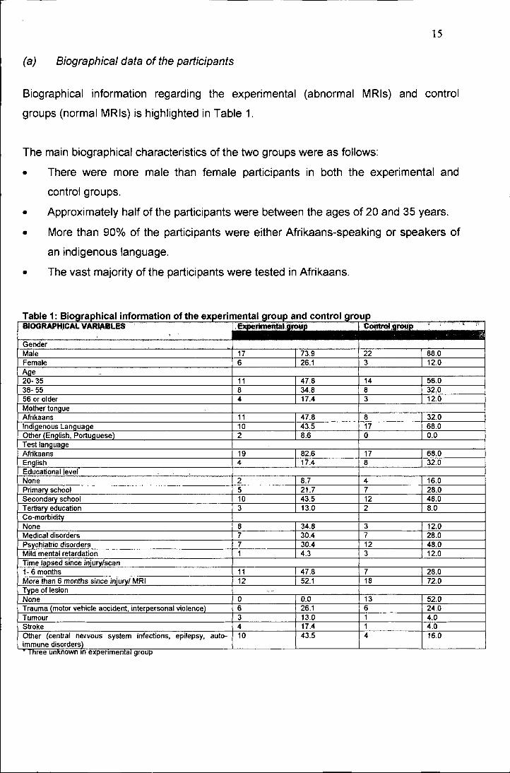
(a) Biographical data of the participants

Biographical information regarding the experimental (abnormal MRls) and control

groups (normal MRls) is highlighted in Table 1.

The main biographical characteristics of the two groups were as follows:

• There were more male than female participants in both the experimental and

control groups.

• Approximately half of the participants were between the ages of 20 and 35 years.

• More than 90% of the participants were either Afrikaans-speaking or speakers of

an indigenous language.

• The vast majority of the participants were tested in Afrikaans.

Table 1: Biographic::al information of the experimental group and control groupJ;Ct ..........ft ·IC~1,9J'f)!.!R " I_.",,-,- ,.,-~-

,GenderMale 17 73.9 22 88.0Female 6 26.1 3 12.0Age20- 35 11 47.8 14 56.036- 55 8 34.8 8 32.056 or older

.. ~.17.4 3 12.0

Mother tongue"'" ,,,aa,,,, 11 47.8 _Il 32.0Indigenous Language 10 43.5 17 68.0Other (English, Portua ..... ,p· 2 8.6 0 0.0TestAfrikaans 19 82.6 17 68.0English 4 17.4 8 32.0I=rllI""'tionallevefNone 2 8.7 4 16.0Primary school 5 21.7 7 28.0!':pl"n"dary school 10 43.5 12 48.0Tertiary "ti",."tinn 3 13.0 2 8.0~". IU"Y

None 8 34.8 3 12.0Medical 7 30.4 7 28.0

7 30.4 12 48.0Mild mental 1 4.3 3 12.0Time lapsed since injury/scan1- 6 months 11 47.8 7 28.0More than 6 months since injury/ MRI 12 52.1 18 72.0Type of lesionNone 0 0.0 13 52.0Trauma (motor vehicle . interperscnal VIVI"""''' 6 26.1 6 24.0Tumour 3 13.0 1 4.0Stroke 4 17.4 1 4.0Other (c~ntral nervous system infections, epilepsy, auto- 10 43.5 4 16.0immuné disorders)

Inree unxnown In ""t''''' '" '''' Ila' group

16

• Very few participants (10.42%) had tertiary education, although the majority did

have secondary school education.

• Psychiatric problems were the most frequent co-morbid condition. DSM-IV

diagnoses included schizophrenia, schizo-affective disorder, bipolar mood

disorder, psychosis not otherwise specified and substance abuse (mainly dagga,

alcohol and analgesics). None of the participants was psychotic at the time of

psychometric testing.

• Only a third of patients were evaluated psychometrically within the first six months

after a diagnosis of a cerebral lesion was made or an MRI done.

• If the specific "mechanism of injury" is taken into consideration, all subgroups

regarding type of lesion were represented more or less equally.

Though there was no deliberate pairing of control and experimental participants, it is

clear that the two groups are comparable.

(b) The agreement between the diagnoses made by MRI versus those made by

means of psychometry for different biographical subgroups

The results regarding the agreement between the diagnoses of cerebral lesions made

by the MRI (absent/present) versus those made by psychometry (absent/present) are

depicted according to biographical subgroups in Table 2. No statistical significant

agreement between the diagnoses of cerebral lesions with MRI and psychometry were

found for subgroups of participants above 55 years, native-tongue speakers, those with

none or only primary school education, the group with a co-morbid psychiatric diagnosis

and the groups of participants where the mechanism of injury was a stroke or due to

miscellaneous factors as discussed. These groups were omitted in the table and will not

be discussed further.

The following agreements ((K)-coefficients) between the MRI and psychometry

diagnoses regarding the presence of cerebral lesions (after correcting for change) were

found in the various subgroups. However, these agreements do not necessarily imply

practical significance, i.e. where statistical significance refers to the ability of a result to

be ascribed to chance or a sample variable. Practical significance refers to the true

usefulness of the result (Steyn, 1999):

17

• In the case of female participants psychometry yielded more accurate results.

• Age did not have a significant influence on the results.

Table 2: Interrater agreement in different biographical subgroups.Diagnosis: MRI Diagnosis: Psychometry ;

Biographical subgroup No Lesion Lesion TotalGender

No Lesion 1 (1.7) 21 22Male Lesion 2 15(15.7) 17(K) = -0.06 Total 3 36 39

No Lesion 0(3.2) 3 3Female Lesion 1 5 (3.2) 6(K) = -0.20 Total 1 8 9Age20-35 years No Lesion 1 (1.7) 13 14(K) = -0.10 Lesion 2 9 (9.7) 11

Total 3 22 2536- 55 years No Lesion 0(0.5) 8 8(K) = -0.13 Lesion 1 7 (7.5) 8

Total 1 15 16Mother tongueAfrikaans No Lesion 1 (1.3) 7 8(K) = -0.07 Lesion 2 9 (9.3) 11

Total 3 16 19Test languageAfrikaans No Lesion 1 (1.4) 16 17(K) = -0.05 Lesion 2 17 (7.4) 19

Total 3 33 36English No Lesion 0(0.7) 8 8(K) = -0.18 Lesion 1 3 (3.7) 4

Total 1 11 12Educational levelSecondary school No Lesion 1 (1.6) 11 12(K) = -0.10 Lesion 2 8 (8.6) 10

Total 3 19 22Tertiary education No Lesion 0(0.4) 2 2(K) = -0.36 Lesion 1 2 (2.4) 3

Total 1 4 5Co-morbidityNone No Lesion 0(0.8) 3 3(K) = -0.29 Lesion 3 5 (5.8) 8

Total 3 8 11Medical No Lesion 1 (0.5) 6 7(K)=0.14 Lesion 0 7 (6.5) 7

Total 1 13 14Time period1-6 months No Lesion 0(1.8) 7 7(K) = -0.21 Lesion 2 9 (9.8) 11

Total 2 16 18>6months No Lesion 1 (1.2) 17 18(K) = -0.02 Lesion 1 11 (11.2) 12

Total 2 28 30Type of lesionNone No Lesion 1 (0.85) 10 11(KJ = 0.03 Lesion 0 2 (1.85) 2

Total 1 12 13Trauma No Lesion 0(1.0) 6 6(K) = -0.33 Lesion 2 4 (5.0) 6

Total 2 10 12Stroke No Lesion 0(0.2) 1 1(K) = -0.25 Lesion 1 3 (3.2) 4

Total 1 4 5

18

• In Afrikaans-speaking participants a slight agreement was found between

diagnoses made by means of psychometry and MRI.

• Assessment in English was more than three times as accurate as assessment in

Afrikaans.

• Tertiary education also had a positive influence on the accuracy of assessment by

means of psychometry.

• Co-morbidity clearly affected the agreement between psychometric and MRI

diagnoses adversely.

• A time lapse of less than 6 months between MRI diagnosis and psychometrical

assessment improved the agreement between the two diagnoses significantly.

• The type of lesion (when present) did not have a significant influence on the

results.

(c) Correlation between psychological test subscales and the MRI diagnoses in

different participant subgroups

The subscales of the four psychological tests with a significant correlation on the 1%

and 5% levels with the MRI diagnosis, for the different subgroups of participants (as

discussed in b), are depicted in Table 3. Subtests with no statistical significant

correlation will not be discussed further.

The main findings for the different subscales were as follows:

• The South African Wechsler Adult Intelligence Scale practical subscales seem

more applicable in the diagnoses of cerebral lesions, especially digit/symbol

coding.

• The Folstein's Mini Mental State Examination also had a very high correlation with

MRI diagnoses in the presence of tertiary education and when no lesions were

present.

• The Bender Gestalt Test continuously revealed high correlation with the MRI

diagnoses through all subgroups. The correlation with education, though, is

conspicuous.

• The Grassi Block Substitute Test behaviour score seems more accurate in the

presence of strokes than trauma.

19

Table 3: Correlation of psychological test subscales with the MRI diagnosis in different participantbsu )groups.

. Biographical Subscale MRI SUbscale MRtgroupGender Co-morbidityMale Bender Gestalt: integration 0.34 None Bender Gestalt: gaps 0.77·

Bender Gestalt: additions 0.34 Medical Grassi: behaviour 0.62Female Bender Gestalt: gaps 0.76 Psychiatric Mini Mental: total 0.48Age Bender Gestalt: gaps 0.4920-35 Grassi: behaviour 0.40 Bender Gestalt: contact 0.5036- 55 Mini Mental: recall 0.52 Wechsler: general comprehension 0.5255 or older Bender Gestalt: gaps 0.75 Wechsler: digit/symbol 0.51Cultural group Time intervalAfrikaans Bender Gestalt: separation 0.55 1-6 months Bender Gestalt: separation 0.37

Bender Gestalt: additions 0.70· Bender Gestalt: integration 0.37

Bender Gestalt: absence erase 0.57 Bender gestalt: additions 0.42Wechsler: picture arrangement 0.47 Mecnanism of

injuryIndigenous Grassi: behaviour 0.40 None Mini Mental: comprehension 0.77·

Wechsler: digit/symbol 0.47 Trauma Bender Gestalt: separation 0.68Test Language Bender Gestalt: integration 0.64Afrikaans Bender Gestalt: separation 0.42 Bender Gestalt: decreases 0.68

Bender Gestalt: additions 0.37 Bender Gestalt: deletions 0.68Wechsler: picture arrangement 0.34 Bender Gestalt: gaps 0.57

English Bender Gestalt: closure 0.63 Bender Gestalt: additions 0.71·Education Stroke Grassi: behaviour 0.99·

None Mini mental: recall 0.87Bender Gestalt: transformation 0.99·Bender Gestalt: absence erasing 0.99·

Tertiary Mini Mental: comprehension 0.92Bender Gestalt: separation 0.99·Bender Gestalt: additions 0.99·

• p <= 0.01 P <= 0.05

DISCUSSION

No statistically significant agreement between the diagnoses of cerebral lesions with

MRI and psychometry were found for subgroups of participants above 55 years, though

the sensitive subscales differed among age groups. Concerning participants above the

age of 55 years, there was a significant correlation between the Bender Gestalt Test

and the MRI. As visual acuity is important in this test, poor vision might have had a

confounding influence. The lack of correlation of injuries in older participants with verbal

subscales supports the findings of previous researchers such as Heaton, Ryan, Grant &

Matthews (1996). Female participants also had a better agreement between MRI and

psychometry diagnoses, although due to the small sample size no definitive conclusion

can be drawn. Afrikaans as mother tongue also seems to be a positive, but still weak,

20

predictor of accuracy of findings. The strong correlation between the use of English astest language and the MRI diagnoses may be due to the small subgroup of participants

tested in English. The practical subscales of the South African Wechsler Adult

Intelligence Scale are less sensitive to cultural influences. The influence of educational

level was marked for the group with no and the group with tertiary education. Thissupports the findings of Lannoo and Vingerhoets (1997) and Reitan and Wolfson

(1995). Again the standardisation of the tests used has to be taken into consideration.

Certain baseline scholastic skills, e.g. writing and copying of designs, are necessary for

the successful completion of the psychological tests used. It is also clear that co-

morbidity is a significant confounding factor in respect of test accuracy that can affect

functioning negatively (Girard et ai., 1996).

Though it seems that the mechanism of injury did not playa significant role in the

agreement between MRI and psychometric diagnosis, the groups did differ, e.g. thegroup with strokes showed a stronger correlation with the Grassi Block Substitute Test,

while those with trauma showed a significant correlation with the Bender Gestalt Test.

This might be due to the different localisation of injuries, as the group with traumatic

lesions were more prone to frontal and temporal lobe injuries. While this group thereforemade more mistakes on the Bender Gestalt Test, they tended to be less aware of their

problems and therefore scored lower on the Grassi Behavioural Subscale, e.g. due to

asking for less repetition of instructions and reassurance. It is also important to note the

strong correlation between injuries less than six months old. This can be attributed tothe plasticity of the brain and also to the rehabilitation process of patients (Kay & Lezak,

1990). After a period it is therefore more difficult to diagnose minor problems in respect

of functioning in patients with cerebral injuries.

It is also clear that the Bender Gestalt Test overshadowed the other tests used in

this study. The Bender Gestalt Test does not require that the testee possess a lot of

skills - only the basic scholastic abilities of holding a pencil, using an eraser and

copying deigns. In the application of the Bender, language also does not seem to be

very important. It is therefore less culturally biased than the other tests used. It is also a

brief test and patients with poor concentration are not unfairly discriminated against.

This finding is comparable to that of previous studies of the Bender's reliability and

validity (Broadhurst & Phillips, 1969). Though the Folstein's Mini Mental State

21

Examination is one of the most widely used tests, it did not seem to have any clear

benefit, except in evaluating a patient's memory.

CONCLUSION

The findings of this study regarding the agreement between the diagnoses of cerebral

lesions made by means of psychological tests and the diagnoses made by MRI in

different subgroups should be interpreted with care. Because of the small sample size

and skewness of the sample, the results may not be an accurate representation of the

reliability and validity of psychoneurological assessment. The use of tests that may not

be culture-fair could also have influenced the results.

Patient variables with a significant influence on the results seem to be educational

level, co-morbidity and culture. It is therefore crucial to develop tests that are culture-fair

or to stick to those tests that seem to be less influenced by culture and education, e.g.

the Bender Gestalt Test.

The type of lesion seems to have an effect on the agreement between the

psychometrical evaluation and the MRI diagnosis, but this can be ascribed to the

different localisation of the lesions and the more widespread effect of trauma (primary

and secondary injuries). The plasticity of the brain must also be kept in mind and a

patient should not be "sentenced" to the current diagnosis, but rather re-evaluated at

intervals to determine the effectiveness of rehabilitation programmes.The use of only one interpreter who had to interpret the results blindly (which is in

contrast with the "real-life" situation), together with the small and skewed sample could

have influenced the results negatively. Better trained professionals could have made

better diagnoses. However, the fact remains that many professionals who are not well-

trained conduct and interpret these tests in practice. This can lead to unreliable and

invalid diagnoses.

The results of this investigation suggest that the injudicious use of psychometrical

instruments not standardised for different cultural groups, especially by untrained

individuals, could result in an unacceptably high diagnostic rate of neuropsychological

impairment in otherwise healthy patients. Though the accuracy of neuropsychological

assessment seems disappointingly low, it could contribute crucial information regarding

22

the functional impairment of the patient as well as the probability of the presence of

structural cerebral lesions and therefore may be useful in deciding whether to request a

neuro-imaging study.

REFERENCES

Anderson, S.J. (2001). On the importance of collecting local neuropsychologicalnormative data. South African Journal of Psychology, 31(3),29-34.

Beaumont, J.G. (1988). Understanding neuropsychology. Oxford: Basil Blackwell.

Bigler, E.D., Johnson, C.J. & Blatter, D.D. (1999). Head trauma and intellectual status:relation to quantitative magnetic resonance imaging findings. Applied Neuropsychology,6(4),217- 225.

Broadhurst, A & Phillips, C.J. (1969). Reliability and validity of the Bender-Gestalt Testin a sample of British School children. British Journal of Clinical Psychology, 8, 253-262.

Cohen, J (1988). Statistical power analysis for the behavioural sciences (2nd ed).Hillsdale, NJ: Lawrence Erlbaum.

Cohen, R.J., Swerdlik, M.E. & Smith, O.K. (1991). Psychological testing andassessment. An introduction to tests and measurement (2nd ed.). Mountain View, CA:Mayfield.

Filley, C.M., Young, D.A, Reardon, M.S. & Wilkening, G.N. (1999). Frontal lobe lesionsand executive dysfunction in children. Neuropsychiatry, Neuropsychology, andBehavioral Neurology, 12(3), 156- 160.

Fontaine, A, Azouvi, P., Remy, P., Bussel, B. & Samson, Y. (1999). Functionalanatomy of neuropsychological deficits after severe traumatic brain injury. Neurology,53, 1963- 1968.

Fouad, N.A & Chan, P.M. (1999). Gender and ethnicity: influence on test interpretationand reception. In J.W. Lichtenberg & R.K. Goodyear (Eds.). Scientist- PractitionerPerspectives on Test Interpretation, pp. 31- 58. Needham Heights, MA: Allyn andBacon.

Francel, P.C. & Snell, B.E. (1999). Age and outcome of traumatic brain injury in infantsand children. Brain Injury Source, 3(2). Retrieved December 30, 2001, from Brain InjuryAssociation of America Web site: http://www.biausa.org/Pages/dbscip%20source/voI.3.issue.2.html

23

Girard, D., Brown, J., Burnet-stolnack, M., Hier-Wellmer, S., Perlman, O.Z. &Seigerman, C. (1996). The relationship of neuropsychological status and outcomesfollowing traumatic brain injury. Brain Injury, 10(9), 663- 676.

Goodglass, H. (1986). The flexible battery in neuropsychological assessment. In T.Incagnoli, G. Goldstein & C.J. Golden (Eds.). Clinical Application of NeuropsychologicalTest Batteries, pp. 121-134. New York, NY: Plenum Press.

Grassi, J.R (1970). The Grassi Block Substitution Test for measuring organic brainpathology. Springfield, Illinois: Charles C Thomas.

Gregory, RJ. (1992). Psychological testing. History, principles, and applications.Needham Heights, MA: Allyn and Bacon.

Heaton,RK., Ryan,L., Grant, I. & Matthews,C.G. (1996). Demographic influences onneuropsychological test performance. In I. Grant (Ed). Neuropsychological Assessmentof neuropsychiatric disorders (2nd ed.), pp.141- 163. Oxford: Oxford University Press.

Herring, S. & Reitan, RM. (1992) Gender influence on neuropsychological performancefollowing unilateral cerebral lesions. The Clinical Neuropsychologist, 6(4),431- 442.

Hornak, J.P. (2002). The basics of MRI. Retrieved August 10, 2002, fromhttp://www.cis.rit.edu/htbooks/mri

Howell, D.C. ( 2002). Statistical methods for psychology (5th ed.). Pacific Grove, CA:Wadsworth.

Kaplan, RM. & Saccuzzo, D.P. (2001). Psychological testing: principles, applications,and issues (5th ed.). Belmont: Wadsworth.

Kay, T. & Lezak, M. (1990). The nature of head injury. In D. Corthell (Ed). TraumaticBrain Injury and Vocational Rehabilitation, pp. 22- 43. Menomie, Wisconsin: StoutVocational Rehabilitation Institute.

Keller, M.B. & Manschreck, T.C. (1981). The bedside mental status examination -reliability and validity. Comprehensive Psychiatry, 22(5), 500 - 511.

Klimidis, S. & Tokgoz, A (n.d.). A transcultural perspective on the Mini Mental StateExamination. Retrieved July 4, 2002, from International Psychogeriatrie Association website: http://www.ipa-online.org/ipaonli ne/htmllPfizer/appendix3. html

Korchin, S.J. & Schuldberg, D. (1981). The future of clinical assessment. AmericanPsychologist, 36(10), 1147-1158.

Lannoo, E. & Vingerhoets, G. (1997). Flemish normative data on commonneuropsychological tests: influence of age, education, and gender. PsychologicaBelgica, 37(3),141- 155.

24

Lezak, M.D. (1995). Neuropsychological assessment (3rd ed.). Oxford: Oxford UniversityPress.

Lloyd, D. (2000). Virtual lesions in the not-so-modular brain. Journal of the InternationalNeuropsychological Society. 6, 627-635.

Louw, D.A. (1997).lntelligence. In D.A. Louwand D.J.A. Edwards (Eds.). Psychology:and Introduction in Southern Africa (2nd ed.), pp. 321- 371. Johannesburg: Heinemann.

Lubin, B & Sands, E.W. (1992). Bibliography of the psychometric properties of theBender Visual- Motor Gestalt Test: 1970- 1991. Perceptual and Motor Skills, 75 ,385-386.

Meyer, G.J., Finn, S.E., Eyde, L.O., Kay, G.G., Moreland, KL., Dies, R,R" Eisman, E.J.,Kubiszyn, T.W. & Reed, G.M. Psychological testing and psychological assessment.American Psychologist, 56(2), 128-165.

Mitrushina, M & Fuld, P.A. (1996). Cognitive screening methods. In I. Grant and KM.Adams (Eds.). Neuropsychological Assessment of Neuropsychiatric Disorders (2nd ed.).New York, NY: Oxford.

Mortensen, E.1.& Gade, A. (1993). On the relation between demographic variables andneuropsychological test performance. Scandinavian Journal of Psychology, 34, 305-317.

Murphy, KR. & Davidshofer, C.O. (1994). Psychological testing. Principles &applications (3d ed.). Englewood Cliffs,NJ: Prentice Hall.

Nell, V. (2000). Cross-cultural neuropsychological assessment: theory and practice.Mahwah, NJ: Lawrence Erlbaum.

Peach, R.K. (1992). Factors underlying neuropsychological test performance in chronicsevere traumatic brain injury. Journal of Speech and Hearing Research, 32(4), 810 -818.

Pieters, H. & Louw, D.A. (1987). Die SAWAIS: 'n kritiese perspektief (The South AfricanWechsler Intelligence Scale: a critical perspective). South African Journal ofPsychology, 17, 145- 159.

Piotrowski, C. (1995). A review of the clinical and research use of the Bender-GestaltTest. Perceptual and Motor Skills, 81, 1272 - 1274.

Plug, C., Louw, O.A., Gouws, L.A. & Meyer, W.F. (1997). Verklarende en VertalendeSielkundewoordeboek. Johannesburg: Heinemann.

Ptacek, J.E. & Young, F.M. (1954). Comparison of the Grassi Block Substitution Testwith the Wechsler-Bellevue in the diagnosis of organic brain damage. Journal of ClinicalPsychology, 10, 375- 378.

25

Reber, A.S. & Reber, E. (2001). The Penguin Ddictionary of Psychology (3rd ed.).London: Penguin.

Reitan, R.M. & Wolfson, D. (1995). Influence of age and education onneuropsychological test results. The Clinical Neuropsychologist, 9(2), 151- 158.

Spraycar, M. (Ed) (1995). Stedman's Medical Dictionary (26th ed.). Baltimore, MD:Williams & Wilkens.

Steyn, H.S. (1999). Praktiese beduidenhuid: die gebruik van effekgroottes.Potchefstroom: Publikasiebeheerkomitee, Potchefstroom Universiteit vir ChristelikeHoër Onderwys.

Stratton, P. & Hayes, N. (1994). A Student's Dictionary of Psychology. Londen: EdwardArnold.

The Mini Mental State Examination (n.d.). Retrieved July 4, 2002, from Tufts University,School of Medicine, Department of Psychiatry web site:http://www.nemc.org/psych/mmse.asp

Voller, B.; Benke, T.; Bendedetto, K.; Schnider, P.; Auff, E. & Aichner, F. (1999).Neuropsychological, MRI and EEG findings after very mild traumatic brain injury. Braininjury, 13(10), 821-827.

Walker, P.M.B. (Ed) (1995). Larousse Dictionary of Science and Technology.Edinburgh: Larousse.

Zilmer, E.A. & Spiers, MV. (2001). Principles of neuropsychology. Belmont, NY:Wadsworth.

\ Ir'f '90(0 4-'(-1iJ!O~.'.1\.\.\OlfP'


Recommended