PHOTO CHALLENGE E10 I CUTIS ® WWW.MDEDGE.COM/DERMATOLOGY PLEASE TURN TO PAGE E11 FOR THE DIAGNOSIS Ulcerated and Verrucous Plaque on the Chest Mahdieh Fazel Jedlowski, MD, PharmD; Patrick Michael Jedlowski, MD A 36-year-old man presented to an emergency department in the southwestern United States with a cough, fatigue, and worsening back pain associated with night sweats of 1 month’s duration. He experi- enced a 9.07-kg weight loss, as well as development of a rough, nontender, nonpruritic rash along the left upper chest over the prior month. The patient was born in West Africa and reported that he had moved to the southwestern United States from the eastern United States approximately 6 years prior to presen- tation. Physical examination on admission revealed a 5×3-cm, purple-brown, verrucous plaque with a central pink cobblestone appearance and ulceration. Chest radiography was notable for perihilar adenopa- thy with no focal infiltrates or cavitary lesions. Com- puted tomography and magnetic resonance imaging of the chest were notable for miliary nodules through- out the lungs; extensive lytic spine lesions of cervical, thoracic, and lumbar vertebral bodies and left twelfth rib; and a left paraspinal thoracic epidural soft tissue phlegmon. Initial laboratory investigations revealed peripheral eosinophilia without absolute leukocytosis and a microcytic anemia. WHAT’S YOUR DIAGNOSIS? a. cutaneous leishmaniasis b. disseminated blastomycosis c. disseminated coccidioidomycosis d. disseminated histoplasmosis e. lupus vulgaris From the University of Arizona College of Medicine, Tucson. The authors report no conflict of interest. Correspondence: Patrick Michael Jedlowski, MD, 7165 N Pima Canyon Dr, Tucson, AZ 85718 ([email protected]). doi:10.12788/cutis.0328 Copyright Cutis 2021. No part of this publication may be reproduced, stored, or transmitted without the prior written permission of the Publisher. CUTIS Do not copy
Transcript
PHOTO CHALLENGE
E10 I CUTIS® WWW.MDEDGE.COM/DERMATOLOGY
PLEASE TURN TO PAGE E11 FOR THE DIAGNOSIS
Ulcerated and Verrucous Plaque on the Chest
Mahdieh Fazel Jedlowski, MD, PharmD; Patrick Michael Jedlowski, MD
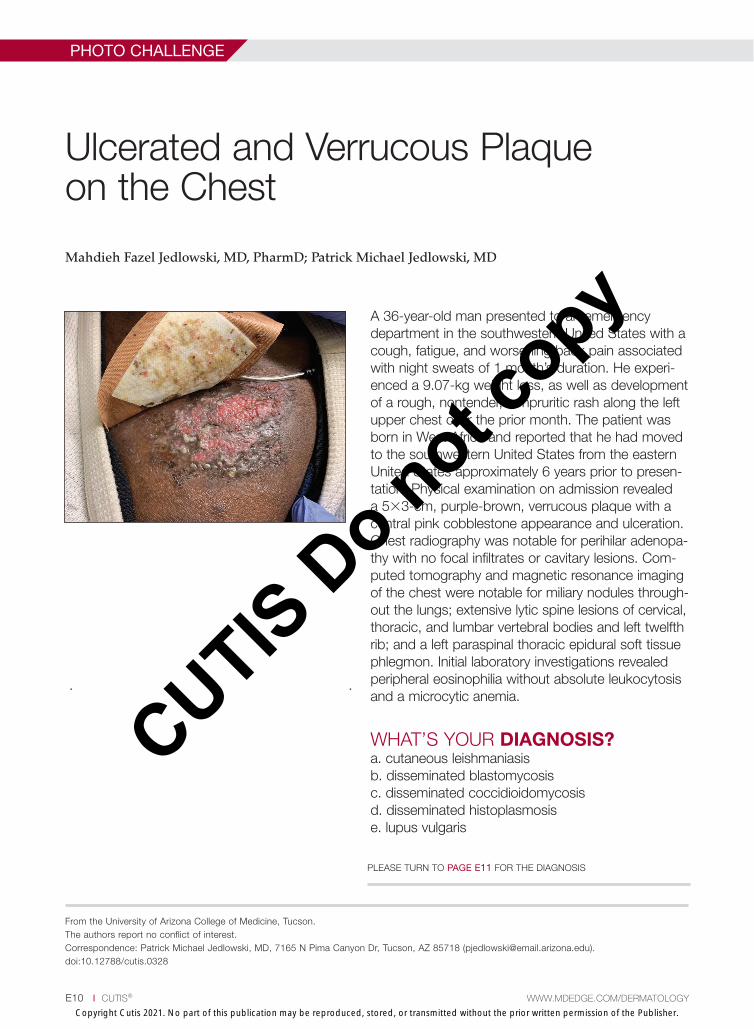
A 36-year-old man presented to an emergency department in the southwestern United States with a cough, fatigue, and worsening back pain associated with night sweats of 1 month’s duration. He experi-enced a 9.07-kg weight loss, as well as development of a rough, nontender, nonpruritic rash along the left upper chest over the prior month. The patient was born in West Africa and reported that he had moved to the southwestern United States from the eastern United States approximately 6 years prior to presen-tation. Physical examination on admission revealed a 5×3-cm, purple-brown, verrucous plaque with a central pink cobblestone appearance and ulceration. Chest radiography was notable for perihilar adenopa-thy with no focal infiltrates or cavitary lesions. Com-puted tomography and magnetic resonance imaging of the chest were notable for miliary nodules through-out the lungs; extensive lytic spine lesions of cervical, thoracic, and lumbar vertebral bodies and left twelfth rib; and a left paraspinal thoracic epidural soft tissue phlegmon. Initial laboratory investigations revealed peripheral eosinophilia without absolute leukocytosis and a microcytic anemia.
From the University of Arizona College of Medicine, Tucson.The authors report no conflict of interest. Correspondence: Patrick Michael Jedlowski, MD, 7165 N Pima Canyon Dr, Tucson, AZ 85718 ([email protected]).doi:10.12788/cutis.0328
Copyright Cutis 2021. No part of this publication may be reproduced, stored, or transmitted without the prior written permission of the Publisher.
CUTIS Do not c
opy
PHOTO CHALLENGE DISCUSSION
VOL. 108 NO. 2 I AUGUST 2021 E11WWW.MDEDGE.COM/DERMATOLOGY
THE DIAGNOSIS:
Disseminated Coccidioidomycosis
A 6-mm punch biopsy was performed at the periph-ery of the ulcerated cutaneous lesion on the chestrevealing extensive spherules. Serum antibody
immunodiffusion for histoplasmosis and blastomycoses both were negative; however, B-D-glucan assay was positive at 364 pg/mL (reference range: <60 pg/mL, negative). Initial HIV-1 and HIV-2 antibody and anti-gen testing was negative as well as repeat testing at 3 weeks. Immunodiffusion for Coccidioides IgM and IgG was positive, and cocci antibody IgG complement fixa-tion assays were positive at titers of 1:64 (reference range: <1:2, negative). A computed tomography needle-guided biopsy of the paravertebral soft tissue was performed. Gram stains and bacterial cultures of the biopsies were negative; however, fungal cultures were notable for growth of Coccidioides. Given the pertinent testing, a diagnosis of disseminated coccidioidomycosis was made.
Cutaneous coccidioidomycosis can occur in 3 situations: direct inoculation (primary cutaneous coc-cidioidomycosis), disseminated infection (disseminated cutaneous coccidioidomycosis), or as a reactive compo-nent of pulmonary infection.1,2 Of them, primary and disseminated cutaneous coccidioidomycosis are organism specific and display characteristic spherules and fungus on histopathology and cultures, respectively. Reactive coccidioidomycosis differs from organism-specific dis-ease, as it does not contain spherules in histopathologic sections of tissue biopsies.1 Reactive skin manifestations occur in 12% to 50% of patients with primary pulmonary infection and include erythema nodosum, erythema mul-tiforme, acute generalized exanthema, reactive interstitial granulomatous dermatitis, and Sweet syndrome.3
Organism-specific cutaneous coccidioidomycosis most often is correlated with hematogenous dissemi-nation of primary pulmonary disease rather than direct inoculation of skin.1 The skin is the most common site of extrapulmonary involvement in disseminated coccidioi-domycosis, and cutaneous lesions have been reported in 15% to 67% of cases of disseminated disease.1,4 In cuta-neous disseminated disease, nodules, papules, macules, and verrucous plaques have been described. In a case series of disseminated cutaneous coccidioidomycosis, nodules were the most common cutaneous presentation and occurred in 39% (7/18) of patients, while verrucous plaques were the rarest and occurred in only 6% (1/18) of patients.5
The rate of coccidioidomycosis dissemination varies based on exposure and patient characteristics. Increased rates of dissemination have been reported in patients of African and Filipino descent, along with individuals that are immunosuppressed due to disease or medical
therapy. Dissemination is clinically significant, as patients with multifocal dissemination have a greater than 50% risk for mortality.6
Disseminated coccidioidomycosis is a relatively rare manifestation of Coccidioides infection; approximately 1.6% of patients exposed to and infected with Coccidioides ultimately will develop systemic or disseminated dis-ease.7,8 Although the rates of primary pulmonary infec-tion are similar between patients of varying ethnicities, the rates of dissemination are higher in patients of African and Filipino ethnicity.8 In population studies of coccidioidomycosis (N=332), Black patients represented 33.3% (4/12) of disseminated cases but only 8.7% of Coccidioides cases overall.7
Population studies of Black patients with coccidioi-domycosis have shown a 4-fold higher predisposition for severe disease compared to mild disease.9 Spondylitis and meningitis also are disproportionately more common in Black patients.8 Black patients comprised 75% of all spondylitis cases in a cohort where only 25% of patients were Black. Additionally, 33% of all meningitis cases occurred in Black patients in a cohort where 8% of total cases were Black patients.8 Within the United States, the highest rates of coccidioidomycosis meningitis are seen in Black patients.10
The pathophysiology underlying the increased sus-ceptibility of individuals of African or Filipino descent to disseminated and severe coccidioidomycosis remains unknown.8 The level of vulnerability within this patient population has no association with increased envi-ronmental exposure or poor immunologic response to Coccidioides, as demonstrated by the ability of these populations to respond to experimental vaccination and skin testing (spherulin, coccidioidin) to a similar extent as other ethnicities.8 Class II HLA-DRB1*1301 alleles have been associated with an increased risk for severe dis-seminated Coccidioides infection regardless of ethnicity; however, these alleles are not overrepresented in these patient populations.8
In patients with primary pulmonary coccidioido-mycosis with no evidence of dissemination, guidelines generally recommend offering treatment to groups at high risk of dissemination, such as pregnant women and patients with diabetes mellitus. Given the high incidence of disseminated and severe disease in Black and Filipino patients, some guidelines endorse treatment of all cases of coccidioidomycosis in this patient population.8 No current data are available to help determine whether this broad treatment approach reduces the development of disseminated infection in these populations. Frequent monitoring for disease progression and/or dissemination
Copyright Cutis 2021. No part of this publication may be reproduced, stored, or transmitted without the prior written permission of the Publisher.
CUTIS Do not c
opy
SWEET SYNDROME
E12 I CUTIS® WWW.MDEDGE.COM/DERMATOLOGY
involving clinical and laboratory reevaluation every 3 months for 2 years is highly recommended.8
Treatment generally is based on location and severity of infection, with disseminated nonmeningeal infection being treated with oral azole therapy (ketoconazole, itraconazole, or fluconazole).11 If there is involvement of the central nervous system structures or rapidly wors-ening disease despite azole therapy, amphotericin B is recommended at 0.5 to 0.7 mg/kg daily. In patients with disseminated meningeal infection, oral fluconazole (800–1000 mg/d) or a combination of an azole with intrathecal amphotericin B (0.01–1.5 mg/dose, interval ranging from daily to 1 week) is recommended to improve response.11
The differential diagnosis of cutaneous disseminated coccidioidomycosis is broad and includes other systemic endemic mycoses (histoplasmosis, blastomycosis) and infections (mycobacteria, leishmania). Lupus vulgaris, a form of cutaneous tuberculosis, presents as a palpable tubercular lesion that may coalesce into erythematous plaques, which may mimic endemic mycoses, especially in patients with risk factors for both infectious etiologies such as our patient.12 Disseminated histoplasmosis may present as polymorphic plaques, pustules, nodules, and ulcerated skin lesions, whereas disseminated blastomy-cosis characteristically presents as a crusted verrucous lesion with raised borders and painful ulcers, both of which may mimic coccidioidomycosis.13 Biopsy would reveal the characteristic intracellular yeast in Histoplasma capsulatum and broad-based budding yeast form of Blastomyces dermatitidis in histoplasmosis and blastomy-cosis, respectively, in contrast to the spherules seen in our patient’s biopsy.13 Localized cutaneous leishmaniasis initially develops as a nodular or papular lesion and can progress to open ulcerations with raised borders. Biopsy and histopathology would reveal round protozoal amas-tigotes.14 Other diagnoses that should be considered
include mycetoma, nocardiosis, and sporotrichosis.15 As the cutaneous manifestations of Coccidioides infec-tions are varied, a broad differential diagnosis should be maintained, and probable environmental and infectious exposures should be considered prior to ordering diag-nostic studies.
REFERENCES 1. Garcia Garcia SC, Salas Alanis JC, Flores MG, et al. Coccidioidomy-
cosis and the skin: a comprehensive review. An Bras Dermatol. 2015; 90:610-619.
2. DiCaudo DJ. Coccidioidomycosis: a review and update. J Am AcadDermatol. 2006;55:929-942; quiz 943-925.
3. DiCaudo DJ, Yiannias JA, Laman SD, et al. The exanthem of acutepulmonary coccidioidomycosis: clinical and histopathologic features of 3 cases and review of the literature. Arch Dermatol. 2006;142:744-746.
4. Blair JE. State-of-the-art treatment of coccidioidomycosis: skin andsoft-tissue infections. Ann N Y Acad Sci. 2007;1111:411-421.
5. Crum NF, Lederman ER, Stafford CM, et al. Coccidioidomycosis: adescriptive survey of a reemerging disease. clinical characteristics andcurrent controversies. Medicine (Baltimore). 2004;83:149-175.
6. Borchers AT, Gershwin ME. The immune response in coccidioidomyco-sis. Autoimmun Rev. 2010;10:94-102.
7. Smith CE, Beard RR. Varieties of coccidioidal infection in relation to the epidemiology and control of the diseases. Am J Public Health NationsHealth. 1946;36:1394-1402.
8. Ruddy BE, Mayer AP, Ko MG, et al. Coccidioidomycosis in AfricanAmericans. Mayo Clin Proc. 2011;86:63-69.
9. Louie L, Ng S, Hajjeh R, et al. Influence of host genetics on the severity of coccidioidomycosis. Emerg Infect Dis. 1999;5:672-680.
10. McCotter OZ, Benedict K, Engelthaler DM, et al. Update on the epi-demiology of coccidioidomycosis in the United States. Med Mycol. 2019;57(suppl 1):S30-S40.
11. Galgiani JN, Ampel NM, Catanzaro A, et al. Practice guideline forthe treatment of coccidioidomycosis. Infectious Diseases Society ofAmerica. Clin Infect Dis. 2000;30:658-661.
12. Khadka P, Koirala S, Thapaliya J. Cutaneous tuberculosis: clinico-pathologic arrays and diagnostic challenges. Dermatol Res Pract. 2018;2018:7201973.
13. Smith JA, Riddell JT, Kauffman CA. Cutaneous manifestations ofendemic mycoses. Curr Infect Dis Rep. 2013;15:440-449.
14. Scorza BM, Carvalho EM, Wilson ME. Cutaneous manifestations ofhuman and murine leishmaniasis. Int J Mol Sci. 2017;18:1296.
PHOTO CHALLENGE DISCUSSION
Copyright Cutis 2021. No part of this publication may be reproduced, stored, or transmitted without the prior written permission of the Publisher.