32
Ulnar Nerve Entrapment Linne0e J Sells, DO, FAOASM
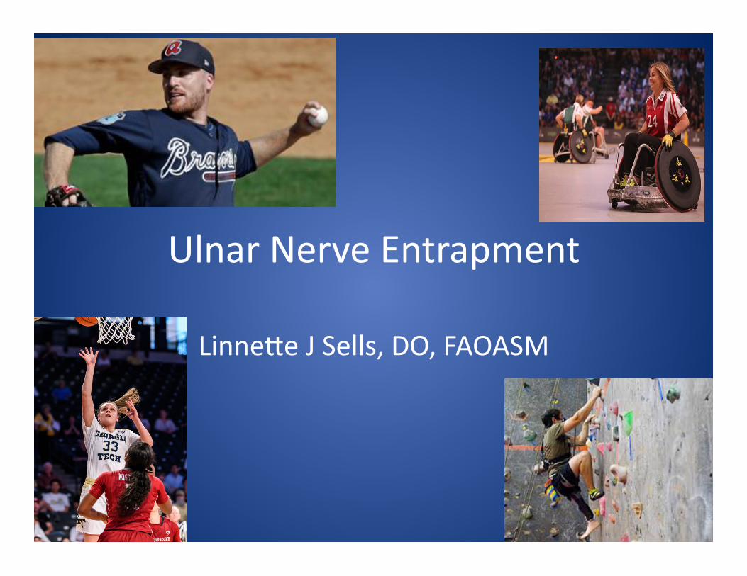
Ulnar Nerve Entrapment
Linne0e J Sells, DO, FAOASM
Learning Objec>ves
• Iden>fy clinical signs and symptoms of ulnar nerve entrapment
• Learn examina>on findings of nerve entrapment
• Learn basic EMG abnormali>es of ulnar nerve entrapment
Case Presenta>on
50 y/o WM c/o weakness and loss of dexterity x several months in R hand
Pa>ent is a physician and needs fine motor skills
Ac>vites include weight liRing, golf, rock climbing, and adult league baseball PMH essen>al nega>ve, no meds
Social ETOH, no smoking
SUBJECTIVE Numbness and >ngling in 4th and 5th digits
with increased sx at night
Medial elbow pain-‐mild, worse with full elbow flexion R hand weakness with pinch, bu0oning, typing
and general loss of dexterity
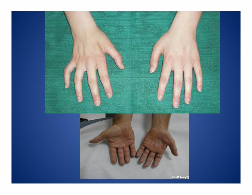
OBJECTIVE Inspec>on shows mild atrophy of 1st dorsal interosseous with no deformity of shoulder, elbow or wrist Palpa>on of ulnar nerve in retrocondylar groove and above elbow finds no subluxa>on FROM shoulder, elbow and wrist with no restric>ons or deformi>es FROM C-‐spine without pain or restric>on
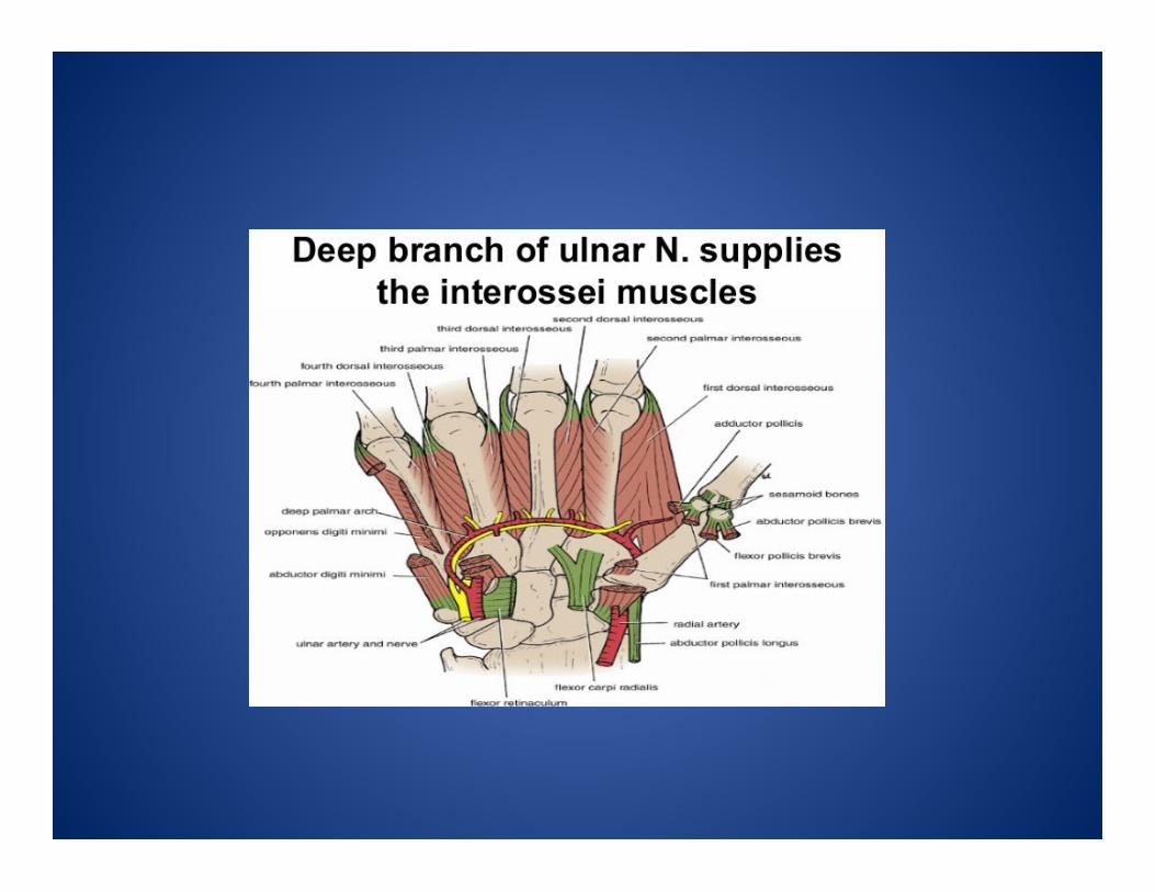
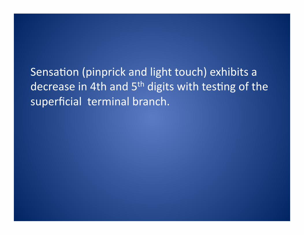
Sensa>on (pinprick and light touch) exhibits a decrease in 4th and 5th digits with tes>ng of the superficial terminal branch.
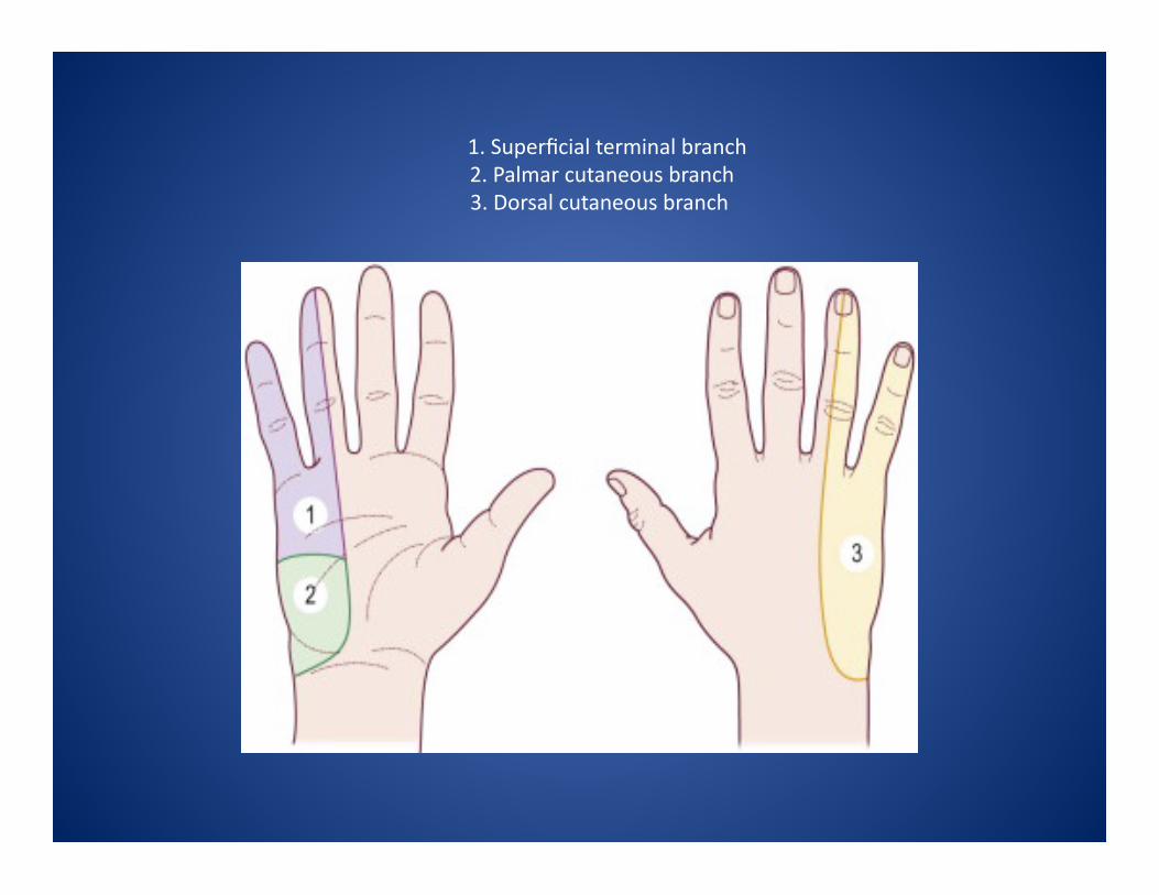
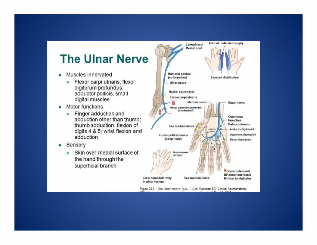
1. Superficial terminal branch 2. Palmar cutaneous branch 3. Dorsal cutaneous branch

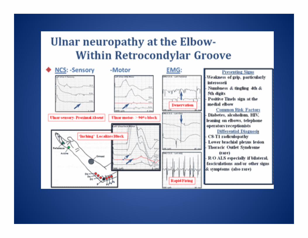
TESTS Posi>ve Tinel’s at elbow nega>ve at Guyon’s canal and upper arm (1 cm proximal to medial epicondyle)
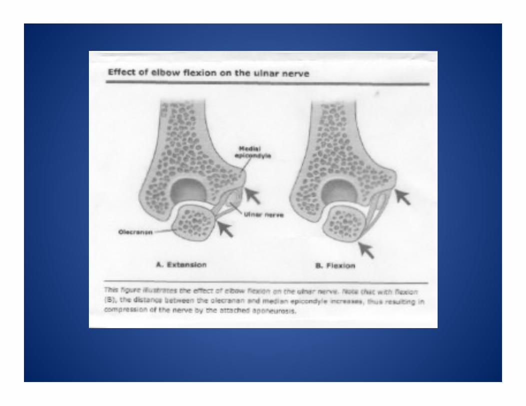
Posi>ve Elbow Flexion (sustained maximal elbow flexion with wrist in neutral x 1 minute) with increased parethesias in fingers
Posi>ve Pressure test (pressure over ulnar nerve at groove with elbow flexion)
POSSIBLE CAUSES/MOI Trauma elbow or wrist (acute or old)
Arthri>s/osteophytes
Mass lesions/ganglion cysts
Repe>>ve overuse
Muscle imbalances and abnormal anatomy
Sports with overuse of elbow or hand ie; bicyclists, pitchers, wheelchair athletes
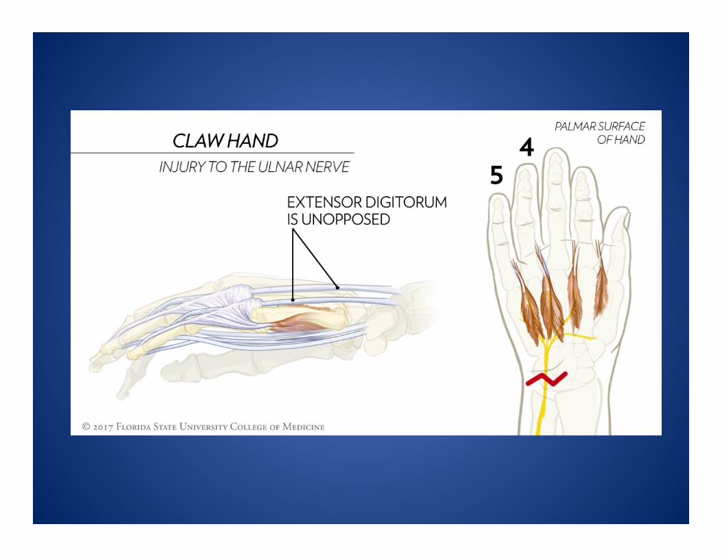
DIFFERENTIAL DIAGNOSIS Proximal lesions C8/T1 or lower trunk of medial cord (usually differs with sensa>on involvement prox. to wrist in distribu>on of medial cutaneous nerve and motor involvment of non-‐ulnar muscles ie; flexor pollicis longus/ext. pollicis and thenar muscles)
Central lesions would show was>ng and weakness in ulnar distribu>on with absence of sensory deficits (ALS or monometric amyotrophic forms of motor neuron disease)


ELECTRODIAGNOSTIC TESTING Nerve Conduc>on Studies and EMG’s
-‐confirm diagnosis
-‐establish baseline results
-‐determine severity
-‐R/O other causes
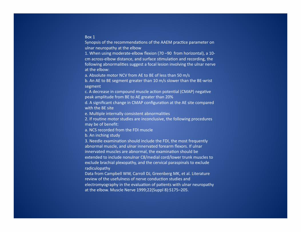
Box 1 Synopsis of the recommenda>ons of the AAEM prac>ce parameter on ulnar neuropathy at the elbow 1. When using moderate-‐elbow flexion (70 –90 from horizontal), a 10-‐cm across-‐elbow distance, and surface s>mula>on and recording, the following abnormali>es suggest a focal lesion involving the ulnar nerve at the elbow: a. Absolute motor NCV from AE to BE of less than 50 m/s b. An AE to BE segment greater than 10 m/s slower than the BE-‐wrist segment c. A decrease in compound muscle ac>on poten>al (CMAP) nega>ve peak amplitude from BE to AE greater than 20% d. A significant change in CMAP configura>on at the AE site compared with the BE site e. Mul>ple internally consistent abnormali>es 2. If rou>ne motor studies are inconclusive, the following procedures may be of benefit: a. NCS recorded from the FDI muscle b. An inching study 3. Needle examina>on should include the FDI, the most frequently abnormal muscle, and ulnar innervated forearm flexors. If ulnar innervated muscles are abnormal, the examina>on should be extended to include nonulnar C8/medial cord/lower trunk muscles to exclude brachial plexopathy, and the cervical paraspinals to exclude radiculopathy Data from Campbell WW, Carroll DJ, Greenberg MK, et al. Literature review of the usefulness of nerve conduc>on studies and electromyography in the evalua>on of pa>ents with ulnar neuropathy at the elbow. Muscle Nerve 1999;22(Suppl 8):S175–205.
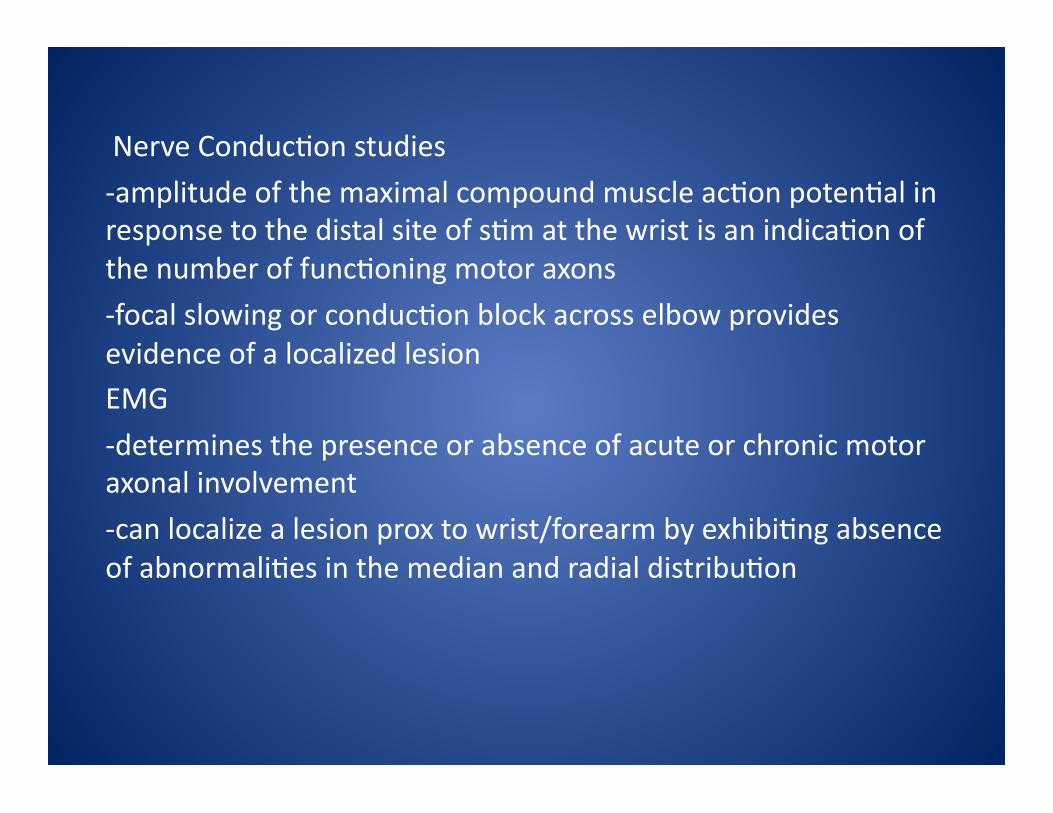
Nerve Conduc>on studies
-‐amplitude of the maximal compound muscle ac>on poten>al in response to the distal site of s>m at the wrist is an indica>on of the number of func>oning motor axons
-‐focal slowing or conduc>on block across elbow provides evidence of a localized lesion EMG
-‐determines the presence or absence of acute or chronic motor axonal involvement
-‐can localize a lesion prox to wrist/forearm by exhibi>ng absence of abnormali>es in the median and radial distribu>on

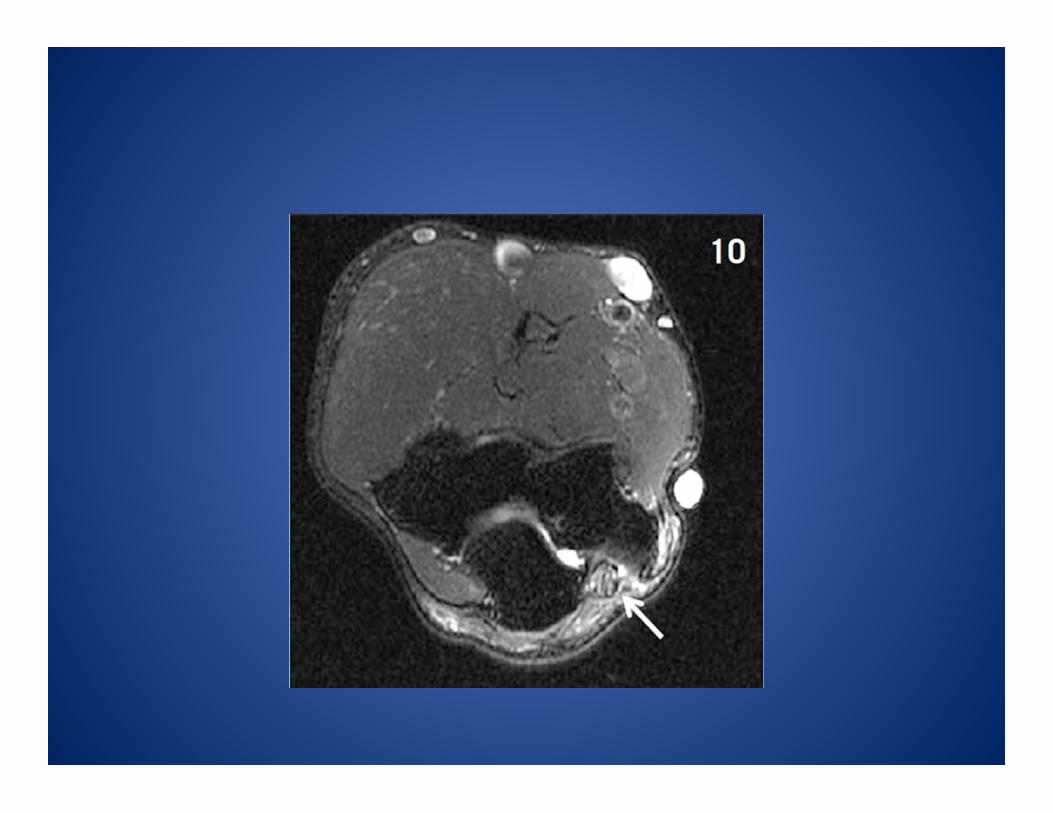
IMAGING MRI – nerve enlargement with increased signal intensity T2 weighted or short T1 inversion recovery sequences Ultrasound – thickening nerve and altered echogenicity -‐excellent for evalua>on of masses or cysts, compression lesions or trauma
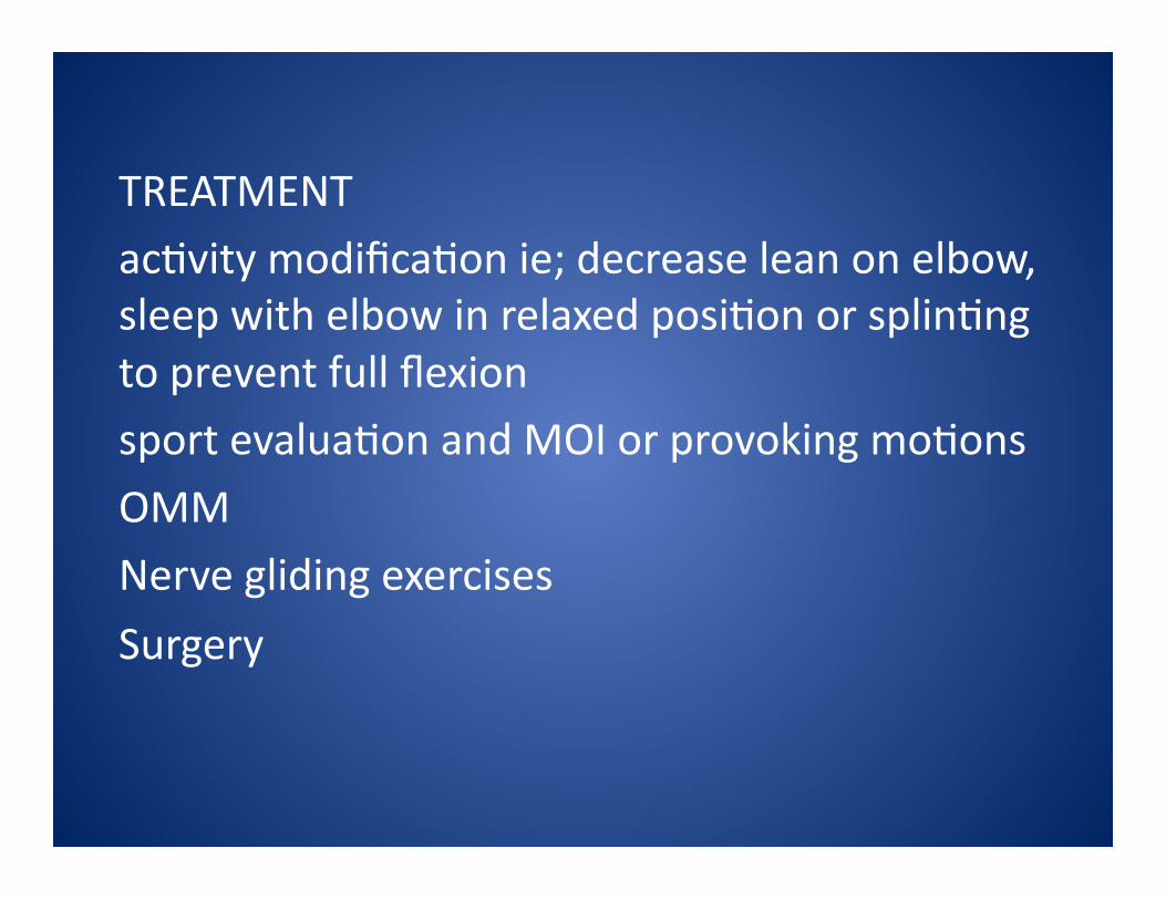
TREATMENT ac>vity modifica>on ie; decrease lean on elbow, sleep with elbow in relaxed posi>on or splin>ng to prevent full flexion sport evalua>on and MOI or provoking mo>ons
OMM
Nerve gliding exercises
Surgery
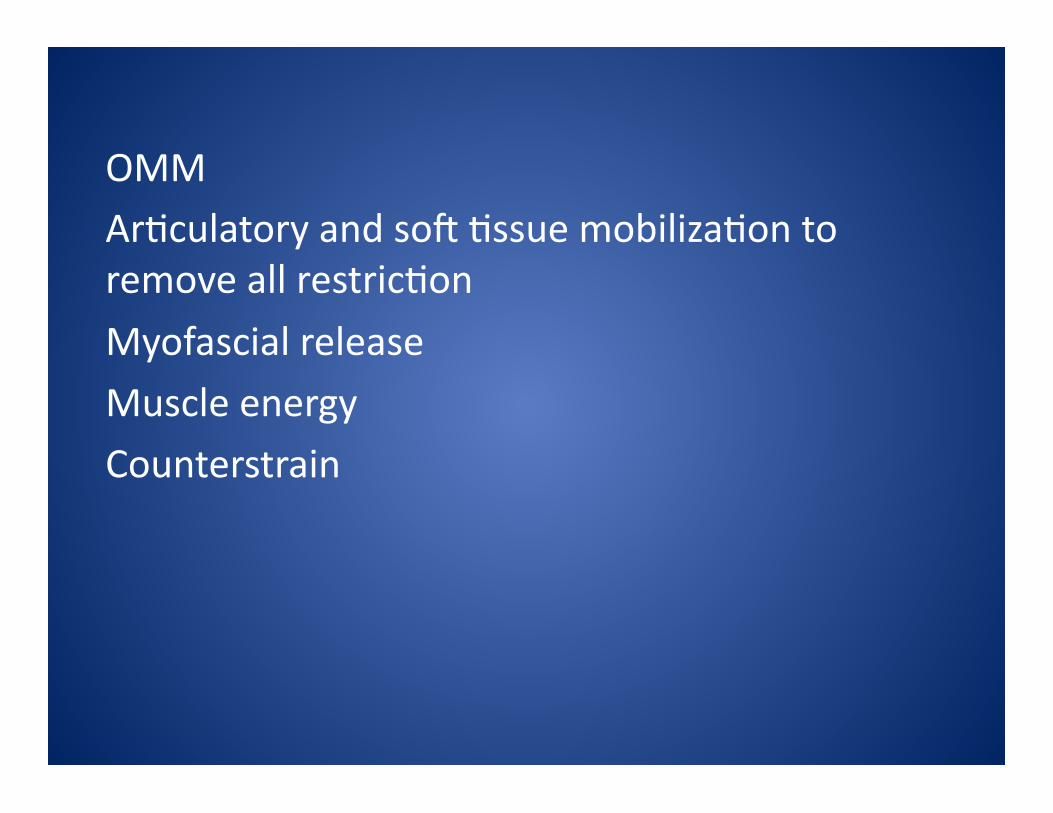
OMM Ar>culatory and soR >ssue mobiliza>on to remove all restric>on
Myofascial release
Muscle energy
Counterstrain
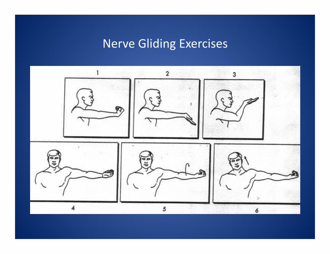
Nerve Gliding Exercises
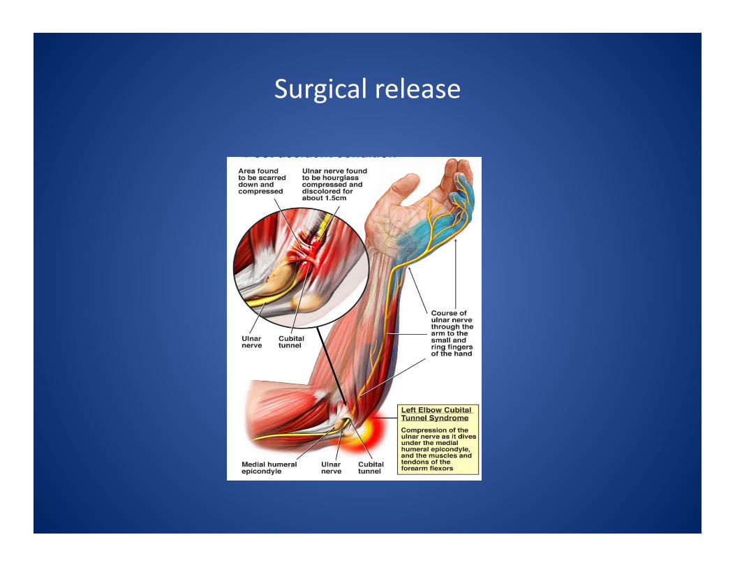
Surgical release

REHAB Healing of 3-‐4 weeks
DISCUSSION Second most common entrapment syndrome vs carpal tunnel
Must be able to Diff. Dx. from C8/T1 or brachial plexus Iden>fy loca>on of entrapment
Advise pa>ent on prolonged sx and complica>ons if treatment is not successful
Thanks


